Preprint
Article

This version is not peer-reviewed.

Inhibition of Jasmonic Acid-Isoleucine Conjugating Enzyme JAR1 Shifts the Local and Systemic Leaf Metabolic Profiles in Arabidopsis

Submitted:

16 November 2025

Posted:

17 November 2025

You are already at the latest version

Abstract

Jasmonates (JAs)-mediated pathways are central signaling hubs in plant defense response. However, the identification of mobile and non-mobile signals involved in downstream systemic signaling is still less studied. Here, we investigate the role of the jasmonic acid-isoleucine conjugating enzyme, JAR1, and the mobility of jasmonic acid-isoleucine (JA-Ile) in wound-induced local and systemic defense using LC-MS/MS for targeted jasmonate analysis and untargeted metabolomics in Arabidopsis thaliana leaves. The use of jarin-1, a specific inhibitor of JA-Ile biosynthesis, suggested that JA-Ile is synthesized de novo in the particular tissues, rather than being a mobile signal. In addition, inhibition of JAR1 enzyme activity affected an array of downstream metabolic pathways, locally and systemically, such as amino acids and carbohydrate metabolism. This study demonstrates that the occurrence and spread of local and systemic downstream signals depend on JAR1 activity, and this enzyme exclusively regulates a series of metabolic pathways under both wounding and non-wounding conditions.

Keywords: 
;  ;  ;  ;  ;  

Introduction

Plants are challenged by various abiotic and biotic stresses across their whole life. Biotic stress, such as infestation by herbivores and pathogens, mainly occurs locally but often spreads to the entire plant. Accordingly, the plant reacts locally and systemically with defense. In this process, plants establish a set of defense mechanisms and accumulate a plethora of defensive metabolites against the attackers. Among them, jasmonic acid (JA) and its derivatives are key phytohormones responding to various kinds of adverse environment (Santino et al., 2013). The amino acid conjugate JA-Ile is the most active endogenous jasmonate (Fonseca et al., 2009). JA-Ile is a key molecule that regulates developmental processes as well as wound response (Nie et al., 2025). To date, there are two views on the occurrence of JA-Ile: one sees JA-Ile as a mobile signal from one to another tissue (Sato et al., 2011; Matsuura et al., 2012); the other as an immobile signal (Bozorov et al., 2017; Schulze et al., 2019; Li et al., 2020). However, it is conceivable that these two possibilities might co-exist in plants and function in different contexts including biotic and abiotic stress conditions. However, there is a large knowledge gap in this field.
Additionally, metabolic changes occurring in cells play an important role in systemic acquired acclimation and could convey specificity to the rapid systemic response of plants (Choudhury et al., 2018). Likewise, JA deficiency and exogenous Methyl-JA (MeJA) treatment has an impact on amino acids and small peptides metabolism, glucosinolates and tryptophan metabolism, lipid metabolism, especially fatty acyls metabolism in Arabidopsis (Cao et al., 2016). Moreover, JA-Ile specifically activates many metabolites in the leaves that are distinct from the ones induced by JA and MeJA (Xu et al., 2018). Until now, there is still limited knowledge about exclusive JA-Ile-induced compounds and metabolic pathways induced by wounding.
For many years, JAR1 was considered the sole enzyme catalyzing the formation of JA-Ile from JA by conjugating isoleucine to JA in Arabidopsis (Staswick & Tiryaki, 2004). More recently, AtGH3.10, a homolog of JAR1 in Arabidopsis, was shown to contribute to JA-Ile biosynthesis and to function redundantly with JAR1 in flower development and the wound response (Delfin et al., 2022). Together, AtGH3.10 and JAR1 catalyze the conversion of 12-hydroxyjasmonic acid (12-OH-JA) to 12-hydroxyjasmonoyl-L-isoleucine (12-OH-JA-Ile) in Arabidopsis (Oki et al., 2025). JAR1 belongs to the GRETCHEN HAGEN 3 (GH3) family of acyl acid amido synthetases, which mediate biochemical modifications such as adenylation across multiple phytohormone signaling pathways, including JA, indole-3-acetic acids (IAA), and salicylic acid (SA) (Staswick et al., 2002; Jez, 2022). Since the GH3 protein family is involved in a variety of catalytic reactions that could blur the line between primary and specialized metabolism (Jez, 2022), it is necessary to gain a comprehensive overview of the metabolic networks regulated by GH3s including JAR1. Untargeted metabolomics provides a powerful approach to achieve this comprehensive understanding.
Here, we first set out to explore the role of JAR1 in the induction of downstream local and systemic signals and their involvement in metabolic pathways and further find out the mobility of JA-Ile upon wounding in our conditions. Thus, we used a well-described specific JA-Ile biosynthesis inhibitor, jarin-1, which was firstly reported to exclusively suppress the JA-conjugating enzyme JAR1 in Arabidopsis, thereby preventing the spreading of JA-Ile signaling (Meesters et al., 2014; Ishimaru et al., 2018). By using this inhibitor and defining a local leaf, we can study the dynamics of JA-Ile in systemic leaves. Here, the application of jarin-1 to inhibit JA-Ile biosynthesis has even more advantages than employing the jar1 mutant. We found that JA-Ile shifted leaf metabolic profiles, and provide the first evidence for an inhibitory effect of jarin-1 on JA-Ile level upon wounding in both a local and systemic manner. Finally, we demonstrated that under our conditions, JA-Ile was not a mobile signal itself but played a role in the transduction of downstream mobile and non-mobile signals.

Materials and Methods

Plant Material and Growth Conditions

Five to six-week-old Arabidopsis thaliana (L.) Heynh. ecotype Col-0 plants were used for all experiments. To ensure the same starting conditions, all plants used for one assay were sown on the same day and kept in the same growth chamber. After stratification for two days at 4℃, plants were grown in growth chambers in 10 cm round pots with soil substrates under short-day conditions (10/14 h light/dark period) with minor differences. For phytohormone or metabolome analyses, plants were grown at the Max Planck Institute for Chemical Ecology or iDiv, respectively. The growth chambers were adjusted to 50-60% humidity and constant 21℃ with a light intensity of 100 μmol m2 s−1 (Scholz et al., 2017).

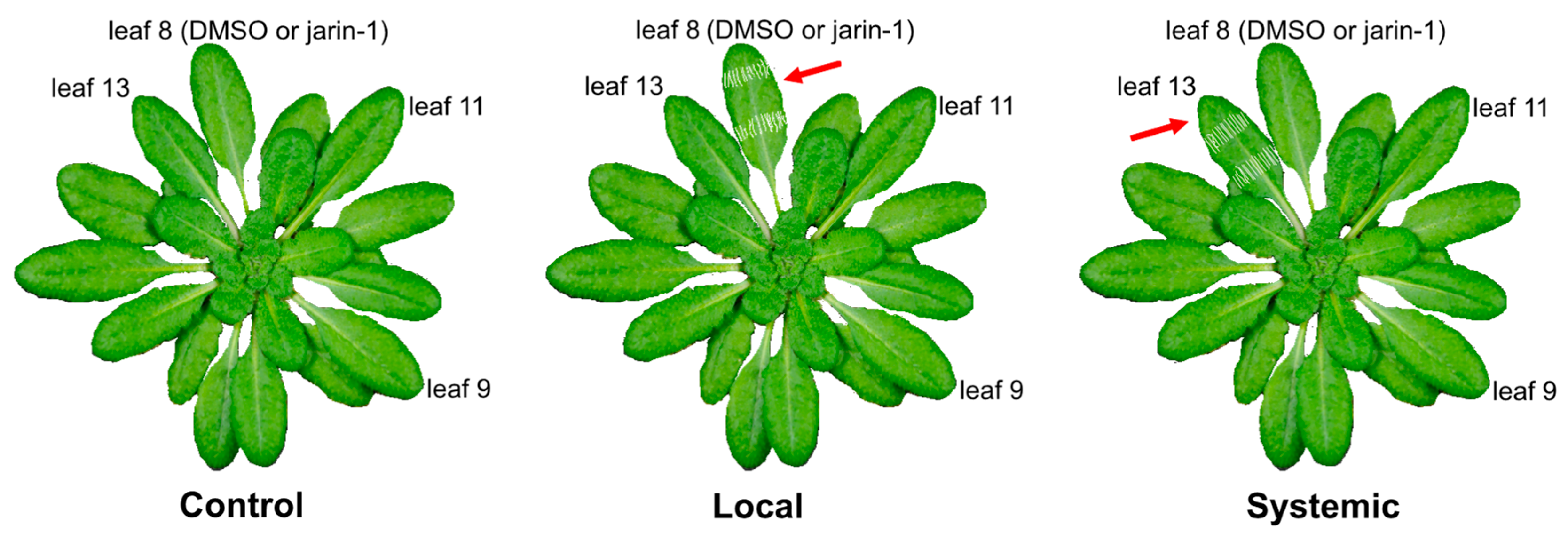
Application of Jarin-1 and Wounding

Jarin-1 (AOBIOUS INC) powder was dissolved in dimethyl sulfoxide (DMSO) to obtain a stock solution of 7.07 mM that was kept at -20°C. For use, it was diluted to 10 µM with ddH2O and subsequently supplied with 0.1% (v/v Tween-20 to facilitate application to the leaf surface (Neumann & Chamel, 1986). The same concentration of DMSO (0.14%, v/v) was used as a chemical control for jarin1. For experiments investigating the systemic response and translocation of metabolites, the leaves of each plant were counted according to their age (Dengler, 2006; Farmer et al., 2013; Kiep et al., 2015). Using leaf 8 as local leaf, leaves 11 and 13 are systemic leaves connected via the vascular system, while leaf 9 is not connected. In all bioassays, leaves directly subjected to wounding were regarded as local leaves, while their vascularly connected leaves were regarded as systemic leaves.
For experiments of measuring phytohormones and metabolites, 50 μl chemical solution was evenly applied on the leaf surface. After 1.5 h, the leaves were wounded in two different ways: (1) the appointed leaf was wounded with a serrated 18/8 tweezers by pinching the surface twice at two different positions, vertically along the midvein (Landgraf et al., 2012); (2) leaf was treated with MecWorm to ensure a continuous wounding process (Mithöfer et al., 2005). This instrument was applied for mechanical wounding of the appointed leaf with punches every 5 s for 48 min. One hour after wounding, the leaves were harvested, flash-frozen in liquid nitrogen and stored at -80ºC for further analysis.

Phytohormone Measurements

Phytohormone extraction and related LC-MS/MS analysis were performed as described previously (Vadassery et al., 2012; Dávila-Lara et al., 2021). The leaves of 5–6-week-old Arabidopsis plants were freshly harvested, frozen in liquid nitrogen and weighed for quantitative analysis. The entire leaf material was extracted in 1.5 ml methanol containing 60 ng D6-JA and 12 ng D6-JA-Ile (HPC Standards GmbH) as internal standards. Samples were agitated on a horizontal shaker at room temperature for 10 min. The homogenate was mixed for 30 min and centrifuged at 13,000 rpm for 20 min at 4°C and the supernatant was collected. The homogenate was re-extracted with 500 μl methanol, mixed and centrifuged and the supernatants were pooled. The combined extracts were evaporated under reduced pressure at 30°C and dissolved in 500 μl methanol. Analysis was performed by LC-MS/MS on an Agilent 1260 series HPLC system (Agilent Technologies) coupled to a tandem mass spectrometer QTRAP 6500 (SCIEX).

Untargeted Metabolomics Analysis

Metabolite Extraction

After four days of freeze-drying at -80℃ (FreeZone Plus 12 L Cascade Console Freeze Dry System, Labconco, Kansas City, MO, USA), the leaf samples were homogenized in 2 ml tubes with ceramic beads in a ball mill (Retsch MM400, Haan, Germany) for 5 minutes at 30 Hz. After weighing, 10 mg of leaf powdered material was extracted with 500 µl of buffer. The 1 L extraction buffer comprised 75% (v/v) methanol (LC-MS grade, VWR, Germany), 25% (v/v) acetate buffer and 50 µl of 100 mM IAA-Valin stock solution as an internal standard. The acetate buffer was prepared by mixing 2.3 ml acetic acid and 3.41 g ammonium acetate in 1 L Milli-Q water, and then pH was adjusted to 4.8. Afterwards, the samples with extraction solution were homogenized with ceramic beads in a ball mill (Retsch MM400, Haan, Germany) for 5 minutes at 30 Hz. The mixture was then centrifuged at 15,000 g for 15 minutes at a low temperature with -4℃. The supernatant was transferred into a 1.5 ml tube. The leaf material went through a second extraction with the same procedure as above. The two supernatants were mixed and then 200 µl of supernatant diluted with 800 µl extraction buffer with a ratio of 1:5. The diluted extracts were stored at -20°C overnight and centrifugated at 8,000 g for 5 minutes. The clarified supernatant was relocated to LC-MS vials.

LC-MS Measurements

LC-MS analysis was performed following the methods described in Weinhold et al., (2022).

Chromatographic separation was performed by the injection of 3 µl of the final extracts into an UltiMate™ 3000 Standard Ultra-High-Performance Liquid Chromatography system (UHPLC, Thermo Scientific) using a C18 column (Acclaim® RSLC 120, 150 mm×2.1 mm, particle size 2.2 μm, ThermoFischer Scientific Waltham, USA) employing a stepwise water–acetonitrile gradient. An elution program employing two mobile phases consisted of mobile phase A (water:formic acid, 99.9:0.1%, v/v) and mobile phase B (acetonitrile:formic acid, 99.9/0.1%, v/v). The flow rate of solvents was set at 0.4 ml/min, and the column temperature was kept at 40°C.
Metabolite detection was conducted within a mass range of 90-1,600 m/z at a spectra rate of 5 Hz (line spectra) using an ESI-UHR-Q-ToF-MS instrument (maXis impact, Bruker Daltonics, Hamburg, Germany). The MS measurements were featured an electrospray ionization source run in positive ion mode with data-dependent collision-induced dissociation (Auto-MSMS mode). Mass calibration for each chromatogram was performed by an automated infusion at the end of the gradient using an HPC mode with a flow rate of 0.1 ml/h. The calibration was achieved by infusing a 10 mM sodium formate cluster of NaOH solution prepared in a 50:50 (v/v) mixture of isopropanol and water containing 0.2% formic acid.

Data Processing

The LC-qToF-MS raw data were processed in Bruker Compass MetaboScape software (2022b; V. 9.0.1; Build 11878; Bruker Daltonics, Hamburg, Germany). Mass recalibration, peak picking, peak alignment, region complete feature extraction, and grouping of isotopes, adduct, and charge states were operated with T-ReX algorithm provided by MetaboScape program. The settings were used for the peak detection: Intensity threshold: 1000 counts; Minimum peak length: 7 spectra; Feature signal: intensity; Minimum peak length for recursive feature extraction: 7 spectra; Mass range: 90-1,600 m/z; Retention time range: 0-18 min; MS/MS import method: average, grouped by collision energy. The parameters for the ion deconvolution were set as below: EIC correlation: 0.8; Primary ion: [M+H]+; Common ions: [M+H-H2O]+; Seed ions: [M+Na]+, [M+K]+; T-ReX-Positive Recalibration: Auto-Detect. Quality checks were implemented to inspect the stability of retention time and signal intensity, examination for residue effect, and verification of group identity. The settings for feature filtering were employed as below: Minimum number of samples: present in 3 of 112, minimum for recursive feature extraction: present in 3 of 112, group filter: present in at least all samples (100%) of at least one group (1 group = all replicates in one treatment). Finally, a feature list including 6178 features was created. Features from blanks were excluded when the ratio of maximum signal of samples to blanks was ≤3.

Compound Annotation

We conducted de novo feature annotation and compound classification based on MS/MS fragmentation patterns using the SIRIUS software (version 5.7.2; Dührkop et al., 2019). The resulting annotations were ranked according to ZODIAC scores (Ludwig et al., 2020). Molecular structures were predicted with CSI:FingerID (Dührkop et al., 2015; Hoffmann et al., 2021), and compound classes were assigned using the Natural Product Classifier (NPClassifier; Kim et al., 2021). De novo compound class prediction was performed with the CANOPUS module (Feunang et al., 2016; Dührkop et al., 2021). For SIRIUS analyses, a mass accuracy tolerance of 5 ppm was applied, the elemental composition was restricted to C, H, N, P, O, and S, and all other parameters were kept at default settings.

Data Analysis

Data analysis was performed in R version 4.4.1 (R Core Team, 2024) within RStudio version 2024.04.2+764 (Posit Software, 2024) unless otherwise stated. For the principal component analysis (PCA), it was performed based on the intensity of the features at MetaboAnalyst 6.0 platform (Pang et al., 2024). The settings for the data normalization were as follows: Sample normalization: normalization by median; Data transformation: square root transformation; Data scaling: pareto scaling. For Figure 3B (left panel) and Figure 4B, the average intensity of four or five biological replicates was used for the heatmap analysis, while the feature intensity of each biological replicate was used for the heatmap analysis in Figure 3B (right panel). These features were selected through one-way ANOVA analysis (FDR-corrected P value ≤0.05 cutoff). For Figure 5 and Table S2, features with high foldchange were extracted at a threshold of FDR-corrected P value ≤0.05 after comparing the peak list derived from DMSO and jarin-1 treatment on the same leaf from the same experiment design. For Figure 5C, Figure 8, and Figure S3, the pairwise comparison was examined by a two-tailed Student´s t test.
To perform a functional analysis of the metabolomics data, the original format files (.d) were transformed into the format of mzML in ProteoWizard (Chambers et al., 2012). Then, mzML files were uploaded to XCMS platform (Tautenhahn et al., 2012) and performed a pairwise comparison: the data from DMSO treatment was set as dataset 1 (control) and the data from jarin1 treatment was set as dataset 2 (treatment). The peak lists of alignment results were then transferred to MetaboAnalyst 6.0 platform (Pang et al., 2024) for functional analysis. In Figure 6, pathway enrichment analysis of differential metabolites was carried out through mapping their KEGG identifiers to the Kyoto Encyclopedia of Genes and Genomes database. Figures were finalized in Inkscape 1.0.2-2 (www.inkscape.org). Figure 5A and Figure 5C were built in Omicstudio (www.omicstudio.cn) and ChipPlot (www.chiplot.online), respectively.

Results

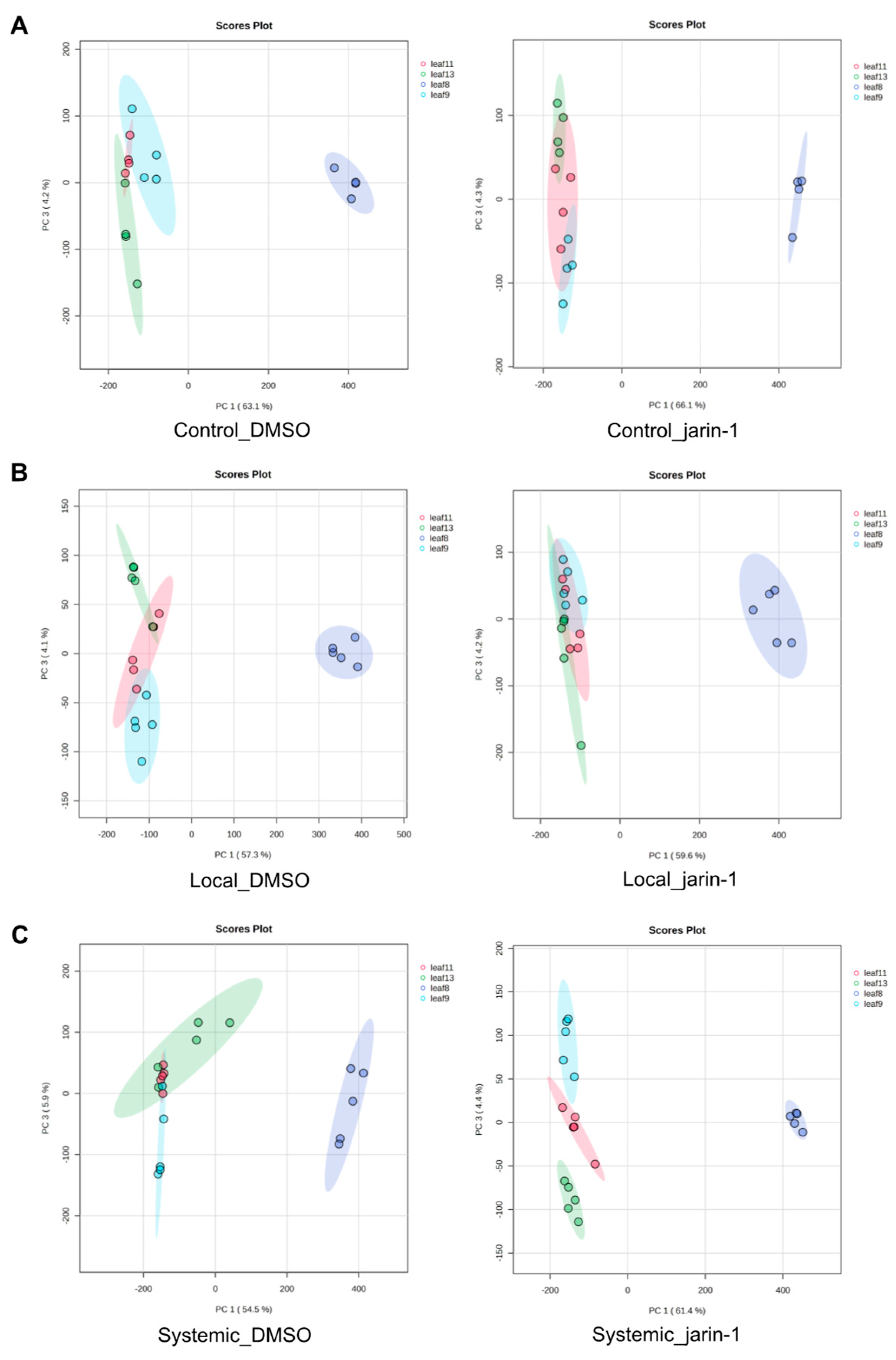
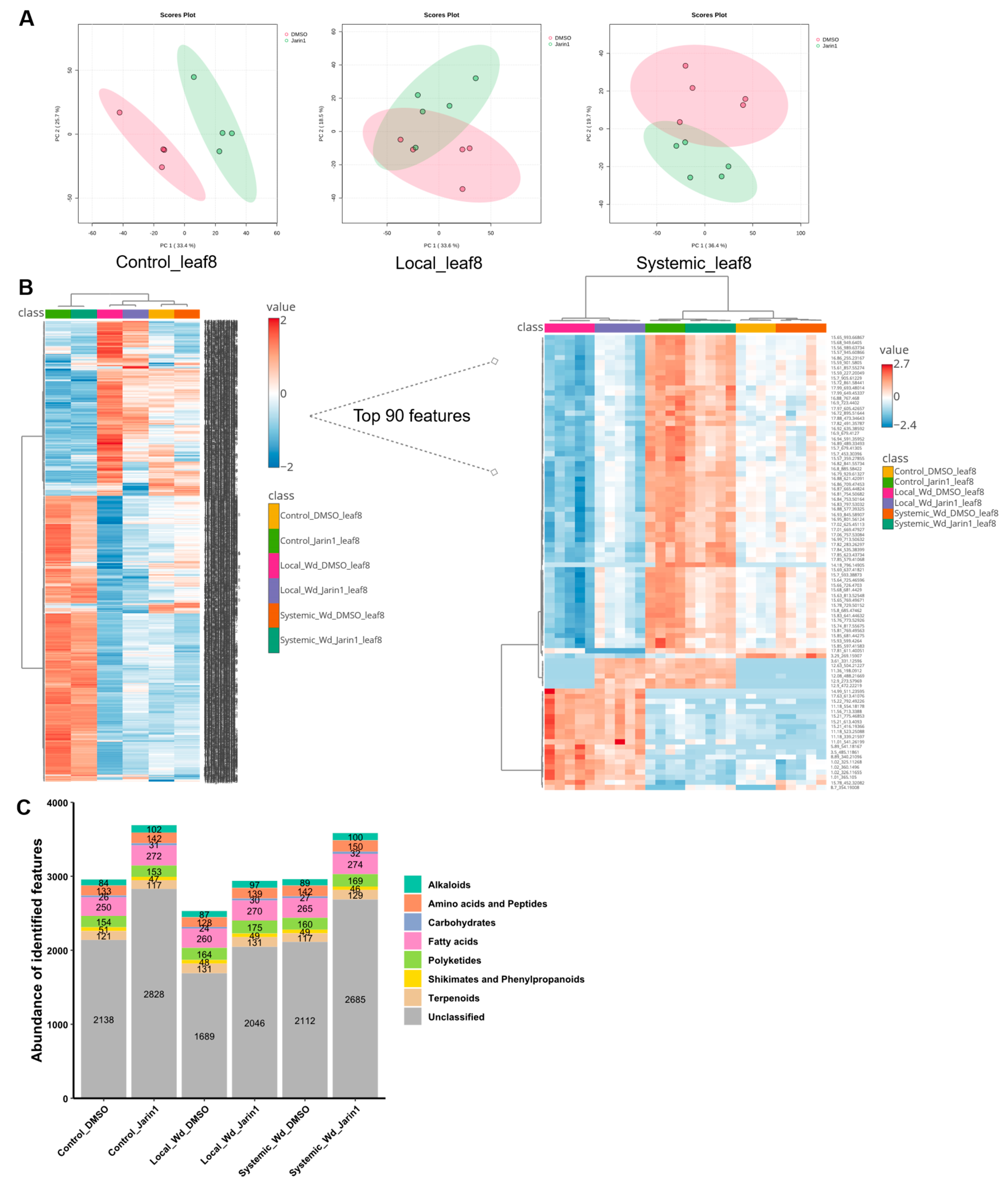
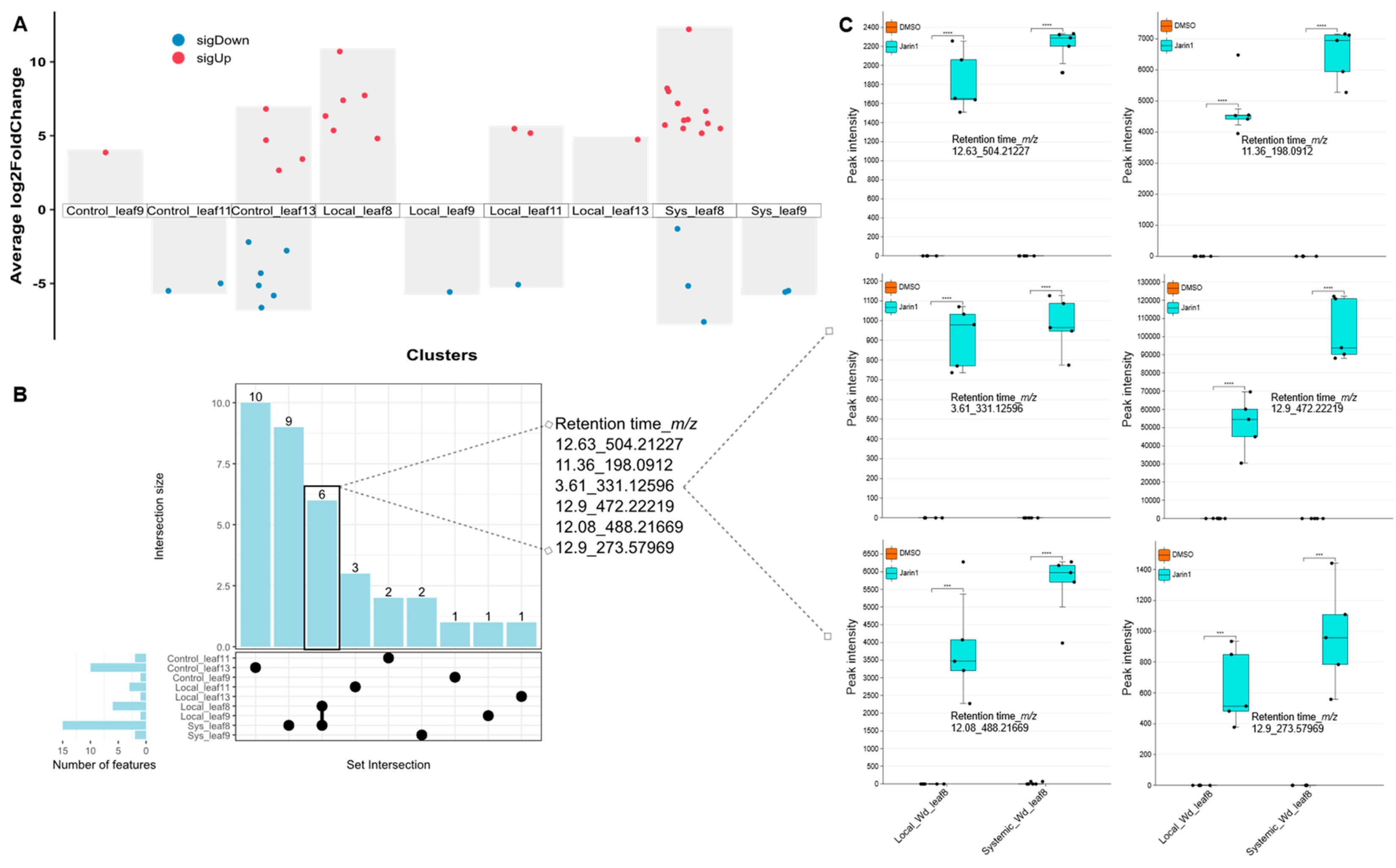
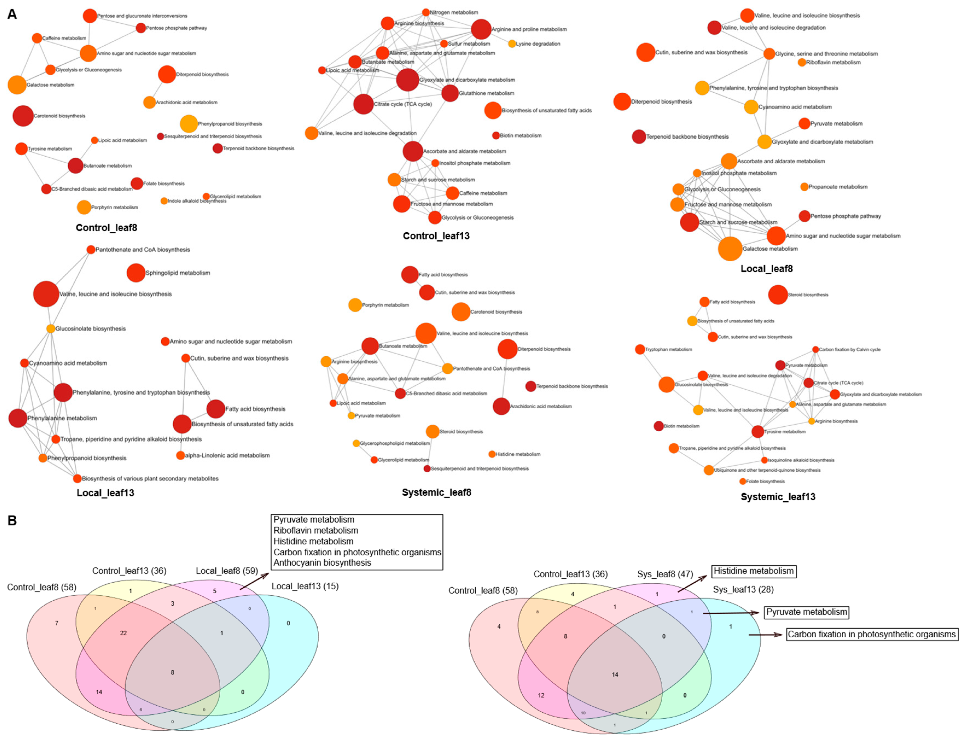
Inhibition of JAR1 Alters the Local and Systemic Leaf Metabolic Profiles

First, we wanted to analyze whether jarin-1 can suppress herbivory- or wound-induced JA-Ile production in vivo in Arabidopsis leaves. Therefore, the effect of different exogenously applied jarin-1 concentrations on wound-induced JA-Ile accumulation was tested. We found that jarin-1 reduced JA-Ile levels at 7 µM and 21 µM in the youngest fully expanded leaves by around 50% (Figure S1). Considering that too high concentration of jarin-1 itself exerts a negative effect on plant growth (Zeng et al., 2023), we selected 10 µM jarin-1 as the working concentration for subsequent experiments. Next, to determine whether jarin-1-mediated JAR1 inhibition affects the leaf metabolic profiles of Arabidopsis following wounding or not, we performed an experiment illustrated in Figure 1, inoculating leaves with different chemical and wounding treatment. After the indicated time, leaves were harvested and used for metabolome analysis. Principal component analysis (PCA) of the overall features in the different leaves compared the maximal variance among all leaves across 6 treatments (Figure 2A-C). Across all treatments, the metabolic pattern of leaf 8 was clearly separated from that of the other leaves (Figure 2A-C), probably due to the infiltration effects of solvents such as DMSO and Tween-20 on the chemically treated leaves. Compared to the controls, jarin-1 caused only minor alterations in leaf chemistry in the local wounding experiment, but induced substantial metabolic shifts in the systemic wounding experiment (Figure 2B, C). Focusing on the local leaf 8, both the PCA plots (Figure 3A) and the feature richness analysis (Table S1) indicated that jarin-1 predominantly affected the metabolite composition of leaf 8 under control conditions, but to a lesser extent upon local or systemic wounding (Figure 3A; Table S1). This might be because intense wounding responses attenuated the jarin-1-mediated metabolic changes in leaf 8. In total, 1,503 metabolic features were identified as significant biomarkers in leaf 8 (Figure 3B). Among them, the top 90 features were primarily induced by wounding (Figure 3B). In contrast, no complete separation of metabolic profiles was observed in the systemic leaf 13 under any of the three experimental designs (Figure 4A). However, this does not imply that jarin-1 had no impact on leaf 13; rather, it partially shifted the overall metabolic composition (Figure 2C; Table S1). Among them, 201 metabolic features were significantly altered (Figure 4B). The features in leaves 8 and 13 were classified into alkaloid, amino acid and peptide, fatty acid, terpenoids, polyketides, fatty acids and other natural product pathways (Figure 3C and Figure 4C). The metabolic features detected as significant ones in Figure 3B and Figure 4B were dependent on the treatment.
Figure 1. Experimental setup for leaf metabolic profiling. Control: leaf 8 was treated with the chemical DMSO or jarin-1 on the surface. There was no wounding treatment on control plants. Local: leaf 8 was treated with the chemical DMSO or jarin-1 on the surface. One hour and a half after chemical treatment, leaf 8 was wounded twice continuously by tweezers. Systemic: leaf 8 was treated with the chemical DMSO or jarin-1 on the surface. One hour and a half after chemical treatment, leaf 13 was wounded twice continuously by tweezers. In all experiments, leaf 8, 9, 11 and 13 were harvested separately one hour after wounding while control plants were waiting still for one hour. The red arrows indicate the wounding site. DMSO, dimethyl sulfoxide.
Preprints 185403 g001
Figure 2. Principal component analysis (PCA) of overall metabolites in leaf 8, 9, 11, 13 within control (A), local (B) and systemic (C) experimental setup. Control: leaf 8 was treated with the chemical DMSO or jarin-1 on the surface. There was no wounding treatment on control plants. Local: leaf 8 was treated with the chemical DMSO or jarin-1 on the surface. One hour and a half after chemical treatment, leaf 8 was wounded twice continuously by tweezers. Systemic: leaf 8 was treated with the chemical DMSO or jarin-1 on the surface. One hour and a half after chemical treatment, leaf 13 was wounded twice continuously by tweezers. In all experiments, leaf 8, 9, 11 and 13 were harvested separately one hour after wounding while control plants were waiting still for one hour. DMSO, dimethyl sulfoxide.
Preprints 185403 g002
Figure 3. The metabolic profile of leaf 8. (A) Ordination analysis of overall metabolites in leaf 8 under different treatments. (B) Heatmap of foldchange of metabolic features in leaf 8 under different treatments. The average intensity of four or five biological replicates was used for the heatmap analysis. 1503 features were selected through one-way ANOVA analysis (FDR-corrected P value ≤0.05 cutoff). The left panel indicates the foldchange of the total 1503 features, while the right panel shows the foldchange of top 90 features. In the right panel, the feature intensity of each biological replicate was used for the heatmap analysis. (C) Annotation of features by SIRIUS in leaf 8. Classified features were sorted into the Natural Products (NP) pathways as shown in the legend. The values shown in the bar plot represent the number of features in the respective NP pathway. A feature was counted as "present" when the average intensity of each group was ≥1000 counts. The features in the respective blanks were removed. Wd: wounding treatment. DMSO, dimethyl sulfoxide.
Preprints 185403 g003
Figure 4. The metabolic profile of leaf 13. (A) Ordination analysis of overall metabolites in leaf 13 under different treatments. (B) Heatmap of foldchange of metabolic features in leaf 13 under different treatments. The average intensity of four or five biological replicates was used for the heatmap analysis. 201 features were selected through one-way ANOVA analysis (FDR-corrected P value ≤0.05 cutoff). The panel indicates the foldchange of the total 201 features. (C) Annotation of features by SIRIUS in leaf 13. Classified features were sorted into the Natural Products (NP) pathways as shown in the legend. The values shown in the bar plot represent the number of features in the respective NP pathway. A feature was counted as "present" when the average intensity of each group was ≥1000 counts. The features in the respective blanks were removed. Wd: wounding treatment. DMSO, dimethyl sulfoxide.
Preprints 185403 g004
In order to rule out the possible influence of the experimental setup on the detection of metabolite profiles altered by jarin-1, we performed pairwise comparisons of metabolic profiles within the same leaf type and under the same experimental conditions. We found that JAR1 inhibition following wounding predominantly shifted the metabolic composition of leaf 8, compared with that of leaf 13 (Figure 5A; Tables S1 and S2). This is evident from the fact that leaf 8 exhibited a considerably higher number of significant features than leaf 13, when the features from different treatments are combined (Figure 5A; Table S2). In contrast, no differential metabolites were detected in leaf 8 of the control group, indicating that the observed changes were specifically triggered by the interaction between jarin-1 treatment and wounding. This further suggests that JAR1-generated JA-Ile plays a central role in regulating the metabolic response of wounded leaves.
Figure 5. Features with high foldchange. (A) Multi-volcano plots presenting significantly changed features by the application of jarin-1 compared to counterparts with DMSO treatment. There were no significant features detected in the leaves of control_leaf8, sys_leaf11, sys_leaf13. The labels “sigDown” and “sigUp” indicate those features significantly downregulated or upregulated in the respective leaves. The significant features were extracted at a threshold of FDR-corrected P value ≤0.05 after comparing the peak list of DMSO and jarin-1 treatment on the same leaf from the same experiment design. (B) Upset plot of significant features across different treatments and leaves. The numbers above the bars represent common features shared by two treatments or specific features in a single leaf. (C) Comparison of intensities of 6 features shared by local_leaf8 and sys_leaf8. Asterisks indicate significant differences between groups (Student’s t-test, ***P ≤0.001, ****P ≤0.0001,). DMSO, dimethyl sulfoxide. Sys: systemic. Wd: wounding treatment. DMSO, dimethyl sulfoxide.
Preprints 185403 g005
Notably, 6. metabolic features were shared by leaf 8 between the local and systemic experiments, all of which were strongly upregulated by jarin-1 (Figure 5B, C). Next, we performed a functional analysis of the differential metabolic features, and we found that these features were associated with diverse metabolic pathways, including valine, leucine, and isoleucine metabolism, fatty acid biosynthesis, and others (Figure 6A). Among these, histidine metabolism, pyruvate metabolism, and carbon fixation in photosynthetic organisms were typically altered by jarin-1 treatment in both local and systemic experiments (Figure 6B).
Figure 6. Pathway analysis of differential metabolites. (A) Enrichment network of leaf differential metabolites by the application of jarin-1. (B) Venn diagram of leaf differential metabolic pathways. The left and right panels show the overlapped and differentiated metabolic pathways among leaves by the application of jarin-1. The value in the diagram and bracket indicate the number of differential metabolic pathways in each leaf by the application of jarin-1. In (A) and (B), differentially expressed metabolites with matching KEGG IDs were annotated to the KEGG database. The KEGG pathways were selected through Mummichog algorithms (P ≤0.05 cutoff). Sys: systemic. DMSO, dimethyl sulfoxide.
Preprints 185403 g006

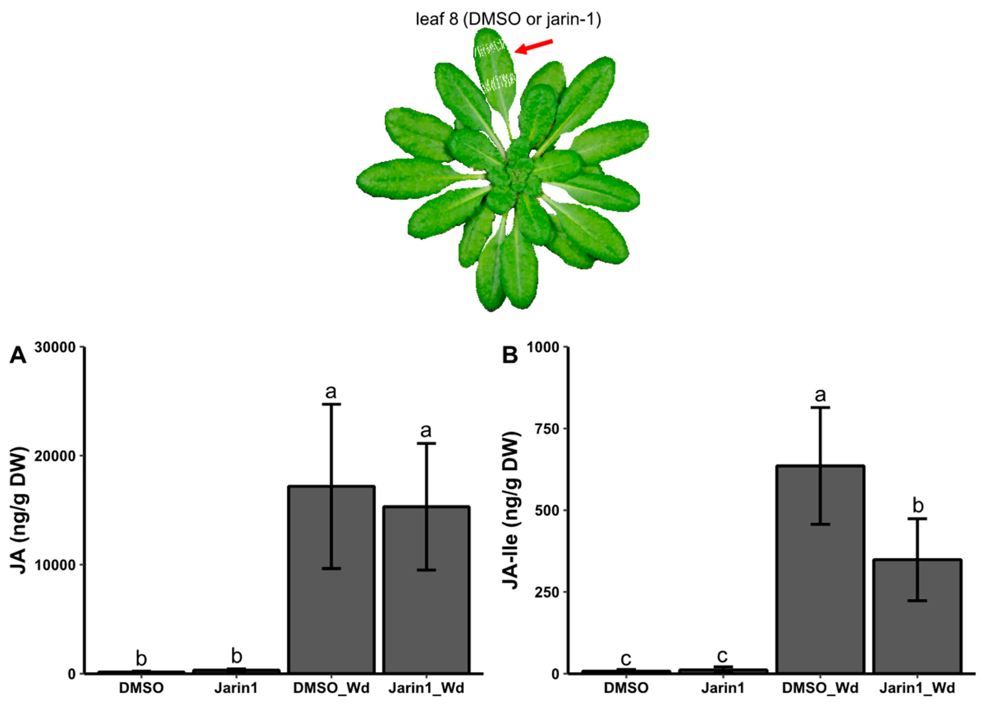
Jarin-1 Partly Inhibits Wound-Stimulated JA-Ile Biosynthesis in a Local Manner

We previously tested the dose effect of jarin-1 on the accumulation of JA-Ile in local leaves after wounding (Figure S1). Next, to further investigate the effect of jarin-1 on JA-Ile level of leaf 8 upon wounding, we developed an in planta assay, where leaf 8 was treated with jarin-1 and wounding (Figure 7). As a result, we found that there was no difference of JA level before and after jarin-1 application (Figure 7A). However, jarin-1 reduced JA-Ile level triggered by wounding in the local leaves, compared to JA-Ile level in DMSO control-treated leaves by wounding (Figure 7B). Overall, these results suggest that jarin-1 functioned specifically by preventing wound-induced JA-Ile production in a local manner.
Figure 7. Effect of jarin1 on the accumulation of JA and JA-Ile of local leaves after wounding. (A) JA and (B) JA-Ile in local leaves of 6 weeks old plants treated without and with DMSO or 10 µM jarin-1 and subjected to wounding or not. As indicated in the upper panel, the young fully expanded leaf was chosen for chemicals and wounding treatment. The leaf was treated without chemicals (Control) or with the same amount of DMSO or jarin1, and one and a half hours later, each plant was not wounded (Control) or leaf wounded (DMSO/Jarin1) with tweezers. JA and JA-Ile were assessed one hour after wounding. Data are mean ± SD (n≥4). Each biological replicate represents leaf material from one pot. Different letters indicate significant differences between treatments according to generalized linear model (GLM) and post hoc tests adjusted by Bonferroni method at P ≤0.05. DMSO, dimethyl sulfoxide.
Preprints 185403 g007

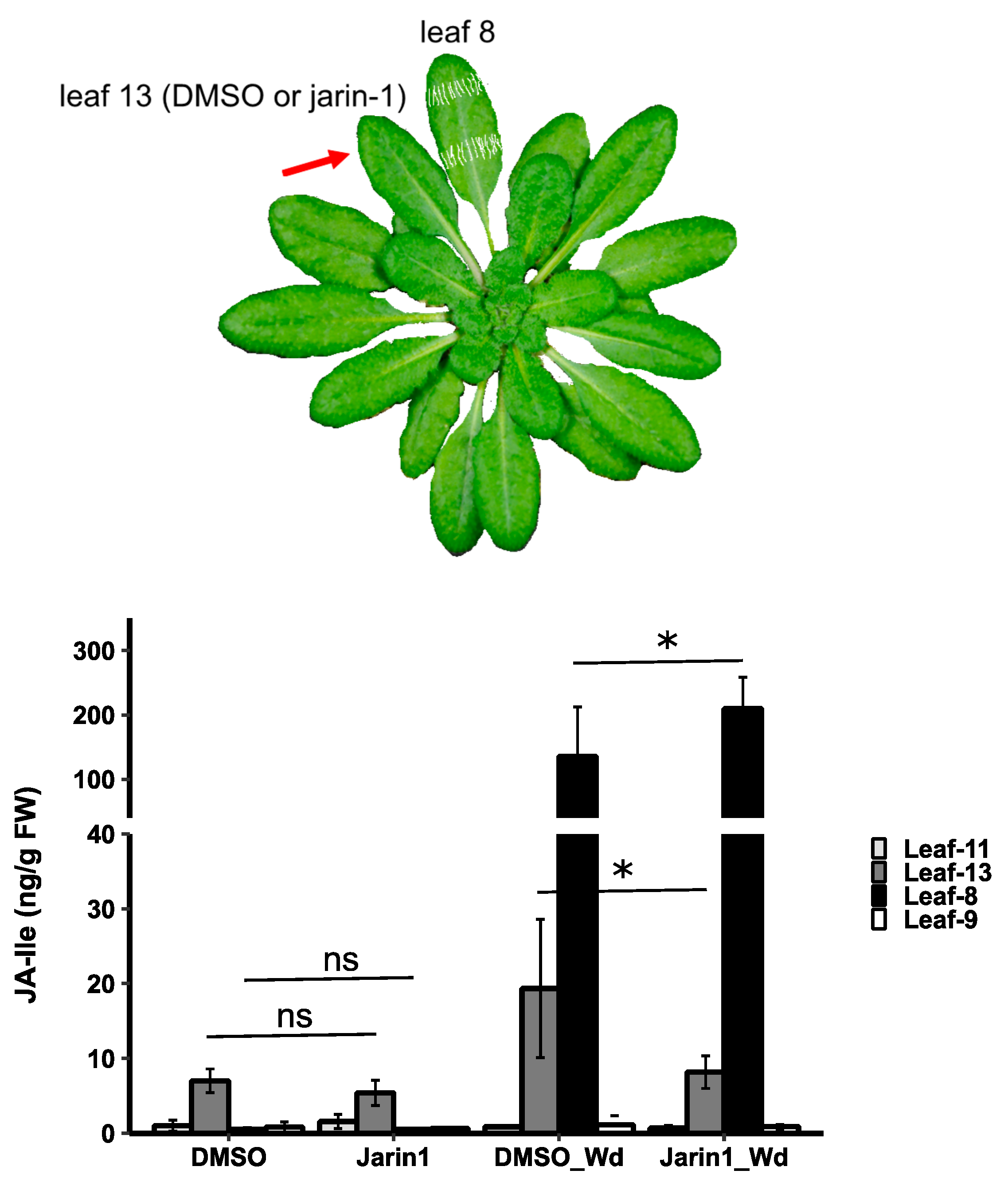
Jarin-1 Inhibits Wound-Stimulated JA-Ile Biosynthesis in a Systemic Manner

To further test whether jarin-1 systemically affects JA-Ile accumulation upon wounding, we developed another in planta assay, where leaf 13, treated with 10 µM jarin-1, was regarded as the systemic leaf; leaf 8 with wounding treatment was regarded as the local leaf (Figure 8). Without wounding, the application of DMSO or jarin-1 chemical itself caused slightly the rise of JA-Ile level. However, there was no difference between the two levels. With wounding, there was a significant increase of JA-Ile level in leaf 8 and 13, compared to the non-wounded plants (Figure 8). Interestingly, jarin-1 led to a downregulation of JA-Ile level of leaf 13; however, JA-Ile level of leaf 8 remained high. This suggests that jarin-1 can undermine the burst of JA-Ile of the systemic leaf which has a vascular connection with the wounded leaf.
Figure 8. Effect of jarin1 on the accumulation of JA-Ile of systemic leaves after wounding. JA-Ile in systemic leaves (leaf 13) of 6 weeks old plants treated with DMSO or 10 µM jarin-1 and subjected to wounding (Wd) or not on leaf 8. As indicated in the upper panel, the leaves from one pot were numbered, and leaf 8 was chosen for wounding treatment while leaf 13 for chemical treatment. Leaf 13 was treated with the same amount of DMSO or jarin1, and one and a half hours later, leaf 8 was wounded with tweezers. JA-Ile was assessed in leaf 8, 9, 11 and 13 one hour after wounding. Data are mean ± SD (n≥4). Each biological replicate represents leaf material from one pot. Asterisks indicate significant differences between groups (Student’s t-test, *P ≤0.05, ns, not significant). DMSO, dimethyl sulfoxide.
Preprints 185403 g008
Similarly, to test the effect of jarin-1 on JA-Ile accumulation upon mimicked herbivore feeding, we performed an experiment using MecWorm (Mithöfer et al., 2005). As for this experimental setup, leaf 8 chemically treated with 10 µM jarin-1 was regarded as the systemic leaf, whereas leaf 13 mechanically treated with MecWorm was regarded as the local leaf (Figure S3). Aligning with Figure 2, wounding led to a strong increase of JA-Ile level in leaf 13 and its vascular-connected leaf 8, compared to non-vascular-connected leaf 9 and 11 (Figure S3). As a result, jarin-1 led to a decrease of JA-Ile level of the chemically treated leaf 8, while JA-Ile level of the wounded leaf 13 stayed high (Figure S3). Based on the results of the above two independent experiments, it implies that jarin-1 can offset the burst of JA-Ile in the systemic leaves upon wounding on the local leaves.

Discussion

Inhibition of JAR1 Alters the Leaf Metabolic Profile

JAR1 belongs to the GH3 acyl acid amido synthetase enzyme family in plants (Staswick et al., 2002; Staswick & Tiryaki, 2004; Suza & Staswick, 2008). Members of the GH3 family are cytosolic acyl acid amido synthetases of the firefly luciferase enzyme group, which catalyze the conjugation of JAs, auxins, salicylic acid, and benzoic acid derivatives to a broad range of amino acids (Jez, 2022). Jarin-1 was first reported to specifically inhibit JAR1 in Arabidopsis and as a consequence thereof, e.g. partially alleviates MeJA-induced root growth inhibition in Arabidopsis (Meesters et al., 2014; Ishimaru et al., 2018). Until now, jarin-1 was applied in many plant species, such as strawberry, tomato and potato (Delgado et al., 2018; Liu et al., 2022; Munawar et al., 2023). However, it does not always have the same effect of relieving root growth inhibition by MeJA on other species (Zeng et al., 2023). Additionally, auxin signaling and other metabolic pathways involving GH3 enzymes may also be affected by jarin-1 treatment (Delgado et al., 2018). Moreover, in addition to their well-known roles in wound responses, JAs also participate in various plant developmental processes (Nie et al., 2025). Therefore, we hypothesized that inhibition of JAR1 could influence a range of leaf metabolic pathways.
Upon boron deficiency, JAR1-dependent JA signaling is involved in root growth inhibition in Arabidopsis (Huang et al., 2021), and JAR1 is proposed to influence growth and development in Medicago sativa by regulating photosynthesis (Dai et al., 2025). Although several partial functions of JAR1 have been characterized in different plant species (Svyatyna et al., 2014; Huang et al., 2021; Dai et al., 2025); however, so far, comprehensive information regarding the global metabolic networks regulated by JAR1, both under resting and stimulated conditions, remains limited.
To address this gap, we performed untargeted metabolomic analyses and found that the presence or reduced level of JA-Ile selectively modified a range of metabolites and their associated pathways (Figure 5 and Figure 6; Table S1). Furthermore, JA-Ile level influenced metabolite abundance under both wounded and non-wounded conditions (Figure 3 and Figure 4; Table S1). Functional enrichment analysis revealed that JAR1 modulation significantly affected several key metabolic pathways, including pyruvate metabolism, amino acid metabolism, sugar metabolism, fatty acid metabolism, and carbon fixation (Figure 6). Among these, pyruvate metabolism serves as a central metabolic hub linking primary metabolism and amino acid biosynthesis and degradation (Fataftah et al., 2018; Figure S2). It also regulates nitrogen (N) starvation response and contributes to the remobilization of N sources under low-N conditions by linking glycolysis and TCA cycle (Fataftah et al., 2018). Additionally, we found that JA-Ile level specifically influenced the anthocyanin biosynthetic pathway in leaf 8 during the local wounding experiment (Figure 6B). This finding aligns with previous studies demonstrating that JAs enhance total anthocyanin accumulation in several plant species (Shan et al., 2009; Concha et al., 2013; Shin et al., 2013; Yu et al., 2020). Collectively, our results suggest that JAR1 as JA-Ile generating enzyme acts as a key integrator of metabolic regulation, functioning not only in constitutive plant growth and development, but also in spatial and temporal responses to wounding stress.
Both mechanical wounding and insect herbivory trigger a rapid increase in cytosolic Ca2+ and a following boost in JA/JA-Ile levels in local and systemic leaves of Arabidopsis (Vadassery et al., 2012, 2014; Kiep et al., 2015). In this study, JAR1 activity not only modified JA-Ile-dependent metabolic pathways, also coordinated the JA-Ile-mediated wounding response at both local and systemic levels (Figure 7 and Figure 8). Recently, JAR1 and GH3.10, two members of the GH3 synthetases family, were shown to jointly catalyze JA conjugation in Arabidopsis (Delfin et al., 2022; Ni et al., 2025; Oki et al., 2025). Accordingly, a residual level of JA-Ile was observed following jarin-1 treatment (Figure 7 and Figure 8; S1 and S3), consistent with the fact that jarin-1 might not affect GH3.10 activity. Notably, JAR1 specifically influenced JA-Ile accumulation, whereas JA levels remained unaffected (Figure 7). A previous study suggests that JA and probably MeJA, rather than JA-Ile, function as long-distance transmissible signals, or interact with other long-distance signals to propagate systemic defense response (Bozorov et al., 2017). Also, mobile signals such as endogenous OPDA and its derivatives, rather than JA-Ile, translocate from wounded shoot to non-wounded root, thereby coordinating systemic wounding response (Schulze et al., 2019). Consequently, JA-Ile precursors that accumulate upon wounding may indirectly affect the biosynthesis of downstream-induced metabolites associated with defense responses. However, this does not imply that JA-Ile is dispensable in systemic defense regulation. Exogenous JA-Ile application has been shown to induce a few metabolites with high fold changes compared with JA and MeJA treatments, including lactic acid, β-glucose, alanine, threonine, steroids, 3,4-dihydroxypyridine and others in the leaves of Leucaena leucocephala (Xu et al., 2018). Moreover, JA-Ile formation has been demonstrated to regulate a specific subset of the JA-dependent soluble metabolome (Schuman et al., 2018). Together, these findings suggests that JAR1-mediated JA-Ile biosynthesis might also play a crucial role in fine-tuning both local and systemic defense metabolism in Arabidopsis.

JA-Ile is Probably Not a Mobile Signal in This Context

Wounding and mechanical stimulation are well-known triggers of plant defense responses (reviewed in Waterman et al., 2019). Similarly, repetitive touch can activate metabolite-based signaling pathways, including JA signaling pathways (Chehab et al., 2012). These stimuli probably activate a set of responses in local and systemic leaves through the direct and indirect vascular connections among the rosettes. In this study, we observed that combined jarin-1 and wounding treatment led to a reduction in JA-Ile levels in jarin-1–treated leaves, both locally and systemically, compared with DMSO and wounding controls. However, this treatment did not significantly affect JA levels (Figure 7), nor did it alter JA-Ile levels in wounded leaves during the systemic experiment (Figure 8 and Figure S3). This very likely is due to the following facts: (i) jarin-1 is a specific JAR1 inhibitor (Meesters et al., 2014), so it did not affect the JA, but the JA-Ile level; (ii) wounding in the leaves might be so strong that the remaining JAR1 activity in the local leaf is sufficient to generate JA-Ile despite the presence of jarin-1; (iii) The systemic response/resistance is probably activated by JA-dependent and-independent mobile signals (Gasperini et al., 2015; Bozorov et al., 2017). Some systemic signals, independent of the JAs pathway, such as electrical signaling, calcium ions, reactive oxygen species (ROS), were also involved in the long-distance transmission of wounding or herbivory stress signals (Mousavi et al., 2013; Kiep et al., 2015; Nguyen et al., 2018; Toyota et al., 2018; Wang et al., 2019). In Arabidopsis, a JA-independent wound signal triggers the rapid de novo biosynthesis of JA and JA-Ile in distal leaves (Koo et al., 2009). Moreover, the rapid electrical signal trigger JAs upsurge in systemic leaves (Mousavi et al., 2013; Toyota et al., 2018). GLUTAMATE RECEPTOR-LIKE 3.3 (GLR3.3)-modulated electrical signal is speculated to be converted subsequently into JA or JA-Ile (Li et al., 2020). In addition, calcium and ROS may be relayed in a self-propagating manner by the generation of JA waves along the phloem pathway (Hilleary & Gilroy, 2018). And a core set of transcripts associated with the ROS wave are linked with Ca2+ signaling (Zandalinas et al., 2019). Some JA-independent signals can interact with each other and are involved in plant stress response together with JA-dependent signals. Also, JA-dependent and-independent mobile signals could be influenced by each other.
In summary, our data support the view that JA-Ile is not a mobile signal. While mobile immune signals transmit to systemic tissues (non-wounded leaves), JAR1 is essential to stimulate local responses and catalyze JA-Ile biosynthesis locally. Our findings indicate that other wound-induced signals move to the systemic parts of the plants and induce JAR1-mediated JA-Ile synthesis. Here. Further investigation, for example an approach with isotope-labelled JA-Ile to trace its mobility, may provide new insights in the regulation of plant defense mechanisms as well as growth and developmental processes.

Supplementary Materials

The following supporting information can be downloaded at the website of this paper posted on Preprints.org.

Acknowledgments

This article was partly supported by iDiv funded by the German Research Foundation (DFG–FZT 118, 202548816). We gratefully acknowledge Dr. Nicole Sachmerda-Schulz and Johanna Müller for yDiv administrative assistance, Dr. Michael Reichelt (MPI-CE) for help in phytohormone analysis and Andrea Lehr (MPI-CE) for technical assistance. We acknowledge Dr. Van cong Doan, Dr. Pierre Pétriacq and Dr. Abigail Moreno-Pedraza for their comments and also Prof. Dr. Bettina Hause for reading the previous version of this manuscript.

Competing interests

None declared.

Author Contributions

MZ performed the experiments, analyzed the data and wrote the first version of this manuscript. AM supervised the study and contributed to the first version of the manuscript. All authors contributed to the final version.

Data availability statement

Data available on request from the authors.

References

  1. Bozorov, T.A.; Dinh, S.T.; Baldwin, I.T. JA but not JA-Ile is the cell-nonautonomous signal activating JA mediated systemic defenses to herbivory in Nicotiana attenuata. Journal of Integrative Plant Biology 2017, 59, 552–571. [Google Scholar] [CrossRef]
  2. Cao, J.; Li, M.; Chen, J.; Liu, P.; Li, Z. Effects of MeJA on Arabidopsis metabolome under endogenous JA deficiency. Scientific Reports 2016, 6, 37674. [Google Scholar] [CrossRef]
  3. Chambers, M.C.; MacLean, B.; Burke, R. A Cross-platform Toolkit for Mass Spectrometry and ProteomicsZwane’s mining charter lunacy. Nature Biotechnology 2012, 30, 918–920. [Google Scholar] [CrossRef]
  4. Chehab, E.W.; Yao, C.; Henderson, Z.; Kim, S.; Braam, J. Arabidopsis Touch-Induced Morphogenesis Is Jasmonate Mediated and Protects against Pests. Current Biology 2012, 22, 701–706. [Google Scholar] [CrossRef]
  5. Choudhury, F.K.; Devireddy, A.R.; Azad, R.K.; Shulaev, V.; Mittler, R. Local and systemic metabolic responses during light-induced rapid systemic signaling. Plant Physiology 2018, 178, 1461–1472. [Google Scholar] [CrossRef] [PubMed]
  6. Concha, C.M.; Figueroa, N.E.; Poblete, L.A.; Oñate, F.A.; Schwab, W.; Figueroa, C.R. Methyl jasmonate treatment induces changes in fruit ripening by modifying the expression of several ripening genes in Fragaria chiloensis fruit. Plant Physiology and Biochemistry 2013, 70, 433–444. [Google Scholar] [CrossRef]
  7. Dai, R.; Chen, Q.; Wang, X.Y.; Huo, X.W.; Li, J.W.; An, J.B.; Mi, F.G.; Shi, F.L.; Zhang, Z.Q. Overexpression MsJAR1 gene increased lateral branches and plant height in alfalfa (Medicago sativa L.). Scientific Reports 2025, 15, 16956. [Google Scholar] [CrossRef] [PubMed]
  8. Dávila-Lara, A.; Rahman-Soad, A.; Reichelt, M.; Mithöfer, A. Carnivorous Nepenthes x ventrata plants use a naphthoquinone as phytoanticipin against herbivory. PLoS ONE 2021, 16, e0258235. [Google Scholar] [CrossRef]
  9. Delfin, J.C.; Kanno, Y.; Seo, M.; Kitaoka, N.; Matsuura, H.; Tohge, T.; Shimizu, T. AtGH3.10 is another jasmonic acid-amido synthetase in Arabidopsis thaliana. Plant Journal 2022, 110, 1082–1096. [Google Scholar] [CrossRef]
  10. Delgado, L.D.; Zúñiga, P.E.; Figueroa, N.E.; Pastene, E.; Escobar-Sepúlveda, H.F.; Figueroa, P.M.; Garrido-Bigotes, A.; Figueroa, C.R. Application of a JA-Ile biosynthesis inhibitor to methyl jasmonate-treated strawberry fruit induces upregulation of specific MBW complex-related genes and accumulation of proanthocyanidins. Molecules 2018, 23, 1433. [Google Scholar] [CrossRef] [PubMed]
  11. Dengler, N.G. The shoot apical meristem and development of vascular architecture. Canadian Journal of Botany 2006, 84, 1660–1671. [Google Scholar] [CrossRef]
  12. Dührkop, K.; Fleischauer, M.; Ludwig, M.; Aksenov, A.A.; Melnik, A.V.; Meusel, M.; Dorrestein, P.C.; Rousu, J.; Böcker, S. SIRIUS 4: A rapid tool for turning tandem mass spectra into metabolite structure information. Nature Methods 2019, 16, 299–302. [Google Scholar] [CrossRef] [PubMed]
  13. Dührkop, K.; Nothias, L.; Fleischauer, M.; Reher, R.; Ludwig, M.; Hoffmann, M.A.; Petras, D.; Gerwick, W.H.; Rousu, J.; Dorrestein, P.C.; et al. Systematic classification of unknown metabolites using high-resolution fragmentation mass spectra. Nature Biotechnology 2021, 39, 462–471. [Google Scholar] [CrossRef] [PubMed]
  14. Dührkop, K.; Shen, H.; Meusel, M.; Rousu, J.; Böcker, S. Searching molecular structure databases with tandem mass spectra using CSI : FingerID. Proceedings of the National Academy of Sciences 2015, 112, 12580–12585. [Google Scholar] [CrossRef]
  15. Farmer, E.; Farmer, E.; Mousavi, S.; Lenglet, A. Leaf numbering for experiments on long distance signalling in Arabidopsis. Protocol Exchange 2013, 071. [Google Scholar] [CrossRef]
  16. Fataftah, N.; Mohr, C.; Hajirezaei, M.R.; Wirén Nvon Humbeck, K. Changes in nitrogen availability lead to a reprogramming of pyruvate metabolism. BMC Plant Biology 2018, 18, 77. [Google Scholar] [CrossRef]
  17. Feunang, Y.D.; Eisner, R.; Knox, C.; Chepelev, L.; Hastings, J.; Owen, G.; Fahy, E.; Steinbeck, C.; Subramanian, S.; Bolton, E.; et al. ClassyFire : Automated chemical classification with a comprehensive, computable taxonomy. Journal of Cheminformatics 2016, 8, 61. [Google Scholar] [CrossRef]
  18. Fonseca, S.; Chini, A.; Hamberg, M.; Adie, B.; Porzel, A.; Kramell, R.; Miersch, O.; Wasternack, C.; Solano, R. (+)-7-iso-Jasmonoyl-L-isoleucine is the endogenous bioactive jasmonate. Nature Chemical Biology 2009, 5, 344–350. [Google Scholar] [CrossRef] [PubMed]
  19. Gasperini, D.; Chételat, A.; Acosta, I.F.; Goossens, J.; Pauwels, L.; Goossens, A.; Dreos, R.; Alfonso, E.; Farmer, E.E. Multilayered Organization of Jasmonate Signalling in the Regulation of Root Growth. PLoS Genetics 2015, 11, e1005300. [Google Scholar] [CrossRef]
  20. Hilleary, R.; Gilroy, S. Systemic signaling in response to wounding and pathogens. Current Opinion in Plant Biology 2018, 43, 57–62. [Google Scholar] [CrossRef]
  21. Hoffmann, M.A.; Nothias, L.; Ludwig, M.; Fleischauer, M.; Emily, C.; Witting, M.; Dorrestein, P.C.; Dührkop, K.; Böcker, S. Assigning confidence to structural annotations from mass spectra with COSMIC. bioRxiv 2021. [CrossRef]
  22. Huang, Y.; Wang, S.; Shi, L.; Xu, F. JASMONATE RESISTANT 1 negatively regulates root growth under boron deficiency in Arabidopsis. Journal of Experimental Botany 2021, 72, 3108–3121. [Google Scholar] [CrossRef]
  23. Ishimaru, Y.; Hayashi, K.; Suzuki, T.; Fukaki, H.; Prusinska, J.; Meester, C.; Quareshy, M.; Egoshi, S.; Matsuura, H.; Takahashi, K.; et al. Jasmonic acid inhibits auxin-induced lateral rooting independently of the CORONATINE INSENSITIVE1 receptor. Plant Physiology 2018, 177, 1704–1716. [Google Scholar] [CrossRef] [PubMed]
  24. Jez, J.M. Connecting primary and specialized metabolism: Amino acid conjugation of phytohormones by GRETCHEN HAGEN 3 (GH3) acyl acid amido synthetases. Current Opinion in Plant Biology 2022, 66, 102194. [Google Scholar] [CrossRef]
  25. Kiep, V.; Vadassery, J.; Lattke, J.; Maaß, J.P.; Boland, W.; Peiter, E.; Mithöfer, A. Systemic cytosolic Ca2+ elevation is activated upon wounding and herbivory in Arabidopsis. New Phytologist 2015, 207, 996–1004. [Google Scholar] [CrossRef] [PubMed]
  26. Kim, H.W.; Wang, M.; Leber, C.A.; Nothias, L.; Reher, R.; Kang KBin Hooft JJJVan Der Dorrestein, P.C.; Gerwick, W.H.; Cottrell, G.W. NPClassi fi er : A Deep Neural Network-Based Structural Classi fi cation Tool for Natural Products. Journal of natural products 2021, 84, 2795–2807. [Google Scholar] [CrossRef] [PubMed]
  27. Koo, A.J.K.; Gao, X.; Daniel Jones, A.; Howe, G.A. A rapid wound signal activates the systemic synthesis of bioactive jasmonates in Arabidopsis. Plant Journal 2009, 59, 974–986. [Google Scholar] [CrossRef]
  28. Landgraf, R.; Schaarschmidt, S.; Hause, B. Repeated leaf wounding alters the colonization of Medicago truncatula roots by beneficial and pathogenic microorganisms. Plant Cell and Environment 2012, 35, 1344–1357. [Google Scholar] [CrossRef]
  29. Li, M.; Wang, F.; Li, S.; Yu, G.; Wang, L.; Li, Q.; Zhu, X.; Li, Z.; Yuan, L.; Liu, P. Importers Drive Leaf-to-Leaf Jasmonic Acid Transmission in Wound-Induced Systemic Immunity. Molecular Plant 2020, 13, 1485–1498. [Google Scholar] [CrossRef]
  30. Liu, X.; Cheng, L.; Li, R.; Cai, Y.; Wang, X.; Fu, X.; Dong, X.; Qi, M.; Jiang, C.Z.; Xu, T.; et al. The HD-Zip transcription factor SlHB15A regulates abscission by modulating jasmonoyl-isoleucine biosynthesis. Plant Physiology 2022, 189, 2396–2412. [Google Scholar] [CrossRef]
  31. Ludwig, M.; Nothias, L.; Dührkop, K.; Koester, I.; Fleischauer, M.; Hoffmann, M.A.; Petras, D.; Vargas, F.; Morsy, M.; Böcker, S.; et al. Database-independent molecular formula annotation using Gibbs sampling through ZODIAC. Nature Machine Intelligence 2020, 2, 629–641. [Google Scholar] [CrossRef]
  32. Matsuura, H.; Takeishi, S.; Kiatoka, N.; Sato, C.; Sueda, K.; Masuta, C.; Nabeta, K. Transportation of de novo synthesized jasmonoyl isoleucine in tomato. Phytochemistry 2012, 83, 25–33. [Google Scholar] [CrossRef] [PubMed]
  33. Meesters, C.; Mönig, T.; Oeljeklaus, J.; Krahn, D.; Westfall, C.S.; Hause, B.; Jez, J.M.; Kaiser, M.; Kombrink, E. A chemical inhibitor of jasmonate signaling targets JAR1 in Arabidopsis thaliana. Nature Chemical Biology 2014, 10, 830–836. [Google Scholar] [CrossRef] [PubMed]
  34. Mithöfer, A.; Wanner, G.; Boland, W. Effects of feeding Spodoptera littoralis on lima bean leaves. II. Continuous mechanical wounding resembling insect feeding is sufficient to elicit herbivory-related volatile emission. Plant Physiology 2005, 137, 1160–1168. [Google Scholar] [CrossRef]
  35. Mousavi, S.A.R.; Chauvin, A.; Pascaud, F.; Kellenberger, S.; Farmer, E.E. GLUTAMATE RECEPTOR-LIKE genes mediate leaf-to-leaf wound signalling. Nature 2013, 500, 422–426. [Google Scholar] [CrossRef]
  36. Munawar, A.; Xu, Y.; Abou El-Ela, A.S.; Zhang, Y.; Zhong, J.; Mao, Z.; Chen, X.; Guo, H.; Zhang, C.; Sun, Y.; et al. Tissue-specific regulation of volatile emissions moves predators from flowers to attacked leaves. Current Biology 2023, 33, 2321–2329.e5. [Google Scholar] [CrossRef]
  37. Neumann, P.M.; Chamel, A. Comparative Phloem Mobility of Nickel in Nonsenescent Plants. Plant Physiology 1986, 81, 689–691. [Google Scholar] [CrossRef]
  38. Nguyen, C.T.; Kurenda, A.; Stolz, S.; Chételat, A.; Farmer, E.E. Identification of cell populations necessary for leaf-to- leaf electrical signaling in a wounded plant. Proceedings of the National Academy of Sciences 2018, 115, 10178–10183. [Google Scholar] [CrossRef]
  39. Ni, B.; Klein, M.; Hossbach, B.; Feussner, K.; Hornung, E.; Herrfurth, C.; Hamberg, M.; Feussner, I. Arabidopsis GH3.10 conjugates jasmonates. Plant Biology 2025, 27, 476–491. [Google Scholar] [CrossRef]
  40. Nie, R.; Chen, D.; Hu, T.; Zhang, S.; Qu, G. A Review: The Role of Jasmonic Acid in Tomato Flower and Fruit Development. Plant Molecular Biology Reporter 2025, 43, 474–483. [Google Scholar] [CrossRef]
  41. Oki, K.; Enoki, A.; Yokota, Y.; Kurihara, T.; Shimizu, T.; Saburi, W.; Tohge, T.; Mori, H.; Van den Ackerveken, G.; Kitaoka, N.; et al. AtGH3.10 and JAR1 Produce 12-Hydroxyjasmonoyl-l-isoleucine from 12-Hydroxyjasmonic Acid in Arabidopsis thaliana. ChemBioChem 2025, 26, e202500151. [Google Scholar] [CrossRef] [PubMed]
  42. Pang, Z.; Lu, Y.; Zhou, G.; Hui, F.; Xu, L.; Viau, C.; Spigelman, A.F.; Macdonald, P.E.; Wishart, D.S.; Li, S.; et al. MetaboAnalyst 6.0: Towards a unified platform for metabolomics data processing, analysis and interpretation. Nucleic Acids Research 2024, 52, W398–W406. [Google Scholar] [CrossRef] [PubMed]
  43. Posit Software, P.B.C. 2024. RStudio: Integrated development environment for R. Boston, MA, USA: Posit Software, PBC. https://posit.co/.
  44. R Core Team. 2024. R: A language and environment for statistical computing. Vienna, Austria: R Foundation for Statistical Computing. https://www.R-project.org/.
  45. Santino, A.; Taurino, M.; De Domenico, S.; Bonsegna, S.; Poltronieri, P.; Pastor, V.; Flors, V. Jasmonate signaling in plant development and defense response to multiple (a)biotic stresses. Plant Cell Reports 2013, 32, 1085–1098. [Google Scholar] [CrossRef] [PubMed]
  46. Sato, C.; Aikawa, K.; Sugiyama, S.; Nabeta, K.; Masuta, C.; Matsuura, H. Distal Transport of Exogenously Applied Jasmonoyl—Isoleucine with Wounding Stress. Plant & cell physiology 2011, 52, 509–517. [Google Scholar]
  47. Scholz, S.S.; Malabarba, J.; Reichelt, M.; Heyer, M.; Ludewig, F.; Mithöfer, A. Evidence for GABA-induced systemic GABA accumulation in arabidopsis upon wounding. Frontiers in Plant Science 2017, 8, 388. [Google Scholar] [CrossRef]
  48. Schulze, A.; Zimmer, M.; Mielke, S.; Stellmach, H.; Melnyk, C.W.; Hause, B.; Gasperini, D. Wound-Induced Shoot-to-Root Relocation of JA-Ile Precursors Coordinates Arabidopsis Growth. Molecular Plant 2019, 12, 1383–1394. [Google Scholar] [CrossRef]
  49. Schuman, M.C.; Meldau, S.; Gaquerel, E.; Diezel, C.; McGale, E.; Greenfield, S.; Baldwin, I.T. The active jasmonate JA-Ile regulates a specific subset of plant jasmonate-mediated resistance to herbivores in nature. Frontiers in Plant Science 2018, 9, 787. [Google Scholar] [CrossRef]
  50. Shan, X.; Zhang, Y.; Peng, W.; Wang, Z.; Xie, D. Molecular mechanism for jasmonate-induction of anthocyanin accumulation in arabidopsis. Journal of Experimental Botany 2009, 60, 3849–3860. [Google Scholar] [CrossRef]
  51. Shin, D.H.; Choi, M.G.; Lee, H.K.; Cho, M.; Choi, S.B.; Choi, G.; Park, Y.I. Calcium dependent sucrose uptake links sugar signaling to anthocyanin biosynthesis in Arabidopsis. Biochemical and Biophysical Research Communications 2013, 430, 634–639. [Google Scholar] [CrossRef]
  52. Staswick, P.E.; Tiryaki, I. The oxylipin signal jasmonic acid is activated by an enzyme that conjugate it to isoleucine in Arabidopsis W inside box sign. Plant Cell 2004, 16, 2117–2127. [Google Scholar] [CrossRef]
  53. Staswick, P.E.; Tiryaki, I.; Rowe, M.L. Jasmonate response locus JAR1 and several related Arabidopsis genes encode enzymes of the firefly luciferase superfamily that show activity on jasmonic, salicylic, and indole-3-acetic acids in an assay for adenylation. Plant Cell 2002, 14, 1405–1415. [Google Scholar] [CrossRef] [PubMed]
  54. Suza, W.P.; Staswick, P.E. The role of JAR1 in Jasmonoyl-l-isoleucine production during Arabidopsis wound response. Planta 2008, 227, 1221–1232. [Google Scholar] [CrossRef] [PubMed]
  55. Svyatyna, K.; Jikumaru, Y.; Brendel, R.; Reichelt, M.; Mithöfer, A.; Takano, M.; Kamiya, Y.; Nick, P.; Riemann, M. Light induces jasmonate-isoleucine conjugation via OsJAR1-dependent and -independent pathways in rice. Plant, Cell and Environment 2014, 37, 827–839. [Google Scholar] [CrossRef] [PubMed]
  56. Tautenhahn, R.; Patti, G.; Rinehart, D.; Siuzdak, G. Metabolomic Data. Analytical Chemistry 2012, 84, 5035–5039. [Google Scholar] [CrossRef]
  57. Toyota, M.; Spencer, D.; Sawai-toyota, S.; Jiaqi, W.; Zhang, T.; Koo, A.J.; Howe, G.A.; Gilroy, S. Glutamate triggers long-distance, calcium-based plant defense signaling. Science 2018, 361, 1112–1115. [Google Scholar] [CrossRef]
  58. Vadassery, J.; Reichelt, M.; Hause, B.; Gershenzon, J.; Boland, W.; Mithöfer, A. CML42-Mediated Calcium Signaling Coordinates Responses to Spodoptera Herbivory and Abiotic Stresses. Plant Physiology 2012, 159, 1159–1175. [Google Scholar] [CrossRef]
  59. Vadassery, J.; Reichelt, M.; Jimenez-Aleman, G.H.; Boland, W.; Mithöfer, A. Neomycin Inhibition of (+)-7-Iso-Jasmonoyl-L-Isoleucine Accumulation and Signaling. Journal of Chemical Ecology 2014, 40, 676–686. [Google Scholar] [CrossRef]
  60. Wang, G.; Hu, C.; Zhou, J.; Liu, Y.; Cai, J.; Pan, C.; Wang, Y.; Wu, X.; Shi, K.; Xia, X.; et al. Systemic Root-Shoot Signaling Drives Jasmonate-Based Root Defense against Nematodes. Current Biology 2019, 29, 3430–3438.e4. [Google Scholar] [CrossRef]
  61. Waterman, J.M.; Cazzonelli, C.I.; Hartley, S.E.; Johnson, S.N. Simulated Herbivory: The Key to Disentangling Plant Defence Responses. Trends in Ecology and Evolution 2019, 34, 447–458. [Google Scholar] [CrossRef]
  62. Weinhold, A.; Döll, S.; Liu, M.; Schedl, A.; Pöschl, Y.; Xu, X.; Neumann, S.; Dam, N.M.V. Tree species richness differentially affects the chemical composition of leaves, roots and root exudates in four subtropical tree species. Journal of Ecology 2022, 110, 97–116. [Google Scholar] [CrossRef]
  63. Xu, Y.; Tao, Z.; Jin, Y.; Chen, S.; Zhou, Z.; Gong, A.G.W.; Yuan, Y.; Dong, T.T.X.; Tsim, K.W.K. Jasmonate-Elicited Stress Induces Metabolic Change in the Leaves of Leucaena leucocephala. Molecules 2018, 23, 188. [Google Scholar] [CrossRef] [PubMed]
  64. Yu, J.; Zhu, M.; Wang, M.; Xu, Y.; Chen, W.; Yang, G. Transcriptome analysis of calcium-induced accumulation of anthocyanins in grape skin. Scientia Horticulturae 2020, 260, 108871. [Google Scholar] [CrossRef]
  65. Zandalinas, S.I.; Sengupta, S.; Burks, D.; Azad, R.K.; Mittler, R. Identification and characterization of a core set of ROS wave-associated transcripts involved in the systemic acquired acclimation response of Arabidopsis to excess light. Plant Journal 2019, 98, 126–141. [Google Scholar] [CrossRef]
  66. Zeng, M.; Krajinski, F.; van Dam, N.M.; Hause, B. Jarin-1, an inhibitor of JA-Ile biosynthesis in Arabidopsis thaliana, acts differently in other plant species. Plant Signaling and Behavior 2023, 18, e2273515. [Google Scholar] [CrossRef] [PubMed]
Disclaimer/Publisher’s Note: The statements, opinions and data contained in all publications are solely those of the individual author(s) and contributor(s) and not of MDPI and/or the editor(s). MDPI and/or the editor(s) disclaim responsibility for any injury to people or property resulting from any ideas, methods, instructions or products referred to in the content.
Copyright: This open access article is published under a Creative Commons CC BY 4.0 license, which permit the free download, distribution, and reuse, provided that the author and preprint are cited in any reuse.
Prerpints.org logo

Preprints.org is a free preprint server supported by MDPI in Basel, Switzerland.

Subscribe

Disclaimer

Terms of Use

Privacy Policy

Privacy Settings

© 2025 MDPI (Basel, Switzerland) unless otherwise stated