Preprint
Article

This version is not peer-reviewed.

Gap Junction Plaques Fragmented in the Cochlea of a Mouse Model of the Compound Heterozygous p.D50N Variant in Gjb2

Submitted:

26 November 2024

Posted:

26 November 2024

You are already at the latest version

Abstract

The GJB2 gene, which encodes the Connexin26 (Cx26), is the gene most frequently linked to hearing loss. Individuals carrying variants in the GJB2 gene may experience either congenital hearing loss or last-onset progressive hearing loss. The underlying reasons for the variability in hearing remain poorly understood. Current research primarily focuses on conditional knockout mouse models, which generally exhibit moderate to severe hearing loss. To elucidate the mechanisms by which Cx26 variants contribute to mild to moderate hearing loss, this study established a mouse model, the Gjb2D50N/- mice. Auditory brainstem response (ABR) indicated that the Gjb2D50N/- mice led to a mild to moderate increase in hearing thresholds. Further investigations demonstrated that in the Gjb2D50N/- mice, the gap junction plaques (GJPs) between supporting cells were fragmented, and the capacity of outer hair cells (OHCs) to uptake 2-NBDG (a glucose analog) was reduced. In conclusion, we hypothesize that the mild to moderate hearing loss associated with Cx26 variants may not be attributable to structural abnormalities within the cochlea, but rather to dysfunction of GJPs, which compromises the energy supply to OHCs. In this study, we first established a Gjb2 compound heterozygous mouse model that causes mild to moderate hearing loss, for subsequent intervention and treatment.

Keywords: 
;  ;  ;  

1. Introduction

Hearing loss is the most prevalent sensory impairment, affecting at least 1.1 out of every 1,000 newborns with permanent hearing loss [1]. In developed countries, genetic factors contribute to 50-60% of hearing loss cases in children [2]. To date, over 120 genes linked to non-syndromic hearing loss have been identified [3], with GJB2 variants are highly prevalent around the world [4]. The GJB2 gene encodes gap junction beta-2 protein, as known as Connexin26 (Cx26). Cx26 aggregates on the cell membrane to form gap junction plaques (GJPs) that facilitate intercellular communication and material transfer, playing a crucial role in maintaining the inner ear's homeostasis [5,6,7,8].
As gene sequencing increasingly becomes a standard approach for diagnosing congenital hearing loss, the heterogeneity of auditory phenotypes has attracted heightened scrutiny, suggesting that the extent of hearing impairment may be significantly modulated by genetic factors [9]. The 35delG variant is widely distributed across various populations and is frequently associated with profound congenital hearing loss [4,10,11]. In contrast, the V37I variant exhibits a high carrier frequency and typically presents with mild to moderate hearing loss [12]. The D50N variant is predominantly linked to Keratitis-Icthyosis-Deafness (KID) syndrome, with carriers potentially experiencing late-onset mild to moderate hearing loss or profound hearing loss at birth [13,14,15]. The substantial variability observed in auditory phenotypes implies that Cx26 may be involved in diverse mechanisms of deafness. Prior research has largely concentrated on Cx26 conditional knockout mice, revealing that early Cx26 deficiency can result in profound hearing loss due to structural abnormalities in the organ of Corti, loss of hair cells (HCs), and degeneration of spiral ganglion neurons (SGNs) [16,17,18,19]. Variants in Cx26 can be classified into truncating mutations, frameshift mutations, and missense mutations. Depending on the specific variant types and their locations, Cx26 variants can be categorized into several groups: those that are incapable of forming GJPs, those that produce non-functional GJPs, those exhibiting diminished ion permeability or substance exchange in the formed GJPs, and those that do not display functional abnormalities in GJPs. Consequently, the development of novel models is imperative for a more thorough and comprehensive investigation into the potential mechanisms associated with Cx26-related hearing loss.
Clinically, patients frequently present with two distinct types of allele variants. Due to the homozygous lethality linked to the GJB2 gene, we utilized CRISPR-Cas9 technology to generate the Gjb2D50N/+ mice, which were subsequently crossed with the Gjb2loxP/loxP; Rosa26CreER mice to produce the Gjb2D50N/- mice. We conducted auditory brainstem response (ABR) assessments and performed pathological examinations on these mice. Our results indicated that the Gjb2D50N/- mice exhibited mild to moderate progressive hearing impairment, which may not be attributed to the loss of HCs or SGNs. Importantly, we observed significant fragmentation of GJPs within the supporting cells (SCs), and the notably shortened GJPs were associated with diminished glucose uptake in HCs. We hypothesize that insufficient nutrition supply may further exacerbate HCs dysfunction and contribute to the hearing loss. In our study, we provided an ideal mouse model of mild to moderate hearing loss, which are of great significance for the development of subsequent therapy.

2. Materials and Methods
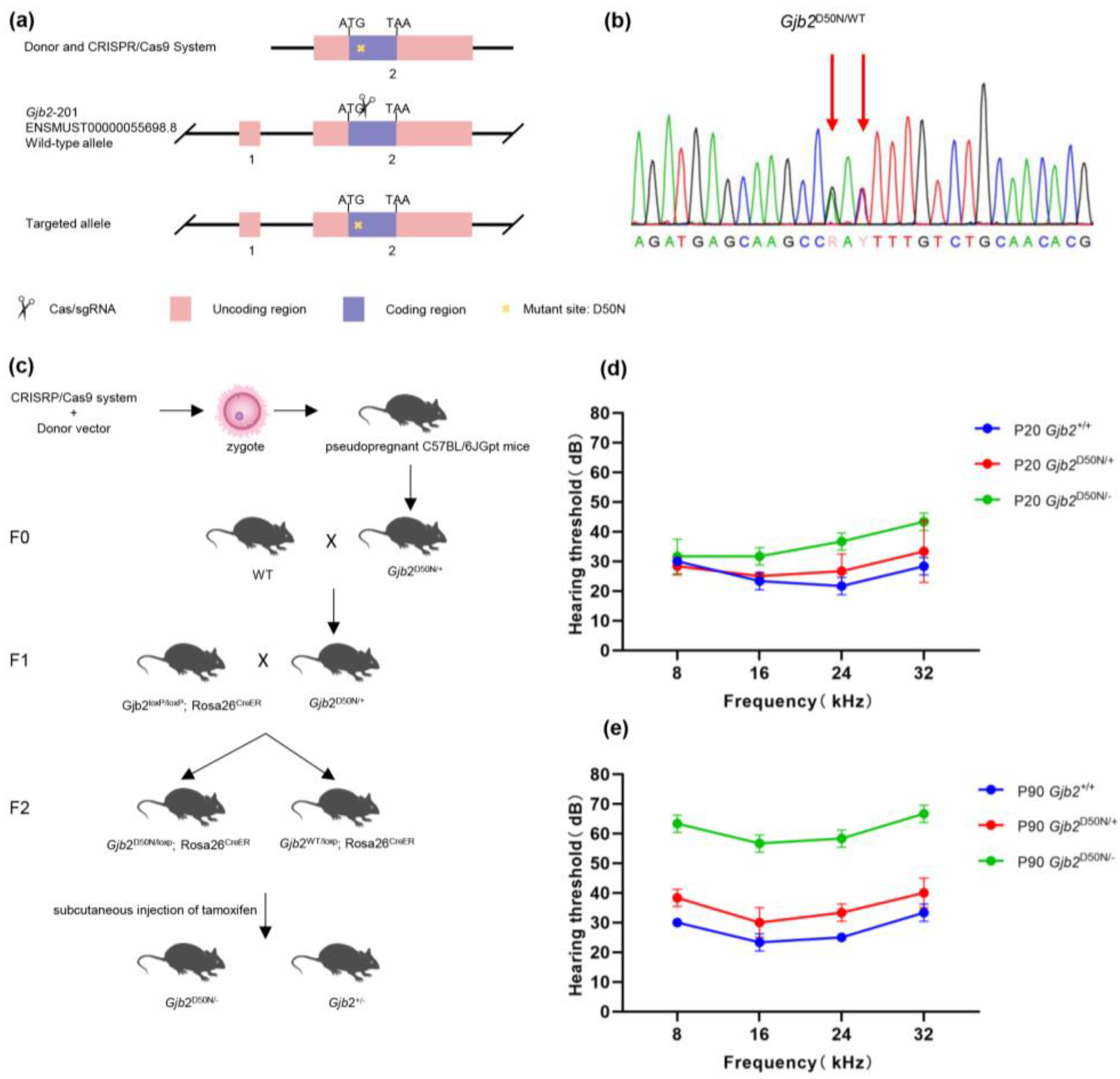
2.1. Mouse Models

According to the Ensembl database, the mouse Gjb2-201 transcript (transcript length 2406 bp, encoding 226 amino acids, spanning 2 exons) was selected for further analysis. Based on the mouse Gjb2 gene sequence and the requirement for a point mutation, guide RNAs (gRNA) were designed to replace the codon GAT with AAC in Exon 2, corresponding to the Gjb2 p.D50N variant. Meanwhile, the donor vector was synthesized and verified for accuracy through sequencing. Subsequently, the CRISPR-Cas9 system and the donor vector were mixed at appropriate concentrations and microinjected into C57BL/6JGpt mouse zygotes. The zygotes were then implanted into the uteri of pseudopregnant C57BL/6JGpt female mice for gestation and offspring production. Genomic DNA was extracted from the tails or toes of the mice 5–7 days post-birth using the One Step Mouse Genotyping Kit (Vazyme, China; PD101-01). The target region was amplified by PCR using the following primers: F1: 5ʹ- TTG TCA CCT ATC AGC AGC CTA GAGG-3ʹ; R1: 5ʹ- TTT CAT GTC TCC GGT AGG CCA-3ʹ. Sanger sequencing of the amplified products was then performed to confirm the genotype of the mice. Finally, mice with genotypes Gjb2D50N/wt or Gjb2D50N/+ were obtained through breeding.
Mice were raised in a specific-pathogen-free experimental animal center at Huazhong University of Science and Technology. Due to the homozygous lethality in the GJB2 gene, the Gjb2D50N/+ mice were crossbred with Gjb2loxP/loxP; Rosa26CreER mice. Consequently, the Gjb2D50N/loxp; Rosa26CreER mice were obtained. In our study, the mouse model of Gjb2D50N/- were generated by subcutaneous injection of tamoxifen (1.5mg/10g body weight; Sigma Aldrich, Germany; T5648-1G) diluted in a fat emulsion at P0 and P1.
All experimental procedures were conducted under the policies established by the Committee on Animal Research at Tongji Medical College, Huazhong University of Science and Technology.

2.2. Auditory Brainstem Response (ABR)

The hearing thresholds were measured using Auditory Brainstem Response (ABR) at postnatal days 20 (P20) and 90 (P90). A 1.25% solution of tribromoethanol (0.2ml/10g, intraperitoneally; AibeiBio, China; M2910) was administered to anesthetize the mice. The mice were then placed on a 37°C thermostatic electric blanket to maintain their body temperature within a sound-attenuating chamber. The Tucker-Davis Technology (TDT) System was employed to measure the hearing threshold, as previously published [16]. Tone burst stimuli were generated by the TDT System at frequencies of 8 kHz, 16 kHz, 24 kHz, and 32 Hz, and the responses were amplified and averaged over 1024 times. SigGen32 software (Tucker- Davis Technologies) was used to record the ABR signals. The lowest sound level that elicited a repeatable wave was considered the threshold.

2.3. Cochlear Tissue Preparation and Immunofluorescent Labeling

Animals were anesthetized with 1.25% tribromoethanol (0.2ml/10g, intraperitoneally) and perfused with 4% paraformaldehyde (PFA) in phosphate-buffered saline (PBS). The cochleas were carefully dissected from the temporal bones and fixed in 4% PFA at 4°C overnight. Following decalcification in 10% ethylenediaminetetraacetic acid disodium salt (disodium EDTA), the cochleas were dehydrated sequentially with 10%, 20%, and 30% sucrose solutions. Tissue-Tek® O.C.T. Compound (Sakura Finetek, America; 4583) was used to embed the processed tissue specimens, allowing for the cutting of sections with a thickness of 10 μm for morphological examination.
For the flattened cochlear preparation, the cochleae were meticulously dissected from the decalcified tissue using a stereomicroscope. Three segments of the basilar membrane- apical, middle, and basal turn were selected for staining. After permeabilization with 0.1% Triton X-100 in 0.01 M PBS for 20 minutes, the samples were immunolabeled overnight at 4°C with a 1:500 dilution of anti-Cx26 antibody (Rabbit; Invitrogen, America; 71-0500) and anti-CD45 antibody (Goat; R&D system, America; AF114). The secondary antibodies used were Alexa Fluor® 488 donkey anti-rabbit (Antgene, China; ANT024S) and Alexa Fluor® 647 donkey anti-goat (Antgene, China; ANT041S) which were applied at room temperature (22–25°C) for 90 minutes, and nuclei were stained with 4′,6–diamidino-2-phenylindole (DAPI, Antgene, China; ANT165) for 15 minutes. Images were obtained using a laser scanning confocal microscope (Nikon; Japan).

2.4. Assessment of In Vivo 2-NBDG Uptake in the HCs

Mice with different genotypes were anesthetized via an injection of 1.25% tribromoethanol (0.2 ml/10g, i.p.). A needle penetrated the left ventricle to introduce solutions into the cardiovascular system of the mice. A 3.8 mM solution of 2-NBDG (Invitrogen, America; N13195) in stroke-physiological saline solution was perfused into the mice for 2 minutes. After 5 minutes, the basilar membranes from the apical turn were rapidly dissected from the cochleae prepared for a flattened cochlear preparation. Finally, images were obtained using a laser scanning confocal microscope, and the fluorescent intensity of the area of interest was measured with ImageJ software.

2.5. Data Analysis

Statistical analyses were conducted using GraphPad Prism version 9.5 (GraphPad Software Inc., La Jolla, CA, USA). Data presented in the figures are expressed as mean ± standard deviation (SD). The least significant difference (LSD) post hoc test was employed to compare differences between two of the groups. In all analyses, P < 0.05 was considered statistically significant.

3. Results

3.1. The Gjb2D50N/- Mice Showed Mile to Moderate Hearing Loss

The model involved a mouse that expresses the Gjb2 p.D50N variant. First, we generated the Gjb2D50N/+ mice using CRISPR-Cas9 technology (Figure 1a) which was confirmed by Sanger sequencing (Figure 1b). However, homozygous lethality has been documented in the literature for variants in the GJB2 gene [20], which is consistent with our inability to obtain homozygous mice through long-term breeding. So, we crossed the Gjb2D50N/+ mice with the Gjb2loxP/loxP; Rosa26CreER mice, resulting in the Gjb2D50N/loxp; Rosa26CreER genotype. Following tamoxifen injection, we successfully obtained the Gjb2D50N/- mice (Figure 1c).
To evaluate if there was hearing loss of the Gjb2D50N/- mice, the ABRs of all the mice were measured at postnatal days 20 and 90 (P20 and P90). At P20, the ABR thresholds evoked by tone-bursts across a range of frequencies (8, 16, 24, 32kHz) in the Gjb2+/+ mice were 30±0, 23.33±2.89, 21.76±2.89, 28.33±2.89 dB SPL, while the ABR thresholds in the Gjb2D50N/- mice were 31.67±5.58, 31.67±2.89, 36.67±2.89, 43.33±2.89 dB SPL (Figure 1d). Compared to age-matched Gjb2+/+ mice, the Gjb2D50N/- mice showed 8.33, 15 and 15 dB increase in hearing thresholds at 16, 24, and 32 kHz at P20, respectively. The Gjb2D50N/+ mice did not show significant hearing loss. At P90, there were no significant hearing loss in the Gjb2+/+ mice. While, the ABR thresholds in the Gjb2D50N/- mice were 63.33±2.89, 56.67±2.89, 58.33±2.89, 66.67±2.89 dB SPL, which mean there were about 30 dB increase in hearing thresholds at all frequencies we measured (Figure 1e). The Gjb2D50N/+ mice showed 5-10 dB increase. To summarize, the Gjb2D50N/- mice have progressive hearing loss that gradually progresses to full frequency, and the Gjb2D50N/+ mice showed last-onset hearing loss.

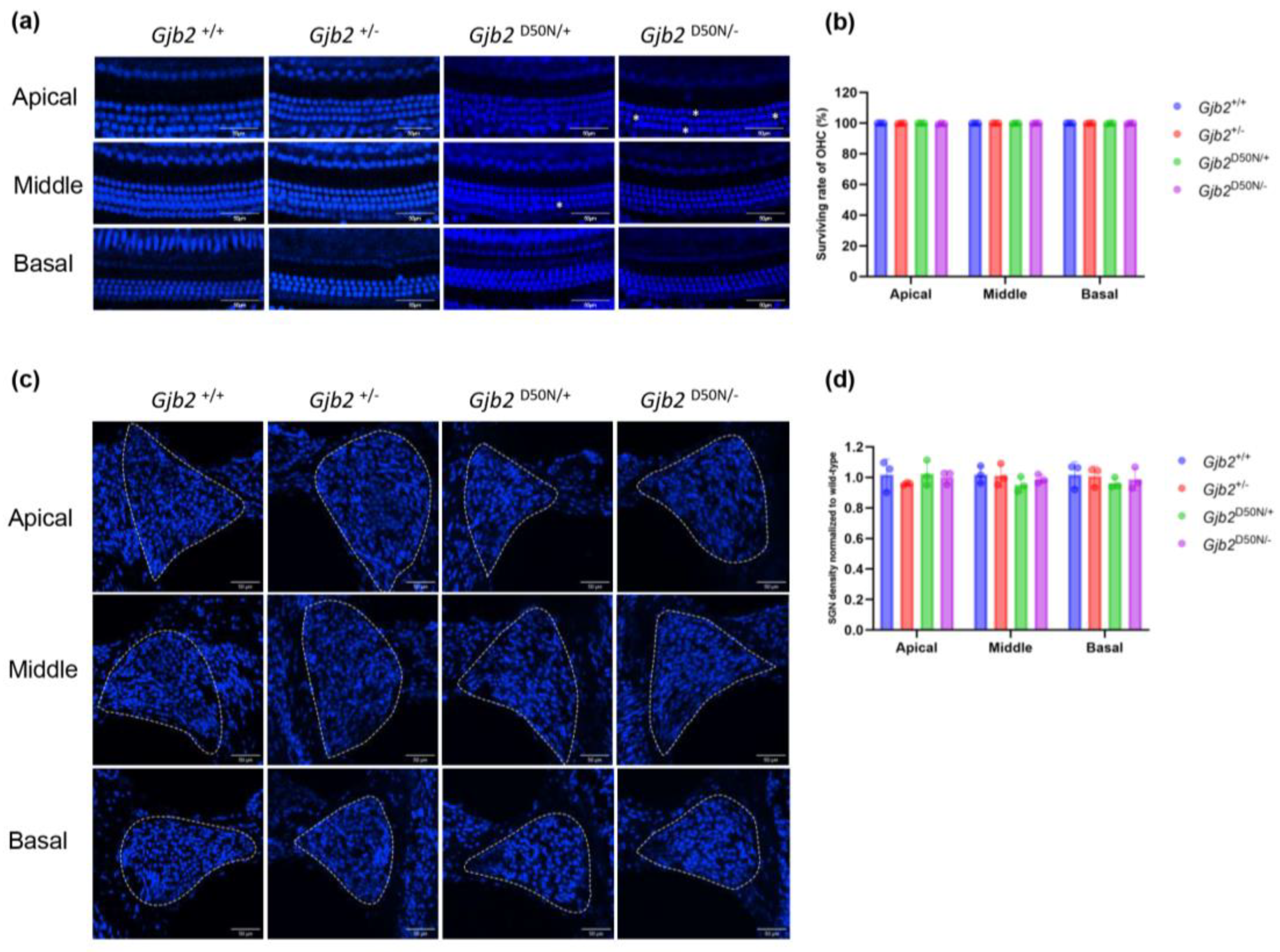
3.2. The Gjb2D50N/- Mice Showed no Significant HCs and SGNs Loss

Whole-mount preparations were utilized to investigate HCs counting (Figure 2a). At P20, the Gjb2 D50N/- mice exhibited sporadic OHCs loss in the apical, middle and basal turns, and we did not observe loss of OHCs in the Gjb2 D50N/+ mice(Figure 2b). Compared to other mouse models, Gjb2 V37I knock-in mice [21] and Gjb2V37M/V37M mice [22] showed similar results. There was also no significant HCs loss in Gjb235delG/delG mice at P14, while significant degeneration of the OHCs had occurred, especially in the basal turn at P35 [23]. In R75 W+ mice, microtubules of inner pilar cells (IPCs) were poorly formed and hypoplasia, tunnels of Corti did not open and even collapse. Interestingly, the supporting cells were incompletely developed, but HCs were not affected [24]. SGNs degeneration was quantified through SGNs counting (Figure 2c). In all groups, we did not observe significant losses of SGNs (Figure 2d). Therefore, we speculate that early in hearing formation, the D50N variant may not yet affect the survival of SGNs. In conclusion, we speculated the loss of HCs and SGNs is not the primary factor contributing to the elevation of hearing thresholds.

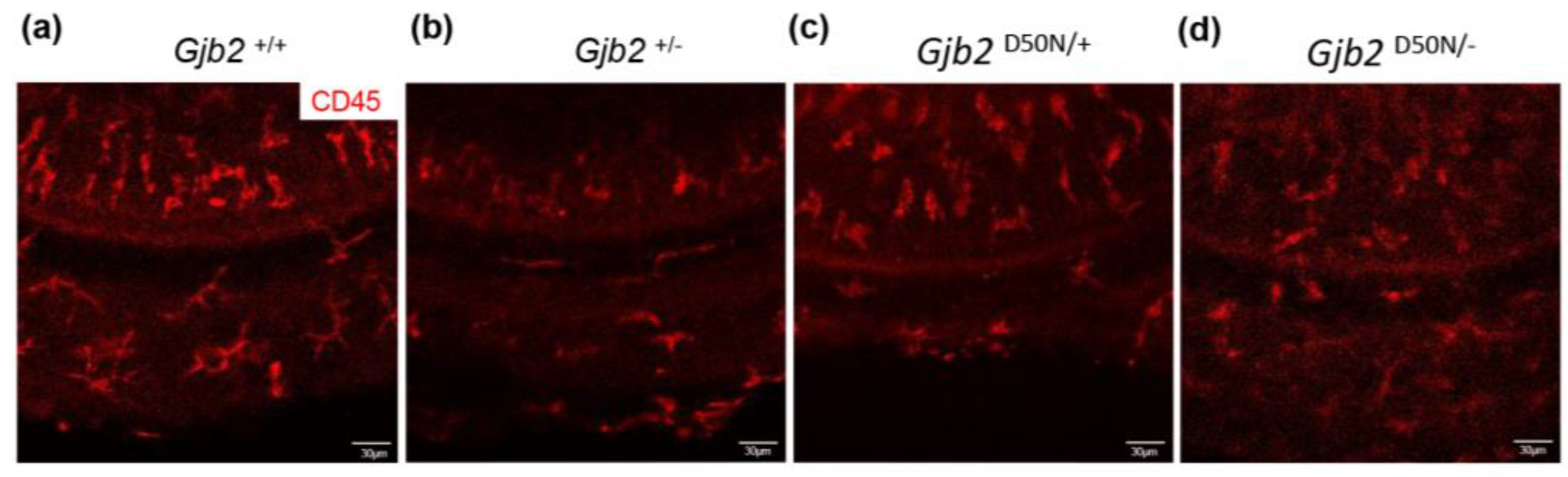
3.3. CD45+Cells in the Organ of Corti in the Gjb2D50N/- Mice Underwent Morphological Changes

In our preliminary observations in the Cx26-null mice, we found that inflammatory cells in the cochlea may play a role in the mechanism of hair cell damage [25]. To further validate this mechanism in the Gjb2D50N/- mice, we labeled cochlear macrophages with CD45. The CD45+cells located at the cochlear basilar membrane in the Gjb2+/+ mice (Figure 3a), the Gjb2+/- mice (Figure 3b), and the Gjb2 D50N/+ mice (Figure 3c) showed irregular appearance, while the Gjb2D50N/- mice (Figure 3d) exhibited morphological changes. Therefore, we speculate that the sporadic loss of HCs in the Gjb2D50N/- mice may be associated with the activation of CD45+cells.

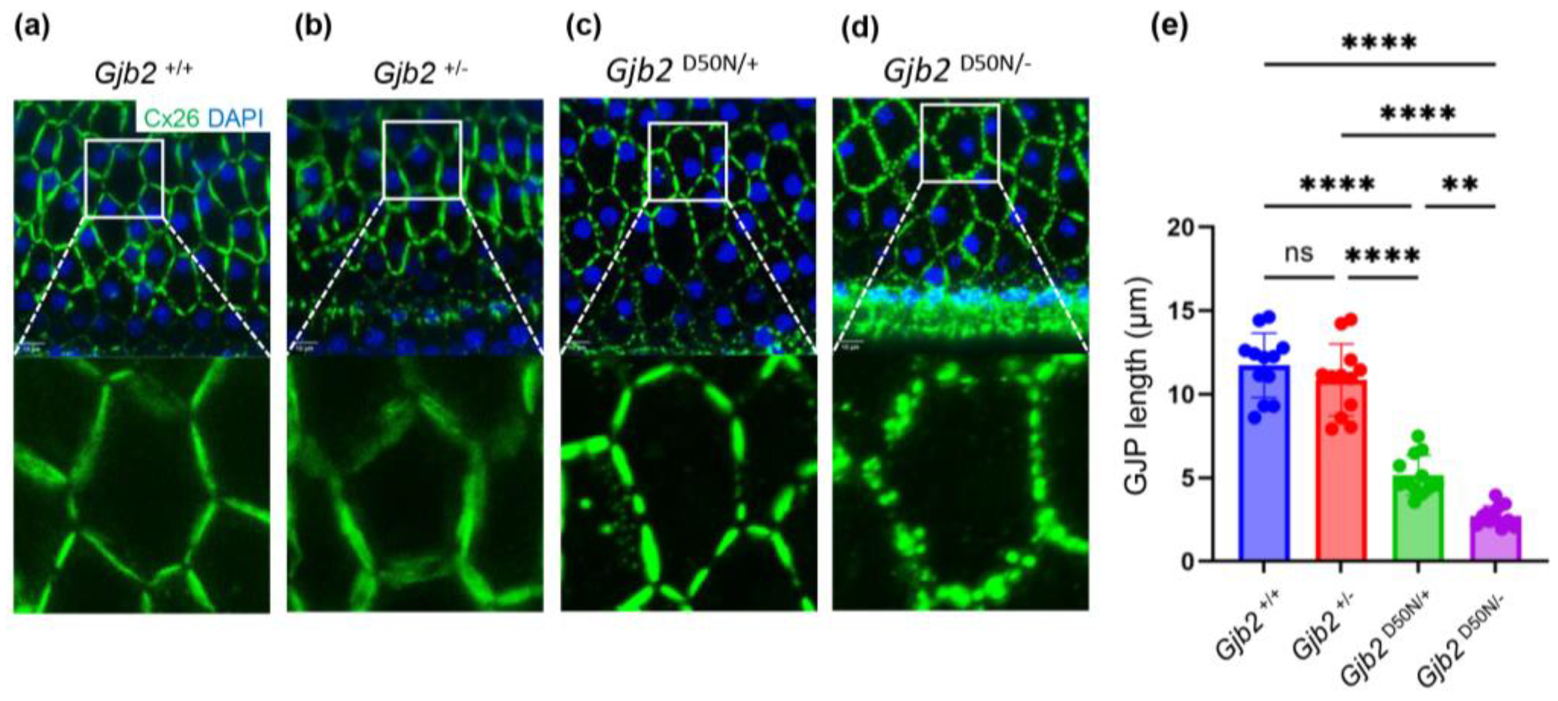
3.4. The Gjb2D50N/- Mice Show Shortened GJPs

According to the results of in vitro experiments, Cx26 variants can be classified into the following categories: those that cannot form GJPs on the cell membrane, those that form non-functional GJPs, those formed GJPs exhibit reduced ion permeability or material exchange capabilities, and those that form functionally GJPs. In our study, at P20, large and regular GJPs were observed on the cell membrane of the inner sulcus cells (ISCs) of the Gjb2+/+ mice (Figure 4a) and the Gjb2+/- mice (Figure 4b), with maximum lengths were 11.73±1.93μm and 10.87±2.14μm. The longest GJPs in the Gjb2 D50N/+ mice (Figure 4c) and the Gjb2 D50N/- mice (Figure 4d) measured 5.15±1.19μm and 2.73±0.60μm respectively, which were shortened 56% and 77% compared to the Gjb2+/+ mice (Figure 4e). Consequently, we propose that fragmented GJPs impair their function, resulting in diminished energy supply between sensory epithelial cells in the inner ear. Interestingly, the D50N variant had a perinuclear localization in NEB1 keratinocytes and corneal epithelial cells, which is different from our findings [26,27]. However, plasma membrane localization can be observed in the sweat gland cells for a KID patient who carried D50N variant[26]. This suggests that D50N variant may exhibit inconsistent behavior in different cells or in vitro versus in vivo.

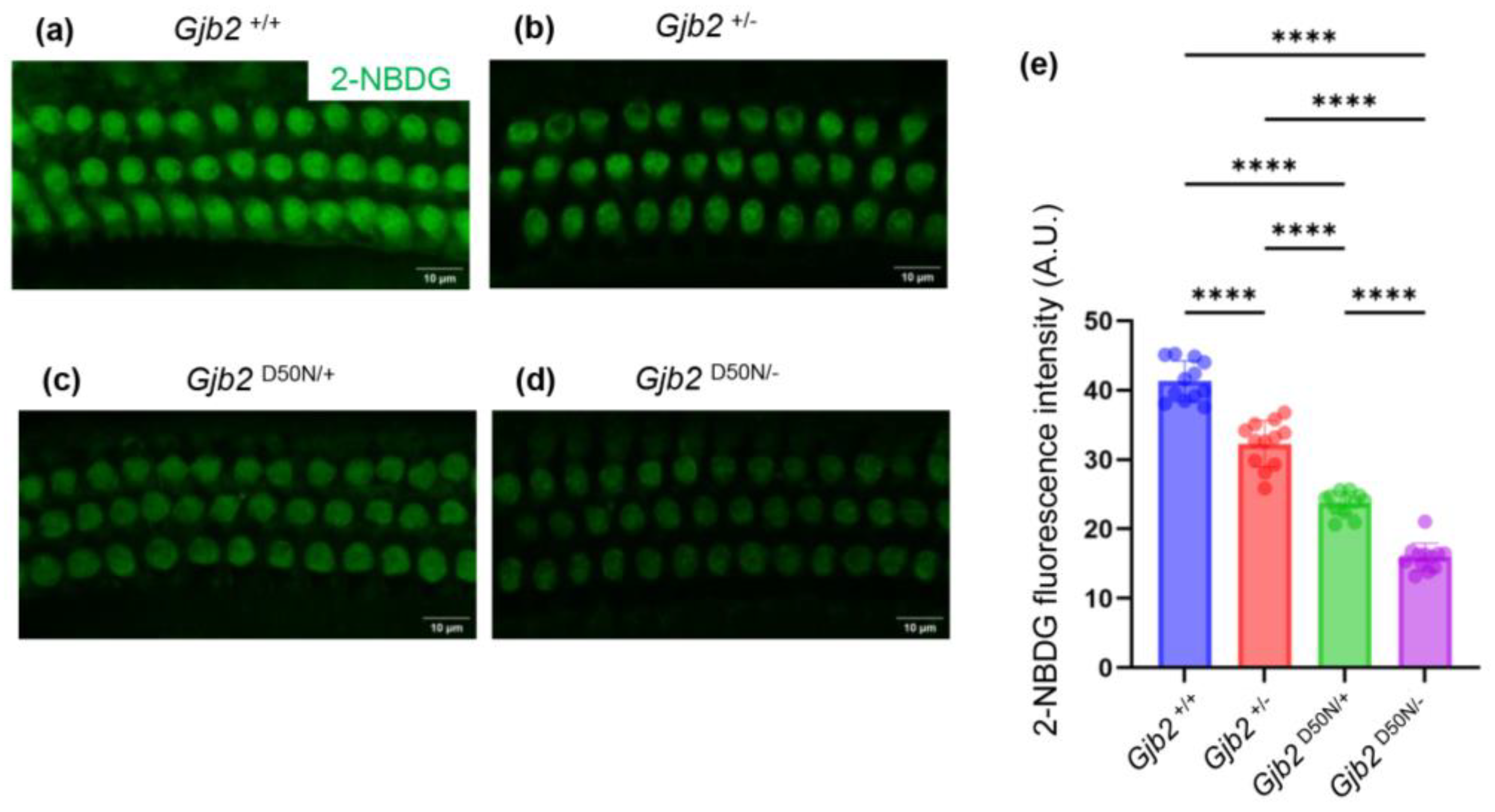
3.5. The Gjb2D50N/- Mice Decreased Intake of 2-NBDG

The sensory epithelium of the inner ear is avascular; consequently, it is strongly believed that these GJPs play a crucial role in the transfer of cellular signals and metabolites. In vitro experiments demonstrated that the D50N variant has abnormally high hemichannel activity and may lead to aberrant Ca2+ regulation[28,29,30]. Previous studies have demonstrated that gap junction mediated intercellular metabolite transfer in the cochlea was compromised in the Cx30-null mice [31]. Therefore, we investigated if there were differences in glucose uptake among OHCs, focusing on the glucose analog 2-NBDG. We conducted fluorescence quantification of 2-NBDG in the OHCs and observed that compared to the Gjb2 +/+ mice (Figure 5a), the fluorescence intensity of the Gjb2 +/- mice (Figure 5b), the Gjb2 D50N/+ mice (Figure 5c), and the Gjb2 D50N/- mice (Figure 5d) had decreased by 21.88%, 42.52%, and 61.42% (Figure 5e). Our study observed the Cx26 variant reduces glucose uptake, and OHCs thus suffer from impaired energy supply, which may lead to decreased OHCs function.

4. Discussion

The GJB2 gene is recognized as a significant contributor to deafness [4,32]. The ClinVar database catalogs over 500 variants of the GJB2 gene (https://www.ncbi.nlm.nih.gov/clinvar), encompassing a range of variants and variant sites that result in considerable variability in the hearing phenotypes of affected individuals [4]. Presently, the primary interventions for hearing loss include the hearing aids and cochlear implants. Research suggests that early intervention with hearing aid devices is correlated with improved outcomes in language development for patients [33,34]. However, for individuals experiencing late-onset progressive hearing loss, the early adoption of cochlear implants may not be feasible due to the substantial costs associated with the devices and their upkeep, as well as the potential for unpredictable complications that could adversely affect patients’ adherence to treatment. Given the heterogeneity of hearing phenotypes and the unique circumstances of each patient, a blanket approach to cochlear implantation may not represent the most effective or singular solution. Consequently, it is imperative to pursue or develop effective and cost-efficient pharmacological treatments tailored to individual needs.
Targeted therapy necessitates a deeper investigation into the underlying mechanisms, as the presence of various hearing phenotypes may indicate multiple pathogenic mechanisms linked to the GJB2 gene. The currently hypotheses struggle to fully explain the pathogenesis of Cx26-related hearing loss. The hemichannels, or gap junction channels, formed by Cx26 appear to play multiple roles in the structure and function of the inner ear [35,36]. Current in vivo studies primarily focus on various conditional knockout mice, which frequently display severe to profound hearing loss and exhibit significant pathological defects. The integrity of the organ of Corti is crucial for the cochlea's active amplification process [37,38,39]. In Cx26-null mice, the developmental arrest of pillar cells results in the abnormal morphology and the Corti tunnel failure to open properly, which may contribute to hearing loss [18,19,40]. Mice with Cx26 knocked-out early after birth show a progressive loss of HCs beginning in the middle turn, accompanied by a reduction in the density of SGNs [16,41,42]. Conditional knockout of Cx26 in supporting cells leads to an early loss of Deiters' cells [42]. However, these pathological changes were not observed in the Gjb2 D50N/- mice in our study. The Gjb2 D50N/- mice exhibited mild to moderate progressive hearing loss. These differing findings underscore the importance of developing various Cx26 variant mouse models, as they may more accurately reflect the conditions experienced by certain patients.
The cochlear basilar membrane is characterized by the absence of a direct blood supply, with GJPs playing a critical role in the signal transduction and substances transportation. In vitro investigations have demonstrated that various variants in the Cx26 gene can result in a range of effects, including the failure to establish GJPs between cells, the production of non-functional GJPs, or a reduction in ion permeability or substance exchange; it is also possible that some variants do not lead to any functional abnormalities[43,44,45,46,47]. A lot of researches indicated that variants in Cx26 can modify the microenvironment of the inner ear. The elevated concentration of potassium (K+) in the endolymph is essential for mechanical-electrical transduction processes within the HCs [48,49]. Mice with a knockout of Cx26 at an early developmental stage exhibited a diminished endolymphatic potential (EP), suggesting that the proper functioning of Cx26 in the sensory epithelium and the lateral wall is crucial for the establishment of EP [24,50]. Previously, researchers introduced the hypothesis of metabolic product transport impairment in Cx30 knock-out mice [31], and we have also noted a decrease in glucose uptake in OHCs in the Gjb2 D50N/- mice. A compromised glucose supply can lead to increased ATP consumption and the subsequent production of elevated levels of ROS [51]. Although various connexin family proteins are expressed within the cochlea, a defect in Cx26 may not entirely inhibit the transport of energy substrates; however, the accumulation of ROS may exacerbate cochlear dysfunction and contribute to hearing loss [52]. Functional impairments, rather than structural abnormalities, may contribute to those mild to moderate progressive hearing loss.
In GJPs, newly synthesized connexins continuously replace older connexins to maintain a dynamic equilibrium [53,54]. However, under pathological conditions, the synthesis and degradation of connexins may become imbalanced [55]. Previous studies have shown that the expression of Cx26 decreases in aged rats [56], and a partial deficiency of Cx26 accelerates age-related hearing loss by inducing redox imbalance and dysregulation of the Nrf2 pathway [52]. Furthermore, Cx26 haploinsufficiency has been shown to heighten sensitivity to noise through enhancing cochlear amplification [57].
The success of OTOF gene therapy has enhanced researchers' confidence in the potential of gene therapy for hereditary hearing loss [58,59]. Research on the mechanisms and treatment of other deafness-causing genes is also in full swing[60,61,62,63]. Adeno-associated viral (AAV) vectors mediated gene transfer of GJB2 had made some progress [64]. However, in mature variant cochlear of the mice with inducible Sox10iCreERT2 -mediated loss of Gjb2, AAV-mediated gene transfer of Gjb2 did not show hearing improvement and even exacerbated hearing loss and resulted in HCs loss in some mice [65]. Consequently, further investigation is required to assess the safety and efficacy of GJB2 gene therapy. Our previous studies demonstrated that dexamethasone and PARP inhibitors exhibit a protective effect against hearing loss in mice with Cx26 defects [25,66]. Besides, protection of HCs and SGNs by a number of small molecule drugs has been demonstrated in drug-induced hearing loss[67,68]. It is posited that for individuals experiencing mild to moderate progressive hearing loss, it is both feasible and beneficial to investigate or develop suitable pharmacological interventions aimed at preventing further deterioration or mitigating the progression of their condition. Such pharmacological agents should exhibit: i) high permeability, allowing for direct action within the inner ear by traversing the blood-labyrinth barrier; ii) possess high targeting capability to minimize off-target effects on other tissues and organs through localized administration; iii) demonstrate a prolonged half-life to decrease the frequency of dosing and improve patient adherence; iv) be economically accessible to reduce the financial burden on patients.

5. Conclusions

In conclusion, our study established a Gjb2 compound heterozygous variants mouse model that exhibit mild to moderate hearing loss, which is associated with compromised glucose supply. Additionally, we propose further research directions for the exploration or development of therapeutic interventions aimed at addressing mild to moderate progressive hearing loss attributable to GJB2 variants.

Author Contributions

X.S. and X.L. designed the study. X.S., X.L., Y.X. and X.X. were involved in the experiments. X.S. and X.L. wrote the manuscript. Y.S. provided technical support and modified the manuscript. All authors critically reviewed the final version of the manuscript.

Funding

This research was funded by the Key Program of the National Natural Science Foundation of China (No. 82430035), the Foundation for Innovative Research Groups of Hubei Province (No. 2023AFA038), the National Key Research and Development Program of China (Nos. 2021YFF0702303, 2023YFE0203200), the Fundamental Research Funds for the Central Universities (No.2024BRA019).

Acknowledgments

We are grateful to Union Hospital of Tongji Medical College, Huazhong University of Science and Technology for providing equipment support for this study. We would like to thank the specific pathogen-free Experimental Animal Center of Huazhong University of Science and Technology for providing assistance.

Conflicts of Interest

The authors declare no conflicts of interest.

References

  1. Butcher, E.; Dezateux, C.; Cortina-Borja, M.; Knowles, R.L. Prevalence of permanent childhood hearing loss detected at the universal newborn hearing screen: Systematic review and meta-analysis. PLoS One 2019, 14, e0219600. [Google Scholar] [CrossRef] [PubMed]
  2. Koffler, T.; Ushakov, K.; Avraham, K.B. Genetics of Hearing Loss: Syndromic. Otolaryngol Clin North Am 2015, 48, 1041–1061. [Google Scholar] [CrossRef] [PubMed]
  3. Shearer, A.E.; Hildebrand, M.S.; Schaefer, A.M.; Smith, R.J.H. Genetic Hearing Loss Overview. In GeneReviews(®); Adam, M.P., Feldman, J., Mirzaa, G.M., Pagon, R.A., Wallace, S.E., Bean, L.J.H., Gripp, K.W., Amemiya, A., Eds.; University of Washington: Seattle, WA, USA.
  4. Copyright © 1993-2024, University of Washington, Seattle. GeneReviews is a registered trademark of the University of Washington, Seattle. All rights reserved.: Seattle (WA), 1993.
  5. Chan, D.K.; Chang, K.W. GJB2-associated hearing loss: systematic review of worldwide prevalence, genotype, and auditory phenotype. Laryngoscope 2014, 124, E34–E53. [Google Scholar] [CrossRef] [PubMed]
  6. Kikuchi, T.; Kimura, R.S.; Paul, D.L.; Takasaka, T.; Adams, J.C. Gap junction systems in the mammalian cochlea. Brain Res Brain Res Rev 2000, 32, 163–166. [Google Scholar] [CrossRef] [PubMed]
  7. Kamiya, K.; Yum, S.W.; Kurebayashi, N.; Muraki, M.; Ogawa, K.; Karasawa, K.; Miwa, A.; Guo, X.; Gotoh, S.; Sugitani, Y.; et al. Assembly of the cochlear gap junction macromolecular complex requires connexin 26. J Clin Invest 2014, 124, 1598–1607. [Google Scholar] [CrossRef]
  8. Jagger, D.J.; Forge, A. Connexins and gap junctions in the inner ear--it's not just about K⁺ recycling. Cell Tissue Res 2015, 360, 633–644. [Google Scholar] [CrossRef]
  9. Beltramello, M.; Piazza, V.; Bukauskas, F.F.; Pozzan, T.; Mammano, F. Impaired permeability to Ins(1,4,5)P3 in a mutant connexin underlies recessive hereditary deafness. Nat Cell Biol 2005, 7, 63–69. [Google Scholar] [CrossRef]
  10. Korver, A.M.H.; Smith, R.J.H.; Van Camp, G.; Schleiss, M.R.; Bitner-Glindzicz, M.A.K.; Lustig, L.R.; Usami, S.-I.; Boudewyns, A.N. Congenital hearing loss. Nat Rev Dis Primers 2017, 3, 16094. [Google Scholar] [CrossRef]
  11. Liu, X.Z.; Xia, X.J.; Ke, X.M.; Ouyang, X.M.; Du, L.L.; Liu, Y.H.; Angeli, S.; Telischi, F.F.; Nance, W.E.; Balkany, T.; et al. The prevalence of connexin 26 ( GJB2) mutations in the Chinese population. Hum Genet 2002, 111, 394–397. [Google Scholar] [CrossRef]
  12. Mahdieh, N.; Rabbani, B. Statistical study of 35delG mutation of GJB2 gene: a meta-analysis of carrier frequency. Int J Audiol 2009, 48, 363–370. [Google Scholar] [CrossRef]
  13. Shen, N.; Peng, J.; Wang, X.; Zhu, Y.; Liu, W.; Liu, A.; Lu, Y. Association between the p.V37I variant of GJB2 and hearing loss: a pedigree and meta-analysis. Oncotarget 2017, 8, 46681–46690. [Google Scholar] [CrossRef] [PubMed]
  14. Markova, T.G.; Brazhkina, N.B.; Bliznech, E.A.; Bakhshinyan, V.V.; Polyakov, A.V.; Tavartkiladze, G.A. Phenotype in a patient with p.D50N mutation in GJB2 gene resemble both KID and Clouston syndromes. International journal of pediatric otorhinolaryngology 2016, 81, 10–14. [Google Scholar] [CrossRef]
  15. Janecke, A.R.; Hennies, H.C.; Günther, B.; Gansl, G.; Smolle, J.; Messmer, E.M.; Utermann, G.; Rittinger, O. GJB2 mutations in keratitis-ichthyosis-deafness syndrome including its fatal form. Am J Med Genet A 2005, 133a, 128–131. [Google Scholar] [CrossRef] [PubMed]
  16. Cohen-Barak, E.; Mwassi, B.; Zagairy, F.; Danial-Farran, N.; Khayat, M.; Tatour, Y.; Ziv, M. Parental mosaic cutaneous-gonadal GJB2 mutation: From epidermal nevus to inherited ichthyosis-deafness syndrome. The Journal of dermatology 2022, 49, 379–382. [Google Scholar] [CrossRef] [PubMed]
  17. Chen, S.; Sun, Y.; Lin, X.; Kong, W. Down regulated connexin26 at different postnatal stage displayed different types of cellular degeneration and formation of organ of Corti. Biochem Biophys Res Commun 2014, 445, 71–77. [Google Scholar] [CrossRef] [PubMed]
  18. Chen, S.; Xie, L.; Xu, K.; Cao, H.Y.; Wu, X.; Xu, X.X.; Sun, Y.; Kong, W.J. Developmental abnormalities in supporting cell phalangeal processes and cytoskeleton in the Gjb2 knockdown mouse model. Disease models & mechanisms 2018, 11. [Google Scholar] [CrossRef]
  19. Qiu, Y.; Xu, K.; Xie, L.; Chen, S.; Sun, Y. The Reduction in Microtubule Arrays Caused by the Dysplasia of the Non-Centrosomal Microtubule-Organizing Center Leads to a Malformed Organ of Corti in the Cx26-Null Mouse. Biomedicines 2022, 10. [Google Scholar] [CrossRef]
  20. Liu, X.Z.; Jin, Y.; Chen, S.; Xu, K.; Xie, L.; Qiu, Y.; Wang, X.H.; Sun, Y.; Kong, W.J. F-Actin Dysplasia Involved in Organ of Corti Deformity in Gjb2 Knockdown Mouse Model. Frontiers in molecular neuroscience 2021, 14, 808553. [Google Scholar] [CrossRef]
  21. Gabriel, H.D.; Jung, D.; Bützler, C.; Temme, A.; Traub, O.; Winterhager, E.; Willecke, K. Transplacental uptake of glucose is decreased in embryonic lethal connexin26-deficient mice. J Cell Biol 1998, 140, 1453–1461. [Google Scholar] [CrossRef]
  22. Lin, X.; Li, G.; Zhang, Y.; Zhao, J.; Lu, J.; Gao, Y.; Liu, H.; Li, G.-L.; Yang, T.; Song, L.; et al. Hearing consequences in Gjb2 knock-in mice: implications for human p.V37I mutation. Aging (Albany NY) 2019, 11, 7416–7441. [Google Scholar] [CrossRef]
  23. Tsai, C.-Y.; Lu, Y.-C.; Chan, Y.-H.; Radhakrishnan, N.; Chang, Y.-Y.; Lin, S.-W.; Liu, T.-C.; Hsu, C.-J.; Chen, P.-L.; Yang, L.-W.; et al. Simulation-predicted and -explained inheritance model of pathogenicity confirmed by transgenic mice models. Comput Struct Biotechnol J 2023, 21, 5698–5711. [Google Scholar] [CrossRef] [PubMed]
  24. Li, Q.; Cui, C.; Liao, R.; Yin, X.; Wang, D.; Cheng, Y.; Huang, B.; Wang, L.; Yan, M.; Zhou, J.; et al. The pathogenesis of common Gjb2 mutations associated with human hereditary deafness in mice. Cellular and molecular life sciences : CMLS 2023, 80, 148. [Google Scholar] [CrossRef] [PubMed]
  25. Inoshita, A.; Iizuka, T.; Okamura, H.O.; Minekawa, A.; Kojima, K.; Furukawa, M.; Kusunoki, T.; Ikeda, K. Postnatal development of the organ of Corti in dominant-negative Gjb2 transgenic mice. Neuroscience 2008, 156, 1039–1047. [Google Scholar] [CrossRef] [PubMed]
  26. Xu, K.; Chen, S.; Xie, L.; Qiu, Y.; Liu, X.Z.; Bai, X.; Jin, Y.; Wang, X.H.; Sun, Y. The protective effects of systemic dexamethasone on sensory epithelial damage and hearing loss in targeted Cx26-null mice. Cell death & disease 2022, 13, 545. [Google Scholar] [CrossRef]
  27. Di, W.-L.; Gu, Y.; Common, J.E.A.; Aasen, T.; O'Toole, E.A.; Kelsell, D.P.; Zicha, D. Connexin interaction patterns in keratinocytes revealed morphologically and by FRET analysis. J Cell Sci 2005, 118, 1505–1514. [Google Scholar] [CrossRef]
  28. Shurman, D.L.; Glazewski, L.; Gumpert, A.; Zieske, J.D.; Richard, G. In vivo and in vitro expression of connexins in the human corneal epithelium. Investigative ophthalmology & visual science 2005, 46, 1957–1965. [Google Scholar] [CrossRef]
  29. Lopez, W.; Gonzalez, J.; Liu, Y.; Harris, A.L.; Contreras, J.E. Insights on the mechanisms of Ca(2+) regulation of connexin26 hemichannels revealed by human pathogenic mutations (D50N/Y). The Journal of general physiology 2013, 142, 23–35. [Google Scholar] [CrossRef]
  30. Sanchez, H.A.; Kraujaliene, L.; Verselis, V.K. A pore locus in the E1 domain differentially regulates Cx26 and Cx30 hemichannel function. The Journal of general physiology 2024, 156. [Google Scholar] [CrossRef]
  31. Taki, T.; Takeichi, T.; Sugiura, K.; Akiyama, M. Roles of aberrant hemichannel activities due to mutant connexin26 in the pathogenesis of KID syndrome. Scientific reports 2018, 8, 12824. [Google Scholar] [CrossRef]
  32. Chang, Q.; Tang, W.; Ahmad, S.; Zhou, B.; Lin, X. Gap junction mediated intercellular metabolite transfer in the cochlea is compromised in connexin30 null mice. PLoS One 2008, 3, e4088. [Google Scholar] [CrossRef]
  33. Snoeckx, R.L.; Huygen, P.L.M.; Feldmann, D.; Marlin, S.; Denoyelle, F.; Waligora, J.; Mueller-Malesinska, M.; Pollak, A.; Ploski, R.; Murgia, A.; et al. GJB2 mutations and degree of hearing loss: a multicenter study. Am J Hum Genet 2005, 77, 945–957. [Google Scholar] [CrossRef] [PubMed]
  34. Wu, C.-M.; Ko, H.-C.; Tsou, Y.-T.; Lin, Y.-H.; Lin, J.-L.; Chen, C.-K.; Chen, P.-L.; Wu, C.-C. Long-Term Cochlear Implant Outcomes in Children with GJB2 and SLC26A4 Mutations. PLoS One 2015, 10, e0138575. [Google Scholar] [CrossRef] [PubMed]
  35. Chang, Q.; Tang, W.; Kim, Y.; Lin, X. Timed conditional null of connexin26 in mice reveals temporary requirements of connexin26 in key cochlear developmental events before the onset of hearing. Neurobiol Dis 2015, 73, 418–427. [Google Scholar] [CrossRef] [PubMed]
  36. Chen, P.; Wu, W.; Zhang, J.; Chen, J.; Li, Y.; Sun, L.; Hou, S.; Yang, J. Pathological mechanisms of connexin26-related hearing loss: Potassium recycling, ATP-calcium signaling, or energy supply? Frontiers in molecular neuroscience 2022, 15, 976388. [Google Scholar] [CrossRef]
  37. Posukh, O.L.; Maslova, E.A.; Danilchenko, V.Y.; Zytsar, M.V.; Orishchenko, K.E. Functional Consequences of Pathogenic Variants of the GJB2 Gene (Cx26) Localized in Different Cx26 Domains. Biomolecules 2023, 13. [Google Scholar] [CrossRef]
  38. Zhu, Y.; Liang, C.; Chen, J.; Zong, L.; Chen, G.-D.; Zhao, H.-B. Active cochlear amplification is dependent on supporting cell gap junctions. Nat Commun 2013, 4, 1786. [Google Scholar] [CrossRef]
  39. Zong, L.; Chen, J.; Zhu, Y.; Zhao, H.-B. Progressive age-dependence and frequency difference in the effect of gap junctions on active cochlear amplification and hearing. Biochem Biophys Res Commun 2017, 489, 223–227. [Google Scholar] [CrossRef]
  40. Dallos, P.; Wu, X.; Cheatham, M.A.; Gao, J.; Zheng, J.; Anderson, C.T.; Jia, S.; Wang, X.; Cheng, W.H.Y.; Sengupta, S.; et al. Prestin-based outer hair cell motility is necessary for mammalian cochlear amplification. Neuron 2008, 58, 333–339. [Google Scholar] [CrossRef]
  41. Xie, L.; Chen, S.; Xu, K.; Cao, H.Y.; Du, A.N.; Bai, X.; Sun, Y.; Kong, W.J. Reduced postnatal expression of cochlear Connexin26 induces hearing loss and affects the developmental status of pillar cells in a dose-dependent manner. Neurochemistry international 2019, 128, 196–205. [Google Scholar] [CrossRef]
  42. Chen, S.; Xu, K.; Xie, L.; Cao, H.Y.; Wu, X.; Du, A.N.; He, Z.H.; Lin, X.; Sun, Y.; Kong, W.J. The spatial distribution pattern of Connexin26 expression in supporting cells and its role in outer hair cell survival. Cell death & disease 2018, 9, 1180. [Google Scholar] [CrossRef]
  43. Qiu, Y.; Xie, L.; Wang, X.; Xu, K.; Bai, X.; Chen, S.; Sun, Y. Abnormal Innervation, Demyelination, and Degeneration of Spiral Ganglion Neurons as Well as Disruption of Heminodes are Involved in the Onset of Deafness in Cx26 Null Mice. Neuroscience bulletin 2024. [CrossRef]
  44. Haack, B.; Schmalisch, K.; Palmada, M.; Böhmer, C.; Kohlschmidt, N.; Keilmann, A.; Zechner, U.; Limberger, A.; Beckert, S.; Zenner, H.P.; et al. Deficient membrane integration of the novel p.N14D-GJB2 mutant associated with non-syndromic hearing impairment. Hum Mutat 2006, 27, 1158–1159. [Google Scholar] [CrossRef] [PubMed]
  45. Albuloushi, A.; Lovgren, M.L.; Steel, A.; Yeoh, Y.; Waters, A.; Zamiri, M.; Martin, P.E. A heterozygous mutation in GJB2 (Cx26F142L) associated with deafness and recurrent skin rashes results in connexin assembly deficiencies. Experimental dermatology 2020, 29, 970–979. [Google Scholar] [CrossRef] [PubMed]
  46. Thönnissen, E.; Rabionet, R.; Arbonès, M.L.; Estivill, X.; Willecke, K.; Ott, T. Human connexin26 (GJB2) deafness mutations affect the function of gap junction channels at different levels of protein expression. Hum Genet 2002, 111, 190–197. [Google Scholar] [CrossRef] [PubMed]
  47. Schütz, M.; Auth, T.; Gehrt, A.; Bosen, F.; Körber, I.; Strenzke, N.; Moser, T.; Willecke, K. The connexin26 S17F mouse mutant represents a model for the human hereditary keratitis-ichthyosis-deafness syndrome. Human molecular genetics 2011, 20, 28–39. [Google Scholar] [CrossRef]
  48. Zong, Y.J.; Liu, X.Z.; Tu, L.; Sun, Y. Cytomembrane Trafficking Pathways of Connexin 26, 30, and 43. International journal of molecular sciences 2023, 24. [Google Scholar] [CrossRef]
  49. Cohen-Salmon, M.; Ott, T.; Michel, V.; Hardelin, J.P.; Perfettini, I.; Eybalin, M.; Wu, T.; Marcus, D.C.; Wangemann, P.; Willecke, K.; et al. Targeted ablation of connexin26 in the inner ear epithelial gap junction network causes hearing impairment and cell death. Curr Biol 2002, 12, 1106–1111. [Google Scholar] [CrossRef]
  50. Wangemann, P. K+ cycling and the endocochlear potential. Hear Res 2002, 165, 1–9. [Google Scholar] [CrossRef]
  51. Kudo, T.; Kure, S.; Ikeda, K.; Xia, A.-P.; Katori, Y.; Suzuki, M.; Kojima, K.; Ichinohe, A.; Suzuki, Y.; Aoki, Y.; et al. Transgenic expression of a dominant-negative connexin26 causes degeneration of the organ of Corti and non-syndromic deafness. Human molecular genetics 2003, 12. [Google Scholar] [CrossRef]
  52. Moley, K.H.; Mueckler, M.M. Glucose transport and apoptosis. Apoptosis 2000, 5. [Google Scholar]
  53. Fetoni, A.R.; Zorzi, V.; Paciello, F.; Ziraldo, G.; Peres, C.; Raspa, M.; Scavizzi, F.; Salvatore, A.M.; Crispino, G.; Tognola, G.; et al. Cx26 partial loss causes accelerated presbycusis by redox imbalance and dysregulation of Nfr2 pathway. Redox biology 2018, 19, 301–317. [Google Scholar] [CrossRef] [PubMed]
  54. Carette, D.; Gilleron, J.; Denizot, J.-P.; Grant, K.; Pointis, G.; Segretain, D. New cellular mechanisms of gap junction degradation and recycling. Biol Cell 2015, 107, 218–231. [Google Scholar] [CrossRef] [PubMed]
  55. Sun, G.; Zheng, Y.; Fu, X.; Zhang, W.; Ren, J.; Ma, S.; Sun, S.; He, X.; Wang, Q.; Ji, Z.; et al. Single-cell transcriptomic atlas of mouse cochlear aging. Protein Cell 2023, 14, 180–201. [Google Scholar] [CrossRef] [PubMed]
  56. Xu, K.; Chen, S.; Bai, X.; Xie, L.; Qiu, Y.; Liu, X.Z.; Wang, X.H.; Kong, W.J.; Sun, Y. Degradation of cochlear Connexin26 accelerate the development of age-related hearing loss. Aging cell 2023, 22, e13973. [Google Scholar] [CrossRef]
  57. Wu, X.; Wang, Y.; Sun, Y.; Chen, S.; Zhang, S.; Shen, L.; Huang, X.; Lin, X.; Kong, W. Reduced expression of Connexin26 and its DNA promoter hypermethylation in the inner ear of mimetic aging rats induced by d-galactose. Biochem Biophys Res Commun 2014, 452, 340–346. [Google Scholar] [CrossRef]
  58. Liu, L.-M.; Liang, C.; Chen, J.; Fang, S.; Zhao, H.-B. Cx26 heterozygous mutations cause hyperacusis-like hearing oversensitivity and increase susceptibility to noise. Sci Adv 2023, 9, eadf4144. [Google Scholar] [CrossRef]
  59. Lv, J.; Wang, H.; Cheng, X.; Chen, Y.; Wang, D.; Zhang, L.; Cao, Q.; Tang, H.; Hu, S.; Gao, K.; et al. AAV1-hOTOF gene therapy for autosomal recessive deafness 9: a single-arm trial. Lancet 2024, 403, 2317–2325. [Google Scholar] [CrossRef]
  60. Qi, J.; Tan, F.; Zhang, L.; Lu, L.; Zhang, S.; Zhai, Y.; Lu, Y.; Qian, X.; Dong, W.; Zhou, Y.; et al. AAV-Mediated Gene Therapy Restores Hearing in Patients with DFNB9 Deafness. Adv Sci (Weinh) 2024, 11, e2306788. [Google Scholar] [CrossRef]
  61. Tan, F.; Chu, C.; Qi, J.; Li, W.; You, D.; Li, K.; Chen, X.; Zhao, W.; Cheng, C.; Liu, X.; et al. AAV-ie enables safe and efficient gene transfer to inner ear cells. Nat Commun 2019, 10, 3733. [Google Scholar] [CrossRef]
  62. Tao, Y.; Liu, X.; Yang, L.; Chu, C.; Tan, F.; Yu, Z.; Ke, J.; Li, X.; Zheng, X.; Zhao, X.; et al. AAV-ie-K558R mediated cochlear gene therapy and hair cell regeneration. Signal Transduct Target Ther 2022, 7, 109. [Google Scholar] [CrossRef]
  63. Zhang, L.; Chen, X.; Wang, X.; Zhou, Y.; Fang, Y.; Gu, X.; Zhang, Z.; Sun, Q.; Li, N.; Xu, L.; et al. AAV-mediated Gene Cocktails Enhance Supporting Cell Reprogramming and Hair Cell Regeneration. Adv Sci (Weinh) 2024, 11, e2304551. [Google Scholar] [CrossRef] [PubMed]
  64. Zhang, B.; Hu, Y.; Du, H.; Han, S.; Ren, L.; Cheng, H.; Wang, Y.; Gao, X.; Zheng, S.; Cui, Q.; et al. Tissue engineering strategies for spiral ganglion neuron protection and regeneration. J Nanobiotechnology 2024, 22, 458. [Google Scholar] [CrossRef] [PubMed]
  65. Yu, Q.; Wang, Y.; Chang, Q.; Wang, J.; Gong, S.; Li, H.; Lin, X. Virally expressed connexin26 restores gap junction function in the cochlea of conditional Gjb2 knockout mice. Gene Ther 2014, 21, 71–80. [Google Scholar] [CrossRef] [PubMed]
  66. Guo, J.; Ma, X.; Skidmore, J.M.; Cimerman, J.; Prieskorn, D.M.; Beyer, L.A.; Swiderski, D.L.; Dolan, D.F.; Martin, D.M.; Raphael, Y. GJB2 gene therapy and conditional deletion reveal developmental stage-dependent effects on inner ear structure and function. Mol Ther Methods Clin Dev 2021, 23, 319–333. [Google Scholar] [CrossRef] [PubMed]
  67. Wang, X.; Chen, S.; Qiu, Y.; Xu, K.; Bai, X.; Xie, L.; Kong, W.; Sun, Y. PARP inhibitor rescues hearing and hair cell impairment in Cx26-null mice. View 2023, 4. [Google Scholar] [CrossRef]
  68. Zhang, Y.; Fang, Q.; Wang, H.; Qi, J.; Sun, S.; Liao, M.; Wu, Y.; Hu, Y.; Jiang, P.; Cheng, C.; et al. Increased mitophagy protects cochlear hair cells from aminoglycoside-induced damage. Autophagy 2023, 19, 75–91. [Google Scholar] [CrossRef]
  69. Liu, W.; Xu, L.; Wang, X.; Zhang, D.; Sun, G.; Wang, M.; Wang, M.; Han, Y.; Chai, R.; Wang, H. PRDX1 activates autophagy via the PTEN-AKT signaling pathway to protect against cisplatin-induced spiral ganglion neuron damage. Autophagy 2021, 17, 4159–4181. [Google Scholar] [CrossRef]
Figure 1. Mouse models and hearing loss in different mouse models. (a, b) Gjb2 p.D50N variant site and Sanger sequencing result. (c) The process of constructing different mouse models. (d, e) The averaged ABR thresholds of the 20-days-old and 90-days-old Gjb2+/+, Gjb2D50N/+ and Gjb2D50N/- mice (n=3 in each group) at 8, 16, 24, and 32 kHz.
Preprints 140848 g001
Figure 2. Hair cells count and SGNs count in different mouse models. (a) Representative images of HCs (DAPI, blue) of different turns in the Gjb2+/+, Gjb2D50N/+ and Gjb2D50N/- mice. Scale bars: 50μm. (b) OHCs count in each group. P>0.05. (c) Representative images of SGNs (DAPI, blue) of different turns in the Gjb2+/+, Gjb2+/- Gjb2D50N/+ and Gjb2D50N/- mice. Scale bars: 50μm. (d) SGNs count in each group. P>0.05.
Preprints 140848 g002
Figure 3. CD45+cells in the organ of Corti. (a-d) Representative images of CD45+cells (CD45, red) in the organ of Corti in the Gjb2+/+, Gjb2+/- Gjb2D50N/+ and Gjb2D50N/- mice. Scale bars: 30μm.
Preprints 140848 g003
Figure 4. Cx26 expression patterns. (a-d) Representative confocal images of connexin 26 (Cx26, green; DAPI, blue) from apical turns of the basilar membrane. Scale bars: 10μm. (e) Histograms presenting the average lengths of the largest GJPs along a single cell border (mean±SD, n=12 for each group). ns, no significant difference; **p≤0.01; ****p≤0.0001.
Preprints 140848 g004
Figure 5. 2-NBDG uptake of OHCs. (a-d) Representative confocal images of 2-NBDG (green) as a fluorescent tracer of glucose from apical turns of the basilar membrane. Scale bars: 10μm. (e) Statistics of 2-NBDG fluorescence intensity of OHCs (mean±SD, n=12 for each group). ****p≤0.0001.
Preprints 140848 g005
Disclaimer/Publisher’s Note: The statements, opinions and data contained in all publications are solely those of the individual author(s) and contributor(s) and not of MDPI and/or the editor(s). MDPI and/or the editor(s) disclaim responsibility for any injury to people or property resulting from any ideas, methods, instructions or products referred to in the content.
Copyright: This open access article is published under a Creative Commons CC BY 4.0 license, which permit the free download, distribution, and reuse, provided that the author and preprint are cited in any reuse.
Prerpints.org logo

Preprints.org is a free preprint server supported by MDPI in Basel, Switzerland.

Subscribe

Disclaimer

Terms of Use

Privacy Policy

Privacy Settings

© 2025 MDPI (Basel, Switzerland) unless otherwise stated