
Submitted:
06 November 2024
Posted:
07 November 2024
You are already at the latest version
Abstract
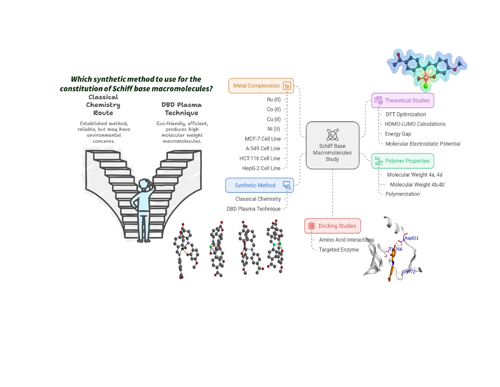
Schiff base macromolecules have been successfully synthesized, utilizing a classical chemistry route and a dielectric barrier discharge (DBD) plasma technique. The synthesis of monomeric units has been accomplished through typical reactions of aldehyde and amine functional molecules. The condensation polymerization of the Schiff base molecules has been instigated chemically, using p-toluene sulfonyl chloride in refluxed ethanol. The molecular weight of the obtained polymers was discovered to be 524,664 and 1,503,228 for polymers 4a and 4b, respectively. Additionally, the polymerization reactions were prompted, employing a Dielectric Barrier Discharge (DBD) atmospheric pressure air plasma technique. The DBD plasma demonstrated a very powerful routine to produce high molecular weights macromolecules, which could be an ecofriendly strategy to acquire this class of materials. Moreover, the complexation of polymer 4b with various metal moieties, Ru (II), Co (II), Cu (II), and Ni (II), has been executed in order to have a comparative study of their antitumor activity against MCF-7, A-549, HCT-116, and HepG-2 cell lines. Furthermore, the density functional theory was exploited to optimize the polymers and their complexes, and their HOMO-LUMO and energy gap were calculated, which was utilized to examine the inter/intra molecular charge transfer. The molecular electrostatic potential map was similarly quantified to investigate the reactive sites that are present in the investigated molecules. The result for the docking study confirmed that these complexed polymers adopted numerous important interactions with the amino acid of the targeted enzyme.
Keywords:
Schiff base macromolecular
; DBD plasma
; metal complexes
; antitumor activity
; docking study
1. Introduction
Polymeric macromolecules have secured remarkable recognition over the years due to their versatile architectures and functionalities, which enable the alteration and modification of the material’s characteristics (physical, chemical, mechanical, biological, etc.) in order to satisfy operative demands [1-9]. These materials have been central frameworks in a cascade of scientific and technological fields, including biophysics, electronics, and medicine [1-10]. Alongside the abundant applications of these polymeric matrices, their implementation in medicinal domains has correspondingly garnered consideration for their utilization as catheter-synonymous equipment, gastric pouches, implants, injury dressing, sutures, and valves [6,8]. The investigation into their biomedical employment has additionally resulted into the establishment of a medicinal class of biocompatible polymers, which yield successful synthetic organ transplantation, drug-like agents, and drug delivery mechanisms [6,8,9,11-16].
Of these three advanced applications of biomedical polymers, the design and synthesis of desirable drug candidates are one of the most critical functionalities that possesses promise in generating a more efficient drug system especially for drug resistant cases [17-20]. This integration into the drug design and synthesis biosphere enables its employment in cancer therapeutics to address and inhibit the detrimental impacts of these treatments. To enhance and ameliorate the influence of these polymeric drug-analogous agents, the configuration process mandates the incorporation of a functional precursor to ensure the necessary antagonistic and inhibitory characters [21-25].
Schiff base moieties have precedingly been investigated for their applications and have manifested as one of the most highly demanded precursors to construct a series of polymeric analogues that accommodate various medical purposes, such as tissue regeneration, biosensors, wound healing, drug delivery, tissue adhesive, and bioprinting [26-30]. Moreover, this class of molecules demonstrated a great impact in treating several types of adenocarcinoma cell lines, including human hepatoma, colorectal carcinoma, breast, leukemia, and HL-60 cells [26-31]. Schiff base molecules are formed through the nucleophilic interaction of the amine substituents with aldehyde precursors [32,33], and Schiff base polymer has been attained via polycondensation [26-28, 30]. Furthermore, the chelating character of Schiff base moieties is responsible for the modification of their properties such as the enhancement of their antioxidant activities and subsequently the manufacture of antitumor agents [29, 34-36].
In order to establish these Schiff base-incorporating polymeric matrices, the classical approach was assumed alongside a newly-implemented DBD non-thermal plasma methodology. Initially, plasma polymerized materials proclaimed notoriety for their byproducts, which bore unpredictable architecture [37-39]. However, their standing in the modern scientific scope was altered with their integration in nuclear batteries as dielectric separation membranes [37-42]. Consequently, examinations within the plasma polymerization procedure have revealed exceptional attributes regarding this approach, which yielded newfound prestige for this technique [37-42]. The distinctive features of plasma polymerization encompass solvent-independent (dry) reactivity and the polymerization of novel polymeric materials whose polymerization cannot proceed through the conventional methodologies (essentially the generation of products whose properties are not conceivable through wet chemistry) [37-42]. Furthermore, extensive research into plasma polymers disclosed that the main determinant of their structural configuration as well as their operative characteristics (bioadhesive, elastic modulus, permeability, roughness, solubility, and wettability) is their deposition conditions.
Herein, this report describes the prosperous synthesis of high molecular weight Schiff base polymers, executing a classical chemistry and a DBD atmospheric pressure air plasma approaches. The DBD plasma demonstrated a powerful mechanism in fabricating high molecular weights macromolecules, which proved to be more beneficial in efficiency and economical mannerisms in order to acquire this class of materials. In addition, the Schiff base polymer 4b, which has been produced chemically, was subjected to a metal complexation process with Co, Ni, Ru, and Cu moieties. The antitumor activities of the newly-synthesized polymer and its complex counterparts have been investigated, followed by their docking estimation.
2. Materials and Methods
2.1. Materials and Characterization
All of the chemicals were obtained from Sigma-Aldrich Chemical Co. (Taufkirchen, Germany) and were used without further purification. Solvents were high performance liquid chromatography (HPLC) grade. The chemical polymerization was performed in anhydrous ethanol. 1H NMR spectra were recorded at 400 MHz, on Bruker 400 MHz ultrashield TM NMR spectrometer (Bruker, Fällanden, Switzerland), with chemical shifts calculated in Hz, referenced to solvent residues. The IR spectra of the new compounds were recorded on a Jasco FTIR-300 E Fourier Transform Infrared Spectrometer (Jasco, Tokoyo, Japan). Thermogravimetric analysis (TGA) of the polymeric materials was carried out under nitrogen atmosphere at a heating rate 10 oC/min-1 using TA instrument, model SDT600 (New Castle, DE, USA). Differential Scanning Calorimeter (DSC) measurements were performed using a Q2000 apparatus with T zero® techniques from TA instruments (New Castle, DE, USA).
Scanning Electron Microscope (SEM) measurements have been carried out using SEM Model Philips XL 30 (FEI company, Hillsboro, OR, USA) with accelerating voltage 30 KV, magnification 10x up to 4000x, and resolution for W. The number-average molecular weights (Mn), weight-average molecular weights (Mw) and dispersities (Mw/Mn) were determined using gel permeation chromatography (GPC). Agilent 1260 Infinity GPC system equipped with refractive index detectors operating at 50 °C and two Agilent Technologies PLgel 5 µm MIXED-C and PLgel 10 µm MIXED-B organic mixed bed columns, with dimensions of 7.5 mm (ID) × 300 mm (L) were used with DMF containing 0.1 mol% LiBr at a flow rate of 1.0 mL/min. The GPC instrument was calibrated with narrowly dispersed linear poly(methyl methacrylate) (PMMA) standards. The solutions were filtered using a 0.25 mm PTFE filter to remove any impurities. Analysis of the chromatograms was performed with the Agilent GPC/SEC software.
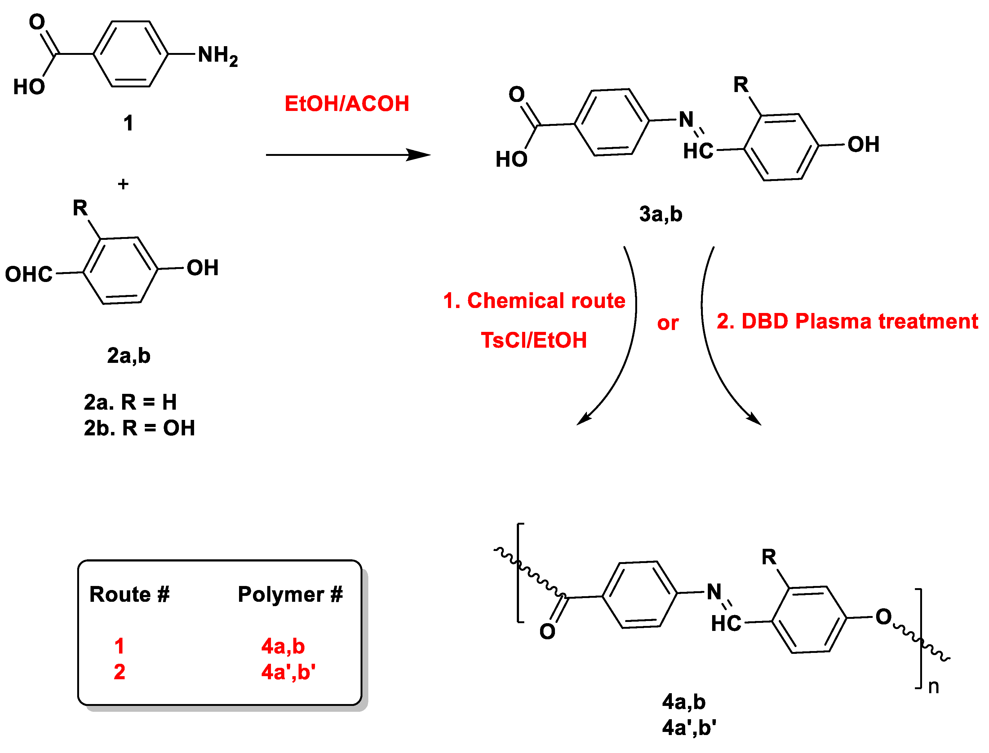
2.2. Synthesis of Schiff Base Monomers 3a,b
Compounds 3a,b were synthesized via the reaction of equimolar amounts of P-aminobenzoic acid (1mmol) with aldehyde molecules 2a,b (1 mmol) in refluxed ethanol solution (20 mL) and in the presence of few drops of acetic acid. The reaction was proceeded for 3 hr. then the solution was precipitated in distilled water, filtered and crystalized from ethanol.
(4-(4-hydroxyphenyl)imino)methyl)benzoic acid (3a). It was obtained in 90% yield as yellow solid, m.p.: 145 ºC; IR: 3396 (OH), 2995 (CH), 1664 (C=O), 1630 (C=N), 1497 (C=C aromatic). 1H NMR (DMSO-d6, ppm) δ: 9.78 (s, 1H, OH), 8.48 (s, 1H, CH=N), 7.95 (d, 2H, Ar-CH), 7.80 (d, 2H, Ar-CH), 7.25 (d, 2H, Ar-CH), 6.90 (d, 2H, Ar-CH). Anal. Calcd. for C14H11NO3; C, 69.70; H, 4.6; N, 5.81. Found: 69.68; H, 4.62; N, 5.78.
(4-(2,4-dihydroxyphenyl)imino)methyl)benzoic acid (3b). It was obtained in 86% yield as yellowish orange solid, m.p.: 155 ºC; IR: 3405 (OH), 3055 (CH), 1672 (C=O), 1656 (C=N), 1545 (C=C aromatic). 1H NMR (DMSO-d6, ppm) δ: 9.92 (s, 1H, OH), 8.85 (s, 1H, CH=N), 7.99 (d, 2H, Ar-CH), 7.61 (d, 1H, Ar-CH), 7.48 (d, 1H, Ar-CH), 7.43 (d, 2H, Ar-CH ), 6.54 (d, 1H, Ar-CH), 6.43 (d, 1H, Ar-CH), 6.31 (s, 1H, Ar-CH), 5.85 (s, 1H, OH). Anal. Calcd. for C14H11NO4; C, 65.37; H, 4.31; N, 5.45. Found: 65.35; H, 4.33; N, 5.43.
2.3. Condensation Polymerization of Schiff Base Monomers
Esterification polymerization of the two Schiff base monomers was executed via refluxing an ethanolic solution of the desired monomers 3a,b in the presence of catalytic amount of P-toluene sulphonyl chloride. The reactions were proceeded for overnight. The polymeric materials 4a and 4b were precipitated in distilled water, filtered and washed with water and ethanol to remove unreacted monomers.
polymer (4a). IR data showed υ(cm-1) at 3402 (OH), 2995 (CH), 1712 (C=O), 1576 (C=N), 1565 (C=C aromatic).1H NMR (DMSO-d6, ppm) δ: 9.79 (s, 1H, CH=N), 7.77 (d, 2H, Ar-CH), 7.65 (d, 2H, Ar-CH), 7.48 (d, 2H, Ar-CH), 7.12 ( d, 2H, Ar-CH), 6.93 (d, 1H, Ar-CH), 6.60 (d, 1H, Ar-CH).
polymer (4b). IR data showed υ (cm-1) at 3268 (OH), 2995 (CH), 1597 (C=O), 1576 (C=N), 1513 (C=C aromatic).1H NMR (DMSO-d6, ppm) δ: 9.92 (s, 1H, OH); 8.13 (s, 1H, CH=N), 7.64 (d, 1H, Ar-CH), 7.53 (d, 1H, Ar-CH), 7.48 (d, 2H, Ar-CH), 7.11 ( d, 2H, Ar-CH ), 6.57 (d, 1H, Ar-CH), 6.32 (s, 1H, Ar-CH).
2.4. Complexation of Polymer 4b
Polymer 4b (0.5 gm) was dissolved in 15 mL ethanol and the solution was purged with N2 atmosphere. The selected metal salts were dissolved in 25 mL of hot ethanol then added to polymeric solution and the mixtures were refluxed for 6 hr. The reactions were left to cool down and the formed precipitate was filtered washed with ethanol and dried under vacuum. The metal containing polymers were characterized using FTIR spectroscopy.
Cu-complex with polymer 4b. IR data showed υ (cm-1) at 3468 (OH), 2995 (CH), 1643 (C=O), 1584 (C=N), 1463 (C=C aromatic), 704 (Metal-O), 611 (Metal-N).
Co-complex with polymer 4b. IR data showed υ (cm-1) at 3367 (OH), 2995 (CH), 1656 (C=O), 1593 (C=N), 1287 (C=C aromatic), 701 (Metal-O), 601 (Metal-N).
Ni-complex with polymer 4b. IR data showed υ (cm-1) at 3332 (OH), 2995 (CH), 1723 (C=O), 1617 (C=N), 1205 (C=C aromatic), 814 (Metal-O), 751 (Metal-N).
Ru-complex with polymer 4b. IR data showed υ (cm-1) at 3352 (OH), 2995 (CH), 1721 (C=O), 1608 (C=N), 1224 (C=C aromatic), 814 (Metal-O), 739 (Metal-N).
2.5. Dielectric Barrier Discharge Plasma System
A dielectric barrier discharge (DBD) atmospheric pressure air plasma system consists of two parallel metallic electrodes that separated by 4 mm gape, Figure 2. The upper electrode is cylindrical shape brass electrode that covered from bottom by a dielectric alumina sheet of 45 mm diameter, 1 mm thickness, and 80x80 mm dimension. The brass is connected to AC high voltage power-supply system that provide up to 30 kV and 30 kHz sinusoidal waveform signal. The lateral side of brass electrode covered by Teflon insulator of 3 mm thickness to protect the users. The lower electrode is grounded-stainless steel disc with 45 mm diameter. The treated samples were placed on the grounded electrodes that have about 3 mm gap with high voltage electrode.
The voltage and current waveforms illustrated that the generated plasma is inhomogeneous plasma and it contain streamers. The discharge breakdowns at ~ 12 kV and the plasma started to fill the 3 mm gap between the two electrodes with increasing the applied voltage which is atypical for DBD plasma. The plasma homogeneity increases visually with decreasing the gap distance or increasing the applied voltage due to the diffusivity and interference of the streamers.
The electric characteristics of the plasma was studied using a Tektronix oscilloscope, DPO7354 C −3.5 GHz, was used to record the current and voltage pulses that measured by a 1:1 Pearson current probe, model: 6585 and calibrated high voltage probe 1:1000, P6015A-Tektronix probe. The applied voltage pulses were measured across the discharge gap and the consumed current pulses were measured through the grounded electrode, Figure 1. The plasma images were captured using Nikon digital camera D3200 with AF-S Micro NIKKOR 105 mm lens.
Figure 1.
Dielectric Barrier Discharge (DBD) plasma system.
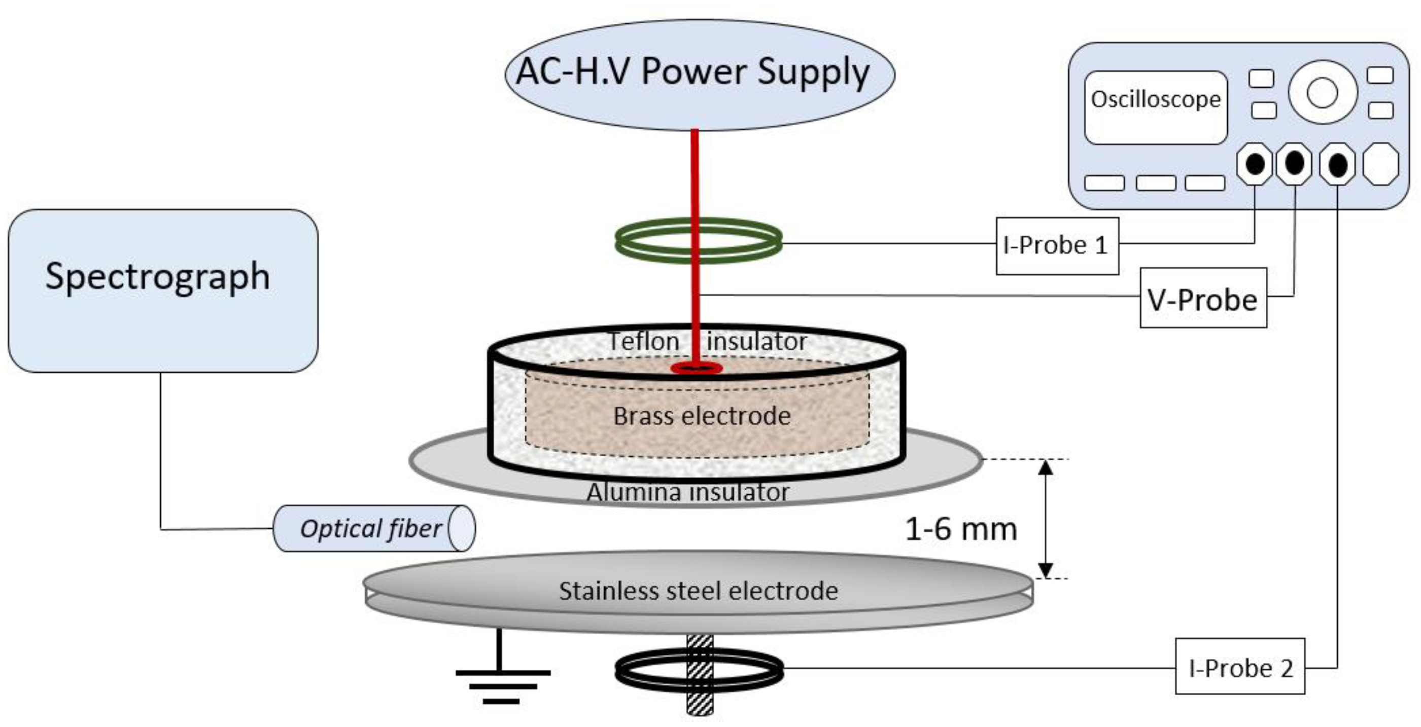
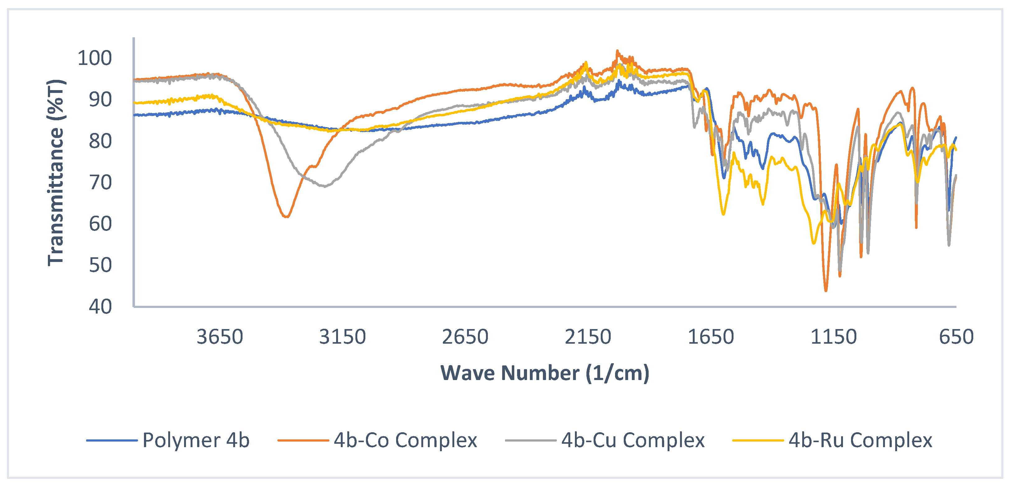
Figure 2.
FT-IR of polymer 4b and its complexes.
A 0.5 m imaging triple grating SP2500i-spectrograph that has three gratings 3600G/mm 1800 G/mm 150 G/mm which are blazed at 240 nm, 500 nm, 500 nm respectively. The spectrograph is coupled to 3 m fiber optics bundle that was used to collect and investigate the generated plasma emission spectra. The fiber optics was placed at the middle distance between electrodes of each generated plasma and far for form plasma edge by ~3 mm. The sensitive rang of the spectrograph was between 200 and 850 nm.
Polymer (4a’).1H NMR (DMSO-d6, ppm) δ: 8.48 (s, 1H, CH=N), 7.95 (d, 2H, Ar-CH), 7.80 (d, 2H, Ar-CH), 7.26 (d, 2H, Ar-CH), 6.90 (d, 2H, Ar-CH).
Polymer (4b’).1H NMR (DMSO-d6, ppm) δ: 9.92 (s, 1H, OH); 8.85 (s, 1H, CH=N), 7.99 (d, 2H, Ar-CH), 7.53 (d, 2H, Ar-CH), 7.43 (d, 2H, Ar-CH), 6.32 (d, 2H, Ar-CH); 6.31 (s, 1H, Ar-CH).
2.6. Antitumor Assay
The cytotoxic evaluation of the new polymeric materials and their complexed counterparts was demonstrated, employing the viability assay as described formerly [43-52]. The inhibitory activity of the desired polymers was inspected against three cancer cell lines: breast carcinoma (MCF-7), colonic carcinoma (HCT-116), and hepatocellular carcinoma (HepG-2).
2.7. Molecular Modeling
The structural model was built using the BUILDER module in wave function package (Spartan’16, Wavefunction, Inc. Irvine, CA) optimization conformational analyses of the built molecules were performed in a two-step procedure. First, these compounds were submitted to energy minimization tool using the included MOPAC 7.0. The geometry of the compounds was optimized using the semiempirical DFT/B3ylp/6311G* [53,54].
3. Results
3.1. Synthesis of Schiff Base Monomers
The synthesis of the Schiff base monomers was accomplished via the reaction of the 4-aminobenzoic acid with 4-hydroxybenzaldehyde or 2,4-dihydroxybezaldehyde in a refluxed ethanolic solution in the presence of a catalytic amount of acetic acid, Scheme 1. Upon crystalization, the attained monomers were characterized, using NMR and FT-IR spectrosopy. For instance, the FT-IR spectra for 3a and 3b displayed resonance peaks at 1664 and 1672 cm-1, corresponding to the vibration of the C=O of the carboxylic groups, and peaks at 1259 and 1218 cm-1, corresponding to the C-O resonance. The resonances at 3396 and 3405 cm-1 are attributed to the broad band exhibited by the OH group. Meanwhile, the 1H-NMR of the isolated monomers revealed that the OH and CH=N protons resonated at 9.78 and 8.48 ppm for monomer 3a, respectively. The same groups displayed resonances further downfield at 9.92 and 8.85 ppm for 3b due to the impact of the additional OH group at the ortho position, which demonstatred a peak at 5.85 ppm. Furthermore, the Ar-CH protons for both monomers exhibited resonaces in the range of 6.31- 7.99 ppm.
3.2. Synthesis of Schiff Base Polymers
3.2.1. Classical Methodology
Monomers 3a and 3b have been subjected to two different styles of polymerization, Scheme 1. The first polymerization process was implemented classically through an estrefication reaction of the desired monomers with p-toluene sulphonyl chloride in refluxed ethanol, where the reactions proceeded for 13 hours and the polymeric materials were precipitated in cold distilled water, Scheme 1. The FT-IR of the isolated polymers displayed a characteristic intense peak at 1712 and 1722 cm-1 for polymers 4a and 4b, respectively, representing the stretching vibration of the ester carbonyl within the polymeric chains. The vibrational frequencies of the C-O appeared at 1148 and 1155 cm-1, and the OH group appeared as broad bands at 3402 and 3268 cm-1 for 4a and 4b, respectively. Meanwhile, the CH=N vibration band occurred at 1576 cm-1 for both polymers, Table 1 and Figure 2. The 1H-NMR of the acquired polymers displayed a desertion of the characteristic carboxylic and phenolic protons of the utilized monomers. Moreover, the CH=N proton appeared at 9.79 and 8.13 ppm for polymers 4a and 4b, respectively. The ortho-positioned OH proton for polymer 4b revealed a resonated peak at 9.92 ppm while the aromatic Ar-CH protons exhibited resonace peaks further downfield in the range of 6.32 and 7.77 ppm for both isolated polymers.
3.2.1. DBD Plasma Methodology
The second polymerization strategy was achieved, utilizing a dielectric barrier discharge (DBD) atmospheric pressure air plasma. The monomeric units were subjected to the plasma stream for various exposure times. During the polymerization process, the treated samples were placed on a movable-swinging stage in the discharge zone to upsurge the homogeneity of the plasma exposure. The obtained polymers were isolated as a film on the stainless steel electrode. The characterization of the polymeric materials was accomplished, using routinely spectroscopic techniques. For instance, The 1H-NMR spectra of the prepared polymers 4a’ and 4b’ demonstrated resonances at 8.48 and 8.85 ppm for the CH=N protons, respectively. The OH proton of 4b’ has a singlet resonance downfield at 9.92 ppm. In addition, the aromatic protons of both polymers displayed similar resonances to their classically-prepared counterparts, polymers 4a and 4b.
The plasma polymerization methodology yields distinct and diverse outcomes to that of the conventional technique in accordance with the distinctive mechanism, which enables the cross-linkage, fragmentation, and re-arrangement of the monomeric units to implement an asymmetrically-structured polymeric pattern as opposed to a consistent, recurring unit structure throughout the polymer [55]. This irregularity in the polymeric framework advances the films’ mechanical characteristics and aging, oxidative, and shrinkage resistance [55, 56]. This behavior could be noticed in the thermal performance of the obtained polymers.
3.3. Complexation of Polymer 4b with Metal Moieties
The complexation of polymer 4b has been accomplished in order to have a comparable inquiry to scrutinize its antitumor influence on the screened cancer cell lines. The complexation of 4b was carried out with four metal salts [Ru (II), Co(II), Cu (II), and Ni (II)] where the desired polymer was refluxed with the selected metallic moieties for 6 hours, and the anticipated products were collected from the corresponding cooled ethanolic solutions. The proposed structures of the complexed polymers is illustrated in Scheme 2. The complexation process was verified, employing the FT-IR spectroscopy, Table 1 and Figure 2. Case in point, the vibratinal frequencies of the OH group revealed a noticable shift due to the complexation with the metallic moities. Polymer 4b exhibited an OH vibrational frequency at 3268 cm-1, which has been shifted to 3367, 3468, 3352, and 3332 cm-1 for its complex counterparts of the Co (II), Cu (II), Ru (II), and Ni (II) moieties, respectively. Moreover, this successful complexation resulted in a distinctive shift of the CH=N vibration to yield values at 1593, 1584, 1608, and 1617 cm-1 for the respected complexes.
3.2. Molecular Weight Determination
The weight average molecular weights of the newly synthesized polymers have been obtained, utilizing a gel permeation chromatography. The acquired results demonstrated that the classical synthetic methodology yielded superior polymeric molecular weights with an average molecular weight of 524,664 and 1,503,322 for polymers 4a and 4b, respectively. Meanwhile, the DBD plasma treatment generated polymeric macromolecules with a mean molecular weight of 162,949 and 874,000 for 4a’ and 4b’, respectively (Table 2).
3.3. Thermal Analysis
The thermal behaviour of the desired polymers (4a, 4b, 4a’,4b’) has been investigated, utilizing the Thermal Gravimetric Analysis (TGA). The samples were subjected to a heat program starting from room temperature to 900oC at a heating rate of 20oC min-1. In general, the obtained thermograms revealed a degradation performance with three weight loss regimes. The first degradation regime was carried out in the range of 209-375oC with the second degradation routine occurring between 316-563oC and the third degradation occuring between 638-863oC, Table 3. The percentage of mass loss varied, according to the structure of the polymeric materials and the procedure of the polymerization. For instance, polymer 4a’ displayed higher stability regarding its mass loss percentage in comparison with its counterpart, polymer 4a, which could be attributed to the possibility of having partial crosslinking and/or rearrangement within its chain matrices as a result of the utilized plazma technique. Similar behavior has been witnessed for polymers 4b’ and 4b, Table 3.
The transition temperatures of the newly synthesized polymers have been scrutinized, employing the differential scanning colorimeter (DSC) technique. The DSC thermogram was obtained under a nitrogen blanket for the second heat run in order to omit the thermal history of the polymeric materials and improve their thermal contact. The phase transition of the polymer is reliant on their molecular weight and the chain matrices arrangement. For instance, polymer 4a displayed only one endothermic phase transition at 80oC followed by an exothermic stage at 145oC. Similary, polymer 4b revealed the same phase transition at 113oC. Contrastingly, the thermal character of the polymeric materials attained through the plasma approach demonstrated a liquid crystal performance in their DSC thermograms, which could be attributed to the possibility of having crosslinking segments and different entanglements of the polymeric chains. Case in point, polymer 4a’ displayed three endothermic regimes at 131, 144, and 170oC. Meanwhile, polymer 4b’ exhibited two endothermic phase transitions at 132 and 177oC. Figure 3 illustrates the thermograms of polymers 4a, 4a’ and 4b’ as representative examples.
3.4. Scanning Electron Microscope
Several micrographs of the polymeric materials and their complexation counterparts have been captured employing the scanning electron microscope (SEM), Figure 4. The following selected images portray the surface morphology of polymer 4b and its Co(II) and Cu(II) complexes, which displayed an amorphous surface. The SEM micrograph demonstrated that the polymeric material appears as marble blocks, Figure 4a. Meanwhile, the coordination with the metal moietes alters their morpholgical shape; for instance, the Schiff base polymer coordinated with the Co(II) moiety exhibited a platelet-like structure, Figure 4b. Additionally, the polymeric matrix containing the Cu(II) moiety revealed a flattered surface with fine particles distributed on the solid matrix, Figure 4c.
3.5. Antitumor Assay
The antitumor activities of the utilized monomeric units, the obtained polymers, and the complexed analogues have been investigated against three cancer cell lines, including: breast carcinoma (MCF-7), colonic carcinoma (HCT-116), and hepatocellular carcinoma (HepG-2), through the employment of the MTT, 3-(4,5-dimethylthiazol-2-yl)-2,5-diphenyl tetrazolium bromide”colorimetric assay [48-50]. The MTT colorimetric examination was conducted with varying concentrations and performed in evaluation against two standard reference drugs: Vinblastine and Colchicine. According to the attained results, the polymer 4a demonstrated the most efficacious apoptotic yields againt the screened tumor cell lines in comparison to its monomeric analogue alongside the reference drug Colchcine. Similarly, polymer 4b demonstrated vastly superior apoptotic activity in assessment with its monomeric counterpart 3b. Additionally, among the polymeric matrices acquired through the plasma pathway, polymer 4a’ exhibited adequate impact in appraisal with the investigated cell lines. Lastly, the metal complexes configured with the 4b polymer revealed an enhancement in behavior regarding the HCT-116 and HepG-2 malignant cells. These results are illustrated in Figure 5 and Table 4.
3.6. Molecular Modeling Studies
3.6.1. Molecular Geometry
The optimization geometry was performed, employing a DFT molecular orbital as reported earlier [57-59]. All calculated energies are listed in Table 5. In the monomer units for 4b, the dihydroxy benzaldehyde are linked in a perpendicular mode with p-amino benzoic acid, Figure 6. Additionally, the monomers are arranged together in chelation with a metal in three possible geometries. Thus, the optimization geometry and their calculated energy levels, measured through the utilization of DFT/BY3LP/6-311G*, predicted the most preferable linkage mode. The calculated energy for these three possible modes revealed that the CuII and NiII copolymers are arranged in a parallel mode. Furthermore, the NiII and RuII complexes demonstrated the most stable geometry, incorporating a linkage with H2O and Cl, Table 5, with total energy (-833.71 and -445.9 Kcal./mol.) respectively.
In the monomer unit of 4b-CuII and 4b-NiII assembled in a parallel pattern, the two phenyl rings are arranged with metal core in a perpendicular mode. The 4b-NiII and 4b-RuII copolymers were chelated with water and stabilized spatially by a coplanarity form between two phenyl and metal rings, Figure 6.
3.6.2. Stability Inter- and Intra-molecular Interaction Profile
Chemical Reactivity & Frontier orbital analysis
FMOs (frontier molecular orbitals) were extensively investigated, regarding their critical role in understanding the interaction methodology of the tested compounds with different chemical systems. The FMOs gap has been calculated to determine the chemical reactivity and kinetic stability of the targeted molecules. The chemical interaction was stabilized inversely with the energy gap.
The stabilization interaction was improved by raising the EHOMO for a molecule and decreasing the ELUMOenergy in another [60]. The EHOMO and ELUMO have been estimated through the DFT theory and are listed in Table 6. The raising of the EHOMO was correlated to the elevated ability of donating electron that corresponded with the detached electrons from the valance shell and reveales a high oxidizing ability, and vice versa [61]. The EHOMO for the ligand exhibited a superior value in comparison with its metal complexes with the complexes arrayed in a decreasing order including 4b-CuII> 4b-NiII> 4b-CoII> 4b-RuII, Table 6. The HOMO region was caped in the CuII polymers over an oxygen linker between the monomer units. Additionally, these orbitals in the NiII polymer cover the C=N linkage, as illustrated in Figure 7. The LUMO zone was condensed over the metal core for the CuII and NiII ring. Moreover, in the Cu-chelate, this zone was situated on the pyridine and metal rings, displayed in Figure 7.
The HOMO and LUMO orbitals in the case of 4b-CoII, localized over metal cores, were distributed between two phenyls along the molecular skeleton for 4b-RuII, as demonstrated in Figure 7, while the HOMO/ LUMO orbitals of 4a and 4b were stabilized over all skeletons.
The EHOMOs and ELUMOs for the 4a, 4b, 4b-CuII, 4b-NiII, 4b-CoII, and 4b-RuII possess negative values. These values promote a high stability interaction with biological media and the migration of electrons over all molecular skeletons for the aforementioned copolymers through the intramolecular transfer mechanism. In addition, FMOs are directly linked via softness and hardness. Thus, electrophiles and nucleophiles can be interpreted through these concepts. 4a, 4b, 4b-CuII, 4b-NiII, 4b-CoII, and 4b-RuII revealed low ΔG values and were ordered as 4a> 4b-RuII > 4b-CuII > 4b > 4b-CoII > 4b-NiII, revealing that growing softness trended as 4b-Ni> 4b-Co> 4b> 4b-Cu> 4b-Ru. The nucleophilicity increased accordingly as 4b< 4b-Ru< 4b-Cu< 4b-Co< 4b-Ni. 4b was represented through the highest electrophilicity index, which easily explained the complexation with metal ions. The intermolecular reactivity has been analyzed, founded on group-phylicity (ΔρKEle and ΔρKNU). These terms are utilized when electrophile ΔρKEle & nucleophile ΔρKNU were simultaneously hitting for the particles. The positive values possessed by the investigated polymer and its complexes with CuII, NiII, CoII, and RuII are likely attributed to the electrophile attacking and the charge shift of media → molecule and vice versa. The results from Table 6 suggested that these ligands and copolymers are promising as nucleophiles with a high ability for receiving electrons from biological media. In addition, ΔNmax has -ve values between -0.6 to -4.1 ev., and the electron cloud migrated as biological media → polymer, Table 6.
Molecular Electrostatic Potential Map (EPM)
The electrostatic potential map (EPM) identifies dye by the equilibrium between repulsive (+ve charge) and attractive (-ve charge) forces [62] and are related to nucleophilic/electrophilic reactivities, respectively. The EPM was mapped for the ligands and metal complexes, drafted in Figure 7. The area with high electron density was distinguished (in orange, yellow, & red colors). The positive efficiency region color is leaning towards the color blue while the intermediate potential region is shaded by the green color. The positive charge was enclosed over the carbon atom in the carbonyl moiety for 4a, and 4b whilst in the 4b-Cu, 4b-Ni, and 4b-Co complexes; the positive cloud is distributed over the chelating sites. The difference between color in the EPMs morphology demonstrated the electrostatic variation between the potential values. Growing the red zone for the 4b-Ru compound has indicated the high ability of the electrophilic process and recognizes the nucleophile groups in the biological media, Figure 7. The 4b, 4b-CuII, 4b-NiII, and 4b-CoII have depicted distribution in the blue zone, which demonstrated the nucleophile site attack of electrophilic media.
The Local Reactivity Profile
The local reactivity “LR” parameters are identified by the variation of electronic systems, through distortion on the electronic cloud at the exact atomic region concerning accepting or donating of electrons. LRs were accomplished to examine site-selectivity toward chemical reactions, as displayed in Figure 8.
The “” is signified through a red color corner, which exhibits the rising of electron density after gaining a charge while a +ve charge is represented in the blue color. In “” , the red cite revealed a reduction in electron density after providing an electron (gaining a -ve charge). Consequently, PVA-NA revealed that their C=O group possesses the highest sensitive site against earning or contributing electrons ( and ), which was depicted as the electron cloud migrating between phenyl and C=O and caused the stabilization interaction with metal. The negative charges in the copolymers are more condensed over M-cores for both cases and f+, which enhanced the chelation power with the biological media.
3.7. Molecular Docking Simulations
The in-silico examination was conducted to scrutinize biological findings through the employment of Glide’s module® and docking experiments against the wild-type EGFR-TKD (PDB ID: 1M17 [63]) and co-crystal erlotinib (AQ44) as reference inhibitors, as disclosed in Figure 9. The binding sites for protein incorporated GLN767, MET769, THR830, and ASP831 [63]. The designed molecules 4a, and 4b and its complexes counterparts 4b-Cu, 4b-Ni, 4b-Co, and 4b-Ru exhibited superior docking scores and orientations in comparison with Erlotinib, Table 5. To validate the docking protocol, the original inhibitor [Erlotinib “AQ44”] was redocked into the crystal structures of EGFR. Furthermore, the original inhibitor was suitably fitted into their own binding sites for their crystal structures. The most potent pose selected was based on the lowest binding free energy ΔG (listed in Table 7) and rmsd.
AQ44 interacted with the vital MET 769 amino acid at a distance of 2.70 Å with the H-interaction energy = −0.8 (kcal/mol). The investigated 4a, 4b, 4b-Cu complex, 4b-Ni complex, 4b-Co complex, and 4b-Ru complex interacted with the same binding manner. The free binding energy was predicted for further investigation of the targeted molecules’ biological findings. Based on the energy framework analysis, the term E_int. is the most important contributor to ΔG, although the central molecule forms hydrogen bonds with its neighboring backbone residue. Compounds 4a, 4b, 4b-Cu complex, 4b-Ni complex, 4b-Co complex, and 4b-Ru complex displayed higher total binding affinities ΔG, ranging from −5.69 to −7.88 kcal/mol, which is higher than that of AQ44 (ΔE = −5.37 kcal/mol) and explains the promising potency. Based on the results in Table 7, the H.B. interaction for the molecules (H.B. = −9.34 to −11.51 Kcal/mol) is the vital factor in the interaction.
Furthermore, the electrostatic energy framework (E_ele. = −14.34 to −20.21 Kcal/mol) displayed that the interaction between 4a, 4b, CuII, NiII, CoII, and RuII and the other residues was mainly contributed by the dispersion interaction. The ΔG for 4b exhibited a lower affinity in evaluation with 4b-Cu complex, 4b-Ni complex, 4b-Co complex, and 4b-Ru complex. The 4b formed two H-bonds between the OH group and (COOH) of Asp831 and Thr766 with distances of 2.09 and 2.08Å and the corresponding ΔE of −0.9 and -1.2(kcal/mol). On the other hand, the Cu metal interacted with Cys773 while NiII is stabilized in the binding pocket by the interaction with Gly772 and Asp831. Moreover, CoII and RuII interacted with Asp831 by a strong H-bond with corresponding distances 2.12 Å and 2.16 Å, Table 7.
3.8. In-silico Pharmacokinetics and Toxicity Studies
Computational studies are viable approaches that possess several advantages over in-vivo studies, particularly in terms of reduction of cost, time, and animal testing. Currently, these studies are widely employed to study the physicochemical and pharmacokinetics properties of a compound in medicinal chemistry
Lipinski rule of 5 (Ro5) is considered as a benchmark in drug development34 and violation of Ro5 integrates MW > 500, lipophilicity (LogPo/w) > 5, hydrogen bond acceptor (HBA) fewer than 10, and hydrogen bond donor (HBD) less than 5. These violations lead to poor intestinal absorption, permeation, and/or solubility. The Lipinski Ro5 was further extended by including polar surface area (PSA less than 140 Å2), molecule flexibility (nROTB less than/equal to 10), and important predictors of oral bioavailability for a drug candidate. The polymers and its complexes have been assessed in-silico by Swiss ADME software.
All molecules except those bearing CuII and NiII in the skeleton acquire the desired molecular weight (MW < 500) while the remaining parameters such as HBA, HBD, and lipophilicity were unveiled to be in the desired range for all the derivatives, indicating good oral absorption and permeation. All compounds displayed molecular flexibility less than 10 and TPSA < 140 Å2, suggesting that these molecules possess decent oral bioavailability as drug candidates.. It can be seen that all the synthesized molecules were non-permeable to BBB, signifying that these molecules will not cause any adverse effects on brain, Table 8. 4a, 4b, 4b-CuII, 4b-NiII, 4b-CoII, and 4b-RuII retain excellent oral drug likeness properties with all the physiochemical and pharmacokinetics parameters in an acceptable range without any Ro5 violations, good TPSA, and without any permeability to brain, Table 8.
Over the last few years, synthetic molecules have increased tremendously causing environmental contamination and toxicity. Additionally, the in-silico toxicity prediction has been carried out to see the adverse effects and the toxicity of the Eugenol derivatives 4a, 4b, 4b-CuII, 4b-NiII, 4b-CoII, and 4b-RuII, and the results are presented in Table 9. According to the guidelines by NICEATM in collaboration with EPA and NCCT, the compounds with LD50 ≤ 50 mg/kg are highly toxic; LD50 in between 50 ≤ 500 mg/kg are moderately toxic; LD50 in between 500 ≤ 5000 mg/kg are slightly toxic; and LD50 > 5000 mg/kg are considered safe. On this evidence, all the synthesized compounds were found to be highly safe, as they displayed LD50 in the range of 2.38-2.70 mole/kg. 4a, 4b, 4b-CuII, 4b-NiII, 4b-CoII, and 4b-RuII were found to be non-carcinogenic and non-mutagenic, as depicted by the AMES toxicity.
4. Conclusions
This work elucidates a comparison investigation of producing Schiff base macromolecules exploiting two synthetic strategies. The first strategy engaged a classical chemistry routine through condensation polymerization, which led to the formation of high molecular weight polyester. In addition, the DBD atmospheric pressure air plasma was employed to construct this class of macromolecules. Additionally, polymer 4b has been coordinated to different metal moieties, Cu, Ni, Co, and Ru. All the polymeric materials and the coordination polymers have been examined as antitumor agents against four cell lines, MCF-7, A-549, HCT-116, and HepG-2. Lastly, the docking appraisal was performed on acquired macromolecules and their metal analogues and exhibited numerous important interactions with the amino acid of the enzyme being targeted.
Author Contributions
R.M.O., T.H.A. and conceived and designed the research; Z.H. and I.I. performed the experiments; A.-A.H.M. designed the plasma experiments. A.A.H., W.H.A. M.O.A. performed the docking studies; and R.M.O., F.F.A., E.A.A., and F.S.A. performed the chemistry and implemented the biological study. All authors analyzed the data, wrote the paper, edited the English language and discussed the results, and commented on the manuscript. All authors have read and agreed to the published version of the manuscript.
Funding
This research was funded by the Deputyship for Research and Innovation, Ministry of Education in Saudi Arabia, project number 194/442.
Acknowledgments
The authors extend their appreciation to the Deputyship for Research and Innovation, Ministry of Education in Saudi Arabia for funding this research work, project number 194/442. Also, the authors would like to extend their appreciation to Taibah University for the supervision support.
Conflicts of Interest
The authors declare no conflict of interest. The funders had no role in the design of the study; in the collection, analyses, or interpretation of data; in the writing of the manuscript; or in the decision to publish the results.
References
- Fu1, C.; Zhu, C.; Synatschke, C.V.; Zhang, X. Editorial: Design, Synthesis and Biomedical Applications of Functional Polymers. Front. Chem. 2021, 9, 681189. [CrossRef]
- Ahmed, S.A.; Okasha, R.M.; Khairou, K.S.; Afifi, T.H.; Mohamed, A.-A. H.; Abd-El-Aziz, A.S. Design of Thermochromic Polynorbornene Bearing Spiropyran Chromophore Moieties: Synthesis, Thermal Behavior and Dielectric Barrier Discharge Plasma Treatment. Polymers 2017, 9, 630-645. [CrossRef]
- Caló, E.; Khutoryanskiy, V. V. Biomedical applications of hydrogels: A review of patents and commercial products. European Polymer Journal 2015, 65, 252– 267. [CrossRef]
- Love, B. Polymeric Biomaterials. In B. Love (Ed.), Biomaterials: A Systems Approach to Engineering Concepts. 2017, 205– 238, Michigan: Elsevier. [CrossRef]
- Abd-El-Aziz, A.S.; El-Ghezlani, E.G.; Elaasser, M.M.; Afifi, T.H.; Okasha, R.M. First Example of Cationic Cyclopentadienyliron Based Chromene Complexes and Polymers: Synthesis, Characterization, and Biological Application. J. Inorg. Organomet. Poly. Mater. 2020, 30, 131. [CrossRef]
- Maitz, M. F. (2015). Applications of synthetic polymers in clinical medicine. Biosurface and Biotribology 2015, 1(3), 161– 176. [CrossRef]
- Joraid, A.A.; Okasha, R.M.; Al-Maghrabi, M.A.; Afifi, T.H.; Agatemor, C.; Abd-El-Aziz, A.S. Thermodynamic Parameters of Non-isothermal Degradation of a New Family of Organometallic Dendrimer with Isoconversional Methods. J. Inorg. Organomet. Poly. Mater. 2022, 32, 2653–2663. [CrossRef]
- Teo, A. J. T.; Mishra, A.; Park, I.; Kim, Y. J.; Park, W. T.; Yoon, Y. J. Polymeric Biomaterials for Medical Implants and Devices. ACS Biomaterials Science and Engineering 2016, 2(4), 454– 472. [CrossRef]
- Gupta, P.; Vermani, K.; Garg, S. Hydrogels: From controlled release to pH-responsive drug delivery. Drugs Discovery Today 2002, 7(10), 569– 579. [CrossRef]
- Hwangbo, J.; Seo, H.; Sim, G.; Avila, R.; Nair, M.; Kim, B.; Choi, Y. Bioresorbable polymers for electronic medicine. Cell Reports Physical Science 2024, 5(8), 102099. [CrossRef]
- Yılmazoğlu, E.; Karakuş, S. Synthesis and specific biomedical applications of polymer brushes. App. Surf. Sci. Adv. 2023, 18, 100544. [CrossRef]
- Sun, D.; Babar Shahzad, M.; Li, M.; Wang, G.; Xu, D. Antimicrobial materials with medical applications. Materials Technology 2015, 30(B2), B90– B95. [CrossRef]
- Sall, C.; Ayé, M.; Bottzeck, O.; Praud, A.; Blache, Y. Towards smart biocide-free anti-biofilm strategies: Click-based synthesis of cinnamide analogues as anti-biofilm compounds against marine bacteria. Bioorganic and Medicinal Chemistry Letters 2018, 28(2), 155– 159. [CrossRef]
- Schönemann, E.; Koc, J.; Aldred, N.; Clare, A.S.; Laschewsky, A.; Rosenhahn, A.; Wischerhoff, E. Synthesis of novel sulfobetaine polymers with differing dipole orientations in their side chains, and their effects on the antifouling properties. Macromolecular Rapid Communications 2019, 41, 1900447. [CrossRef]
- Utrata-Wesoek, A. Antifouling surfaces in medical application. Polimery/Polymers, 2013, 58(9), 685– 695. [CrossRef]
- Yue, Z.; Liu, X.; Molino, P. J.; Wallace, G. G. (2011). Bio-functionalisation of polydimethylsiloxane with hyaluronic acid and hyaluronic acid – Collagen conjugate for neural interfacing. Biomaterials 2011, 32(21), 4714– 4724. [CrossRef]
- Althakfi, S.H.; Suhail, M.; Locatelli, M.; Hsieh, M.-F.; Alsehli, M.; Hameed, A.M. Advances in Polymeric Colloids for Cancer Treatment. Polymers 2022, 14, 5445. [CrossRef]
- Rabha, B.; Bharadwaj, K.K.; Pati, S.; Choudhury, B.K.; Sarkar, T.; Kari, Z.A.; Edinur, H.T.; Baishya, D.; Atanase, L.I. Development of polymer-based nanoformulations for glioblastoma brain cancer therapy and diagnosis: An update. Polymers 2021, 13, 4114. [CrossRef]
- Saeedi, T.; Alotaibi, H.F.; Prokopovich, P. Polymer colloids as drug delivery systems for the treatment of arthritis. Adv. Colloid Interf. Sci. 2020, 285, 102273. [CrossRef]
- Xin, J.; Lu, X.; Cao, J.; Wu, W.; Liu, Q.; Wang, D.; Zhou, X.; Ding, D. Fluorinated Organic Polymers for Cancer Drug Delivery. Adv. Mater. 2024, 36(30), 2404645. [CrossRef]
- Misiak, P.; Markiewicz, K.H.; Szymczuk, D.; Wilczewska, A.Z. Polymeric Drug Delivery Systems Bearing Cholesterol Moieties: A Review. Polymers 2020, 12, 2620. [CrossRef]
- Jelonek, K.; Kasperczyk, J. Polyesters and polyester carbonates for controlled drug delivery. Part II. Implantable systems. Polimery 2013, 58, 858–863. [CrossRef]
- Jones, D. Pharmaceutical Applications of Polymers for Drug Delivery; Rapra Review Reports; Rapra Technology: Shrewsbury, UK, 2004; ISBN 978-1-85957-479-9.
- Cheng, L.-C.; Jiang, Y.; Xie, Y.; Qiu, L.-L.; Yang, Q.; Lu, H.-Y. Novel amphiphilic folic acid-cholesterol-chitosan micelles for paclitaxel delivery. Oncotarget 2017, 8, 3315–3326. [CrossRef]
- Borandeh, S.; Bochove, B.V.; Teotia, A.; Seppälä, J. Polymeric drug delivery systems by additive manufacturing. Adv. Drug Deliv. Rev. 2021, 137, 349-373. [CrossRef]
- Xu, J.; Liu, Y.; Hsu, SH. Hydrogels Based on Schiff Base Linkages for Biomedical Applications. Molecules 2019, 24(16), 3005. [CrossRef]
- Kenawy, E.-R.; El-Khalafy, S.H.; Abosharaf, H.A.; El-nshar, E.M.; Ghazy, A.R.; Azaam, M.M. Synthesis, Characterization, and Anticancer Potency of Branched Poly (p-Hydroxy Styrene) Schiff-Bases. Macromol. Biosci. 2023, 23, 2300090. [CrossRef]
- Mighani, H. Schiff Base polymers: synthesis and characterization. J. Polym. Res. 2020, 27, 168. [CrossRef]
- Demirbağ, B.; Büyükafşar, K.; Kaya, H.; Yıldırım, M.; Bucak, Ö.; Ünver, H.; Erdoğan, S. Investigation of the anticancer effect of newly synthesized palladium conjugate Schiff base metal complexes on non-small cell lung cancer cell line and mouse embryonic fibroblast cell line. Biochem. Biophys. Res. Comm. 2024, 735, 150658. [CrossRef]
- Huang, J.; Deng, Y.; Ren, J.; Chen, G.; Wang, G.; Wang, F.; Wu, X. Novel in situ forming hydrogel based on xanthan and chitosan re-gelifying in liquids for local drug delivery. Carbohydr. Polym. 2018, 186, 54–63. https://doi.10.1016/j.carbpol.2018.01.025.
- Desai, S.B.; Desai, P.B.; Desai, K.R. Synthesis of some Schiff bases, thiazolidones, and azetidinones derived from 2,6-diaminobenzo [1,2-d:4,5-d′]bisthiazole and their anticancer activities. Heterocycl Commun. 2001, 7(1), 83–90. [CrossRef]
- Thakor, P.M.; Patel, J.D.; Patel, R.J.; Chaki, S.H.; Khimani, A.J.; Vaidya, Y.H.; Chauhan, A.P.; Dholakia, A.B.; Patel, V.C.; Patel, A.-K.J.; Bhavsar, N.H.; Patel, H.V. Exploring New Schiff Bases: Synthesis, Characterization, and Multifaceted Analysis for Biomedical Applications. ACS Omega 2024, 9 (33), 35431-35448. [CrossRef]
- Azam, F.; Singh, S.; Khokhra, S.L.; Prakash, O. Synthesis of Schiff bases of naphtha [1,2-d]thiazol-2-amine and metal complexes of 2-(2’-hydroxy)benzylideneaminonaphthothiazole as potential antimicrobial agents. J Zhejiang Univ Sci B. 2007, 8(6), 446-52. [CrossRef]
- Trávníček, Z.; Maloň, M.; Šindelář, Z.; Doležal, K.; Rolčik, J.; Kryštof, V.; Strnad, M.; Marek, J. Preparation, physicochemical properties and biological activity of copper(II) complexes with 6-(2-chlorobenzylamino) purine (HL) or 6-(3-chlorobenzylamino)purine (HL). The single-crystal X-ray structure of [Cu(H+L2)2Cl3]Cl·2H2O. J Inorg Biochem. 2001, 84(1-2), 23–32. [CrossRef]
- Tadele, K.T.; Tsega, T.W. Schiff Bases and their Metal Complexes as Potential Anticancer Candidates: A Review of Recent Works. Anticancer Agents Med Chem. 2019, 19(15), 1786-1795. [CrossRef]
- Jain, S.; Rana, M.; Sultana, R.; Mehandi, R.; Rahisuddin. Schiff Base Metal Complexes as Antimicrobial and Anticancer Agents. Polycyclic Aromatic Compounds 2022, 43(7), 6351–6406. [CrossRef]
- Levchenko, I.; Xu, S.; Baranov, O.; Bazaka, O.; Ivanova, E.P.; Bazaka, K. Plasma and Polymers: Recent Progress and Trends. Molecules 2021, 26(13), 4091. [CrossRef]
- Jang, H.J.; Jung, E.Y.; Parsons, T.; Tae, H.S.; Park, C.S. A Review of Plasma Synthesis Methods for Polymer Films and Nanoparticles under Atmospheric Pressure Conditions. Polymers (Basel) 2021, 13(14), 2267. [CrossRef]
- Aziz, G.; Ghobeira, R.; Morent, R.; Geyter, N.D. Plasma Polymerization for Tissue Engineering Purposes. Recent Research in Polymerization. In Tech 2018, ISBN: 978-953-51-3747-4. http://dx.doi.org/10.5772/intechopen.72293.
- Krtouš, Z.; Hanyková, L.; Krakovský, I.; Nikitin, D.; Pleskunov, P.; Kylián, O.; Sedlaříková, J.; Kousal, J. Structure of Plasma (re)Polymerized Polylactic Acid Films Fabricated by Plasma-Assisted Vapour Thermal Deposition. Materials 2021, 14, 459. [CrossRef]
- Kima, J.Y.; Leea, Y.; Limb, D.Y. Plasma-modified polyethylene membrane as a separator for lithium-ion polymer battery. Electrochimica. Acta 2009, 54, 3714–3719. 10.1016/j.electacta.2009.01.055.
- Sabetzadeh, N.; Falanaka, C.; Riahifar, R.; Yaghmaee, M.S.; Raissi, B. Plasma treatment of polypropylene membranes coated with zeolite/organic binder layers: Assessment of separator performance in lithium-ion batteries. Solid State Ion. 2021, 363, 115589. [CrossRef]
- Mosmann, T. Rapid colorimetric assay for cellular growth and survival: Application to proliferation and cytotoxicity assays. J. Immunol. Methods 1983, 65, 55–63.
- Gangadevi, V.; Muthumary, J. Preliminary studies on cytotoxic effect of fungal taxol on cancer cell lines. Afr. J. Biotechnol. 2007, 6, 1382–1386. [CrossRef]
- Bensaber, S.M.; Allafe, H.A.; Ermeli, N.B.; Mohamed, S.B.; Zetrini, A.A.; Alsabri, S.G.; Erhuma, M.; Hermann, A.; Jaeda, M.I.; Gbaj, A.M. Chemical synthesis, molecular modelling, and evaluation of anticancer activity of some pyrazol-3-one Schiff base derivatives. Med. Chem. Res. 2014, 23, 5120–5134.
- Eltayeb, N.E.; Lasri, J.; Soliman, S.M.; Mavromatis, C.; Hajjar, D.; Elsilk, S.E.; Babgi, B.A.; Hussien, M.A. Crystal structure, DFT, antimicrobial, anticancer and molecular docking of (4E)-4-((aryl)methyleneamino)-1,2-dihydro-2,3-dimethyl-1-phenylpyrazol-5-one. J Mol Struct 2020, 1213, 128185.
- Fayed, E.A.; Eldin, R.R.E.; Mehany, A.; Bayoumi, A.H.; Ammar, Y.A. Isatin-Schiff’s base and chalcone hybrids as chemically apoptotic inducers and EGFR inhibitors; design, synthesis, anti-proliferative activities and in silico evaluation. J Mol Struct 2021, 1234, 130159.
- Ghasemi, M.; Liang, S.; Luu, Q. M.; Kempson, I. The MTT Assay: A Method for Error Minimization and Interpretation in Measuring Cytotoxicity and Estimating Cell Viability. Springer Protocols, 2024. [CrossRef]
- Assays to Assess the Proliferative Behavior of Cancer Cells. ScienceDirect Topics, 2023. (Sigma Aldrich). [CrossRef]
- MTT Assay Protocol for Cell Viability and Proliferation. Sigma-Aldrich, 2023. https://www.sigmaaldrich.com/US/en/technical-documents/protocol/cell-culture-and-cell-culture-analysis/cell-based-assays/mtt-assay-for-cell-viability-and-proliferation (Sigma Aldrich).
- Vistica, V. T.; Skehan, P.; Scudiero, D.; Monks, A.; Pittman, A.; Boyd, M. R. Tetrazolium-based Assays for Cellular Viability: A Critical Examination of Selected Parameters Affecting Formazan Production. Cancer Res. 1991, 51 (10), 2515-2520.
- Mosmann, T. Rapid Colorimetric Assay for Cellular Growth and Survival: Application to Proliferation and Cytotoxicity Assays. J. Immunol. Methods 1983, 65 (1-2), 55-63. [CrossRef]
- Hehre WJ, Huang WW. Chemistry with computation: an introduction to SPARTAN. Wavefunction, Incorporated; 1995.
- Alam, W., Khan, H., Jan, M.S., W. Darwish, H., Daglia, M. and A. Elhenawy, A., 2024. In vitro 5-LOX inhibitory and antioxidant potential of isoxazole derivatives. PloS one, 19(10), p.e0297398. [CrossRef]
- Friedrich, J. Mechanisms of Plasma Polymerization –Reviewed from a Chemical Point of View. Plasma Process. Polym. 2011, 8, 783–802. [CrossRef]
- Friedrich, J.; Ku¨hn, G.; Mix, R.; Retzko, I.; Gerstung, V.; Weidner, S.T.; Schulze, R.-D.; Unger, W. in: Polyimides and other High Temperature Polymers: Synthesis, Characterization and Applications, K. L. Mittal, Ed., VSP, Utrecht, 2003, 359– 388.
- Khan, F., Alam, A., Rehman, N.U., Ullah, S., Elhenawy, A.A., Ali, M., Islam, W.U., Khan, A., Al-Harrasi, A., Ahmad, M. and Haitao, Y., 2025. Synthesis, anticancer, α-glucosidase inhibition, molecular docking and dynamics studies of hydrazone-Schiff bases bearing polyhydroquinoline scaffold: In vitro and in silico approaches. Journal of Molecular Structure, 1321, p.139699. [CrossRef]
- Rafik, A., Tüzün, B., Zouihri, H., Poustforoosh, A., Hsissou, R., Elhenawy, A. and Guedira, T., 2024. Morphology Studies, Optic Proprieties, Hirschfeld Electrostatic Potential Mapping, Docking Molecular Anti-Inflammatory, and Dynamic Molecular Approaches of Hybrid Phosphate. Journal of the Indian Chemical Society, p.101419. [CrossRef]
- Asad, M., Arshad, M.N., Azum, N., Alzahrani, K.A., Marwani, H.M., Elhenawy, A.A., Alam, M.M., Nazreen, S., Snigdha, K. and TN, M.M., 2024. Chitosan/La catalyzed synthesis of novel ferrocenated spiropyrrolizidines: Green synthesis, crystallographic, DFT and Hirshfeld surface studies. Journal of Molecular Structure, p.140240. [CrossRef]
- Mphahlele, M.J., Magwaza, N.M., More, G.K. and Elhenawy, A.A., 2024. Synthesis, structure of the N-(Alkyl/Arylsulfonyl) substituted 5-(Bromo/Iodo)-3-methylindazoles and bioactivity screening against some of the biochemical targets linked to type 2 diabetes mellitus. Journal of Molecular Structure, 1312, p.138636. [CrossRef]
- Gul, S., Elhenawy, A.A., Ali, Q., Rehman, M.U., Alam, A., Khan, M., AlAsmari, A.F. and Alasmari, F., 2024. Discovering the anti-diabetic potential of thiosemicarbazone derivatives: In vitro α-glucosidase, α-amylase inhibitory activities with molecular docking and DFT investigations. Journal of Molecular Structure, 1312, p.138671. [CrossRef]
- Abdalrazaq, E.A., Abu-Yamin, A.A., Taher, D., Hassan, A.E. and Elhenawy, A.A., 2024. Zn (II) and Cd (II) complexes of dithiocarbamate ligands: synthesis, characterization, anticancer, and theoretical studies. Journal of Sulfur Chemistry, 45(5), pp.714-739. [CrossRef]
- Stamos, J., Sliwkowski, M.X. and Eigenbrot, C., 2002. Structure of the epidermal growth factor receptor kinase domain alone and in complex with a 4-anilinoquinazoline inhibitor. Journal of biological chemistry, 277(48), pp.46265-46272. [CrossRef]
Scheme 1.
Synthesis of Schiff base monomers and their corresponding polymers.
Scheme 2.
Metal complexes of polymer 4b.
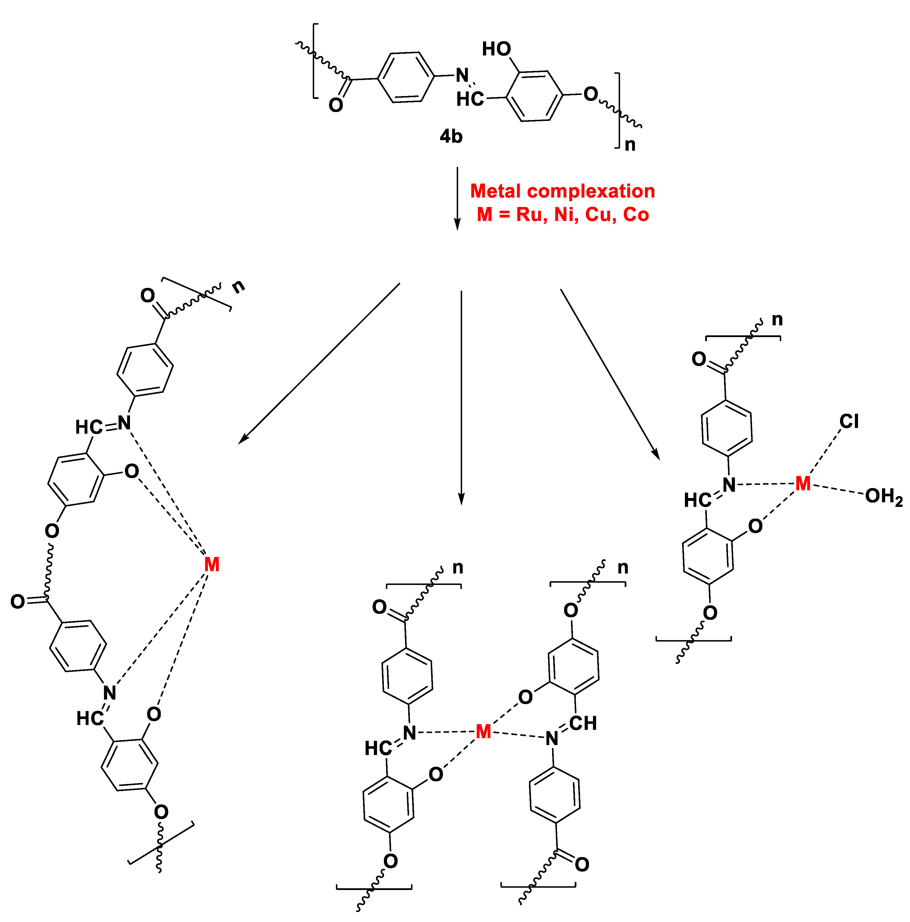
Figure 3.
DSC thermogram of polymers 4a (classic polymerization) and 4a’ (plasma polymerization).
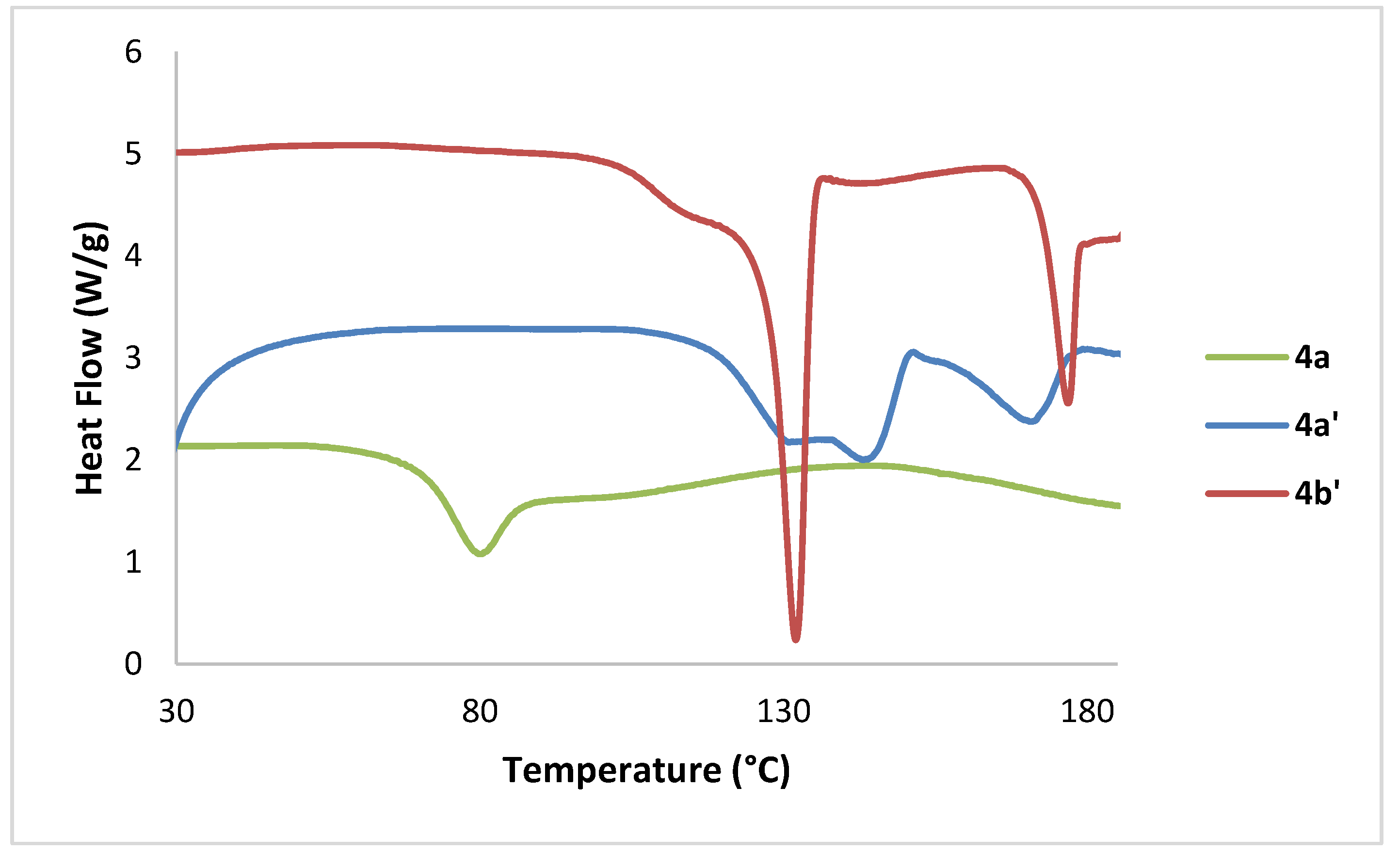
Figure 4.
SEM micrograph of poymer 4b (a), 4b-Co complex (b), and 4b-Cu complex (c).
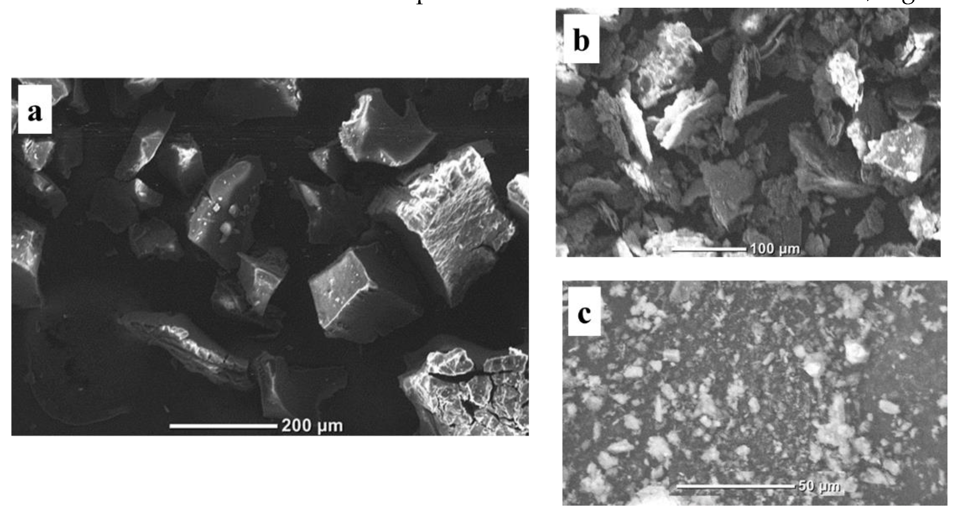
Figure 5.
Cytotoxic activity of Schiff base monomers, polymers, and complexed analogs. .
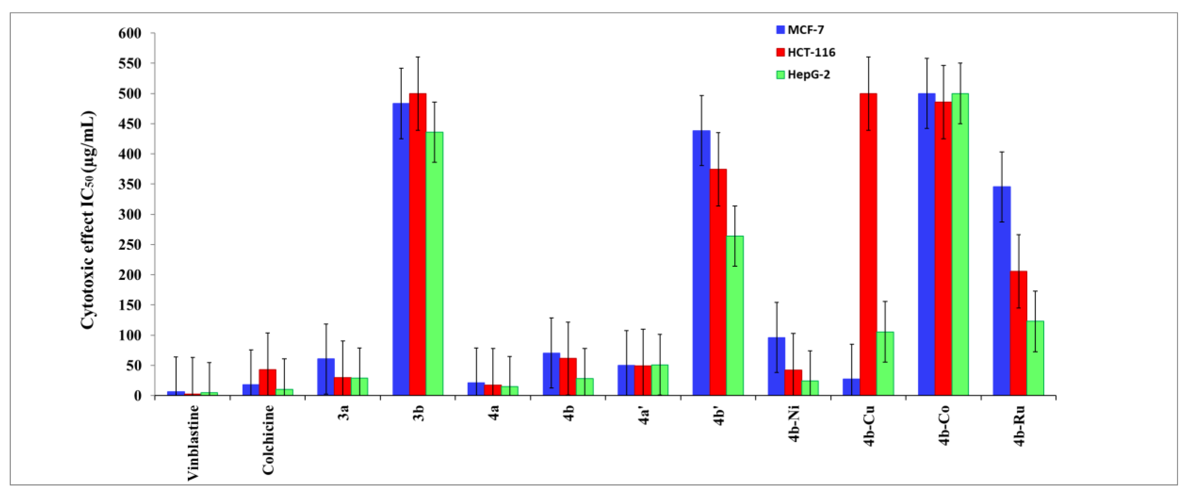
Figure 6.
Optimization Geometry ligands and their metal complexes which represented in ball and stick model, the hydrogen atoms removed for clarifying.
Figure 6.
Optimization Geometry ligands and their metal complexes which represented in ball and stick model, the hydrogen atoms removed for clarifying.
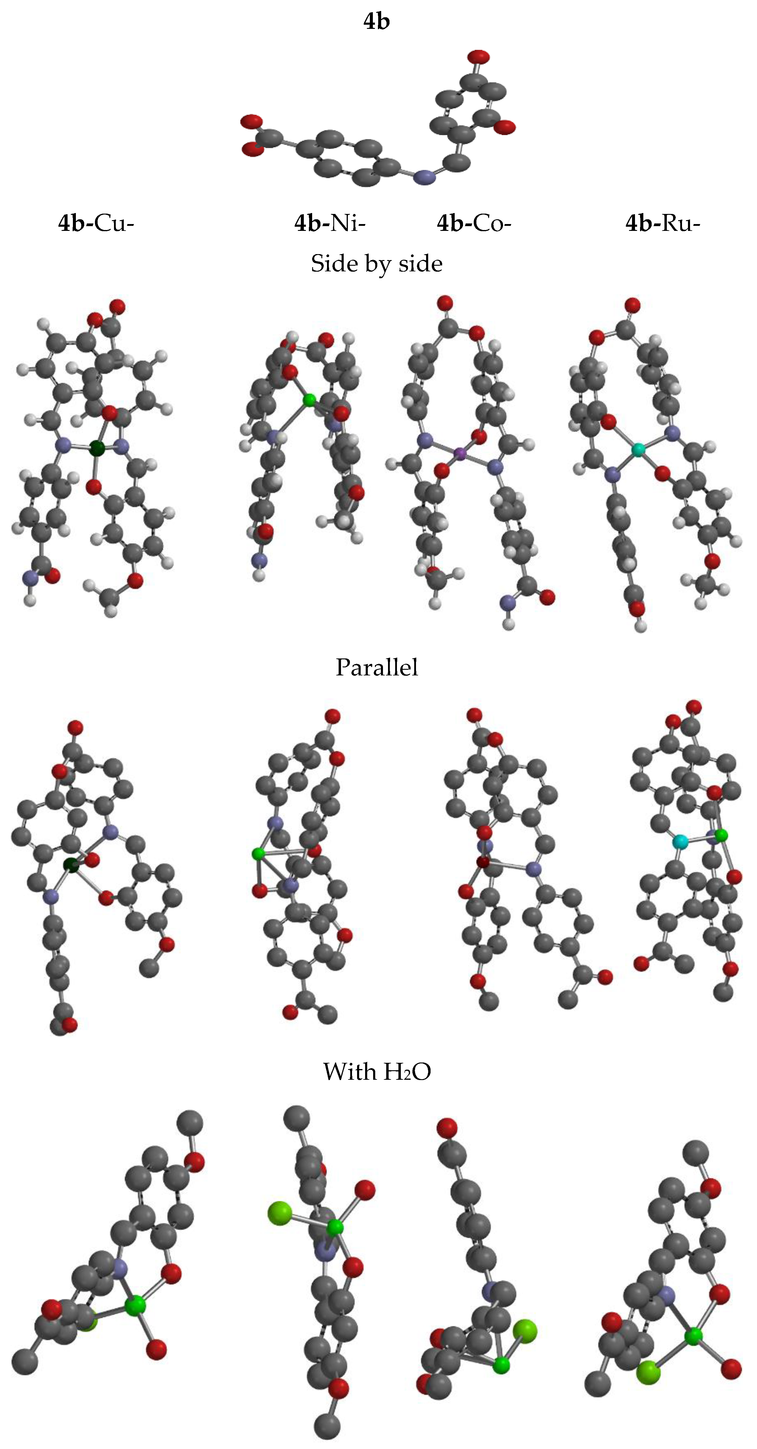
Figure 7.
Frontier molecular orbitals (HOMO and LUMO) for ligand and its complexes (ii)Molecular electrostatic potential map(EPM).
Figure 7.
Frontier molecular orbitals (HOMO and LUMO) for ligand and its complexes (ii)Molecular electrostatic potential map(EPM).
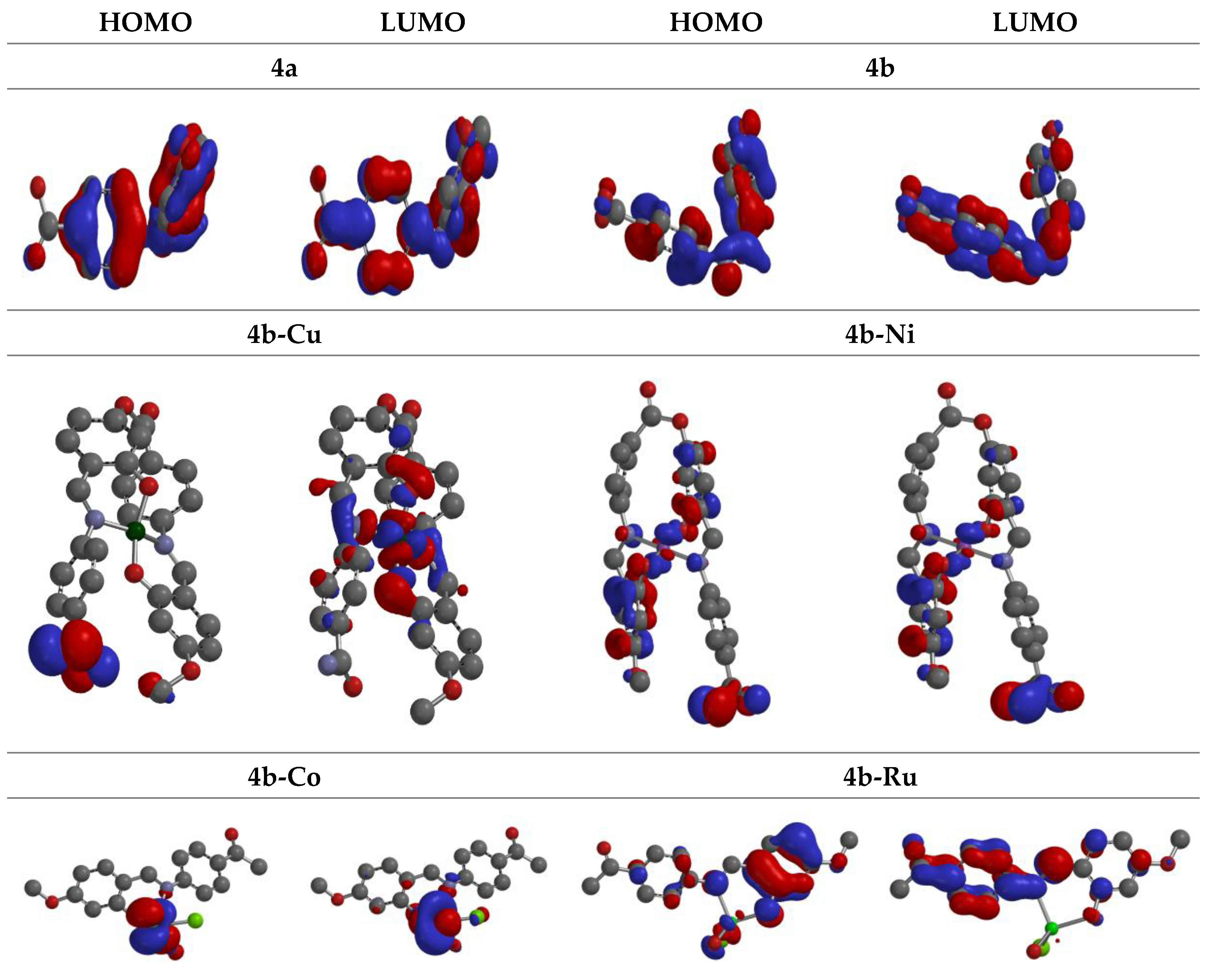
Figure 7.
Electrostatic potential for 4a, 4b and its complexes at DFT/BY3LP.
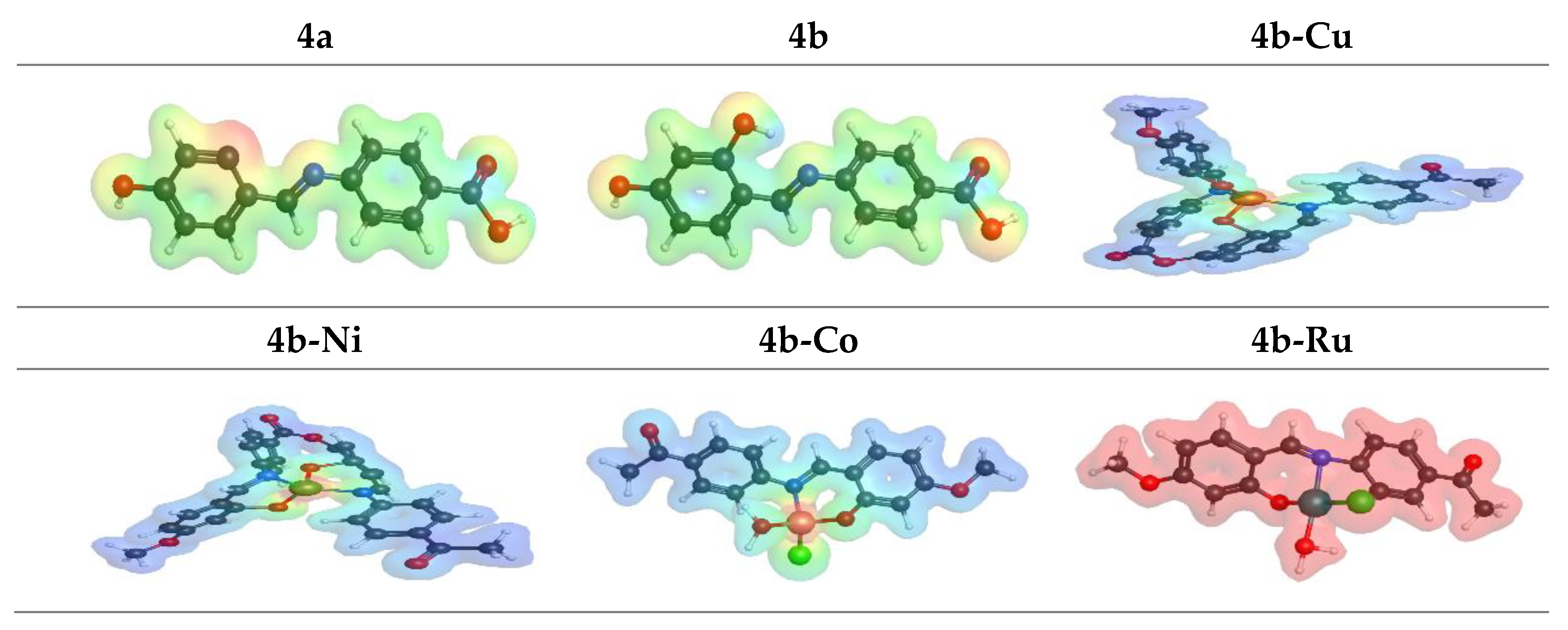
Figure 8.
Fuki function (electrophile, nucleophile and radical) for Ligands and its complexes using DFT/BY3LP.
Figure 8.
Fuki function (electrophile, nucleophile and radical) for Ligands and its complexes using DFT/BY3LP.
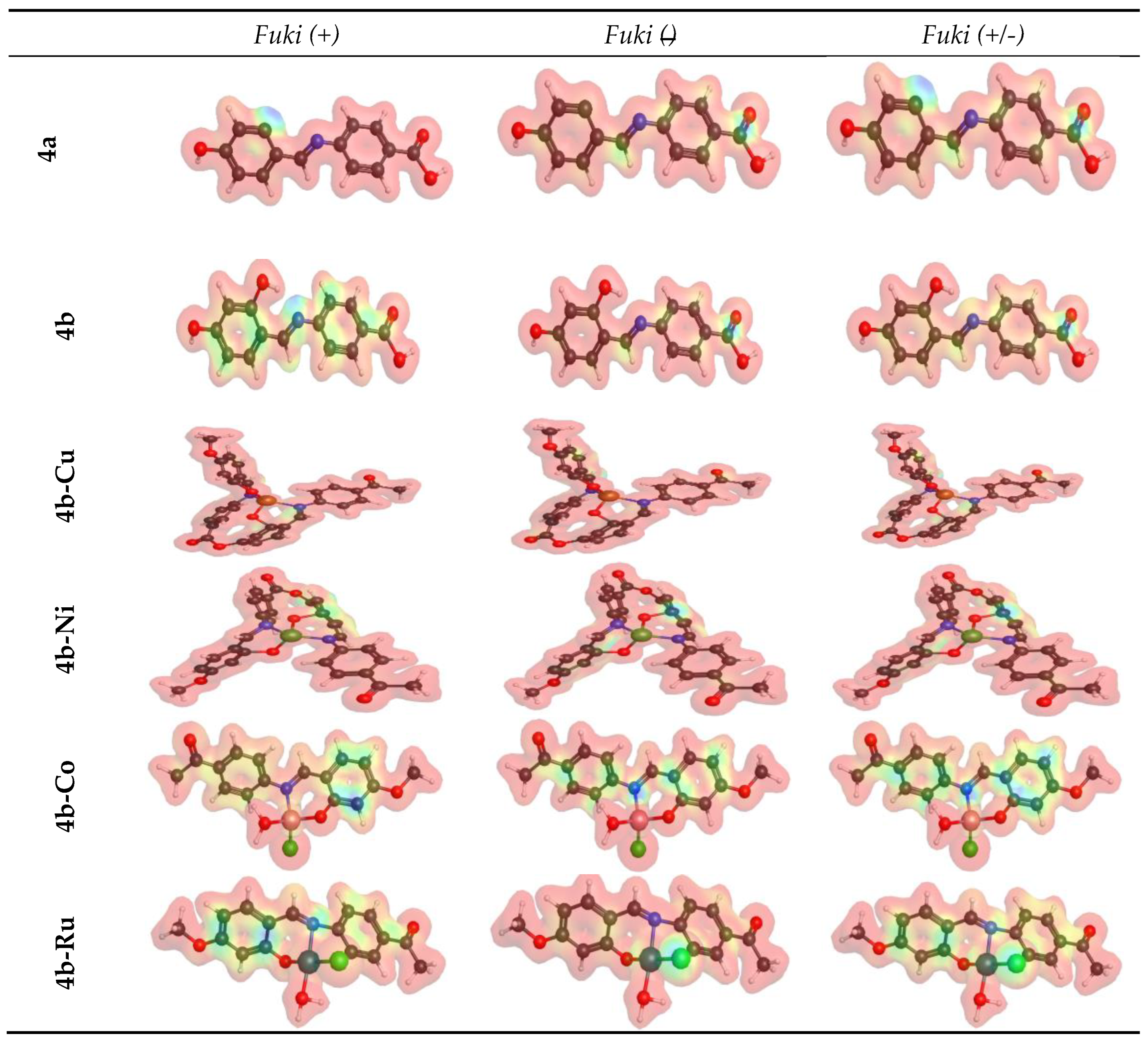
Figure 9.
Binding manner for 4a, 4b, 4b-Cu complex, 4b-Ni complex, 4b-Co complex, and 4b-Ru complex over 1M17.
Figure 9.
Binding manner for 4a, 4b, 4b-Cu complex, 4b-Ni complex, 4b-Co complex, and 4b-Ru complex over 1M17.
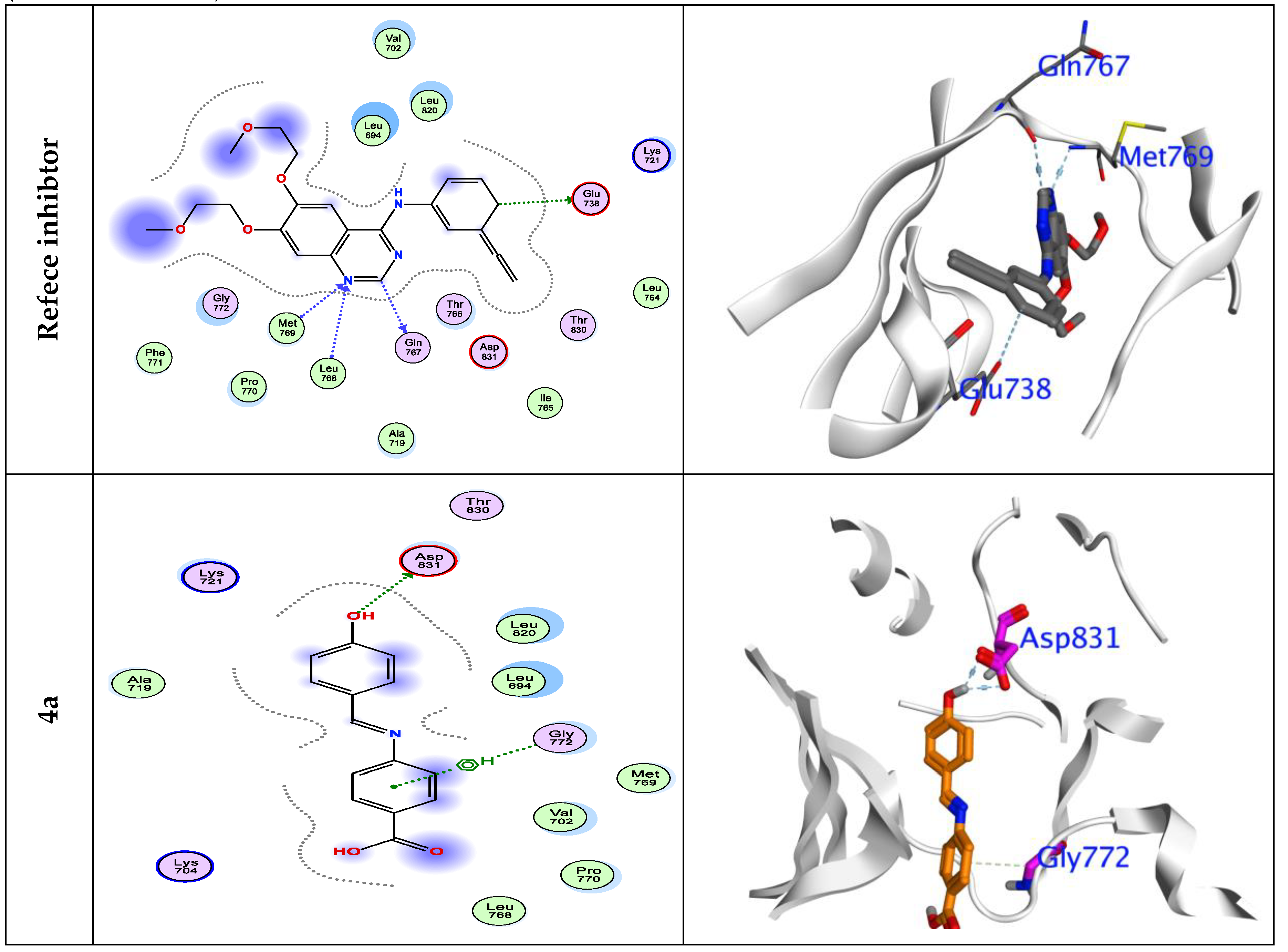
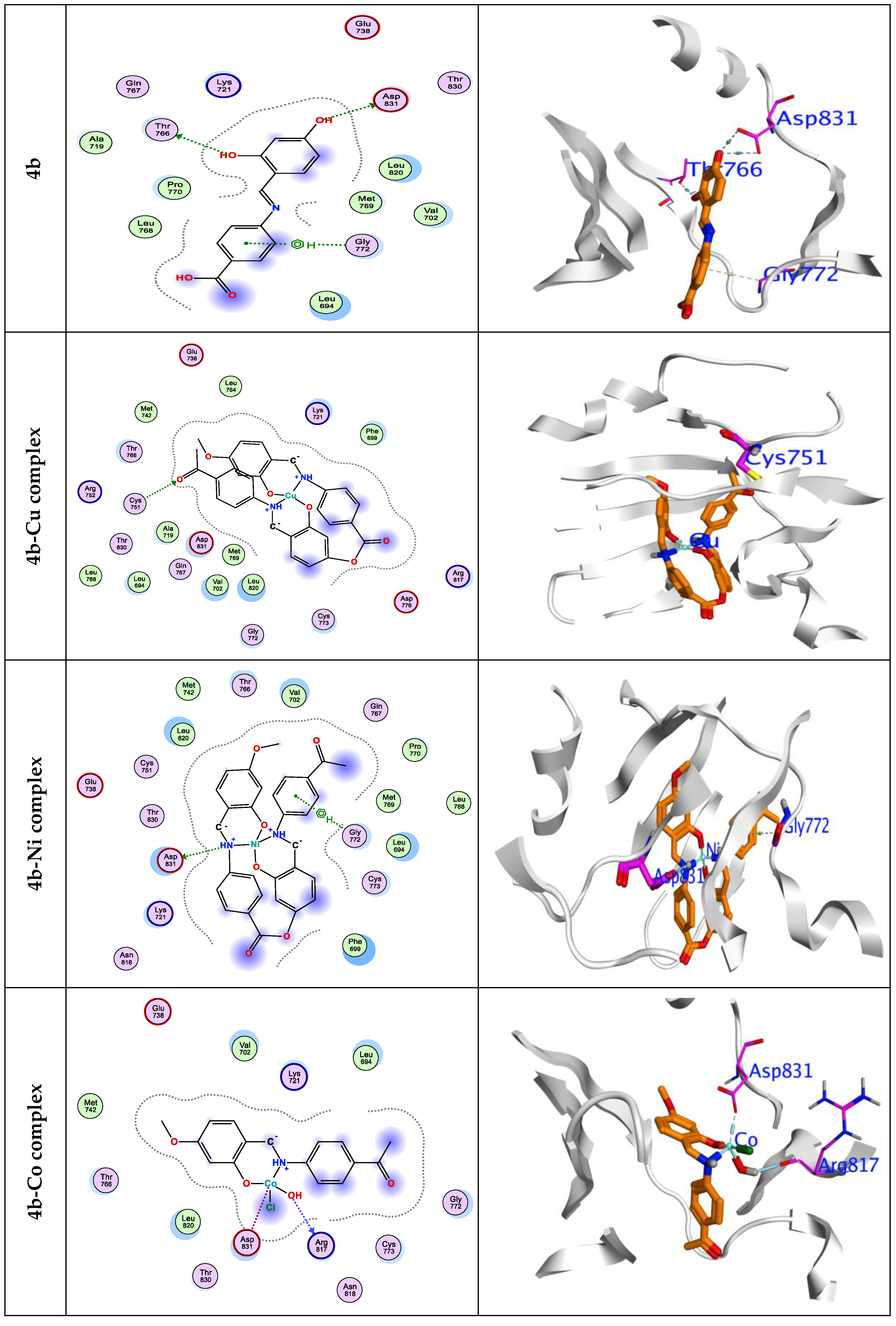
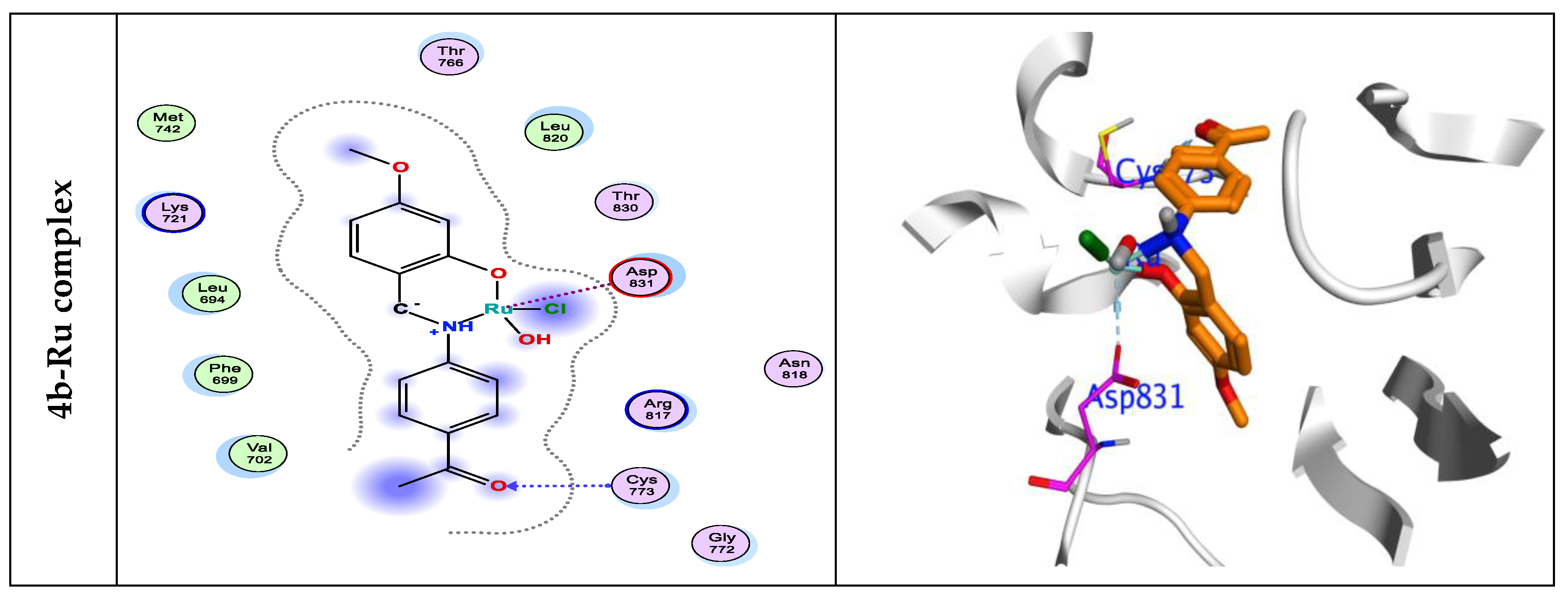

Table 1.
FT-IR of Schiff base monomers, polymers, and their complexes analogues.
| No. | Compounds | υ(OH) | υ(C=O) | υ(CH=N) | υ(C-O) | M-O | M-N |
| 1 | 3a | 3396 | 1664 | 1630 | 1259 | - | - |
| 2 | 3b | 3405 | 1672 | 1656 | 1218 | - | - |
| 3 | 4a | 2402 | 1712 | 1576 | 1148 | - | - |
| 4 | 4b | 3268 | 1722 | 1576 | 1155 1155 |
- | - |
| 5 | 4b-Co complex | 3367 | 1656 | 1593 | 1187 | 701 | 601 |
| 6 | 4b-Cu complex | 3468 | 1643 | 1584 | 1120 | 704 | 611 |
| 7 | 4b-Ru complex | 3352 | 1721 | 1608 | 1224 | 814 | 734 |
| 8 | 4b-Ni complex | 3332 | 1723 | 1617 | 1205 | 814 | 751 |
Table 2.
Molecular weight distribution of the obtained polymers.
| Polymer | Mw | Mn | PDI |
| 4a | 524,664 | 303,388 | 1.042 |
| 4b | 1,503,228 | 1,500,349 | 1.002 |
| 4a’ | 162949 | 161039 | 1.012 |
| 4b’ | 874877 | 560517 | 1.561 |
Table 3.
Thermal gravimetric analysis of the newly polymers.
| Polymer | Weight loss (%) | Tonset (°C) | Tendset (°C) |
| 4a | 22 | 238 | 375 |
| 43 | 378 | 511 | |
| 29 | 763 | 841 | |
| 4b | 20 | 243 | 355 |
| 45 | 398 | 541 | |
| 26 | 784 | 863 | |
| 4a’ | 17 | 209 | 359 |
| 14 | 460 | 563 | |
| 35 | 638 | 716 | |
| 4b’ | 17 | 225 | 286 |
| 32 | 316 | 400 | |
| 29 | 775 | 800 |
Table 4.
Antitumor activity of Schiff base monomers and polymers.
| No. | Compounds | IC50 | ||
| HepG-2 | HCT-116 | MCF-7 | ||
| 1 | 3a | 28.72 | 29.82 | 60.77 |
| 2 | 3b | 435.84 | > 500 | 483.40 |
| 3 | 4a | 14.93 | 17.03 | 21.16 |
| 4 | 4b | 28.16 | 61.26 | 70.47 |
| 5 | 4a‘ | 51 | 49.02 | 49.61 |
| 6 | 4b‘ | 264.03 | 374.31 | 438.39 |
| 7 | 4b-Ni complex | 24.43 | 42.28 | 95.99 |
| 8 | 4b-Cu complex | 105.41 | >500 | 27.25 |
| 9 | 4b-Co complex | >500 | 485.89 | >500 |
| 10 | 4b-Ru complex | 122.82 | 205.65 | 345.37 |
| 11 | Colchicine | 10.6 | 42.8 | 17.7 |
| 12 | vinblastin | 4.6 | 2.6 | 6.1 |
Table 5.
Total energy in Kcal/mol. for prepend complexes.
| Side by side | Parallel | With H2O | |
| 4b-Cu | -1184.46 | -539.89 | -280.04 |
| 4b-Ni | -1175.59 | -734.14 | -445.95 |
| 4b-Co | -586.46 | -127.65 | -833.71 |
| 4b-Ru | 1085.81 | -128.88 | -445.98 |
Table 6.
Calculated energetic of reactivity parameters for at DFT with a B3LYP\6-311G* Basics.
| Cpd. | 4a | 4b | 4b-Cu | 4b-Ni | 4b-Co | 4b-Ru |
| HOMO | -7.02 | -7.653 | -12.11 | -10.23 | -10.13 | -8.73 |
| LUMO | -1.11 | -2.953 | -7.02 | -8.05 | -7.57 | -1.68 |
| ΔG | 5.91 | 4.7 | 5.09 | 2.18 | 2.56 | 7.05 |
| I | 7.02 | 7.653 | 12.11 | 10.23 | 10.13 | 8.73 |
| A | 1.11 | 2.953 | 7.02 | 8.05 | 7.57 | 1.68 |
| η | 2.96 | 2.35 | 2.55 | 1.09 | 1.28 | 3.53 |
| S | 0.34 | 0.43 | 0.39 | 0.92 | 0.78 | 0.28 |
| χ | -4.07 | -5.3 | -9.57 | -9.14 | -8.85 | -5.21 |
| ωi | 4.07 | 5.2 | 9.57 | 9.14 | 8.85 | 5.31 |
| ω± | 7.62 | 10.62 | 40.73 | 81.21 | 65.61 | 14.29 |
| ΔΝμαξ | -0.69 | -1.13 | -1.88 | -4.19 | -3.46 | -0.74 |
| ΔρKNU | 1.38 | 2.26 | 3.76 | 8.39 | 6.91 | 1.48 |
| ΔρKEle | 2.75 | 4.51 | 7.52 | 16.77 | 13.83 | 2.95 |
E:energy (kcal/mol).,E-ele: electrostatic energy (kcal/mol), HF: heat of formation (kcal/mol), HOMO: Highest Occupied Molecular Orbital (eV), LUMO: Lowest Occupied Molecular Orbital (eV), ΔG: energy Gap (eV), I: Ionization potential, A; electron affinity; η: Hardness(eV), S: Softness(eV), χ: Electronegativity (eV), ωi: electrophilicity index; ω±: nucleophilicity (eV); ΔNmax:maximum number of electrons transfer in ground state. ΔρKEle: Group electrophilicity; ΔρKNU: Group nucleophilicity
Table 7.
The energetic Docking-affinity (kcal/mol) of compounds 4a, 4b, 4b-CuII, 4b-NiII, 4b-CoII and 4b-RuII against 1M17.
Table 7.
The energetic Docking-affinity (kcal/mol) of compounds 4a, 4b, 4b-CuII, 4b-NiII, 4b-CoII and 4b-RuII against 1M17.
| ΔG | RMSD | EInt. | Eele | H. B | LE | Ki | BindingSite | distance | Interactionenergy | |
| 4a | -5.69 | 1.67 | -21.28 | -15.85 | -11.51 | -7.24 | 1.98 | ASP831 | 2.09 | -0.9 |
| ASP831 | 2.08 | -1.2 | ||||||||
| GLY772 | 2.81 | -0.5 | ||||||||
| 4b | -5.77 | 0.83 | -21.17 | -19.45 | -11.04 | -4.67 | 1.54 | ASP831 | 2.18 | -0.6 |
| ASP831 | 2.08 | -0.6 | ||||||||
| THR 766 | 2.21 | -0.6 | ||||||||
| GLY772 | 2.92 | -0.6 | ||||||||
| 4b-Cu | -6.60 | 1.48 | -46.31 | -20.21 | -8.82 | -8.65 | 2.16 | GLY772 | 3.49 | -0.6 |
| 4b-Ni | -7.88 | 1.65 | -48.58 | -25.25 | -9.34 | -5.66 | 1.73 | ASP831 | 2.90 | -3.7 |
| GLY772 | 2.99 | -0.6 | ||||||||
| 4b-Co | -6.52 | 1.99 | -33.39 | -14.34 | -9.41 | -6.93 | 1.94 | ASP831 | 2.12 | -2.6 |
| GLY772 | 2.88 | -0.5 | ||||||||
| 4b-Ru | -6.83 | 1.15 | -23.16 | -17.01 | -9.44 | -4.77 | 1.56 | CYS773 | 2.28 | -1.3 |
| ASP831 | 2.61 | -1.0 |
Where, ΔG: Free binding energy of the ligand, RMSD: root-mean-square deviation; H.B.: H-bonding energy between protein and ligand; EInt.: Binding affinity of H-bond interaction with receptor; Eele: Electrostatic interaction over the receptor; Ki: inhibition constant; LE: Lignad efficiency; 5FU: 5-fluorouracil (standard reference drug).
Table 8.
In-silico physicochemical/pharmacokinetic studies of polyemr 4a, and 4b and its metallated counterparts.
Table 8.
In-silico physicochemical/pharmacokinetic studies of polyemr 4a, and 4b and its metallated counterparts.
| Compd.No. | Lipinski parameters | nROTBe | TPSAf | BSg | BBBh | GI ABSi | ||||
| MWa | HBAb | HBDc | LogPo/w | Violations | ||||||
| 4a | 558.15 | 5 | 3 | 1.58 | 0 | 3 | 90.12 | 97.0 | No | High |
| 4b | 553.15 | 4 | 2 | 2.14 | 0 | 3 | 69.89 | 95.2 | No | High |
| 4b-Cu | 408.8 | 7 | 2 | 2.45 | 1 | 2 | 127.01 | 94.5 | No | Low |
| 4b-Ni | 366.66 | 7 | 2 | 1.91 | 1 | 2 | 127.01 | 96.6 | No | Low |
| 4b-Co | 257.24 | 4 | 2 | 0.61 | 0 | 2 | 68.12 | 100 | No | High |
| 4b-Ru | 241.24 | 4 | 2 | 0.61 | 0 | 2 | 68.12 | 94.5 | No | High |
a Molecular weight; b Hydrogen Bond Acceptor ; c Hydrogen Bond Donor; d Partition Coefficient; eNumber of rotable bonds; fTopological Polar Surface Area; g Bioavailability Score %; hBlood Brain Barrier; iGastro-intestinal absorption.
Table 9.
In-silico toxicity studies of the polymer 4a, 4b and its metal complexes.
| Compds | AMES toxicity |
LD50 (mole/kg) | Oral rat chronic toxicity (logmg/kgb.w/day) |
Hepato toxicity |
Skin sensitization |
|---|---|---|---|---|---|
| 4a | No | 2.292 | 0.530 | No | No |
| 4b | No | 2.094 | 0.602 | No | No |
| 4b-Cu | No | 2.539 | 0.691 | No | No |
| 4b-Ni | No | 2.539 | 0.691 | No | No |
| 4b-Co | No | 2.572 | 1.007 | No | No |
| 4b-Ru | No | 2.572 | 1.002 | No | No |
Disclaimer/Publisher’s Note: The statements, opinions and data contained in all publications are solely those of the individual author(s) and contributor(s) and not of MDPI and/or the editor(s). MDPI and/or the editor(s) disclaim responsibility for any injury to people or property resulting from any ideas, methods, instructions or products referred to in the content. |
© 2024 by the authors. Licensee MDPI, Basel, Switzerland. This article is an open access article distributed under the terms and conditions of the Creative Commons Attribution (CC BY) license (http://creativecommons.org/licenses/by/4.0/).
Copyright: This open access article is published under a Creative Commons CC BY 4.0 license, which permit the free download, distribution, and reuse, provided that the author and preprint are cited in any reuse.


