
Submitted:
01 October 2025
Posted:
02 October 2025
You are already at the latest version
Abstract
Metal complexes have been utilized in medicine for thousands of years in a variety of ways, with positive benefits and well recognized as anti-infective, anti-inflammatory, antimicrobial, antidiabetic, neurological agents, delivery prob and diagnostic agents. Based on that Co, Zn, Cu complexes were synthesized and fully characterized. The complexes were then assessed with MDA-MB-453, human breast cancer cell line and the data showed that Cu- ligand complex was the most potent anticancer agent followed by Zn then Co complex. The DFT theoretical calculations were parallel with the biological assessment and support it. In addition, the complexes were tested for their biological activities in both gram positive and negative bacteria and the data revealed that Cu>Zn>Co with strong inhibitory effect on both bacterial growths. In sum, copper complex proved to be potent anticancer and antibacterial agent and can be used for future therapies.
Keywords:
transition metal complexes
; synthesis
; anticancer effect
; DFT calculations
; antibacterial activity
1. Introduction
The field of medical chemistry holds metals in high regard. Groups three through twelve on the periodic table are known as the "d block" elements, or transition metals [1]. Filling up their d shells led to the development of coordination complexes. A metal complex, also known as a coordination compound, is a structure made up of a central metal atom bound to a variety of nearby ligands or anions [1,2]. These complexes have the capacity to interact and react with biomolecules in novel ways and through different modes of action. Metal ions have significant effects on biological processes, and the study of how transition metal chemistry might be used to treat or diagnose illnesses is attractive field in medicinal chemistry [3] which started from the past decades with platinum complex (cis platinum) as antitumor drug [1]. There is a global epidemic of diseases brought on by bacteria, fungi, and viruses’ infections [4]. Other significant health issues include diabetes [5], cancer [6], and malaria [6,7]. Various metals and nonmetals in the periodic table have demonstrated their significance for the healthy functioning of the human body [8,9]. Especially, d-block transition metal elements such as manganese (Mn), iron (Fe), cobalt (Co), copper (Cu), zinc (Zn), and molybdenum (Mo) are necessary for regular biological functioning and homeostasis [8,10]. Either a deficiency, or an excess of transition metals may result in various disease in human body [8] such as Alzheimer’s disorders [11], Parkinson’s disorders [12], cardiovascular diseases [13], cancer [14], and diabetes [8,15]. Therefore, it becomes necessary to discover new agents that may be employed as therapeutic medications for bacteria and cancer treatments Figure 1.
Researchers synthesized many transitions metal complexes for disease treatment, and diagnosis such as cobalt, copper and zinc complexes [16,17,18]. First, cobalt is naturally occurring compound for the coenzyme vitamin B-12 and has distorted octahedral geometries, and forms stable complexes with many ligands including water, chloride ion and ammonia [19,20]. Studies proved that cobalt complexes used as antiviral, antibacterial agents [21], anticancer agent [22] and antimalarial agents [8,23]. Second, copper is essential for several vital biological activities [8], such as photosynthesis, iron metabolism, mitochondrial respiration, catalytic cofactor, scavenging of free radicals, the transmission of electrons inside biological molecules, certain neurological processes [24,25] and the redox chemistry of enzymes, all are essential to cell physiology and can happen because of the two different redox states of copper reduced (I) and oxidized Cu (II) [26]. Studies also showed that copper complexes had anticancer activities [27,28] and function as antibacterial [28], antimalarial agents [29]. Third, zinc is catalytic metal and important for biological signaling systems [30], protein metabolism, and immune system [31]. In addition, zinc is structural metal occur in a variety of RNA and DNA binding proteins known as zinc fingers [32]. Also, research studies proved that zinc complexes behave as anticancer and antibacterial agents [33,34].
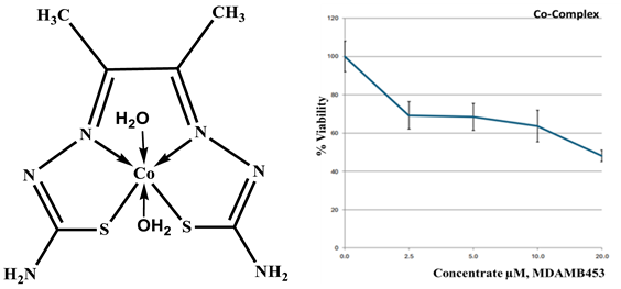
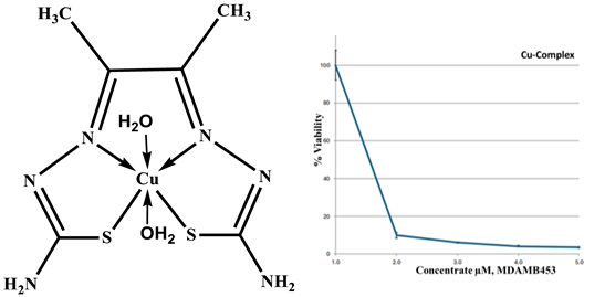
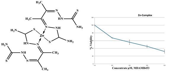
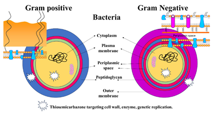
Several ligands have played a significant role in the development of coordination chemistry and form stable complexes with many transition metals [16,17,18]. In this study thiosemicarbazone was used to form Co, Cu, and Zn complexes. Thiosemicarbazone’s inherent biological activity, structural flexibility, and multifunctional metal-chelating capabilities have attracted a lot of attention [35]. Before their anti-tumor effect was discovered, thiosemicarbazones were known to be metal chelators. It has a versatile thiourea structure that allows it to incorporate other substituents or functional groups in addition to the C=N and C=S bonds in its own structure, which is advantageous for metal coordination [35,36] and it exhibited a wide variety of biological activity, such as antiviral, antifungal, and anticancer properties [36] Figure 2.
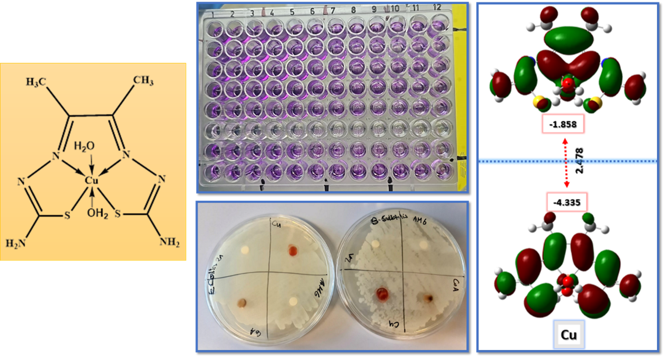
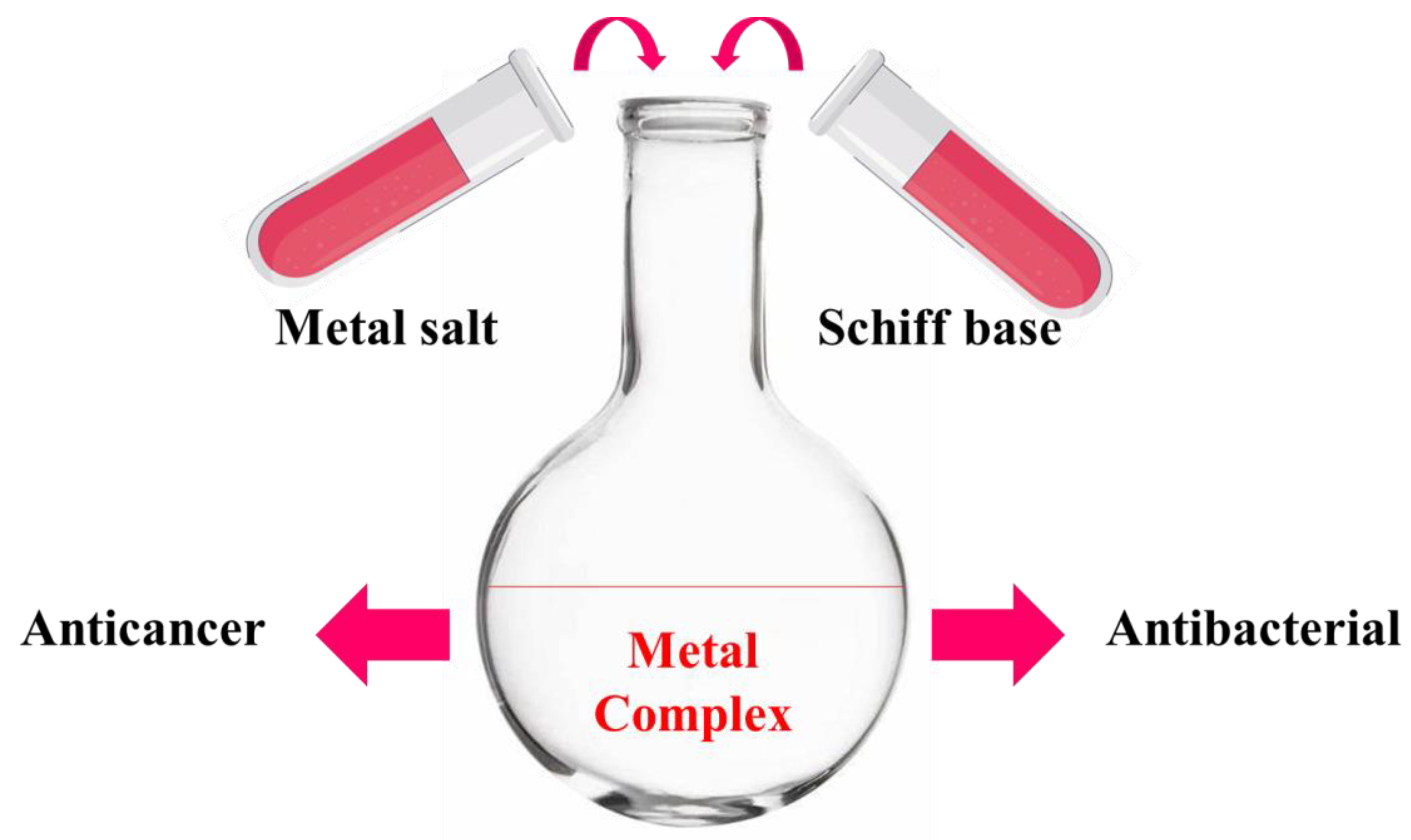
Therefore, cobalt (II), copper (II), and zinc (II) metal complexes have been synthesized, characterized by CHNS, mass spectroscopy, proton, and carbon NMR, FTIR, thermal gravimetric analysis, molar conductivity, differential scanning calorimetry technique, scanning electron microscope and dynamic light scattering. The metal complexes biologically evaluated for their anticancer activity using breast cancer cell line and this was supported by molecular geometry and computational methodology study. In addition, the antibacterial activity of the metal complexes was assessed using gram positive and negative bacteria as demonstrated in Figure 3.
2.1. Materials and Reagents
We synthesized the metal ligand complexes in our laboratory at Kuwait University. All chemicals and biological supplies were graded as analytical reagents and were pure and ready for direct use such as hydrazine carbothioamide, ethanol, diethyl ether, DMSO, CuCl2, CoCl2, ZnCl2, DMEM medium, FBS, penicillin/streptomycin, 1% L-glutathione, PBS, WST-1 test Kit (ab65473 Abcam, USA), and nutrient agar, all were purchased from Sigma-Aldrich, Germany, and E. Merk. The strains Escherichia coli and Bacillus-subtills (ISO 17034 from ATCC) strains from biology department. All complexes were dissolved in dimethyl sulfoxide to prepare chemical stock solutions (μg/mL) for the antibacterial treatment and assessment.
2.2. Instruments Used
All chemical spectral analysis were carried out at the Research Sector Project Unit (RSPU) in Kuwait University. Thin-layer chromatography (TLC pre-coated aluminum plate silica gel, Merk 9385) was used to detect the endpoints of the reactions. The molecular weights of the samples were determined using the EI (70 ev) mode of the VG Auto spec QMS 30 and MSg (AEI) spectrometer and were then confirmed by mass spectroscopy by LCMS. On an Elemental uni-cube superuser, elemental analysis (CHNS) was carried out. NMR spectrophotometer Bruker, DPX 600 was used to collect 1H and 13C nuclear magnetic resonance (NMR) spectra at 600 MHz and 150 MHz in DMSO and CDCL3, respectively. The fourier-transform infrared spectroscopy (FTIR) spectra were collected in the 4000-400 cm-1 range using a Jasco 6300 FTIR. Scanning electron microscope was used to check the morphology (SEM). Dynamic light scattering technique was used to determine the metal complexes size -DLS (the Malvern Zeta sizer -ZS). In addition, heat flow is measured using DSC to characterize the metal ligand complexes. Also, the sample’s weight change in relation to temperature is measured by the thermal gravimetric analysis (TGA).
2.3. Synthesis
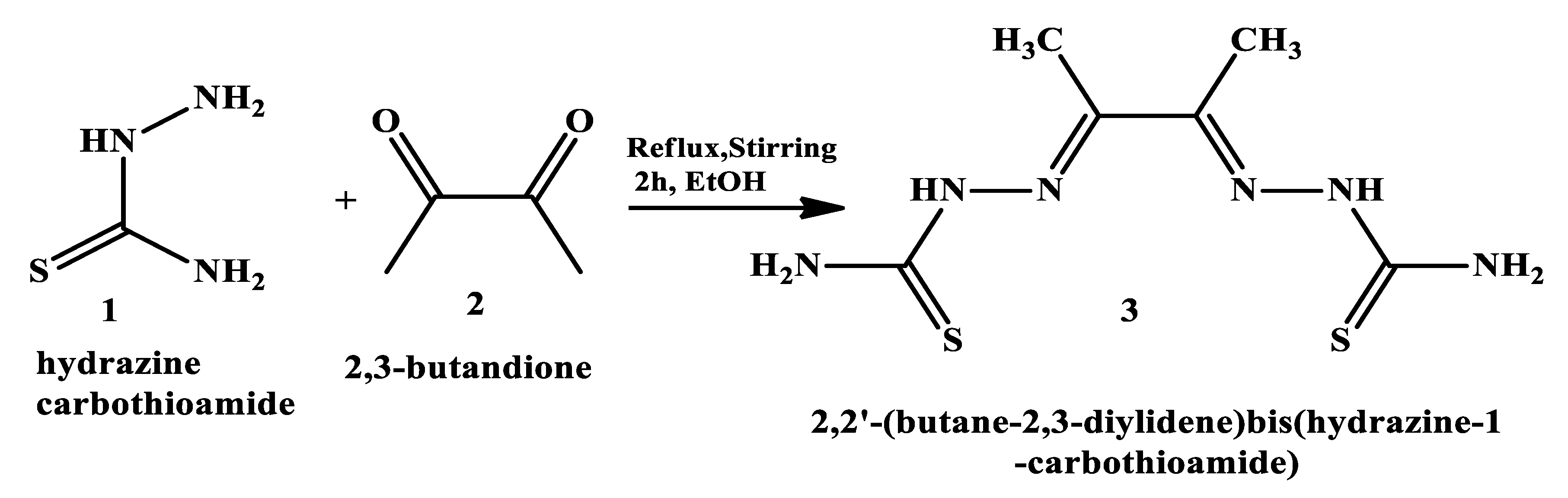
2.3.1. General synthesis of the ligand: 2,2’-(butane-2,3-diylidene) bis(hydrazine-1-carbothioamide)
2,2’-(butane-2,3-diylidene) bis(hydrazine-1-carbothioamide) (H2L) (3) was synthesised as shown in Scheme 1, by addation of 30 ml hot ethanol solution of hydrazine carbothioamide (1) (18.12 g, 0.2 mol) slowly to 30 ml hot ethanollic solution of 2,3-butandione (2) (23.2 g, 0.1 mol). This followed heating the mixture under reflux, with continuous stirring, utilizing heating mantle for two hours. The formed precipitate was collected by filtration, washed with absolute ethanol, diethyl ether and finally dried under vacuum over silica gel to produce (H2L) (3) with yield 77%.
2.3.2. General Synthesis of the Metal-Ligand Complex
The synthesized 2,2’-(butane-2,3-diylidene) bis(hydrazine-1-carbothioamide) (H2L) (3) was used as ligand for the reaction with metal chloride salt (MCl2) to form our target complex. Co, Cu and Zn complexes were prepared by refluxing 1 mmol of the ligands (3) under investigation with 1 mmol of the metal salt CuCl2, and CoCl2 in an ethanoic solution on a water bath for 2 - 3 hrs as shown in Scheme 2. The resulting solid metal ligand complexes were filtered off, washed several times with absolute ethanol followed by diethyl ether and finally dried in a vacuum desiccator over anhydrous calcium chloride with yeild 80%.
2.4. Thermal Gravimetric Analysis (TGA)
This technique provides more insights into the stabilization and water molecule characteristics of the complexes, the TGA analysis was conducted for all metal ligand complexes weighing five mg were placed in the Pt pan and heated in a nitrogen environment from between 25 to 800 °C within ten minutes. According to the TGA statistics, there is a clear correlation between the calculated and recommended formulas for weight loss that were recorded [37].
2.5. Differential Scanning Calorimetry Technique (DSC)
A thermal analysis tool known as a DSC monitors a sample’s temperature and physical properties over time in both hot and cold conditions. Five milligrams of each sample were weighed on a sealed Al pan heated from 25 to 500 oC in ten minutes at a heating rate in an argon environment, and the DSC curves for the metal ligand complexes were then recorded [38,39].
2.6. The Triazole Agents’ Morphology Investigation by SEM
The surface morphology of the complexes was assessed by utilizing the (JEOL-EDS-KU-RSPU) system to take several SEM readings at 15.0 kV, at various micrographs magnifications (X100, X500, X1000, X2000, and X5000). Uniform morphologies were discovered by the morphological characterization [40].
2.7. Particles Size Measurements by DLS
The Malvern Zeta Sizer (ZS) is analytical instrument used to measure the particles size, zeta potential, and molecular weight in solutions. It uses dynamic light scattering (DLS) for particles with size in the range (0.3 nm to 10 µm). The electrophoretic light scattering (ELS) is (> ± 500 mV). The temperature control range of 0 oC to 90 oC, sensitive and can work with small sample volumes. This technique is ideal for nano-particles analysis and stability studies in pharmaceuticals and material sciences. All metal complexes were solubilized in DMSO, and readings were measured ± standard deviation [40,41].
2.8. Cytotoxicity Assessment & Statistical Analysis
MDA-MB-453 (HTB-131 from ATCC), is a human breast cancer cell line, which was employed in in-vitro screening investigations. The cells were cultured in 5% CO2 humidified air with 10% FBS, 1% penicillin/streptomycin, and 1% L-glutathione added in DMEM medium. Before usage, fetal bovine serum (FBS) is inactivated for 30 minutes at 37 oC in a water bath. The WST-1 assay Kit (ab65473 Abcam, USA) manufacturer’s protocol was followed to conduct the in-vitro cell proliferation assay. To put it briefly, after seeding the cells in day one, the cells were exposed to the metal ligand complexes for 72 hours while they were between 75% and 85% confluent. Subsequently, each well received ten µl of WST-1 reagent. Using a plate reader (SoftMax Pro 9.0 Flex Station-Molecular Devices), the absorbance at 440 nm was measured to calculate the amount of formazan product that had generated. There were four duplicates of each concentration used in the studies. Statistical analysis was performed using GraphPad Prism 09. Every in-vitro data was displayed as the average ± standard error. For statistical significance, p values ˂0.05 were used [41,42,43].
2.9. Molecular Geometry and Computational Methodology
All computations were conducted using gaussian 16 program [44]. The molecular geometry for the studied compounds was fully optimized using density functional theory B3LYP method. Where (B3) stands for Becke’s three parameters exact exchange functional combined with gradient-corrected correlation functional of Lee, Yang and Parr (LYP) [44,45,46,47], and DFT/GENECP level had been obtained by implementing Def2TZVP. No symmetry constraints were applied during the geometry optimization. The choice of mixed basis set was due to flexibility, accuracy, consistent and better performance of the Alrich’s effective core potentials basis set (def2-TZVP) with Gaussian type triple- ζ potential(6-311++G(d,p). By using HOMO and LUMO energy values for the metal ligand complexes, electronegativity and chemical hardness can be calculated as follows: X= (I+A)/2 (electronegativity), ɳ=(I-A)/2 (chemical hardness), S = 1/2ɳ (chemical softness) where I and A are ionization potential and electron affinity, and I= - EHOMO and A= - ELUMO, respectively. NBO calculations had been performed at the same level of calculation by using NBO 3.1 program as implemented in the Gaussian 09W software package. Throughout this work optimized structures were visualized using Chemcraft version 1.6 package and Gauss View version 5.0.9.
2.10. In-Vitro Antibacterial Activity
As directed by Difco, the pure culture plates were prepared two days before to usage. Bacterial suspension was prepared by nutrient broth with bacterial culture incubated at 37 ± 1 °C. A few mL of the bacterial suspensions of each strain was added to 20 mL of fresh broth until the final concentration is approximately 5 × 10⁵ CFU/mL. A suspension containing 0.2 ml of each of the investigated compounds (5 and 10 mg/ml) was cultured for 24 hours. In nutrient agar media (NA), the strains of the microorganism were uniformly distributed. The studied samples were placed inside discs of Whatman paper, which had a diameter of 5 mm. After the socked discs were placed on the NA, the diameters (in millimeters) of the clear inhibition zone encircling the sample were measured by ruler to determine the inhibition power against those specific organisms. Using the agar disk diffusion method with minimum inhibitory concentration, the metal ligand complexes were administered in triplicate to both gram-positive and gram-negative bacteria for a duration of 24 hours to test for antibacterial activity. To avoid contamination, all experimental work was conducted close to the flame. Four mm wells were created using a cork borer and a 100 μl of the metal ligand complex was added by micropipette to each well and incubated at 37 °C overnight. Three replicas were used to assess the inhibition zones surrounding the wells and report the average. To compare the potency and importance of the metal ligand complex as antibacterial agents, free ligand was utilized as the reference control in the current investigation. [48].
3. Results and Discussion
As stated in the experimental section, a schiff base reaction was applied to synthesize the ligand thiosemicarbazone, and then form the metal complexes in the nano scale. The obtained ligand and its metal complexes are air stable. The complexes possess assorted colors, the solubility test illustrates that these complexes are water insoluble and soluble in polar aprotic solvent like DMSO and DMF. The value of molar conductivity of Co, Cu, Zn complexes are 5.83, 4.34, and 21.52 Ω-1cm2mol−1 respectively indicating their non-electrolytic nature. The Co, Cu, Zn complexes were created, synthesized, and their structures were fully characterized. The complexes formulas, physical characteristics, elemental analyses, thermal, molar conductivity, magnetic moment, and formula weights (LCMS) derived from mass spectra are mentioned together as shown in Table 1. The data illustrated that Co2+ and Cu2+ complexes were composed in molar ratio (1L:1M) with formulae [M(L)].2H2O where M= Co2+ or Cu2+. While Zn2+ acts as (2L:1M) with formula [Zn(H2L)2].
3.1. Elemental Analysis, Electronic Spectra, Magnetic and Physical Measurements
The elemental analysis is in the good agreement with that calculated results for the proposed formula of the metal complexes as described in Table 1.
3.2. Mass Spectra of Metal Ligand Complexes (MS).
The MS of the H2L (Figure 4) showed a molecular peak at m/z = 232.1 (7%) and weak peaks surround it due to 13C and 15N isotopes. The other positive ions give peaks at 215.1 (15%), 174.2 (3%), 116.0 (100%), 86.1 (6%) and 56.07 (32%) mass numbers. The intensities of these peaks indicate the stabilities of these fragments. The MS of [Co(L)].2H2O displayed the molecular ion peak of intensity 100% at e/m = 288.97 corresponding to [Co(L)] as shown in Figure 5. The species after may be formed after the removal of 2H2O while the small peaks at 250, 163 and 158 representing sequential degradation for the molecule. The MS of [Cu(L)].2H2O showed a peak of intensity 100% at e/m = 293.97 corresponding exactly [Cu(L)], this species may be formed after the removal of 2H2O. The positive ions peaks at 235 (2%) may be due to the removal of methyl and amino groups. The other positive ions give peaks at mass numbers 192 (4%), 178.0 (1%) and 164 (65%), 141 (13%), 124 (16%) mass numbers representing sequential degradation for the molecule (Figure 6). The spectrum of [Zn(H2L)2] the peak appeared at m/e = 532.06 (Calcd. 535.6) representing the molecular ion peak with intensity 75%, the peak of intensity 100% at 296.3 corresponding to [Zn(H2L)] as shown in Figure 7.
3.3. Nuclear Magnetic Resonance (1H and 13C)
The 1H NMR and 13C NMR spectrum of the free ligand and Zn ligand complex were recorded and summarized in Table 2 and Figure 8, Figure 9, Figure 10 and Figure 11. The ligand 1H-NMR spectrum in DMSO-d6 agree with the proposed chemical structure. The two chemical shifts noted as singlet at 11.061 and 10.197 ppm (s, 1H, NH6) and (t, 1H, NH7), while the doublet chemical shifts were noted at 8.390 (dd, 2H, 10H2N) and 8.101(d, 2H, 12H2N). The spectrum also showed chemical shifts at δ = 1.947 ppm for CH3 group at position (3 & 14) were imputed to the methyl group protons. 1H-NMR spectrum clarifies, that the thiosemicarbazone offered only a thio-keto form; there’s no evidence for existence of the thio-enol form as shown in Figure 8. This conclusion was supported by the existence of chemical shifts relevant to (NH) and the complete lack of a chemical shift relevant to (SH) of the thio-enol form [50]. The 13C-NMR spectrum of the ligand thiosemicarbazone (H2L) displayed a chemical shift at 183.12 ppm assigning to the thioketone carbon atom (8,9C=S) [51,52]. The imine carbon (1,2C=N) chemical shift was appeared at 148.37 ppm [51,52]. The peaks appearing at 11.55, 13.81 ppm can be related the methyl group carbon atoms (3,14CH3). It is important to mention that [Cu(L)].2H2O and [Co(L)].2H2O are paramagnetic complexes which significantly impact the interpretation of NMR spectra by causing broadening and large chemical shifts. The 1H-NMR spectra of the Zn2+ complex showed that the thio-amide protons are appeared at 10.208 ppm clarifying that the Zn2+ is bonded to the thio-keto tautomer via the deprotonated thio-amide sulfur atom Figure 10. The 13C-NMR spectrum of Zn2+ complex showed the thio-amide δ(C=S) signal shift to up field by about 5 ppm in comparing with the parent ligand thiosemicarbazone Figure 11. This up field shift may be due to the movement of electron density from the thio-amide moiety to the Zn2+ ions upon chelation that could have caused the thio-amide carbon nuclei to be de-shielded hence the up field shift [53].
3.4. Infrared (FTIR) Spectra of Co, Cu, Zn Complexes
The FTIR bands of the thiosemicarbazone ligand spectrum (Figure 12) showed peaks at 3408, 3248 and 3194, 3149 cm-1 which could be attributed to the imine ν(N-H) and ν(NH2) [54,55,56]. The weak bands which appeared at 3000, 2950, 2990 cm-1 could be attributed to the azomethine and aliphatic protons [57,58]. The spectrum also displayed bands at 1594 and 833 cm-1 which are related to imine ν(C=N), and thio-amide ν(C=S) [59,60] groups respectively (Table 3). These observations indicated that, the thiosemicarbazone adopted only a thio-keto form in the solid state. This conclusion is endorsed by the absence of the band related to the ν(S–H) linkage (generally shows in the 2500–2600 cm-1) [59,60,61,62,63]. The coordination manner can be obvious by matching the complexes’ FtIR spectra (Figure 13, Figure 14 and Figure 15) with the thiosemicarbazone (H2L) IR spectrum. The matching clarifies that the ligand (H2L) worked as dibasic bidentate bonded with the Cu2+ and Co2+ ions via the imine nitrogen atom, thio-amide group in its thio-enol form in Cu2+ and Co2+ complexes. This way of chelation was mended by four proofs as follow:
- i.
- The negative shift in position (4-30 cm-1) and intensity of the band of imine group C=N).[64]
- ii.
- The misplaced of the imine group ν(N-H6,7) band upon chelation, signifying that the thiosemicarbazone in these complexes interacted in the thio-enol form, that was endorsed by the appearing of new band in the region 1640-1690 cm-1, assignable to ν(C=N-N=C(.[64]
- iii.
- iv.
- The being of new bands at 451-476 and 497-519 cm-1 could be attributed to the ν(M-S) and ν(M←N) consecutively [66].
On the other hand, the ligand (H2L) worked as neutral bidentate bonded with the Zn2+ ion via the imine nitrogen atom, thio-amide group in its thio-keto form.
3.5. Thermal Gravimetric Analysis (TGA)
To achieve further data about the stabilization and water molecules nature of the complexes, the thermo-gravimetric analysis was measured in the 25 to 800 °C range. The data were summarized in (Table 4) and as shown in (Figure 16, Figure 17, Figure 18 and Figure 19) which proved that there is an obvious matching in the losing weight between the suggested and calculated formulae. The TG thermogram showed that the ligand is stable up to 191 and losing amino group at 191-221 °C consequentially decomposed completely in the temperature range 230-650°C with weight loss 91.67 % (calcd. 93.10%) Figure 16. Co2+and Cu2+ complexes displayed weight loss between 70-73 oC and refereeing to two water molecules abroad the coordination sphere. Co2+and Cu2+ complexes decomposed in one step within temperature ranged from 250 to 800 °C with weight loss equal to 63.58 and 64.19 % (calcd. 62.53% and 63.41%) leaving Cu+2C and Co+2C as shown in Figure 17 and Figure 18. While the TG thermogram for Zn2+ complex showed that it is stable up to 157°C, then start decomposition to eliminate C12H26N12S2 at 256 to 800°C with wight loss 73.83 % (calcd. 75.56%) leaving Zn+2S as a residue as shown in Figure 19 [43,49].
3.6. Differential Scanning Calorimetry (DSC) Characterization
The results of the measured DSC curves of Co, Cu, Zn complexes were measured to start melting between minimum 50 oC and maximum 450 oC as plotted in Figure 20, Figure 21 and Figure 22. The metal complexes showed different behaviors, first sharp endothermic peaks at 200-350 oC range which corresponds to the metal complexes melting points and second broad exothermic peaks in Co+2 and Cu+2 complexes at 350-450°C represent their decomposition to form the respective metal sulfides. In Co-ligand complex the decomposition peak was at 264 oC (Figure 20). Cu-ligand complex three peaks were observed at 247.0 oC, 259.0 oC, and 350.4 oC (Figure 21). While in Zn- ligand complex the two peaks were observed at 257.5 oC and 259 oC (Figure 22). In depth, the first complex to melt is Cu-ligand complex, followed by Zn-ligand complex and the last harder complex to decompose is Co-ligand complex. Generally, all complexes were stable until 240 oC. It is important to mention that normal body temperature is 37 oC, and the fever is from 38 oC to 41 oC which means our complexes are stable if they will considered as chemotherapy drugs in the future.
3.7. The Complexes’ Morphology Investigation by SEM
The surface morphology of the metal complexes was assessed, and several micrograph images were taken as shown in Table 5. The morphological characterization of Co, Cu, Zn complexes showed smooth nanoparticles shapes without pores or cracks but with sharp edges. Generally, the nanoparticles size better than micro size [67]. In accordance with a study, the nanoparticles easily accumulate in the cancer cell [40,42]. Furthermore, these agents are excellent choices for delivering anticancer medications to cancer tumors and tissues due to their small size nanoparticles and biocompatible [68]. Studies proved that many nanoparticles were selective, promising anticancer, antimicrobial, and anti-Alzheimer agents such as ZnO, [69], AgNPs [70], SnO, CuO, and FeO [71,72,73]. Generally, due to their better efficacy and capacity for selective targeting, nanoparticles are beginning to replace more conventional cancer treatments including radiation, chemotherapy, and surgery [73].
3.8. Particles Size Measurements of Metal Ligand Complexes
The metal complexes’ particles sizes were summarized in Table 6. The data showed that all metal complexes were in the nano-seize range between 226 and 266 nm. Cu-complex exhibited the smallest particle size followed by Zn complex then Co-complex. Current chemotherapy for cancer causes cytotoxicity, lack of selectivity, multidrug resistance, and stem-like cell growth [40]. For a range of cancer treatments, these nanomaterials which target the immune system, DNA, tumor microenvironment, and cancer cells have been altered to increase drug capacity and bioavailability, lower toxicity, and enhance selectivity [2]. According to recent studies, therapeutic nanoparticles can improve efficacy and reduce side effects when compared to conventional cancer therapy drugs. Scientists are constantly developing novel and effective nanoparticles for drug delivery. Nanotechnology is widely used in medicine for targeted drug delivery [40,42], imaging, and diagnostics [74]. Nanoparticles have recently received a lot of attention in the field of medication delivery due to their unique properties and potential applications in the treatment of cancer. In addition, studies proved that nano-complexes have antibacterial and antifungal properties [75,76,77]. In conclusion, given all these findings, further research is needed to reduce toxicity and enhance clinical outcomes in medication delivery using nanoparticles (Figure 19, Figure 20 and Figure 21).
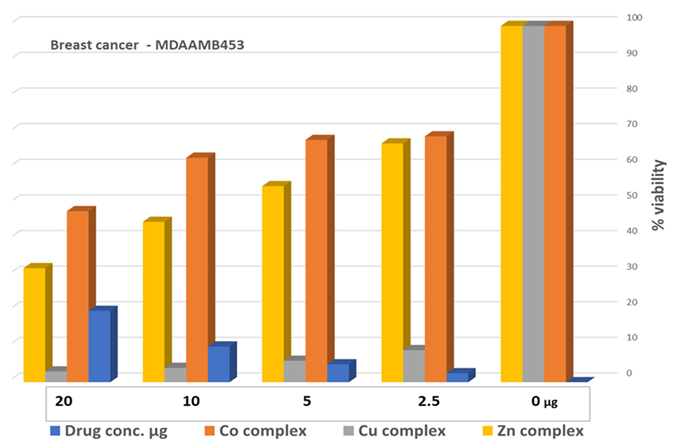
3.9. Cell Proliferation Assessments
The MTT assay of the metal complexes tested against breast cancer cell line (MDA-MB-453). The data were at different concentrations 0, 2.5, 5, 10, and 20 µM of metal complexes and results were summarized in Table 7. According to the biological evaluation, there is no detectable toxicity prior to the 48-hour treatment period [37], and cancer cell proliferation is prevented during 72 hrs. The findings showed that the metal ligand complexes have strong anticancer properties. In depth, Cu- L complex was the most potent agent that killed MDA-MB-453 cells with IC50 values of 3.31±0.34 µM. The second potent agent was Zn-L complex followed by Co-L complex with IC50 values in the range of 32.1±4.80 and 48.1±3.06 respectively. The synthesized complexes proved their strength as anticancer agents. Recent study in 2023 supported our finding that copper complex had the superior anticancer activity and selectivity against cisplatin resistant lung cancer cell line [78,79]. Previously, studies showed that thiosemicarbazones complexes have antimicrobial, antiviral activities, antibacterial, antifungal, anticonvulsant, antimicrobial, antiallergenic and anticancer activities [35,36,37,38,39,49,77,78,79,80]. Additionally, research has shown that metal complexes have stronger anticancer effects than the metal-free ligand [49]. The coordination, orientation, aromatic moiety, ligand binding [3] and solubility of metal ions all contribute to the overall stability of complexes [49]. In addition, to the practical biomedical focuses to synthesize new transition metal complexes in nanoscale to creates new materials with unique physical, chemical, and biological properties [81] that has effective potent biological activities that can benefit the medical field.
3.10. Computational Analysis of Optimized Geometry and Energy Gap
Several physical properties, including frontier molecular orbital energies (eV), ionization potential (IE), electron affinity (EA), electronegativity (χ), global electrophilicity (ω), chemical potential (µ), global softness (S), and dipole moment, can be determined by optimizing the structure and frequency of compounds using quantum chemical calculations. For metal ligand complexes, the geometric characteristics are summarized in Figure 23, Figure 24 and Figure 25 and the physical parameters are given in Table 7. In this work, the lowest unoccupied molecular orbital (LUMO) which acts as electron donor and the highest occupied molecular orbital (HOMO) which acts as electron acceptor, structural parameters, dipole moments and nonlinear optical properties of the Co, Cu and Zn complexes were calculated by both B3LYP/LANL2DZ and B3LYP/GENECP/LANL2DZ-6-311G (B3LYP/MIX) methods. In the optimized structures of complexes, the metal salt was complicated with two water molecules as confirmed by different spectroscopic tools. The frontier molecular orbital HOMO of the complexes has exhibited similar behavior, and the charge density has localized in the metallic region of all complexes. The energy gap (ΔEHOMO-LUMO) is the difference in energy between the HOMO and LUMO (Figure 24) [82]. A molecule is more stable, hard, and less reactive when its energy gap (ΔE) is larger, and vice versa [83]. Therefore, the order of the energy gap values is arranged in the following order Co> Zn> Cu complex. The calculation of Cu-ligand complex proved that it’s the least hard complex (η) with the highest softness (σ) parameter which proved that Cu-ligand complex recognized as the most reactive complex biologically followed by Zn then Co complexes. The DFT calculation agreed with the biological anticancer results. A study in 2021 conducted by Howsaui et al., found that Cu (II) complex presents good reactivity and reveals good toxicity and inhibition activity against examined cell lines [84]. Relational to electron density, the molecular electrostatic potential (MEP) is a crucial metric for identifying potential sites of electrophilic/nucleophilic attack and hydrogen-bonding interactions of the newly synthesized complexes. Figure 25. Showing the electrostatic potential map illustrated in the three-dimensional distribution of the charge in each metal ligand complex. The electrical potential at the surface is represented by several colors; the most electronegative is red, the most positive is blue, and the zero potential is green [85]. Red < orange < yellow < green < blue is the order in which the potential progressively rises from negative to positive. The lone pair of electronegative atoms is typically found in regions with the highest electronegative potentials. From the MEP map of the metal ligand complexes, it can be observed that the negative regions in the molecule considered as nucleophiles such as the oxygen, sulfur, and nitrogen atoms of the complex. However, maximum positive regions are localized around the electrophile the hydrogen atoms region and these data were supported by these studies [86,87].

Table 7.
Theoretical energy calculations and dipole moment of the studied metal ligand complexes and their interaction products.
Table 7.
Theoretical energy calculations and dipole moment of the studied metal ligand complexes and their interaction products.
| ID | Etotal (Hartree) |
E* HOMO |
ELUMO | ΔE | I=-E HOMO | A=-E LUMO | η=(EHOMO-ELUMO)\2 |
S= 1\η |
µ=-(1+A)\2 |
X= (I+A)/2 |
|
Co-L complex |
-1175.560 | -5.667 | -1.498 | 4.169 | 5.667 | 1.498 | -2.084 | -0.480 | -1.249 | 3.582 |
|
Cu-L complex |
-1918.132 | -4.335 | -1.858 | 2.478 | 4.335 | 1.858 | -1.239 | -0.807 | -1.429 | 3.097 |
|
Zn-L complex |
-1762.038 | -5.668 | -2.674 | 2.995 | 5.668 | 2.674 | -1.497 | -0.668 | -1.837 | 4.171 |
*E = energy (eV), The lowest unoccupied molecular orbital energy (ELUMO), The difference between HOMO and LUMO energy levels (ΔE), Mulliken electronegativity (χ), Global hardness (η), Global softness (S).
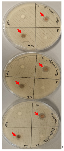
3.11. Antibacterial Evaluation
Antibacterial activity of the newly synthesized free ligand and Co, Cu, Zn ligand complexes was evaluated using the disk diffusion method against gram-positive (Bacillus-subtills) and gram-negative bacteria (E. coli). Trials were conducted three times, the values recorded represent the mean average as shown in Table 8. The results showed that the free ligand is ineffective in both gram positive and negative bacteria which is proved in a study of chelating activity of (2E,2′E)-2,2′-(pyridine-2,6-diylbis(ethan-1-yl-1-ylidene) bis(N-ethylhydrazinecarbothioamide [88]. In gram positive bacteria, the most potent nano metal-ligand complex was copper followed by the cobalt complex and no effect of the zinc complex. This is consistent with chelation theory, which postulated that the metal complex chelation would enhance the complexes’ capacity to cross a cell membrane and explain the antibacterial action of metal complexes [89,90,91]. In our study the Cu-L complex had more potent inhibition zones than penicillin, and ampicillin drugs (drugs against bacterial diseases [48]) with 6 and 7mm respectively against the gram-positive bacteria bacillus subtills [88]. On the other hand, the data showed in gram negative bacteria at different tested concentrations (5 and 10 mg weak effect when using cu complex with no effect of the other complexes [88]. One study revealed that nano Co and Zn ligand complexes showed antibacterial activity higher than the free ligand. All these data were supported by a study done by Hoda El-Shafiy in 2017, when the nano-complexes of Zn (II), Cu (II), Ni (II) and Co (II) ions where much potent than the ligand itself [92]. Additionally, while considering metal complexes with antibacterial properties, several elements should be considered, including the chelate effect, the ligands’ nature, the complex’s overall charge, and the type of ion that neutralizes the specific ionic complex [93,94]. In sum according to the findings, when compared to the free ligand, copper complexes showed promise as antibacterial agents in gram positive and negative bacteria.
4. Conclusions
Over the past decades, Co (II), Cu (II), and Zn (II) complexes have become increasingly popular in chemical biology, especially as powerful sensory tools, and novel antibacterial and anticancer medications. After the synthesis, a variety of analytical methods were used to customize and thoroughly describe three novel metal ligand complexes. Using a breast cancer cell line, the nanoscale complexes demonstrated stable and strong anticancer activity. Theoretical DFT calculations showed results that were equivalent to those of cell proliferation. The metal ligand complexes had strong antibacterial action against both gram positive and gram-negative bacteria, according to the antibacterial investigation. In general, the Cu-ligand complex exhibited the strongest antibacterial and anticancer properties, followed by Zn then Co complexes. Cu complex is promising agent for future disease treatments.
Acknowledgements
The authors are grateful to the graduate studies to support this project. Also, we acknowledge and appreciate RSPU at Kuwait University for providing the facilities especially instrument no: GS01/01, GS01/03, GS01/05, GS02/01. In addition, we are thankful to the Nanoscopy Science Center (NSC) for performing the SEM images. We also thank Dasman Diabetes Institute, in particular Dr. Dania Alhadad, for providing the facilities needed to conduct the biological studies.
Conflict of Interest
The authors declare no conflict of interest.
References
- .
- Karges J, Stokes RW, Cohen SM. (2021). Metal Complexes for Therapeutic Applications. Trends Chem. 3(7):523-534. [CrossRef]
- Klaudia Jomova, Marianna Makova, Suliman Y. Alomar, Saleh H. Alwasel, Eugenie Nepovimova, Kamil Kuca, Christopher J. Rhodes, Marian Valko. (2022). Essential metals in health and disease. Chemico-Biological Interactions. 367,110173. [CrossRef]
- Church DL. (2004). Major factors affecting the emergence and re-emergence of infectious diseases. Clin Lab Med. 24(3):559-86. [CrossRef]
- Van Crevel R, van de Vijver S, Moore DAJ. (2017). The global diabetes epidemic: what does it mean for infectious diseases in tropical countries? Lancet Diabetes Endocrinol. 5(6):457-468.
- Ellis, T., Eze, E. & Raimi-Abraham, B.T. (2021). Malaria and Cancer: a critical review on the established associations and new perspectives. Infect Agents Cancer. 16,33. [CrossRef]
- Liu Q, Jing W, Kang L, Liu J, Liu M. (2021). Trends of the global, regional and national incidence of malaria in 204 countries from 1990 to 2019 and implications for malaria prevention. J Travel Med. 7,28(5). [CrossRef]
- Byrne, C., Divekar, S.D., Storchan, G.B. et al. (2013). Metals and Breast Cancer. J Mammary Gland Biol Neoplasia. 18,63–73. [CrossRef]
- Maret W. (2016). The Metals in the Biological Periodic System of the Elements: Concepts and Conjectures. Int J Mol Sci. 2016 Jan 5,17(1):66. [CrossRef]
- Maria Antonietta Zoroddu, Jan Aaseth, Guido Crisponi, Serenella Medici, Massimiliano Peana, Valeria Marina Nurchi. (2019). The essential metals for humans: a brief overview, Journal of Inorganic Biochemistry. 195,120-129.
- Ejaz HW, Wang W, Lang M. (2020). Copper Toxicity Links to Pathogenesis of Alzheimer’s Disease and Therapeutics Approaches. Int J Mol Sci. 16,21(20):7660. [CrossRef]
- Pradhan SH, Liu JY, Sayes CM. (2023). Evaluating Manganese, Zinc, and Copper Metal Toxicity on SH-SY5Y Cells in Establishing an Idiopathic Parkinson’s Disease Model. Int J Mol Sci. 24(22):16129. [CrossRef]
- Houtman JP. (1996). Trace elements and cardiovascular diseases. J Cardiovasc Risk. 3(1):18-25.
- Górska A, Markiewicz-Gospodarek A, Trubalski M, Żerebiec M, Poleszak J, Markiewicz R. (2024). Assessment of the Impact of Trace Essential Metals on Cancer Development. Int J Mol Sci. 25(13):6842. [CrossRef]
- Bjørklund G, Dadar M, Pivina L, Doşa MD, Semenova Y, Aaseth J. (2020). The Role of Zinc and Copper in Insulin Resistance and Diabetes Mellitus. Curr Med Chem. 27(39):6643-6657. [CrossRef]
- Sousa, C.; Freire, C.; De Castro, B. (2003). Synthesis and Characterization of Benzo-15-Crown-5 Ethers with Appended N2O Schiff Bases. Molecules. 8,894-900. [CrossRef]
- L.T. Yildirm, O. Atakol. Cryst. Res. Technol. (2002). Crystal structure analysis of Bis{(N,N′-dimethylformamide)-[μ-bis-N,N′-(2-oxybenzyl)-1,3-propanediaminato](μ- asetato) nickel(II)}nickel(II). Crystal Research and Technology 37(12):1352-1359.
- Tesauro D. (2022). Metal Complexes in Diagnosis and Therapy. Int J Mol Sci. 23(8):4377.
- M. Hapke, G. Hilt. (2020). Introduction to cobalt chemistry and catalysis. Cobalt Catalysis in Organic Synthesis: Methods and Reactions. Wiley. 1-23.
- M. Valko, R. Klement, P. Pelikan, R. Boca, L. Dlhan, A. Bottcher, H. Elias, L. Muller. (1995). Copper (II) and Cobalt (II) complexes with derivatives of Salen and Tetrahydrosalen: an electron spin resonance, magnetic susceptibility, and quantum chemical study. J. Phys. Chem. 99,137-143. [CrossRef]
- Chang EL, Simmers C, Knight DA. (2010). Cobalt Complexes as Antiviral and Antibacterial Agents. Pharmaceuticals (Basel). 3(6):1711-1728. [CrossRef]
- Sopbué Fondjo, E., Songmi Feuze, S., Tamokou, JdD. et al. (2024). Synthesis, characterization, and antibacterial activity studies of two Co(II) complexes with 2-[(E)-(3-acetyl-4-hydroxyphenyl)diazenyl]-4-(2-hydroxyphenyl)thiophene-3-carboxylic acid as a ligand. BMC Chemistry. 18,75. [CrossRef]
- P. Kamalakannan, D. Venkappayya. (2002). Synthesis and characterization of cobalt and nickel chelates of 5-dimethylaminomethyl-2-thiouracil and their evaluation as antimicrobial and anticancer agents. Journal of Inorganic Biochemistry. 90, (1-2):22-37. [CrossRef]
- W. Kaim, B. Schwederski. (1994). Bioinorganic Chemistry: Inorganic. lements in the Chemistry of Life. Wiley. 1-432.
- S.J. Lippard, J.M. Berg. (1994). Principles of Bioinorganic Chemistry. University Science Books. Mill Valley. CA. 1-411.
- L. Virag, F. Erdodi, P. Gergely. (2016). Bioinorganic Chemistry for Medical Students. Scriptum. University of Debrecen. Hungary. 1-104.
- Simunkova M, Lauro P, Jomova K, Hudecova L, Danko M, Alwasel S, Alhazza IM, Rajcaniova S, Kozovska Z, Kucerova L, Moncol J, Svorc L, Valko M. (2019). Redox-cycling and intercalating properties of novel mixed copper (II) complexes with non-steroidal anti-inflammatory drugs tolfenamic, mefenamic and flufenamic acids and phenanthroline functionality: Structure, SOD-mimetic activity, interaction with albumin, DNA damage study and anticancer activity. J Inorg Biochem. 194:97-113. [CrossRef]
- Olar, R., Badea, M., Bacalum, M. et al. (2021). Antiproliferative and antibacterial properties of biocompatible copper (II) complexes bearing chelating N,N-heterocycle ligands and potential mechanisms of action. Biometals. 34,1155-1172. [CrossRef]
- Villarreal, W.; Castro, W.; González, S.; Madamet, M.; Amalvict, R.; Pradines, B.; Navarro, M. (2022). Copper (I)-Chloroquine Complexes: Interactions with DNA and Ferriprotoporphyrin, Inhibition of β-Hematin Formation and Relation to Antimalarial Activity. Pharmaceuticals. 15, 921. [CrossRef]
- J. Benters, U. Flogel, T. Schafer, D. Leibfritz, S. Hechtenberg, D. Beyersmann. (1997). Study of the interactions of cadmium and zinc ions with cellular alcium homoeostasis using 19F-NMR spectroscopy. Biochem. J. 322:793-799. [CrossRef]
- R. Ye, C. Tan, B. Chen, R. Li, Z. Mao. (2020). Zinc-containing metalloenzymes: inhibition by metal-based anticancer agents. Front. Chem. 8:402. [CrossRef]
- A.Klug, D. Rhodes. (1987). Zinc fingers: a novel protein fold for nucleic acid recognition. Cold Spring Harbor Symp. Quant. Biol. 52:473-482. [CrossRef]
- Mousa, A.B., Moawad, R., Abdallah, Y. et al. (2023). Zinc Oxide Nanoparticles Promise Anticancer and Antibacterial Activity in Ovarian Cancer. Pharm Res 40, 2281-2290. [CrossRef]
- Khashan, K.S., Sulaiman, G.M., Hussain, S.A. et al. (2020). Synthesis, Characterization and Evaluation of Anti-bacterial, Anti-parasitic and Anti-cancer Activities of Aluminum-Doped Zinc Oxide Nanoparticles. J Inorg Organomet Polym.30, 3677-3693. [CrossRef]
- Bai XG, Zheng Y, Qi J. (2022). Advances in thiosemicarbazone metal complexes as anti-lung cancer agents. Front Pharmacol. 13:1018951. [CrossRef]
- Serda M, Kalinowski DS, Rasko N, Potůčková E, Mrozek-Wilczkiewicz A, Musiol R, Małecki JG, Sajewicz M, Ratuszna A, Muchowicz A, Gołąb J, Simůnek T, Richardson DR, Polanski J. (2014). Exploring the anti-cancer activity of novel thiosemicarbazones generated through the combination of retro-fragments: dissection of critical structure-activity relationships. PLoS One. 9(10):e110291. [CrossRef]
- Jaragh-Alhadad LA, Ali MS. (2022). Methoxybenzamide derivative of nimesulide from anti-fever to anti-cancer: Chemical characterization and cytotoxicity. Saudi Pharm J. 30(5):485-493. [CrossRef]
- Laila A. Jaragh-Alhadad, Gamaleldin I. Harisa, Fars K. Alanazi, (2022). Development of nimesulide analogs as a dual inhibitor targeting tubulin and HSP27 for treatment of female cancers. Journal of Molecular Structure. 1248:131479.
- Laila A. Jaragh-Alhadad and Mayada S. Ali. (2022). Nimesulide derivatives reduced cell proliferation against breast and ovarian cancer: synthesis, characterization, biological assessment, and crystal structure. Kuwait J.Sci. 49(3):1-17.
- Laila Jaragh-Alhadada,b, Haider Behbehania and Sadashiva Karnik, (2022). Cancer targeted drug delivery using active low-density lipoprotein nanoparticles encapsulated pyrimidines heterocyclic anticancer agents as microtubule inhibitors. Drug Delivery. 29:1, 2759-2772. [CrossRef]
- Laila A. Jaragh-Alhadad, Mayada S. Ali, Moustafa S. Moustafa, Gamaleldin I. Harisa, Fars K. Alanazi, Sadashiva Karnik. (2022). Sulfonamide derivatives mediate breast and lung cancer cell line killing through tubulin inhibition. Journal of Molecular Structure. 1268:133699.
- Jaragh-Alhadad L, Samir M, Harford TJ, Karnik S. (2022). Low-density lipoprotein encapsulated thiosemicarbazone metal complexes is active targeting vehicle for breast, lung, and prostate cancers. Drug Delivery. 29(1):2206-2216. [CrossRef]
- Mayada S. Ali, Fathy A. El-Saied, Mohamad ME. Shakdofa, Sadashiva Karnik, Laila A. Jaragh-Alhadad. (2023). Synthesis and characterization of thiosemicarbazone metal complexes: Crystal structure, and antiproliferation activity against breast (MCF7) and lung (A549) cancers. Journal of Molecular Structure. 1274(1):13448.
- Vázquez-Valadez Víctor Hugo, Hernández-S. Manuel Alejandro, Velázquez-S. Ana María, Rosales-H. María, Leyva-R. Marco Antonio, Prado-O. María Guadalupe, Muñoz-G. Marco Antonio, Alba-H. Fernando, Abrego Víctor, Cruz-A. Diego, Ángeles Enrique (2018). Molecular Modeling and Synthesis of Ethyl Benzyl Carbamates as Possible Ixodicide Activity. Computational Chemistry. 7(1).
- Andersson, M. P.; Uvdal, P., (2005). New Scale Factors for Harmonic Vibrational Frequencies Using the B3LYP Density Functional Method with the Triple-ζ Basis Set 6-311+G(d,p). The Journal of Physical Chemistry A . 109(12):2937-2941. [CrossRef]
- Bhat, A. R.; Dongre, R. S.; Almalki, F. A.; Berredjem, M.; Aissaoui, M.; Touzani, R.; Hadda, T. B.; Akhter, M. S., (2021). Synthesis, biological activity and POM/DFT/docking analyses of annulated pyrano [2,3-d]pyrimidine derivatives: Identification of antibacterial and antitumor pharmacophore sites. Bioorganic Chemistry. 106,104480. [CrossRef]
- Guerfi, M.; Berredjem, M.; Bahadi, R.; Djouad, S.-E.; Bouzina, A.; Aissaoui, M., (2021). An efficient synthesis, characterization, DFT study and molecular docking of novel sulfonylcycloureas. Journal of Molecular Structure. 1236,130327.
- Mounyr, B.; Moulay, S.; saad, K.I. (2015). Methods for invitro evaluating antimicrobial activity: A review. J Pharm Anal. 6(2):71-79.
- Mayada S. Ali, Dhanachandra Kuraijam, Sadashiva Karnik, Laila A. Jaragh-Alhadad. (2023). Hydrazone bimetallic complex: synthesis, characterization, in silico and biological evaluation targeting breast and lung cancer cells’ G-quadruplex DNA. Kuwait J.Sci.. 50,(3):1-17.
- M. M. E. Shakdofa, H. A. Mousa, A. M. A. Elseidy, A. A. Labib, M. M. Ali, A. S. Abd-El-All, Appl. (2018). Synthesis, characterization, and density functional theory studies of hydrazone–oxime ligand derived from 2,4,6-trichlorophenyl hydrazine and its metal complexes searching for new antimicrobial drugs. Organomet. Chem. 32,e3936.
- A.Sethukumar, C. U. Kumar, R. Agilandeshwari, B. A. Prakasam, J. Mol. Struct. (2013). Synthesis, stereochemical, structural and biological studies of some 2, 6-diarylpiperidin-4-one N (4′)-cyclohexyl thiosemicarbazones. 1047,237. [CrossRef]
- F. P. Andrew, P. A. Ajibade, (2018). Synthesis, characterization and anticancer studies of bis(1-phenylpiperazine dithiocarbamato) Cu(II), Zn(II) and Pt(II) complexes: Crystal structures of 1-phenylpiperazine dithiocarbamato-S,S′ zinc(II) and Pt(II). J. Mol. Struct. 1170,24.
- S. M. Emam, I. E. T. El Sayed, M. I. Ayad, H. M. R. Hathout. (2017). Synthesis, characterization and anticancer activity of new Schiff bases bearing neocryptolepine. J. Mol. Struct. 1146,600.
- S. Chandra, S. Bargujar, R. Nirwal, N. Yadav, Spectrochim. (2013). Synthesis, spectral characterization and biological evaluation of copper(II) and nickel(II) complexes with thiosemicarbazones derived from a bidentate Schiff base. Acta Part A Mol. Biomol. Spectrosc. 106,91. [CrossRef]
- L. A. Saghatforoush, S. Hosseinpour, M. W. Bezpalko, W. S. Kassel. (2019). Synthesis, spectroscopic studies and X-ray structure determination of two mononuclear copper complexes derived from the Schiff base ligand N,N-dimethyl-N’-((5-methyl-1H-imidazol-4-yl)methylene)ethane-1,2-diamine. Inorg. Chim. Act. 484,527.
- E. Shahsavani, A. D. Khalaji, N. Feizi, M. Kučeráková, M. Dušek. (2015). Synthesis, characterization, crystal structure and antibacterial activity of new sulfur-bridged dinuclear silver(I) thiosemicarbazone complex [Ag2(PPh3)2(μ-S-Brcatsc)2(η1-S-Brcatsc)2](NO3)2. Inorg. Chim. Act. 429,61.
- N. M. Rageh, A. M. A. Mawgoud, H. M. Mostafa. (1999). Transition Metal Complexes Derived From 2-hydroxy-4-(p-tolyldiazenyl)benzylidene)-2-(p-tolylamino)acetohydrazide Synthesis, Structural Characterization, and Biological Activities. Chem. Pap. 53,107.
- A.B. P. Lever. (1969). ’Inorganic electronic spectroscopy’. Elsevier science. 46(9).
- Sethukumar, C. Udhaya Kumar, R. Agilandeshwari, B. Arul Prakasam. (2013). Synthesis, stereochemical, structural and biological studies of some 2,6-diarylpiperidin-4-one N(4′)-cyclohexyl thiosemicarbazones. J. Mol. Struct. 1047, 37.
- Şen, B. (2021). 2-Acetyl-5-chloro-thiophene thiosemicarbazone and its nickel(II) and zinc(II) complexes: Hirshfeld surface analysis and Density Functional Theory calculations for molecular geometry, vibrational spectra and HOMO-LUMO studies. Turkish Computational and Theoretical Chemistry. 5(1):27-38.
- Gaber, M., El-Ghamry, H.A and Mansour, M.A. (2018). Pd (II) and Pt (II) chalcone complexes. Synthesis, spectral characterization, molecular modeling, biomolecular docking, antimicrobial and antitumor activities. Journal of Photochemistry and Photobiology A: Chemistry. 354, pp. 163-174. [CrossRef]
- D. C. Ilies, E. Pahontu, S. Shova, R. Georgescu, N. Stanica, R. Olar, A. Gulea, T. Rosu. (2014). Synthesis, structure and biological properties of a series of dicopper(bis-thiosemicarbazone) complexes. Polyhedron. 81,123.
- Vijayan, P., Vijayapritha, S., Ruba, C. et al. (2019). Ruthenium(II) carbonyl complexes containing thiourea ligand: Enhancing the biological assets through biomolecules interaction and enzyme mimetic activities. Monatsh Chem 150, 1059–1071.
- Şen, H. K. Kalhan, V. Demir, E. E. Güler, H. A. Kayalı, E. Subaşı, Mater. (2019). Crystal structures, spectroscopic properties of new cobalt(II), nickel(II), zinc(II) and palladium(II) complexes derived from 2-acetyl-5-chloro thiophene thiosemicarbazone: Anticancer evaluation. Sci. Eng. C. 98,550. [CrossRef]
- Z. Piri, Z. Moradi–Shoeili, A. Assoud. (2019). Ultrasonic assisted synthesis, crystallographic, spectroscopic studies and biological activity of three new Zn(II), Co(II) and Ni(II) thiosemicarbazone complexes as precursors for nano-metal oxides. Inorg. Chim. Acta. 484,338. [CrossRef]
- A.Akbari, H. Ghateazadeh, R. Takjoo, B. Sadeghi-Nejad, M. Mehrvar, J. T. Mague. (2019). Synthesis & crystal structures of four new biochemical active Ni(II) complexes of thiosemicarbazone and isothiosemicarbazone-based ligands: In vitro antimicrobial study. J. Mol. Struct. 2019, 1181, 287. [CrossRef]
- Chehelgerdi M, Chehelgerdi M, Allela OQB, Pecho RDC, Jayasankar N, Rao DP, Thamaraikani T, Vasanthan M, Viktor P, Lakshmaiya N, Saadh MJ, Amajd A, Abo-Zaid MA, Castillo-Acobo RY, Ismail AH, Amin AH, Akhavan-Sigari R. (2023). Progressing nanotechnology to improve targeted cancer treatment: overcoming hurdles in its clinical implementation. Mol Cancer. 22(1):169.
- Hembram KC, Kumar R, Kandha L, Parhi PK, Kundu CN, Bindhani BK. Therapeutic prospective of plant-induced silver nanoparticles: application as antimicrobial and anticancer agent. Artif Cells Nanomed Biotechnol. 2018;46(sup3):S38-S51. [CrossRef]
- Bisht G, Rayamajhi S. (2016). ZnO Nanoparticles: A Promising Anticancer Agent. Nanobiomedicine (Rij). 1;3:9.
- Arshad F, Naikoo GA, Hassan IU, Chava SR, El-Tanani M, Aljabali AA, Tambuwala MM. (2024). Bioinspired and Green Synthesis of Silver Nanoparticles for Medical Applications: A Green Perspective. Appl Biochem Biotechnol. 196(6):3636-3669.
- Jaragh-Alhadad LA, Falahati M. (2022). Tin oxide nanoparticles trigger the formation of amyloid β oligomers/protofibrils and underlying neurotoxicity as a marker of Alzheimer’s diseases. Int J Biol Macromol. 204:154-160.
- Jaragh-Alhadad LA, Falahati M. (2022). Copper oxide nanoparticles promote amyloid-β-triggered neurotoxicity through formation of oligomeric species as a prelude to Alzheimer’s diseases. Int J Biol Macromol. 207:121-129.
- Zheng Nie, Yasaman Vahdani, William C. Cho, Samir Haj Bloukh, Zehra Edis, Setareh Haghighat, Mojtaba Falahati, Rasoul Kheradmandi, Laila Abdulmohsen Jaragh-Alhadad, Majid Sharifi. (2022). 5-Fluorouracil-containing inorganic iron oxide/platinum nanozymes with dual drug delivery and enzyme-like activity for the treatment of breast cancer. Arabian Journal of Chemistry. 15(8):103966.
- Rümenapp, C., Gleich, B. & Haase, A. (2012). Magnetic Nanoparticles in Magnetic Resonance Imaging and Diagnostics. Pharm Res. 29,1165–1179.
- Hoda F. El-Shafiy, M. Saif, Mahmoud M. Mashaly, Shimaa Abdel Halim, Mohamed F. Eid, A.I. Nabeel, R. Fouad. (2017). New nano-complexes of Zn(II), Cu(II), Ni(II) and Co(II) ions; spectroscopy, thermal, structural analysis, DFT calculations and antimicrobial activity application. Journal of Molecular Structure. 1147, 452-461. [CrossRef]
- M. Imran, L. Mitu, S. Latif, Z. Mahmood, I. Naimat, S.S. Zaman, S. Fatima. (2010). antibacterial Co(II), Ni(II), Cu(II) and Zn(II) complexes with biacetyl-derived Schiff bases. J. Serb. Chem. Soc. 75:1075-1084.
- Jain, S., Rana, M., Sultana, R., Mehandi, R., & Rahisuddin. (2022). Schiff Base Metal Complexes as Antimicrobial and Anticancer Agents. Polycyclic Aromatic Compounds, 43(7), 6351–6406. [CrossRef]
- Gai S, He L, He M, Zhong X, Jiang C, Qin Y, Jiang M. (2023). Anticancer Activity and Mode of Action of Cu(II), Zn(II), and Mn(II) Complexes with 5-Chloro-2-N-(2-quinolylmethylene)aminophenol. Molecules. 28(12):4876.
- S. Rafique, M. Idrees, A. Nasim, H. Akbar, A. Athar. (2010). Transition metal complexes as potential therapeutic agents. Biotech. Mol. Biol. Rev. 5:38-45.
- Agnieszka Dziewulska-Kułaczkowska, Liliana Mazur (2011). Structural studies and characterization of 3-formylchromone and products of its reactions with chosen primary aromatic amines. J. Mol. Struct. 985:233-242.
- M. Saif, Hoda F. El-Shafiy, Mahmoud M. Mashaly, Mohamed F. Eid, A.I. Nabeel, R. Fouad. (2016). Synthesis, characterization, and antioxidant/cytotoxic activity of new chromone Schiff base nano-complexes of Zn(II), Cu(II), Ni(II) and Co(II). Journal of Molecular Structure. 1118:75-82. [CrossRef]
- Kotova, K. Lyssenko, A. Rogachev, S. Eliseeva, I. Fedyanin, L. Lepnev, L. Pandey, A. Burlov, A. Garnovskii, A. Vitukhnovsky, M.V.D. Auweraer, N. Kuzmin, Photochem. Photobiol. (2011). Low temperature X-ray diffraction analysis, electronic density distribution and photophysical properties of bidentate N,O-donor salicylaldehyde Schiff bases and zinc complexes in solid state. A Chem. 218:117-129. [CrossRef]
- V. Chiş, S. Filip, V. Miclăuş, A. Pîrnău, C. Tănăşelia, V. Almas, M. Vasilescu. (2005). Vibrational spectroscopy and theoretical studies on 2,4-dinitrophenylhydrazine J. Mol. Struct. 744:363. [CrossRef]
- Howsaui, H.B.; Sharfalddin, A.A.; Abdellattif, M.H.; Basaleh, A.S.; Hussien, M.A. (2021). Synthesis, Spectroscopic Characterization and Biological Studies of Mn(II), Cu(II), Ni(II), Co(II) and Zn(II) Complexes with New Schiff Base of 2-((Pyrazine-2-ylimino)methyl)phenol. Appl. Sci. 2021, 11, 906.
- M. Karabacak, E. Kose, A. Atac. (2012). Molecular structure (monomeric and dimeric structure) and HOMO–LUMO analysis of 2-aminonicotinic acid: a comparison of calculated spectroscopic properties with FT-IR and UV–Vis. Spectrochim. Acta A. 91:83. [CrossRef]
- M. Karabacak, E. Kose, A. Atac. (2012). Molecular structure (monomeric and dimeric structure) and HOMO–LUMO analysis of 2-aminonicotinic acid: a comparison of calculated spectroscopic properties with FT-IR and UV–Vis. Spectrochim. Acta A, 91:83.
- E. Porchelvi, S. Muthu Spectrochim. Acta A. (2015). Vibrational spectra, molecular structure, natural bond orbital, first order hyperpolarizability, thermodynamic analysis and normal coordinate analysis of Salicylaldehyde p-methylphenylthiosemicarbazone by density functional method. Spectrochimica Acta Part A: Molecular and Biomolecular Spectroscopy. 134:453-464.
- Mayada S. Ali, Mohamad Hasan. (2021). Chelating activity of (2E,2′E)-2,2′-(pyridine-2,6-diylbis(ethan-1-yl-1-ylidene)bis(N-ethylhydrazinecarbothioamide). Journal of Molecular Structure. 1238:130436.
- Damena T.; Zeleke D.; Desalegn T.; B Demissie T.; Eswaramoorthy R. (2022). Synthesis, Characterization, and Biological Activities of Novel Vanadium(IV) and Cobalt(II) Complexes. ACS Omega. 7:4389-4404.
- Hasan M. M.; Ahsan H. M.; Saha P.; Naime J.; Kumar Das A.; Asraf M. A.; Nazmul Islam A. B. M.(2021). Antioxidant, Antibacterial and Electrochemical Activity of (E)-N-(4 (Dimethylamino) Benzylidene)-4H-1,2,4-Triazol-4-Amine Ligand and Its Transition Metal Complexes. Results Chem. 3:00115.
- Chebout O.; Trifa C.; Bouacida S.; Boudraa M.; Imane H.; Merzougui M.; Mazouz W.; Ouari K.; Boudaren C.; Merazig H. (2022). Two New Copper (II) Complexes with Sulfanilamide as Ligand: Synthesis, Structural, Thermal Analysis, Electrochemical Studies and Antibacterial Activity. J. Mol. Struct. 1248:131446. [CrossRef]
- Hoda F. El-Shafiy, M. Saif, Mahmoud M. Mashaly, Shimaa Abdel Halim, Mohamed F. Eid, A.I. Nabeel, R. Fouad. (2017). New; spectroscopy, thermal, structural analysis, DFT calculations and antimicrobial activity application. Journal of Molecular Structure. 1147:452-461.
- Mandewale M. C.; Kokate S.; Thorat B.; Sawant S.; Yamgar R. (2022). Zinc Complexes of Hydrazone Derivatives Bearing 3,4-Dihydroquinolin-2(1H)-One Nucleus as New Anti-Tubercular Agents. Arab. J. Chem. 12,4479–4489.
- Damena T, Alem MB, Zeleke D, Desalegn T, Eswaramoorthy R, Demissie TB. Novel Zinc(II) and Copper(II) Complexes of 2-((2-Hydroxyethyl)amino)quinoline-3-carbaldehyde for Antibacterial and Antioxidant Activities: A Combined Experimental, DFT, and Docking Studies. ACS Omega. 7(30):26336-26352.
Figure 1.
Transition metals used clinically for diagnosis and therapies.
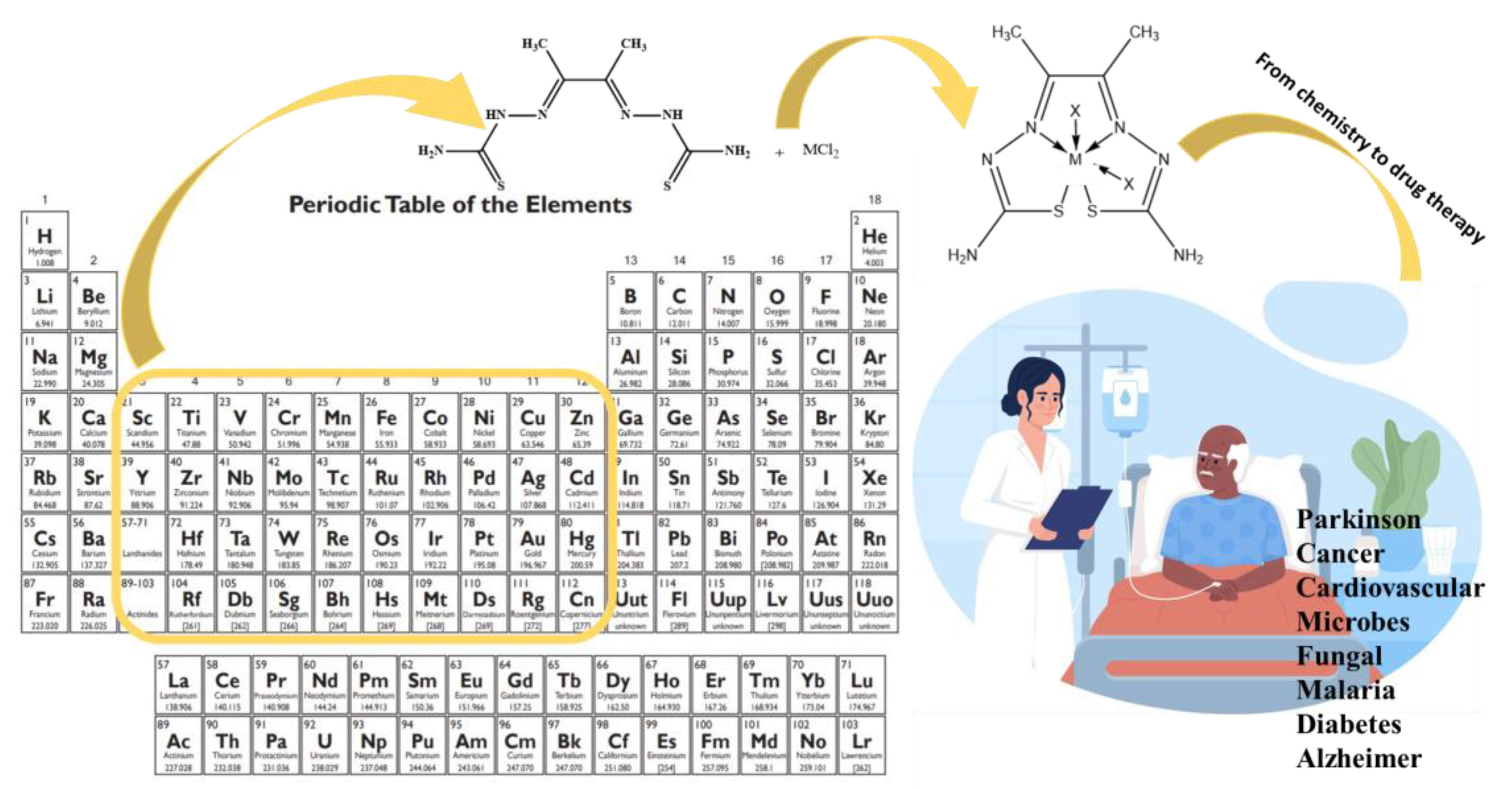
Figure 2.
Thiosemicarbazone metal chelator because of the carbon nitrogen and carbon sulfur bonds.
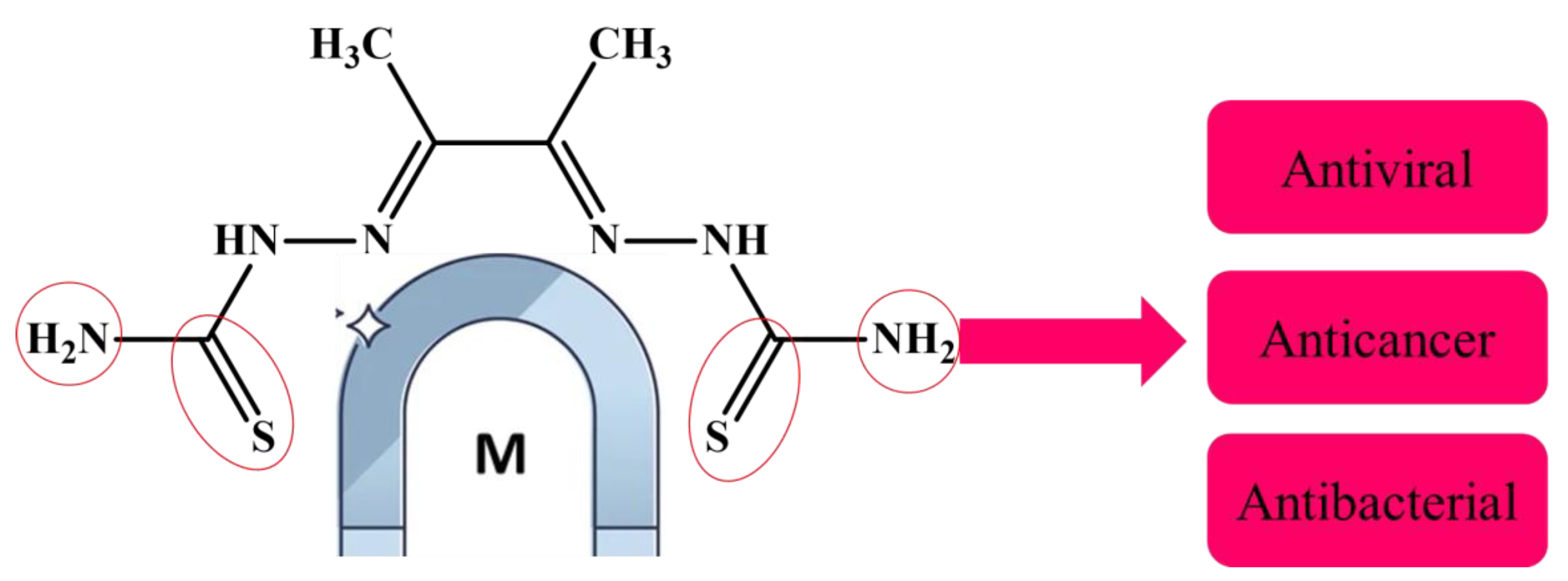
Figure 3.
Schematic representation starting from synthesis to biological evaluation.
Scheme 1.
The starting materials to synthesiz the ligand H2L (3).
Scheme 2.
General made of chelation of the metal ligand complex.
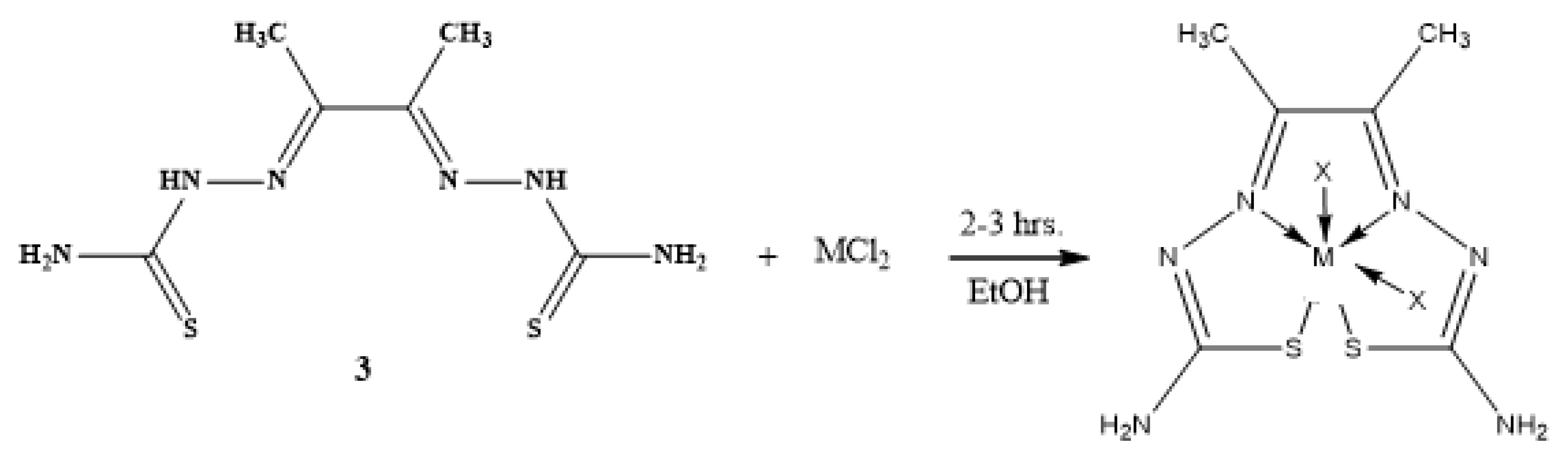
Figure 4.
Mass spectrum of ligand thiosemicarbazone (H2L).
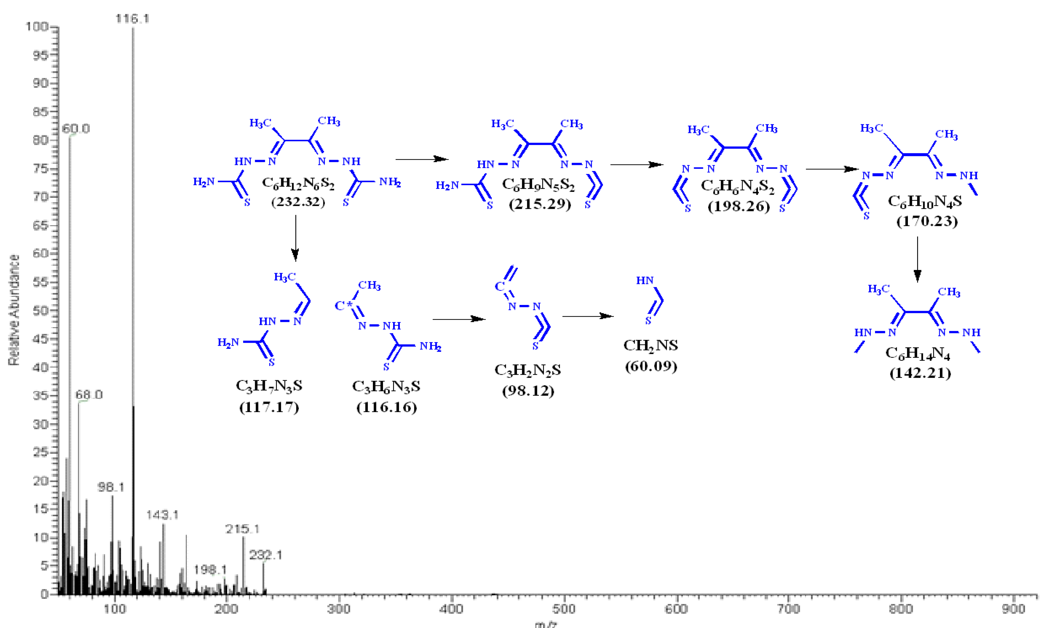
Figure 5.
Mass spectrum of [Co(L)].2H2O.
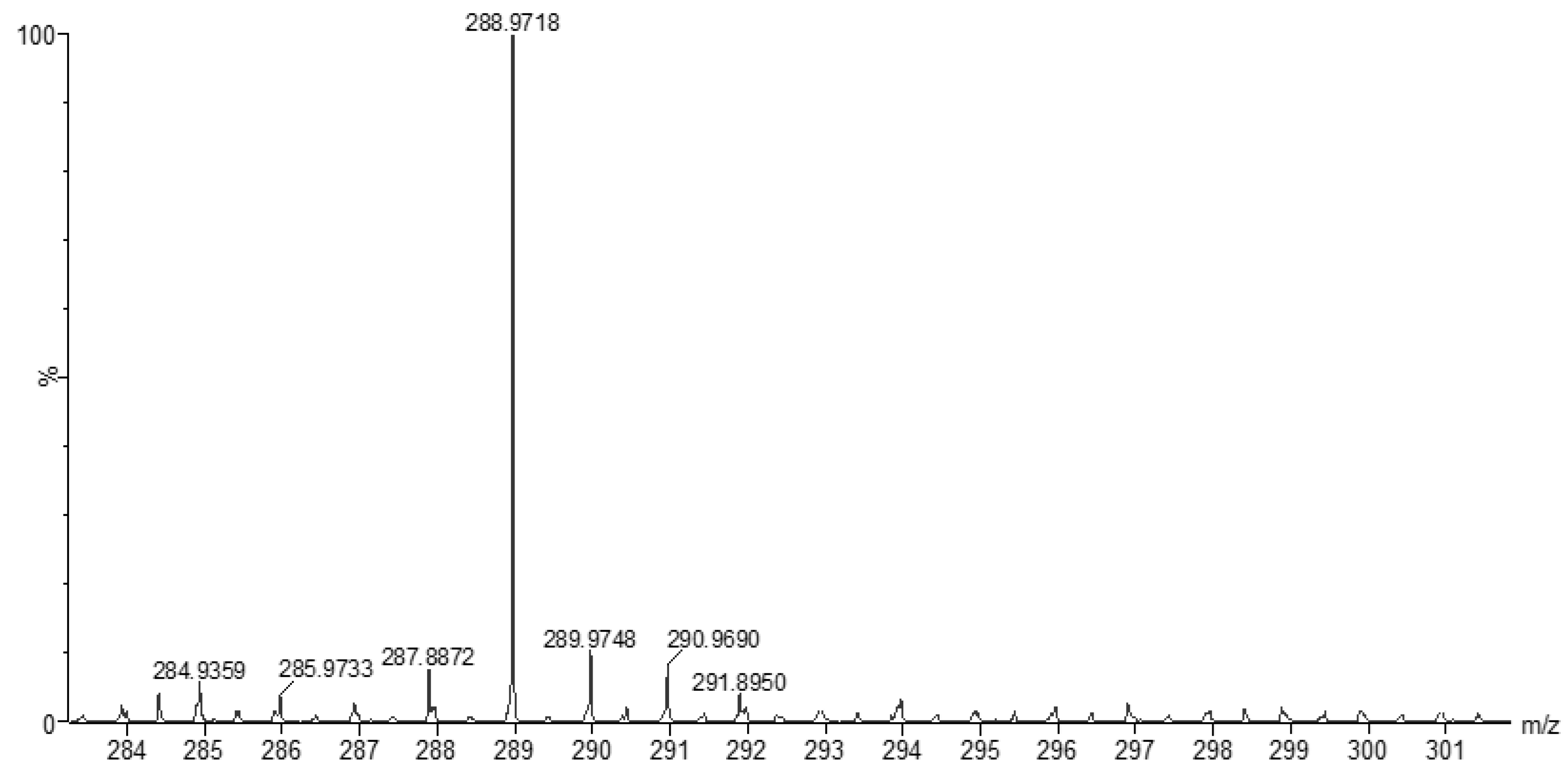
Figure 6.
Mass spectrum of [Cu(L)].2H2O.
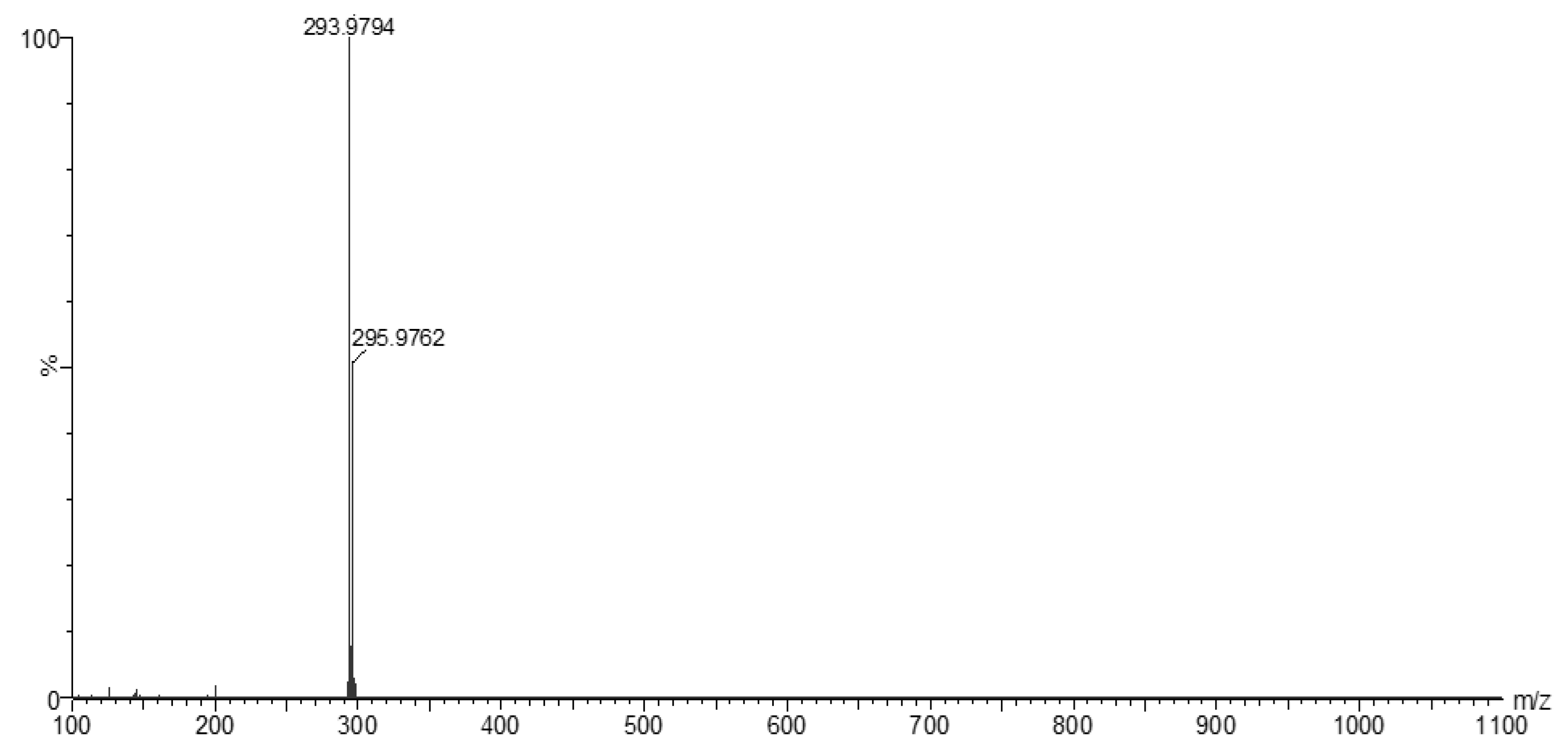
Figure 7.
Mass spectrum of [Zn(H2L)2].
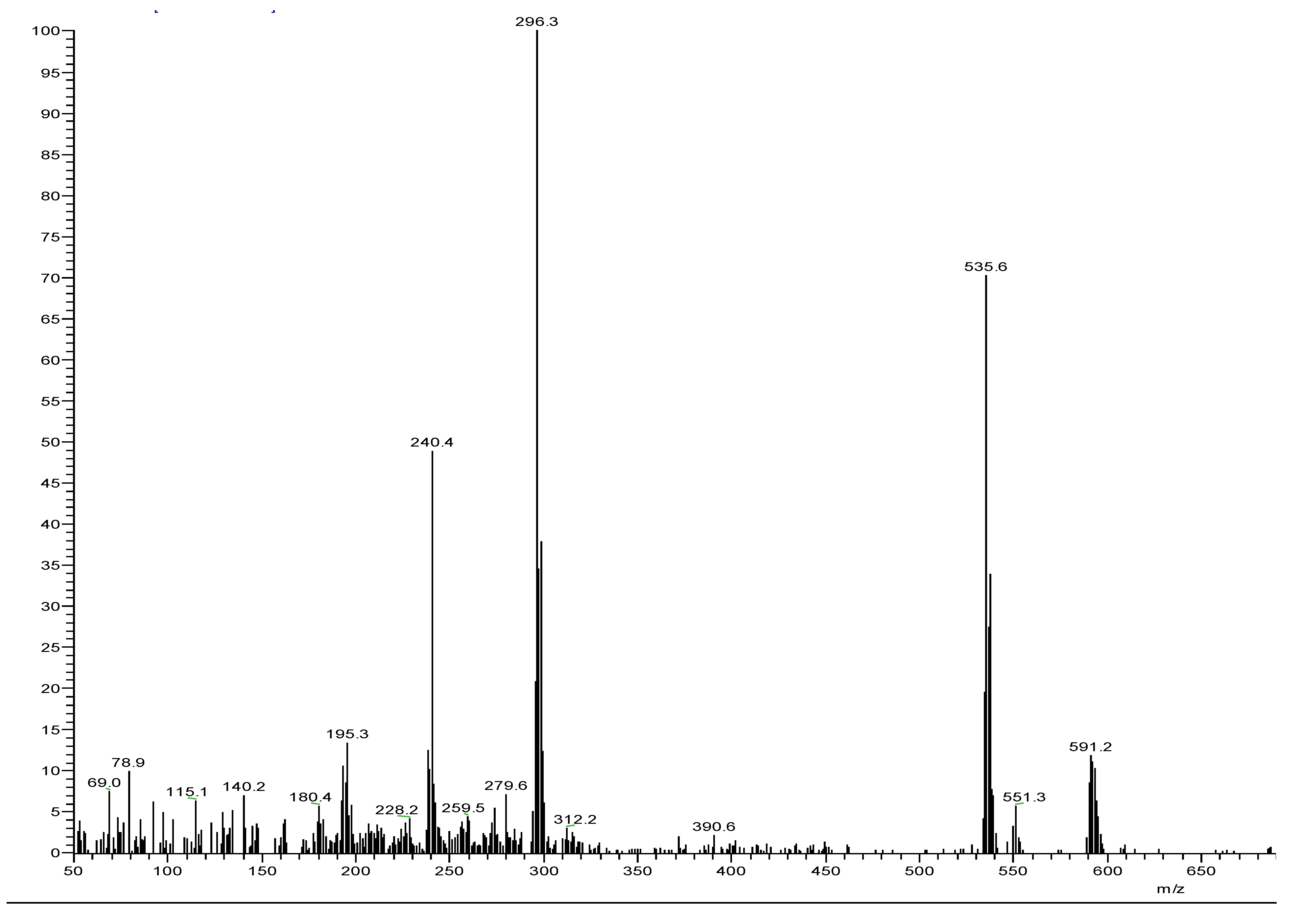
Figure 8.
The 1H NMR spectrum of the thiosemicarbazone ligand (H2L).
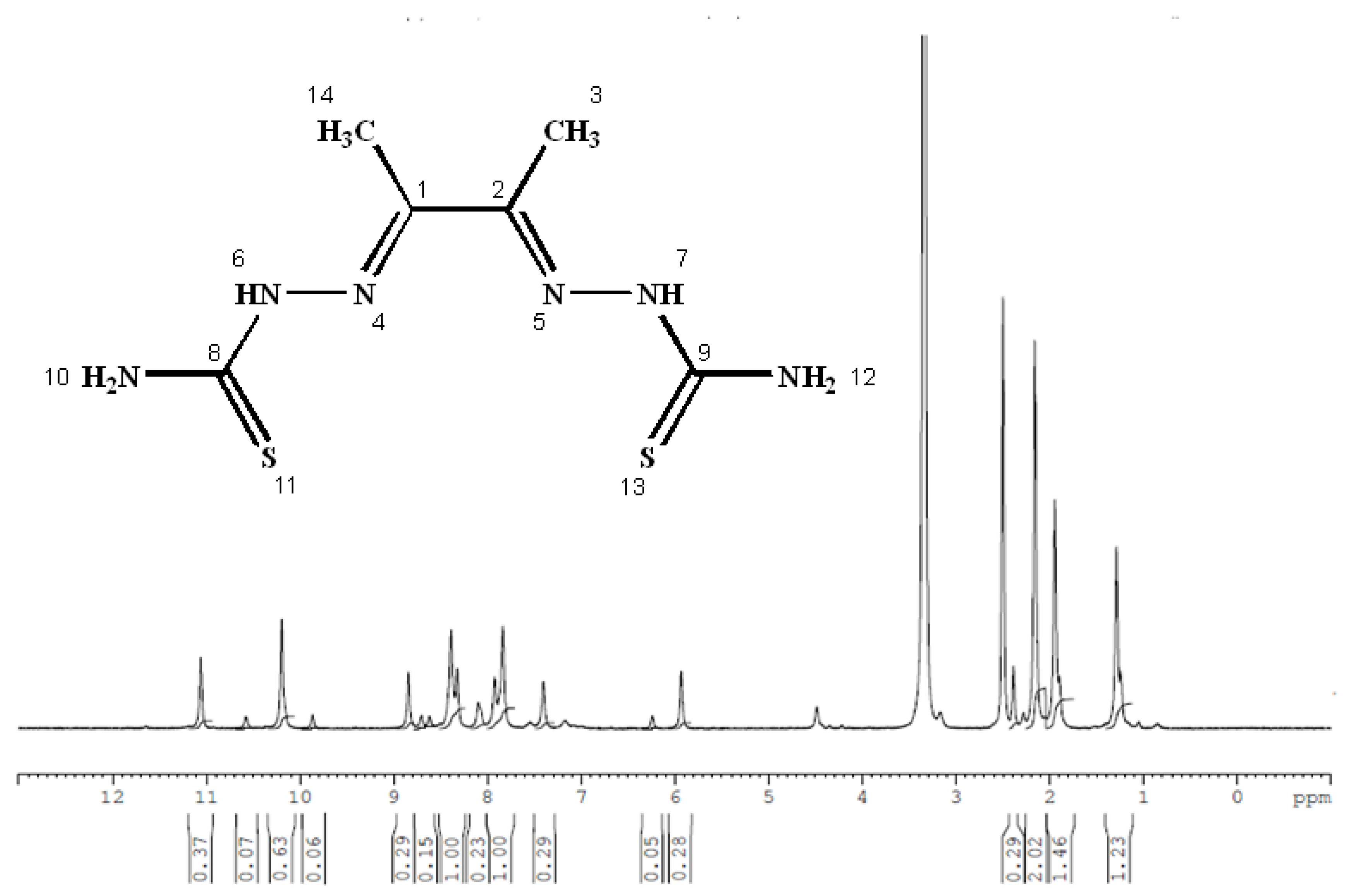
Figure 9.
The 13C NMR spectrum of the thiosemicarbazone ligand (H2L).
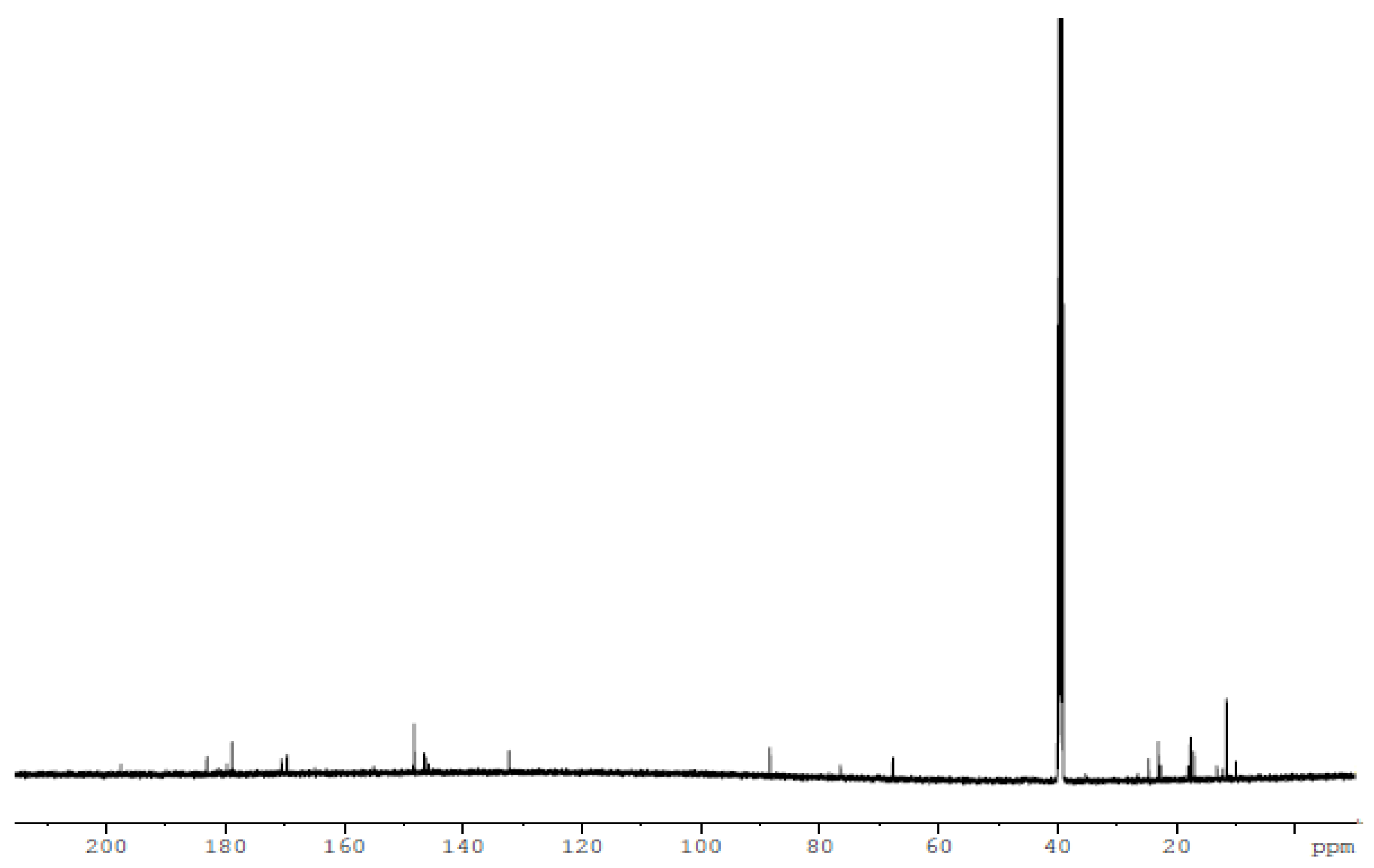
Figure 10.
The 1H NMR spectrum of [Zn(H2L)2] complex.
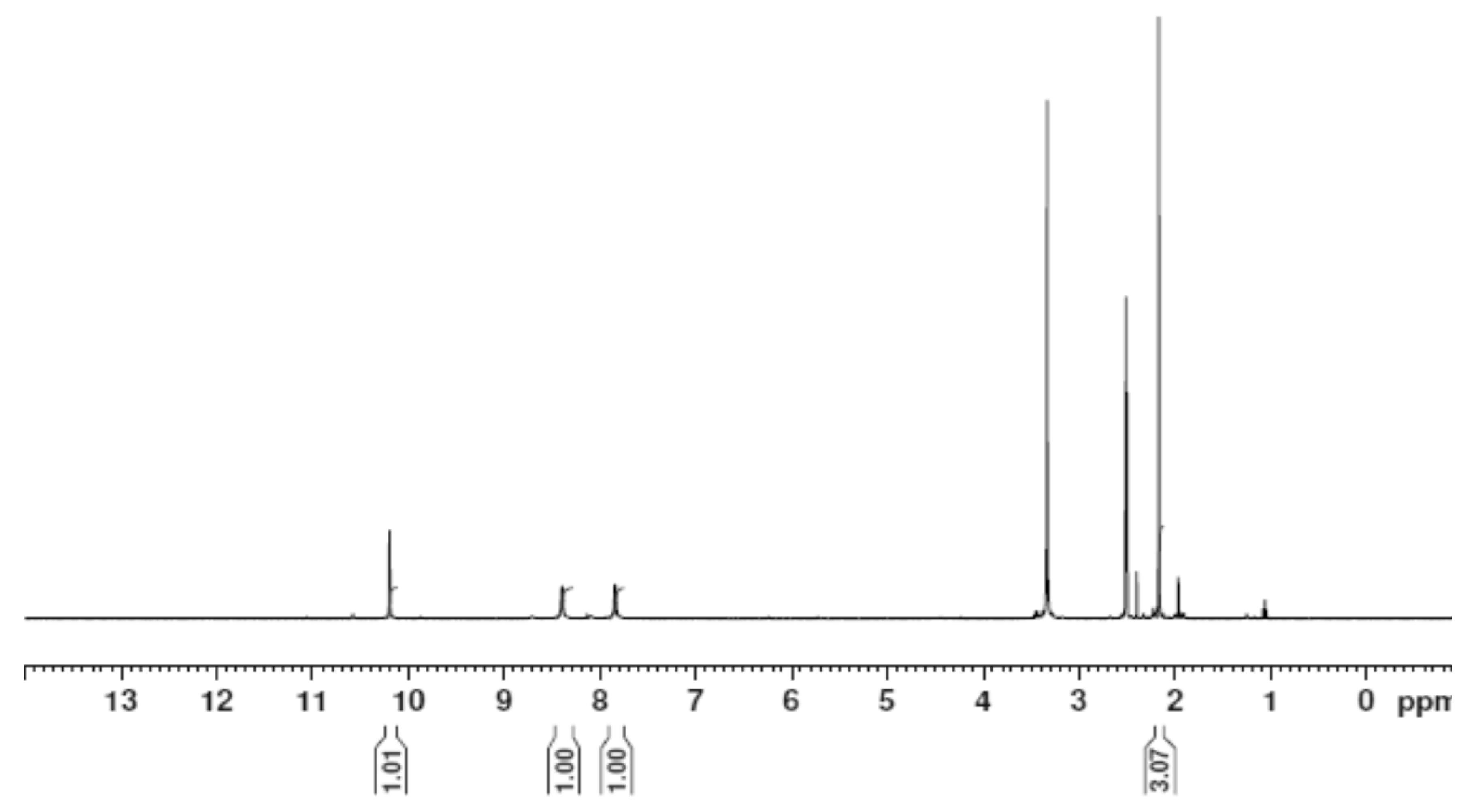
Figure 11.
The 13C NMR spectrum of the [Zn(H2L)2] complex.
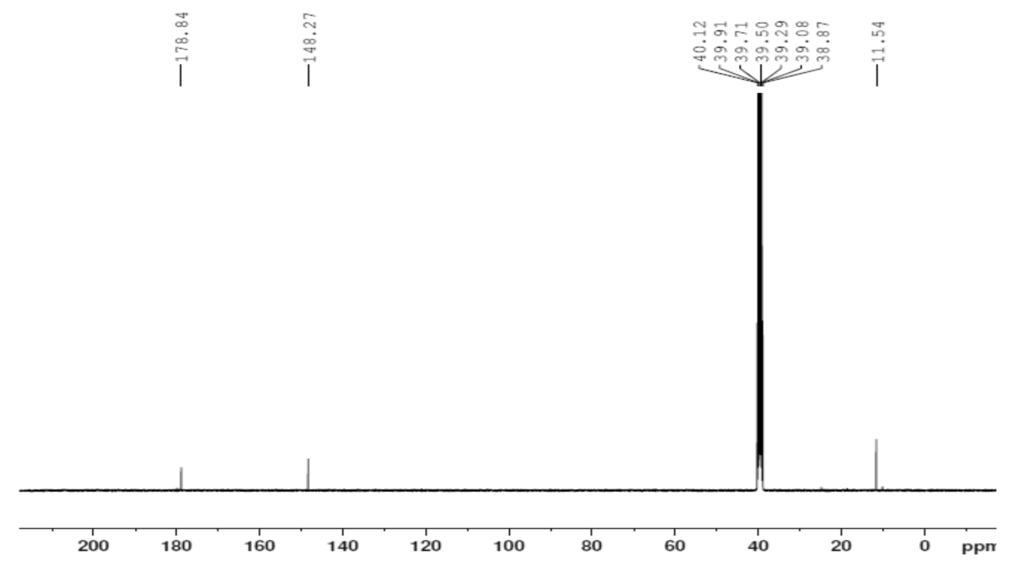
Figure 12.
Infrared spectrum of the ligand thiosemicarbazone (H2L).
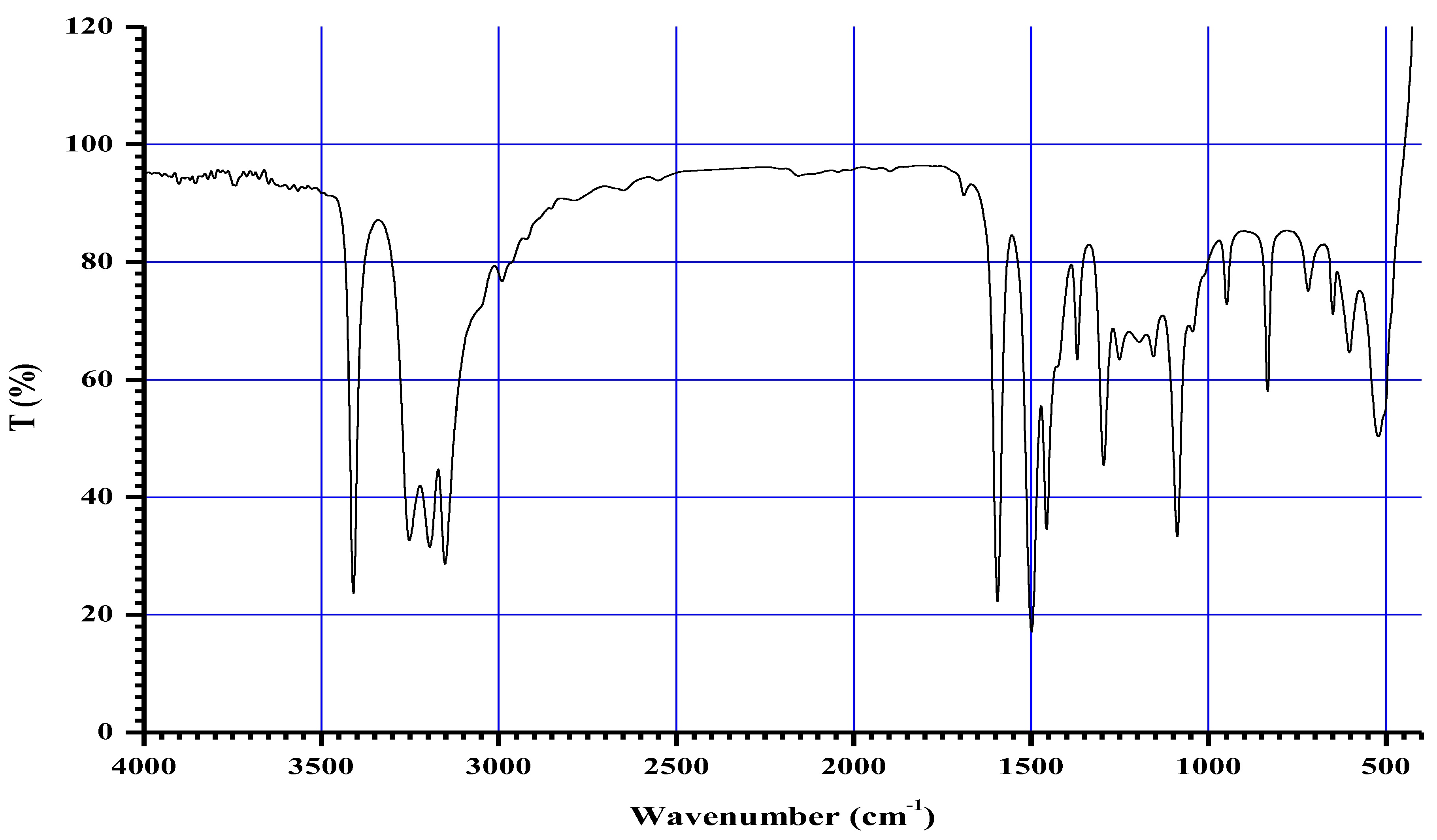
Figure 13.
Infrared spectrum of the Co+2 complex.
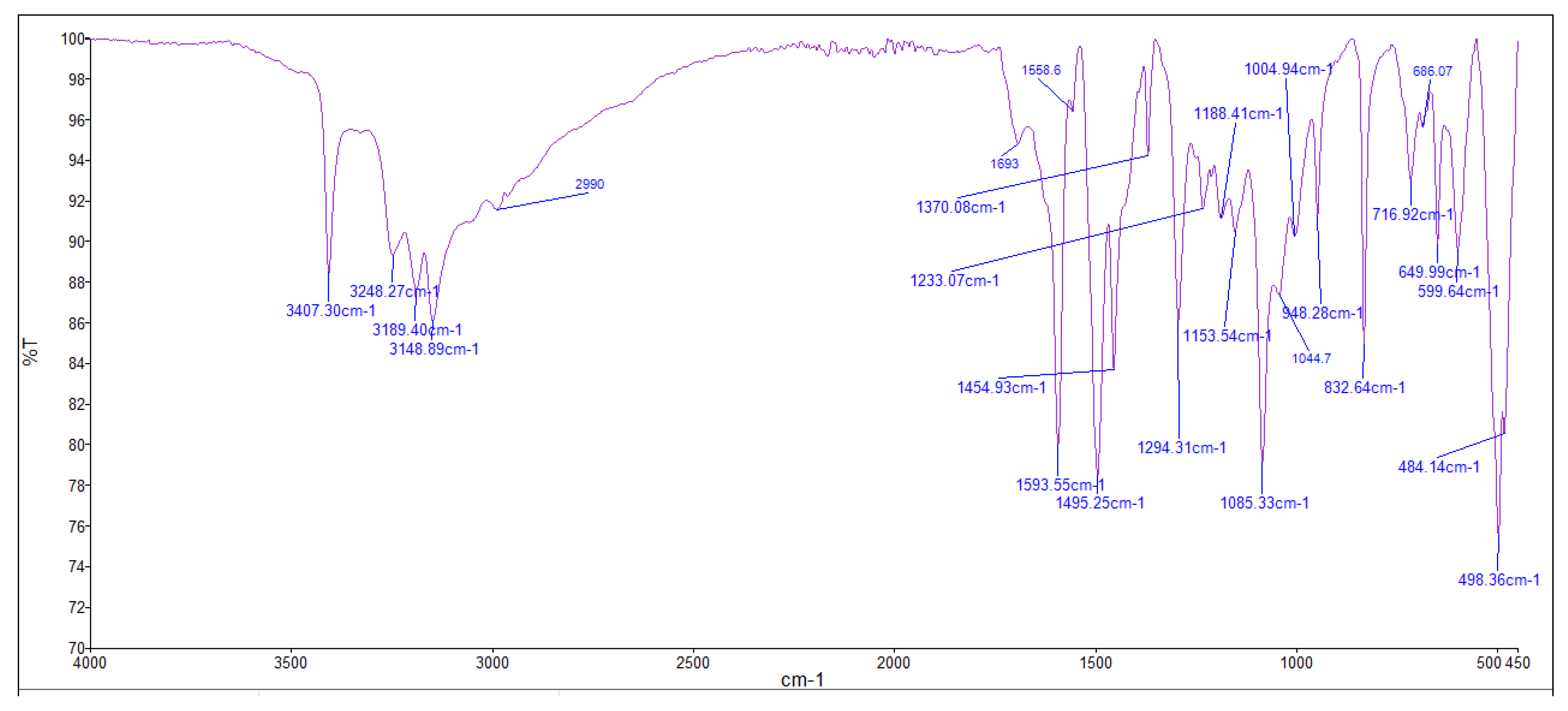
Figure 14.
Infrared spectrum of the Cu+2 complex.

Figure 15.
Infrared spectrum of the Zn+2 complex.
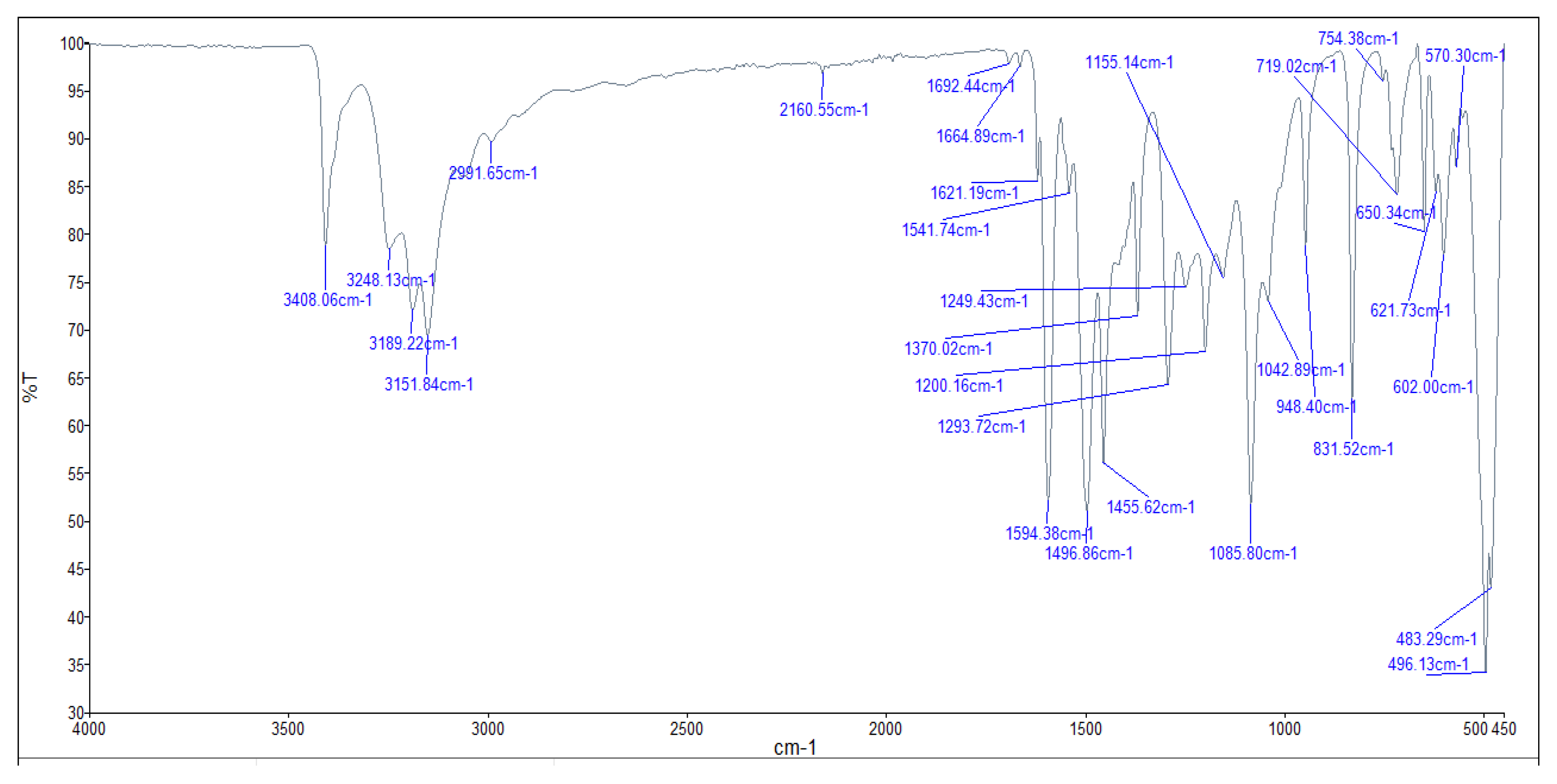
Figure 16.
TGA spectrum of ligand thiosemacarbazone.
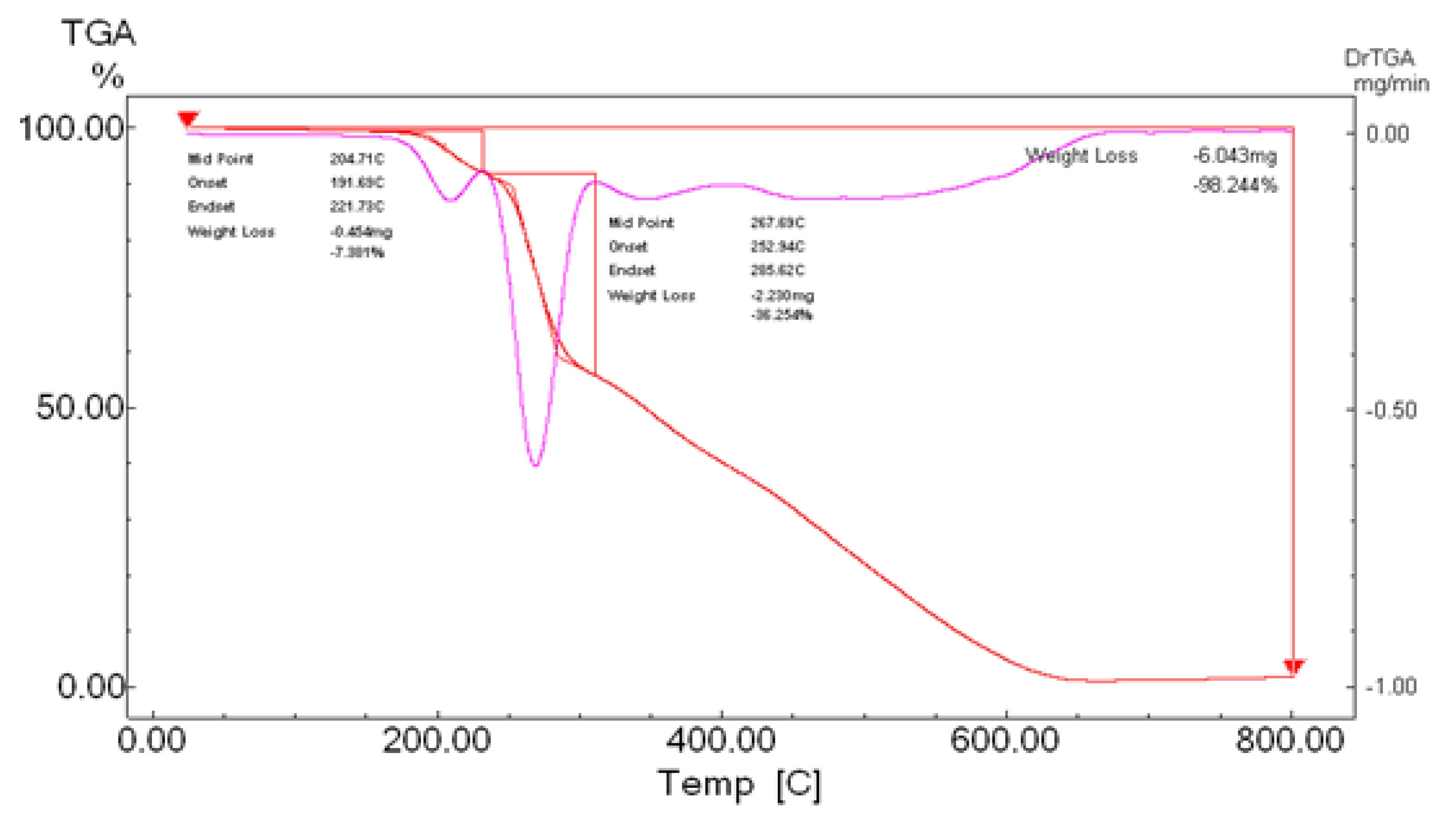
Figure 17.
TGA spectrum of Co- ligand complex.
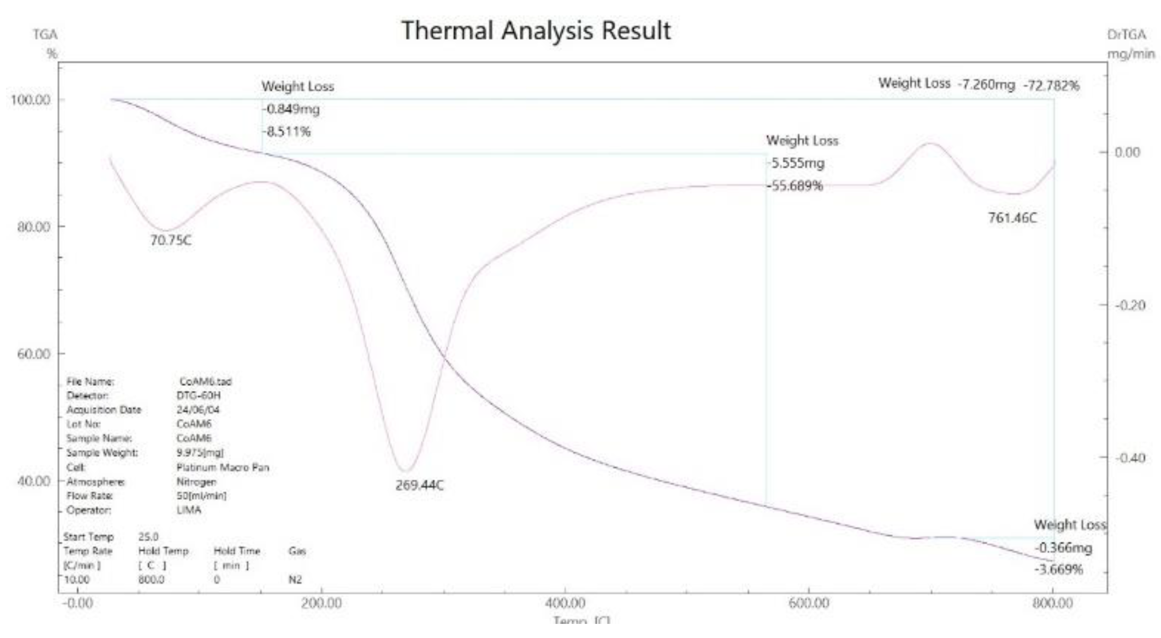
Figure 18.
TGA spectrum of Cu- ligand complex.
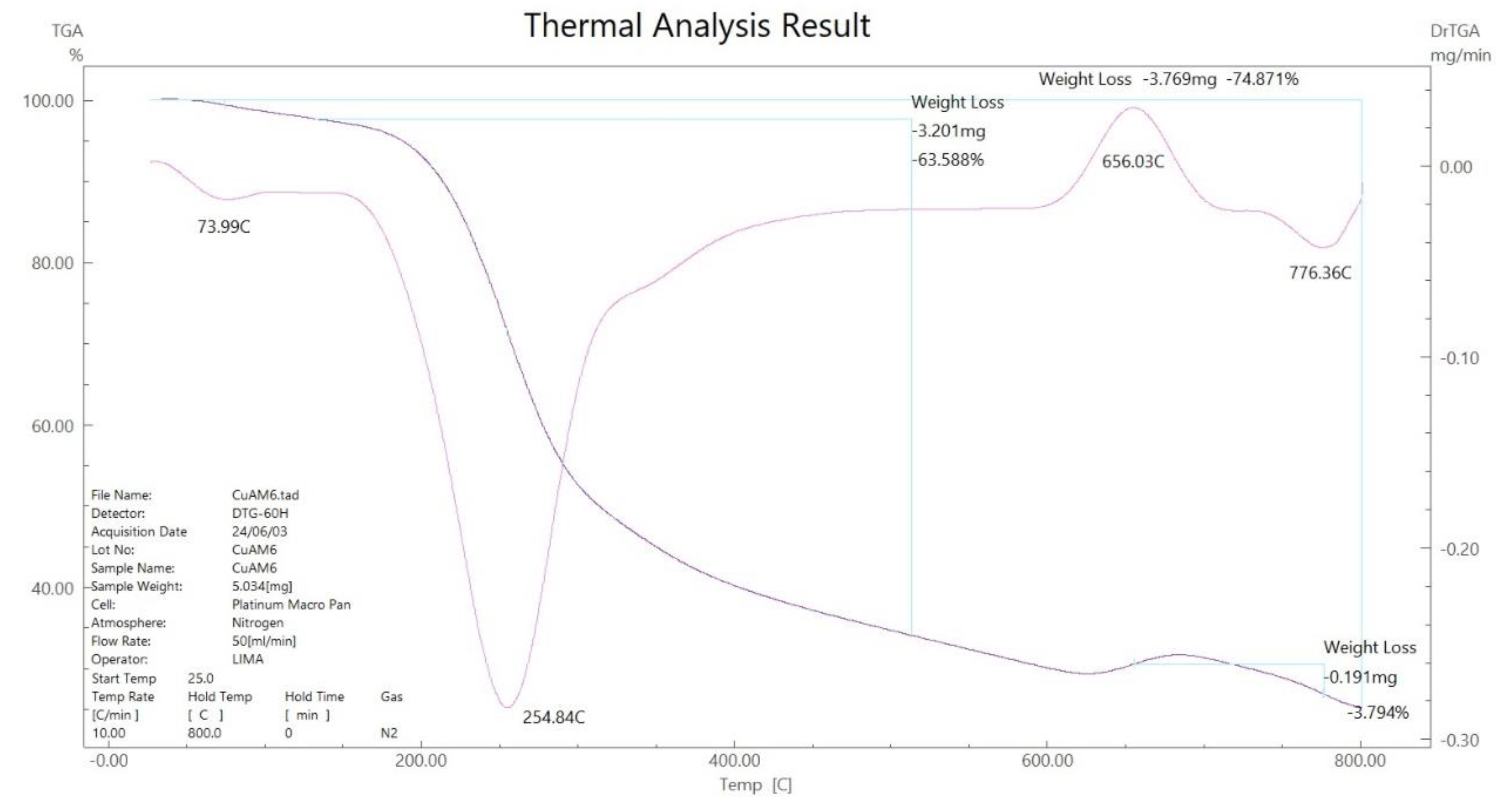
Figure 19.
TGA spectrum of Zn- ligand complex.
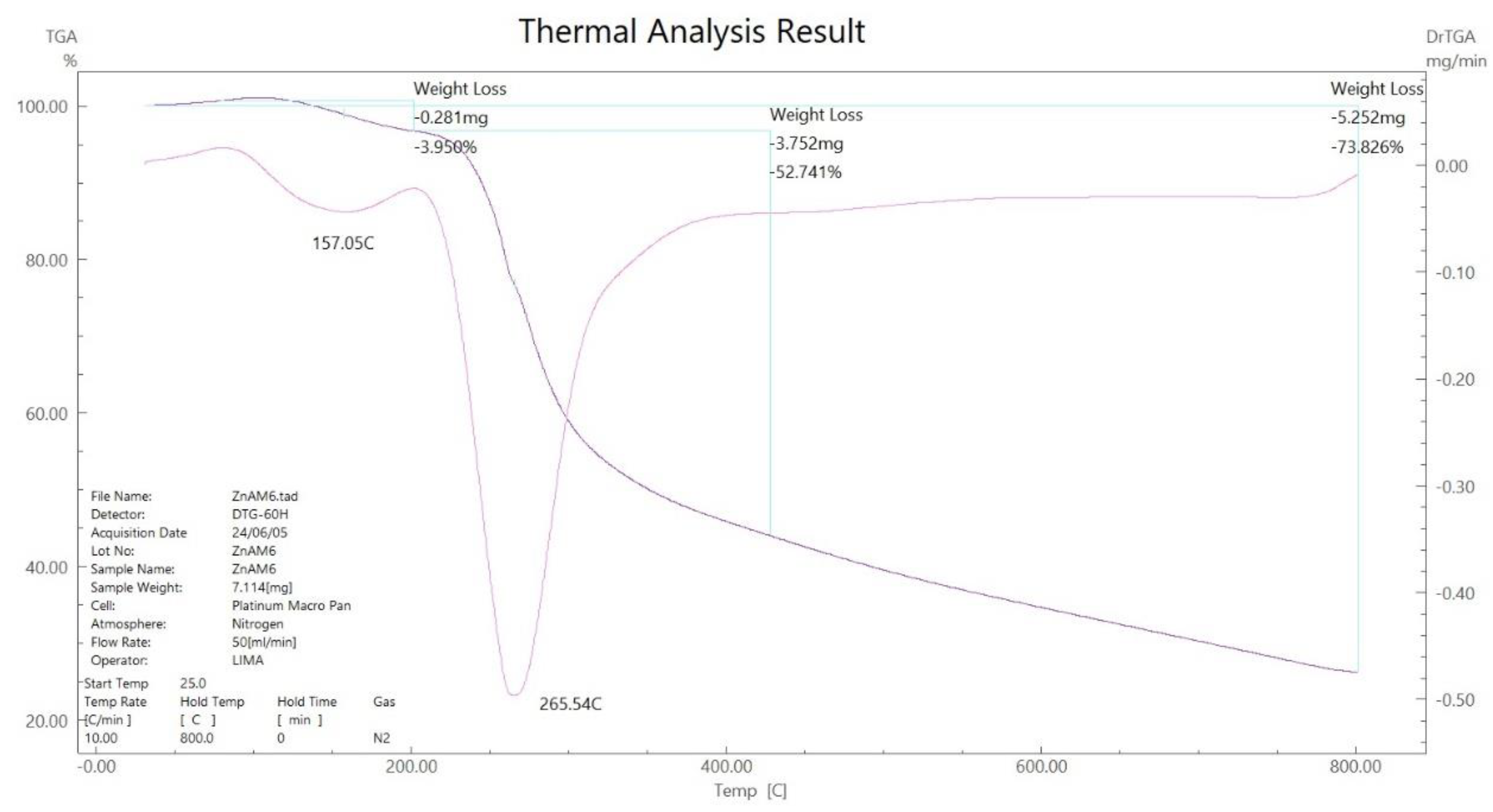
Figure 20.
TG thermogram of Co+2 complex.
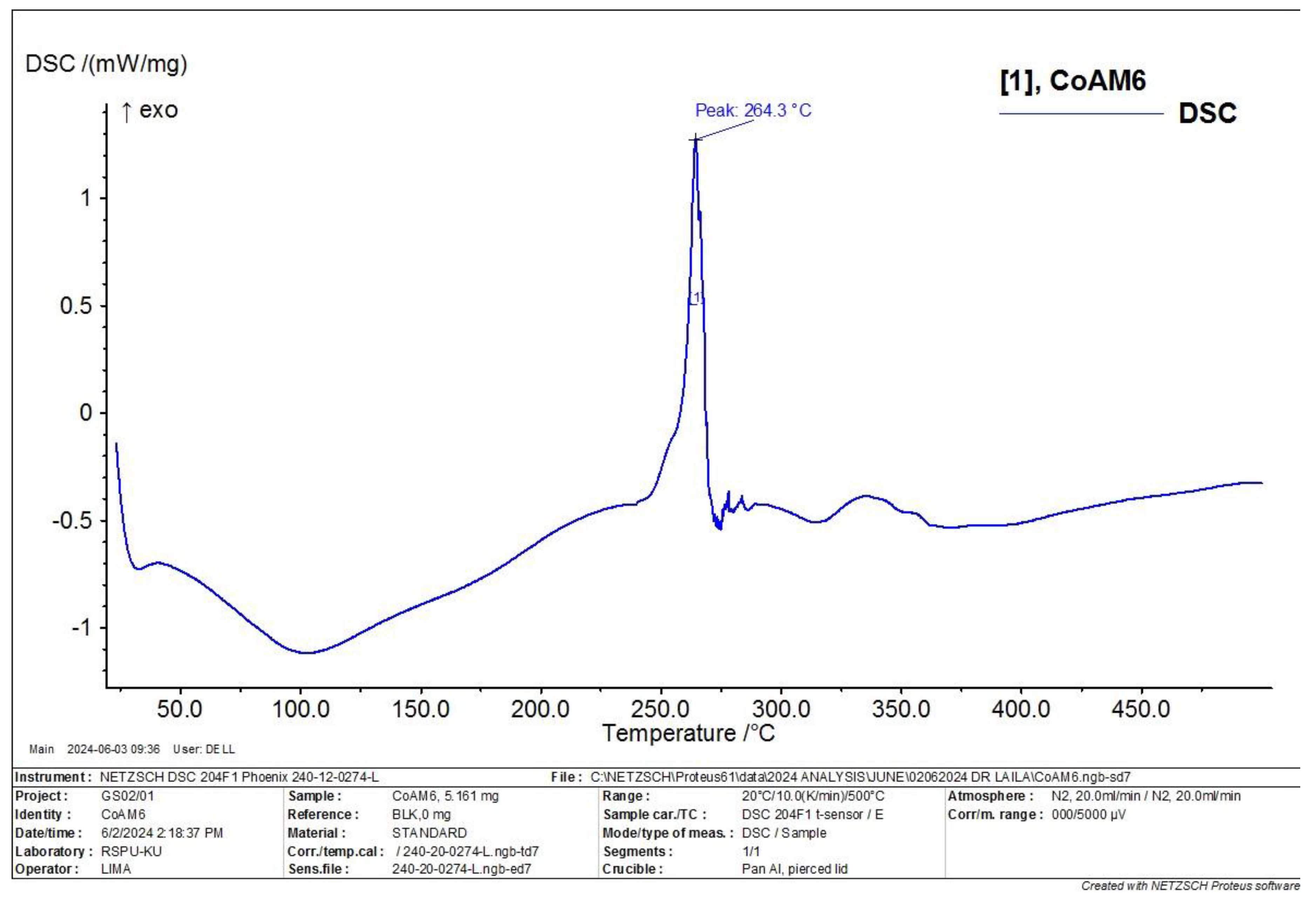
Figure 21.
TG thermogram of Cu+2 complex.
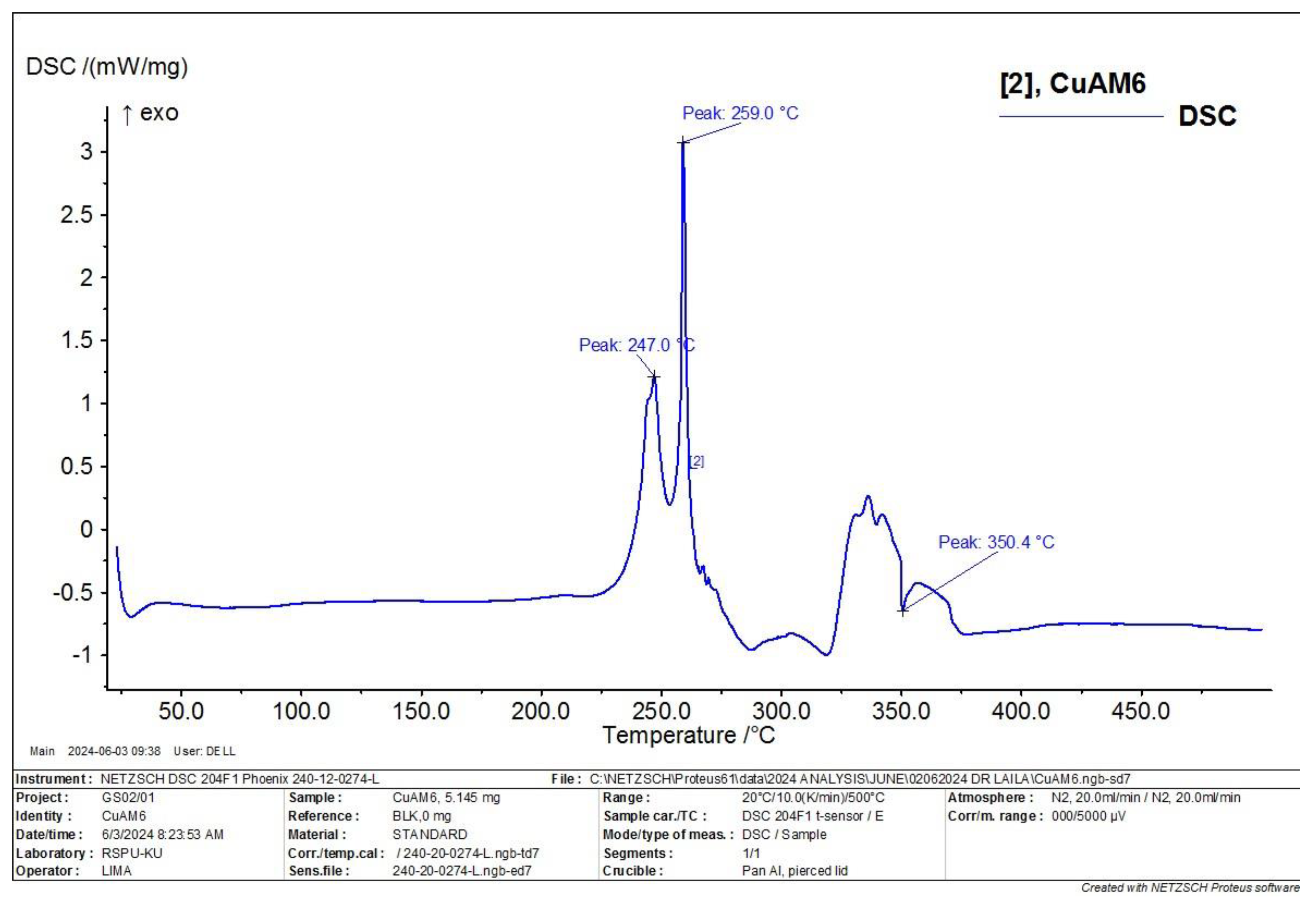
Figure 22.
TG thermogram of Zn+2 complex.
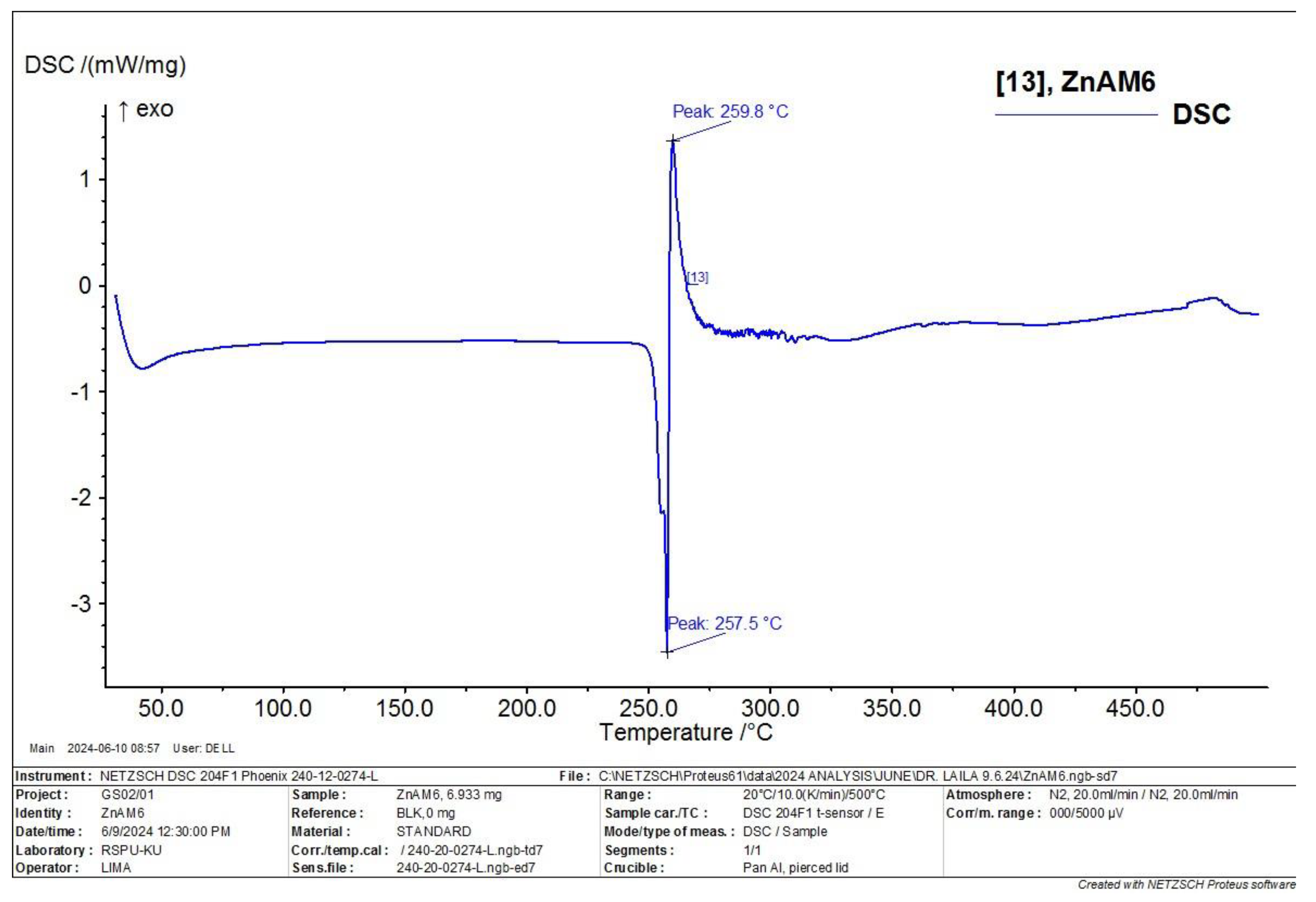
Figure 23.
The optimized molecular structure of Co, Cu and Zn ligand complexes as a ball and stick model.
Figure 23.
The optimized molecular structure of Co, Cu and Zn ligand complexes as a ball and stick model.
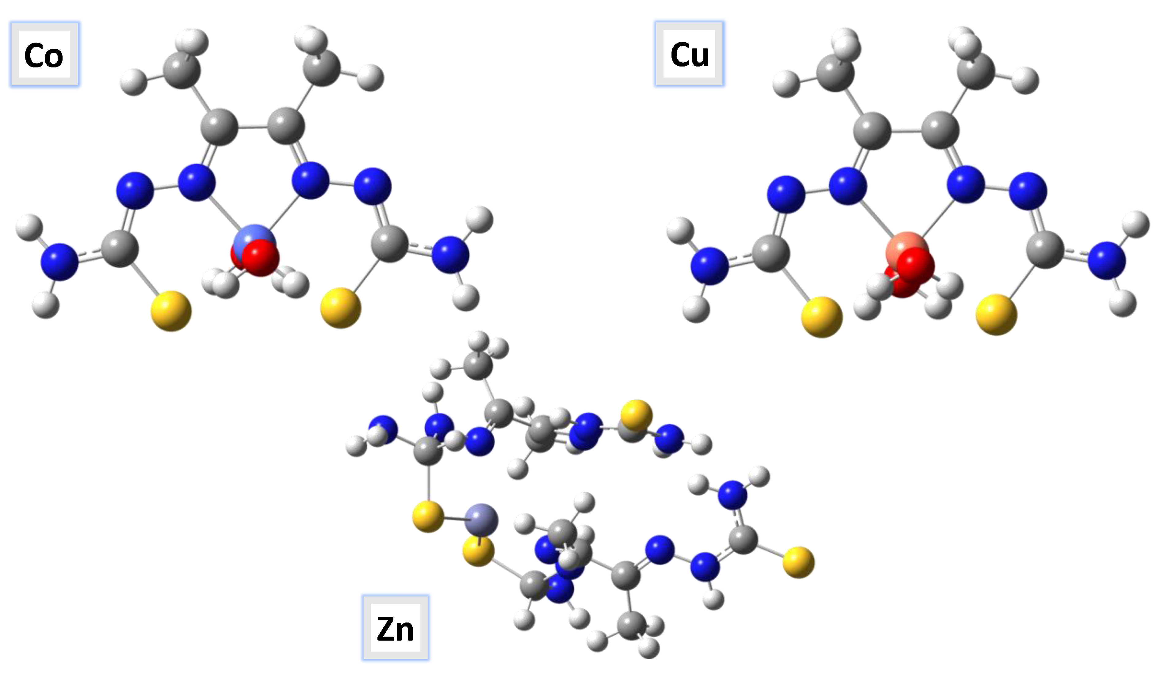
Figure 24.
Frontier molecular orbitals for Co, Cu and Zn ligand complexes calculated in gas phase at the B3LYP/GENECP method; energy level of HOMOs and LUMOs; and Egap (ΔE).
Figure 24.
Frontier molecular orbitals for Co, Cu and Zn ligand complexes calculated in gas phase at the B3LYP/GENECP method; energy level of HOMOs and LUMOs; and Egap (ΔE).
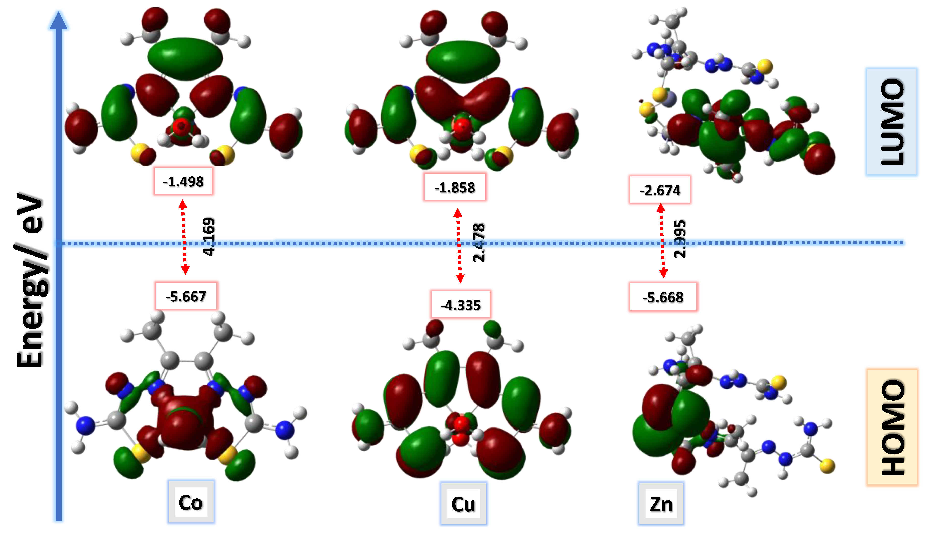
Figure 25.
ESP maps of Co, Cu and Zn ligand complexes by mapping the total density over the molecular electrostatic potential calculated in the gas phase.
Figure 25.
ESP maps of Co, Cu and Zn ligand complexes by mapping the total density over the molecular electrostatic potential calculated in the gas phase.
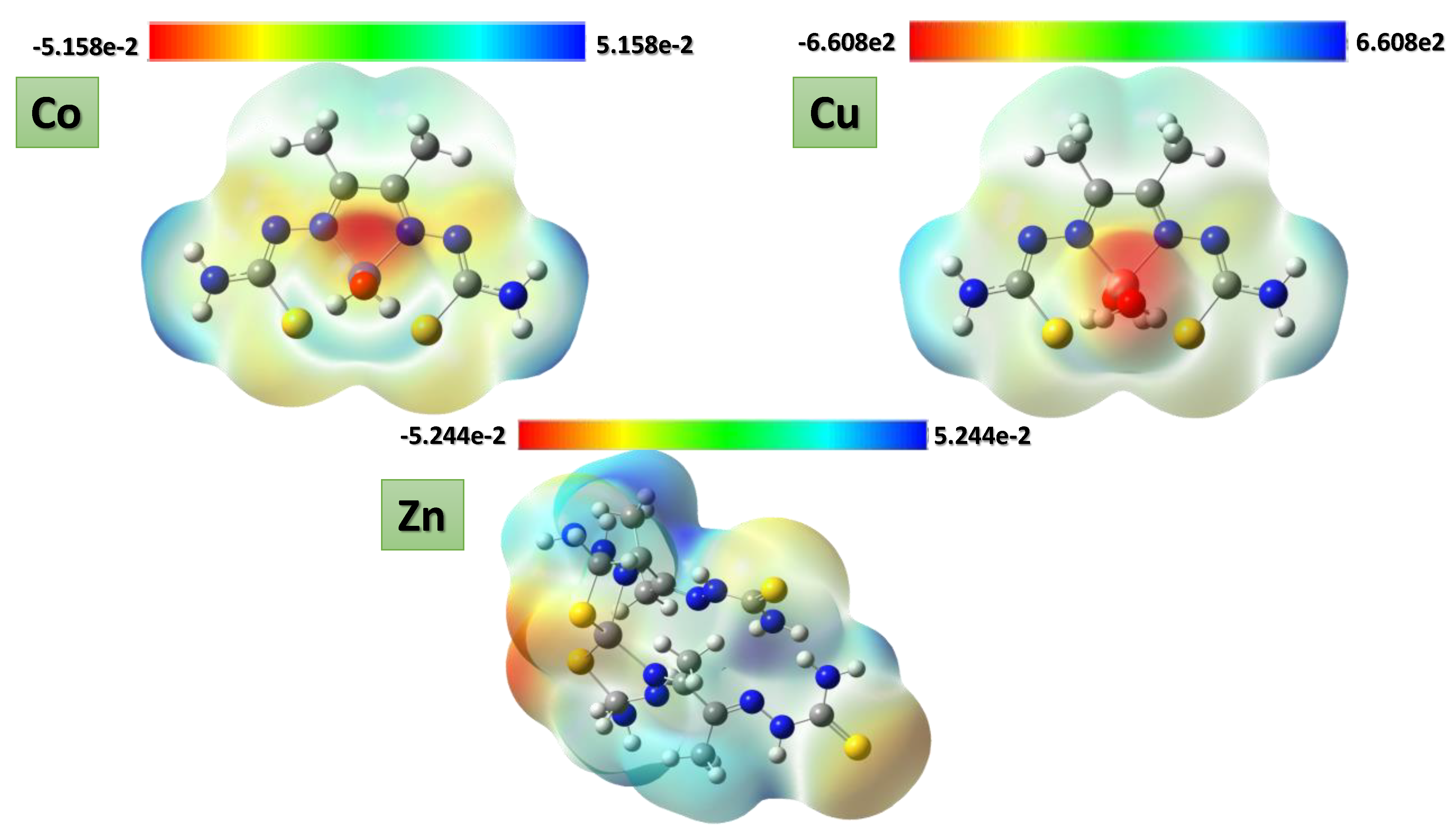
Table 1.
The analytical and physical characteristics for the ligand H2L and its metal complexes.
| Compounds | Color | M.Wt. | M.P. (oC) |
Λa | Calcd. (Found) (%) | ||||
|---|---|---|---|---|---|---|---|---|---|
| C | H | N | S | M | |||||
|
H2L (C6H12N6S2) |
Pale yellow | 232.32 | 225 | -- | 31.02 (31.04) |
5.21 (5.16) |
36.17 (35.07) |
27.60 (27.83) |
-- |
|
[Cu(L)].2H2O (C6H14CuN6O2S2) |
Dark brown | 329.89 (293.97)* |
263-270 | 4.34 | 21.84 (21.98) | 4.28 (3.80) | 25.48 (24.33) | 19.44 (18.95) | 20.31 (19.89) |
|
[Co(L)].2H2O (C6H14CoN6O2S2) |
Brown | 325.28 (288.97)* |
245-248 | 5.83 | 22.15 (22.22) | 4.34 (4.65) | 25.84 (24.70) | 19.72 (19.68) | 31.60 (31.28) |
| [Zn(H2L)2] (C12H26N12S4Zn) | Beige | 532.06 (481.28) |
268-272 | 21.52 | 27.09 (27.83) | 4.93 (4.95) | 31.59 (31.77) | 24.11 (25.38) | 19.26 (18.95) |
Λa is molar conductivity in Ω-1 cm2 mol-1. CHNS is average of three runs.
Table 2.
1H and 13C assignments of the ligand [H2L]and Zn+2 complex.
| Position | H2L | [Zn(H2L)2] | ||
|---|---|---|---|---|
| 1H (ppm) | 13C (ppm) | 1H (ppm) | 13C (ppm) | |
| 1 | -- | 148.37 | -- | 148.27 |
| 2 | -- | -- | ||
| 3 | 1.947 | 13.81 | 2.168 | 11.54 |
| 4 | -- | -- | -- | -- |
| 5 | -- | -- | -- | -- |
| 6 | 10.197,11.061 | -- | 10.208 | -- |
| 7 | -- | -- | ||
| 8 | -- | 183.12 | -- | 178.84 |
| 9 | -- | -- | ||
| 10 | 8.390,8.101 | -- | 7.850, 8.404 | -- |
| 12 | -- | -- | ||
| 13 | -- | -- | -- | -- |
| 14 | 1.947 | 11.55 | 2.508 | 11.54 |
Table 3.
IR spectral data for the H2L and its metal complexes and their assignments.
| Compounds | ν(H2O) ν(NH/NH2) |
ν(C=N) ν(C=N)* |
ν(C=S) ν(C-S) |
ν(N-N) | ν(M-N) | ν(M-S) |
| H2L (C6H12N6S2) |
3408,3248, 3194, 3149 |
1594 -- |
833 -- |
948 | -- | -- |
| [Cu(L)].2H2O (C6H14CuN6O2S2) |
3407, 3250, 3187, 3144 |
1595, 1648 |
-- 649 |
1005 | 603 | 485 |
| [Co(L)].2H2O (C6H14CoN6O2S2) |
3401, 3289 3142 |
1564, 1693 |
-- 949 |
1004 | 599 | 484 |
| [Zn(H2L)2] (C12H26N12S4Zn) |
3408, 3248, 3189, 3151 |
1594 -- |
831 -- |
948 | 602 | 483 |
Table 4.
Thermogravimetric data of thiosemicarbazone and its complexes.
|
Compounds |
Temp. (°C) | Weight loss (%) Found (calcd.) |
assignment |
|---|---|---|---|
|
H2L (C6H12N6S2) |
30-191 191-221 230-650 |
-- 7.38(6.90) 91.67 (93.10) |
Stable Elimination of NH2 Complete decomposition of the ligand |
|
[Cu(L)].2H2O (C6H14CuN6O2S2) |
73.99 254-776 800 |
11.28 (10.91) 63.58 (62.53) 25.13 (26.54) |
- 2H2O - C4H10N6S2 Cu + 2C (Residue) |
|
[Co(L)].2H2O (C6H14CoN6O2S2) |
70 269-761 800 |
08.51 (11.07) 64.19 (63.41) 27.22 (25.50) |
- 2H2O - C4H10N6S2 Co + 2C (Residue) |
| [Zn(H2L)2] (C12H26N12S4Zn) | 30-157 265-750 800 |
-- 73.83 (75.56) 26.17 (24.34) |
Stable - C12H26N12S2 Zn +2S (Residue) |
Table 5.
The SEM morphologies of metal complexes at different micrographs magnifications (5,10,50 and 100 µM).
Table 5.
The SEM morphologies of metal complexes at different micrographs magnifications (5,10,50 and 100 µM).
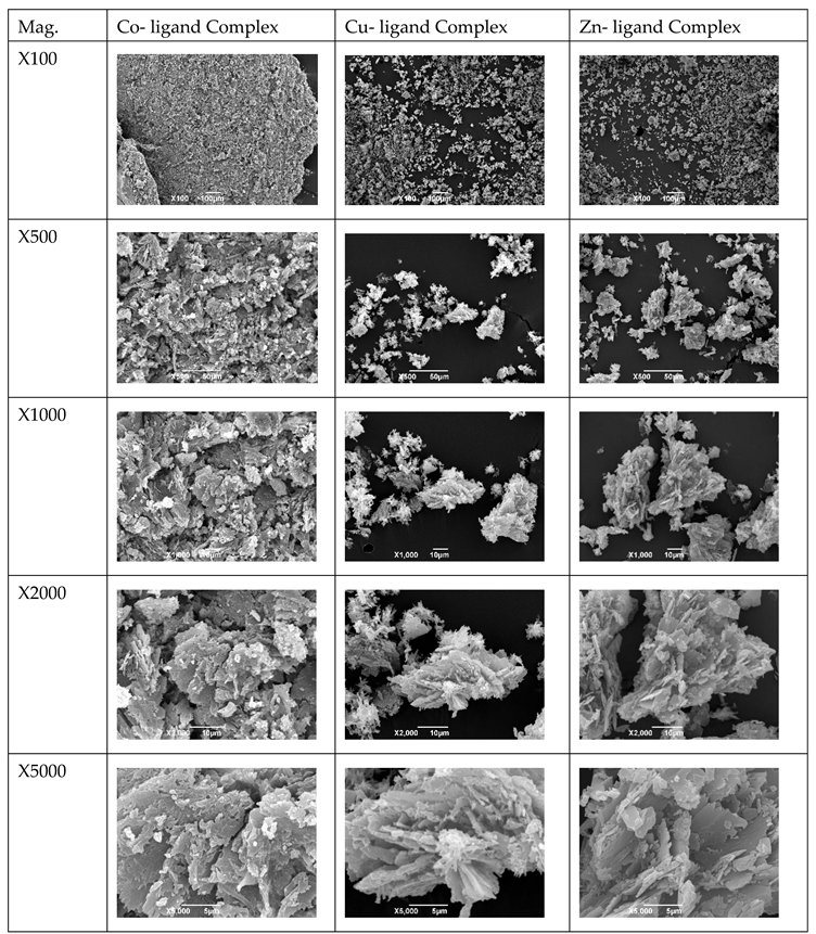 |
X: different SEM magnifications.
Table 6.
Particle size measurements of the synthesized metal ligand complexes.
| Metal complex |
Particle size ± standard deviation nm |
Graph |
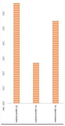 |
|
Co- ligand complex |
266.7±83.31 |
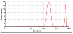 |
|
|
Cu- ligand complex |
226.9±75.18 |
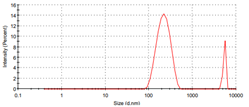 |
|
|
Zn- ligand complex |
254.9±68.87 |
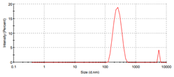 |
Table 7.
IC50 data of breast cancer cell lines treated with metal complexes.
| Parameter | Dilution used to treat the cells with the metal complexes/ µg | ||||
 | |||||
|
Co- ligand complex 1:1 |
 |
||||
| Drug conc µg | 0 | 2.5 | 5 | 10 | 20 |
| mean - blank | 100 | 69.2 | 68.4 | 63.6 | 48.1 |
| Standard deviation | 8.00 | 7.15 | 7.08 | 8.29 | 3.07 |
|
Cu-ligand complex 1:1 |
 |
||||
| Drug conc µg | 0 | 2.5 | 5 | 10 | 20 |
| mean - blank | 100 | 9.80 | 6.06 | 3.91 | 3.31 |
| Standard deviation | 7.97 | 1.58 | 0.34 | 0.44 | 0.34 |
|
Zn-ligand complex 2:1 |
 |
||||
| Drug conc µg | 0 | 2.5 | 5 | 10 | 20 |
| mean - blank | 100 | 67.1 | 55.7 | 45.2 | 32.1 |
| Standard deviation | 2.45 | 0.92 | 6.14 | 3.17 | 4.80 |
MDA-MB-453: breast cancer cell line with high androgen expression. X= (I + A)/2 (electronegativity), ɳ=(I-A)/2 (chemical hardness), S = 1/2ɳ (chemical softness) where I and A are ionization potential and electron affinity, and I= - EHOMO and A= - ELUMO, respectively.
Table 8.
Antibacterial activity of synthesized nano metal ligand complexes (5, 10 mg/ml) with the test organisms E. coli and Bacillus-subtills.
Table 8.
Antibacterial activity of synthesized nano metal ligand complexes (5, 10 mg/ml) with the test organisms E. coli and Bacillus-subtills.
| Strain/concentration | 5mg* | 10mg |
 * * |
|
Bacillus subtills (gram-positive) |
Inhibition zone mm |
||
| Co-L complex | 9 | 9.3 | |
| Cu-L complex | 12.3 | 14 | |
| Zn-L Complex | Nill | Nill | |
| Free ligand | Nill | Nill | |
| Strain/concentration | 5mg | 10mg | |
|
E. coli (gram-negative) |
inhibition zone mm |
||
| Co-L complex | Nill | Nill | |
| Cu-L complex | 5.6 | 6 | |
| Zn-L Complex | Nill | Nill | |
| Free ligand | Nill | Nill | |
 | |||
The zone inhibition numbers’ diameter in millimeters (mm) together with the standard deviation of three separate tests for each strain.
Disclaimer/Publisher’s Note: The statements, opinions and data contained in all publications are solely those of the individual author(s) and contributor(s) and not of MDPI and/or the editor(s). MDPI and/or the editor(s) disclaim responsibility for any injury to people or property resulting from any ideas, methods, instructions or products referred to in the content. |
© 2025 by the authors. Licensee MDPI, Basel, Switzerland. This article is an open access article distributed under the terms and conditions of the Creative Commons Attribution (CC BY) license (http://creativecommons.org/licenses/by/4.0/).
Copyright: This open access article is published under a Creative Commons CC BY 4.0 license, which permit the free download, distribution, and reuse, provided that the author and preprint are cited in any reuse.


