
Submitted:
08 May 2025
Posted:
08 May 2025
You are already at the latest version
Abstract
This study investigates the cytotoxic properties of metal complexes incorporating thio-uracil de-rivatives, specifically 2,4-dithiouracil and 6-propyl-2-thiouracil. The research focuses on the cyto-toxic effects of Cu(II) and Pd(II) complexes with 6-propyl-2-thiouracil, as well as mixed-ligand transition metal Cu(II) and Au(III) complexes of 2,4-dithiouracil with 2-thiouracil and uracil. Cy-totoxic activity was assessed against human cervical carcinoma cells (HeLa) and normal kidney cells from the African green monkey. The results demonstrated that incorporating Cu(II) and Au(III) into the compound structures significantly enhanced their cytotoxic effects. Notably, all tested complexes exhibited a stronger inhibitory effect on cancer cell proliferation compared to normal cells, with the gold(III) complex of 6-propyl-2-thiouracil showing the lowest CD50 value against the tumor cell line. These findings suggest that thio-uracil-based metal complexes, partic-ularly those containing gold(III), hold significant potential for further development as anticancer agents.
Keywords:
6-propyl-2-thiouracil
; 2
; 4-dithiouracil
; copper(II) complexes
; palladium(II) complexes
; gold(III) complexes
; cytotoxic properties
1. Introduction
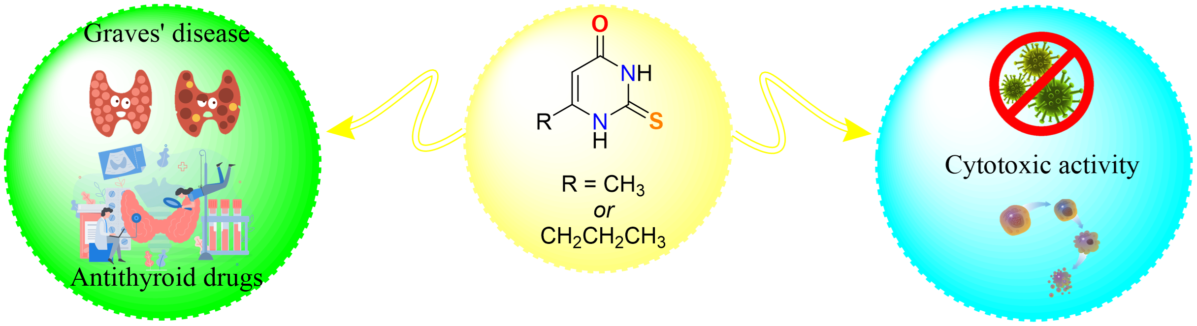
Thionamides, a category of relatively straightforward molecules, function as antithyroid medications, incorporating a sulfhydryl group and a thiourea unit within a heterocyclic structure. The interest in platinum and palladium compounds arises from their notable cytostatic properties. In Scheme 1 the different applications of bioactive pyrimidines, such as uracil, 2,4-dithiouracil, 6-propyl-2-thiouracil and other and their metal complexes is given.
Recently, cis-dihalogenated complexes of Pd(II) and Pt(II) were investigated in combination with 6-tert-butyl-2-thiouracil [1]. Vetter et al. described the synthesis, characterization, and in vitro study of the cytotoxic potential of platinum(IV) complexes with thiouracil-based ligands [2]. Ru(II)-centered complexes incorporating 2-thiouracil derivatives—specifically 6-methyl-2-thiouracil (6m2TU) and 2-thiouracil (2TU) —were tested against HepG2, K-562, HL-60 and B16-F10 cancer cells, as well as non-cancerous cells (PBMCs). The biological evaluation indicated that complex 2 demonstrated greater potential than complex 1. Ultimately, this investigation suggests that complexes 1 and 2 induce apoptosis, significantly increasing the proportion of apoptotic HL-60 cells by disrupting the cell cycle and decreasing the presence of cells in the G1/G0, G2/M, and S phases [3]. Correa et al. presented the chemical and cytotoxic investigations of four novel ruthenium(II) complexes incorporating uracil derivatives. All synthesized compounds are neutral, described by the formulas [Ru(PPh3)2(2TU)2] (1), [Ru(PPh3)2(6m2TU)2] (2), [Ru(dppb)(2TU)2] (3), and [Ru(dppb)(6m2TU)2] (4), where PPh3 = triphenylphosphine, dppb = 1,4-bis(diphenylphosphino)butane, 6m2TU = 6-methyl-2-thiouracil and 2TU = 2-thiouracil [4]. The coordination of 2-thiouracil derivatives with ruthenium enhances regions capable of forming hydrogen bonds with biological targets, such as DNA. The interaction between these complexes and DNA was investigated via UV-Vis spectrophotometric titration, yielding DNA-binding constant values in the range of 0.8–1.8 × 10⁴ M⁻¹. Furthermore, their interaction with bovine serum albumin (BSA) was explored [4]. In vitro studies tested their activity against HL-60 (human promyelocytic leukemia), HepG2 (human liver cancer), B16-F10 (murine melanoma) and K562 (human chronic myeloid leukemia) cells, along with non-cancerous PBMC (human peripheral blood mononuclear cells activated with concanavalin A—human lymphoblasts) [4]. Cytotoxicity tests revealed that complexes (2) and (4) exhibited biological activity against tumor cells comparable to oxaliplatin, the standard platinum-based drug, positioning them as promising candidates for new anticancer agents [4]. Bomfim et al. synthesized Ru(II) complexes incorporating 6-methyl-2-thiouracil, demonstrating potential as innovative antileukemic drug candidates [5]. Oladipo and Isola carried out a comprehensive review of uracil's coordination chemistry and the practical uses of several of its complexes [6]. 5-Substituted uracils are recognized as key structural components in various therapeutics [7]. Prachayasittikul et al. were synthesized a novel mixed-ligand transition metal (Cu, Ni, Mn) complexes of 5-iodouracil (5Iu) with 8-hydroxyquinoline (8HQ) (1-3) and 5-nitrouracil (5Nu) with 8HQ (4-6). These metal complexes showed notable cytotoxic effect against HuCCA-1, A-549, HepG2, and MOLT-3 cell lines [7]. Specifically, the cytotoxic activities of the tested complexes on HepG2 cells resulted in IC50 values lower than the standard drug. The copper complex of 5Nu (5Nu-Cu-8HQ, 5) proved to be the most effective cytotoxic agent, whereas the manganese complex of 5Iu (5Iu-Mn-8HQ, 1) exhibited the highest antioxidant activity [7]. These results underscore the potential of using simple and easily accessible bioactive ligands, such as 5Iu, 5Nu, and 8HQ, in the design and development of new lead compounds with notable and promising biological properties [7]. Al-Halbosy et al. presented the cytotoxic effect of the N-Phenylmorpholine-4-carbothioamide (HPMCT) and PdCl2(HPMCT)2] (2), and [PtCl2(HPMCT)2] (3) complexes. The compounds were assisted on breast cancer cell lines (MCF-7), and complex (3) reveals the most promising activity with an IC50 value 12.72 ± 0.4 μM [8]. Copper complexes exhibit cytotoxic activity through mechanisms distinct from those employed by cisplatin, a platinum-based chemotherapeutic agent currently in clinical use [9]. The biological efficacy of these copper compounds varies significantly depending on the nature of the coordinated ligands. Investigations into their cytotoxic potential are often grounded in the hypothesis that endogenous copper may exert lower toxicity on normal cells compared to malignant ones [10]. However, this assumption is nuanced; copper ions are redox-active and capable of competing with other metal ions for essential biological binding sites [10]. As an essential trace element, copper plays a critical role in numerous cellular processes and functions as a cofactor in various enzymatic catalytic reactions [10]. The involvement of copper in angiogenesis remains a matter of scientific debate, and the broader role of transition metals in this process continues to be actively explored [10]. Notably, copper complexes have been shown to mimic the activity of superoxide dismutase (SOD) [11], a key antioxidant enzyme that catalyzes the dismutation of superoxide radicals into less reactive molecular species. SOD enzymes are ubiquitous across nearly all forms of life [11]. Endogenous SOD is highly efficient in neutralizing reactive oxygen species (ROS) [11]; however, under conditions of oxidative stress—characterized by an imbalance between ROS production and antioxidant defense systems—the use of low molecular weight SOD mimetics with potent free radical-scavenging capabilities becomes imperative [11].
Low molecular weight copper complexes are of particular interest due to their effective mimicry of superoxide dismutase activity. Studies have reported that combinations of copper salts with dithiocarbamates (DTCs) and clioquinol (CQ) can spontaneously chelate intracellular copper, resulting in the formation of complexes that act as proteasome inhibitors and apoptosis inducers [10]. Recently, complex with general formulae [Cu(C20H22NO3)2]·H2O was obtained and cytotoxic properties was assessed [12]. The complex was studied for its interaction with calf thymus DNA and bovine serum albumin using spectroscopic methods. The results revealed that the binding mechanism is a static quenching process [12]. The in vitro cytotoxic investigation was conducted using MTT assay, and the result revealed that a metal complex exhibited enhanced cytotoxicity, high selectivity and dose-dependent cytotoxicity [12]. Four novel mononuclear Schiff base copper(II) complexes were obtained and characterized by X-ray analysis, which included [Cu(L)(OAc)]·H2O, [Cu(HL)(C2O4)(EtOH)]·EtOH, [Cu(L)(Bza)] and [Cu(L)(Sal)](HL=1-(2-(2-hydroxypropyl)(aminoethyl)(imino)(methyl)naphthalene-2-ol), Bza = benzoic acid and Sal = salicylic acid) [13]. Lian et al. were evaluated the antiproliferative effects of these complexes. The result showed that all complexes demonstrated good cytotoxicity against cancer cell lines [13]. The cytotoxic property of gold complexes has attracted attention recently. This may not be unconnected to the various level of challenges witnessed with the clinical use of platinum compounds. Gold(III) complexes are an emerging class of metal complexes with potential antitumor properties alternative to cis-DDP. This is mainly due to their outstanding cytotoxic properties exhibited through non-cisplatin antitumor mechanism. Zou et al. have described gold complexes in detail in a recent review [14]. The main objective of designing these drugs is to have a product that is very effective, less toxic and selectively binds to the active site of enzymes [14]. The potential of selectivity of gold(III) complexes to thiol-containing enzymes such as thioredoxin reductase (TrxR) makes it an attractive probe in designing compounds that can selectively bind to residues in the active site of the enzyme [14]. Many Аu(III) complexes were synthesized, and the anticancer effect was assisted against different cancer cell lines. In most cases, the ligands contained donor atom either Cl, Br, S or P. da Silva Maia et al. have also been synthesized a gold(III) complexes and their cytotoxic effect was studies [15]. Most of the cited gold(III) complexes have a profound effect on cisplatin-resistant cell lines. The cytotoxic properties of a complex with general formulae [(η-C5H5)2Ti{OC(O)CH2PPh2AuCl}2] was investigated in vitro against prostate and renal cell lines as potential chemotherapeutics drugs [16]. The result showed that the complex acts synergistically because the resulting cytotoxic effect is more pronounced when compared to [{HOC(O)RPPh2}AuCl] (R = −CH2− 6, −4-C6H4− 7) in renal cancer cell lines [16]. Metal complexes tend to associate with specific residues, many of which play key roles in the enzyme’s catalytic function. This interaction can disrupt cellular activities, ultimately leading to programmed cell death (apoptosis) [17]. Rana et al. investigated a gold(III) complexes who known to bind to TrxR [17]. In this enzyme, the catalytic residues are positioned between two subunits, each contributing to the gold(III) complex attachment. Additional binding regions have been found in the enzyme, but they exhibit weaker binding capacity. To date, numerous metal complexes have been developed utilizing uracil and thiouracil derivatives, incorporating various metals such as Cu, Fe, Co, Ni, Zn, Mn, Cd, and V [18,19,20], along with Pd, Pt, and Au, with analyses conducted on their composition and structural characteristics [21]. Novel thiolate gold(I) complexes, incorporating P(NMe₂)₃ (HMPT) as a phosphine ligand, were successfully synthesized [22], with two of these gold(I) thiolate complexes exhibiting potential as promising chemotherapy agents. Additionally, the cytotoxic effect of various metal complexes derived from thiouracil have been investigated against different cancer cell lines [22,23,24,25,26,27]. Recently, Marinova et al. successfully synthesized copper(II), palladium(II), and gold(III) complexes with 2-thiouracil [28], along with novel palladium(II) and copper(II) complexes containing 6-propyl-2-thiouracil and 6-methyl-2-thiouracil [29]. Additionally, Au(III) and Cu(II) complexes incorporating 2,4-dithiouracil (2,4-DTu) were developed [30], as well as an Au(III) complex with 6-methyl-2-thioxo-2,3-dihydropyrimidin-4(1H)-one [31]. Furthermore, the biological effect and synthesis of several metal complexes derived from 2-thiouracil and its derivatives were explored [32].
This article describes the cytotoxic properties of metal compounds with 2,4-dithiouracil and 6-propyl-2-thiouracil. In this study we provide details regarding the cytotoxic effect of Pd(II) and Cu(II) complexes with 6-propyl-2-thiouracil and Au(III) and Cu(II) complexes with 2,4-dithiouracil. The possible structures of the gold and copper complexes with 2,4-dithiouracil are given in Figure 1 and Figure 2 and Cu(II) and Pd(II) complexes with 6-propyl-2-thiouracil – Figure 3, respectively. All complexes that are the subject of this study have been previously published by us in two articles [29,30]. In our previous study, it was proposed that 2,4-dithiouracil might undergo desulfurization due to the action of NaOH employed during the synthesis of the gold complex. This process could lead to the substitution of one or both sulfur atoms with oxygen, thereby yielding 2-thiouracil and uracil as potential products within the reaction mixture [30].
2. Materials and Methods
2.1. Cytotoxicity Assay
To assess the in vitro biocompatibility of the tested compounds, a series of cell viability assays were conducted on human cervical carcinoma cell lines (HeLa), as well as normal Vero cells (kidney cells from the African green monkey). All cell lines were obtained from the German Collection of Microorganisms and Cell Cultures (DSMZ GmbH, Braunschweig, Germany). The cell cultures were maintained in DMEM (Dulbecco's Modified Eagle Medium) (Gibco Thermofisher Scientific, NY, USA) growth medium, supplemented with 10% fetal bovine serum (FBS, Sigma-Aldrich, Darmstadt, Germany) and 5% L-glutamine (Sigma-Aldrich, Darmstadt, Germany), and incubated under controlled conditions of 37 °C in a humidified atmosphere with 5% CO₂.
Cell Viability Assessment
The experimental approach included a series of cytotoxicity assays to determine the extent of cell proliferation inhibition by the synthesized complexes and their free ligands. Cell viability was quantified using a standard MTT-based colorimetric assay. Exponentially growing cells were harvested and seeded (100 μL per well) into 96-well plates at an appropriate density—3 × 10⁵ for HeLa and Vero cells. The cells were exposed to various concentrations of the tested compounds, ranging from 0.0001 to 10 mg/mL, and incubated for 72 hours.
Following the incubation period, a filter-sterilized MTT substrate solution (5 mg/mL in PBS, (Sigma-Aldrich, Darmstadt, Germany),) was introduced into each well. A subsequent incubation of 1–4 hours facilitated the formation of insoluble purple formazan crystals, which were then dissolved in an isopropanol solution containing 5% formic acid. Absorbance measurements were taken at 550 nm using a microplate reader (Labexim LMR-1). The absorbance values were corrected against MTT and isopropanol controls and normalized to the mean value of the untreated control (100% cell viability). Semi-logarithmic dose–response curves were generated, and the cytotoxic dose CD₅₀ (causing a 50 % reduction of cell viability) for each tested compound against the respective cell lines was determined. Statistical significance was established at p ≤ 0.05.
3. Results and Discussion
Two thiouracil derivatives and their complexes with Au(III), Pd(II) and Cu(II) were tested for cytotoxicity against two cell lines – a normal cell line of kidney cells from African green monkey and a tumor cell line of human cervical carcinoma. The concentration-response curves for 2,4-dithiouracil and its metal complexes against the normal cell line are presented on Figure 4. The results show that the tested compounds reduced the proliferation of the treated cells in a concentration-dependent manner, but the addition of Cu(II) and Au(III) improves cell viability at the higher tested concentrations. A similar tendency was observed in the treatment of the tumor cell line (human cervical carcinoma) (Figure 5) with the same compounds.
The CD50 values of the 2,4-dithiouracil ligand and its metal complexes were determined through MTT testing and are presented in Table 1. The hierarchal order of cytotoxicity against the normal cell line revealed that the addition of AuIII) increased the cytotoxic activity of the ligand and the addition of Cu(II) to the complex lead to an even lower CD50. The tumor cell line was also less sensitive to 2,4-dithiouracil in comparison with its complexes, the Au(III) complex being the stronger cytotoxic agent. Other authors also report an improvement of cytotoxicity after the addition of copper and gold ions to ligands [28,37]. The comparison between the normal and tumor line sensitivity towards 2,4-dithiouracil and its metal complexes show much higher cytotoxicity against human cervical carcinoma cells, which is a good prerequisite for possible chemotherapy applications.
Similar results were obtained in the investigation of the cytotoxic effect of 6-propyl-2-thiouracil and its complexes with copper(II) and paladium(II). The concentration-response curves depicted on Figure 6 reveal that the cell viability of the normal cell line is also concentration-dependent and the metal complexes are cytotoxic in lower concentration in comparison with the ligand. In the experiments with tumor cells and the same compounds the cell viability was affected by even lower test concentrations (Figure 7).
The determined CD50 values of 6-propyl-2-thiouracil and its complexes were lower than those of 2,4-DTu and its Pd(II) and Cu(II) compounds. The highest cytotoxic concentration against the normal cell line was detected for 6-Pro-2Tu, followed by its Cu(II) and Pd(II) complex (Table 2). Similarly, the most potent compound preventing the proliferation of the tumor cells was the Pd(II) complex of 6-propyl-2-thiouracil, followed by the Cu(II) complex and the ligand. This second set of compounds also showed better effect on tumor cells in comparison with the normal cells.
Cisplatin is a widely used chemotherapeutic agent, and its cytotoxic effects on HeLa cell lines have been extensively studied [33,34,35,36,37,38]. On average a concentration of 0,003 mg/mL is reported to lead to a significant loss of cell viability in HeLa cell lines. Becit et al. reported that for complete inhibition of the viability of a human cervical carcinoma cell line was needed a concentration of cisplatin higher than 1,5 mg/mL [39]. Another study reported that 0,15 mg/mL of cisplatin caused about 10% reduction on cell viability in the same cell line after 24 h. [40]. In comparison, the compaound included in this study inhibit completely the proliferation of the tumor line at concentrations between 0,001 and 0,1 mg/mL. Another platinum-based chemotherapeutic agent, exhibits cytotoxic effects on HeLa cells is carboplatin. Various potency is reported across different studies. Aborehab and Osama [41] investigated the effect of combining gallic acid with carboplatin on HeLa cells, employing concentrations of carboplatin ranging from 0,00781 mg/mL to 1 mg/mL. The application of 1 mg/mL yielded complete inhibition of cell viability, which is a significantly higher concentration than the ones reported in this study.
4. Conclusions
The conducted study on 2-thiouracil derivatives and their metal complexes showed that the inclusion of Cu(II), Pd(II) and Au(III) in the compounds structure improved significantly their cytotoxicity. All tested compounds exhibited higher capability to reduce the proliferation of human cervical carcinoma cells in comparison with normal kidney cells from African green monkey. The lowest CD50 was detected for the complex of 6-propyl-2-thiouracil with Pd(II) against the tumor cell line. These findings suggest that the coordination of metal ions, especially Cu(II), Au(III) and Pd(II) to 2-thiouracil derivatives plays a significant role in the improvement of their cytotoxicity towards tumor cells while maintaining lower toxicity towards normal cells. This makes the promising candidates for further investigation as anticancer agents.
Author Contributions
Conceptualization, P.M. and D.B.; methodology, P.M. and P. G.-K.; formal analysis, P. G.-K..; investigation, P.M.; D.B.; P. G.-K.; A.S.; resources, P. G.-K.; data curation, P.M.; writing—original draft preparation, P.M; D. B.; A.S.; writing—review and editing, P.M.; D. B.; A.S.; supervision, P.M.; project administration, funding acquisition, P.M.
Conflicts of Interest
The authors declare no conflict of interest.
References
- Golubyatnikova L., G.; Khisamutdinov R., А.; Grabovskii S., А.; Kabal’nova N., N.; Murinov Yu., I. Complexes of Palladium(II) and Platinum(II) with 6-tert-Butyl-2-thiouracil. Russ. J. Gen.Chem. 2017, 87, 117–121. [Google Scholar] [CrossRef]
- Vetter, C.; Kaluđerović, G. N.; Paschke, R.; Kluge, R.; Schmidt, J.; Steinborn, D. Synthesis, characterization and in vitro cytotoxicity studies of platinum(IV) complexes with thiouracil ligands, Inorg. Chim. Acta 2010, 363, 2452–2460. [Google Scholar] [CrossRef]
- Diogo E. L. Carvalho, Katia M. Oliveira, Larissa M. Bomfim, Milena B. P. Soares, Daniel P. Bezerra, Alzir A. Batista, and Rodrigo S. Correa, Nucleobase Derivatives as Building Blocks to Form Ru(II)-BasedComplexes with High Cytotoxicity ACS Omega 2020, 5, 122-130. [CrossRef]
- Rodrigo S.. Correa, Larissa M. Bomfim , Katia M. Oliveira , Diogo R.M. Moreira , Milena B.P. Soares , Javier Ellena , Daniel P. Bezerra, Alzir A. Batista. Ru(II) complexes containing uracil nucleobase analogs with cytotoxicity against tumor cells. Journal of Inorganic Biochemistry 2019, 198, 110751. [CrossRef]
- Bomfim L.M.; de Araujo F. A.; Dias R. B.; Sales C. B. S.; Gurgel Rocha C.A.; Correa R. S.; Soares M. B. P.; Batista A. A.; Bezerra D. P. Ruthenium(II) complexes with 6-methyl-2-thiouracil selectively reduce cell proliferation, cause DNA double-strand break and trigger caspase-mediated apoptosis through JNK/p38 pathways in human acute promyelocytic leukemia cells. Sci. Rep. 2019, 9, 11483.
- Oladipo, M. A.; Isola, K. T., Coordination Possibility of Uracil and Applications of Some of Its Complexes: A Review — Res. J. Pharm., Biol.Chem. Sci. 2013, 4, 386-394. [CrossRef]
- Prachayasittikul, S.; Worachartcheewan, A.; Pingaew, R.; Suksrichavalit, T.; Isarankura-Na-Ayudhya, C.; Ruchirawat, S.; Prachayasittikul, V. Metal Complexes of Uracil Derivatives with Cytotoxicity and Superoxide Scavenging Activity, Letters in Drug Design & Discovery 2012, 9, 282–287. [CrossRef]
- Al-Halbosy, A.T.F.; Hamada, A.A.; Faihan, A.S.; Saleh, A.M.; Yousef, T.A.; Abou-Krisha, M.M.; Alhalafi, M.H.; Al-Janabi, A.S.M. Thiourea Derivative Metal Complexes: Spectroscopic, Anti-Microbial Evaluation, ADMET, Toxicity, and Molecular Docking Studies. Inorganics 2023, 11, 390. [Google Scholar] [CrossRef]
- Palma, G.; D’Aiuto, M.; Rea, D.; Bimonte, S.; Lappano, R.; Sinicropi, M. S.; Maggiolini, M.; Longo, P.; Arra, C.; Saturnino1, C. Organo-metallic compounds: novel molecules in cancer therapy. Biochem Pharmacol Open Access. 2014, 13, 1603–1615. [Google Scholar] [CrossRef]
- Santini, C.; Pellei, M.; Gandin, V.; Porchia, M.; Tisato, F.; Marzano, C. Advances in copper complexes as anticancer agents. Chem Rev. 2014, 114, 815–862. [Google Scholar] [CrossRef]
- Khalid, H.; Hanif, M.; Hashmi, M.A.; Mahmood, T.; Ayub, K.; Monim-Ul-Mehboob, M. Copper complexes of bioactive ligands with superoxide dismutase activity. Mini Rev Med Chem. 2013, 13, 1944–1956. [Google Scholar] [CrossRef] [PubMed]
- Shokohi-Pour, Z.; Chiniforoshan, H.; Momtazi-Borojeni, A.A.; Notash, B. A novel Schiff base derived from the gabapentin drug and copper (II) complex: synthesis, characterization, interaction with DNA/protein and cytotoxic activity. J Photochem Photobiol B. 2015, 162, 34–44. [Google Scholar] [CrossRef]
- Lian, W-J.; Wang, X-T.; Xie, C-Z.; He Tian, Xue-Qing Song, He-Ting Pan, Xin Qiao, Jing-Yuan Xu. Mixed-ligand copper Schiff base complexes: the role of the co-ligand in DNA binding, DNA cleavage, protein binding and cytotoxicity. Dalton Trans. 2016, 45, 9073–9087. [CrossRef]
- Zou, T.; Ching, A.; Lum, T.; Lok, C-N.; Zhang, J-J.; Che, C-M. Chemical biology of anticancer gold(III) and gold(I) complexes. Chem Soc Rev. 2015, 44, 8786–8801. [CrossRef]
- da Silva Maia, P.I.; Deflon, V.M.; Abram, U. Gold(III) complexes in medicinal chemistry. Future Med Chem. 2014, 6, 1515–1536. [Google Scholar] [CrossRef]
- Ferna, J.; Elie, B.T.; Sulzmaier, F.J.; Sanau, M.; Ramos, J.W.; Contel, M. Organometallic titanocene-gold compounds as potential chemotherapeutics in renal cancer. Study of their protein kinase inhibitory properties. Organometallics. 2014, 33, 6669–6681. [Google Scholar] [CrossRef]
- Bidyut Kumar Rana, Abhishek Nandy, Valerio Bertolasi, Christopher W. Bielawski, Krishna Das Saha, Joydev Dinda Novel gold(I) − and gold(III) − N-heterocyclic carbene complexes: synthesis and evaluation of their anticancer properties. Organometallics 2014, 33, 2544–2548. [CrossRef]
- Abou-Melha K. S. Elaborated studies for the ligitional behavior of thiouracil derivative towards Ni(II), Pd(II), Pt(IV), Cu(II) and UO22 2 ions. Spectrochim. Acta Part (A): Molecular and Biomolecular Spectroscopy 2012, 97, 6-16.
- Masoud M.S.; Amira M.F.; Ramadan A.M.; El- Ashry G.M. Synthesis and characterization of some pyrimidine, purine, amino acid and mixed ligand complexes. Spectrochim. Acta Part(A) 2008, 69, 230-238.
- Singh U. P.; Ghose R.; Ghose A. K.; Sodhi A.; Singh S. M.; Singh R. K. J Inorg Biochem 1989, 37, 325-329.
- El-Morsy F.A.; Jean-Claude B.J.; Butler I.S.; El- Sayed S.A.; Mostafa S.I. Synthesis, characterization and anticancer activity of new zinc(II), molybdate(II), palladium(II), silver(I), rhodium(III), ruthenium(II) and platinum(II) complexes of 5,6-diamino-4-hydroxy2-mercaptopyrimidine. Inorg. Chim. Acta 2014, 423, 144-155.
- Abás, E.; Pena-Martínez, R.; Aguirre-Ramírez, D.; Rodríguez-Diéguez, A.; Laguna, M.; Grasa, L. New selective thiolate gold(I) complexes inhibit proliferation of different human cancer cells and induce apoptosis in primary cultures of mouse colon tumors. Dalton Trans. 2020, 49, 1915–1927. [Google Scholar] [CrossRef]
- Singh U. P.; Singh S.; Singh S. M. Synthesis, characterization and antitumour activity of metal complexes of 5-carboxy-2-thiouraci. Metal-Based Drugs, 1998, 5, p. 35-39. [CrossRef]
- Papazoglou, I.; Cox, P.J.; Hatzidimitriou, A.G.; Kokotidou, C.; Choli-Papadopoulou, T.; Aslanidis, P. Copper(I) halide complexes of 5-carbethoxy-2-thiouracil: Synthesis, structure and in vitro cytotoxicity. Eur. J. Med. Chem. 2014, 78, 383. [Google Scholar] [CrossRef] [PubMed]
- Hoeschele J., D.; Piscataway, N.J. Ethylenediamineplatinum(II) 2,4-dioxopyrimidine complexes, US Patent 4 207 416, 1980. [Google Scholar]
- Supaluk, P.; Apilak, W.; Ratchanok, P.; Thummaruk, S.; Chartchalerm, I.; Somsak, R.; Virapong, P. Metal Complexes of Uracil Derivatives with Cytotoxicity and Superoxide Scavenging Activity. Lett. Drug Des. Discov. 2012, 9, 282–287. [Google Scholar] [CrossRef]
- Illán-Cabeza N. A.; García-García A. R.; Moreno-Carretero M. N.; Martínez-Martos J. M.; Ramírez-Expósito M. J. Synthesis, characterization and antiproliferative behavior of tricarbonyl complexes of rhenium(I) with some 6-amino-5-nitrosouracil derivatives: Crystal structure of fac-[ReCl(CO)3(DANU-N5,O4 )] (DANU = 6-amino-1,3-dimethyl-5-nitrosouracil). J. Inorg. Biochem. 2005, 99, 1637-1645.
- Marinova, P.; Tsoneva, S.; Frenkeva, M.; Blazheva, D.; Slavchev, A.; Penchev, P. New Cu(II), Pd(II) and Au(III) complexes with 2-thiouracil: Synthesis, Characteration and Antibacterial Studies, Russ. J. Gen. Chem. 2022, 92, 1578–1584. [Google Scholar] [CrossRef]
- Marinova, P.; Hristov,M.; Tsoneva, S.; Burdzhiev,N.; Blazheva, D.; Slavchev, A.; Varbanova, E.; Penchev, P. Synthesis, Characterization, and Antibacterial Studies of New Cu(II) and Pd(II) Complexes with 6-Methyl-2-Thiouracil and 6-Propyl-2-Thiouracil. Appl. Sci. 2023, 13, 13150-13168. [CrossRef]
- Marinova, P.; Stoitsov, D.; Burdzhiev, N.; Tsoneva, S.; Blazheva, D.; Slavchev, A.; Varbanova, E.; Penchev, P. Investigation of the Complexation Activity of 2,4-Dithiouracil with Au(III) and Cu(II) and Biological Activity of the Newly Formed Complexes. Appl. Sci. 2024, 14, 6601. [Google Scholar] [CrossRef]
- Marinova, P.; Burdzhiev, N.; Blazheva, D.; Slavchev, A. Synthesis and Antibacterial Studies of a New Au(III) Complex with 6-Methyl-2-Thioxo-2,3-Dihydropyrimidin-4(1H)-One. Molbank 2024, 2024, M1827. [Google Scholar] [CrossRef]
- Marinova, P.E.; Tamahkyarova, K.D. Synthesis and Biological Activities of Some Metal Complexes of 2-Thiouracil and Its Derivatives: A Review. Compounds 2024, 4, 186–213. [Google Scholar] [CrossRef]
- Qi, Y.Y.; Gan, Q.; Liu, Y.X.; Xiong, Y.H.; Mao, Z.W.; Le, X.Y. Two new Cu(II) dipeptide complexes based on 5-methyl-2-(2’-pyridyl)benzimidazole as potential antimicrobial and anticancer drugs: Special exploration of their possible anticancer mechanism. Eur. J. Med. Chem. 2018, 154, 220–232. [CrossRef]
- Reddy, T.S.; Privér, S.H.; Mirzadeh, N.; Bhargava, S.K. Synthesis of gold(I) phosphine complexes containing the 2-BrC6F4PPh2 ligand: Evaluation of anticancer activity in 2D and 3D spheroidal models of HeLa cancer cells. Eur. J. Med. Chem. 2018, 145, 291–301. [Google Scholar] [CrossRef]
- Fei, B.L.; Tu, S.; Wei, Z.; Wang, P.; Qiao, C.; Chen, Z.F. Optically pure chiral copper(II) complexes of rosin derivative as attractive anticancer agents with potential anti-metastatic and anti-angiogenic activities. Eur. J. Med. Chem. 2019, 176, 175–186. [CrossRef]
- Khan, T.M.; Gul, N.S.; Lu, X.; Wei, J.H.; Liu, Y.C.; Sun, H.; Liang, H.; Orvig, C.; Chen, Z.F. In vitro and in vivo anti-tumor activity of two gold(III) complexes with isoquinoline derivatives as ligands. Eur. J. Med. Chem. 2019, 163, 333–343. [Google Scholar] [CrossRef] [PubMed]
- Pérez-Villanueva, J.; Matadamas-Martínez, F.; Yépez-Mulia, L.; Pérez-Koldenkova, V.; Leyte-Lugo, M.; Rodríguez-Villar, K.; Cortés-Benítez, F.; Macías-Jiménez, A.P.; González-Sánchez, I.; Romero-Velásquez, A.; et al. Synthesis and Cytotoxic Activity of Combretastatin A-4 and 2,3-Diphenyl-2H-indazole Hybrids. Pharmaceuticals 2021, 14, 815. [Google Scholar] [CrossRef]
- Zeng, Z.-F.; Huang, Q.-P.; Cai, J.-H.; Zheng, G.-J.; Huang, Q.-C.; Liu, Z.-L.; Chen, Z.-L.; Wei, Y.-H. Synthesis, Characterization, DNA/HSA Interactions, and Anticancer Activity of Two Novel Copper(II) Complexes with 4-Chloro-3-Nitrobenzoic Acid Ligand. Molecules 2021, 26, 4028. [Google Scholar] [CrossRef]
- Becit, M.; Aydın Dilsiz, S.; Başaran, N. Interaction of curcumin on cisplatin cytotoxicity in HeLa and HepG2 carcinoma cells. Istanbul J.Pharm. 2020, 50, 202–210. [Google Scholar] [CrossRef]
- Ganesan, B. S.; Prabhakaran, P. Effect of HeLa Cell Density Towards Cisplatin Treatment. Proc. Sci. Math. 2022, 12, 58-65. https://science.utm.my/procscimath/wp-content/uploads/sites/605/2022/11/07_Barthi-S-Ganesen_58-65new-1.pdf.
- Aborehab, N.M.; Osama, N. Effect of Gallic acid in potentiating chemotherapeutic effect of Paclitaxel in HeLa cervical cancer cells. Cancer Cell Int. 2019, 19, 154. [Google Scholar] [CrossRef] [PubMed]
Scheme 1.
Different applications of uracil, 2-thiouracil, 2,4-dithiouracil, 6-metyl-2-thiouracil, 6-propyl-2-thiouracil, 5-fluorouracil and their metal complexes.
Scheme 1.
Different applications of uracil, 2-thiouracil, 2,4-dithiouracil, 6-metyl-2-thiouracil, 6-propyl-2-thiouracil, 5-fluorouracil and their metal complexes.
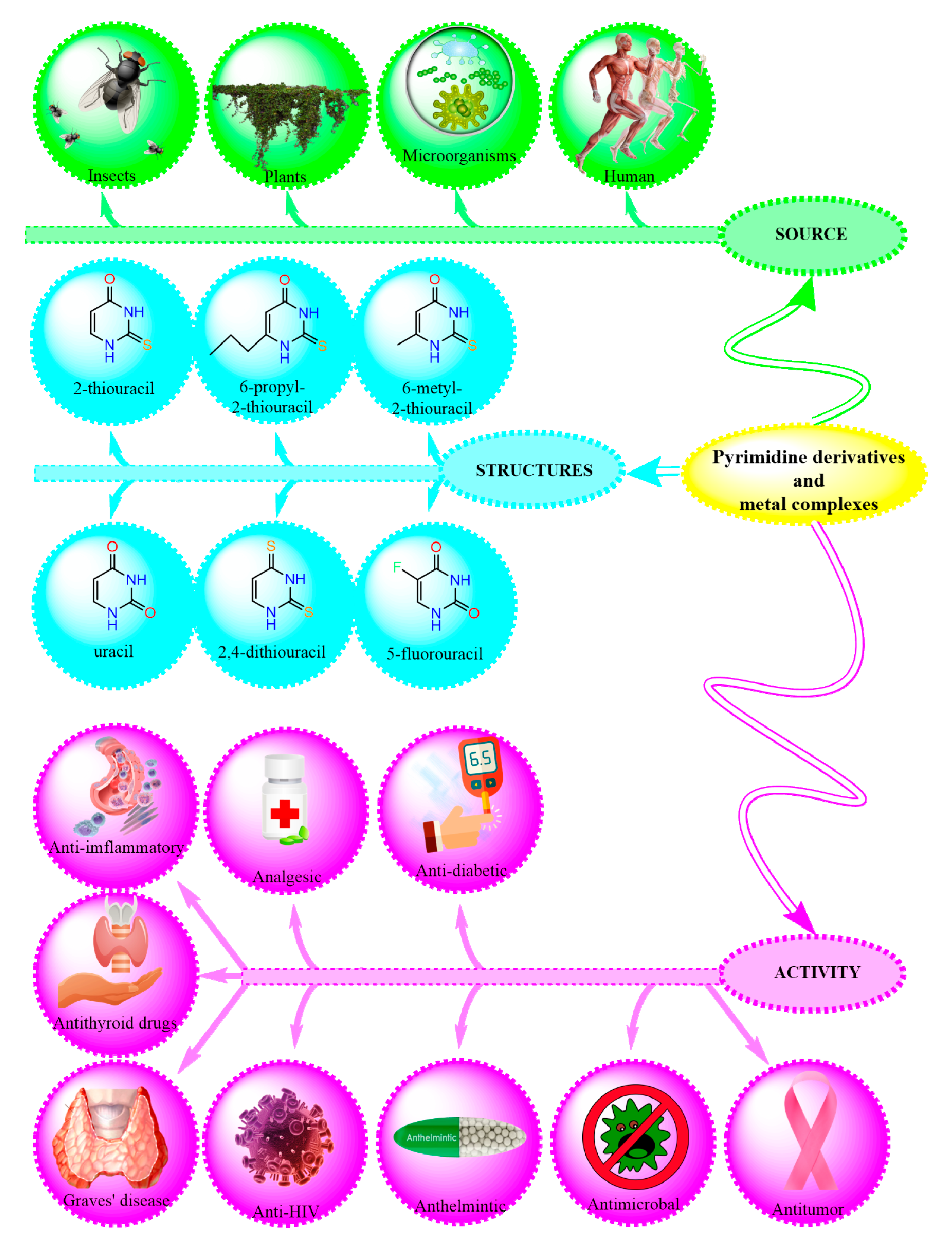
Figure 1.
Тhe probable structureof the gold complex with 2,4-dithiouracil, 2-Tu, and U [30].
Figure 1.
Тhe probable structureof the gold complex with 2,4-dithiouracil, 2-Tu, and U [30].
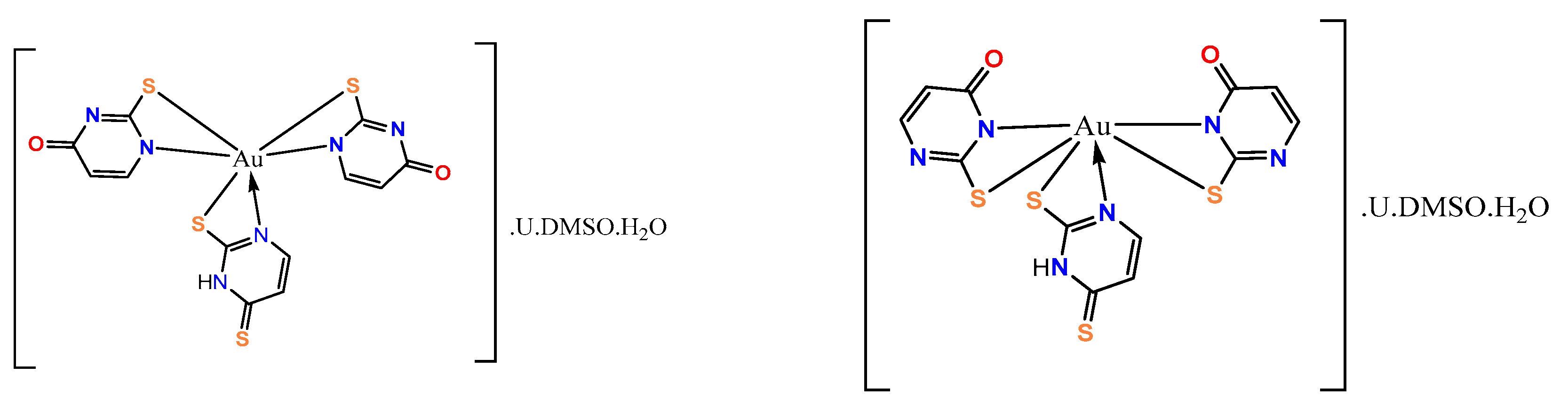
Figure 2.
A proposed structural arrangement and the corresponding coordination binding sites in the copper complex. If Z1 = H2O or DMSO-h6, then Z2 = H2O or DMSO-h6 [30].
Figure 2.
A proposed structural arrangement and the corresponding coordination binding sites in the copper complex. If Z1 = H2O or DMSO-h6, then Z2 = H2O or DMSO-h6 [30].
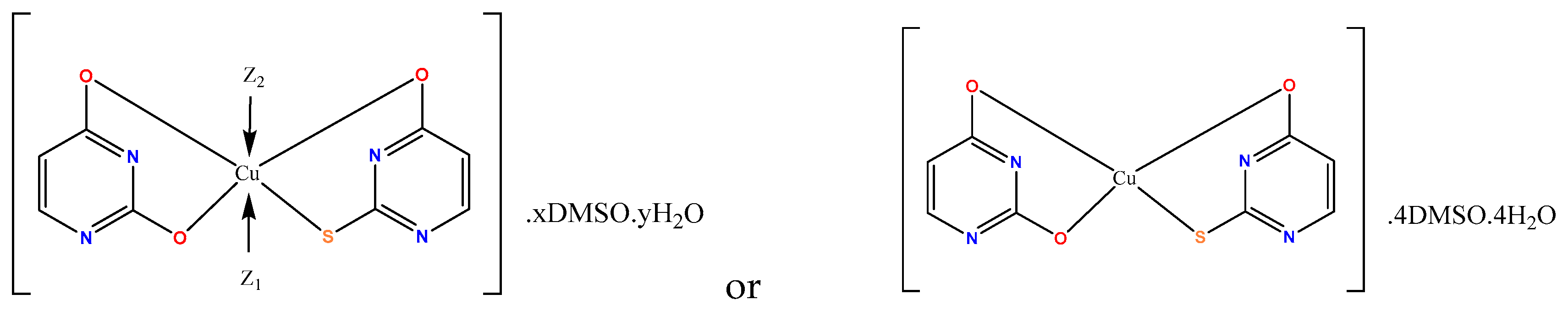
Figure 3.
The illustration of the suggested coordination binding sites for 6-propyl-2-thiouracil with copper and palladium [29].
Figure 3.
The illustration of the suggested coordination binding sites for 6-propyl-2-thiouracil with copper and palladium [29].
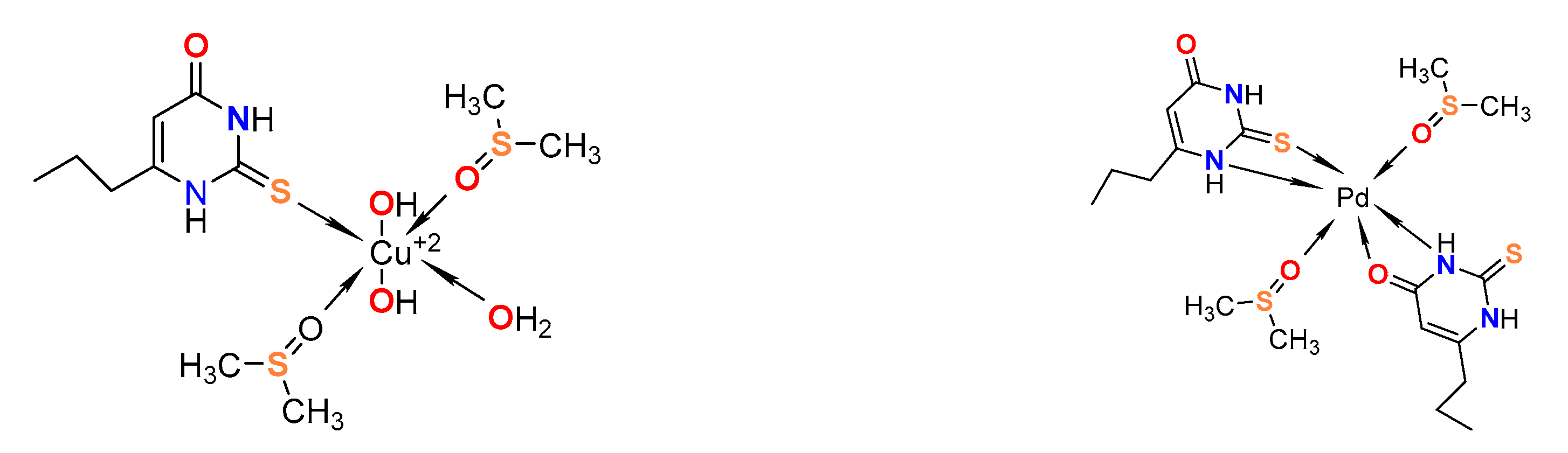
Figure 4.
Concentration-response curves of a) 2,4-dithiouracil and it’s b) Cu(II) and c) Au(III) complex against Vero cell line (kidney cells from African green monkey).
Figure 4.
Concentration-response curves of a) 2,4-dithiouracil and it’s b) Cu(II) and c) Au(III) complex against Vero cell line (kidney cells from African green monkey).
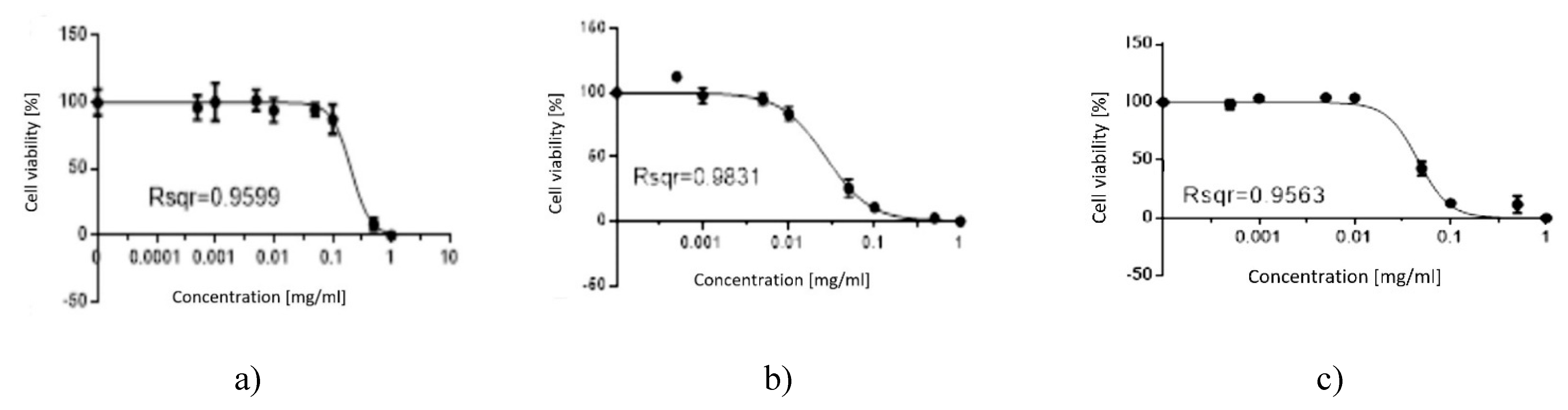
Figure 5.
Concentration-response curves of a) 2,4-dithiouracil and it’s b) Cu(II) and c) Au(III) complex against HeLa cell line (human cervical carcinoma).
Figure 5.
Concentration-response curves of a) 2,4-dithiouracil and it’s b) Cu(II) and c) Au(III) complex against HeLa cell line (human cervical carcinoma).
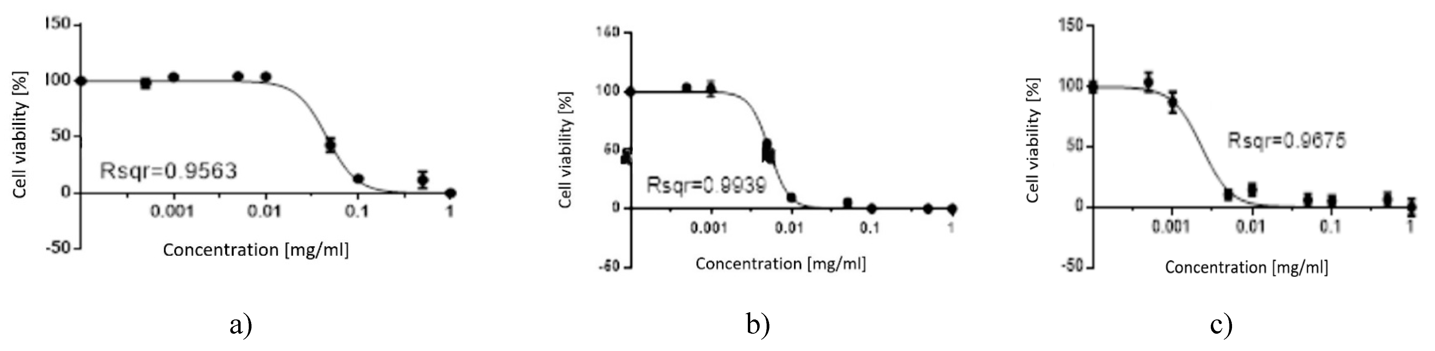
Figure 6.
Concentration-response curves of a) 6-propyl-2-thiouracil and it’s b) Cu (II) and c) Pd (II) complex against Vero cell line (kidney cells from African green monkey).
Figure 6.
Concentration-response curves of a) 6-propyl-2-thiouracil and it’s b) Cu (II) and c) Pd (II) complex against Vero cell line (kidney cells from African green monkey).
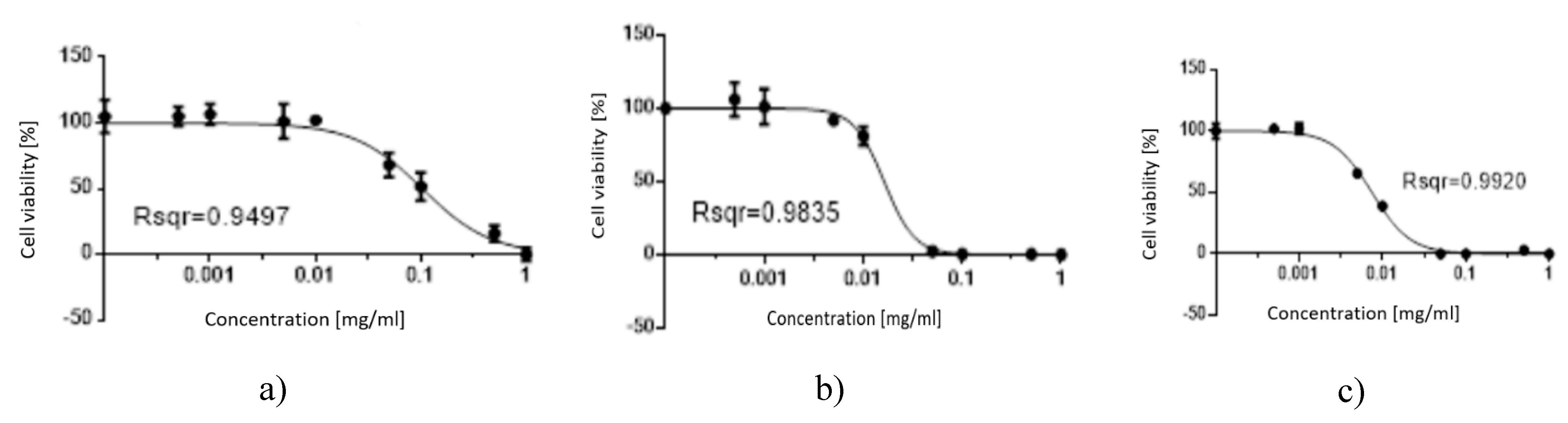
Figure 7.
Concentration-response curves of a) 6-propyl-2-thiouracil and it’s b) Cu(II) and c) Pd(II) complex against HeLa cell line (human cervical carcinoma).
Figure 7.
Concentration-response curves of a) 6-propyl-2-thiouracil and it’s b) Cu(II) and c) Pd(II) complex against HeLa cell line (human cervical carcinoma).
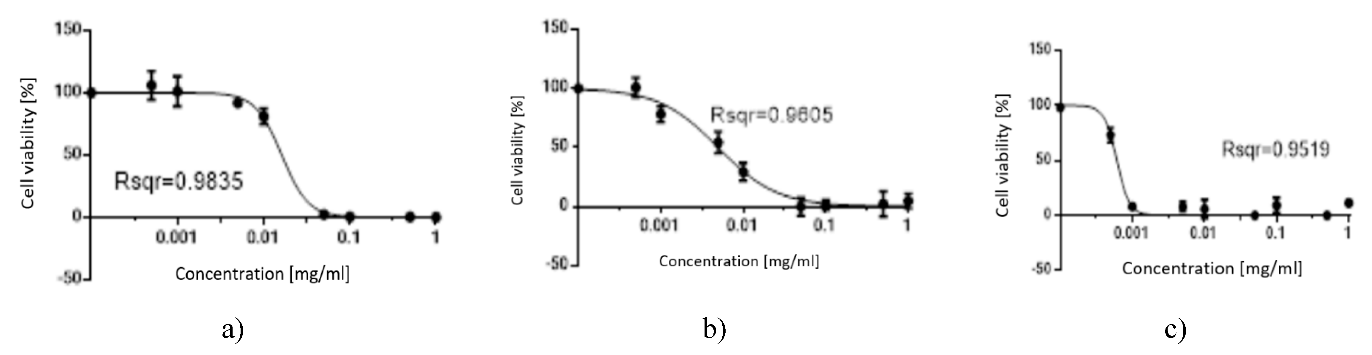

Table 1.
Hierarchical order of 2,4-dithiouracil and its complexes according to their cytotoxicity against Vero cell line (kidney cells from African green monkey).
Table 1.
Hierarchical order of 2,4-dithiouracil and its complexes according to their cytotoxicity against Vero cell line (kidney cells from African green monkey).
| Compound | Cytotoxic concentration (CD50), mg/mL | |
|---|---|---|
| Normal cell line | Tumor cell line | |
| Vero cell line (kidney cells from African green monkey) | HeLa cell line (human cervical carcinoma) | |
| 2,4-dithiouracil | 0,2049 | 0,04568 |
| Cu(II) complex | 0,02708 | 0,00534 |
| Au(III) complex | 0,04568 | 0,002327 |
| Order | 2,4-DTu > Au(III) complex > Cu(II) complex | 2,4-DTu > Cu(II) complex > Au(III) complex |
Table 2.
Hierarchical order of 6-Pro-2Tu and its complexes according to their cytotoxicity against Vero cell line (kidney cells from African green monkey).
Table 2.
Hierarchical order of 6-Pro-2Tu and its complexes according to their cytotoxicity against Vero cell line (kidney cells from African green monkey).
| Compound | Cytotoxic concentration (CD50), mg/mL | |
|---|---|---|
| Normal cell line | Tumor cell line | |
| Vero cell line (kidney cells from African green monkey) | HeLa cell line (human cervical carcinoma) | |
| 6-propyl-2-thiouracil | 0,1033 | 0,01626 |
| Cu(II) complex | 0,01626 | 0,004976 |
| Pd(II) complex | 0,007487 | 0,0006124 |
| Order | 6-Pro-2Tu > Cu(II) complex > Pd(II) complex | 6-Pro-2Tu > Cu(II) complex > Pd(II) complex |
Disclaimer/Publisher’s Note: The statements, opinions and data contained in all publications are solely those of the individual author(s) and contributor(s) and not of MDPI and/or the editor(s). MDPI and/or the editor(s) disclaim responsibility for any injury to people or property resulting from any ideas, methods, instructions or products referred to in the content. |
© 2025 by the authors. Licensee MDPI, Basel, Switzerland. This article is an open access article distributed under the terms and conditions of the Creative Commons Attribution (CC BY) license (http://creativecommons.org/licenses/by/4.0/).
Copyright: This open access article is published under a Creative Commons CC BY 4.0 license, which permit the free download, distribution, and reuse, provided that the author and preprint are cited in any reuse.


