Submitted:
29 January 2026
Posted:
02 February 2026
You are already at the latest version
Abstract
Ovarian tissue cryopreservation and transplantation represent promising strategies for fertility preservation in domestic species, laboratory models, and wildlife. However, follicular survival after transplantation remains limited by ischemia–reperfusion injury, delayed revascularization, and microenvironmental disruption. Thus, this review provides an update on cryopreservation methods, grafting approaches, angiogenic modulation, and microscopical tools used to evaluate ovarian tissue viability. Evidence indicates that follicular outcomes are influenced by the type of cryopreservation technique, ischemic duration, grafting site, and factors governing angiogenesis and extracellular matrix remodeling. Pro-angiogenic molecules, antiapoptotic agents, mesenchymal stem cell co-transplantation, and emerging biomaterials have shown potential to enhance vascular perfusion and reduce follicular loss. Histological and histochemical techniques, including PAS and Picrosirius Red staining, remain essential for assessing follicular morphology, stromal integrity, and collagen organization. In addition, immunostaining facilitates the investigation of key biological processes, including angiogenesis, cell death, and cellular proliferation. Collectively, these findings highlight that refining cryopreservation methodologies, improving revascularization, and adopting standardized morphological evaluation are crucial steps toward increasing the efficiency of ovarian tissue transplantation in livestock species. Continued integration of reproductive biotechnology, tissue engineering, and molecular analyses is expected to expand the applicability of this technique in animal reproduction, production systems, and biodiversity conservation.
Keywords:
ovary
; grafting
; angiogenesis
; follicular viability
; morphology evaluation
1. Introduction
The continuous advancement of diagnostic and therapeutic approaches in oncology has substantially improved survival rates among young and adult women diagnosed with neoplasms [1]. However, anticancer treatments frequently impair reproductive function and may result in premature ovarian failure [2]. In this context, cryopreservation combined with ovarian tissue transplantation has emerged as a promising strategy for fertility preservation and for the restoration of endocrine activity [3]. Beyond human applications, these techniques also hold considerable potential for conservation programs involving species with reproductive challenges or those threatened with extinction, thereby contributing to biodiversity preservation [4,5].
Depending on the type of recipient, ovarian tissue transplantation may be classified as autotransplantation (within the same individual), isotransplantation (between genetically identical individuals), allotransplantation (between individuals of the same species), or xenotransplantation (between individuals of different species) [6]. Xenotransplantation has been widely used in experimental studies and in reproductive research involving domestic species.
Ovarian tissue transplantation involves the reimplantation of cortical fragments into different anatomical sites. These sites may be classified as orthotopic, when the tissue is positioned near its original location, or heterotopic, such as in the subcutaneous tissue, intraperitoneal cavity, or beneath the renal capsule, all of which can support effective revascularization [7,8]. Evidence indicates that heterotopic transplantation offers practical advantages related to anesthesia and surgical procedures, thereby facilitating endocrine recovery [9]. According to Oktay & Marin [10], orthotopic ovarian transplantation results in higher gamete and embryo quality. However, the endocrine function restoration rate and longevity are similar between orthotopic and heterothopic approaches. When feasible, orthotopic should be preferred for those who intend to conceive, although a less invasive heterotopic can be performed for those who primarily desire ovarian endocrine function [10].
Despite encouraging outcomes, including more than 200 births reported worldwide, the technique still faces limitations related to ischemia and delayed revascularization, which may lead to necrosis, fibrosis, and the loss of stromal and follicular cells [11,12,13]. Ischemia begins immediately after tissue removal and is typically alleviated only around seven days after transplantation [14,15]. Consequently, selecting biologically suitable implantation sites is essential to ensure graft survival and functionality [16]. Several factors influence this process, including oxidative stress [17], hypoxia [18], hormonal and molecular interactions [19], and variations in grafting protocols [16].
The pre-transplantation phase, particularly the method of tissue preservation, plays a central role in minimizing cellular damage. Vitrification has emerged as an efficient approach to ovarian tissue cryopreservation because it reduces ice crystal formation and offers operational practicality. It has yielded favorable results in different species, including humans [20,21], small ruminants [22,23,24], felids [25,26,27], and canids [28,29,30,31,32]. However, elevated oxidative stress in vitrified samples may compromise cellular viability due to the high concentrations of cryoprotectants and the rapid cooling rates used. The incorporation of antioxidants has therefore been proposed as a strategy to mitigate these deleterious effects [33].
After vitrification, ovarian tissue can resume its physiological function either through in vivo or in vitro culture, supporting follicular development and the production of viable oocytes for fertilization [22,24,34]. Understanding the combined effects of vitrification and transplantation is thus essential for advancing reproductive biotechnologies and promoting fertility preservation. This review aims to integrate recent evidence on ovarian tissue cryopreservation and transplantation in veterinary species, with emphasis on the cellular mechanisms underlying follicular survival and activation, the factors that modulate graft revascularization, and the histological and histochemical approaches used for tissue assessment. The originality of this contribution lies in its comparative synthesis of cryopreservation methods, strategies for preserving the follicular microenvironment, the use of angiogenic modulators, and grafting protocols, providing an integrated perspective not yet consolidated in the veterinary literature.
2. Ovarian Tissue Cryopreservation
Ovarian tissue cryopreservation consists of storing cells and tissues at ultra-low temperatures (−150°C to −196°C), allowing metabolic activity to decrease and enabling long-term preservation until normal development can later resume [35]. This powerful technique has emerged as an effective strategy for maintaining ovarian function and fertility, providing protection against premature ovarian failure and other causes of follicular reserve depletion. Among its advantages are the preservation of the entire pool of primordial follicles, its applicability at any age or estrous cycle stage [36], the safeguarding of genetic material in women prior to oncologic treatments, reduced ethical concerns compared with oocyte and embryo cryopreservation [37], and the conservation of genetic material from high-value animals or endangered species [38]. Collectively, these factors underscore their broad reproductive potential and value for genetic preservation.
Cryopreservation can be performed using either slow freezing or vitrification (Figure 1), which differ primarily in cooling rate [32]. In slow freezing, temperature is gradually reduced (~1°C/min) with the use of low concentration cryoprotectants; however, ice crystal formation still occurs and may damage cellular structures. In vitrification, ice formation is essentially prevented by achieving a glass-like state through the application of high concentrations of cryoprotectants combined with rapid cooling to -196°C. This method is faster, simpler, and does not require specialized equipment [37].
Several ovarian tissue vitrification protocols employing intracellular cryoprotectants (ethylene glycol and dimethyl sulfoxide) combined with extracellular cryoprotectants (sucrose), in stepwise increasing concentrations of intracellular agents (10% and 20%), have been successfully applied in different domestic species, including sheep [39], goats [22], and cattle [40]. Among these protocols, the Ovarian Tissue Cryosystem (OTC) can be highlighted as a metallic, hermetically sealed device that prevents direct contact between the tissue and liquid nitrogen, thereby avoiding contamination. Additionally, the system allows the vitrification solution to be removed before the OTC is immersed in liquid nitrogen [33,39].
Following cryopreservation, ovarian tissue may be transplanted (in vivo culture), enabling the recovery of both endocrine and gametogenic functions [11]. Alternatively, the tissue can be used in in vitro culture systems to support the development of preantral follicles within the ovarian cortex (Figure 1) [41], or for the culture of isolated follicles retrieved from previously cryopreserved fragments, which may progress to maturity and yield fertilizable oocytes [24]. Moreover, preantral follicles isolated directly from ovarian tissue and subsequently cryopreserved may also progress to the antral stage, producing viable oocytes suitable for in vitro embryo production [42].
3. Ovarian Tissue Transplantation
The ovarian tissue transplantation technique consists of a surgical approach in which an ovarian tissue fragment is transplanted in vivo into a host, where it functions as a biological incubator, stimulating the resumption of folliculogenesis [43]. Unlike in vitro environments, tissue transplantation offers the advantage of providing an optimal environment for follicular activation and development, supplying hormones, growth factors, and biological conditions that in vitro culture systems are not yet fully capable of reproducing [44]. The primary aim of this methodological approach is the restoration of fertility and endocrine activity.
In small ruminants, the first studies on ovarian tissue freezing and transplantation were conducted by Gosden et al. [45] and Baird et al. [46]. Since then, several births in sheep have been reported following the transplantation of frozen hemi-ovaries or whole ovaries [47,48,49], as well as after the transplantation of vitrified ovarian tissue [50,51]. Despite these advances, transplantation as a method for fertility restoration in production species such as sheep and goats remains limited by the need for immunosuppression, the invasive nature of the procedure, and high operational costs, which make its implementation largely unfeasible for small-scale farmers [52]. Table 1 provides a compilation of relevant scientific studies reporting advances in ovarian tissue cryopreservation and transplantation in small ruminants.
Ovarian tissue transplantation can be classified according to several parameters, among which the total size of the grafted material [34], the type of host selected for transplantation [57], and the anatomical implantation site [60] are particularly noteworthy, as summarized in Table 2. In addition to these criteria, factors such as the degree of graft vascularization, the cryopreservation method employed, and the time required for the recovery of ovarian function also play decisive roles in the success of the procedure and are frequently addressed in experimental and clinical studies.
The selection of graft size, host type, and implantation site must be made carefully, as these factors directly influence transplantation success. Graft size affects both the time required for revascularization and the extent of follicular loss due to ischemia: smaller grafts exhibit faster functional recovery, whereas larger grafts, such as whole ovaries, carry a greater ischemic risk if revascularization does not occur promptly [69].
In this context, whole-ovary transplantation emerges as an alternative capable of mitigating some of these effects, as it allows for immediate vascular anastomosis, thereby reducing ischemia–reperfusion injury and promoting preservation of the follicular reserve. However, this approach requires appropriate cryopreservation of the vascular pedicle and involves high surgical complexity [70]. Among the primary mechanisms of follicular loss are acute ischemia, premature activation of primordial follicles, and hypoxia-induced apoptosis. Strategies such as the use of angiogenic factors, antiapoptotic agents, erythropoietin, or co-transplantation of mesenchymal stem cells have been shown to reduce these losses and enhance revascularization [71].
From a clinical standpoint, restoration of endocrine function can be achieved through the transplantation of cortical ovarian tissue fragments or, in specific situations, through whole-ovary transplantation. However, reproductive outcomes vary according to the preservation method, implantation site, and surgical technique employed. Among fragment-based transplants, orthotopic implantation consistently yields the most favorable reproductive results, primarily due to the possibility of permitting natural ovulation and spontaneous pregnancy. Whole-ovary transplantation, although capable of preserving a larger quantity of follicles, remains limited by technical complexity and challenges associated with maintaining the viability of the vascular pedicle [70].
Given these limitations, the use of ovarian cortical fragments has emerged as a promising alternative, as the reduced tissue volume enhances the diffusion of nutrients and oxygen, thereby minimizing ischemia and complications associated with the need for vascular reanastomosis. Additionally, the procedure is less invasive, faster to perform, and allows for the possibility of retransplantation in cases of graft failure [61,72].
4. Maintenance and Follicular Activation: Recent Evidence in Production Species
Studies on ovarian tissue transplantation have highlighted the critical roles of the post-transplant microenvironment, vascular perfusion, and angiogenic factors in follicular survival and activation. Souza et al. [68] evaluated ovaries from mares that were cooled or cryopreserved and subsequently xenotransplanted into mice, demonstrating that cooling—either alone or followed by transplantation—preserved follicular morphology, whereas the cryopreservation–cooling–transplantation sequence markedly reduced follicular survival. Furthermore, transplantation triggered follicular activation, likely as a response to post-surgical hypoxia.
In goats, Vieira et al. [67] examined intra-auricular grafting and observed a higher proportion of healthy and activated primordial follicles seven days after transplantation. However, prolonged ischemia reduced follicular viability, emphasizing the importance of both graft site and post-transplantation interval. In cattle, Morais et al. [73] reported that short-term pre-exposure of ovarian tissue to VEGF before heterotopic autotransplantation enhanced neovascularization but partially affected follicular morphology: transplantation without VEGF resulted in better preservation of primordial follicles, whereas VEGF treatment promoted greater activation of developing follicles. Collectively, these findings indicate that variables such as vascular supply, duration of ischemia, graft location, and angiogenic stimulation interact in a complex and integrated manner to modulate follicular survival, activation, and development following ovarian tissue transplantation.
5. Blood Flow of the Ovarian Tissue Implantation Site
Ultrasonography has proven to be a valuable tool for real-time, non-invasive monitoring of ovarian structures in small ruminants, both in non-stimulated animals [74] and in those subjected to hormonal stimulation [75]. In the context of ovarian tissue transplantation in humans, two-dimensional ultrasonography has been widely used to monitor follicular [63] and embryonic development [76,77].
An important variation of this method, still underexplored in studies involving ovarian transplantation, is color Doppler ultrasonography, which enables real-time visualization of vascular dynamics by detecting the movement of erythrocytes within vessels near the transducer [78]. This approach not only allows identification of anatomical structures but also provides an assessment of blood perfusion, offering essential information regarding post-transplant revascularization, a process that is critical for follicular survival, maintenance, and activation and therefore fundamental to graft success.
In the study conducted by Pinto et al. [79], color Doppler ultrasonography was used to assess local blood flow within and around implanted tissues. The perimeter and number of colored pixels were measured on alternating days over seven days (right side) and fifteen days (left side). The authors observed that blood perfusion was similar among the different regions evaluated around the implants during the first two days following transplantation, suggesting a positive correlation between vascular perfusion and the presence of normal and primordial follicles. These findings reinforce the applicability of color Doppler imaging as a non-invasive tool for monitoring perfusion in superficial grafts.
Complementarily, Vieira et al. [59] performed daily assessments of blood flow from day 0 (pre-transplant) to day 15 (post-transplant) at the graft site in goats subjected to ovarian allotransplantation. The images obtained revealed a progressive and significant increase in vascularization in the analyzed regions, particularly when comparing day 0 with days 7 and 15 after the procedure (Figure 2).
The effectiveness of Doppler ultrasonography as a non-invasive method for evaluating blood flow in ovarian implants has also been demonstrated in other species, including equine [80] and bovine [73]. Taken together, these findings establish color Doppler imaging as an essential tool for monitoring vascularization and the viability of transplanted ovarian tissues, contributing to the advancement of techniques aimed at preserving and restoring reproductive function across different species.
6. Histology and Immunohistochemistry Applied to Ovarian Tissue
The appropriate choice of staining method is essential for accurate histological assessment, as different dyes allow the visualization of specific cellular structures and components of the extracellular matrix [81]. In ovarian tissue, Periodic Acid–Schiff Hematoxylin (PAS) staining is widely used, producing a purple–blue coloration in the stroma and follicular cells and a pink coloration in carbohydrate-rich structures such as the zona pellucida and the basement membrane, thereby facilitating the identification of potential abnormalities [82]. Among these abnormalities are thickening or duplication of the basement membrane, discontinuities or irregularities in the zona pellucida, pyknotic oocytes, granulosa cells exhibiting cytoplasmic degeneration, and stromal alterations such as fibrotic areas or abnormal accumulation of extracellular matrix, which are critical parameters for evaluating tissue integrity and follicular viability after preservation or culture procedures [83].
Another relevant technique is Picrosirius Red staining, which is particularly indicated for the characterization of collagen fibers [84]. Under conventional light microscopy, the total collagen content in the section appears pink, whereas under polarized light, type I collagen fibers exhibit a red coloration and type III fibers appear in shades of green [84]. This approach is useful for identifying regeneration, structural damage, or fibrosis following in vitro culture [85] or transplantation [13], thereby providing complementary information on extracellular matrix remodeling in preserved or grafted ovarian tissue.
Immunohistochemistry (IHC) is based on morphological, molecular, and immunological principles to identify specific antigens in tissues through highly specific antigen–antibody interactions. This technique enables the detection, localization, and semi-quantitative assessment of proteins in cellular and tissue compartments, and it is widely used in both experimental research and diagnostic pathology [86]. In the context of ovarian biology, IHC has been employed to identify proteins expressed in the stroma or parenchyma, allowing the evaluation of key processes such as angiogenesis, apoptosis, and cell proliferation, particularly in tissues subjected to ovarian transplantation [73,87]. A summary of the most used immunohistochemical indicators in studies of transplanted ovarian tissue, highlighting their respective applications is presented in Table 3.
The assessment of blood vessel density is essential for analyzing tissue perfusion and the viability of ovarian grafts. The markers CD31 and α-smooth muscle actin (αSMA) allow the distinction between newly formed and mature vessels, respectively [19,57,73]. Combined analysis of these markers enables the correlation of revascularization with follicular development and stromal integrity, reflecting the efficiency of the graft in supporting adequate diffusion of nutrients and oxygen.
Angiogenic factors such as vascular endothelial growth factor (VEGF) have shown significant potential to improve vascularization in cryopreserved and transplanted ovarian tissue. Experimental studies demonstrate that VEGF supplementation increases the density of newly formed and mature vessels, reduces apoptosis, and contributes to the preservation of primordial follicles after transplantation [73,90,91]. In murine models, the combined use of VEGF and basic fibroblast growth factor (FGF2) further enhanced follicular survival and revascularization, suggesting promising perspectives for clinical application and for use in domestic species of zootechnical interest [19,92].
In addition to angiogenesis, the evaluation of cell proliferation is commonly performed using markers such as Ki-67 [89] and proliferating cell nuclear antigen (PCNA) [73], whereas the follicular reserve can be investigated with germ cell markers such as DDX4 [88]. Increased Ki-67 [89] and PCNA [73] expression in granulosa and stromal cells has been associated with regenerative activity and follicular activation in transplanted tissues treated with VEGF or other modulatory agents. Taken together, these findings reinforce immunohistochemistry as a fundamental approach to evaluate angiogenesis, cellular viability, and the maintenance of ovarian function in preserved and transplanted ovarian tissue, enabling the establishment of robust correlations between therapeutic interventions and graft quality, vascular integration, and follicular survival.
7. Future Perspectives
Emerging investigative approaches driven by recent advances are reshaping the future of ovarian tissue cryopreservation and transplantation, with emphasis on multiple complementary strategies aimed at improving graft performance, follicular preservation, and reproductive outcomes. Current directions highlight the need to: i) accelerate revascularization and reduce post-transplant ischemia by incorporating pro-angiogenic molecules, biomaterials, or cellular therapies; ii) modulate follicular activation to preserve the pool of primordial follicles and avoid premature depletion; iii) optimize cryopreservation protocols through the use of less toxic cryoprotectants and more efficient cooling–warming curves; iv) integrate tissue engineering approaches, including three-dimensional scaffolds and hydrogels capable of supporting the follicular microenvironment; v) apply molecular and omics technologies to precisely map cellular, metabolic, and inflammatory processes associated with thermal and hypoxic injury; vi) explore new implantation sites that are minimally invasive, well vascularized, and capable of sustaining endocrine and reproductive functions; vii) conduct long-term functional evaluations correlating perfusion, morphology, and actual reproductive performance, especially in species of zootechnical and conservation interest; and viii) expand the use of ovarian tissue transplantation in wildlife species, developing species-specific strategies that account for anatomical, physiological, and reproductive differences.
Taken together, these perspectives underscore an expanding field in which the integration of basic and applied science has the potential to enhance the effectiveness of ovarian tissue transplantation in animal production, clinical medicine, and biodiversity conservation. Continued multidisciplinary efforts involving reproductive physiology, biotechnology, tissue engineering, and molecular analysis will be essential for transforming current limitations into viable opportunities for fertility restoration.
8. Conclusion
Ovarian tissue cryopreservation and transplantation have advanced significantly in recent decades, establishing themselves as central strategies for fertility preservation in various species of zootechnical and clinical interest. The success of these techniques depends on meticulous attention across all procedural stages, from tissue collection and processing to transplantation and postoperative evaluation, particularly regarding revascularization, tissue repair, and cellular proliferation.
In this context, Doppler ultrasonography has emerged as a valuable tool for the continuous assessment of blood perfusion in transplanted ovarian tissues, enabling real-time, in vivo monitoring of vascular changes that are essential for graft survival and follicular development. Following tissue recovery, histological analysis combined with immunohistochemical markers remains indispensable for elucidating the underlying biological processes from a structural and molecular perspective. These tools provide detailed insights into tissue remodeling, angiogenesis, apoptosis, and the maintenance of follicular reserves.
Despite significant progress and promising results reported in the literature, current evidence indicates that follicular survival remains limited by complex and still insufficiently understood pathophysiological mechanisms. Continued research is therefore necessary to standardize protocols, refine grafting methodologies, optimize microenvironmental support, and broaden the applicability of ovarian tissue transplantation across domestic and wild species.
Author Contributions
writing—original draft preparation, A.R.S.V. and F.C.S.; writing—review and editing, F.C.S. and D.Í.A.T; funding acquisition, D.Í.A.T. All authors have read and agreed to the published version of the manuscript.
Funding
This research was funded by Coordination for the Improvement of Higher Education Per-sonnel—Brazil (CAPES)—Funding Code 001.
Conflicts of Interest
The authors declare that the research was conducted in the absence of any commercial or financial relationships that could be construed as potential conflicts of interest.
References
- Assogba, E.L.F.; Kamga, A.M.; Costaz, H.; Jankowski, C.; Dumas, A.; Roignot, P.; Jolimoy, G.; Coutant, C.; Arveux, P.; Dabakuyo-Yonli, T.S. What are young women’s living conditions after breast cancer? Health-related quality of life, sexual and fertility issues, and professional reinsertion. Cancers 2020, 12, 1564. [Google Scholar] [CrossRef]
- Meirow, D.; Hardan, I.; Dor, J.; Fridman, E.; Elizur, S.; Raianani, H.; Slyusarevsky, E.; Amariglio, N.; Schiff, E.; Rechavi, G.; Nagler, A.; Ben Yehuda, D. Searching for evidence of disease and malignant cell contamination in ovarian tissue stored from hematologic cancer patients. Hum. Reprod. 2008, 23, 1007–1013. [Google Scholar] [CrossRef]
- Kim, S.S. Revisiting the role of heterotopic ovarian transplantation: Futility or fertility? Reprod. Biomed. Online 2014, 28, 141–145. [Google Scholar] [CrossRef]
- Santos, R.R.; Amorim, C.; Cecconi, S.; Fassbender, M.; Imhof, M.; Lornage, J.; Paris, M.; Schoenfeldt, V.; Martinez-Madrid, B. Cryopreservation of ovarian tissue: An emerging technology for female germline preservation of endangered species and breeds. Anim. Reprod. Sci. 2010, 122, 151–163. [Google Scholar] [CrossRef]
- Rodrigues, A.P.R.; Rocha, R.M.P.; Rodrigues, G.Q.; Brito, D.C.C. Cryopreservation of ovarian tissue and/or preantral follicles: An alternative to safeguard the genetic material and fertility of valuable sheep and goats. In Proceedings of the XXIII Congresso Brasileiro de Reprodução Animal (CBRA 2019), Gramado, Brazil, 15–17 May 2019. [Google Scholar]
- Krohn, P.L. Transplantation of the ovary. In The Ovary, 2nd ed.; Zuckerman, L., Weir, B.J., Eds.; Academic Press: New York, NY, USA, 1977; Volume 2, pp. 101–128. [Google Scholar]
- Donfack, N.J.; Alves, K.A.; Araújo, V.R.; Cordova, A.; Figueiredo, J.R.; Smitz, J.; Rodrigues, A.P.R. Expectations and limitations of ovarian tissue transplantation. Zygote 2017, 25, 391–403. [Google Scholar] [CrossRef] [PubMed]
- Amorim, C.A.; David, A.; Van Langendonckt, A.; Dolmans, M.M.; Donnez, J. Vitrification of human ovarian tissue: Effect of different solutions and procedures. Fertil. Steril. 2011, 95, 1094–1097. [Google Scholar] [CrossRef]
- Demeestere, I.; Simon, P.; Emiliani, S.; Delbaere, A.; Englert, Y. Orthotopic and heterotopic ovarian tissue transplantation. Hum. Reprod. Update 2009, 15, 649–665. [Google Scholar] [CrossRef]
- Oktay, K.H.; Marin, L. Comparison of orthotopic and heterotopic autologous ovarian tissue transplantation outcomes. Fertil. Steril. 2024, 121, 72–79. [Google Scholar] [CrossRef]
- Liu, J.; Van der Elst, J.; Van den Boecke, R.; Dhont, M. Early massive follicle loss and apoptosis in heterotopically grafted newborn mouse ovaries. Hum. Reprod. 2002, 17, 605–611. [Google Scholar] [CrossRef] [PubMed]
- Dath, C.; Van Eyck, A.S.; Dolmans, M.M.; Romeu, L.; Delle Vigne, L.; Donnez, J.; Van Langendonckt, A. Xenotransplantation of human ovarian tissue to nude mice: Comparison between four grafting sites. Hum. Reprod. 2010, 25, 1734–1743. [Google Scholar] [CrossRef] [PubMed]
- Scalercio, S.R.; Amorim, C.A.; Brito, D.C.; Percário, S.; Oskam, I.C.; Domingues, S.F.S.; Santos, R.R. Trolox enhances follicular survival after ovarian tissue autograft in squirrel monkey (Saimiri collinsi). Reprod. Fertil. 2015, 28, 1854–1864. [Google Scholar] [CrossRef]
- Van Eyck, A.S.; Jordan, B.; Gallez, B.; Heilier, J.F.; Van Langendonckt, A.; Donnez, J. Electron paramagnetic resonance as a tool to evaluate human ovarian tissue reoxygenation after xenografting. Fertil. Steril. 2009, 92, 374–381. [Google Scholar] [CrossRef]
- Duncan, F.E.; Zelinski, M.; Gunn, A.H.; Pahnke, J.E.; O’Neill, C.L.; Songsasen, N.; Woodruff, R.I.; Woodruff, T.K. Ovarian tissue transport to expand access to fertility preservation: From animals to clinical practice. Reproduction 2016, 152, 201–210. [Google Scholar] [CrossRef]
- Youm, H.W.; Lee, J.R.; Lee, J.; Jee, B.C.; Suh, C.S.; Kim, S.H. Transplantation of mouse ovarian tissue: Comparison of transplantation sites. Theriogenology 2015, 83, 854–861. [Google Scholar] [CrossRef]
- Nugent, D.; Newton, H.; Gallivan, L.; Gosden, R.G. Protective effect of vitamin E on ischaemia-reperfusion injury in ovarian grafts. J. Reprod. Fertil. 1998, 114, 341–346. [Google Scholar] [CrossRef]
- Damous, L.L.; Nakamuta, J.S.; Soares, J.M.; Maciel, G.A.; Simões, R.S.; Montero, E.F.; Krieger, J.E.; Baracat, E.C. Females transplanted with ovaries subjected to hypoxic preconditioning show impairment of ovarian function. J. Ovarian Res. 2014, 7, 34–41. [Google Scholar] [CrossRef]
- Li, S.H.; Hwu, Y.M.; Lu, C.H.; Chang, H.H.; Hsieh, C.E.; Lee, R.K. VEGF and FGF2 improve revascularization, survival, and oocyte quality of cryopreserved, subcutaneously transplanted mouse ovarian tissues. Int. J. Mol. Sci. 2016, 17, 1–13. [Google Scholar] [CrossRef] [PubMed]
- Sanfilippo, S.; Canis, M.; Smitz, J.; Sion, B.; Darcha, C.; Janny, L.; Brugnon, F. Vitrification of human ovarian tissue: A practical and relevant alternative to slow freezing. Reprod. Biol. Endocrinol. 2015, 13, 67. [Google Scholar] [CrossRef]
- Yuzhakov, V.V.; Malinova, I.V.; Kiseleva, M.V.; Fomina, N.K.; Bandurko, L.N.; Komarova, E.V.; Sevan’kaeva, L.E.; Ingel, I.E.; Yakovleva, N.D.; Kaprin, A.D. Effect of vitrification on functional morphology and viability of the ovarian tissue. Bull. Exp. Biol. Med. 2018, 164, 502–507. [Google Scholar] [CrossRef] [PubMed]
- Carvalho, A.A.; Faustino, L.R.; Silva, C.M.; Castro, S.V.; Lopes, C.A.; Santos, R.R.; Báo, S.N.; Figueiredo, J.R.; Rodrigues, A.P. Novel wide-capacity method for vitrification of caprine ovaries: Ovarian Tissue Cryosystem (OTC). Anim. Reprod. Sci. 2013, 138, 220–227. [Google Scholar] [CrossRef] [PubMed]
- Bandeira, F.T.; Carvalho, A.A.; Castro, S.V.; Lima, L.F.; Viana, D.A.; Evangelista, J.S.; Pereira, M.J.; Campello, C.C.; Figueiredo, J.R.; Rodrigues, A.P. Two methods of vitrification followed by in vitro culture of the ovine ovary: Evaluation of follicular development and ovarian extracellular matrix. Reprod. Domest. Anim. 2015, 50, 177–185. [Google Scholar] [CrossRef]
- Lunardi, F.O.; De Aguiar, F.L.; Duarte, A.B.; Araújo, V.R.; De Lima, L.F.; Correia, H.H.V.; Domingues, S.F.; Campello, C.C.; Smitz, J.; De Figueiredo, J.R.; Rodrigues, A.P.R. Ovine secondary follicles vitrified outside the ovarian tissue grow and develop in vitro better than those vitrified within ovarian fragments. Theriogenology 2016, 85, 1203–1210. [Google Scholar] [CrossRef]
- Brito, D.C.; Domingues, S.F.; Silva, J.K.; Wu, X.; Santos, R.R.; Pieczarka, J.C. Detrimental effect of phenol red on the vitrification of cat (Felis catus) ovarian tissue. Biopreserv. Biobank 2016, 14, 17–22. [Google Scholar] [CrossRef]
- Brito, D.C.C.; Domingues, S.F.S.; Rodrigues, A.P.R.; Maside, C.; Lunardi, F.O.; Wu, X.; Figueiredo, J.R.; Pieczarka, J.C.; Santos, R.R. Cryopreservation of domestic cat (Felis catus) ovarian tissue: Comparison of two vitrification methods. Theriogenology 2018, 111, 69–77. [Google Scholar] [CrossRef]
- Brito, D.C.C.; Domingues, S.F.S.; Rodrigues, A.P.R.; Figueiredo, J.R.; Santos, R.R.; Pieczarka, J.C. Vitrification of domestic cat (Felis catus) ovarian tissue: Effects of three different sugars. Cryobiology 2018, 83, 97–99. [Google Scholar] [CrossRef] [PubMed]
- Ishijima, T.; Kobayashi, Y.; Lee, D.S.; Ueta, Y.; Matsui, M.; Lee, J.Y.; Suwa, Y.; Miyahara, K.; Suzuki, H. Cryopreservation of canine ovaries by vitrification. J. Reprod. Dev. 2006, 52, 293–299. [Google Scholar] [CrossRef] [PubMed]
- Suzuki, H.; Ishijima, T.; Maruyama, S.; Ueta, Y.; Abe, Y.; Saitoh, H. Beneficial effect of desialylated erythropoietin administration on frozen–thawed canine ovarian xenotransplantation. J. Assist. Reprod. Genet. 2008, 25, 571–575. [Google Scholar] [CrossRef]
- Ishijima, T.; Abe, Y.; Suzuki, H. Follicular loss of the cryopreserved canine ovary after xenotransplantation. J. Mamm. Ova Res. 2009, 26, 61–65. [Google Scholar] [CrossRef]
- Ackermann, C.L.; Asa, C.S.; Krisher, R.; Bauman, K.; Casey, S.; Lopes, M.D. Evaluation of follicular growth and tissue viability in vitrified/warmed domestic dog ovaries after in vitro culture. Reprod. Domest. Anim. 2016, 52, 77–81. [Google Scholar] [CrossRef] [PubMed]
- Jivago, J.L.P.R.; Paulini, F.; Silva, R.C.; Araujo, M.S.; Marinho, A.P.S.; Lucci, C.M. Cryopreservation and characterization of canine preantral follicles. Cryobiology 2018, 81, 34–42. [Google Scholar] [CrossRef]
- Ñaupas, L.V.S.; Brito, D.C.C.; De Souza, S.S.; Brandão, F.A.S.; Da Silva, R.F.; Raposo, R.S.; Rodrigues, A.P.R. Alpha lipoic acid supplementation improves ovarian tissue vitrification outcome: An alternative to preserve the ovarian function of Morada Nova ewes. Reprod. Sci. 2021, 28, 3109–3122. [Google Scholar] [CrossRef] [PubMed]
- Gavish, Z.; Peer, G.; Roness, H.; Cohen, Y.; Meirow, D. Follicle activation and “burn-out” contribute to post-transplantation follicle loss in ovarian tissue grafts: The effect of graft thickness. Hum. Reprod. 2014, 29, 989–996. [Google Scholar] [CrossRef] [PubMed]
- Pegg, D.E. Principles of cryopreservation. In Methods in Molecular Biology; Humana Press: Totowa, NJ, USA, 2007; Volume 368, pp. 39–57. [Google Scholar]
- Shaw, J.M.; Oranratnachai, A.; Trounson, A.O. Fundamental cryobiology of mammalian oocytes and ovarian tissue. Theriogenology 2000, 53, 59–72. [Google Scholar] [CrossRef]
- Zhang, J.M.; Sheng, Y.; Cao, Y.Z.; Wang, H.Y.; Chen, Z.J. Effects of cooling rates and ice-seeding temperatures on the cryopreservation of whole ovaries. J. Assist. Reprod. Genet. 2011, 28, 627–633. [Google Scholar] [CrossRef]
- Amstislavsky, S.; Lindeberg, H.; Luvoni, G.C. Reproductive technologies relevant to the genome resource bank in Carnivora. Reprod. Domest. Anim. 2012, 47, 164–175. [Google Scholar] [CrossRef] [PubMed]
- Silva, L.M.; Mbemya, G.T.; Guerreiro, D.D.; Brito, D.C.C.; Donfack, N.J.; Morais, M.L.G.S.; Rodrigues, G.Q.; Bruno, J.B.; Rocha, R.M.P.; Alves, B.G.; Apgar, G.A.; Cibin, F.W.S.; Figueiredo, J.R.; Ribeiro, A.P.R. Effect of catalase or alpha lipoic acid supplementation in the vitrification solution of ovine ovarian tissue. Biopreserv. Biobank 2018, 16, 258–269. [Google Scholar] [CrossRef]
- Bizarro-Silva, C.; Bergamo, L.Z.; Costa, C.B.; González, S.M.; Yokomizo, D.N.; Rossaneis, A.C.; Verri, W.A., Jr.; Sudano, M.J.; Andrade, E.R.; Alfieri, A.A.; et al. Evaluation of cryopreservation of bovine ovarian tissue by analysis of reactive oxygen species, toxicity, morphometry, and morphology. Vet. Sci. 2024, 11, 579. [Google Scholar] [CrossRef]
- Candelaria, J.I.; Botigelli, R.C.; Guiltinan, C.; et al. Three-dimensional culture in a bioengineered matrix and somatic cell complementation to improve growth and survival of bovine preantral follicles. J. Assist. Reprod. Genet. 2025, 42, 1509–1523. [Google Scholar] [CrossRef]
- Lopes, E.P.F.; Rodrigues, G.Q.; Brito, D.C.C.; Rocha, R.M.P.; Ferreira, A.C.A.; Sá, N.A.R.; Alcântara, G.L.H.; Alves, B.G.; Figueiredo, J.R.; Zelinski, M.; Rodrigues, A.P.R. Vitrification of caprine secondary and early antral follicles as a perspective to preserve fertility function. Reprod. Biol. 2020, 20, 371–378. [Google Scholar] [CrossRef]
- Tahaei, L.S.; Eimani, H.; Hajmusa, G.; Fathi, R.; Rezazadeh Valojerdi, M.; Shahverdi, A.; Eftekhari-Yazdi, P. Follicle development of xenotransplanted sheep ovarian tissue into male and female immunodeficient rats. Int. J. Fertil. Steril. 2015, 9, 354–360. [Google Scholar]
- Kim, S.S. Revisiting the role of heterotopic ovarian transplantation: Futility or fertility? Reprod. Biomed. Online 2014, 28, 141–145. [Google Scholar] [CrossRef] [PubMed]
- Gosden, R.G.; Baird, D.T.; Wade, J.C.; Webb, R. Restoration of fertility to oophorectomized sheep by ovarian autografts stored at −196 °C. Hum. Reprod. 1994, 9, 597–603. [Google Scholar] [CrossRef]
- Baird, D.T.; Webb, R.; Campbell, B.K.; Harkness, L.M.; Gosden, R.G. Long-term ovarian function in sheep after ovariectomy and transplantation of autografts stored at −196 °C. Hum. Reprod. 1999, 14, 2539–2546. [Google Scholar]
- Imhof, M.; Bergmeister, H.; Lipovac, M.; Rudas, M.; Hofstetter, G.; Huber, J.C. Orthotopic transplantation of cryopreserved ovarian tissue in a sheep model: Effects on follicular development, angiogenesis, and ovarian function. Reproduction 2006, 131, 809–821. [Google Scholar]
- Campbell, B.K.; Hernandez-Medrano, J.; Onions, V.; Pincott-Allen, C.; Albon, S.; Clutton-Brock, T.H.; Webb, R. Ovarian tissue cryopreservation and transplantation in sheep: Effects on follicular development and ovarian function. Reproduction 2014, 147, 889–898. [Google Scholar]
- Torre, A.; Vertu-Ciolino, D.; Mazoyer, C.; Selva, J.; Lornage, J.; Salle, B. Safeguarding fertility with whole ovary cryopreservation and microvascular transplantation. Transplantation 2016, 100, 1889–1897. [Google Scholar] [CrossRef] [PubMed]
- Bordes, A.; Lornage, J.; Demirci, B.; Franck, M.; Guerin, J.F.; Salle, B. Normal gestations and live births after orthotopic autograft of vitrified-warmed hemi-ovaries into ewes. Hum. Reprod. 2005, 20, 2745–2748. [Google Scholar] [CrossRef]
- Lornage, J.; Courbière, B.; Mazoyer, C.; Odagescu, V.; Baudot, A.; Bordes, A.; Poirel, M.T.; Franck, M.; Salle, B. Ovarian tissue vitrification: Cortex and whole ovary in sheep. Gynecol. Obstet. Fertil. 2006, 34, 746–753. [Google Scholar] [CrossRef]
- Diehl, R.; Ferrara, F.; Müller, C.; Dreyer, A.Y.; Mcleod, D.D.; Fricke, S.; Boltze, J. Immunosuppression for in vivo research: State-of-the-art protocols and experimental approaches. Cell. Mol. Immunol. 2017, 14, 146–179. [Google Scholar] [CrossRef]
- Arav, A.; Revel, A.; Nathan, Y.; Bor, A.; Gacitua, H.; Yavin, S.; Gavish, Z.; Uri, M.; Elami, A. Oocyte recovery, embryo development and ovarian function after cryopreservation and transplantation of whole sheep ovary. Hum. Reprod. 2005, 20, 3554–3559. [Google Scholar] [CrossRef]
- Grazul-Bilska, A.T.; Banerjee, J.; Yazici, I.; et al. Morphology and function of cryopreserved whole ovine ovaries after heterotopic autotransplantation. Reprod. Biol. Endocrinol. 2008, 6, 16. [Google Scholar] [CrossRef]
- Arav, A.; Gavish, Z.; Elami, A.; Natan, Y.; Revel, A.; Silber, S.; Gosden, R.G.; Patrizio, P. Ovarian function six years after cryopreservation and transplantation of whole sheep ovaries. Reprod. Biomed. Online 2010, 20, 48–52. [Google Scholar] [CrossRef]
- Vatanparast, M.; Khalili, M.A.; Yari, N.; Omidi, M.; Mohsenzadeh, M. Evaluation of sheep ovarian tissue cryopreservation with slow freezing or vitrification after chick embryo chorioallantoic membrane transplantation. Cryobiology 2018, 81, 178–184. [Google Scholar] [CrossRef]
- Donfack, N.J.; Alves, K.A.; Alves, B.G.; Rocha, R.M.P.; Bruno, J.B.; Lima, L.F.; Lobo, C.H.; Santos, R.R.; Domingues, S.F.S.; Bertolini, M.; Smitz, J.; Rodrigues, A.P.R. In vivo and in vitro strategies to support caprine preantral follicle development after ovarian tissue vitrification. Reprod. Fertil. Dev. 2018, 30, 1055–1065. [Google Scholar] [CrossRef]
- Widad, S.; Nurdiati, D.S.; Ayuandari, S.; et al. Primordial follicle survival of goat ovarian tissue after vitrification and transplantation on chorioallantoic membrane. Middle East Fertil. Soc. J. 2020, 25, 34. [Google Scholar] [CrossRef]
- Vieira, A.R.S.; Sousa, F.C.; Barros, C.H.S.C.; Santana, M.J.; Alves, B.G.; Teixeira, D.Í.A. Color Doppler ultrasonographic examination of ovarian grafts in goats. Vet. Sci. 2024, 11, 580. [Google Scholar] [CrossRef] [PubMed]
- Damasio, L.C.; Soares-Junior, J.M.; Iavelberg, J.; Maciel, G.A.; De Jesus Simoes, M.; Dos Santos Simoes, R.; Da Motta, E.V.; Baracat, M.C.; Baracat, E.C. Heterotopic ovarian transplantation results in less apoptosis than orthotopic transplantation in a minipig model. J. Ovarian Res. 2016, 9, 1–14. [Google Scholar] [CrossRef] [PubMed]
- Donnez, J.; Dolmans, M.M. Transplantation of ovarian tissue. Best Pract. Res. Clin. Obstet. Gynaecol. 2014, 28, 1188–1197. [Google Scholar] [CrossRef] [PubMed]
- Gastal, G.D.A.; Alves, B.G.; Alves, K.A.; Souza, M.E.M.; Vieira, A.D.; Varela, A.S., Jr.; Figueiredo, J.R.; Feugang, J.M.; Lucia, T., Jr.; Gastal, E.L. Ovarian fragment sizes affect viability and morphology of preantral follicles during storage at 4 °C. Reproduction 2017, 153, 577–587. [Google Scholar]
- Oktay, K.; Turkcuoglu, I.; Rodriguez-Wallberg, K.A. Four spontaneous pregnancies and three live births following subcutaneous transplantation of frozen-banked ovarian tissue. Fertil. Steril. 2011, 95, 804.e7–804.e10. [Google Scholar] [CrossRef]
- Terazono, T.; Kaedei, Y.; Tanihara, F.; Namula, Z.; Viet, V.; Takagi, M.; Inoue, M.; Sato, Y.; Taniguchi, M.; Otoi, T. Follicle formation in the canine ovary after autografting to a peripheral site. Reprod. Domest. Anim. 2012, 47, 16–21. [Google Scholar] [CrossRef] [PubMed]
- Youm, H.W.; Lee, J.R.; Lee, J.; Jee, B.C.; Suh, C.S.; Kim, S.H. Transplantation of mouse ovarian tissue: Comparison of transplantation sites. Theriogenology 2015, 83, 854–861. [Google Scholar] [CrossRef]
- Silber, S.J.; Grudzinskas, G.; Gosden, R.G. Successful pregnancy after microsurgical transplantation of an intact ovary. N. Engl. J. Med. 2008, 359, 2617–2618. [Google Scholar] [CrossRef]
- Vieira, A.R.S.; Bersano, L.M.C.P.; Brandão, F.A.S.; Barros, C.H.S.C.; Sousa, F.C.; Rodrigues, A.L.S.; Alves, B.G.; Gomes, F.D.R.; Rodrigues, A.P.R.; Teixeira, D.Í.A. Heterotopic ovarian allotransplantation in a caprine model: Effects of implant area on morphological parameters. Anim. Reprod. Sci. 2024, 267, 107509. [Google Scholar] [CrossRef] [PubMed]
- Souza, S.S.; Aguiar, F.L.N.; Alves, B.G.; Alves, K.A.; Brandão, F.A.S.; Brito, D.C.C.; Raposo, R.S.; Gastal, M.O.; Rodrigues, A.P.R.; Figueiredo, J.R.; Teixeira, D.Í.A.; Gastal, E.L. Equine ovarian tissue xenografting: Impacts of cooling, vitrification, and VEGF. Reprod. Fertil. 2021, 2, 251–266. [Google Scholar] [CrossRef]
- Diaz, A.A.; Kubo, H.; Handa, N.; Hanna, M.; Laronda, M.M. A systematic review of ovarian tissue transplantation outcomes by ovarian tissue processing size for cryopreservation. Front. Endocrinol. 2022, 13, 918899. [Google Scholar] [CrossRef] [PubMed]
- Lee, S.; Ozkavukcu, S.; Ku, S.-Y. Current and future perspectives for improving ovarian tissue cryopreservation and transplantation outcomes for cancer patients. Reprod. Sci. 2021, 28, 1746–1758. [Google Scholar] [CrossRef]
- Roness, H.; Meirow, D. Fertility preservation: Follicle reserve loss in ovarian tissue transplantation. Reproduction 2019, 158, F35–F44. [Google Scholar] [CrossRef]
- Grynberg, M.; Poulain, M.; Sebag-Peyrelevade, S.; Le Parco, S.; Fanchin, R. Ovarian tissue and follicle transplantation as an option for fertility preservation. Fertil. Steril. 2012, 97, 1260–1268. [Google Scholar] [CrossRef]
- Morais, A.N.P.; Souza, S.S.; Aguiar, F.L.N.; Gastal, G.D.A.; Brandão, F.A.S.; Souza, J.A.; Ñaupas, L.V.S.; Alves, K.A.; Alves, B.G.; Gastal, M.O.; Rodrigues, A.P.R.; Figueiredo, J.R.; Teixeira, D.Í.A.; Gastal, E.L. Short-term bovine ovarian tissue heterotopic autotransplantation: VEGF beneficial and detrimental effects. Mol. Reprod. Dev. 2025, 92, 70009. [Google Scholar] [CrossRef]
- Ginther, O.J.; Kot, K. Follicular dynamics during the ovulatory season in goats. Theriogenology 1994, 42, 987–1001. [Google Scholar] [CrossRef]
- Sousa, F.C.; Melo, C.H.S.; Teles Filho, A.C.A.; Avelar, S.R.G.; Moura, A.A.A.; et al. Ovarian follicular response to different hormonal stimulation treatments in Canindé goats. Anim. Reprod. Sci. 2011, 125, 88–93. [Google Scholar] [CrossRef]
- Stern, C.J.; Gook, D.; Hale, L.G.; Agresta, F.; Oldham, J.; et al. First clinical pregnancy following heterotopic grafting of cryopreserved ovarian tissue in a woman after bilateral oophorectomy. Hum. Reprod. 2013, 28, 2996–2999. [Google Scholar] [CrossRef] [PubMed]
- Suzuki, N.; Yoshioka, N.; Takae, S.; Sugishita, Y.; Tamura, M.; et al. Successful fertility preservation following ovarian tissue vitrification in patients with primary ovarian insufficiency. Hum. Reprod. 2015, 30, 608–615. [Google Scholar] [CrossRef]
- Ginther, O.J. Ultrasonic Imaging and Animal Reproduction: Book 2 – Horses; Equiservices Publishing: Cross Plains, WI, USA, 2007; p. 394p. [Google Scholar]
- Pinto, Y.; Alves, K.A.; Alves, B.G.; Souza, S.S.; Brandão, F.A.S.; et al. Heterotopic ovarian allotransplantation in goats: Preantral follicle viability and tissue remodeling. Anim. Reprod. Sci. 2020, 215, 106310. [Google Scholar] [CrossRef] [PubMed]
- Souza, S.S.; Alves, B.G.; Alves, K.A.; Brandão, F.A.S.; Brito, D.C.C.; Gastal, M.O.; et al. Heterotopic autotransplantation of ovarian tissue in a large animal model: Effects of cooling and VEGF. PLoS ONE 2020, 15, e0241442. [Google Scholar] [CrossRef] [PubMed]
- Suvarna, K.; Layton, C.; Bancroft, J. Bancroft’s Theory and Practice of Histological Techniques, 7th ed.; Elsevier: Rio de Janeiro, Brazil, 2013. [Google Scholar]
- Alves, K.A.; Alves, B.G.; Gastal, G.D.A.; et al. The mare model to study the effects of ovarian dynamics on preantral follicle features. PLoS ONE 2016, 11, e0149693. [Google Scholar] [CrossRef]
- Amorim, C.A.; David, A.; Van Langendonckt, A.; Dolmans, M.M.; Donnez, J. Vitrification of human ovarian tissue: Effect of different solutions and procedures. Fertil. Steril. 2011, 95, 1094–1097. [Google Scholar] [CrossRef]
- Junqueira, L.C.; Cossermelli, W.; Brentani, R. Differential staining of collagens type I, II and III by Sirius Red and polarization microscopy. Arch. Histol. Jpn. 1978, 41, 267–274. [Google Scholar] [CrossRef]
- Bandeira, F.T.; Carvalho, A.A.; Castro, S.V.; Lima, L.F.; Viana, D.A.; Evangelista, J.S.; Pereira, M.J.; Campello, C.C.; Figueiredo, J.R.; Rodrigues, A.P. Two methods of vitrification followed by in vitro culture of the ovine ovary: Evaluation of follicular development and extracellular matrix. Reprod. Domest. Anim. 2015, 50, 177–185. [Google Scholar] [CrossRef]
- Taylor, C.R.; Levenson, R.M. Quantification of immunohistochemistry: Issues concerning methods, utility and semiquantitative assessment. Histopathology 2018, 73, 373–385. [Google Scholar] [CrossRef]
- Donfack, N.J.; Alves, K.A.; Alves, B.G.; Rocha, R.M.P.; Bruno, J.B.; Bertolini, M.; Dos Santos, R.R.; Domingues, S.F.S.; Figueiredo, J.R.; Smitz, J.; Rodrigues, A.P.R. Stroma cell-derived factor 1 and connexins (37 and 43) are preserved after vitrification and in vitro culture of goat ovarian cortex. Theriogenology 2018, 116, 83–88. [Google Scholar] [CrossRef] [PubMed]
- Bindels, J.; Squatrito, M.; Bernet, L.; Nisolle, M.; Munaut, C. Ovarian cryopreservation with rapamycin improves fertility restoration in a murine orthotopic transplantation model. Sci. Rep. 2025, 15, 9441. [Google Scholar] [CrossRef] [PubMed]
- Terren, C.; Bindels, J.; Nisolle, M.; Noël, A.; Munaut, C. Evaluation of an alternative heterotopic transplantation model for ovarian tissue to test pharmaceutical improvements for fertility restoration. Reprod. Biol. Endocrinol. 2022, 20, 35. [Google Scholar] [CrossRef] [PubMed]
- Kim, S.S. Revisiting the role of heterotopic ovarian transplantation: Futility or fertility? Reprod. Biomed. Online 2014, 28, 141–145. [Google Scholar] [CrossRef]
- Zand-Vakili, M.; Eimani, H.; Golkar-Narenji, A.; Eftekhari-Yazdi, P.; Shahverdi, A.; Mozdziak, P. Histological evaluation of the effect of VEGF on autotransplanted mouse ovaries. Anim. Cells Syst. 2016, 20, 260–266. [Google Scholar] [CrossRef]
- Gao, J.; Huang, Y.; Li, M.; Zhao, H.; Zhao, Y.; Li, R.; Yan, J.; Yu, Y.; Qiao, J. Effect of local basic fibroblast growth factor on subcutaneously allotransplanted ovarian tissue in ovariectomized mice. PLoS ONE 2015, 10, e0134035. [Google Scholar] [CrossRef]
Figure 1.
Main methods of ovarian tissue cryopreservation. In the upper left panel, tissue fragments are prepared in a petri dish. In the upper right panel, the slow-freezing process is shown, performed using a controlled-rate freezer (~1 °C/min), alongside the vitrification method, in which tissue fragments are exposed to cryoprotectant solutions before storage in liquid nitrogen (-196°C). After thawing, the fragments may be used for in vitro culture of ovarian tissue or for isolated follicle culture, maintained in an incubator at 38.5°C and CO₂ 5%, conditions that simulate the physiological environment and promote follicular development. Source: Prepared by the authors.
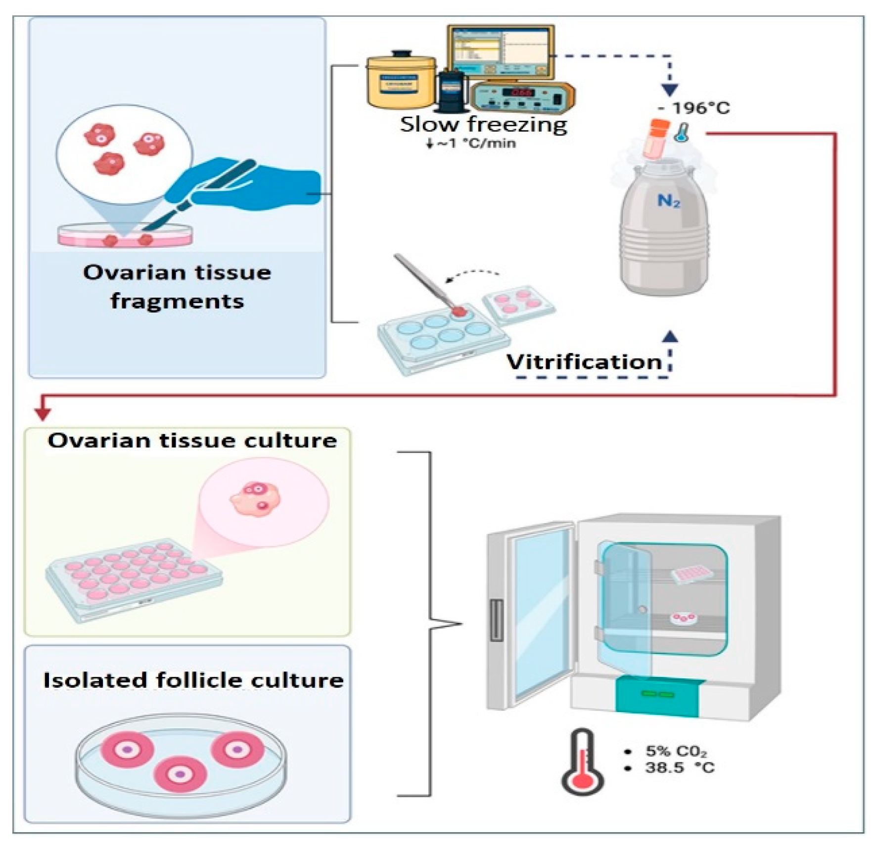
Figure 2.
Color Doppler ultrasonographic assessment of blood flow in regions adjacent to and surrounding transplanted caprine ovarian tissue. The images illustrate the progression of tissue blood perfusion (“hyperemia”) between days 0 and 7 after transplantation, highlighting the gradual increase in vascularization within the implant areas. Source: Prepared by the authors.
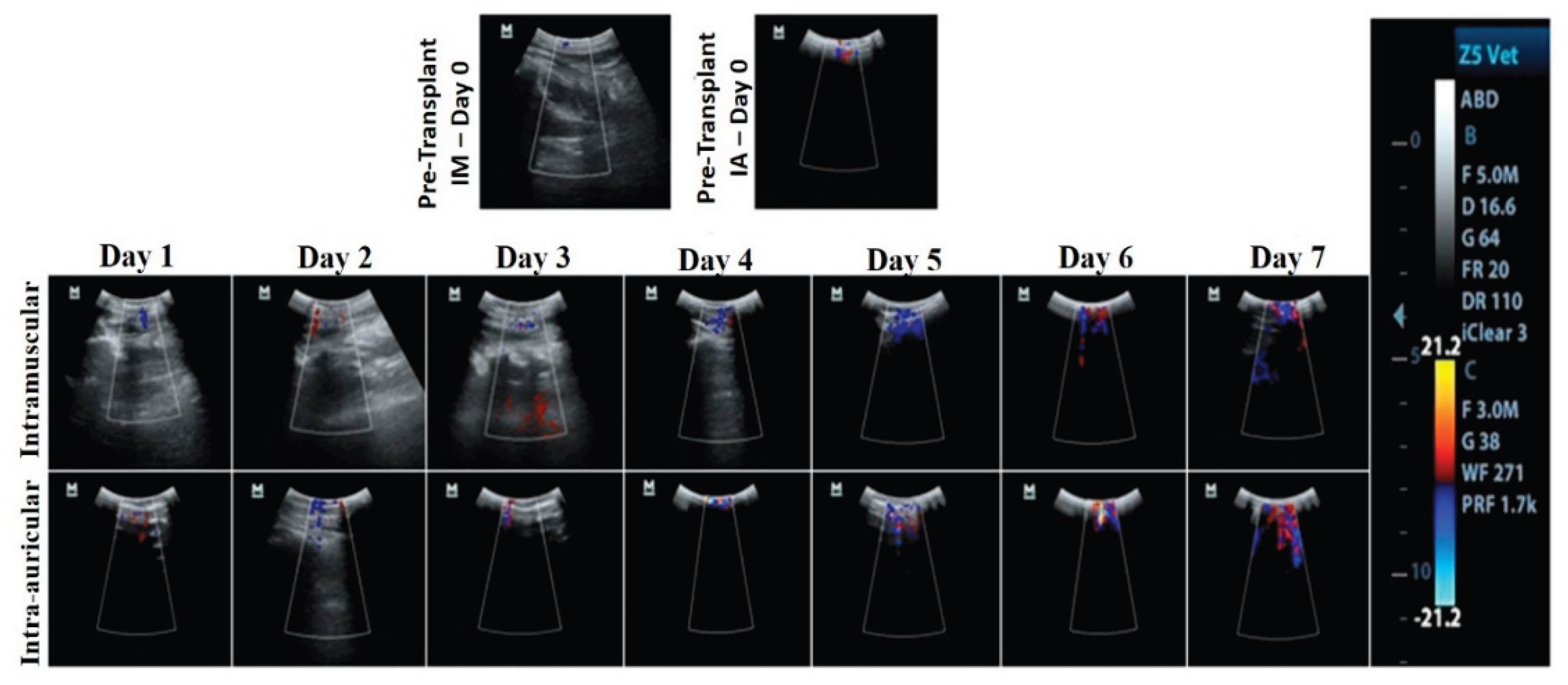

Table 1.
Experimental evidence on ovarian tissue cryopreservation and transplantation in sheep and goat species.
Table 1.
Experimental evidence on ovarian tissue cryopreservation and transplantation in sheep and goat species.
| Species | Investigated Aspects | Main Findings | Reference |
|---|---|---|---|
| Sheep | Evaluation of hormonal recovery, follicular and embryonic development after transplantation of whole frozen ovaries. | Re-establishment of ovarian function in the medium/long term. | [53] |
| Sheep | Assessment of FSH response, fertilization, morphology, and vascularization following heterotopic autotransplantation of whole frozen ovaries. | Presence of follicles at multiple stages, good vascularization and proliferation, although with lower yield compared to controls. | [54] |
| Sheep | Long-term functional evaluation of orthotopic transplantation of whole frozen ovaries using microvascular anastomosis. | Preservation of ovarian function in some animals, presence of follicles at different stages, ovulations, and substantial structural and hormonal recovery. | [55] |
| Sheep | Comparison between slow freezing and vitrification of ovarian tissue transplanted onto the chorioallantoic membrane (CAM) of chicken embryos. | Improved preservation of follicular integrity after slow freezing; more pronounced necrosis following vitrification. | [56] |
| Goat | Medium-term functional analysis of orthotopic transplantation of fresh or vitrified ovarian cortex in ovariectomized goats. | Maintenance of a high percentage of morphologically normal preantral follicles and restoration of endocrine function. No follicles were identified in vitrified tissue after grafting. | [57] |
| Goat | Analysis of primordial follicle survival after vitrification and transplantation of ovarian tissue onto the CAM. | Reduction in primordial follicle density after preservation and grafting, but without increased DNA fragmentation. | [58] |
| Goat | Doppler ultrasonographic evaluation of blood perfusion in ovarian fragments implanted in the ear and neck. | Greater blood flow area in ear implants at 7 and 15 days, indicating that implantation site influences revascularization. | [59] |
Table 2.
Classification of ovarian tissue transplantation types according to graft size, recipient host type, and implantation site.
Table 2.
Classification of ovarian tissue transplantation types according to graft size, recipient host type, and implantation site.
| Category | Transplant type | Reference |
|---|---|---|
| Graft size | Whole ovary | [61,62] |
| Hemi-ovary | ||
| Cortical fragment | ||
| Recipient host type | Autotransplantation¹ | [63,64,65] |
| Isotransplantation² | [66] | |
| Allotransplantation³ | [11,61,67] | |
| Xenotransplantation⁴ | [68] | |
| Implantation site | Orthotopic | [61] |
| Heterotopic | [67] |
Footnotes: ¹Autotransplantation: graft performed within the same individual. ²Isotransplantation: performed between genetically identical individuals (e.g., monozygotic twins). ³ Allotransplantation: performed between individuals of the same species but genetically distinct. ⁴ Xenotransplantation: performed between individuals of different species.
Table 3.
Main markers used in immunohistochemical studies of transplanted ovarian tissue and their respective purposes.
Table 3.
Main markers used in immunohistochemical studies of transplanted ovarian tissue and their respective purposes.
| Marker | Purpose | Reference |
|---|---|---|
| αSMA | Identification of mature blood vessels | [19,57,73] |
| SDF-1α | Evaluation of angiogenic or reparative activity | [87] |
| CD31 | Identification of newly formed blood vessels | [19,80] |
| Caspase-3 | Detection of apoptosis in stromal cells | [80] |
| DDX4 | Identification of primordial follicles | [88] |
| Ki-67 | Assessment of cell proliferation | [89] |
| PCNA | Assessment of cell proliferation | [73] |
Legend: αSMA: α–smooth muscle actin; SDF-1α: stromal cell–derived factor 1α; CD31: cluster of differentiation 31, also known as PECAM-1 (platelet endothelial cell adhesion molecule-1); Ki-67: antigen identified by clone 67 from the city of Kiel; PCNA: proliferating cell nuclear antigen.
Disclaimer/Publisher’s Note: The statements, opinions and data contained in all publications are solely those of the individual author(s) and contributor(s) and not of MDPI and/or the editor(s). MDPI and/or the editor(s) disclaim responsibility for any injury to people or property resulting from any ideas, methods, instructions or products referred to in the content. |
© 2026 by the authors. Licensee MDPI, Basel, Switzerland. This article is an open access article distributed under the terms and conditions of the Creative Commons Attribution (CC BY) license (http://creativecommons.org/licenses/by/4.0/).
Copyright: This open access article is published under a Creative Commons CC BY 4.0 license, which permit the free download, distribution, and reuse, provided that the author and preprint are cited in any reuse.


