
Submitted:
27 January 2026
Posted:
28 January 2026
You are already at the latest version
Abstract
Upconverting nanoparticles, which transform low-energy infrared radiation into high-energy visible or UV light, show great potential in today’s technology. High-quality upconversion colloid (UCC) consisting of lanthanide-based nanoparticles with a diameter of ~10 nm was obtained using a combination of two processes, high-temperature coprecipitation and hydrothermal treatment in an autoclave. The UCC was then PEGylated with PEG-alendronate (PEG-Ale) to facilitate its dispersion in aqueous cell culture media intended for in vitro cell uptake assays. The surface modification of the nanoparticles increased both the colloidal stability in water and the upconversion emission by mitigating surface quenching. UCC@Ale-PEG was characterized by transmission and scanning electron microscopy, dynamic light scattering, and by fluorescence microscopy detecting upconversion photoluminescence emission. The results of an in vitro assay revealed that this new generation of UCC can be internalized by various cell types including epithelial cells, macrophages, and glioma cells, upon several hours of exposure, suggesting broad application potential of this type of UCC in biomedicine, bioengineering, and environmental sciences.
Keywords:
upconversion
; colloid
; surface modification
; epithelial cells
; macrophages
; glioma cells
; internalization
1. Introduction
Advances in the development of nanoparticle-based technologies currently often exploit photon upconversion, in which excitation by low-energy near-infrared radiation (NIR) results in the emission of high-energy visible or ultraviolet light due to anti-Stokes shift, offering great potential for many applications [1]. The mechanism of upconversion luminescence is quite complex and, depending on the type of particles, involves at least three basic processes: absorption of the excited state, photon avalanche, and energy transfer upconversion [2]. In this regard, lanthanide-based upconversion colloid (UCC) is particularly interesting. It consists of an inert host matrix (typically NaYF4) doped with a sensitizer (usually Yb3+, Nd3+) and an activator (e.g., Tm3+, Er3+, Ho3+). UCC has many advantages, the major one being that it has no background in biological tissues and NIR light can penetrate relatively deep into the body [3]. In addition, it has no photobleaching, non-blinking, sharp emission bands, long photoluminescence lifetime, low photodamage to surrounding tissues, and tunable emission color [4].
Various approaches are being used for the synthesis of the UCC with controllable morphology of nanoparticles (i.e., shape and size), specific chemical composition, crystal structure, and optical properties [5]. In terms of size, the prevailing trend in manufacturing is to reduce it in order to facilitate internalization and diffusion of the nanoparticles into cells and tissues. In addition, if the size is <10 nm, the particles can be easily biodegraded or excreted from the body, but at the same time they can be prone to surface quenching, which reduces the efficiency of upconversion luminescence [6,7]. Nevertheless, both biodegradation and excretion of particles also depend on their composition, surface charge, hydrophilic properties, etc. [8]. Common methods of UCC synthesis include thermal decomposition or coprecipitation of metal salts in high-boiling solvents in the presence of compounds with polar groups and long hydrocarbon chains (e.g., oleylamine and oleic acid), hydrothermal/solvothermal techniques, microwave heating, etc. [9,10,11]. However, particles prepared in organic solvents in this way are hydrophobic, undispersible in water, and therefore not suitable for biological applications. Moreover, despite their promising optical properties, bioapplications of UCC are limited by other challenges such as chemical and colloidal stability, resistance to aggregation and non-specific interactions, biocompatibility, reproducibility, and functionalization [12]. A significant concern relates to the dissolution of UCC nanoparticles in physiological aqueous environments, which is accompanied by the release of potentially cytotoxic fluoride and lanthanide ions [13]. For example, fluoride ions can inhibit mitochondrial activity, cell growth, protein synthesis, and proliferation in cultured human pulp cells [14].
It is therefore necessary to choose a suitable strategy for transferring hydrophobic UCC nanoparticles from the organic to the aqueous phase and to modify their surface using various ligands and polymer coatings of sufficient thickness [15]. Here, ligand exchange and conjugation with a number of different low- and high-molecular weight hydrophilic molecules and chelating agents such as citric acid, poly(ethylene glycol) (PEG), poly(maleic anhydride-alt-1-octadecene)-PEG, poly(isobutylene-alt-maleic) anhydride-PEG, polyvinylpyrrolidone, poly(acrylic acid), polyethyleneimine, and encapsulation in a silica shell have been described [16,17,18]. At the same time, it is important that the coating is not washed off from the surface of the particles. Therefore, the polymer should contain effective anchoring groups that attach to the surface of UCC nanoparticles through non-covalent or covalent interactions; examples include bisphosphonate functional groups of PEG-alendronate [19].
Due to their remarkable physicochemical properties, upconversion nanoparticles are used in a number of industrial applications as well as in the life sciences [20]. In the former case, these include photovoltaics (solar cells), displays, photocatalysis, security (anti-counterfeiting, fingerprinting), and barcoding. In nanomedicine, they include biosensing, theranostics, in vitro/in vivo bioimaging, drug delivery (photodynamic and photothermal therapy), and optogenetics [4]. In these situations, it is always important to know the detection limit, i.e., the lowest functional concentration of nanoparticles in UCC that can be determined from the upconversion luminescence signal.
In this paper, new UCC based on small (~10 nm) PEGylated NaYF4:Yb,Er nanoparticles was designed for future bioapplications including environmental sciences. The goal of this study was to monitor the internalization of this model colloid into different cell lines.
2. Experimental
2.1. Materials
YCl3, YbCl3, ErCl3∙6H2O, octadec-1-ene, oleic acid, paraformaldehyde, Hoechst 33342 stain, and fetal bovine serum (FBS) were purchased from Sigma-Aldrich (St. Louis, MO, USA). DMEM medium was obtained from Merck (Darmstadt, Germany) and N-hydroxylsuccinimide-functionalized methoxy poly(ethylene glycol) (MeO-PEG-NHS; Mn = 5,000 g/mol) was from Rapp Polymere (Tübingen, Germany). The sodium salt of 4-amino-1-hydroxy-1-phosphonobutyl phosphonic acid trihydrate (alendronate; Ale) was purchased from TCI (Tokyo, Japan). 4′,6-Diamidino-2-phenylindole (DAPI) and CellMask Deep Red were purchased from Thermo Fisher Scientific (St. Louis, MO, USA). Fetal bovine serum (FBS) was purchased from Biosera (Cholet, France). All other chemicals and solvents were from Lachema (Brno, Czech Republic). Deionized water was prepared using a Milli-Q IQ7000 system (Merck).
2.2. Synthesis of PEG-Alendronate (PEG-Ale)
PEG-Ale was obtained by modifying a previously published method [21]. A solution of sodium alendronate (0.64 g; 2.0 mmol) in phosphate-buffered saline (PBS; pH = 7.4) was cooled to 5 °C. MeO-PEG-NHS (1.0 g; 0.2 mmol NHS groups) was added to the solution with stirring and the reaction mixture was maintained at 5 °C for 5 h to facilitate reaction between the NHS ester and the amino group of alendronate under formation of an amide bond. The resulting PEG-Ale was then purified by size exclusion chromatography using a Sephadex G-25 column with water as eluent and lyophilized.
2.3. Synthesis of UCC
The synthesis of UCC was performed in two stages. First, in a 100 ml three-necked flask, lanthanide chlorides (YCl3, YbCl3, and ErCl3·6H2O in the molar ratio of 0.78:0.20:0.02, respectively) were dissolved in octadec-1-ene (30 ml) and oleic acid (12 ml) at 160 °C for 30 min with stirring (350 rpm) under an Ar atmosphere. After cooling the reaction mixture to 40 °C, a methanolic solution (12 ml) of NaOH (2.5 mmol) and NH4F (4 mmol) was added dropwise. To evaporate the methanol and residual water, the temperature was gradually increased to 120 °C and then maintained for 30 min with stirring in an Ar atmosphere. In the second step, the mixture was cooled to 60 °C in the above atmosphere, transferred to an autoclave equipped with a magnetic stirrer, and 0.75 ml of water was added. The sealed autoclave was subsequently heated in the sand bath at 310 °C for 1.5 h with stirring at 350 rpm. After cooling to RT, ethanol was added to the resulting UCC, the particles were separated by centrifugation (3,460 rcf) for 30 min and then redispersed in hexane.
For biological experiments, it was necessary to transfer UCC into water. This was achieved by gradually replacing hexane with ethanol (ethanol/hexane 1:3 and 2:3 v/v, and then ethanol alone) with centrifugation (3,460 rcf) for 30 min after each step. The ethanol was then gradually replaced with deionized water using the same gradient procedure. Finally, the particles were washed twice with deionized water.
2.4. PEGylation of UCC
PEG-Ale (30 mg) was added to an aqueous UCC (1 ml; 22.2 mg particles/ml), the mixture was stirred at RT for 12 h and dialyzed for 48 h against deionized water using a cellulose Spectra/Por® membrane (MWCO = 100 kDa; Spectrum Laboratories; Rancho Dominquez, CA, USA) to remove unbound components. The resulting PEG-functionalized nanoparticles (UCC@Ale-PEG) were collected by centrifugation (3,460 rcf) and resuspended in water.
2.5. Characterization Methods
Ultrastructural analysis of UCC by transmission electron microscopy (TEM) has been done as follows: first, the surface of grids for electron microscopy (carbon-coated copper 400 mesh—EMS #215-412-8400) was hydrophilized by glow-discharge using Plasma HPT-100 cleaner (Henniker; Runcorn, UK), grids were then floated on a drop of a sample for 3 min, blotted and air-dried. TEM micrographs were acquired on JEM2100-Plus apparatus (JEOL; Tokyo, Japan) operated at 200 kV using TVIPS TemCam XF416 camera (Gauting, Germany). The number-average (Dn), weight-average diameter (Dw), and dispersity (Ð) of at least 300 particles were defined as:
where ni and Di are the number and diameter of the particle, respectively.
Dn = Σ ni∙Di/Σ ni,
Dw = Σ ni∙Di4/Σ ni∙Di3,
Ð = Dw/Dn,
Ultrastructural analysis of UCC by scanning electron microscopy (SEM) has been done as follows: first, serial dilution of a sample was done in Milli-Q water and 1 µl drops from each dilution were applied on a carbon-coated glass coverslip, air dried and mounted on SEM pins. SEM micrographs were acquired on Helios NanoLab 660 G3 UC apparatus (Thermo Fischer Scientific; Waltham, MA, USA) using retractable high contrast solid-state backscatter electron detector.
Dynamic light scattering (DLS) using a ZSU 5700 Zetasizer Ultra (Malvern Instruments; Malvern, UK) provided hydrodynamic diameter Dh, polydispersity PD values, and ζ-potential for the nanoparticles.
Photoluminescence spectra were recorded using an FS5 spectrofluorometer (Edinburgh Instruments; Edinburgh, UK) equipped with a 980 nm CW laser and 2 W output power.
2.6. Cells Experiments
2.6.1. Cell Culture and Labeling.
Cell lines Caco-2 (ATCC #HTB-37, Manassas, VA, USA), RAW264.7 (ATCC #TIB71; Manassas), and C6 (ATCC #CCL-107, Manassas) were cultured in DMEM medium enriched with 10% FBS supplemented with non-essential amino acids (Thermo Fisher Scientific), L-glutamine, and a mixture of antibiotics (penicillin, streptomycin, and amphotericin B from Merck). Cells were grown in a thermostat at 37 °C set to 5% CO2 with medium changed twice a week.
For the cell uptake assay, RAW264.7 cells were seeded in 8-well glass-bottom chamber (Ibidi µ-Slide) without coating (Gräfelfing, Germany). The UCC@Ale-PEG was added to the cells after media exchange and incubated for 16 and 24 h. Cells were then washed with PBS, stained with CellMask Deep Red (1 µg/ml), fixed with 4% paraformaldehyde in PBS, washed in PBS, and subjected to DNA (cell nuclei) staining by Hoechst 33342 (20 µM) followed by microscopy imaging.
Caco-2 cells were seeded at low density in 35-mm µ-dishes with glass bottom coated with collagen. The cells were then grown to full confluence and cultivated for another two weeks to differentiate into enterocytes forming polarized monolayer. After that, cells were starved overnight in serum-free medium for synchronization of cell cycle (G0/G1 phase arrest). The UCC@Ale-PEG was added in FBS-containing medium to trigger the uptake and incubated for 16 and 24 h. Cells were then washed with PBS, plasma membrane stained with CellMask Deep Red, DNA (cell nuclei) stained with Hoechst 3342, and finally fixed with 4% paraformaldehyde in PBS.
C6 cells were seeded into a six-well plate on collagen-coated slides. The UCC@Ale-PEG was added to the cells after media exchange and incubated for 24 h. Cells were then fixed by 3% paraformaldehyde, washed in PBS, and subjected to DNA staining by DAPI followed by microscopy imaging.
2.6.2. Microscopy.
Samples prepared from all three cell types were analyzed on Carl Zeiss LSM 880 NLO fluorescence optical microscope (Oberkochen, Germany) equipped with a tunable femtosecond laser Chameleon Ultra II (Coherent, PA, USA). The UCC@Ale-PEG was excited with focused 966 nm laser beam, pulse frequency 80 MHz, pulse width 320 fs, and average power 1 mW at sample plane, using 40× 1.1 NA water immersion objective. The bidirectional scanning was set to the lowest possible speed of 66 µs per pixel and 140 nm pixel size, with the pinhole fully open to allow the upconversion signal—emitted up to milliseconds after the excitation—reach the confocal detector. The spectral emission profile of upconversion signal was verified by lambda detection on 32 channels (8.8 nm per channel) in 410-690 nm range of GaAsP spectral detector operated in photon counting mode. For standard UCC@Ale-PEG detection, two channel settings (540-570 nm and 640-670 nm), using the same detector in photon counting mode, were used. Due to the opened pinhole, the axial resolution for UCC@Ale-PEG imaging was similar to wide-field type of acquisition, which is several µm. CellMask Deep Red and Hoechst 33342 signals were one-photon excited with 633 or 405 nm lasers, respectively, and detected also on spectral GaAsP detector using channel mode, with confocal pinhole size set to 1 Airy unit. The axial resolution for DAPI and CellMask Deep Red corresponded to the confocal mode, which was ~1 µm. UCC@Ale-PEG and DAPI with CellMask Deep Red images were acquired in frame (2D experiment) or stack (3D experiment) sequential scanning mode. Orthogonal projections from confocal z-stacks were used to determine precise localization of UCC@Ale-PEG within cells and to discriminate them from surface-bound particles.
3. Results and Discussion
3.1. UCC
NaYF4:Yb,Er-based UCC was prepared using our newly developed hybrid strategy integrating high-temperature coprecipitation of lanthanide chlorides in a high-boiling solvent (octadec-1-en) at 310 °C with subsequent hydrothermal treatment in an autoclave. This combination of two synthetic approaches was designed to enable control of particle size, dispersity, and crystallinity. It is based on a previous study that showed the significant influence of water added to the reaction feed on particle size [22]. Under normal synthesis conditions at atmospheric pressure in a reaction vessel containing undried lanthanide precursors, an uncontrollable amount of water is present, originating from erbium chloride hexahydrate and a methanolic solution of NaOH and NH4F; the size of the resulting particles is then typically in the range of 20-30 nm. In contrast, if the reactants are rigorously dried, particles >100 nm are formed [23]. In this report, in order to significantly reduce the particle size, a controlled amount of water was added to the reaction mixture with completion of the reaction in the autoclave. This approach ensured that water was retained in the feed even at high temperature (310 °C) and elevated pressure and promoted the nucleation of primary particles at the expense of their growth. The ultrastructural analysis of UCC dried from hexane using TEM showed relatively well-dispersed nanoparticles with a high concentration in a monolayer with Dn = 9 nm and a dispersity Ð = 1.09 (Figure 1 a) indicating a narrow particle size distribution. The particles prepared by the hybrid method of high-temperature coprecipitation and hydrothermal treatment in an autoclave were thus much smaller than the upconverting nanoparticles obtained by common techniques, such as coprecipitation of lanthanide chlorides, thermal decomposition of lanthanide oleates, or microwave-assisted synthesis. This can be explained by the nucleation mechanism of particle formation. The nuclei formed during the above-mentioned hybrid approach were in more quantity, but smaller in size than when using conventional coprecipitation. Moreover, in the latter method, nuclei rapidly grew into mature particles at the expense of small ones due to Ostwald ripening. Dynamic light scattering (DLS) measurement of the UCC in hexane showed hydrodynamic diameter Dh = 18 nm with polydispersity PD = 0.17 (Table 1). The relatively small hydrodynamic diameter of UCC in hexane confirmed the positive role of the residual oleic acid on the particle surface, which makes them well-dispersed in a nonpolar solvent, as the hydrophobic interactions minimize aggregation. The somewhat larger Dh value of the particles than Dn was ascribed to the fact that TEM measured the particles in a dry state, whereas DLS characterized them in a solvated state, which increased their size. DLS then revealed pronounced difference in the hydrodynamic behavior of UCC depending on the dispersion medium. UCC in water exhibited Dh = 105 nm, which was significantly more than in hexane, indicating the tendency of unmodified nanoparticles to form aggregates in aqueous environments. Such aggregation arises from high surface energy and insufficient colloidal stabilization. Polydispersity PD = 0.19 then remained practically unchanged from that of UCC in hexane.
The upconversion luminescence spectrum of UCC excited by a 980-nm laser exhibited typical Er3+ emission peak at 408 nm attributed to the 2H9/2 → 4I15/2 electron transition (Figure 2). The high-intensity peaks at 520 nm (green), 545 nm (green) and 650 nm (red) were induced by the 2H11/2 → 4I15/2, 2H9/2 → 4I11/2, and 4F9/2 → 4I15/2 transitions, respectively. The spectra clearly showed that UCC in hexane exhibited high emission intensity due to the fact that hexane is a nonpolar solvent without high-energy O–H vibrational modes. In contrast, in water, strong O–H stretching vibrations (at ~3,400 cm-1) could effectively couple with Er3+ excited states, inducing multiphoton non-radiative relaxation and significantly quenching luminescence. In addition, in water, the residues of oleic acid on the surface of particles were removed and the surface was susceptible to quenching and surface defects. In contrast, in hexane, oleic acid remained intact, preserving surface passivation and ensuring colloidal stability, which enhanced emission.
3.2. PEGylation of UCC
To render UCC colloidally stable in aqueous environments over a long-term period, it was PEGylated using PEG-Ale, whose bisphosphonate groups complex with the lanthanide ions of the particles, thereby firmly anchoring the PEG to their surface. TEM micrograph of UCC@Ale-PEG dried from water showed the dispersed nanoparticles with Dn = 11 nm and Ð = 1.13 confirming a relatively narrow particle size distribution (Table 1; Figure 1 b). Based on particle size measurements, the PEGylated particles appeared to be ~2 nm larger than the starting particles.
Upon PEGylation of UCC, the Dh decreased to 90 nm with a low PD = 0.14, reflecting improved colloidal stability and reduced aggregation in water compared to unmodified UCC (Table 1). Also, when UCC@Ale-PEG was transferred to DMEM medium, the nanoparticles remained dispersed, which is important for further biological utilization (Figure 1 c). This can be ascribed to the PEG chains acting as a hydrophilic steric barrier that prevents close particle-particle contacts, thereby narrowing the particle size distribution. The ζ-potential measurements then showed a decrease from 25 mV for neat UCC to 14 mV for UCC@Ale-PEG. This reduction of surface charge was consistent with partial shielding of surface lanthanide ions by conjugated PEG chains, which decrease the contribution of positively charged lanthanides to the electro-kinetic potential. Importantly, despite this reduction, the ζ-potential of PEGylated particles accompanied by steric stabilization contributed to preserving colloidal stability. When UCC was coated with Ale-PEG and dispersed in water, the emission intensity modestly but noticeably increased compared to uncoated particles in water (Figure 2). This improvement can be attributed to the fact that the Ale-PEG shell partially shielded the UCC surface from contact with water, thereby reducing non-radiative quenching by O–H vibrations. In addition, PEG contributed to improved colloidal stability, which prevented aggregation and excessive scattering. Although the emission in water was weaker than in hexane, surface functionalization restored some of the lost brightness by mitigating the effects of surface quenching.
3.3. Cell Uptake Assay of the UCC@Ale-PEG
Internalization of UCC@Ale-PEG into the cells is a prerequisite for its potential use as a long-term in vivo tracer and/or marker, as the particles must first cross one of the epithelial barriers to enter the body and then spread by body fluids. In order to assess whether and to what extent the UCC@Ale-PEG is internalized into cells, we performed in vitro cell uptake assay evaluated by fluorescence microscopy. The nature of the UCC@Ale-PEG and the method of its detection in cells by measuring emission from photon upconversion has important advantages. First, the emission is not dependent on pH, which drops significantly in the endosomal compartment when the late endosome (with internalized cargo) fuses with lysosome; second, the particles do not undergo fast enzymatic degradation in endo-lysosomal compartments (due to their composition), neither proteolytic cleavage in the cytosol and thus have essentially unlimited lifetime and fluorescence quantum yield. The rate of endocytosis, which is main pathway for particle internalization, varies significantly depending on the cell type. Cells performing specialized functions, such as gut epithelial cells and macrophages, have exceptionally high uptake rate due to their role in nutrient absorption and immune defense, respectively.
In the experiments, three complementary in vitro models were employed to investigate cellular interactions with UCC@Ale-PEG: Caco-2 intestinal epithelial cells, macrophages, and C6 rat glioma cell line. Caco-2 cells, which are human gut tissue-derived epithelial cells undergoing differentiation into enterocytes and forming polarized monolayer in vitro mimicking the small intestine in the gastrointestinal system, were tested first. The choice of this cell line was to probe the ability of nanoparticles to cross the intestine epithelial barrier, the primary port of their entry into the body in vivo. Caco-2 cells were grown into high confluency and left to differentiate into stratified epithelium with formation of an apical brush border, the site of endocytosis and potential uptake of the UCC@Ale-PEG.
The second cell line was RAW264.7 macrophages, which are commonly used as a model of inflammation in mice. Since macrophages can cross the blood-brain barrier under certain conditions, they represent a convenient model to study foreign particle transfer throughout the body and their potential accumulation in target organs (including brain). In addition to the three major modes of endocytosis (clathrin-mediated endocytosis, caveolae-mediated endocytosis, and macropinocytosis) that are common to both selected cell models, RAW macrophages are very potent in phagocytosis, namely, the internalization (and digestion) of large solid objects, usually pathogens, foreign particles, or apoptotic bodies. This allows the internalization of even very large UCC@Ale-PEG aggregates to be monitored, which would otherwise not be internalized by classical endocytosis.
Finally, C6 glial cells of the brain and peripheral nervous system widely used in neurobiology and cancer research were employed as an astrocyte-like central nervous system model to assess particle contact and internalization in neural-relevant cells. C6 cells are pleomorphic with variable nuclear shapes, being an ideal model similar to human glioblastoma in terms of invasiveness, immune microenvironment, and vascular behavior [24].
Orthogonal views of z-stacks of both UCC@Ale-PEG and uncoated UCC incubated with Caco-2 cells and Raw264.7 macrophages discriminated intracellular (internalized) particles from particles still adhered to the cell surface (extracellular; Figure 3 and Figure 4). The results confirmed that both types of cells internalized both PEGylated and uncoated UCC, although to various extent and efficiency. A substantial proportion of particles remained on the cell surface after 16-24 h of incubation, whereby the pattern of the fluorescence signal, namely the size and intensity of the fluorescent spots, suggested that most of them were aggregates that were poorly internalized by endocytosis. Confocal micrograph of C6 cells incubated with UCC@Ale-PEG showed that the cells could engulf the particles, which could then distribute around the DAPI-stained cell nuclei (Figure 5 a), as indicated by the same micrograph taken with a red laser (Figure 5 b). Some particles (or their clusters) could also be adsorbed onto the cell surface.
In order to assess the sensitivity of detection of UCC@Ale-PEG and determine whether upconversion emission can be tracked from single particles or only from their smaller aggregates or from intracellular endosomes where they naturally accumulate upon internalization, the UCC@Ale-PEG detection limit assay was designed. A series of dilutions of UCC@Ale-PEG was prepared and transferred in 1 µl drops onto glass coverslips, one uncoated for fluorescence measurement and the other coated with carbon for SEM analysis. The goal was to determine the dilution of UCC@Ale-PEG that provides the distribution of single particles on a coverslip with a spacing between particles >1 µm (using SEM with back-scattered electron detector), and then to assess whether single dots of emission can be detected in the same dilution using fluorescence microscopy. The results showed that higher concentration of UCC@Ale-PEG formed an almost continuous monolayer of particles after drying on the coverslip surface, which was confirmed by SEM. The emission signal from this monolayer was significantly higher than the non-specific background signal from the regions of interest (ROI) on the coverslip that did not contain UCC used as negative control (Figure 6).
Upconversion fluorescence emission spots were detected in each of the tested dilutions yet with intensities (peak gray values in the histogram) decreasing proportionally with the dilution and with the size of the spots (aggregates) until dilution of 1:1000, where little upconversion signal (if any) was detected. The smallest discrete spots distinguishable by fluorescence had diameter between 6-10 pixels (pixel size 86 nm) (Figure 6).
Analysis of corresponding dilutions by SEM revealed that separation of individual nanoparticles to ~1 μm spacing between particles was achieved in sample diluted 1:1000 resulting in ~3-4 nanoparticles per μm2. When the same dilution was analyzed by fluorescence (upconversion emission) within the ROI area comparable to the SEM image (6 × 8 μm rectangle), signal from individual UCC was not detected (Figure 6). We suppose that the bright spots detected by the fluorescence microscopy originated from small aggregates rather than from individual nanoparticles, but it was not possible to discriminate whether these aggregates formed during drying of the colloid on the glass surface or whether they were already present in the colloid.
4. Conclusions
Previous studies have pointed to limitations in size control of the prepared UCC, which was typically in the range of 30-160 nm [25]. In this report, upconversion colloid based on small PEGylated NaYF4:Yb,Er nanoparticles (~10 nm) was designed using newly developed combined coprecipitation in the presence of water and hydrothermal treatment in an autoclave. The small size resulted from increased nucleation during the particle formation process. Such a size, together with high dispersibility in water-based carriers, is advantageous for achieving high endocytosis efficiency. It is also important to mention that the resulting narrow particle size distribution ensures their identical physicochemical and biological properties, reproducible and predictable behavior, and improved colloidal stability. The colloid was characterized not only in terms of particle size and size distribution using TEM and DLS, but also upconversion fluorescence. Modification of the UCC surface with PEG-Ale improved upconversion emission in water by reducing surface quenching. The results then demonstrated that the dispersion medium and surface functionalization critically affected the colloidal stability of UCC. While UCC containing oleic acid residues on the particle surface remained well dispersed in nonpolar hexane, it tended to aggregate in polar water. PEGylation mitigated this problem by providing hydrophilic steric stabilization, reducing both hydrodynamic size and polydispersity, and enabling the use of such particles for biological applications in an aqueous environment.
Finally, three cellular systems (epithelial Caco-2 cells, Raw264.7 macrophages, and tumor C6 cells) provided a framework to characterize UCC–cell interactions and nanoparticle internalization across intestinal, immune, and neural-like contexts. Differences between epithelial cells, tumor cells, and macrophages reflect distinct endocytic and phagocytic mechanisms, which are highly relevant for the rational design of nanoparticle-based diagnostic or therapeutic platforms. In particular, uptake by macrophages highlights the relevance of this system for modeling interactions with the innate immune system, including nanoparticle clearance and intracellular fate, which is critical for assessing biocompatibility and translational potential of nanomaterials. Cell uptake assays with the UCC@Ale-PEG confirmed its capacity to be internalized by various cell types after co-incubation in growing medium. Interpolating results of these in vitro experiments to undoubtedly more complex environment in vivo, we propose that UCC@Ale-PEG would be able to cross the epithelial barrier and potentially reach the brain, where it could accumulate. In a prospective study, it will be interesting to assess whether internalization of UCC@Ale-PEG by these cells has any long-term impact on their viability or growth rate. It may also be important to determine what is the fate of this intracellular UCC after internalization, whether it slowly undergoes some degradation or whether it is expelled from the cells by exocytosis as a non-degradable substance. Further experiments should finally be transferred to in vivo models. Although nanoparticle internalization, transport within tissues and organs, and long-term fate and persistence in the organism are complex processes, the unique nature of UCC@Ale-PEG, namely colloidal stability, fluorescent properties with high photostability, and NIR excitation, enable sustained cell labeling with minimal background signal. These characteristics are particularly suitable for applications in nanomedicine, such as in vivo bioimaging, sensing, NIR-activated photodynamic therapy, or intracellular drug delivery.
Author Contributions
Conceptualization, D.H. and D.L.; methodology, D.M.; validation, A.B., formal analysis, H.M.; investigation, M.N. and P.P.; data curation, L.M.U.; writing—oiginal draft preparation, D.H., D.L. and L.M.U.; writing—review and editing, D.H.; visualization, M.N.; supervision, D.H.; funding acquisition, D.H. All authors have read and agreed to the published version of the manuscript.
Acknowledgments
Support of the Czech Science Foundation (No. 25-16155S) is acknowledged.
Conflicts of Interest
The authors declare no conflicts of interest.
Abbreviations
The following abbreviations are used in this manuscript:
| Ð | Dispersity |
| Dh | Hydrodynamic diameter |
| Dn | Number-average diameter |
| Dw | Weight-average diameter |
| DAPI | 4′,6-Diamidino-2-phenylindole |
| DLS | Dynamic light scattering |
| DMEM | Dulbecco’s modified Eagle’s medium |
| DNA | Deoxyribonucleic acid |
| FBS | Fetal bovine serum |
| MeO-PEG-NHS | N-Hydroxylsuccinimide-functionalized methoxy poly(ethylene glycol) |
| MWCO | Molecular weight cut-off |
| NIR | Near-infrared radiation |
| PBS | Phosphate-buffered saline |
| PD | Polydispersity |
| PEG-Ale | Poly(ethylene glycol)-alendronate |
| ROI | Region of interest |
| SEM | Scanning electron microscopy |
| TEM | Transmission electron microscopy |
| UCC | Upconversion colloid |
References
- Zhang, Y.; Du, W.; Liu, X. Photophysics and its application in photon upconversion. Nanoscale 2024, 16, 2747. [Google Scholar] [CrossRef]
- Geng, S.; Li, H.; Lv, Z.; Zhai, Y.; Tian, B.; Luo, Y.; Zhou, Y.; Han, S.-T. Challenges and opportunities of upconversion nanoparticles for emerging NIR optoelectronic devices. Adv. Mater 2025, 2419678. [Google Scholar] [CrossRef]
- Ruggiero, E.; Alonso-de-Castro, S.; Habtemariam, A.; Salassa, L. Upconverting nanoparticles for the near infrared photoactivation of transition metal complexes: New opportunities and challenges in medicinal inorganic photochemistry. Dalton Trans. 2016, 45, 13012–13020. [Google Scholar] [CrossRef]
- Chen, G.; Qiu, H.; Prasad, P.; Chen, X. Upconversion nanoparticles: Design, nanochemistry, and applications. Chem. Rev. 2014, 114, 5161–5214. [Google Scholar] [CrossRef]
- Zhang, Z.; Zhang, Y. Orthogonal emissive upconversion nanoparticles: Material design and applications. Small 2021, 17, 2004552. [Google Scholar] [CrossRef]
- Liu, J.; Yu, M.; Zhou, C.; Zheng, J. Renal clearable inorganic nanoparticles: A new frontier of bionanotechnology. Mater. Today 2013, 38, 676–681. [Google Scholar] [CrossRef]
- Gargas, D.; Chan, E.; Ostrowski, A.; Aloni, S.; Altoe, M.; Barnard, E.; Sanii, B.; Urban, J.; Milliron, D.; Cohen, B.; Schuck, P. Engineering bright sub-10-nm upconverting nanocrystals for single-molecule imaging. Nat. Nanotechnol. 2014, 9, 300–305. [Google Scholar] [CrossRef] [PubMed]
- Albanese, A.; Tang, P.; Chan, W. The effect of nanoparticle size, shape, and surface chemistry on biological systems. Annu. Rev. Biomed. Eng. 2012, 14, 1–16. [Google Scholar] [CrossRef] [PubMed]
- Liu, X.; Liu, M.; Chen, J.; Li, Z.; Yuan, Q. Rational design and biomedical applications of DNA-functionalized upconversion nanoparticles. Chin. Chem. Lett. 2018, 29, 1321–1332. [Google Scholar] [CrossRef]
- Boyer, J.C.; Vetrone, F.; Cuccia, L.A.; Capobianco, J.A. Synthesis of colloidal upconverting NaYF4 nanocrystals doped with Er3+, Yb3+ and Tm3+, Yb3+ via thermal decomposition of lanthanide trifluoroacetate precursors. J. Am. Chem. Soc. 2006, 128, 7444–7445. [Google Scholar] [CrossRef]
- Kang, N.; Ai, C.-C.; Zhou, Y.-M.; Wang, Z.; Ren, L. Nanotechnology 2018, 29, 075601. [CrossRef] [PubMed]
- Phuong, H.; Huong, T.; Vinh, L.; Thao, D.; Tu, L.; Cong, T.; Van, N.; Tien, T. SCN–IgG functionalized NaYF4:Yb3+/(Er3+,Tm3+) upconversion nanoparticles for targeted fluorescence imaging of liver cancer cells. RSC Adv. 2025, 15, 22682–2268. [Google Scholar] [CrossRef] [PubMed]
- Salomão, P.M.A.; Oliveira, F.A.; Rodrigues, P.D.; Al-Ahj, L.P.; Gasque, K.C.D.S.; Jeggle, P.; Buzalaf, M.A.R.; Oliveira, R.C.; Edwardson, J.M.; Magalhães, A.C. The cytotoxic effect of TiF4 and NaF on fibroblasts is influenced by the experimental model, fluoride concentration and exposure time. PLOS ONE 2017, 12, e0179471. [Google Scholar] [CrossRef]
- Chang, Y.C.; Chou, M.Y. Cytotoxicity of fluoride on human pulp cell cultures in vitro. Oral Surg. Oral Med. Oral Pathol. Oral Radiol. Endod. 2001, 91, 230–234. [Google Scholar] [CrossRef]
- Ansari, A.A.; Parchur, A.K.; Chen, G. Surface modified lanthanide upconversion nanoparticles for drug delivery, cellular uptake mechanism, and current challenges in NIR-driven therapies. Coord. Chem. Rev. 2022, 457, 214423. [Google Scholar] [CrossRef]
- Chang, H.; Xie, J.; Zhao, B.; Liu, B.; Xu, S.; Ren, N.; Xie, X.; Huang, L.; Huang, W. Rare earth ion-doped upconversion nanocrystals: Synthesis and surface modification. Nanomaterials (Basel) 2014, 25, 1–25. [Google Scholar] [CrossRef]
- Malhotra, K.; Kumar, B.; Piunno, P.A.E.; Krull, U.J. Cellular uptake of upconversion nanoparticles based on surface polymer coatings and protein corona. ACS Appl. Mater. Interfaces 2024, 16, 35985–36001. [Google Scholar] [CrossRef]
- Muhr, V.; Wilhelm, S.; Hirsch, T.; Wolfbeis, O.S. Upconversion nanoparticles: From hydrophobic to hydrophilic surfaces. Acc. Chem. Res. 2014, 47, 3481–3493. [Google Scholar] [CrossRef] [PubMed]
- Vasylyshyn, T.; Patsula, V.; Větvička, D.; Shapoval, O.; Pankrác, J.; Kabešová, M.; Beneš, J.; Horák, D. Intraperitoneal versus intravenous administration of Flamma®-conjugated PEG-alendronate-coated upconversion nanoparticles in mice pancreatic cancer model. Nanoscale Adv. 2025, 7, 144–154. [Google Scholar] [CrossRef]
- Zhou, B.; Shi, B.; Jin, D.; Liu, X. Controlling upconversion nanocrystals for emerging applications. Nat. Nanotechnol. 2015, 10, 924–936. [Google Scholar] [CrossRef]
- Kostiv, U.; Lobaz, V.; Kučka, J.; Švec, P.; Sedláček, O.; Hrubý, M.; Janoušková, O.; Francová, P.; Kolářová, V.; Šefc, L.; Horák, D. A simple neridronate-based surface coating strategy for upconversion nanoparticles: Highly colloidally stable 125I-radiolabeled NaYF4:Yb3+/Er3+@PEG nanoparticles for multimodal in vivo tissue imaging. Nanoscale 2017, 9, 16680–16688. [Google Scholar] [CrossRef]
- Nahorniak, M.; Horák, D.; Šlouf, M.; Steinhart, M.; Shapoval, O.; Engstová, H.; Ježek, P. Lanthanide-based UCNPs: Toxicity evaluation and interaction of ultrasmall core vs. core–shell nanoparticles with cells. Mater. Adv. 2025, 6, 6907–6918. [Google Scholar] [CrossRef]
- Nahorniak, M.; Oleksa, V.; Vasylyshyn, T.; Pop-Georgievski, O.; Rydvalová, E.; Filipová, M.; Horák, D. Cytotoxicity evaluation of photosensitizer-conjugated hexagonal upconverting nanoparticles. Nanomaterials 2023, 13, 1535. [Google Scholar] [CrossRef] [PubMed]
- Giakoumettis, D.; Kritis, A.; Foroglou, N. C6 cell line: The gold standard in glioma research. Hippokratia 2018, 22, 105–112. [Google Scholar] [PubMed]
- Wang, M.; Abbineni, G.; Clevenger, A.; Mao, C.; Xu, S. Upconversion nanoparticles: Synthesis, surface modification and biological applications. Nanomed. Nanotechnol. Biol. Med. 2011, 7, 710–729. [Google Scholar] [CrossRef] [PubMed]
Figure 1.
TEM micrographs of (a) UCC and (b, c) UCC@Ale-PEG dried from (a) hexane, (b) water and (c) DMEM.
Figure 1.
TEM micrographs of (a) UCC and (b, c) UCC@Ale-PEG dried from (a) hexane, (b) water and (c) DMEM.
Figure 2.
Upconversion emission spectra of NaYF4:Yb,Er nanoparticles before and after their surface modification with PEG-alendronate at 980 nm excitation. UCC in water (black) and hexane (red) and UCC@Ale-PEG in water (blue); particle concentration 4 mg/ml.
Figure 2.
Upconversion emission spectra of NaYF4:Yb,Er nanoparticles before and after their surface modification with PEG-alendronate at 980 nm excitation. UCC in water (black) and hexane (red) and UCC@Ale-PEG in water (blue); particle concentration 4 mg/ml.
Figure 3.
Uptake of (A) UCC@Ale-PEG and (B) UCC in Caco-2 cells (intestine epithelial barrier model) after 16-24 h of incubation. Upconversion signal of nanoparticles is pseudo-coloured in red and cell membrane stained by CellMask Deep Red is pseudo-coloured in green. Five selected regions of interest (ROI) with orthogonal (side) views of confocal z-stacks of cell monolayer (basal membrane at bottom, apical side at the top). Scale bar 5 µm.
Figure 4.
Uptake of (A) UCC@Ale-PEG and (B) UCC in Raw264.7 TIB-71 macrophages (inflammation and blood-brain barrier crossing model) after 16-24 h of incubation. Upconversion signal of nanoparticles is pseudo-coloured in red and cell membrane stained by CellMask Deep Red is pseudo-coloured in green. Five selected ROI with orthogonal (side) views of confocal z-stacks of cell monolayer (basal membrane at bottom, apical side at the top). Scale bar 5 µm.

Figure 5.
(a) Overlay of C6 cells in transmitted light (grey) at 405 nm excitation and 420 nm emission (nucleus is blue; DAPI staining). (b) Upconversion signal of UCC@Ale-PEG nanoparticles (excitation at 966 nm and emission at 660 nm).
Figure 5.
(a) Overlay of C6 cells in transmitted light (grey) at 405 nm excitation and 420 nm emission (nucleus is blue; DAPI staining). (b) Upconversion signal of UCC@Ale-PEG nanoparticles (excitation at 966 nm and emission at 660 nm).
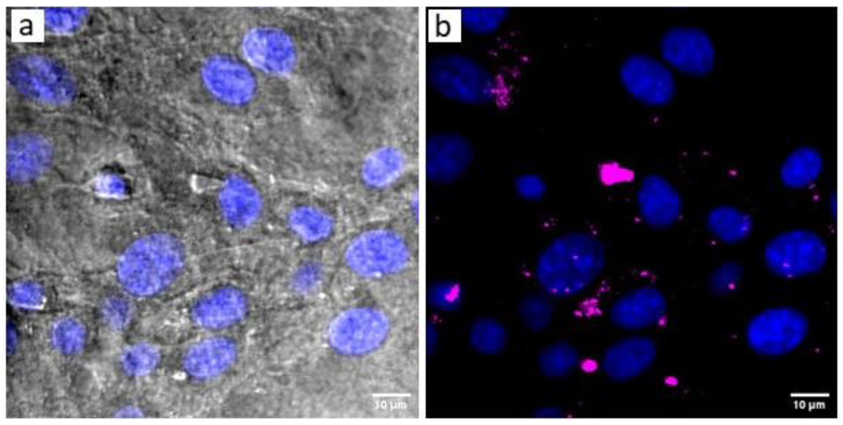
Figure 6.
Upconversion emission signal of UCC@Ale-PEG at various concentrations: undiluted (UD) and 10×, 100×, and 1000× diluted. Left panel: fluorescence emission of UCC@Ale-PEG adsorbed on glass surface upon air-drying of 1 µl drop of the colloid (insets – another three-fold magnification). Middle panel: intensity plot profiles from images (diagonal line). Right: SEM images (back scattered electron detector) at 25,000× magnification (with ROI magnified three times more). The smallest discrete spots in the image of 100× diluted sample have diameter of ~10 nm – corresponding to size of individual upconversion nanoparticles (as revealed by TEM analysis).
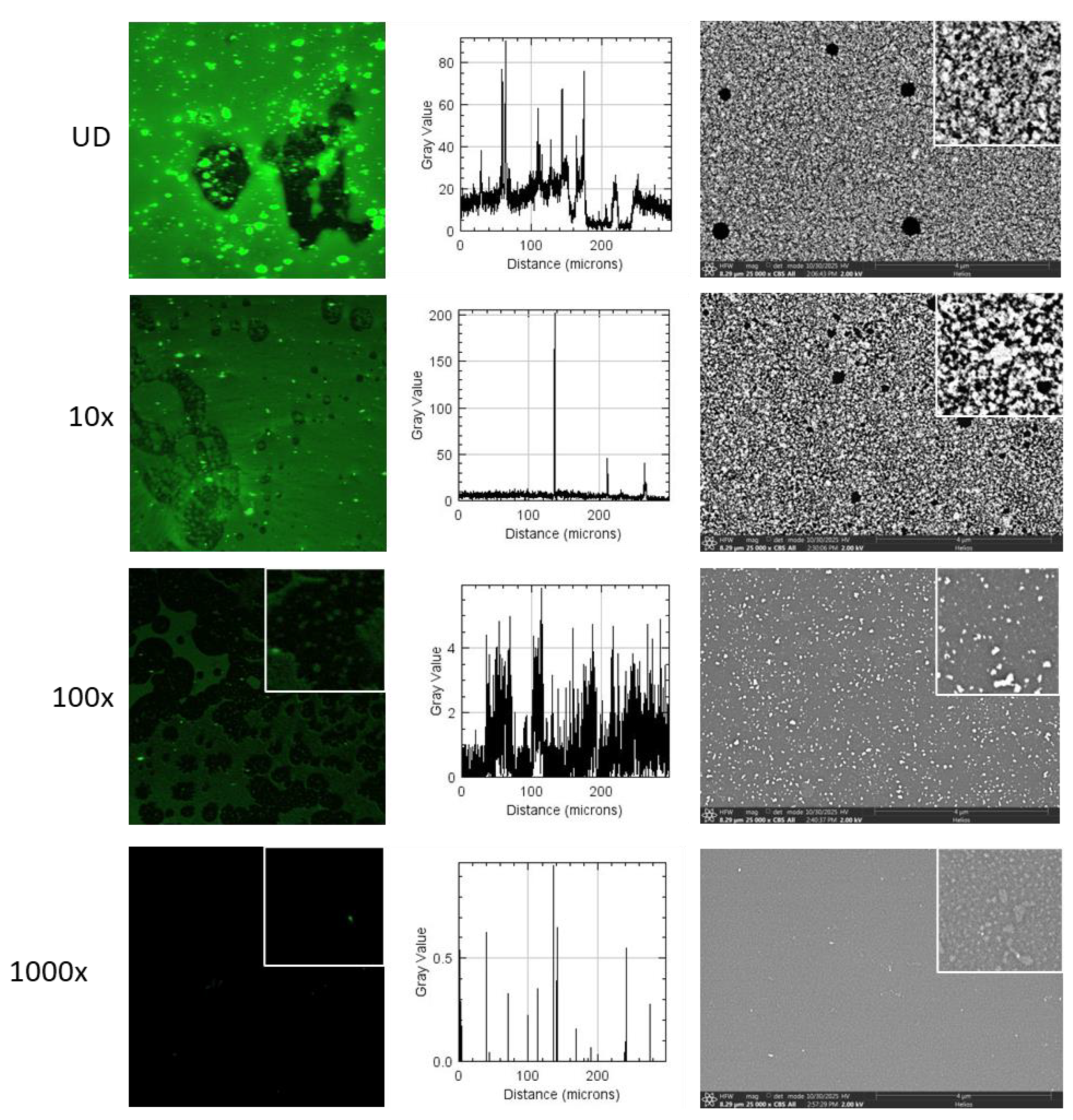

Table 1.
Characterization of colloids.
| Colloid |
Dn (nm) |
Ð | Dh (nm) | PD | ζ-potential, (mV) |
| UCC | 9 | 1.09 | 105 (18*) | 0.19 (0.17*) | 25 |
| UCC@Ale-PEG | 11 | 1.13 | 90 | 0.14 | 14 |
Dn – number-average diameter (TEM); Ð – dispersity (TEM); Dh – hydrodynamic diameter (DLS in water); PD – polydispersity (DLS in water); * in hexane.
Disclaimer/Publisher’s Note: The statements, opinions and data contained in all publications are solely those of the individual author(s) and contributor(s) and not of MDPI and/or the editor(s). MDPI and/or the editor(s) disclaim responsibility for any injury to people or property resulting from any ideas, methods, instructions or products referred to in the content. |
© 2026 by the authors. Licensee MDPI, Basel, Switzerland. This article is an open access article distributed under the terms and conditions of the Creative Commons Attribution (CC BY) license (http://creativecommons.org/licenses/by/4.0/).
Copyright: This open access article is published under a Creative Commons CC BY 4.0 license, which permit the free download, distribution, and reuse, provided that the author and preprint are cited in any reuse.


