Preprint
Article

This version is not peer-reviewed.

Antimicrobial Activity of Medicinal Plants Against Bacteria Causing Bovine Mastitis and Phytochemical Profiling by Paper Spray

Submitted:

22 December 2025

Posted:

24 December 2025

You are already at the latest version

Abstract

Medicinal plants have become increasingly important due to the diversity and bactericidal potential of many species. They can work as an alternative to the use of antimicrobials in the treatment of bacterial infections, which may represent impairment to health. Considering the importance of alternative compounds, we aimed to evaluate the antimicrobial activity in vitro of medicinal plants Stryphnodendron adstringens (Mart.) Coville, known as barbatimão, Baccharis crispa Spreng, known as carqueja and Azadiractha indica, known as neem. S. adstringens and B. crispa were used as extract and obtained from plants collected in the municipality of Bambuí, state of Minas Gerais, Brazil. A. indica was evaluated as extract and oil, and the crushed leaves and oil were purchased from a commercial company. Antimicrobial activity was determined by the minimum bactericidal concentration (MBC) test against Staphylococcus aureus, Streptococcus agalactiae, Streptococcus uberis, Escherichia coli, and Salmonella spp, isolated from bovine mastitis. The bacteria were submitted to the MBC test at concentrations of 100, 50, 25, 12.5, 6.25, 3.12, 1.56, 0.78, 0.39, 0.19 and 0.09 mg/mL. The bacteria evaluated were sensitive to most plant extracts for at least one of the concentrations evaluated, except for Gram-negative bacteria, Escherichia coli, and Salmonella spp. There was no activity of B. crispa extract and A. indica against E. coli and neither of A. indica extract against Salmonella spp. even at the highest concentration evaluated. S. adstringens was considered the extract with the highest activity against the bacteria evaluated and S. uberis the most susceptible to antimicrobial action. The results indicate the antimicrobial activity of the compounds and a possible application of these for the development of biotechnological products against the main bacteria causing bovine mastitis, becoming an alternative to the use of antibiotics.

Keywords: 
;  ;  ;  ;  ;  

1. Introduction

Animal health has a major impact on the quality of the final product in the milk production chain. In this sense, bovine mastitis has great relevance since it is the main infection of dairy cattle, usually caused by bacteria, especially those of the genera Staphylococcus spp. and Streptococcus spp. [1]. Besides representing a worldwide problem, scientific reports show an increase of mastitis’ cases, partly due to the selection of animals for higher milk production and use of broad-spectrum antibiotics [2,3,4]. Due to the high prevalence and impact of losses caused by the disease, mastitis is considered the costliest disease of dairy activity [5].
The emergence of antimicrobial resistance to commonly used antibiotics required new antimicrobial products that would ensure the efficacy of treatment [6]. Besides this necessity, a growing interest in medicinal plants as agents of new bioactive molecules against bacteria has been reported [6,7]. The bioactivity of medicinal plants is inherent to a group of compounds called secondary metabolites. These compounds are synthesized from catabolic, anabolic, and biotransformation reactions from the amino acids, carbohydrates, and lipids produced by the plant, and several crude extracts containing metabolites with antimicrobial activity that can inhibit bacterial growth are reported [6,7].
Stryphnodendron adstringens (Mart.) Coville, popularly known as barbatimão, stands out for being one of the most used plants in medicinal treatments, especially for the number of tannins present in the bark, which confers its main properties such as antimicrobial activity against Staphyloccocus aureus, Pseudomonas spp. and Escherichia coli [8,9,10]. Baccharis crispa Spreng, known by the name of carqueja, is popularly used as an antiseptic [11] and as an antioxidant [12]. Azadiractha indica, known by the name neem, has several proven benefits, such as insecticide [13], a fungicide, and controlling nematodes [14], besides proven antibacterial activity against some species [15].
Considering the potential of these medicinal plants, we aimed to determine, using the minimum bactericidal concentration (MBC), the in vitro antimicrobial effect of plant extracts of Stryphnodendron adstringens (Mart.) Coville, Baccharis crispa Spreng, and Azadiractha indica and of Azadiractha indica oil against strains of Staphylococcus aureus, Streptococcus agalactiae, Streptococcus uberis, Escherichia coli, and Salmonella spp. isolated from bovine mastitis. A complementary objective was to identify the secondary metabolites present in the evaluated extracts using the ambient ionization methodology for paper spray mass spectrometry (PS-MS).Once their antimicrobial potential is detected it is possible to investigate alternatives to the use of antimicrobials against the main bacteria that cause bovine mastitis.

2. Materials and Methods

2.1. Material Sampling and Extract Preparation

Plants S. adstringens (Mart.) Covillee and B. crispa Spreng were collected in the municipality of Bambuí, state of Minas Gerais, and then deposited in the Herbarium of the Agricultural Research Company of Minas Gerais (EPAMIG). The crushed leaves of A. indica were provided by the company GoNeem, and the oil of A. indica (OAi) extracted from the seeds of the plant by cold maceration was acquired from the company Globo Agronegócios e Participações Ltd.a.
The extract of S. adstringens (ESa) was obtained by maceration, from 428.57 g of dry matter (DM) of the ground bark, in one liter of absolute ethyl alcohol PA for eight days, and the solvent was renewed every four days [16,17]. The extract of B. crispa (EBc) was prepared from the aerial part of the dry plant at room temperature and obtained by decoction for 15 minutes from 25 g of DM of the ground plant for each liter of distilled water [18,19]. The extract of A. indica (EAi) was obtained from the maceration of 100 g of DM of the crushed leaves in one liter of absolute ethyl alcohol PA for eight days, with the renewal of the solvent every four days [20,21]. All extracts were filtered in a paper filter with a capacity of 80 g/m² with the aid of a vacuum.
ESa and EAi were concentrated using roto-evaporator Fisatom 801 at 45 ºC. The semi-solid residue was placed in an oven at a temperature of 45 °C for seven days for total drying. The EBc was kept in a freezer at -80 ºC and then freeze-dried using the L101 Liotop®. The extracts and OAi were used to evaluate the bactericidal activity after the sterility certification proposed by the Brazilian Pharmacopoeia (2010) [22].

2.2. Strains

Five strains isolated from bovine mastitis cases were used. They belong to the Collection of Microorganisms of Interest of Agroindustry and Livestock (CMIAP) of Embrapa Dairy Cattle. The following bacterial species were evaluated: Staphylococcus aureus, Streptococcus agalactiae, Streptococcus uberis, Escherichia coli, and Salmonella spp.

2.3. Inoculum Preparation

The bacteria were kept at -20 ºC in Skim Milk culture medium (Difco®), plus glycerol at 10% (v/v). To reactivate them, an aliquot of frozen bacterial suspension was inoculated onto Petri dishes containing Tryptone Soya Agar (TSA) (Difco®), enriched with 5% (v/v) of defibrinated sheep blood. The plates were incubated at 35 ºC ± 2 °C for 24 hours.
The standardization of bacterial suspensions was performed according to the method proposed by the Clinical and Laboratory Standards Institute [23]. The inoculum was prepared by suspending from four to five colonies previously grown in Brain Heart Infusion (BHI, Difco®) in a test tube containing Mueller-Hinton broth (MH). The turbidity was adjusted according to the standard of 0.5 of the McFarland scale by visual comparison, corresponding to approximately 1.5 × 10⁸ CFU/mL. The inoculums were standardized at the concentration of 1 to 3 × 10⁶ CFU/mL for all bacteria and then dispensed in each test tube. MH broth was used as a culture medium for MBC tests. For the bacteria S. agalctiae and S. uberis, 5% sterile defibrinated equine serum was added.

2.4. Determination of Minimum Bactericidal Concentration (MBC)

The MBCs of the extracts and the OAi were determined by the broth macro dilution method as proposed by CLSI (2008). One mL of the extracts and OAi reconstituted in aqueous solution containing DMSO and Tween 80 [24] at the concentration of 200 mg/mL was diluted twice in MH broth to obtain concentrations 100, 50, 25, 12.5, 6.25, 3.12, 1.56 and 0.78, 0.39, 0.19 and 0.09 mg/mL for each extract and OAi.
The solution containing DMSO and TWEEN 80 was used as a negative control, and antibiotics oxacillin or neomycin were used as a positive control, the first for Gram-positive bacteria and the second for Gram-negative bacteria. One mL of the bacterial inoculum standardized in 1 to 3 × 10⁶ CFU/mL was inoculated in each of the test tubes. Then, the test tubes were incubated at 35º C ± 2 °C for 24 hours. Subsequently, an aliquot of 10 μL of each dilution was spread out onto Petri dishes containing BHI agar and incubated at 35º C ± 2° C for 18-24h. The MBC was considered as the lowest concentration of extracts that did not present bacterial growth. The tests to evaluate the bactericidal activity of the extracts and oil were executed in triplicates.

2.5. Paper Spray Mass Spectrometry

A sample of 1 g of each extract (ESa, EBc and OAi) was weighed separately on an analytical balance and added to 8 mL of methanol (HPLC grade) in a Falcon flask. These mixtures were vortexed for 30 seconds and left at rest for one hour at room temperature.
The chemical profile of the extracts was determined according to the methodology described by Ramos et al. [18] using an LCQ Fleet mass spectrometer (ThermoScientific, USA) coupled with an ionization source for paper pulverizing. For this, chromatographic paper in the shape of an equilateral triangle (1.5 cm) was used, placed at a distance of 0.5 cm from the spectrometer entrance in a metal connector. 2 μL of the extract, and 40 μL of methanol was applied to the paper, reading in triplicate in positive and negative ionization modes.
The instrumental conditions of the analyses were: source voltage at 4.5 kV for the positive mode and 3.5 kV for the negative mode; capillary voltage of 40 V; transfer tube temperature of 275 °C; tube lens voltage of 120 V; mass range from 100 to 1000 m/z. To identify the compounds, the charge mass ratios from literature data were compared with the instrumental signals obtained and subsequent fragmentation with collision energies of 15 to 30 eV

2.6. Statistical Analysis

The statistical analysis was performed with the help of the statistical program R [25]. For the analysis of the bactericidal action of the extracts and the OAi, the generalized linear model (GLM) of the MASS package was used, with the response modeled by Bernoulli distribution and logit link function, and ANODEV was analyzed in a completely randomized design. The value 0 was assigned when there was bactericidal action and the value 1 when there was bacterial growth. The significance level was established in P <0.01. For graphical analysis, the ggplot function of the ggplot2 package [25] was used. CBM response variable is the following:
Yi~Bernoulli (μi)
Yi: when there was bacterial growth (1) and when there was bactericidal action (0).
μi: Bernoulli distribution with logit link function.

3. Results and Discussion

3.1. Antimicrobial Activity

All evaluated extracts showed some antimicrobial activity against at least one of the tested bacteria. However, differences regarding the concentrations used and regarding the species in question were observed. The ESa showed an inhibitory effect against all bacterial species tested, but at different concentrations. The inhibitory effect against E. coli was observed only with the highest concentration of 100mg/mL(50 mg/mL). The same was observed with OAi since the oil proved efficient against all species under test, but at higher concentrations than those used when the test was performed with ESa. For OAi, the lowest dose considered efficient was 50 mg/mL against S. uberis. Against other species, OAi was only able to inhibit bacterial growth when used at maximum concentration (100 mg/mL). EBc and EAi, even when used at the maximum concentration, were not able to inhibit E. coli growth, and considering the Salmonella spp, the same was observed for the maximum concentration of EBc (Table 1). Thus, according to MBC, the pathogens most susceptible to bactericidal action were S. aureus and S. uberis, and the most effective extract was ESa; on the other hand, the least susceptible species was E. coli, which presented MBC only when exposed to ESa and OAi at high concentrations (P>0.001).
When used at concentrations 0.097, 0.195, 0.39, and 0.78 mg/mL, ESa proved inefficient in inhibiting all bacteria studied. As the concentration was increased, the bactericidal effect of ESa increased, showing different susceptibility of the bacteria studied to this extract. When used at a concentration of 50 mg/mL, ESa was effective against all species studied (Figure 1). Among the bacteria studied, E. coli was the least susceptible (growth inhibited at a concentration of 50 mg/mL), while S. aureus and S. uberis were the most susceptible to this extract (growth inhibited at concentrations of 1.56 mg/mL (Table 1).
Costa et al. [26] evaluated higher concentrations of ESa against bacteria isolated from milk samples, reaching 400 mg/mL, and classified E. coli as not susceptible to the extract since no bactericidal action was observed even at the highest concentration evaluated. The result obtained by these researchers differs from that observed in our study and showed the difference in susceptibility of microorganisms isolated from different sources, especially the environmental pathogen, which may originate from a variety of sources, including cattle bedding, manure, pastures and water [27].
Despite limiting the growth of both Gram-positive and Gram-negative species, lower ESa concentrations have more inhibitory activity in the growth of Gram-positive species, indicating the greater susceptibility of this group to the extract. The difference in sensitivity between Gram-positive and Gram-negative has been discussed previously, and may be related to structural differences between these microorganisms. Gram-negative bacteria have an external phospholipid membrane with lipopolysaccharides, which makes the cell wall more complex and resistant to certain antimicrobial components, different from Gram-positive bacteria, which have only one outer layer of peptidoglycan and may be are, more permeable [26,28,29].
Unlike ESa, EBc did not exhibit significant antibacterial activity against the microorganisms tested. It was not possible to find the MBC for E. coli and Salmonella spp. (Table 1). For these two species, MBC was considered to be higher than 100 mg/mL and was considered not susceptible to the extract. Since EBc did not present bactericidal action against Gram-negative bacteria due to the lower susceptibility of these bacteria, the difference (P<0.001) was observed in the concentrations evaluated against S. aureus, S. agalactiae, and S. uberis. EBc at the concentration of 100 mg/mL presented higher bactericidal potential when compared with the use at the concentration of 50 mg/mL, which in turn was effective only against S. uberis (Figure 2).
Palacios et al. [30] evaluated the antimicrobial power of B. crispa oil by the presence of a disk diffusion test and also found the antimicrobial potential against S. aureus. They also did not find bactericidal action against E. coli. These authors were the first to describe the antimicrobial potential of B. crispa and concluded that this potential is partial and variable by the action of the compounds present according to the species.
The growth inhibition of S. uberis, S. aureus, and S. agalactiae, even at maximum concentration, promoted by EBc is considered important since the extract inhibited the growth of three pathogens commonly associated with bovine mastitis. The species S. aureus, S. uberis, S. agalactiae, and coliforms can represent up to 80% of isolated pathogens [31]. Considering the importance of inhibiting these pathogens, Avancini et al. [19] evaluated the bactericidal power of Baccharis trimera and suggested the possibility of using plant extracts as a natural disinfectant and biological antiseptic in certain problem situations in animal production, specifically related to agents S. aureus and S. uberis.
The medicinal efficacy of the Indian plant Azadiractha indica, popularly known as neem has been the target of several studies, and the biologically active extracts obtained from its leaves, fruits, seeds, and trunk are recognized by their multiple therapeutic properties, insecticides, nematicides, and fungicides [32,33]. In this study, the bactericidal power of the plant was evaluated in oil (OAi) and extract (EAi).
The bactericidal action of the EAi was detected for most of the species studied, except for E.coli, for which MBC was not determined (Table 1). The concentrations of EAi used promoted different bactericidal efficacy according to the bacteria (P<0.01). At a concentration of 100 mg/mL, the bactericidal action was effective against S. ureus, S. agalactiae, S. uberis, and Salmonella spp. As observed when in contact with EBc, the most susceptible pathogen was S. uberis, being inhibited with 50 mg/mL. When used at a concentration of 3.12 mg/mL or lower, EAi was not efficient against any of the species evaluated (Figure 3).
The A. indica oil was more efficient than the extract since lower concentrations were used to inhibit bacterial growth (Table 1). OAi showed satisfactory antibacterial activity only if used at the maximum concentration (100 mg/mL) for most bacteria, except for S. uberis, in which MBC was 50mg/mL (P<0.001, Figure 4).
Although EAi is generally considered more efficient, we found that OAi was more efficient against E. coli than EAi since E. coli was considered not susceptible to the extract. This indicates that oil compounds can be more efficient in certain situations. Arroteia et al. [33] evaluated the effect of A. indica on the inhibition of a mycotoxin production found in apples and reported a higher potential of A. indica in oil, indicating that liposolubility would be an activating potential of its activity. As reported here, the action of oil is not always more efficient, and besides the scope of action against microorganisms, production costs should be considered. The greatest use of the extract may be associated with lower costs since obtaining the oil requires the use of greater resources [34].

3.2. Mass Spectrometry with Paper Spray Ionization

3.2.1. Stryphnodendron adstringens (Mart.) Coville

Fourteen compounds in negative ionization mode and 8 in positive ionization mode were found for Stryphnodendron adstringens (Mart.) Coville detected compounds suggest a phytochemical profile particularly composed of polyphenolic and flavonoid derivatives, which are commonly associated with antioxidant and anti-inflammatory activities. These findings align with previous studies indicating the ethnopharmacological relevance of Stryphnodendron adstringens (Mart.) Coville, traditionally used in antimicrobial and wound-healing applications [35,36]. Identification of multiple hydrolyzable tannins, flavonoid glycosides, and phenolic acids further supports its potential use in functional foods and medicinal applications [37,38].
Related to paper spray analysis, in the positive ionization mode, ellagic acid (m/z 303) was identified, a known polyphenol with strong antioxidant and anticancer properties, commonly found in medicinal plants. Its presence in Stryphnodendron adstringens (Mart.) Coville may contribute to oxidative stress mitigation [39]. HHDP-digalloylglucoside, also known as tellimagrandin I (m/z 787), was detected and tentatively identified based on its fragmentation pattern [M+H]+. This compound has been studied for its antiviral and antimicrobial activities, which could contribute to explaining the traditional use of this plant in wound healing [40]. Riparin III (m/z 288), an alkaloid, has been previously reported for its anxiolytic and neuroprotective properties [41]. Casuariin (m/z 785), a hydrolyzable tannin, further supports the plant’s astringent properties, which are linked to its medicinal uses in gastrointestinal disorders [42].
The negative ionization mode identified several flavonoid and phenolic glycosides, substantiating the bioactive potential of Stryphnodendron adstringens (Mart.) Coville. Caffeic acid-hexoside (m/z 341) was detected, a derivative of caffeic acid known for its anti-inflammatory and neuroprotective properties [43]. Rutin (m/z 609), a well-characterized flavonoid glycoside, has been widely studied for its vascular protective effects [44]. Punicalin (m/z 781), a hydrolyzable tannin, was also identified, contributing to the presence of antioxidant constituents. Quercetin-3,4’-O-diglucoside (m/z 625) and cyanidin-O-glucosyl-O-acetylpentoside (m/z 623) were also tentatively identified and can be considered to build on the plant’s polyphenolic complexity, with quercetin derivatives playing a key role in cardiovascular health and cyanidin derivatives being linked to anti-inflammatory and neuroprotective effects [45,46].

3.2.2. Baccharis crispa Spreng

For Baccharis crispa Spreng, 8 compounds were identified in negative ionization mode and 5 in positive ionization mode, majorly composed of hydrolyzable tannins, flavonoids, and phenolic acids. This phytochemical composition is within the expected profile makeup of species used in traditional hepatoprotective and anti-inflammatory treatments, such as the case for Baccharis crispa Spreng [47,48]. The presence of bioactive polyphenols suggests potential applications in functional foods and nutraceuticals [49,50].
Among the positively ionized compounds tentatively identified, casuariin (m/z 785) was found, a tannin with antimicrobial and antioxidant activities. Octadecanoic acid (m/z 285), a saturated fatty acid, was also detected, known for its emollient properties and potential cardiovascular benefits [42,51]. Tellimagrandin I (m/z 787), a hydrolyzable tannin, was also tentatively identified, indicating a potential yield of a significative result of Baccharis crispa Spreng employment in traditional medicinal use [40,52].
The negative ionization mode tentatively identified ferulic acid (m/z 193), a phenolic compound known for its antioxidant and anti-inflammatory properties. HHDP-digalloylglucose (m/z 785) and procyanidin trimer A (m/z 863) were also identified, both of which have been implicated in cardiovascular protection and metabolic health [53,54,55]. Galloyl-bis-HHDP-glucose (m/z 935), also known as casuarinin, tentatively indicates the presence of potent antioxidant tannins in Baccharis crispa Spreng, presenting hepatoprotective potential [56,57].

3.2.3. Azadiractha indica

The analysis of Azadirachta indica was able to identify 6 compounds in negative ionization mode and 3 in positive ionization mode. Identified compounds suggest antimicrobial, anti-inflammatory, and neuroprotective properties, consistent with the traditional medicinal uses of neem [58,59,60].
In the positive ionization mode, octadecanoic acid (m/z 285) was tentatively identified, a compound known for its dermatological applications and potential role in lipid metabolism [51,61]. Tellimagrandin I (m/z 787) was also identified, with literature citing this compound as possessing antimicrobial properties [40,52].
The negative ionization mode tentatively identified dihydroquercetin-3,5-rhamnoside (m/z 449), a flavonoid with strong antioxidant properties, frequently studied for its cardioprotective effects [62]. Delphinidin-3-O-glucoside (m/z 465), an anthocyanin, has been associated with anti-inflammatory and neuroprotective benefits [63]. Procyanidin trimer A (m/z 863) was also detected, a compound widely studied for its cardiovascular and metabolic health effects [64]. Galloyl-bis-HHDP-glucose (m/z 935), also known as casuarinin, indicates the presence of bioactive tannins in Azadirachta indica, and may contribute to understanding its traditional use and role in antimicrobial treatments [65].
Several compounds were identified across multiple plant extracts, including tellimagrandin I (m/z 787), casuariin (m/z 785), procyanidin trimer A (m/z 863), and galloyl-bis-HHDP-glucose (m/z 935). These shared constituents suggest a common phytochemical basis for their reported biological activities, particularly in antioxidant, antimicrobial, and anti-inflammatory effects. The identification of these recurring bioactive compounds underscores their pharmacological relevance and potential therapeutic applications. These results provide a valuable basis for further pharmacological and clinical investigations [40,42,52,55,56,57].

4. Conclusions

The extracts showed bactericidal activity against bacteria isolated from bovine mastitis. However, the extract of S. adstringens presented a lower MBC for all bacteria. Thus, we can consider it the most efficient among the extracts evaluated. S. uberis strain was shown to be the least resistant, regardless of the extract or oil evaluated. Despite the higher efficiency of S. adstringens, both the extracts and the oil evaluated demonstrated their potential as antimicrobials. Paper Spray Mass Spectrometry (PS-MS) was fundamental for identifying the secondary metabolites responsible for the bactericidal action. Therefore, studies that promote the use of these compounds, preventively or in a combination of pharmaceutical and antibiotic therapy, against the main pathogenic bacteria isolated in cases of bovine mastitis should be disclosed.

5. Patents

In the context of intellectual property protection, the invention patent “Herbal Solution for Pre- and Post-Dipping” also known as “Natural Dipping” has been filed under the Brazilian National Application for invention, utility model, certificate of addition of invention, and entry into the national phase of the Patent Cooperation Treaty (PCT). The patent application, registered under Process BR 10 2021 009874 0, was officially submitted on May 21, 2021.

Author Contributions

Conceptualization, G.C.N., M.L.M.P., A.C.C.F.F.P. and R.B.T.; methodology, G.C.N., A.C.C.F.F.P., M.A.V.P.B., and R.B.T; software, G.C.N., A.C.C.F.F.P., and R.B.T.; validation, G.C.N., A.C.C.F.F.P., R.O.P.; formal analysis, G.C.N., A.C.C.F.F.P.; investigation, G.C.N., M.L.M.P., B.V.D., A.H.O.J, L.L.A., R.A, J.O.F.M., A.C.C.F.F.P., and R.B.T; data curation, G.C.N., B.V.D.; and A.C.C.F.F.P.; writing—original draft preparation G.C.N., M.A.V.P.B.; and A.C.C.F.F.P.; writing—review and editing, G.C.N.; A.C.C.F.F.P.; M.A.V.P.B., B.V.D.; and R.O.P.; visualization, G.C.N.; A.C.C.F.F.P., and R.O.P.; supervision, A.C.C.F.F.P., M.A.V.P.B.; and R.B.T.; project administration, G.C.N..; funding acquisition, G.C.N.; A.C.C.F.F.P; and R.B.T. All authors have read and agreed to the published version of the manuscript.

Institutional Review Board Statement

Not applicable.

Informed Consent Statement

Not applicable.

Data Availability Statement

The original contributions presented in this study are included in the article. Further inquiries can be directed to the corresponding author(s).

Acknowledgments

The authors would like to thank the Instituto Federal de Minas Gerais, the Innovation Agency (IFMG AI-RE/NIT), the Brazilian Agricultural Research Cooperation (EMBRAPA), the Universidade Federal de São João Del-Rei, the Coordination for the Improvement of Higher Education Personnel (CAPES/code 001), the National Council for Scientific and Technological Development (CNPq) (research productivity grant 132217/2023-6, 307787/2022-2 and 404432/2024-7), Minas Gerais State Research Support Foundation (FAPEMIG) - Finance Code APQ-04336-23, APQ-05883-24, PPE-00094-23, APQ-03644-23 and 5.308/15 and the Teaching, Research and Extension Group in Chemistry and Pharmacognosy (GEPEQF).

Conflicts of Interest

The authors declare no conflicts of interest.

References

  1. Makovec, J.A.; Ruegg, P.L. Antimicrobial Resistance of Bacteria Isolated from Dairy Cow Milk Samples Submitted for Bacterial Culture: 8,905 Samples (1994–2001). J. Am. Vet. Med. Assoc. 2003, 222, 1582–1589. [Google Scholar] [CrossRef] [PubMed]
  2. Valde, J.P.; Lawson, L.G.; Lindberg, A.; Agger, J.F.; Saloniemi, H.; Østerås, O. Cumulative Risk of Bovine Mastitis Treatments in Denmark, Finland, Norway and Sweden. Acta Vet. Scand. 2004, 45, 201–210. [Google Scholar] [CrossRef] [PubMed]
  3. Hand, K.J.; Godkin, A.; Kelton, D.F. Milk Production and Somatic Cell Counts: A Cow-Level Analysis. J. Dairy Sci. 2012, 95, 1358–1362. [Google Scholar] [CrossRef]
  4. DANMAP. Use of Antimicrobial Agents and Occurrence of Antimicrobial Resistance in Bacteria from Food Animals, Food and Humans in Denmark. Available online: http://orbit.dtu.dk/files/140535625/DANMAP_2016_LOW_241017.pdf (accessed on 24 April 2021).
  5. Halasa, T.; Nielen, M.; de Roos, A.P.W.; van Hoorne, R.; Jong, G.; Lam, T.J.G.M.; Werven, T.; Hogeveen, H. Production Loss Due to New Subclinical Mastitis in Dutch Dairy Cows Estimated with a Test-Day Model. J. Dairy Sci. 2009, 92, 599–606. [Google Scholar] [CrossRef] [PubMed]
  6. Leite, J.A.; Pereira, H.P.; Borges, C.A.V.; Alves, B.R.C.; Ramos, A.I.A.P.; Martins, M.F.; Arcuri, E.F. Lytic Bacteriophages as a Potential Alternative to Control Staphylococcus aureus. Pesqui. Agropecu. Bras. 2019, 54, 1–9. [Google Scholar] [CrossRef]
  7. Cohen, M.L. Epidemiology of Drug Resistance: Implications for a Post-Antimicrobial Era. Science 1992, 257, 1050–1055. [Google Scholar] [CrossRef]
  8. Ribeiro, A.Q.; Leite, J.P.V.; Dantas-Barros, A.M. Profile of Herbal Medicine Use in Community Pharmacies of Belo Horizonte under the Influence of National Legislation. Rev. Bras. Farmacogn. 2005, 15, 65–70. [Google Scholar] [CrossRef]
  9. Toledo, C.E.M. Estudos Anatômico, Químico e Biológico de Cascas e Extratos Obtidos de Barbatimão (Stryphnodendron adstringens). Master’s Thesis, Universidade Estadual Paulista, Araraquara, Brazil, 2002; p. 115 pp. [Google Scholar]
  10. Macedo, M.; Ferreira, A.R. Medicinal Plants Used for Dermatological Treatments in Communities of the Upper Paraguay River Basin, Mato Grosso. Rev. Bras. Farmacogn. 2004, 14, 40–44. [Google Scholar] [CrossRef]
  11. Del Vitto, L.A.; Petenatti, E.M.; Petenatti, M.E. Introducción a la Herboristería; Serie Técnica Herbario UNSL: San Luis, Argentina, 2002; pp. 10–61. [Google Scholar]
  12. Roig, F. Flora Medicinal Mendocina: Las Plantas Medicinales y Aromáticas de la Provincia de Mendoza; EDIUNC: Mendoza, Argentina, 2001. [Google Scholar]
  13. Abdullah, F.; Subramanian, P. The Feeding Response of Epilachna indica (Coleoptera: Coccinellidae Epilachninae) Towards Extracts of Azadirachta indica. J. Entomol. 2008, 5, 77–90. [Google Scholar] [CrossRef]
  14. Agbenin, N.O.; Emechebe, A.M.; Marley, P.S. Evaluation of Neem Seed Powder for Fusarium Wilt and Meloidogyne Control on Tomato. Arch. Phytopathol. Plant Prot. 2004, 37, 319–326. [Google Scholar] [CrossRef]
  15. Subramaniam, S.K.; Siswomihardjo, W.; Sunarintyas, S. The Effect of Different Concentrations of Neem (Azadirachta indica) Leaves Extract on the Inhibition of Streptococcus mutans (In Vitro). Dent. J. (Majalah Kedokteran Gigi) 2005, 38, 176–179. [Google Scholar] [CrossRef]
  16. Costa, J.P.R.; Almeida, A.C.; Martins, A.C.; Rodrigues, M.N.; Santos, C.A.; Menezes, I.R. Antimicrobial Activity of Rosmarinus officinalis Essential Oil and Stryphnodendron adstringens Extract Against Bacteria Isolated from Milk. Rev. Biotemas 2011, 24, 1–6. [Google Scholar]
  17. Sousa, J.N. Obtenção de Extratos Padronizados a Partir da Casca de Stryphnodendron adstringens (Mart.) Coville (Fabaceae) e Avaliações Biológicas In Vitro. Master’s Thesis, Universidade Federal de Goiás, Goiânia, Brazil, 2014; pp. 34–47. [Google Scholar]
  18. Brazilian Pharmacopedia. Farmacopeia dos Estados Unidos do Brasil, 2nd ed.; Siqueira: São Paulo, Brazil, 1959. [Google Scholar]
  19. Avancini, C.A.M.; Wiest, J.M.; Dall’Agnol, R.; Haas, J.S.; Poser, G.L. Antimicrobial Activity of Plants Used in the Prevention and Control of Bovine Mastitis in Southern Brazil. Lat. Am. J. Pharm. 2008, 27, 894–899. [Google Scholar]
  20. Sukanya, S.L.; Sudisha, J.; Hariprasad, P.; Niranjana, S.R.; Prakash, H.S.; Fathima, S.K. Antimicrobial Activity of Leaf Extracts of Indian Medicinal Plants Against Clinical and Phytopathogenic Bacteria. Afr. J. Biotechnol. 2009, 8, 6677–6682. [Google Scholar]
  21. Mistry, K.S.; Sanghvi, Z.; Parmar, G.; Shah, S. The Antimicrobial Activity of Azadirachta indica, Mimusops elengi, Tinospora cordifolia, Ocimum sanctum and 2% Chlorhexidine Gluconate on Common Endodontic Pathogens: An In Vitro Study. Eur. J. Dent. 2014, 8, 172–177. [Google Scholar]
  22. Farmacopeia Brasileira, 5th ed.; Agência Nacional de Vigilância Sanitária (ANVISA): Brasília, Brazil, 2010; Volume 2.
  23. Clinical & Laboratory Standards Institute (CLSI). Performance Standards for Antimicrobial Disk and Dilution Susceptibility Tests for Bacteria Isolated from Animals; CLSI: Wayne, PA, USA, 2008; Volume 28, Issue 8, pp. 24–35. [Google Scholar]
  24. Aizawa, H. Morphology of Polysorbate 80 (Tween 80) Micelles in Aqueous Dimethyl Sulfoxide Solution. J. Appl. Crystallogr. 2010, 43, 630–631. [Google Scholar] [CrossRef]
  25. R Core Team. R: A Language and Environment for Statistical Computing; R Foundation for Statistical Computing: Vienna, Austria, 2020. Available online: http://www.R-project.org/ (accessed on 10 January 2020).
  26. Costa, E.S.; Hiruma-Lima, C.A.; Lima, E.O.; Sucupira, G.C.; Bertolin, A.O.; Lolis, S.F.; Andrade, F.D.P.; Vilegas, W.; Souza-Brito, A.R.M. Antimicrobial Activity of Some Medicinal Plants of the Cerrado, Brazil. Phytother. Res. 2008, 22, 705–707. [Google Scholar] [CrossRef]
  27. Baskaran, A.S.; Kazmer, G.W.; Hinckley, L.; Andrew, S.M.; Venkitanarayanan, K. Antibacterial Effect of Plant-Derived Antimicrobials on Major Bacterial Mastitis Pathogens In Vitro. J. Dairy Sci. 2009, 92, 1423–1429. [Google Scholar] [CrossRef]
  28. Babita, P.; Hari, D.B.; Jin, S.L.; Soon, G.H.; Hyun, W.S.; Joung, H.Y. Antibacterial Potential of Antarctic Lichens Against Human Pathogenic Gram-Positive Bacteria. Phytother. Res. 2008, 22, 1269–1271. [Google Scholar]
  29. Walter, C.; Shinwari, Z.K.; Afzal, I.; Malik, R.N. Antibacterial Activity in Herbal Products Used in Pakistan. Pak. J. Bot. 2011, 43, 155–162. [Google Scholar]
  30. Palacios, P.; Gutkind, G.; Rondina, R.V.; de Torres, R.; Coussio, J.D. Genus Baccharis. II. Antimicrobial Activity of B. crispa and B. notosergilla. Planta Med. 1983, 48, 128. [Google Scholar] [CrossRef]
  31. Ranjan, R.; Swarup, D.; Patra, R.C.; Nandi, D. Bovine Protothecal Mastitis: A Review. CAB Rev. Perspect. Agric. Vet. Sci. Nutr. Nat. Resour. 2006, 1, 1–7. [Google Scholar]
  32. Mossini, S.A.G.; Kemmelmeier, C. The Neem Tree (Azadirachta indica A. Juss.): Multiple Uses. Acta Farm. Bonaer. 2005, 24, 139–148. [Google Scholar]
  33. Arroteia, C.C.; Kemmelmeier, C.; Junior, M.M. Effect of Aqueous and Oily Extracts of Neem [Azadirachta indica A. Juss (Meliaceae)] on Patulin Production in Apples Contaminated with Penicillium expansum. Ciênc. Rural 2007, 37, 1518–1523. [Google Scholar] [CrossRef]
  34. Kasper, S.; Müller, W.E.; Volz, H.P.; Möller, H.J.; Koch, E.; Dienel, A. Silexan in Anxiety Disorders: Clinical Data and Pharmacological Background. World J. Biol. Psychiatry 2018, 19, 412–420. [Google Scholar] [CrossRef] [PubMed]
  35. Barbosa, C.R.; Pantoja, J.C.; Fernandes, T.; Chagas, R.A.; Souza, C.G.; Santos, A.R.; Vargas Junior, F.M. Bioactive Compounds of Barbatimão (Stryphnodendron sp.) as Dietary Additive in Lamb Diets. Agriculture 2023, 13(3), 664. [Google Scholar] [CrossRef]
  36. Souza-Moreira, T.M.; Queiroz-Fernandes, G.M.; Pietro, R.C.L.R. Stryphnodendron Species Known as “Barbatimão”: A Comprehensive Report. Molecules 2018, 23(4), 910. [Google Scholar] [CrossRef]
  37. Gomes, P.W.; Pamplona, T.C.; Navegantes-Lima, K.C.; Quadros, L.B.; Oliveira, A.L.; Santos, S.M.; da Silva, M.N. Chemical Composition and Antibacterial Action of Stryphnodendron pulcherrimum Bark Extract, “Barbatimão” Species: Evaluation of Its Use as a Topical Agent. Arab. J. Chem. 2021, 14(6), 103183. [Google Scholar] [CrossRef]
  38. Ricardo, L.M.; Dias, B.M.; Mügge, F.L.; Leite, V.V.; Brandão, M.G. Evidence of Traditionality of Brazilian Medicinal Plants: The Case Studies of Stryphnodendron adstringens (Mart.) Coville (Barbatimão) Barks and Copaifera spp. (Copaíba) Oleoresin in Wound Healing. J. Ethnopharmacol. 2018, 219, 319–336. [Google Scholar] [CrossRef]
  39. Polce, S.A.; Burke, C.; França, L.M.; Kramer, B.; Paes, A.M.D.A.; Carrillo-Sepulveda, M.A. Ellagic Acid Alleviates Hepatic Oxidative Stress and Insulin Resistance in Diabetic Female Rats. Nutrients 2018, 10(5), 531. [Google Scholar] [CrossRef]
  40. Suručić, R.; Radović Selgrad, J.; Kundaković-Vasović, T.; Lazović, B.; Travar, M.; Suručić, L.; Škrbić, R. In Silico and In Vitro Studies of Alchemilla viridiflora Rothm—Polyphenols’ Potential for Inhibition of SARS-CoV-2 Internalization. Molecules 2022, 27(16), 5174. [Google Scholar] [CrossRef]
  41. Mallmann, A.S.V.; de Castro Chaves, R.; de Oliveira, N.F.; Oliveira, I.C.M.; Capibaribe, V.C.C.; Valentim, J.T.; de Sousa, F.C.F. Is Riparin III a Promising Drug in the Treatment for Depression? Eur. J. Pharm. Sci. 2021, 162, 105824. [Google Scholar] [CrossRef] [PubMed]
  42. Jiang, C.; Wang, S.; Wang, Y.; Wang, K.; Huang, C.; Gao, F.; Li, Y. Polyphenols from Hickory Nut Reduce the Occurrence of Atherosclerosis in Mice by Improving Intestinal Microbiota and Inhibiting Trimethylamine N-oxide Production. Phytomedicine 2024, 128, 155349. [Google Scholar] [CrossRef] [PubMed]
  43. Formato, M.; Piccolella, S.; Zidorn, C.; Pacifico, S. UHPLC-HRMS Analysis of Fagus sylvatica (Fagaceae) Leaves: A Renewable Source of Antioxidant Polyphenols. Antioxidants 2021, 10(7), 1140. [Google Scholar] [CrossRef]
  44. Morais-Campos, R.; Lima, L.M.A.L.L.; da Silva, A.G.; Santiago, R.O.; Paz, I.A.; Cabral, P.H.B.; do Nascimento, N.R.F. Rutin Ameliorates Nitrergic and Endothelial Dysfunction on Vessels and Corpora Cavernosa of Diabetic Animals. Res. Vet. Sci. 2023, 161, 163–172. [Google Scholar] [CrossRef] [PubMed]
  45. Alizadeh, S.R.; Ebrahimzadeh, M.A. Quercetin Derivatives: Drug Design, Development, and Biological Activities, a Review. Eur. J. Med. Chem. 2022, 229, 114068. [Google Scholar] [CrossRef]
  46. Straßmann, S.; Passon, M.; Schieber, A. Chemical Hemisynthesis of Sulfated Cyanidin-3-O-glucoside and Cyanidin Metabolites. Molecules 2021, 26(8), 2146. [Google Scholar] [CrossRef]
  47. Fraga-Corral, M.; Otero, P.; Cassani, L.; Echave, J.; Garcia-Oliveira, P.; Carpena, M.; Simal-Gandara, J. Traditional Applications of Tannin Rich Extracts Supported by Scientific Data: Chemical Composition, Bioavailability and Bioaccessibility. Foods 2021, 10(2), 251. [Google Scholar] [CrossRef]
  48. Marrone, G.; Di Lauro, M.; Izzo, F.; Cornali, K.; Masci, C.; Vita, C.; Noce, A. Possible Beneficial Effects of Hydrolyzable Tannins Deriving from Castanea sativa L. in Internal Medicine. Nutrients 2023, 16(1), 45. [Google Scholar] [CrossRef]
  49. Zhang, Z.; Li, X.; Sang, S.; McClements, D.J.; Chen, L.; Long, J.; Qiu, C. Polyphenols as Plant-Based Nutraceuticals: Health Effects, Encapsulation, Nano-Delivery, and Application. Foods 2022, 11(15), 2189. [Google Scholar] [CrossRef]
  50. Obeme-Nmom, J.I.; Abioye, R.O.; Flores, S.S.R.; Udenigwe, C.C. Regulation of Redox Enzymes by Nutraceuticals: A Review of the Roles of Antioxidant Polyphenols and Peptides. Food Funct. 2024. [Google Scholar] [CrossRef]
  51. Fujimoto, D.; Shinohara, M.; Kawamori, H.; Toba, T.; Kakizaki, S.; Nakamura, K.; Otake, H. The Relationship between Unique Gut Microbiome-Derived Lipid Metabolites and Subsequent Revascularization in Patients Who Underwent Percutaneous Coronary Intervention. Atherosclerosis 2023, 375, 1–8. [Google Scholar] [CrossRef] [PubMed]
  52. Fernandes, A.S.; de Melo Bisneto, A.V.; Silva, L.S.; Bailão, E.F.L.C.; Cardoso, C.G.; Carneiro, C.C.; Chen-Chen, L. Pedunculagin and Tellimagrandin-I Stimulate Inflammation and Angiogenesis and Upregulate VEGF and TNF-α In Vivo. Microvasc. Res. 2024, 151, 104615. [Google Scholar] [CrossRef] [PubMed]
  53. Li, D.; Rui, Y.X.; Guo, S.D.; Luan, F.; Liu, R.; Zeng, N. Ferulic Acid: A Review of Its Pharmacology, Pharmacokinetics and Derivatives. Life Sci. 2021, 284, 119921. [Google Scholar] [CrossRef] [PubMed]
  54. Fernandes, I.D.A.A.; Maciel, G.M.; Maroldi, W.V.; Bortolini, D.G.; Pedro, A.C.; Haminiuk, C.W.I. Bioactive Compounds, Health-Promotion Properties and Technological Applications of Jabuticaba: A Literature Overview. Meas. Food 2022, 8, 100057. [Google Scholar] [CrossRef]
  55. Hanaka, A.; Dresler, S.; Mułenko, W.; Wójciak, M.; Sowa, I.; Sawic, M.; Strzemski, M. Phenolic-Based Discrimination between Non-Symptomatic and Symptomatic Leaves of Aesculus hippocastanum Infested by Cameraria ohridella and Erysiphe flexuosa. Int. J. Mol. Sci. 2023, 24(18), 14071. [Google Scholar] [CrossRef]
  56. Wang, P.; Zhong, L.; Yang, H.; Hou, X.; Wu, C.; Zhang, R.; Cheng, Y. Systematic Transcriptomic and Metabolomic Analysis of Walnut (Juglans regia L.) Fruit to Trace Variations in Antioxidant Activity during Ripening. Sci. Hortic. 2022, 295, 110849. [Google Scholar] [CrossRef]
  57. Bogahawaththa, S.; Kawamura, T.; Bandaranayake, U.; Hirakawa, T.; Yamada, G.; Ishino, H.; Tsujita, T. Identification and Mechanistic Investigation of Ellagitannins from Osbeckia octandra That Attenuate Liver Fibrosis via the TGF-β/SMAD Signaling Pathway. Biosci. Biotechnol. Biochem. 2023, 87(11), 1295–1309. [Google Scholar] [CrossRef]
  58. Wylie, M.R.; Merrell, D.S. The Antimicrobial Potential of the Neem Tree Azadirachta indica. Front. Pharmacol. 2022, 13, 891535. [Google Scholar] [CrossRef]
  59. Ujah, I.I.; Nsude, C.A.; Ani, O.N.; Alozieuwa, U.B.; Okpako, I.O.; Okwor, A.E. Phytochemicals of Neem Plant (Azadirachta indica) Explain Its Use in Traditional Medicine and Pest Control. GSC Biol. Pharm. Sci. 2021, 14(2), 165–171. [Google Scholar] [CrossRef]
  60. Reddy, I.V.; Neelima, P. Neem (Azadirachta indica): A Review on Medicinal Kalpavriksha. Int. J. Econ. Plants 2022, 9(1), 59–63. [Google Scholar] [CrossRef]
  61. Vazquez de Vasquez, M.G.; Carter-Fenk, K.A.; McCaslin, L.M.; Beasley, E.E.; Clark, J.B.; Allen, H.C. Hydration and Hydrogen Bond Order of Octadecanoic Acid and Octadecanol Films on Water at 21 and 1 °C. J. Phys. Chem. A 2021, 125(46), 10065–10078. [Google Scholar] [CrossRef]
  62. Silva, G.G.; Pimenta, L.P.S.; Melo, J.O.F.; Mendonça, H.D.O.P.; Augusti, R.; Takahashi, J.A. Phytochemicals of Avocado Residues as Potential Acetylcholinesterase Inhibitors, Antioxidants, and Neuroprotective Agents. Molecules 2022, 27(6), 1892. [Google Scholar] [CrossRef]
  63. Sari, D.R.T.; Cairns, J.R.K.; Safitri, A.; Fatchiyah, F. Virtual Prediction of the Delphinidin-3-O-glucoside and Peonidin-3-O-glucoside as Anti-Inflammatory of TNF-α Signaling. Acta Inform. Med. 2019, 27(3), 152. [Google Scholar] [CrossRef]
  64. Chen, W.; Zhang, L.; Zhao, L.; Yan, F.; Zhu, X.; Lu, Q.; Liu, R. Metabolomic Profiles of A-Type Procyanidin Dimer and Trimer with Gut Microbiota In Vitro. J. Funct. Foods 2021, 85, 104637. [Google Scholar] [CrossRef]
  65. Rana, A.; Kumari, A.; Chaudhary, A.K.; Srivastava, R.; Kamil, D.; Vashishtha, P.; Sharma, S.N. An Investigation of Antimicrobial Activity for Plant Pathogens by Green-Synthesized Silver Nanoparticles Using Azadirachta indica and Mangifera indica. Physchem 2023, 3(1), 125–146. [Google Scholar] [CrossRef]
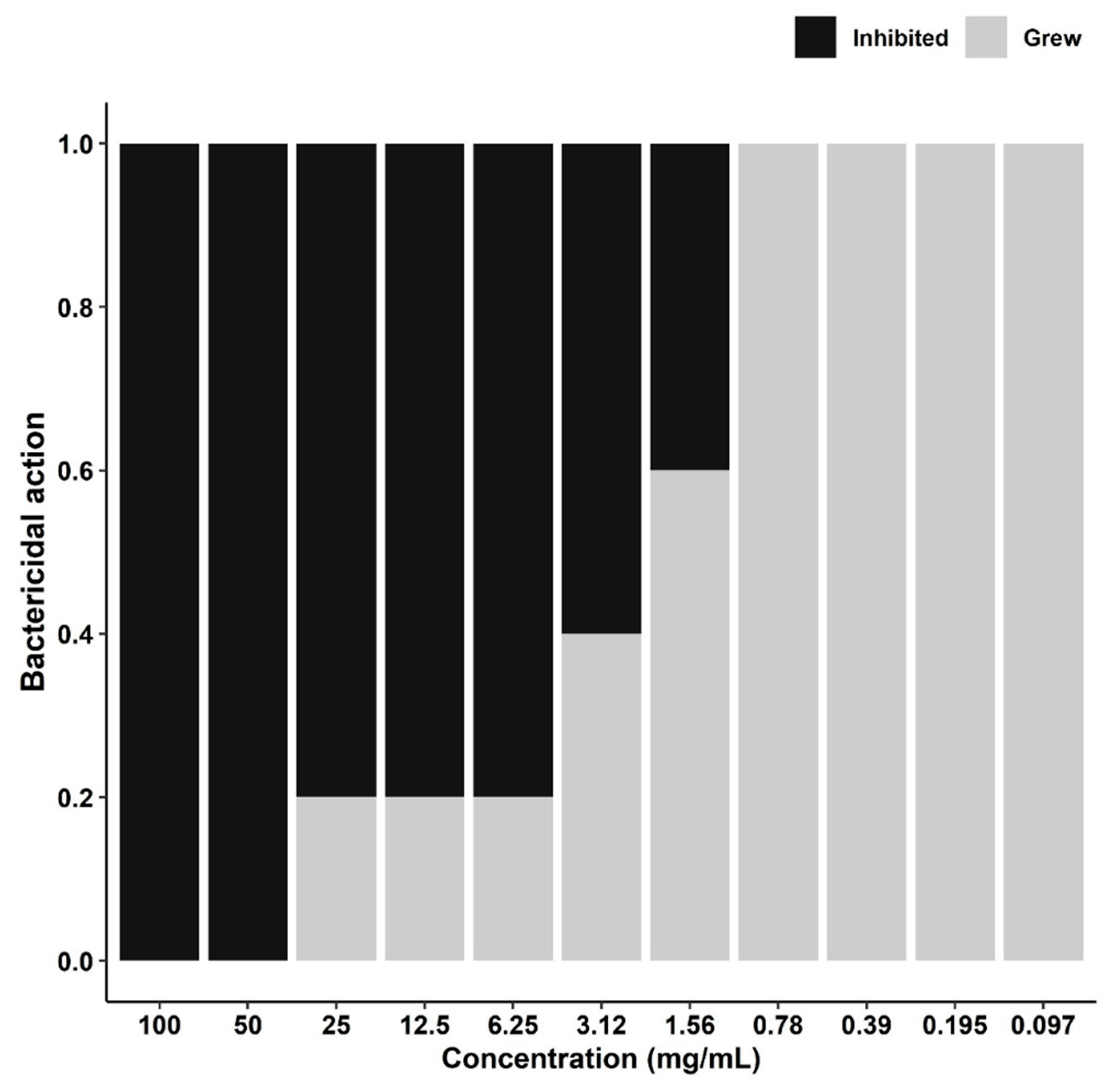
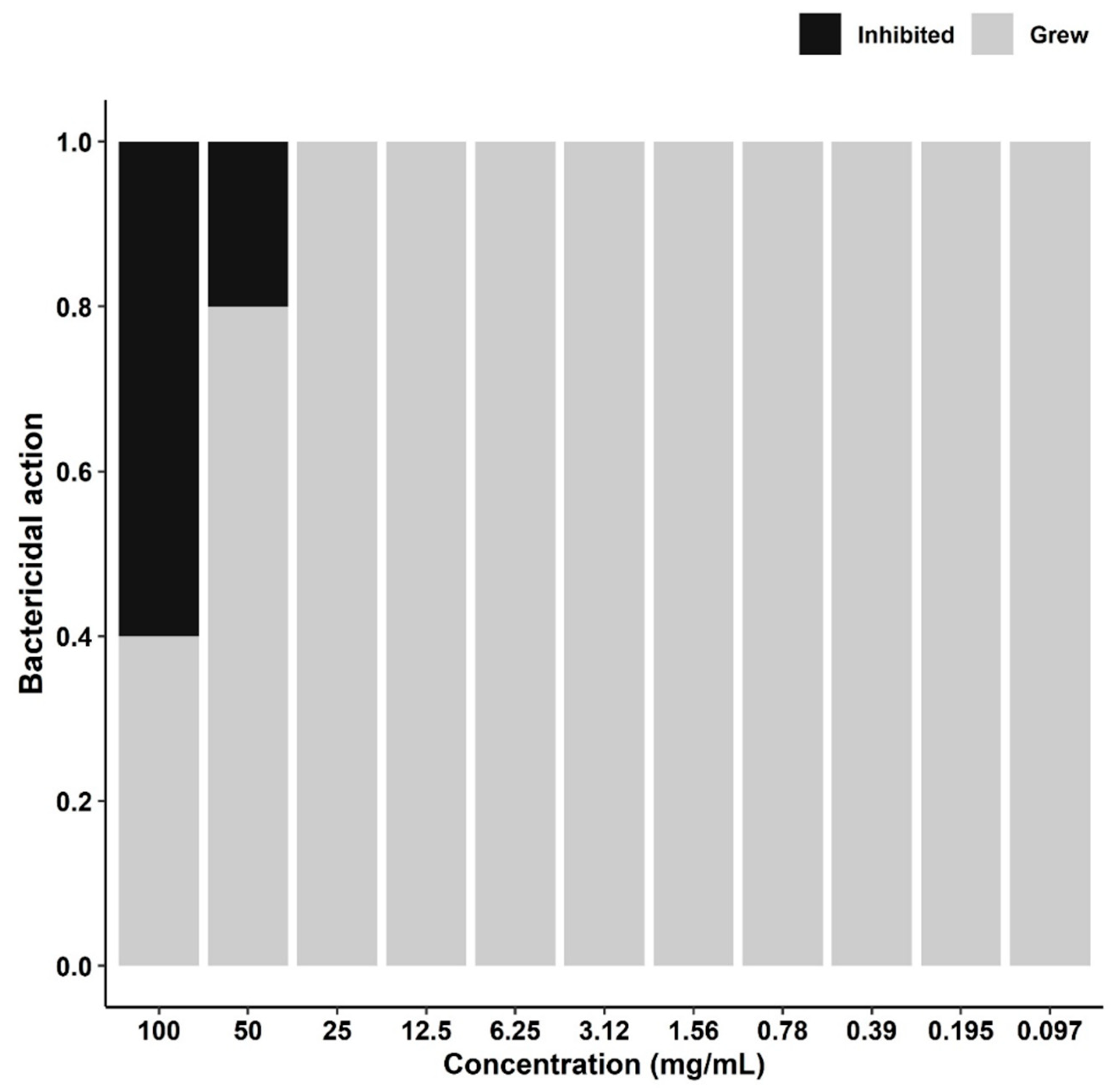
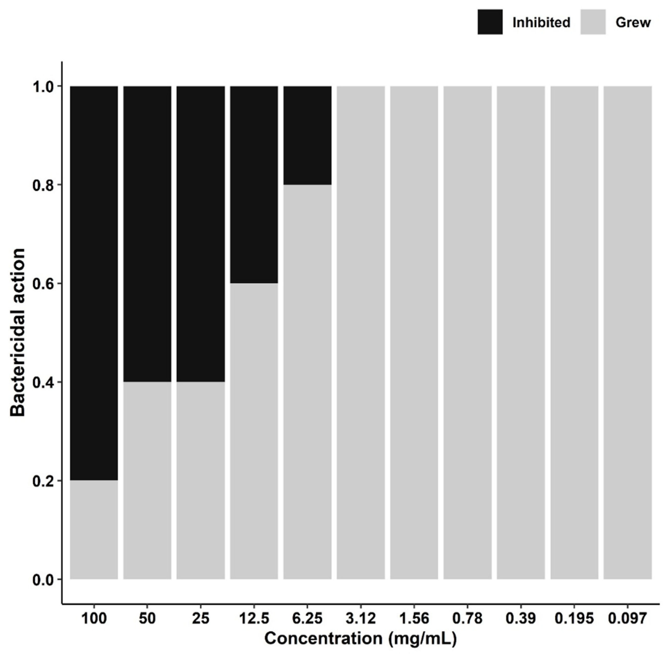
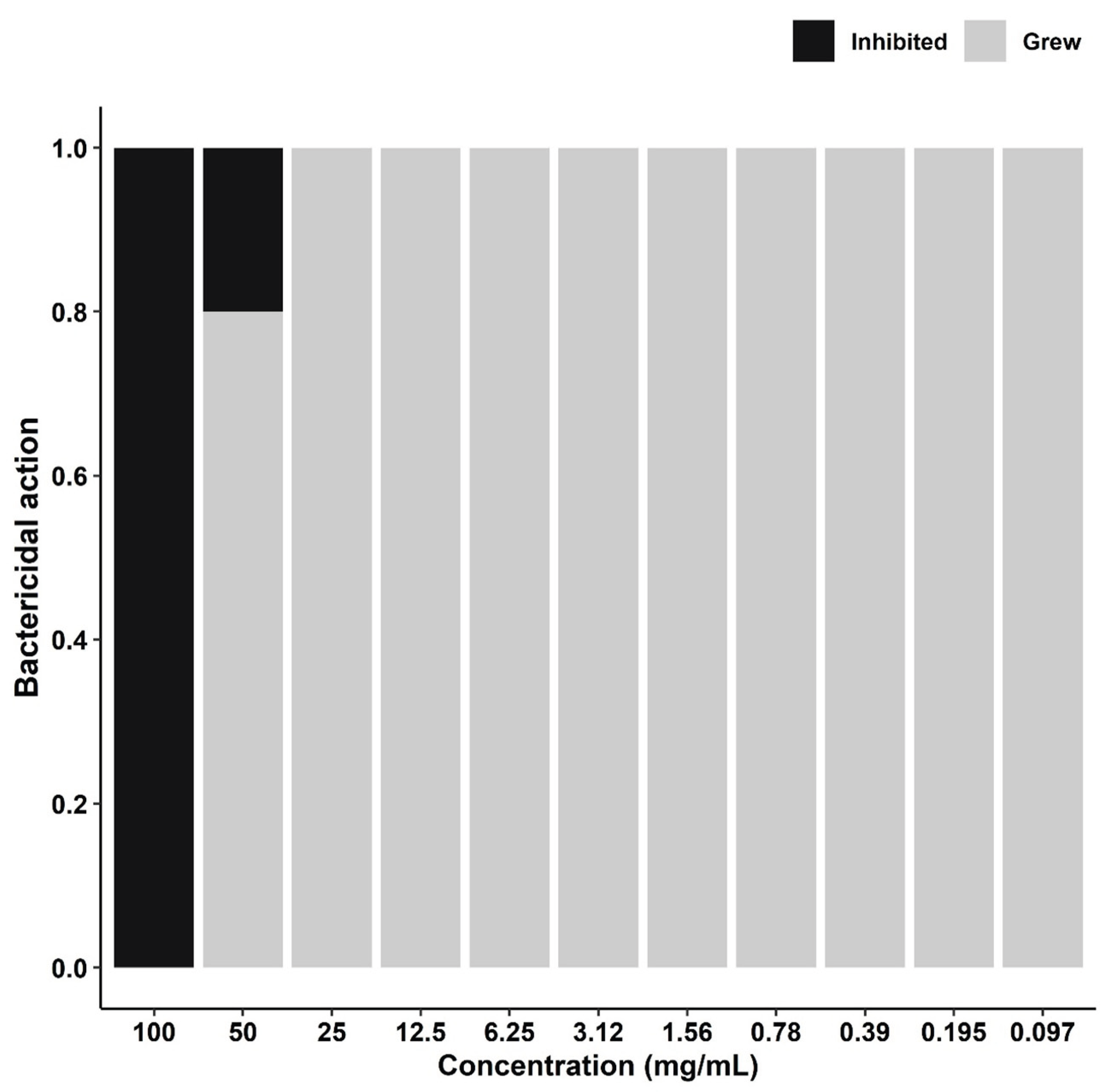
Figure 1. Bactericidal action of the different concentrations of Strypnodentron adistringens against S. aureus, S. agalactiae, S. uberis, E. coli, and Salmonella spp.
Preprints 190969 g001
Figure 2. Bactericidal action among the different concentrations of Bacharis crispa against S. aureus, S. agalactiae, S. uberis, E. coli, and Salmonella spp.
Preprints 190969 g002
Figure 3. Bactericidal action between the different concentrations of Azadractha indica in front of S. aureus, S. agalactiae, S. uberis, E. coli, and Salmonella spp.
Preprints 190969 g003
Figure 4. Bactericidal action of different concentrations of Azadractha indica oil against S. aureus, S. agalactiae, S. uberis, E. coli, and Salmonella spp.
Preprints 190969 g004
Table 1. Minimum bactericidal concentration (MBC) of ESa, EBc, EAi, and OAi against strains of S. aureus, S. agalactiae, S. uberis, E. coli, and Salmonella spp. isolated from cases of bovine mastitis.
Bacterium Concentration of Extracts and Oil (mg/mL)
ESa EBc EAi OAi
S. aureus 1.56 100 25 100
S. agalactiae 3.12 100 12.5 100
S. uberis 1.56 50 6.25 50
E. coli 50 >100* >100* 100
Salmonella spp. 6.25 >100* 100 100
* Showed bacterial growth in all concentrations. ESa= Extract of Strypnodentron adistringens; EBc=Extract of Bacharis crispa; EAi=Extract of Azadractha indica; OAi= Oil of Azadrachta indica. Concentrations do not differ between columns but differ between rows (P<0.001).
Disclaimer/Publisher’s Note: The statements, opinions and data contained in all publications are solely those of the individual author(s) and contributor(s) and not of MDPI and/or the editor(s). MDPI and/or the editor(s) disclaim responsibility for any injury to people or property resulting from any ideas, methods, instructions or products referred to in the content.
Copyright: This open access article is published under a Creative Commons CC BY 4.0 license, which permit the free download, distribution, and reuse, provided that the author and preprint are cited in any reuse.
Prerpints.org logo

Preprints.org is a free preprint server supported by MDPI in Basel, Switzerland.

Subscribe

Disclaimer

Terms of Use

Privacy Policy

Privacy Settings

© 2026 MDPI (Basel, Switzerland) unless otherwise stated