Preprint
Article

This version is not peer-reviewed.

Bari1 Transposon Arrays in Drosophila melanogaster Display High Stability over Time and Limited Impact on Ectopic Heterochromatin Assembly

Submitted:

19 December 2025

Posted:

22 December 2025

You are already at the latest version

Abstract

Transposable elements (TEs) impact genes and genomes in different ways. In Drosophila, TEs play a critical role in establishing heterochromatic loci, such as centromeric and pericentromeric regions, and contribute to ectopic heterochromatin formation especially when found in a tandem repeat configuration. The Bari transposon family belongs to the Tc1/mariner superfamily. Bari1 is featured by a unique tandem repeat arrangement in the heterochromatin of D. melanogaster. Despite its conservation could suggest a structural role in both natural and laboratory populations, little is known about the role of the Bari1 repeat in the heterochromatin. In this paper, we have studied the impact of the Bari1 transposon clusters on chromatin. We obtained transgenic strains of D. melanogaster carrying arrays of either three or seven copies of the Bari1 transposon in a typical heterochromatic tandem repeat configuration. We performed qualitative and quantitative phenotypic assays to evaluate the impact of these arrays on the chromatin status. Besides, we performed Southern Blot Hybridization analyses to evaluate the stability of Bari1 repeats over multiple generations. Our results suggest that Bari1 arrays are very stable over time and can poorly act as heterochromatin seeding sites. Our findings expand the knowledge on the structural and functional impact of TEs, whose potentially neutral, beneficial, or detrimental effect cannot be envisioned without experimental validation.

Keywords: 
;  ;  ;  ;  

1. Introduction

Transposable Elements and Heterochromatin

Transposable elements (TEs) are structural and functional genetic elements found in virtually all the eukaryotic and prokaryotic genomes investigated so far [1]. Decades of studies have demonstrated the role of TEs in genome structure and function and their contribution in evolution [2], although many aspects of their biology are still obscure. TEs can induce genomic rearrangements [3], disrupt genes and regulatory elements [4], or influence the spatial and temporal expression of nearby host genes, through the juxtaposition of their own cis-acting regulatory sequences [5].
As repetitive sequences, TEs constitute a large portion of the constitutive heterochromatin [6], a dense, dynamic, and heterogeneous nucleoprotein complex mainly located at the centromeres and telomeres. The enrichment of TE density at the transition from eu- to heterochromatin in D. melanogaster [7] may reflect insertional preference of TEs into heterochromatin [8], relaxed selection [9], or the TE- driven induction of the heterochromatin [10]. The latter is one of the most intriguing evolutionary scenarios that sets TEs in a leading role in the establishment and maintenance of heterochromatin and represents a possible way to understand how cis-acting sequences and trans-acting factors influence each other to organize chromatin. Evidence that repetitive DNA can actively contribute to heterochromatin formation comes mainly from studies in Drosophila on Position Effect Variegation (PEV), a phenotype first observed in the 1930s associated to the mosaic expression of the white gene [11]. PEV occurs when euchromatic genes are transcriptionally repressed after relocation near heterochromatin by chromosomal rearrangement or transposition [12]. The white gene of D. melanogaster has been extensively used as a PEV reporter [13,14]. Using PEV as a sensitive and sophisticated reporter to investigate how high order chromatin dynamics affect gene regulation, more than 150 genetic modifiers of the PEV phenotype (with possibly more yet to be discovered [15,16]), including dominant suppressors [Su(var)] and enhancer mutations [E(var)][17], have been identified in D. melanogaster [18]. Many of these loci encode chromatin proteins or enzymes that modify chromatin proteins post-translationally [18]. Two major suppressors of PEV are the Su(var)205 and the Su(var)3-9 genes, encoding the HP1 (Heterochromatin Protein 1) and the Histone-lysine N-methyltransferase proteins respectively, two key proteins in heterochromatin formation, maintenance, and spreading [19,20].
Several pieces of evidence elucidate the mechanisms behind the formation of heterochromatin around repeated sequences, resulting in the mosaic expression of nearby genes. Heterochromatin exerts an additive effect on white gene inactivation [21], and the severity of PEV correlates not only with the size of heterochromatic blocks but also with the chromatin conformation determined by the nearby repetitive DNA [22].
To test whether cis-acting factors are required for heterochromatin formation, Dorer and Henikoff [23] exploited P-elements carrying a mini-white reporter [24]. They found that arrays of P-elements induced white variegation and that the PEV phenotype was modulated by classical PEV modifiers. Other studies on transgenes containing white or brown as reporter genes, showed that reporter silencing increases with the number of copies in the array [23,25]. Interestingly, the 1360 transposon has been indicated as the initiation site of heterochromatin formation on the fourth chromosome but only in a subset of genomic locations near existing heterochromatin masses [13,26,27]. It was next demonstrated that 1360 mediates HP1 and H3K9me2 enrichment at its sites, through cis-acting internal sequences with homology to piRNAs (Piwi-interacting RNA [28]. This feature is also shared with the invader4 transposon [28]. Finally, recent evidence indicates that the repetitiveness itself, rather than the sequence and its origin, is sufficient to initiate heterochromatin formation, which can have distinct properties compared with endogenous heterochromatin [29,30,31].
The ability of TEs to establish and maintain heterochromatic domains is well-known in flies, mammals [32], plants [33,34], and in fungi [35,36,37]. But what are the mechanisms that trigger heterochromatin formation around TEs?
Several papers suggest that individual or clustered TE insertion could recruit specific proteins that remodel the chromatin into a silenced state [38], in flies [28,39] and in mammals [40,41]. TE silencing and heterochromatin formation differ between germline and somatic cells. In the germline, piRNAs direct repressive histone marks at the loci where TEs are inserted [42]. In the soma, evidence suggests the involvement of the siRNA (small interfering RNA) pathway in heterochromatin formation during early in embryo development [43], although mutations of key factors in the pathway, including Piwi, affect heterochromatin formation in somatic tissues [44,45]. The main physiological consequence of such molecular events is the heterochromatin-mediated transcriptional silencing of TEs, with the side effect that adjacent genes can be also transcriptionally silenced, due to the spread of the heterochromatin [18]. While the prevailing view supports the role of TEs in heterochromatin formation, studies have also highlighted exceptions to this pattern. Indeed, TEs do not always behave the same way, since there are reported instances demonstrating that chromatin remains unaffected upon TE insertion. Indeed, in contrast with the observation of Dorer and Henikoff, Conte et al. [46] studied the effect of multiple retrotransposon insertions (specifically ZAM and Idefix) into the white locus, showing that the accumulation of four copies of transposable elements at an euchromatic locus does not induce heterochromatin formation and gene silencing. These discrepancies suggest that the outcome of TE insertion may be influenced by multiple factors, including the TE insertion context and the presence of regulatory elements.

Bari1 Transposon

Bari1 is an active transposon [47] of the Bari family [48] belonging to the widespread ITm (IS630-Tc1-mariner) superfamily of Class II transposons [49]. It has been detected in species of the Drosophilidae family [48,50], suggesting it is an ancient genomic component of the Drosophila lineage.
It moves via the cut-and-paste transposition mechanism [51]. The canonical sequence of Bari1 comprises 1726 bp featured by 26 bp-long terminal inverted repeats and a 1020 bp-long gene encoding for the Bari1 transposase [49,51,52]. Bari1 was originally discovered in the genome of D. melanogaster as a block of 80 tandem repeated copies adjacent to the Responder locus (Rsp) [53]. Rsp (also known as XbaI repeat) is a component of the Segregation Distorter meiotic drive gene complex found in D. melanogaster [54]. Both the Bari1 and the Rsp repeats map into the h39 chromosome band, in the deep constitutive heterochromatin of the second chromosome [52]. In addition several copies of Bari1 (varying from 2 to 20 in natural population) are dispersed as single copy in the euchromatin [55]. An additional Bari1 cluster of 6 copies has been mapped in the heterochromatin of the X chromosome, in a region embedded in rDNA [48,56,57]. In both Bari1 arrays the monomers follow one another in a head-to-tail fashion, display a high degree of sequence conservation, and many copies retain their coding potential [48,56,58]. The Bari1 arrays are uniquely found in the D. melanogaster species [48,50,52], suggesting that the events that generated the Bari1 tandem repeat occurred early in the lineage that led to the D. melanogaster species [56]. Population genetics studies and copy number variation analysis in nearly 90 D. melanogaster populations revealed a low variability of the Bari1 cluster [55,57,59]. This is in contrast with similar data regarding other DNA repeat arrays, including the Rsp locus that can vary from zero to thousands copies, which have a high degree of variability in terms of copy number variation, and are also cause of genomic instability [48,56,57]. Compared to the canonical euchromatic Bari1 sequence, Bari1 elements in both arrays share a two-nucleotides deletion in the 5’ terminal inverted repat, making them transposition-incompetent [60]. This feature makes the Bari1 array unique among the transposon arrays discovered so far. Despite its intriguing features no function has been yet associated to the Bari1 heterochromatic cluster. A few papers describe the interaction of trans-acting factors with Bari1. The Bari1 transposase interacts with the Bari1 terminal sequences to operate transposition [47]. The HP1 protein binds the heterochromatic Bari1 cluster [61] and single euchromatic insertions [62]. Also, Bari1 is targeted by specific piRNAs which possibly regulate its mobility [63].
The Bari1 transposon array represents a unique model system to study the effect of a natural transposon tandem repeat on the chromatin composition. Inspired by the unique organization of the Bari1 arrays and with the aim to fill the knowledge gap associated to its functions, in this work we investigated the impact of ectopic Bari1 arrays inserted in the genome of D. melanogaster. Using P-element mediated integration, we randomly inserted two set of Bari1 repeats consisting of three and seven arrayed copies respectively and assessed the effect on the adjacent mini-white reporter gene [23,31]. Repeats of comparable size have been used in previous works to assess the effect of arrayed DNA sequences on the expression of reporter genes and their ability to form ectopic heterochromatin [23]. Collectively, our results demonstrate that Bari1 arrays do not influence the expression of adjacent reporter gene and therefore would not initiate de novo heterochromatin assembly, at least under our experimental conditions. We also report that the structure of ectopically inserted arrays of the Bari1 transposon is very stable over time.

2. Materials and Methods

Plasmids Construction and Molecular Methods

Phage clones containing Bari1 in a heterochromatic arrangement were identified by screening of an Oregon-R genomic library in EMBL4 vector using a labeled Bari1 probe as described in [52] and selected based on both their digestion and hybridization patterns.
An 11,9 Kbp fragment (corresponding to seven Bari1 copies) was subcloned from a positive phage clone into the EcoRI site of the pGEM-T easy vector. This plasmid was further manipulated to obtain Bari1 arrays with three copies.
Inverse PCR (iPCR) [64] was performed using the method described in http://www.fruitfly.org/about/methods/inverse.pcr.html. The iPCR primers used in this study were: Pry4 (CAATCATATCGCTGTCTCACTCA), Pry1 (CCTTAGCATGTCCGTGGGGTTTGAAT), Plac4 (ACTGTGCGTTAGGTCCTGTTCATTGTT), Plac1 (CACCCAAGGCTCTGCTCCCACAAT).
Sanger sequencing of the iPCR products was performed at the BMR-Genomics sequencing facility (Padova-Italy). The BlastN tool implemented in FlyBase [65] was used to map the insertion sequences. The Release 6 plus ISO1 MT assembly version was used as a reference for sequence analyses.
The 5,1 Kbp or the 11,9 Kbp HindIII fragments (hereafter indicated as 5 Kbp e 12 Kbp fragments) containing either three or seven copies of Bari1 in a tandem repeat configuration, were subcloned into the pCaSper3 vector[24] to obtain the pCasp-Ba3 and pCasp-Ba7 transformation vectors.

Drosophila Genetics Methods

Fly Stocks Were Maintained at 25 °C on Standard Culturing Medium.

The pCasp-Ba3 and pCasp-Ba7 plasmids were co-injected with the Pπ25.7 helper plasmid [66] into w1118 recipient embryos, following the procedure for P-mediated transformation described by Rubin and Spradling [67].
G0 adults were crossed to w1118 flies, and the red-eyed progeny was selected for further analyses. Genetic mapping of transgenes was performed by segregation analyses using strains carrying multiple balancer chromosomes. Once established, transgenic lines were continuously monitored to assess the stability of the phenotype observed.
Fly eye phenotypes were imaged under a Leica M165C stereo microscope. At least 50 individuals for each sex were observed.
Two Position Effect Variegation (PEV) modifier strains were used in this work, the Su(var)2055 [68] (an allele of Su(var205), and the Su(var)3-96 [69] (an allele of Su(var)3-9).
Stocks used in this study were obtained from the Bloomington Stock Center (NIH P400D180537).

Southern Blot Hybridization

Genomic DNA was extracted en masse from approximately 500 adult flies, according to standard protocols [70]. Genomic DNA was subjected to restriction analyses using the either EcoRI or SalI (both enzymes release the Bari1 array cloned in the pCasp-Ba3 or pCasp-Ba7 vectors). Standard Southern Blot hybridizations were performed using probes labeled by random priming with α32P-dATP. A 2 Kbp mini-white probe was obtained from a SalI digestion of the pCasper3 vector. A 1,7 Kbp HindIII fragment containing a full-length Bari1 element [52] was also used as a probe.

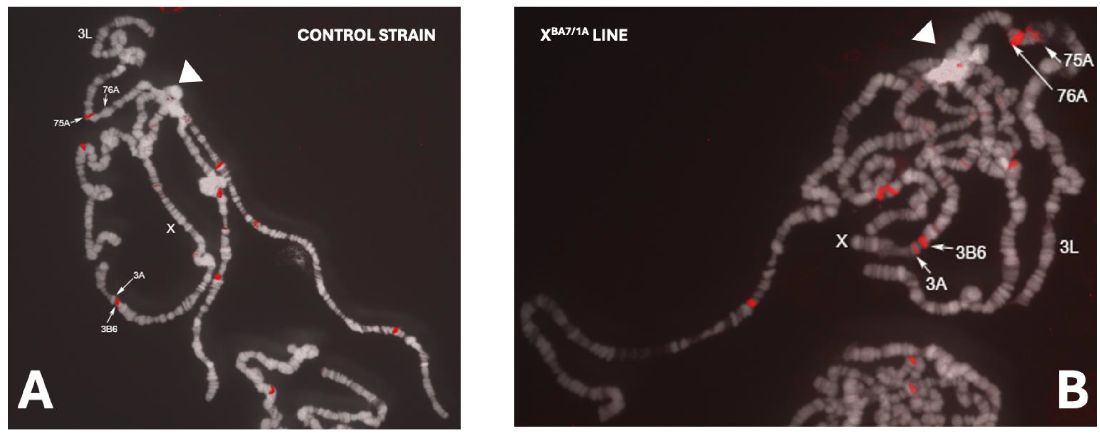
FISH (Fluorescence In Situ Hybridization) Experiments

Polytene chromosomes were prepared from salivary glands isolated from third-instar larvae of the Oregon-R strain and transgenic lines obtained in this study, as described by Kennison [71]. The pCasp-Ba7 plasmid was labeled by nick translation with Cy3-dCTP (Amersham). Chromosomes were stained with DAPI (4′,6-diamidino-2-phenylindole). Digital images were obtained using a Leica DMRXA epifluorescence microscope equipped with a cooled CCD (Charge-Coupled Device) camera (Princeton Instruments, NJ). Gray-scale images, obtained separately recording Cy3 and DAPI fluorescence by specific filters, were computer-colored and merged for the final image using the Adobe Photoshop software.

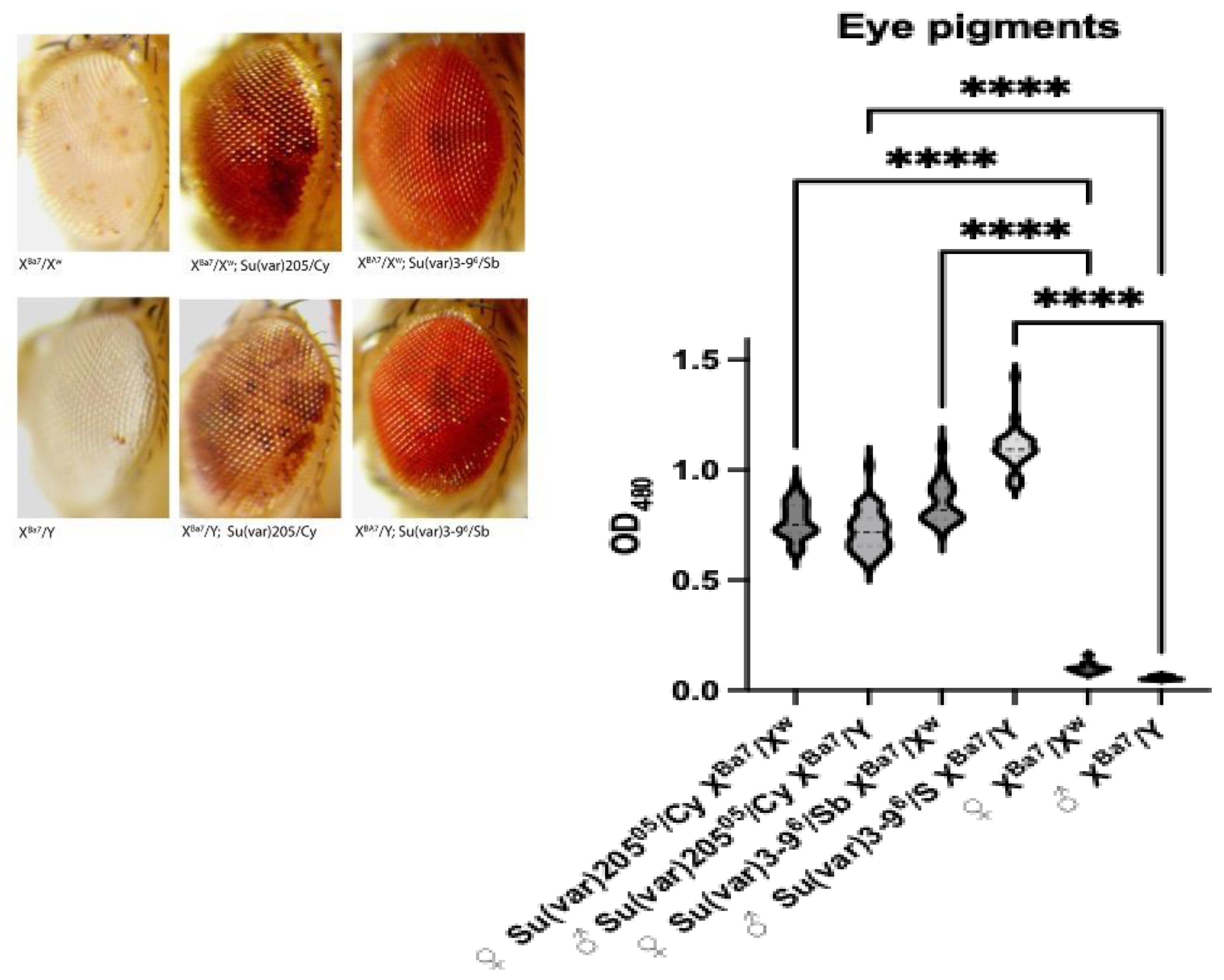
PEV (Position Effect Variegation) and Eye Pigment Quantification

We monitored the impact of the Su(var)2055 allele (carried by the Bloomington strain #6234) and the Su(var)3-96 allele on the PEV phenotype of the transgenic lines analyzing offspring eyes phenotype. Homozygous females carrying the transgenes were crossed either to Su(var)2055/Cy or to Su(var)3-96/TM6 males and the pigmentation extent of heterozygous F1 progeny was observed. Crosses were performed at 25 °C to prevent temperature effects on variegation. Extraction of the eye pigments and measurement were done according to [72]. Newly hatched adults were aged for six-nine days then 15 heads were collected by freezing the flies in liquid nitrogen and vortexing for few seconds; heads were incubated for three days in 1ml of 30% ethanol acidified to pH 2 with HCl. Pigment levels were determined by spectrophotometric assay at 480nm. Measurements for each line contained 35 to 40 replicates. Plots were obtained using Prism (version 10.2.3). One-way Anova test was performed to evaluate statistical significance.

3. Results

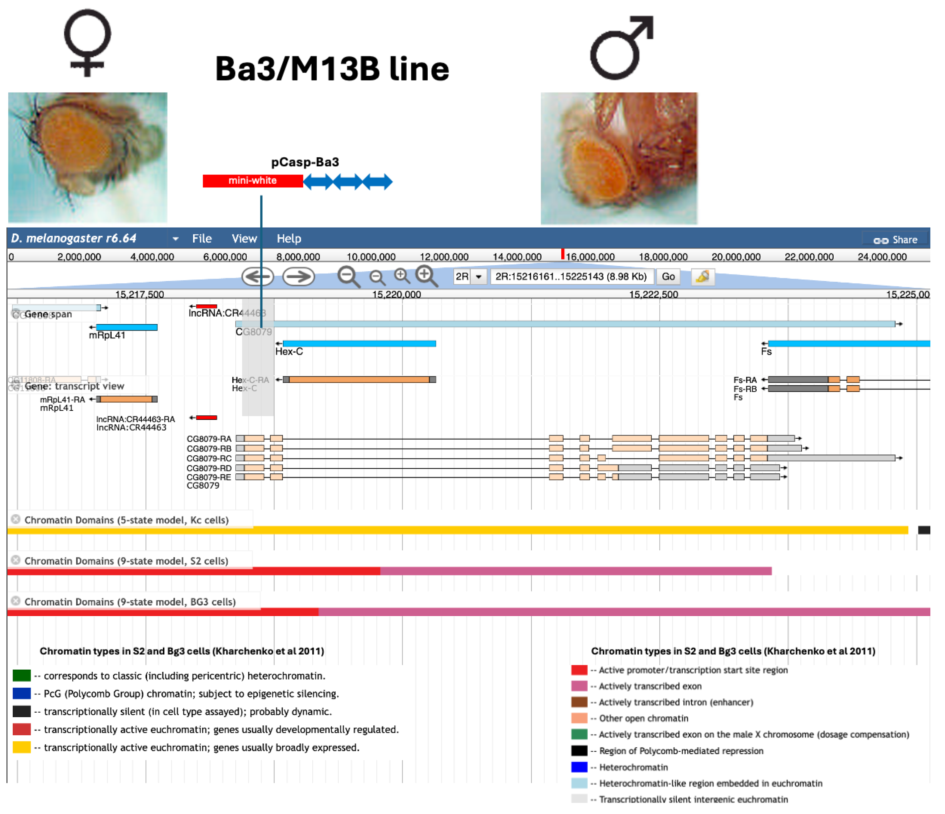
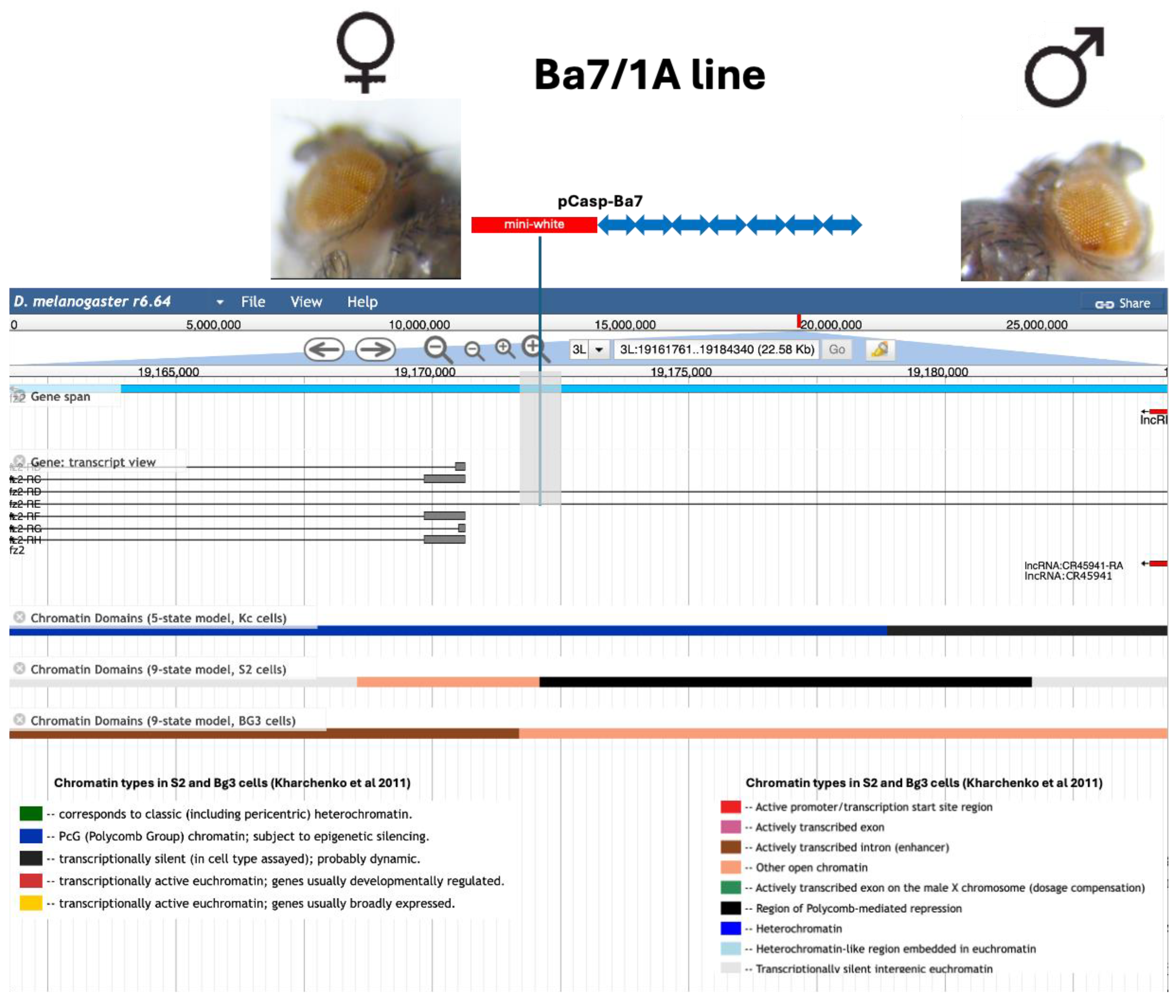
3.1. Strategy for Constructing Artificial Bari1 Arrays

The strategy devised for obtaining constructs carrying three or seven copies of Bari1 made use of a phage clone (λB15) previously isolated from an Oregon-R genomic library during the initial characterization of the Bari1 transposon [52]. The phage clone harbors a 12 Kbp EcoRI fragment (Figure 1 lane 1 panel A) that hybridizes with the Bari1 probe (Figure 1 lane 1 panel B) and is digested as a 1,7 Kbp band with HindIII (Figure 1 lane 2 panel A and panel B). This digestion pattern is consistent with the presence of a tandem array of seven Bari1 copies. The phage-ends sequencing analysis revealed that the Bari1 copies present at both insert sides are truncated. The leftmost Bari1 fragment corresponds to position 748-1726 whereas the rightmost Bari1 fragment corresponds to positions 3-748, if compared to the reference Bari1 sequence (accession number X67681). We sub-cloned the 12 Kbp fragment into the pGEM T-easy vector and obtained the plasmid pTBa7 (Figure 1 lane 4), in order to manipulate the Bari1 copy number in the cloned fragment. Partial digestion with the diagnostic HindIII restriction enzyme of the pTBa7 vector, previously digested with EcoRI, resulted in a ladder-like hybridization pattern (Figure 1 lane 5 panel A, and lane 5 panel B), which is consistently expected due to the repeated configuration of the 1,7 Kbp Bari1 monomer. The partial digestion product was re-ligated to the pGEM-T vector to obtain clones containing three Bari1 copies (pTBa3) that were selected and further processed.
Both the 12 Kbp fragment of the pTBa7 plasmid and the 5 Kbp fragment of the pTBa3 plasmid were independently sub-cloned into the EcoRI site of the destination vector pCaSpeR3 to obtain the pCasp-Ba7 and the pCasp-Ba3 respectively.
These plasmids were microinjected into w1118 embryos collected at the pre-blastoderm stage, and the surviving individuals were crossed to w1118 partners. Progeny was selected based on mini-white reporter gene expression. White-eyed individuals were excluded, as they lacked transgene insertions. However, the reliance on the mini-white-based reporter system may have resulted in an underestimation of integration frequencies, since insertions within heterochromatic regions (including the Y chromosome) may not have been detected due to complete transcriptional silencing of the reporter. Conversely, if the integration is associated to ectopic heterochromatin formation a mottled-like phenotype is expected. Twenty Ba3 lines (containing three arrayed copies of Bari1) and nineteen Ba7 lines (containing seven arrayed copies of Bari1) were established from these crosses. We subsequently genetically mapped the transgenes in all the established transformant lines and carefully inspected them for their eye pigmentation. As showed in Table 1, the recovered insertions are equally distributed among chromosomes, suggesting that the presence of the Bari1 array does not introduce any bias in the choice of the P-element transposase target (chi-square test not significant at the significance level of p<0,05).
A subset of lines was characterized by FISH or by iPCR (Supplementary Table 1). We assumed that if the Bari1 repeats could assemble ectopic heterochromatin, they would influence the closely associated mini-white reporter gene, resulting in a variegated eye pigmentation, which in turn reflects a mosaic mini-white expression. Moreover, if this effect depended on the number of Bari1 copies in the array, differences in the degree of variegation would be expected. However, only two of the transgenic lines described and discussed below displayed a PEV-like phenotype, regardless of the number of Bari1 in the repeat.

3.2. Analysis of Two Transgenic Lines that Display Uneven Eye Pigmentation.

The Ba3/M13B line originated from the integration of the pCasp-Ba3 construct. Adults of the Ba3/M13B line display two distinct and differentially pigmented eye sectors. This eye pigmentation phenotype is more pronounced in males than in females. The insertion sequence analysis by iPCR revealed that the insertion occurred in the CG8079 gene. We will refer to this allele as CG8079Ba3 throughout the manuscript. CG8079 is still an uncharacterized D. melanogaster gene, and it is predicted to encode a nucleic acid binding protein with a possible function in the splicing process [73].
The insertion occurred in the first exon of the CG8079 (at nucleotide 15218444 of the AE013599 accession, Figure 2) and the strain is homozygous viable. Even though we do not have tested the expression level of CG8079Ba3 in the transgenic line Ba3/M13B, the homozygous state suggests that CG8079 is a dispensable gene that potentially affects the pigment distribution in the eye. Furthermore, the second intron of CG8079 contains the Hex-C gene on the antisense strand. This gene, encoding the hexokinase-C protein, was already known to be not essential either for fly viability and fertility [74], and indeed our fly stock is homozygous, viable, and fertile. Finally, this phenotype is not modified by the PEV modifier Su(var)2055 (not shown), strongly supporting the hypothesis that the observed phenotype is independent from chromatin remodeling across the insertion site in the Ba3/M13B line. Moreover, the locus does not display heterochromatic epigenetic marks in proximity of the insertion, as can be inferred from the modEncode data displayed in the FlyBase “JBrowse” genome browser [75] (Figure 2).
From these observations it is possible to conclude that the observed phenotype of the Ba3/M13B strain could possibly be the result of the CG8079 gene inactivation rather than an epigenetic effect exerted on the mini-white transgene by the three tightly linked Bari1 copies.
Among the selected lines in which seven copies of Bari1 have been inserted, the BA7/1A line displays a non-uniform eye pigmentation pattern, reminiscent of the well-known variegated phenotype associated with the white-mottled (wm4) allele [12]. However, the eye pigmentation observed in the Ba7/1A line differs markedly from the wm4 phenotype since it shows darker spots of different areas over a light-red background (Figure 3). We anticipate that this line carries two insertions, both contributing to generating the observed phenotype.
Inverse PCR yielded a single amplification fragment and the subsequent sequence analyses revealed that the insertion occurred within the frizzled 2 (fz2) gene, which maps in the 76A1 region of the polytene chromosomes and encodes a G-protein coupled receptor of the Fz/Smo family (Figure 3). The insertion occurred into the 5th intron of the fz2 (precisely at nucleotide 19171766 of the AE014296.5 accession) gene, exactly 334 bp upstream of a region characterized by heterochromatic epigenetic marks [76], in two cellular models (Figure 3), as defined by the modEncode data displayed in the FlyBase “JBrowse” genome browser [75,78]. State 6 is a repressive chromatin state mediated by the Polycomb group proteins. This repressed chromatin region is flanked by an actively transcribed region (state 4) due to the presence of an alternative promoter of the fz2 gene.
While the position effect, i.e. proximity to a repressed chromatin site, could be the simplest hypothesis explaining the variegated expression of the mini-white reporter gene in the BA7/1A line, we had evidence that the BA7/1A line carried an additional insertion. We mapped genetically the second insertion on chromosome X, and the eye phenotype was analyzed separately in the two segregant lines. The sub-line carrying the insertion on the 3rd chromosome displayed a uniformly pigmented eye, while the insertion on chromosome X showed a strongly variegated phenotype (Figure 4A, left column). To distinguish the two insertions, we will refer to XBA7/1A (X-linked insertion) and to fz2BA7/1A (insertion on the 3rd chromosome) hereafter.
To gain further insights into the nature of the variegated reporter expression observed in the XBA7/1A, we checked if the variegated phenotype could be influenced by the PEV modifiers Su(var)205 and Su(var)3-9. The extreme variegated phenotype displayed by heterozygous females and hemizygous males is mitigated by the Su(var)2055 loss-of-function mutation (Figure 4 central column). Consistently, a nearly uniform red-eye pattern is completely restored in XBA7/1A/+; Su(var)3-96 /+ flies (Figure 4 right column). These observations confirm that the PEV phenotype in the original XBA7/1A line is due to heterochromatin wrapped around the X-linked pCasp-BA7 insertion. The qualitative phenotypic observation reported in Figure 4A are supported by the spectrophotometric quantitation of the pigment level in fly heads isolated from males and females of the same genotype (Figure 4B). Su(var)3-9 acts as a stronger suppressor compared to Su(var)2055 and overall, the pigment content analysis confirms a correlation between the observed eye phenotype and the drosopterin amount detected. We also tested additional PEV modifiers, such as Pol32 [79], that resulted in a similar suppression of the variegated phenotype in this transgenic line (not shown). No significative change in pigment amount was detected when the line carrying the insertion on the third chromosome was combined with PEV suppressors.
Fine mapping of the X-linked insertion by iPCR was unsuccessful, possibly due to a local rearrangement of the insertion (see discussion). However, the insertion site was mapped by recombination very close to yellow (not shown) and this observation was confirmed by FISH mapping, that revealed a hybridization signal in the 3A region of the polytene chromosomes (Figure 5).

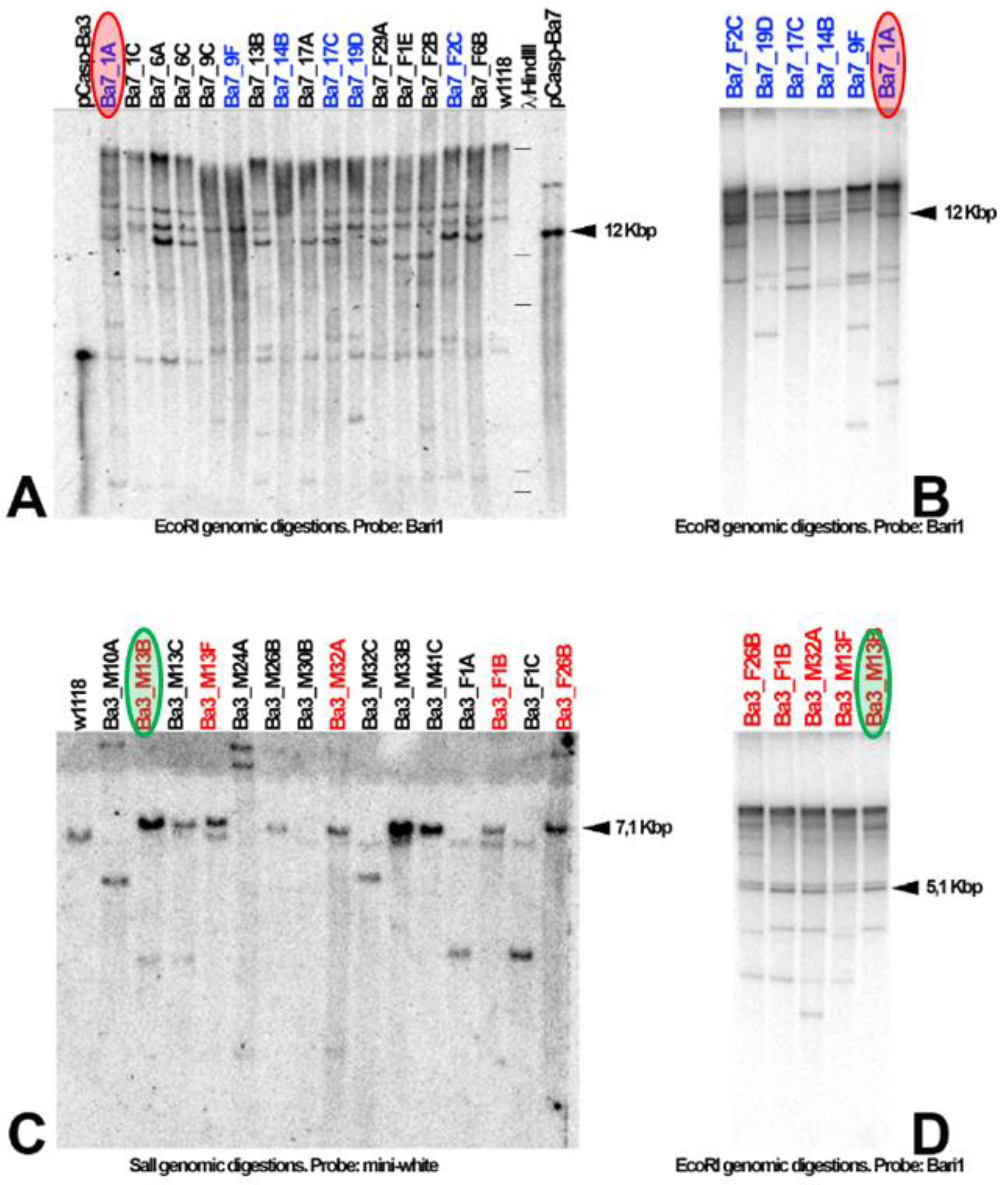
3.3. Bari1 Repeats Stability Over Time

The initial establishment of the Drosophila transgenic lines described in this study occurred in 2003. We performed Southern Blot Hybridization analysis on genomic DNA extracted three generations after the establishment of the transgenic lines, with the aim to verify if the Bari1 copies (three or seven respectively for the Ba3- and Ba7-carrying transgenic strains) were retained in the selected transgenic lines. Most of the transgenic lines of the Ba7 series tested positive for the expected band (Figure 6 A). Ten lines of the Ba7 series, over 15 tested, contain the expected 12 Kbp hybridization fragment. Four lines displayed a hybridization band of smaller size, while a single line showed a heavier hybridization fragment. Among the lines containing unexpected hybridization bands, the Ba7/1A line (that contains two insertions, see description in the paragraphs above) displays several high molecular weight bands which could be suggestive of a rearrangement associated with the X-linked Bari1 array, mapped in the 3A region of the polytene chromosome. It can be concluded that in 31% of the lines tested a rearrangement occurred in the ectopically inserted Bari1 array within three generations after the establishment of the lines.
The Ba3 lines were similarly assayed (Figure 6 C). A Southern Blot with a SalI genomic digestion was hybridized with a mini-white probe. The expected 7 Kbp hybridization fragment is clearly visible in nine lines over 16 lines tested. Four lines displayed a hybridization band smaller than expected while two had a heavier band. In summary, 40% of the lines analyzed had a rearranged transgene soon after the establishment of the lines (within the third generation).
We have next evaluated the stability of the insertions obtained over time. To establish if the rearrangements initially observed in some of the lines analyzed could be symptomatic of a repeat-induced instability, we performed again the experiment after approximately 150 generations (roughly equivalent to 10 years at 18 °C) on a subset of the above-described lines. The hybridization pattern after 150 generations suggests that no rearrangements occurred during this time (Figure 6 B and D) if compared with the hybridization pattern of the third generation, (Figure 6 A and C), leading to the conclusion that the few initially observed rearrangements are the result of ectopic recombination occurred soon after the microinjection, or at least within the three generations after the establishment of the transgenic lines.

4. Discussion

Bari1 Arrays do not Induce Heterochromatin Formation

How tandem repeated sequences impact genome stability and gene expression remains a debated topic in functional genetics and genomics. The current knowledge and technological advancement in the fields of genome sequencing [80], gene expression [81], and epigenetics [82] enable functional studies aimed to get insights into the role of satellite DNA and transposon arrays on chromosome stability, gene expression, and the interaction between genomic loci. Functional studies are also needed to solidify evidence provided by “omics” studies, particularly those concerning repetitive DNA. Overall, heterochromatin remains still poorly studied from a functional point of view. Emerging evidence suggest that TEs play a pivotal role in generating and maintaining heterochromatic structures in the eukaryotic genome [6,33]. In an evolutionary perspective, repositioning of gene clusters and their conversion in heterochromatic blocks is apparently associated with an increase of TE content in the repositioned loci [83]. TEs also contribute to short-term effects on gene expression since they carry cis-acting elements that impact the resident gene expression, and their ability to form heterochromatin de novo.
Genetic evidence demonstrates that tandem arrays of transgenes are able to create or enhance mosaic patterns of gene silencing [23] and as little as three tandem copies of P[lacW] are sufficient to establish a repressive chromatin domain in D. melanogaster [23,25]. The structural analogy of these arrays with heterochromatin has been demonstrated using genetic criteria, such as suppression by modifiers of classical PEV, including mutations in the genes encoding the heterochromatin protein HP1 and the histone methyltransferase Su(var)3-9. However, there are many variables that influence repeat induced heterochromatin formation such as the presence of permissive sites [31] that seem to be required to trigger PEV of reporter genes. Moreover, these observations are challenged by experimental evidence showing that some large transgene arrays do not form heterochromatin and maintain a uniform eye pigmentation [84]. In addition, studies in maize suggest that only a limited number of retrotransposon families act as nucleation center for heterochromatin that can influence the expression of neighboring genes [85].
Based on the above information, we wonder if the tandem repetitive configuration of the Bari1 transposon represents a sufficient condition to trigger heterochromatin seeding.
After nearly seven decades of research on the h39 region, the role of the Bari1 repeat in this genomic region remains unclear, especially its relationship with the Responder (Rsp) repeat. We can bona fide exclude that the Bari1 cluster has no role in the genome of D. melanogaster, since it has been positively selected and it is found in all the populations analyzed so far [55,57]. It is not yet known whether Bari1 influences the physical size of the Rsp locus in different D. melanogaster populations, or whether it affects the responsiveness of the Rsp locus to Sd. Previous evidence suggests that the h39 region influences the nucleosomal organization of the centromere of chromosome 2 without altering its functionality [86]. However, the absence of chromosomal aberrations that physically separate the Rsp and Bari1 repeats makes it difficult to dissect the individual roles of these clusters in centromere function. It is hoped that the combination of modern genetic and molecular strategies will help fill this knowledge gap.
As a first step in this direction, we asked whether the tandemly repetitive configuration of the Bari1 transposon represents a sufficient condition to trigger heterochromatin seeding.
Among the 40 transgenic lines analyzed, 19 harbored a tandem array of seven Bari1 copies. Our observations suggest that this configuration does not support heterochromatin assembly or at least has a limited power of perturbing the native chromatin status. First, a single line - segregated from transgenic line harboring a double insert - exhibited a variegated expression of the mini-white reporter gene – a hallmark of heterochromatin formation. Several lines of evidence suggest that the variegated phenotype observed in the XBA7/1A line might be possibly generated by uncontrolled genetic factors. For example, precise mapping of the insertion associated to the variegated stock remains elusive. Multiple ineffective attempts were made to amplify the flanking sequence of the insertion via iPCR, i.e. using different restriction enzymes and primers. We hypothesize that a possible rearrangement involving the X-linked synthetic transposon may have occurred. This hypothesis is supported by the Southern blot hybridization analysis (Figure 6A, lane Ba7/1A), which reveals additional unexpected hybridization fragments in the BA7/1A (harboring insertions on both the 3rd and the X chromosomes), which are not noticed in the negative control. An additional suggestion of a possible rearrangement comes from FISH hybridization results. Indeed, the hybridization signal at the 3A region is weaker –than expected if compared to the signal at the 76A region (Figure 5 B). However, the low intensity of the hybridization signal could be attributed to under-replication of the locus which could have been induced by the pCasp-BA7 insertion. Indeed, the 3A region of the polytene chromosomes is not a known under-replicated region [87]. Further genetic and molecular characterization will shed light on the molecular basis of this phenotype.
Additional evidence supports the limited chromatin-altering capacity of the Bari1 array. Even when inserted in close proximity of a potential transcriptionally silent genomic locus, seven copies of Bari1 are not able to seed heterochromatin. Indeed, the fz2BA7/1A insertion occurs in a region characterized by a PcG-type (Polycomb Group) chromatin, thus potentially subjected to epigenetic silencing (state 6 type of chromatin according to the 5-state model in Kc cells and the 9-states model in S2 cells [76]). Suggestively, Bari1 contains Polycomb Responsive Elements (PREs) [62] that do not mediate enrichment of the H3K9me3 histone mark in some associated loci [62]. It could be hypothesized that the introduction of seven arrayed PREs should act cooperatively with the local epigenetic marks and finally trigger chromatin compaction around the mini-white reporter gene. However, no difference has been observed between the fz2BA7/1A insertion and other P-based insertions in the same region (Supplementary Table 2). Additional experimental conditions (such as oxidative stress) should be tested to clarify whether Bari1 mediates heterochromatin seeding. Taken together, this information suggests a neutral effect of the Bari1 sequences in chromatin assembly.
In conclusion, the overall trend of the transgenic lines analyzed in this study would suggest that the Bari1 array, at least with this number of repeated copies and in this orientation, is not such a potent heterochromatin assembly site as demonstrated for other TEs [23,28]. While our results may at first appear to contrast with previously published observations, other studies have shown that only certain types of satellite DNA are capable of triggering heterochromatin formation. For example, a cassette carrying human alphoid satellite DNA induced significant silencing of an adjacent reporter gene, consistent with heterochromatin formation [88]. In contrast, when human gamma-satellite DNA was used, the reporter gene remained highly expressed, indicating that this sequence favored an open chromatin state rather than heterochromatinization [88]. These findings provide direct experimental evidence that different satellite DNA sequences can differentially modulate chromatin structure, resulting in either heterochromatic repression or an open chromatin configuration. However, we acknowledge several limitations of our study that highlight opportunities for future improvements. First, it is possible that insertions resulting in complete suppression of reporter expression were not recovered, as they may have been misclassified as lines lacking the insertion. This scenario could have led to the underestimation of the total number of insertions analyzed. This limitation could be addressed in future studies using dual reporter constructs (such as white, yellow and brown), as described in previous works [13,31]. Second, only a single orientation of the array relative to the reporter gene was tested, which may represent an additional source of bias. Third, the number of insertions analyzed in this study was relatively limited. In contrast, other studies have identified permissive sites by analyzing a larger set of insertions. Expanding the number of insertions studied, for example by remobilizing some of the lines identified here, could enable the identification of permissive regions, similar to those observed in other studies [13]. Finally, the length of the array itself may represent a further limitation. This constraint could be overcome in the future by employing longer arrays or by directly manipulating the Bari1 array within the h39 region using genome-editing technologies. Such approaches could also provide direct in vivo experimental evidence for the functional role of the Bari1 repeat.

Bari1 Arrays Are Stable over Time

It is known that DNA repeats represent a source of genomic instability both in prokaryotes [89] and in eukaryotes [90,91]. The most common consequence of such instability is the expansion or contraction of the repeat due to the non-homologous (i.e. out-of-registry) recombination between different monomers of the array, leading to gain/loss of copy number [92]. This phenomenon also results in several types of human genetic disorders [93].
Heterochromatin is very dense in DNA repeats whose variability, in terms of sequence and number of repetitions, is reflected at the population level as chromosome polymorphism, that is sometimes detectable through simple cytogenetical analyses. There is a particular class of satellite DNA in D. melanogaster, which is the target of a segregation distortion systems (the Sd/Rsp system) and this poses a special interest on this DNA repeat class. The Responder locus is indeed the target of the Sd protein that lead to the loss of the chromosome carrying a responsive Rsp allele [94], a DNA satellite locus composed of a variable number of repetition of the 240 bp repeat (or XbaI repeat) in the h39 region of the second chromosome, a cytological band adjacent to the centromere locus (the h38 band). The Responder locus represents one example of satellite DNA exhibiting a strong length polymorphism [54] in D. melanogaster populations. Contrastingly, the Bari1 repeat displays a homogeneous composition in the same region, and a limited length polymorphism in different populations [55,57], suggesting a differential structural role of both repeats in the h39 region. Even the small clusters mapped on the third chromosome (Rsp) and on the X chromosome (Bari1) behave the same as the main cluster, in that the Responder cluster is composed of divergent copies [54], whereas the Bari1 elements in the cluster are very similar to each other and to euchromatic elements [48].
It has been shown that HP1 is a key chromatin component driving heterochromatin phase separation, and that Polycomb complexes mediate long-range interactions between transcriptionally repressed domains. [95]. The documented binding of HP1 [61] and, although under specific conditions, Polycomb protein [62] to Bari1 suggests that this repeated element may act as a structural heterochromatic node. The stability of the Bari1 array over time and the lack of deletions that specifically target the Bari1 cluster in the h39 region further support the possibility that the Bari1 cluster plays a role in maintaining correct nuclear architecture and heterochromatin–euchromatin segregation. From an evolutionarily perspective, it should be noted that the Rsp repeats exist in other species of the melanogaster complex (i.e. D. simulans, D. sechellia, D. mauritiana, D. erecta, and D. yakuba) [54] whereas only single Bari1 insertions are found outside the D. melanogaster species [48,96]. The above observation suggests that the Bari1 clusters originated more recently, possibly after the separation of the D. melanogaster species, and it is therefore possible that the Bari1 clusters are still in the process of being homogenized by ectopic recombination.
The observed stability of the transgenes obtained in this study resembles the stability of the Bari1 cluster in the h39 region in both laboratory and wild collected D. melanogaster populations [52,55,57,97].
While the stability of the Bari1 heterochromatic cluster - in terms of copy number variation – could be in part explained considering the reduced recombination rate in the heterochromatin [98], it should be underlined that the transgenes carrying the Bari1 arrays analyzed in this study map all in euchromatic loci. Therefore, the stability of ectopic Bari1 arrays appears associated to a reduced recombination rate around the insertion point of the constructs. This feature makes the ectopic Bari1 arrays a good model for studying the stability of heterochromatic transposon arrays.

5. Conclusions

Our study on ectopic Bari1 clusters inserted in the fly genome suggests that this kind of arrangement, which is stably maintained over time, may not strongly drive the formation of ectopic heterochromatin. Additional studies are required to understand the functional role of Bari1 repeats especially in the contexts of the structure and the stability of the second chromosome heterochromatin.

Supplementary Materials

The following supporting information can be downloaded at the website of this paper posted on Preprints.org, Table S1. Mapping data of the transgenic lines analyzed in this work. Table S2. List of the available stocks carrying P-element insertions in the fz2 gene. Figure S1-S4: Original southern blot figures.

Author Contributions

MFB, RM, SC, EP, AP, RC, RMM performed the experiments and analyzed the data. RMM and RC conceived and supervised the project. RMM drafted the manuscript. All authors approved the final manuscript.

Funding

This work was funded by Università degli Studi di Bari (Contributo di Ateneo to R.M.M.).

Institutional Review Board Statement

Not applicable.

Informed Consent Statement

Not applicable.

Acknowledgments

We thank Prof. Rodolfo Costa (University of Padova) for granting us access to the microinjection facilities, and Prof. Laura Fanti (University “La Sapienza”, Rome) for providing fly mutant strains. We are also grateful to Prof. Francesca Capezzuto (University of Bari) for kindly providing her support with the image acquisition at the Leica microscope workstation.

Conflicts of Interest

The authors declare no conflicts of interest. The funders had no role in the design of the study; in the collection, analyses, or interpretation of data; in the writing of the manuscript; or in the decision to publish the results.

Abbreviations

The following abbreviations are used in this manuscript:
PEV Position Effect Variegation
piRNA Piwi-interacting RNA
TE Transposable Element
Rsp Responder
iPCR inverse PCR

References

  1. Wells, J.N.; Feschotte, C. A Field Guide to Eukaryotic Transposable Elements. Annu Rev Genet 2020, 54, 539–561. [Google Scholar] [CrossRef]
  2. Kazazian, H.H., Jr. Mobile elements: drivers of genome evolution. Science 2004, 303, 1626–1632. [Google Scholar] [CrossRef]
  3. Bohne, A.; Brunet, F.; Galiana-Arnoux, D.; Schultheis, C.; Volff, J.N. Transposable elements as drivers of genomic and biological diversity in vertebrates. Chromosome Res 2008, 16, 203–215. [Google Scholar] [CrossRef]
  4. Bourque, G.; Burns, K.H.; Gehring, M.; Gorbunova, V.; Seluanov, A.; Hammell, M.; Imbeault, M.; Izsvák, Z.; Levin, H.L.; Macfarlan, T.S.; et al. Ten things you should know about transposable elements. Genome Biol 2018, 19, 199. [Google Scholar] [CrossRef]
  5. Sundaram, V.; Wysocka, J. Transposable elements as a potent source of diverse cis-regulatory sequences in mammalian genomes. Philos Trans R Soc Lond B Biol Sci 2020, 375, 20190347–20190347. [Google Scholar] [CrossRef] [PubMed]
  6. Marsano, R.M.; Dimitri, P. Constitutive Heterochromatin in Eukaryotic Genomes: A Mine of Transposable Elements. Cells 2022, 11. [Google Scholar] [CrossRef] [PubMed]
  7. Marsano, R.M.; Giordano, E.; Messina, G.; Dimitri, P. A New Portrait of Constitutive Heterochromatin: Lessons from Drosophila melanogaster. Trends Genet 2019, 35, 615–631. [Google Scholar] [CrossRef] [PubMed]
  8. Gao, X.; Hou, Y.; Ebina, H.; Levin, H.L.; Voytas, D.F. Chromodomains direct integration of retrotransposons to heterochromatin. Genome Res 2008, 18, 359–369. [Google Scholar] [CrossRef]
  9. Kent, T.V.; Uzunović, J.; Wright, S.I. Coevolution between transposable elements and recombination. Philos Trans R Soc Lond B Biol Sci 2017, 372, 20160458. [Google Scholar] [CrossRef]
  10. Rebollo, R.; Karimi, M.M.; Bilenky, M.; Gagnier, L.; Miceli-Royer, K.; Zhang, Y.; Goyal, P.; Keane, T.M.; Jones, S.; Hirst, M.; et al. Retrotransposon-induced heterochromatin spreading in the mouse revealed by insertional polymorphisms. PLoS Genet 2011, 7, e1002301–e1002301. [Google Scholar] [CrossRef]
  11. Muller, H.J. Types of visible variations induced by X-rays inDrosophila. J Genet 1930, 22, 299–334. [Google Scholar] [CrossRef]
  12. Tartof, K.D.; Hobbs, C.; Jones, M. A structural basis for variegating position effects. Cell 1984, 37, 869–878. [Google Scholar] [CrossRef] [PubMed]
  13. Wallrath, L.L.; Elgin, S.C. Position effect variegation in Drosophila is associated with an altered chromatin structure. Genes Dev 1995, 9, 1263–1277. [Google Scholar] [CrossRef]
  14. Hazelrigg, T.; Levis, R.; Rubin, G.M. Transformation of white locus DNA in Drosophila: Dosage compensation, zeste interaction, and position effects. Cell 1984, 36, 469–481. [Google Scholar] [CrossRef] [PubMed]
  15. Kelsey, K.J.P.; Clark, A.G. Variation in Position Effect Variegation Within a Natural Population. Genetics 2017, 207, 1157–1166. [Google Scholar] [CrossRef]
  16. Wang, S.H.; Elgin, S.C.R. The impact of genetic background and cell lineage on the level and pattern of gene expression in position effect variegation. Epigenetics Chromatin 2019, 12, 70. [Google Scholar] [CrossRef] [PubMed]
  17. Schotta, G.; Ebert, A.; Dorn, R.; Reuter, G. Position-effect variegation and the genetic dissection of chromatin regulation in Drosophila. Semin Cell Dev Biol 2003, 14, 67–75. [Google Scholar] [CrossRef]
  18. Elgin, S.C.R.; Reuter, G. Position-Effect Variegation, Heterochromatin Formation, and Gene Silencing in Drosophila. Cold Spring Harb Perspect Biol 2013, 5. [Google Scholar] [CrossRef]
  19. Li, Y.; Kirschmann, D.A.; Wallrath, L.L. Does heterochromatin protein 1 always follow code? Proc Natl Acad Sci U S A 2002, 99 Suppl 4, 16462–16469. [Google Scholar] [CrossRef]
  20. Greil, F.; van der Kraan, I.; Delrow, J.; Smothers, J.F.; de Wit, E.; Bussemaker, H.J.; van Driel, R.; Henikoff, S.; van Steensel, B. Distinct HP1 and Su(var)3-9 complexes bind to sets of developmentally coexpressed genes depending on chromosomal location. Genes Dev 2003, 17, 2825–2838. [Google Scholar] [CrossRef]
  21. Reuter, G.; Wolff, I.; Friede, B. Functional properties of the heterochromatic sequences inducing wm4position-effect variegation in Drosophila melanogaster. Chromosoma 1985, 93, 132–139. [Google Scholar] [CrossRef]
  22. Howe, M.; Dimitri, P.; Berloco, M.; Wakimoto, B.T. Cis-effects of heterochromatin on heterochromatic and euchromatic gene activity in Drosophila melanogaster. Genetics 1995, 140, 1033–1045. [Google Scholar] [CrossRef]
  23. Dorer, D.R.; Henikoff, S. Expansions of transgene repeats cause heterochromatin formation and gene silencing in Drosophila. Cell 1994, 77, 993–1002. [Google Scholar] [CrossRef] [PubMed]
  24. Pirrotta, V. CHAPTER 22 - Vectors for P-Mediated Transformation in Drosophila. In Vectors, Rodriguez, R.L., Denhardt, D.T., Eds.; Butterworth-Heinemann: 1988; pp. 437-456.
  25. Sabl, J.F.; Henikoff, S. Copy Number and Orientation Determine the Susceptibility of a Gene to Silencing by Nearby Heterochromatin in Drosophila. Genetics 1996, 142, 447–458. [Google Scholar] [CrossRef] [PubMed]
  26. Riddle, N.C.; Shaffer, C.D.; Elgin, S.C.R. A lot about a little dot — lessons learned from Drosophila melanogaster chromosome 4. Biochemistry and Cell Biology 2009, 87, 229–241. [Google Scholar] [CrossRef] [PubMed]
  27. Sun, F.-L.; Haynes, K.; Simpson, C.L.; Lee, S.D.; Collins, L.; Wuller, J.; Eissenberg, J.C.; Elgin, S.C.R. cis-Acting Determinants of Heterochromatin Formation on Drosophila melanogaster Chromosome Four. Mol Cell Biol 2004, 24, 8210–8220. [Google Scholar] [CrossRef]
  28. Sentmanat, M.F.; Elgin, S.C. Ectopic assembly of heterochromatin in Drosophila melanogaster triggered by transposable elements. Proc Natl Acad Sci U S A 2012, 109, 14104–14109. [Google Scholar] [CrossRef]
  29. Ahmad, K.; Wooten, M.; Takushi, B.N.; Vidaurre, V.; Chen, X.; Henikoff, S. Cell-cycle-dependent repression of histone gene transcription by histone H4. bioRxiv 2025, 2024.2012.2023.630206. [Google Scholar] [CrossRef]
  30. Watase, G.J.; Nelson, J.O.; Yamashita, Y.M. Nonrandom sister chromatid segregation mediates rDNA copy number maintenance in Drosophila. Science Advances 2022, 8, eabo4443. [Google Scholar] [CrossRef]
  31. Gu, T.; Gracheva, E.; Lee, M.; Leung, W.; Bieser, S.; Nielsen, A.; Smiley, A.T.; Vuong, N.N.T.; Walther, M.; Reuter, G.; et al. Heterochromatin-based silencing of a foreign tandem repeat in Drosophila melanogaster shows unusual biochemistry and temperature sensitivity. bioRxiv 2025, 2025.2007.2031.667933. [Google Scholar] [CrossRef]
  32. Estécio, M.R.H.; Gallegos, J.; Dekmezian, M.; Lu, Y.; Liang, S.; Issa, J.-P.J. SINE retrotransposons cause epigenetic reprogramming of adjacent gene promoters. Mol Cancer Res 2012, 10, 1332–1342. [Google Scholar] [CrossRef]
  33. Lippman, Z.; Gendrel, A.-V.; Black, M.; Vaughn, M.W.; Dedhia, N.; Richard McCombie, W.; Lavine, K.; Mittal, V.; May, B.; Kasschau, K.D.; et al. Role of transposable elements in heterochromatin and epigenetic control. Nature 2004, 430, 471–476. [Google Scholar] [CrossRef]
  34. Evtushenko, E.V.; Levitsky, V.G.; Elisafenko, E.A.; Gunbin, K.V.; Belousov, A.I.; Šafář, J.; Doležel, J.; Vershinin, A.V. The expansion of heterochromatin blocks in rye reflects the co-amplification of tandem repeats and adjacent transposable elements. BMC Genomics 2016, 17, 337. [Google Scholar] [CrossRef]
  35. Zemach, A.; McDaniel, I.E.; Silva, P.; Zilberman, D. Genome-Wide Evolutionary Analysis of Eukaryotic DNA Methylation. Science 2010, 328, 916. [Google Scholar] [CrossRef]
  36. Jeon, J.; Choi, J.; Lee, G.-W.; Park, S.-Y.; Huh, A.; Dean, R.A.; Lee, Y.-H. Genome-wide profiling of DNA methylation provides insights into epigenetic regulation of fungal development in a plant pathogenic fungus, Magnaporthe oryzae. Scientific reports 2015, 5, 8567–8567. [Google Scholar] [CrossRef] [PubMed]
  37. Montanini, B.; Chen, P.-Y.; Morselli, M.; Jaroszewicz, A.; Lopez, D.; Martin, F.; Ottonello, S.; Pellegrini, M. Non-exhaustive DNA methylation-mediated transposon silencing in the black truffle genome, a complex fungal genome with massive repeat element content. Genome Biol 2014, 15, 411–411. [Google Scholar] [CrossRef]
  38. Minervini, C.F.; Marsano, R.M.; Casieri, P.; Fanti, L.; Caizzi, R.; Pimpinelli, S.; Rocchi, M.; Viggiano, L. Heterochromatin protein 1 interacts with 5’UTR of transposable element ZAM in a sequence-specific fashion. Gene 2007, 393, 1–10. [Google Scholar] [CrossRef] [PubMed]
  39. Shpiz, S.; Ryazansky, S.; Olovnikov, I.; Abramov, Y.; Kalmykova, A. Euchromatic transposon insertions trigger production of novel Pi- and endo-siRNAs at the target sites in the Drosophila germline. PLoS Genet 2014, 10, e1004138–e1004138. [Google Scholar] [CrossRef]
  40. Leung, D.C.; Dong, K.B.; Maksakova, I.A.; Goyal, P.; Appanah, R.; Lee, S.; Tachibana, M.; Shinkai, Y.; Lehnertz, B.; Mager, D.L.; et al. Lysine methyltransferase G9a is required for de novo DNA methylation and the establishment, but not the maintenance, of proviral silencing. Proc Natl Acad Sci U S A 2011, 108, 5718–5723. [Google Scholar] [CrossRef] [PubMed]
  41. Li, P.; Wang, L.; Bennett, B.D.; Wang, J.; Li, J.; Qin, Y.; Takaku, M.; Wade, P.A.; Wong, J.; Hu, G. Rif1 promotes a repressive chromatin state to safeguard against endogenous retrovirus activation. Nucleic Acids Res 2017, 45, 12723–12738. [Google Scholar] [CrossRef]
  42. Brennecke, J.; Aravin, A.A.; Stark, A.; Dus, M.; Kellis, M.; Sachidanandam, R.; Hannon, G.J. Discrete small RNA-generating loci as master regulators of transposon activity in Drosophila. Cell 2007, 128, 1089–1103. [Google Scholar] [CrossRef] [PubMed]
  43. Fagegaltier, D.; Bougé, A.-L.; Berry, B.; Poisot, É.; Sismeiro, O.; Coppée, J.-Y.; Théodore, L.; Voinnet, O.; Antoniewski, C. The endogenous siRNA pathway is involved in heterochromatin formation in Drosophila. Proceedings of the National Academy of Sciences 2009, 106, 21258–21263. [Google Scholar] [CrossRef] [PubMed]
  44. Gu, T.; Elgin, S.C.R. Maternal Depletion of Piwi, a Component of the RNAi System, Impacts Heterochromatin Formation in Drosophila. PLoS Genet 2013, 9, e1003780. [Google Scholar] [CrossRef] [PubMed]
  45. Pal-Bhadra, M.; Leibovitch, B.A.; Gandhi, S.G.; Chikka, M.R.; Bhadra, U.; Birchler, J.A.; Elgin, S.C. Heterochromatic silencing and HP1 localization in Drosophila are dependent on the RNAi machinery. Science 2004, 303, 669–672. [Google Scholar] [CrossRef]
  46. Conte, C.; Calco, V.; Desset, S.; Leblanc, P.; Dastugue, B.; Vaury, C. Impact of multiple insertions of two retroelements, ZAM and Idefix at an euchromatic locus. Genetica 2000, 109, 53–59. [Google Scholar] [CrossRef]
  47. Palazzo, A.; Marconi, S.; Specchia, V.; Bozzetti, M.P.; Ivics, Z.; Caizzi, R.; Marsano, R.M. Functional Characterization of the Bari1 Transposition System. PLoS One 2013, 8, e79385. [Google Scholar] [CrossRef]
  48. Palazzo, A.; Lovero, D.; D’Addabbo, P.; Caizzi, R.; Marsano, R.M. Identification of Bari Transposons in 23 Sequenced Drosophila Genomes Reveals Novel Structural Variants, MITEs and Horizontal Transfer. PLoS One 2016, 11, e0156014. [Google Scholar] [CrossRef]
  49. Tellier, M.; Bouuaert Corentin, C.; Chalmers, R. Mariner and the ITm Superfamily of Transposons. Microbiology Spectrum 2015, 3. [Google Scholar] [CrossRef]
  50. Moschetti, R.; Caggese, C.; Barsanti, P.; Caizzi, R. Intra- and Interspecies Variation Among Bari-1 Elements of the Melanogaster Species Group. Genetics 1998, 150, 239–250. [Google Scholar] [CrossRef]
  51. Davies, D.R.; Goryshin, I.Y.; Reznikoff, W.S.; Rayment, I. Three-dimensional structure of the Tn5 synaptic complex transposition intermediate. Science 2000, 289, 77–85. [Google Scholar] [CrossRef]
  52. Caizzi, R.; Caggese, C.; Pimpinelli, S. Bari-1, a new transposon-like family in Drosophila melanogaster with a unique heterochromatic organization. Genetics 1993, 133, 335–345. [Google Scholar] [CrossRef]
  53. Wu, C.I.; Lyttle, T.W.; Wu, M.L.; Lin, G.F. Association between a satellite DNA sequence and the Responder of Segregation Distorter in D. melanogaster. Cell 1988, 54, 179–189 pii. [Google Scholar] [CrossRef] [PubMed]
  54. Larracuente, A.M. The organization and evolution of the Responder satellite in species of the Drosophila melanogaster group: dynamic evolution of a target of meiotic drive. BMC Evol Biol 2014, 14, 233. [Google Scholar] [CrossRef]
  55. Caggese, C.; Pimpinelli, S.; Barsanti, P.; Caizzi, R. The distribution of the transposable element Bari-1 in the Drosophila melanogaster and Drosophila simulans genomes. Genetica 1995, 96, 269–283. [Google Scholar] [CrossRef] [PubMed]
  56. Marsano, R.M.; Milano, R.; Minervini, C.; Moschetti, R.; Caggese, C.; Barsanti, P.; Caizzi, R. Organization and possible origin of the Bari-1 cluster in the heterochromatic h39 region of Drosophila melanogaster. Genetica 2003, 117, 281–289. [Google Scholar] [CrossRef]
  57. McGurk, M.P.; Barbash, D.A. Double insertion of transposable elements provides a substrate for the evolution of satellite DNA. Genome Res 2018, 28, 714–725. [Google Scholar] [CrossRef] [PubMed]
  58. Palazzo, A.; Caizzi, R.; Moschetti, R.; Marsano, R.M. What Have We Learned in 30 Years of Investigations on Bari Transposons? Cells 2022, 11. [Google Scholar] [CrossRef]
  59. Marsano, R.M.; Marconi, S.; Moschetti, R.; Barsanti, P.; Caggese, C.; Caizzi, R. MAX, a novel retrotransposon of the BEL-Pao family, is nested within the Bari1 cluster at the heterochromatic h39 region of chromosome 2 in Drosophila melanogaster. Mol Genet Genomics 2004, 270, 477–484. [Google Scholar] [CrossRef]
  60. Moschetti, R.; Chlamydas, S.; Marsano, R.M.; Caizzi, R. Conserved motifs and dynamic aspects of the terminal inverted repeat organization within Bari-like transposons. Mol Genet Genomics 2008, 279, 451–461. [Google Scholar] [CrossRef]
  61. van Steensel, B.; Delrow, J.; Henikoff, S. Chromatin profiling using targeted DNA adenine methyltransferase. Nat Genet 2001, 27, 304–308. [Google Scholar] [CrossRef]
  62. Guio, L.; Vieira, C.; Gonzalez, J. Stress affects the epigenetic marks added by natural transposable element insertions in Drosophila melanogaster. Sci Rep 2018, 8, 12197. [Google Scholar] [CrossRef]
  63. Quenerch’du, E.; Anand, A.; Kai, T. The piRNA pathway is developmentally regulated during spermatogenesis in Drosophila. Rna 2016, 22, 1044–1054. [Google Scholar] [CrossRef] [PubMed]
  64. Hartl, D.L.; Ochman, H. Inverse polymerase chain reaction. Methods in molecular biology (Clifton, N J ) 1996, 58, 293–301. [Google Scholar] [CrossRef]
  65. Larkin, A.; Marygold, S.J.; Antonazzo, G.; Attrill, H.; dos Santos, G.; Garapati, P.V.; Goodman, Joshua L.; Gramates, L S.; Millburn, G.; Strelets, V.B.; et al. FlyBase: updates to the Drosophila melanogaster knowledge base. Nucleic Acids Res 2021, 49, D899–D907. [Google Scholar] [CrossRef] [PubMed]
  66. Karess, R.E.; Rubin, G.M. Analysis of P transposable element functions in Drosophila. Cell 1984, 38, 135–146. [Google Scholar] [CrossRef]
  67. Rubin, G.M.; Spradling, A.C. Genetic transformation of Drosophila with transposable element vectors. Science 1982, 218, 348–353. [Google Scholar] [CrossRef] [PubMed]
  68. Eissenberg, J.C.; Morris, G.D.; Reuter, G.; Hartnett, T. The heterochromatin-associated protein HP-1 is an essential protein in Drosophila with dosage-dependent effects on position-effect variegation. Genetics 1992, 131, 345–352. [Google Scholar] [CrossRef]
  69. Reuter, G.; Dorn, R.; Wustmann, G.; Friede, B.; Rauh, G. Third chromosome suppressor of position-effect variegation loci in Drosophila melanogaster. Molecular and General Genetics MGG 1986, 202, 481–487. [Google Scholar] [CrossRef]
  70. Ashburner, M. Drosophila: A laboratory manual; Cold Spring Harbor Laboratory, 1989. [Google Scholar]
  71. Kennison, J.A. Dissection of Larval Salivary Glands and Polytene Chromosome Preparation. Cold Spring Harbor Protocols 2008, 2008, pdb.prot4708. [Google Scholar] [CrossRef]
  72. Henikoff, S.; Dreesen, T.D. Trans-inactivation of the Drosophila brown gene: evidence for transcriptional repression and somatic pairing dependence. Proc Natl Acad Sci U S A 1989, 86, 6704–6708. [Google Scholar] [CrossRef]
  73. Herold, N.; Will, C.L.; Wolf, E.; Kastner, B.; Urlaub, H.; Luhrmann, R. Conservation of the protein composition and electron microscopy structure of Drosophila melanogaster and human spliceosomal complexes. Mol Cell Biol 2009, 29, 281–301. [Google Scholar] [CrossRef]
  74. Burkhart, B.D.; Montgomery, E.; Langley, C.H.; Voelker, R.A. Characterization of Allozyme Null and Low Activity Alleles from Two Natural Populations of Drosophila melanogaster. Genetics 1984, 107, 295–306. [Google Scholar] [CrossRef]
  75. Buels, R.; Yao, E.; Diesh, C.M.; Hayes, R.D.; Munoz-Torres, M.; Helt, G.; Goodstein, D.M.; Elsik, C.G.; Lewis, S.E.; Stein, L.; et al. JBrowse: a dynamic web platform for genome visualization and analysis. Genome Biol 2016, 17, 66. [Google Scholar] [CrossRef]
  76. Kharchenko, P.V.; Alekseyenko, A.A.; Schwartz, Y.B.; Minoda, A.; Riddle, N.C.; Ernst, J.; Sabo, P.J.; Larschan, E.; Gorchakov, A.A.; Gu, T.; et al. Comprehensive analysis of the chromatin landscape in Drosophila melanogaster. Nature 2011, 471, 480–485. [Google Scholar] [CrossRef]
  77. Filion, G.J.; van Bemmel, J.G.; Braunschweig, U.; Talhout, W.; Kind, J.; Ward, L.D.; Brugman, W.; de Castro, I.J.; Kerkhoven, R.M.; Bussemaker, H.J.; et al. Systematic Protein Location Mapping Reveals Five Principal Chromatin Types in Drosophila Cells. Cell 2010, 143, 212–224. [Google Scholar] [CrossRef]
  78. Donlin, M.J. Using the Generic Genome Browser (GBrowse). Current Protocols in Bioinformatics 2007, 17, 9.9.1–9.9.24. [Google Scholar] [CrossRef] [PubMed]
  79. Tritto, P.; Palumbo, V.; Micale, L.; Marzulli, M.; Bozzetti, M.P.; Specchia, V.; Palumbo, G.; Pimpinelli, S.; Berloco, M. Loss of Pol32 in Drosophila melanogaster Causes Chromosome Instability and Suppresses Variegation. PLoS One 2015, 10, e0120859. [Google Scholar] [CrossRef]
  80. Logsdon, G.A.; Vollger, M.R.; Eichler, E.E. Long-read human genome sequencing and its applications. Nature Reviews Genetics 2020, 21, 597–614. [Google Scholar] [CrossRef] [PubMed]
  81. Stark, R.; Grzelak, M.; Hadfield, J. RNA sequencing: the teenage years. Nature Reviews Genetics 2019, 20, 631–656. [Google Scholar] [CrossRef] [PubMed]
  82. Xie, N.; Zhou, Y.; Sun, Q.; Tang, B. Novel Epigenetic Techniques Provided by the CRISPR/Cas9 System. Stem Cells Int 2018, 2018, 7834175–7834175. [Google Scholar] [CrossRef]
  83. Caizzi, R.; Moschetti, R.; Piacentini, L.; Fanti, L.; Marsano, R.M.; Dimitri, P. Comparative Genomic Analyses Provide New Insights into the Evolutionary Dynamics of Heterochromatin in Drosophila. PLoS Genet 2016, 12, e1006212. [Google Scholar] [CrossRef] [PubMed]
  84. Clark, D.V.; Sabl, J.F.; Henikoff, S. Repetitive arrays containing a housekeeping gene have altered polytene chromosome morphology in Drosophila. Chromosoma 1998, 107, 96–104. [Google Scholar] [CrossRef]
  85. Eichten, S.R.; Ellis, N.A.; Makarevitch, I.; Yeh, C.-T.; Gent, J.I.; Guo, L.; McGinnis, K.M.; Zhang, X.; Schnable, P.S.; Vaughn, M.W.; et al. Spreading of Heterochromatin Is Limited to Specific Families of Maize Retrotransposons. PLoS Genet 2012, 8, e1003127. [Google Scholar] [CrossRef] [PubMed]
  86. Chlamydas, S.; Heun, P.; Dimitri, P.; Moschetti, R.; Barsanti, P.; Caizzi, R. The paracentric inversion In(2Rh)PL alters the centromeric organization of chromosome 2 in Drosophila melanogaster. Chromosome Res 2009, 17, 1–9. [Google Scholar] [CrossRef]
  87. Moshkin, Y.M.; Alekseyenko, A.A.; Semeshin, V.F.; Spierer, A.; Spierer, P.; Makarevich, G.F.; Belyaeva, E.S.; Zhimulev, I.F. The Bithorax Complex of Drosophila melanogaster: Underreplication and morphology in polytene chromosomes. Proceedings of the National Academy of Sciences 2001, 98, 570–574. [Google Scholar] [CrossRef]
  88. Kim, J.H.; Ebersole, T.; Kouprina, N.; Noskov, V.N.; Ohzeki, J.; Masumoto, H.; Mravinac, B.; Sullivan, B.A.; Pavlicek, A.; Dovat, S.; et al. Human gamma-satellite DNA maintains open chromatin structure and protects a transgene from epigenetic silencing. Genome Res 2009, 19, 533–544. [Google Scholar] [CrossRef]
  89. Bzymek, M.; Lovett, S.T. Instability of repetitive DNA sequences: the role of replication in multiple mechanisms. Proc Natl Acad Sci U S A 2001, 98, 8319–8325. [Google Scholar] [CrossRef]
  90. Heartlein, M.W.; Knoll, J.H.; Latt, S.A. Chromosome instability associated with human alphoid DNA transfected into the Chinese hamster genome. Mol Cell Biol 1988, 8, 3611–3618. [Google Scholar] [CrossRef]
  91. Stults, D.M.; Killen, M.W.; Williamson, E.P.; Hourigan, J.S.; Vargas, H.D.; Arnold, S.M.; Moscow, J.A.; Pierce, A.J. Human rRNA gene clusters are recombinational hotspots in cancer. Cancer Res 2009, 69, 9096–9104. [Google Scholar] [CrossRef]
  92. Warmerdam, Daniël O.; van den Berg, J.; Medema, René H. Breaks in the 45S rDNA Lead to Recombination-Mediated Loss of Repeats. Cell Reports 2016, 14, 2519–2527. [Google Scholar] [CrossRef] [PubMed]
  93. Depienne, C.; Mandel, J.L. 30 years of repeat expansion disorders: What have we learned and what are the remaining challenges? Am J Hum Genet 2021, 108, 764–785. [Google Scholar] [CrossRef]
  94. Houtchens, K.; Lyttle, T.W. Responder (Rsp) Alleles in the Segregation Distorter (SD) System of Meiotic Drive in Drosophila may Represent a Complex Family of Satellite Repeat Sequences. Genetica 2003, 117, 291–302. [Google Scholar] [CrossRef]
  95. Penagos-Puig, A.; Furlan-Magaril, M. Heterochromatin as an Important Driver of Genome Organization. Front Cell Dev Biol 2020, 8, 579137. [Google Scholar] [CrossRef] [PubMed]
  96. Palazzo, A.; Moschetti, R.; Caizzi, R.; Marsano, R.M. The Drosophila mojavensis Bari3 transposon: distribution and functional characterization. Mob DNA 2014, 5, 21. [Google Scholar] [CrossRef] [PubMed]
  97. Di Franco, C.; Terrinoni, A.; Dimitri, P.; Junakovic, N. Intragenomic Distribution and Stability of Transposable Elements in Euchromatin and Heterochromatin of Drosophila melanogaster: Elements with Inverted Repeats Bari 1, hobo, and pogo. J Mol Evol 1997, 45, 247–252. [Google Scholar] [CrossRef] [PubMed]
  98. Westphal, T.; Reuter, G. Recombinogenic Effects of Suppressors of Position-Effect Variegation in Drosophila. Genetics 2002, 160, 609. [Google Scholar] [CrossRef]
Figure 1. Construction of the P-based integration vectors. Ethidium bromide (panel A) and hybridization pattern (panel B) of the digested clones used to construct the pCasp-Ba3 and pCasp-Ba7 transformation vectors. M: λ/HindIII molecular weight marker; lane 1: λB15/EcoRI; lane 2: λB15/HindIII; lane 3: pGEM-T easy/EcoRI; lane 4: pTBa7/EcoRI; lane 5: HindIII partial digestion of the pTBa7 plasmid digested with EcoRI; lane 6: pTBa-7/HindIII; lane 7: pCasper3/EcoRI; lane 8: pCasp-Ba7/EcoRI; lane 9: pCasp-Ba7/HindIII; lane 10: pTBa-3 /EcoRI; lane 11: pTBa-3 /HindIII; lane 12: pCasp-Ba3/EcoRI; lane 13: pCasp-Ba3/HindIII; M’: 1Kbp ladder (Roche). The arrow in the mini-white gene shows the transcription direction. Clones in pGEM-T (T-series) or pCasper3 (C-series) carrying either seven (T7 and C7) or three (T3 and C3) copies are grouped with black lines. The number of Bari1 repeats represented in each of the hybridization bands of the partial digestion is indicated (lane 5 right panel). Panel C. Overall organization of the two pCasper3-based constructs (drawings not to scale). Panel D. Schematic map of the constructs showing the position and the size (in Kbp) of the probes used. The position of key restriction enzymes is also shown. In panels C and D Bari1 elements are depicted as blue double arrows. Red bars represent the mini-white gene. Terminal inverted repeats of P-element are depicted in yellow.
Preprints 190597 g001
Figure 2. The Ba/M13B line. The CG8079 locus is represented as reported in the JBrowse tool in FlyBase [65]. The eye-color phenotype of males and females is shown in the pictures. The shadowed region corresponds to the sequenced iPCR fragment that allowed mapping the insertion. The orientation of the pCasp-BA3 construct (blue double-headed arrows: Bari1 elements; red bar: mini-white gene) is shown in the upper part of the figure. The picture also shows nearby mapped genes and their transcripts. The epigenetic status of the locus as defined by Kharchenko et al. [76]. Legends show the chromatin types detected in three cellular models [76,77].
Preprints 190597 g002
Figure 3. The Ba7/1A insertion. The fz2 locus representation is as reported in the JBrowse tool in FlyBase [65]. The shadowed region corresponds to the sequenced iPCR fragment that allowed mapping the insertion. The orientation of the pCasp-BA7 construct (blue double-headed arrows: Bari1 elements; red bar: mini-white gene) is shown in the upper part of the figure. Legends show the chromatin types detected in three cellular models [76,77].
Preprints 190597 g003
Figure 4. Suppression of the PEV phenotype associated with the XBA7/1A insertion (XBA7 in the picture). Panel A: the variegated eye phenotype of females (top row) and males (bottom row) carrying one copy of the insert (left column) and the suppression operated by the Su(var)2055 (central column) or by the Su(var)3-96 (right column) alleles are shown. Cy and Sb in the genotype indicate the presence of balancer chromosomes (CyO or TM3 respectively) Panel B: pigment quantitation analysis. ****=p<0,0001.
Preprints 190597 g004
Figure 5. FISH mapping of the pCasp-Ba7 insertions on the polytene chromosomes in the XBA7/1A line. (A) Control strain. (B) XBA7/1A line. In panel (B), the arrow in region 3A indicates the insertion site of the construct on the X chromosome, while the arrow in 76A points to the insertion in the frizzled 2 gene, on the left arm of the 3rd chromosome (3L); both signals are absent at the corresponding positions in the control strain (panel A). The hybridization signal indicated by the arrow in 3B6, present in both samples, corresponds to the white gene and is due to the presence of mini-white in the probe. Other signals correspond to euchromatic Bari1 copies. The signal corresponding to the heterochromatic Bari1 clusters (arrowhead) is faintly visible in the chromocenter, as heterochromatin is under-replicated in polytene chromosomes.
Preprints 190597 g005
Figure 6. Analysis of the Bari1 tandem repeats stability. Southern blotting analyses of the transgenic lines obtained with the pCasp-Ba7 (panels A and B) and pCasp-Ba3 (panels C and D) vectors, at the time of the transgenic lines’ establishment (panels A and C) and after 10 years (panels B and D). The restriction enzymes and probes used in the single experiments are indicated in the figure. Arrowheads point to the expected hybridization bands. The subset of transgenic lines analyzed after 150 generations is shown in blue (Ba7 lines) or red (Ba3 lines) fonts. The BA3/M13 and BA7/1A are circled. Note that the λ/HindIII marker has been manually inserted based on the superimposition of the ethidium bromide and the hybridization images. Original Southern Blot figures are included in the supplementary materials.
Preprints 190597 g006
Table 1. Summary of the microinjection experiments results.
Construct Injected embryos G0 Adults G0 Fertile individuals G0 Sterile individuals G0 w+ individuals Progeny scored Total w+ lines X-linked Autosomal
II III
pCasp-Ba7 186 71 63 8 18 20216 19* 5* 8 6
pCasp-Ba3 220 112 75 37 17 16151 20 5 7 8
Asterisk indicates that an additional X-linked insertion was identified in a transgenic line carrying a double insertion, and it was established as a separate line (see main text).
Disclaimer/Publisher’s Note: The statements, opinions and data contained in all publications are solely those of the individual author(s) and contributor(s) and not of MDPI and/or the editor(s). MDPI and/or the editor(s) disclaim responsibility for any injury to people or property resulting from any ideas, methods, instructions or products referred to in the content.
Copyright: This open access article is published under a Creative Commons CC BY 4.0 license, which permit the free download, distribution, and reuse, provided that the author and preprint are cited in any reuse.
Prerpints.org logo

Preprints.org is a free preprint server supported by MDPI in Basel, Switzerland.

Subscribe

Disclaimer

Terms of Use

Privacy Policy

Privacy Settings

© 2026 MDPI (Basel, Switzerland) unless otherwise stated