Preprint
Article

This version is not peer-reviewed.

Recurrent Duplication, Testis-Biased Expression, and Functional Diversification of Esf2/ABT1 Family Genes in Drosophila

A peer-reviewed article of this preprint also exists.

Submitted:

15 August 2025

Posted:

19 August 2025

You are already at the latest version

Abstract

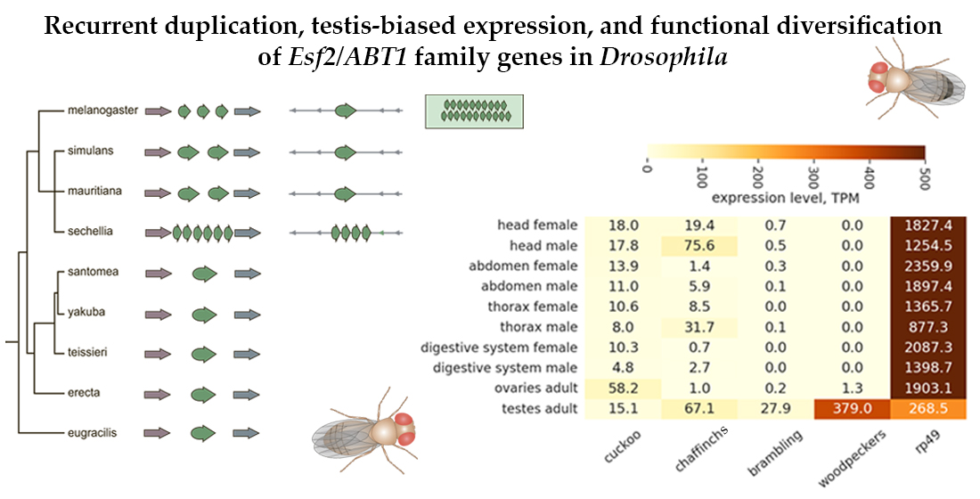
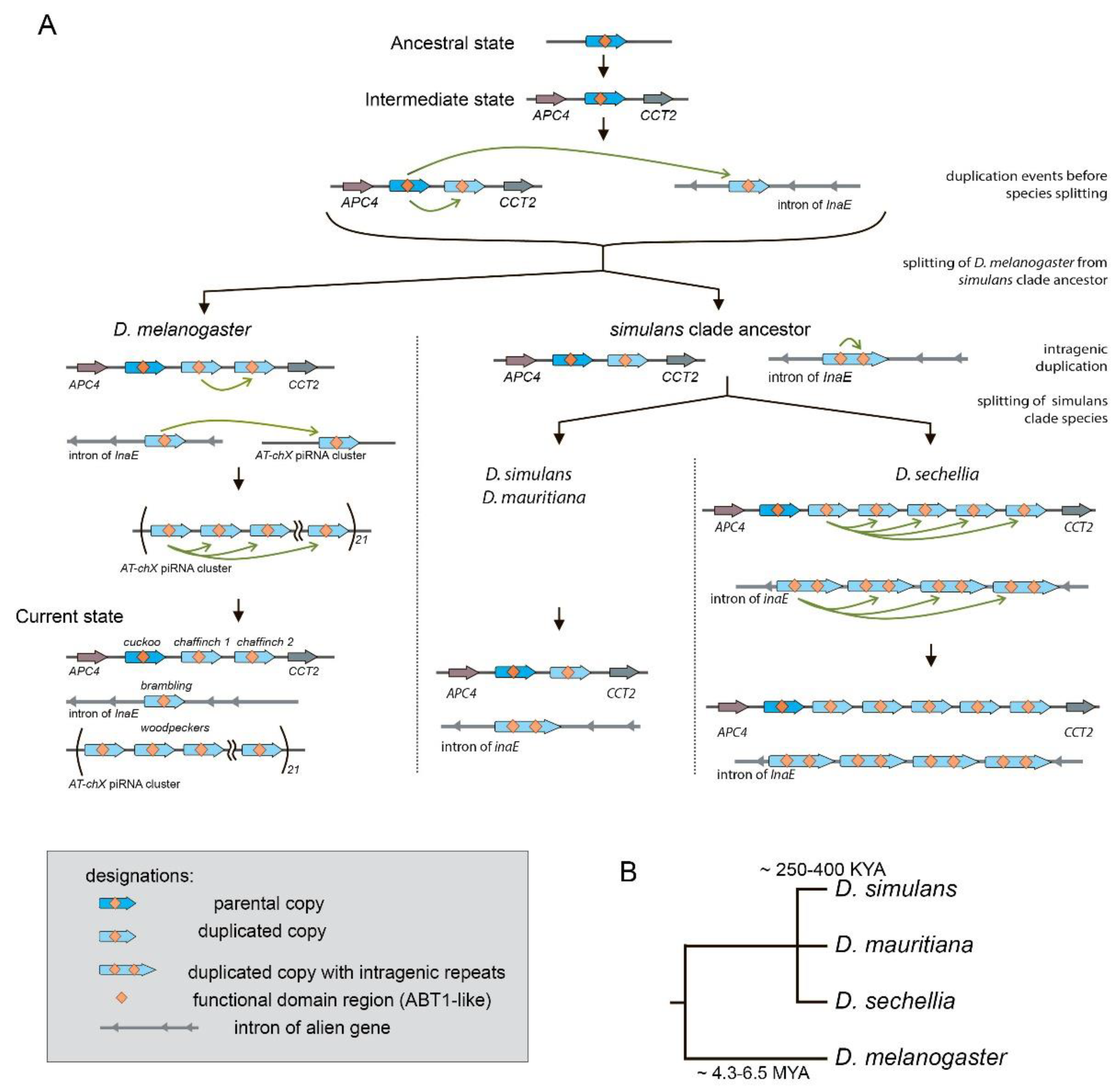
Gene duplications are considered to be the major evolutionary resource of novel functions. The gene family Esf2/ABP1 is conserved in metazoan organisms from yeast to humans. Here we performed a search and characterization of Esf2/ABP1 homologs in the Drosophila genus. Whereas in the majority of Drosophila species this gene family is represented by only a single gene, in the melanogaster and suzukii subgroups recurrent gene duplications arose, providing 47 homologous genes located on the X chromosome. To study the evolutionary history of duplicates, we performed phylogenetic, functional domain, and tissue-specific expression analyses. We revealed a male-specific and testis-biased transcription pattern of duplicated copies in Drosophila melanogaster and Drosophila sechellia compared to ubiquitous expression of the parental gene. The amplification of 21 repeated paralogs within the heterochromatic piRNA cluster resulted in the ovarian-specific transformation of these repeats into piRNAs in D. melanogaster. In three species of the suzukii subgroup, Esf2/ABP1 genes evolved with domain diversification: in addition to RNA-binding ABT1-like domain preservation, all homologous proteins acquired expanded intrinsically disordered regions. By studying the duplicated copies of the Esf2/ABP1 family in Drosophila, we offer insight into how novel gene functions emerge and are maintained, contributing to life's diversity and complexity.

Keywords: 
;  ;  ;  ;  ;  

1. Introduction

Gene duplication followed by divergence is now considered one of the fundamental mechanisms driving the evolution of new functions in metazoan organisms. Duplicated genes provide substrates from which evolution can generate novel functions, and thus duplications contribute to processes of environmental adaptation, speciation, and increasing organismic complexity. Gene duplication can be raised in different ways, including tandem gene duplication, whole-genome and segmental chromosomal duplication, and also retroposition or transposition. Following gene duplication, a newly duplicated gene copy can realize different fates. Most often it is a pseudogenization with accumulation of degenerative changes, e.g., mutations, frameshifting deletions, or premature stop codons owing its functional redundancy [1,2]. However, another part of duplicated genes could acquire new functions and be retained in the genome.
According to the modern view, multiple processes can functionally preserve duplicated genes, including conservation, neofunctionalization, subfunctionalization, specialization, dosage benefit, duplication-degeneration-complementation, and others [3,4,5,6]. When increased gene dosage is beneficial, ancestral functions are conserved in both gene copies. During neofunctionalization, one gene copy carries out its ancestral functions, and the other one acquires a novel function and expression pattern [2,3,6]. Subfunctionalization process means that copies are mutated differently with a maintenance of ancestral gene functions by both genes [2,4]. Concerted action of subfunctionalization and neofunctionalization processes provides a specialization of gene copies that functionally evolved from each other and from the ancestral gene.
The gene family Esf2/ABP1 includes genes that are phylogenetically conserved in metazoan organisms from yeast to humans. These genes encode the nuclear protein ABT1 (activator of basal transcription 1) and its yeast homolog, pre-rRNA-processing protein Esf2 (eighteen S factor 2). The molecular function of ABT1 is described as a TBP-binding regulator of basal transcription for PolII genes in mice [7]. In Saccharomyces cerevisiae, Esf2 is known as a nucleolar RNA-binding protein involved in pre-rRNA processing and the biogenesis of small ribosomal subunit [8]. The functions of Esf2 were found to be essential in yeast [7]. Despite the molecular functions of Esf2/ABP1 proteins in different metazoan organisms not being clearly understood yet, in Drosophila melanogaster the Esf2/ABP1 family gene CG32708 has been found to be essential for fly development and associated with the Notch signaling pathway and cell proliferation [9,10,11,12].
The Drosophila represents a clear and attractive model system to study the evolutionary forces acting on the evolutionary dynamics of gene copies [13,14,15,16,17]. The recent appearance of high-quality genome assemblies of Drosophila species, including challenging heterochromatin genomic regions with arranged genomic repeats [18,19,20], provides a valuable opportunity to understand genetic and evolutionary mechanisms that drive the maintenance and divergence of Esf2/ABP1 family genes. In this study we report our initial functional and evolutionary characterization of Esf2/ABP1 gene family duplications in genomes of Drosophila. We found that in the majority of Drosophila species, this gene family is represented by only a single gene maintaining original functions. However, in the melanogaster subgroup as well as in the suzukii subgroup, we identified recurrent gene duplications providing, in sum, at least 47 homologous genes expanding to distinct genomic locations on the X chromosome. Investigating the origin, divergence, phylogenetic relationships, and expression pattern of Esf2/ABP1 genes, we revealed a sex-specific expression bias of duplicated copies in Drosophila melanogaster and Drosophila sechellia and functional diversification of homologs in the suzukii subgroup species, indicating evolutionary adaptation of newly raised gene copies.

2. Materials and Methods

2.1. Homolog search and identification

We performed a search of Esf2/ABP1 homologs and duplicates in the Drosophila genus using Ensemble Metazoa tool (https://metazoa.ensembl.org/) and CG327708 gene as a query. The analysis of the identity of homologs was performed using the coding and protein sequences identified by Ensembl (https://ftp.ebi.ac.uk/pub/databases/ensembl/genomes/release-60/metazoa/fasta/). Sequences were pairwise aligned using MUSCLE v3.8.1551 [21]. The percent identity was calculated as the number of matching bases/amino acids divided by the number of alignment columns, excluding gaps. The data were visualized as the heat map using the Python libraries, Matplotlib and Seaborn. For microsynteny estimation we analyzed up to three upstream and three downstream surrounding genes for each location using NCBI Genome Data Viewer (https://www.ncbi.nlm.nih.gov/gdv?org) and corresponding genome assemblies. Genes are considered to be in a syntenic location if they share more than 15% flanking homology.

2.2. Phylogenetic analysis

For search Esf2/ABP1 family homologous genes 23 whole genome assemblies of Drosophila species (NCBI RefSeq) were used with BLAST (blastn) algorithm and CG327708 gene as a query. Corresponding genome assemblies are listed in Table S1. For multiple alignment we retrieved the CDS for homologous genes of closely related species with reference genome assemblies include Drosophila simulans Prin_Dsim_3.1 GCA_016746395.2, Drosophila mauritiana ASM438214v1 GCA_004382145.1, Drosophila sechellia ASM438219v2 GCA_004382195.2, D. yakuba Prin_Dyak_Tai18E2_2.1 GCA_016746365.2 and Drosophila melanogaster Release 6 plus ISO1 MT GCA_000001215.4. The nucleotide sequence alignment was carried out using MEGA 12 software [22] by the MUSCLE method [21] for codons. The alignments required minimal manual editing and G-blocks 0.91.1 [23] treatment with following parameters: data type: codon; max number of contiguous non-conserved positions: 8; min length of a block: 10; allowed gap position: with half. Phylogenetic tree constructions were performed using the MEGA 12 software. The Maximum Likelihood method was used with the Tamura-Nei model of nucleotide substitutions [24] for the tree generation with highest log likelihood (-1933.81) and Drosophila yakuba sequence of LOC6525244 gene as the outgroup. The percentage of replicate trees in which the associated taxa clustered together (500 replicates) is shown below the branches. Gamma distribution was used to model evolutionary rate differences across 5 categories (+G, parameter = 1.9616), with 0.00% of sites deemed evolutionarily invariant (+I). The partial deletion option was applied to eliminate all positions with less than 95% site coverage resulting in a final data set comprising 351 positions. The analytical procedure encompassed 42 coding nucleotide sequences using 1st, 2nd, 3rd, and non-coding positions. Evolutionary analyses were conducted in MEGA 12 [22] utilizing up to 4 parallel computing threads.

2.3. Z-test of neutrality

To estimate the type of selection for the Esf2/ABP1 family genes in D. melanogaster, we applied the codon-based test of neutrality, the Z-test, using MEGA 11 software [22,25]. Analyses were conducted using the Nei-Gojobori method [26]. This analysis involved two aligned nucleotide sequences. All ambiguous positions were removed for each sequence pair (pairwise deletion option). The test statistic (dN - dS) is represented as a Z-score, where dS and dN are the numbers of synonymous and nonsynonymous substitutions per site, respectively.

2.4. Search for protein domains

For the determination of known protein domains of Esf2/ABP1 family proteins, NCBI's Conserved Domain Database (CDD) tool (https://www.ncbi.nlm.nih.gov/Structure/cdd/wrpsb.cgi) [27] was used. The search was performed against database CDD v3.21 with an expected value threshold of 0.01. Only specific hits were considered. Intrinsically disordered regions (IDRs) were predicted with the Critical Assessment of Intrinsic Protein Disorder (CAID) Prediction Portal (https://caid.idpcentral.org/) [28] with a default threshold of 0.5. The overall disorder scores for the proteins expressed as 50% of IDRs in the whole protein length were considered as significant.

2.5. Fly stocks

To analyze transcript level in different fly tissues and copy number estimation we used flies of wild-type strains of D. melanogaster Batumi L, Harwich, GH10, and GH16. Batumi L and Harwich strains were obtained from the Collection of laboratory and natural strains of Drosophila of Institute of Developmental Biology, RAS, Russia. Strains from African population (Ghana, Africa), GH10 and GH16 [29], were obtained from the Drosophila collection of Institute of Cytology and Genetics, SB RAS, Russia. D. sechellia stock was obtained from the Gifsur-Yvette CNRS Center collection, France. All flies were reared at 23C on a standard medium. The flies were subjected to a laboratory light–dark cycle of 12:12 h.

2.6. RT-qPCR analysis

For RT-qPCR analysis, total RNA was isolated from gonads, heads and carcasses of 3-5-day adult male and female flies with TRIzol Reagent (Invitrogen). DNA-free Kit DNA Treatment & Removal (Invitrogen) kit was used for removal of genomic DNA from preparations. Reverse transcription was performed using Mint kit (Eurogen) and Oligo(dT) primers. cDNA samples were analyzed by real-time quantitative PCR using the incorporation of SYTO13 (Invitrogen). All experiments were performed with at least three independent RNA samples; each sample was analyzed in triplicate. Fold change ratios of the average expression level were calculated. We used rp49 (rpL32) as a loading control. The following primers were used: rp49 fw 5′-ATGACCATCCGCCCAGCATAC-3′, rev 5′-GCTTAGCATATCGATCCGACTGG-3′; cuckoo fw 5′-CATGGAAGTAGAGGAGGCTGAG-3′, rev 5′-GGTATGTTGGATATGTAGATGATCCC-3′; chaffinch (shared) fw 5′-AAATGACGATAAAAAGGAGCTGG-3′, rev 5′-TGTCCTTGGGTATGTTGGATATTA-3′; brambling fw 5′-CTGAGATGCCGTTCTCTTATAAGACG-3′, rev 5′-CAAGGTCATGTGCTTGGGTAGGT-3′; woodpecker (shared) fw 5′-GGAGGAGATTGAGACCTCGG-3′, rev 5′-GTGCTTGGGTATGTTGGATATGT-3′; D. sechellia parental copy LOC6619759 fw 5′-GGTGCACAAGCAACGCCTA-3′, rev 5′-CAGAGCCTTTTCCGCTTTCTT-3′; D. sechellia duplicated copies of cluster 1 (LOC6621826, LOC116802136, LOC116802134, LOC116802120, LOC6619761) (shared) fw 5′-CTGAGAAACCCGAGAAGATGCA-3′, rev 5′-TTAACAGCTCTTTCGGCTGCAC-3′; D. sechellia cluster 2 duplicates (LOC6618463, LOC116801944, LOC6618464, LOC6618462) (shared) fw 5′-AACGAACAAAAATGAATTCTGAGCC-3′, rev 5′-CATTCGCAGACTCACCTTCCTC-3′.

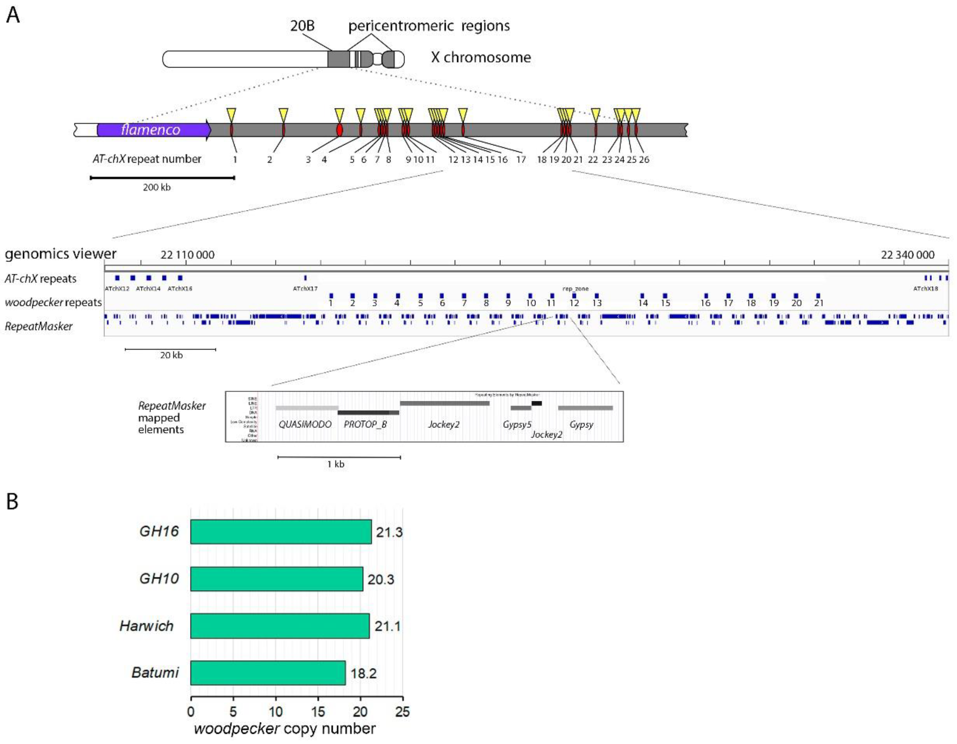
2.7. Copy number estimation of woodpecker genes

To estimate the number of woodpecker gene copies in the D. melanogaster genome, quantitative PCR was performed with genomic DNA of Batumi L, Harwich, GH10, and GH16 wild-type strains to determine the concentration level of amplification products of each of the genes of the family. We used highly specific woodpecker primers (see above), the efficiency of which was previously tested and compared with the efficiency of rp49 primers (normalization control). Measurements for each strain were carried out in three replicates.

2.8. Transcriptomic analyses of Esf2/ABP1 family genes using deep sequencing library data

PolyA+ RNA-seq libraries from w1118 strain of D. melanogaster embryos, third instar larval gonads and sex-separated adult tissues/body parts and gonads of w1118 and aub mutant flies [30,31,32,33,34,35] (Table S2) were used for expression analysis (from duplicate to quadruplicate of libraries for tissue). The libraries were pseudo-aligned using Kallisto v.0.50.0 with following parameters --single -l 100 -s 0.05 [36]. The D. melanogaster transcriptome set obtained from NCBI was used as a reference (GCF_000001215.4). Transcript abundances were used to assess expression in terms of transcripts per million (tpm). The data were visualized using the Python libraries, Pandas, Matplotlib, and Seaborn.

2.9. Bioinformatics analysis of piRNAs

For piRNA distribution analysis in the AT-chX cluster, small RNA libraries from Oregon R ovaries [37] and w1118 (yw) testes [38] of D. melanogaster were used (Table S2). Adapter sequences from small RNA libraries were removed by cutadapt 2.8. The FastQC tool was used for quality control of the sequenced libraries. Reads were filtered by length (23-30) and quality (Phred quality score > 30) using fastq-filter (https://github.com/LUMC/fastq-filter?tab=readme-ovfile# readme). rRNA, snRNA, snoRNA, microRNA and tRNA were filtered off from the libraries. To analyze the reads generated from the AT-chX and woodpecker repeat containing piRNA cluster, locally unique reads (reads could only be mapped to genomic region with coordinates chrX:21631000-22440000 containing AT-chX repeats, woodpecker repeats, and neighboring flamenco piRNA cluster) were used. For this, libraries were mapped to the dm6 genome assembly without contigs and chrX:21631000-22440000 region. Unmapped reads were remapped to the chrX:21631000-22440000 region using bowtie with -n 1 -l 30 parameters. The mapped reads were visualized using Integrative Genomics Viewer (IGV) (https://www.broadinstitute.org/scientific-community/software/integrative-genomics-viewer). To evaluate the potential silencing of Esf2/ABP1 family genes by piRNAs, the Oregon R ovarian library was independently mapped to gene sequences using bowtie (-n 2). The mapped reads were counted and normalized. Ping-pong signatures for sense and antisense piRNA pairs were calculated using the signature.py script [39].

3. Results

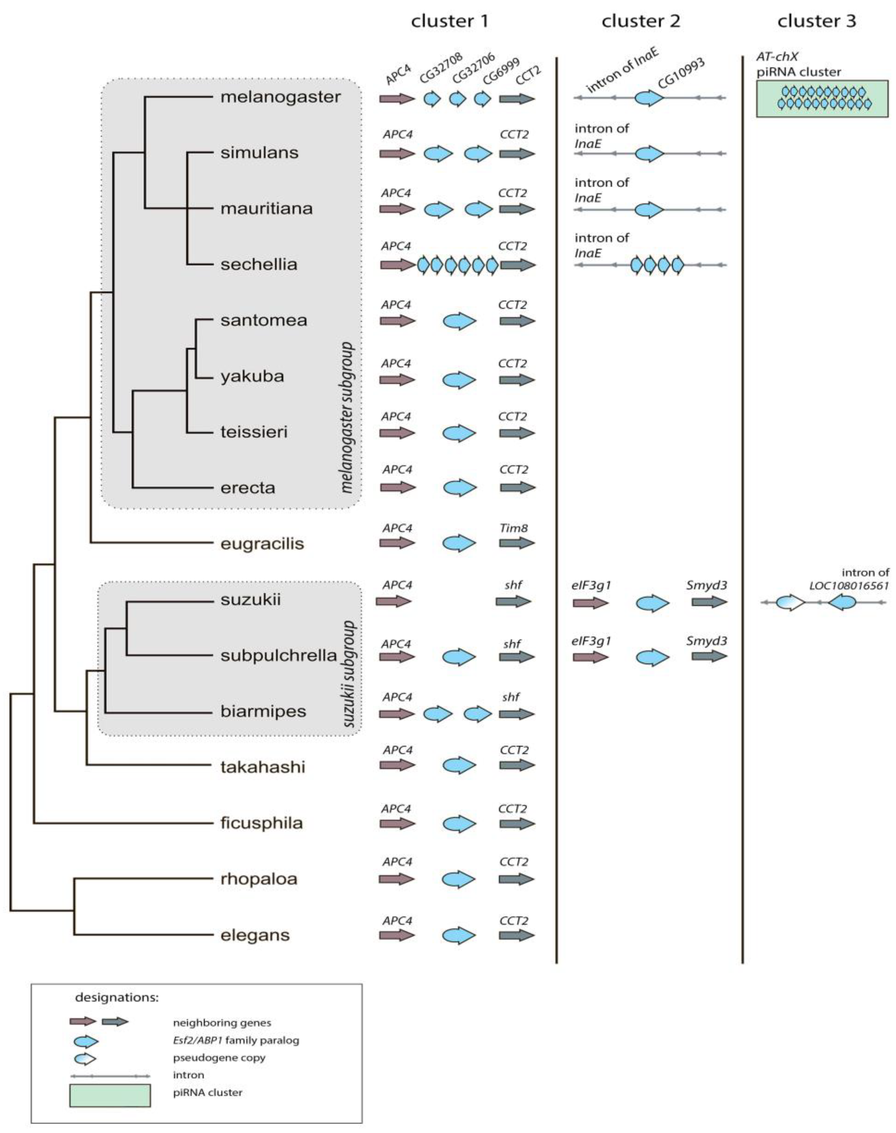
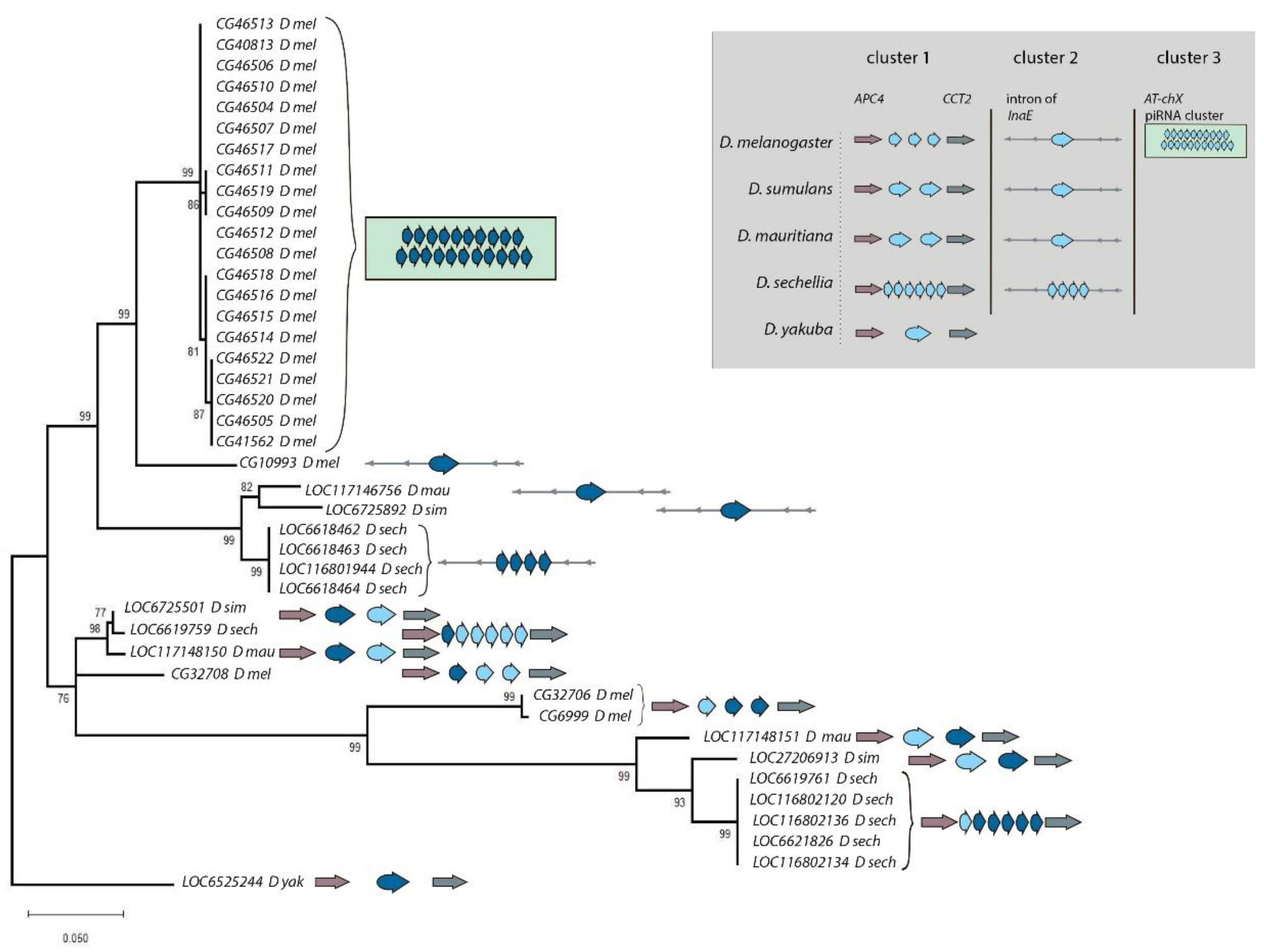
3.1. Survey of recurrent duplications of Esf2/ABP1 family genes in Drosophila

Multiple animal species represent genes belonging to the Esf2/ABP1 family in their genomes as a single homologous copy per genome. Using Ensemble Metazoa tool (https://metazoa.ensembl.org/), we identified a single Esf2/ABP1 gene per genome for the majority of Drosophila genus species; however, we revealed gene duplication events for several species of the melanogaster group (Figure S1). Among them there are four species of the melanogaster subgroup, Drosophila melanogaster, Drosophila simulans, Drosophila mauritiana, and Drosophila sechellia, and species of the suzukii subgroup, Drosophila biarmipes and Drosophila subpulchrella. Some of the Esf2/ABP1 gene duplications for D. melanogaster, D. simulans, and D. mauritiana have been found earlier [40]. With pairwise sequence alignments between homologs, we revealed that high sequence homology is preserved across the genus Drosophila both for nucleotide sequences of their CDSs and for translated amino acid sequences (Figure 1).
To characterize Drosophila Esf2/ABP1 homologs in detail, we focused on 16 species of the melanogaster group of the subgenus Sophophora (∼25 MIA divergence between species). By leverage of high-quality Drosophila genome assemblies (https://www.ncbi.nlm.nih.gov/datasets/genome/), we used blastn-based approach to identify Esf2/ABP1 orthologs and paralogs across the Sophophora species and subjected all found genes to the synteny analysis (Figure 2, Table S1). Our analysis revealed that duplicated gene copies in D. melanogaster and species of the simulans clade are clustered at three distinct genomic locations on the X chromosome. The first region (cluster 1) is located within a gene-rich euchromatin environment between APC4 (Anaphase Promoting Complex subunit 4) and CCT2 (Chaperonin containing TCP1 subunit 2) genes (Figure 2). This region contains at least one copy of the Esf2/ABP1 family genes in most analyzed genomes. This region of D. melanogaster (cytolocation 8C7) contains three paralogous Esf2/ABP1 genes, CG32708, CG32706, and CG6999, as has been described earlier [40]. Among them, the upstream CG32708 gene has a high nucleotide identity with homologous genes of other Drosophila species (Figure 1 block 1). Orthologous genes of the melanogaster group that are predominantly located between APC4 and CCT2 genes on the X chromosome have nucleotide identity to CG32708 gene for their CDSs in the range of 75.2-95.4% (Figure 1, Table S1, Figure 2). CG32708 is characterized by ubiquitous expression in different fly tissues (according to FlyAtlas2 data) and is found to be the essential gene for fly development and cell proliferation [9,10,11,12]. Thus, the most parsimonious explanation is that CG32708 is the progenitor gene copy maintaining original functions in D. melanogaster. Taking all this into account, we assume that CG32708 and its orthologs in this location appear to be parental copies from which the following paralogous genes originated in the melanogaster group. We found the parental copy and one duplicated copy in this genomic location in D. simulans and D. mauritiana genomes. However, in D. sechellia genome we observed in this region the parental copy with the highest homology 94.7% to CG32708 and downstream five almost identical tandem gene duplications with only 66.8-67.3% homology to CG32708 (Figure 1 block 4, Figure 2, Table S1). Considering that D. melanogaster has split from an ancient precursor with the simulans clade about 4.3-6.5 MYA [41,42,43,44,45], we proposed that first duplication event in this genomic region can occurred before D. melanogaster divergence from sibling species.
The second X chromosome region (cluster 2) containing Esf2/ABP1 copies is the intron of the InaE (inactivation no afterpotential E) gene of the melanogaster subgroup (cytolocation 12C5 for D. melanogaster). Whereas, only single Esf2/ABP1 orthologs were found for D. melanogaster, D. simulans, and D. mauritiana species in this genomic location; for D. sechellia, we observed four highly identical tandem duplicated paralogs (Figure 1 block 3, Figure 2, Table S1). The strong syntenic location of these genes also indicates that the initial gene insertion into the InaE intron and subsequent fixation occurred before the splitting of D. melanogaster from the species of the simulans clade.
We uncovered the third region (cluster 3) only in D. melanogaster genome, containing at least 21 Esf2/ABP1 gene copies (Figure 2, Table S1, Figure 3A). This region is located in pericentromeric heterochromatin of the X chromosome (cytolocation 20B). It has been identified earlier as the expanded germline-specific piRNA cluster containing AT-chX repeats [33,38]. piRNA clusters are specialized genomic regions responsible for producing the majority of small non-coding piRNAs in germinal tissues [46,47]. The Esf2/ABP1 paralogs in this region of D. melanogaster are arranged tandemly, with blocks of fused-together transposon fragments of repeating patterns placed between neighboring paralogous copies (Figure 3A). Despite their location within the active piRNA cluster, these duplicated copies are annotated as genes in the reference genome assembly; each copy contains one intron and possesses the highest homology among themselves (more than 99%) (Figure 1 block 2). We designated these repeats as woodpeckers with corresponding numbers (woodpecker 1, woodpecker 2,…, woodpecker 21) (Figure 3A). We estimated woodpecker gene copy number in four wild-type D. melanogaster strains (Batumi, Harwitch, GH10, and GH16) using quantitative PCR of genomic DNA (Figure 3B). There are 18-21 woodpecker copies in the analyzed genomes. The absence of these repeats in the genomes of other Drosophila species, including closely related ones of the simulans clade, indicates their recent arising, fixation, and maintenance only in D. melanogaster.
According to our analysis, duplication events for the Esf2/ABP1 gene family have occurred also in the suzukii subgroup. Both D. biarmipes and D. subpulchrella genomes contain parental Esf2/ABP1 copies with 76.2% and 81.5% nucleotide identity to CG32708, respectively, in the genomic location flanked upstream by APC4 gene, and, besides that, D. biarmipes has a second paralog in this location (Figure S1; Figure 2 cluster 1, Table S1). D. subpulchrella genome also contains a single Esf2/ABP1 copy in a non-syntenic region of the X chromosome, between eIF3g1 and Smyd3 genes (Figure 2 cluster 2, Table S1). Our search revealed that in their closely related species D. suzukii, the parental Esf2/ABP1 gene near APC4 gene has been apparently lost, but instead we identified one copy between eIF3g1 and Smyd3 genes, and additionally two duplicated copies, a gene and a pseudogene, in the intron of X-linked LOC108016561 gene encoding the ortholog of D. melanogaster CG42594 gene (Figure 2 clusters 2 and 3, Table S1).
The pattern of Esf2/ABP1 homolog distribution in the suzukii subgroup species indicates that gene arising and loss is a rather independent process compared with species of the melanogaster subgroup. However, for both subgroups we observed that the parental copy and all duplicated copies are located exclusively on the X chromosome. It should be noted that the Y chromosome is not consistently represented in the available genome assemblies; for this reason, we were unable to systematically search of gene duplication on the Y chromosome, except for D. melanogaster and D. suzukii assemblies (see Table S1). However, for the simulans clade, we used gene annotation data of recently improved Y chromosome long-read assemblies [48] and also did not observe Y-linked Esf2/ABP1 homologs. Beyond the melanogaster group, we did not detect Esf2/ABP1 duplications in the obscura, virilis, and repleta groups considering only RefSeq annotated genome assemblies (data are shown in Table S1). Whereas in the obscura group we observed a partial synteny of the gene location; in the species of the virilis group of the distinct subgenus Drosophila, Drosophila virilis, the single Esf2/ABP1 copy was found on the X chromosome at a non-syntenic location (Table S1). This suggests that the ancestral Esf2/ABP1 gene changed its location after the divergence about 40-60 MYA [42,43] from a most recent common ancestor of this species and D. melanogaster. Taken together, we revealed several gene duplication events for this gene family in Drosophila, with the parental copy located nearby APC4 gene being the Esf2/ABP1 gene that remains a single-copy gene per genome across the majority of the melanogaster group species.

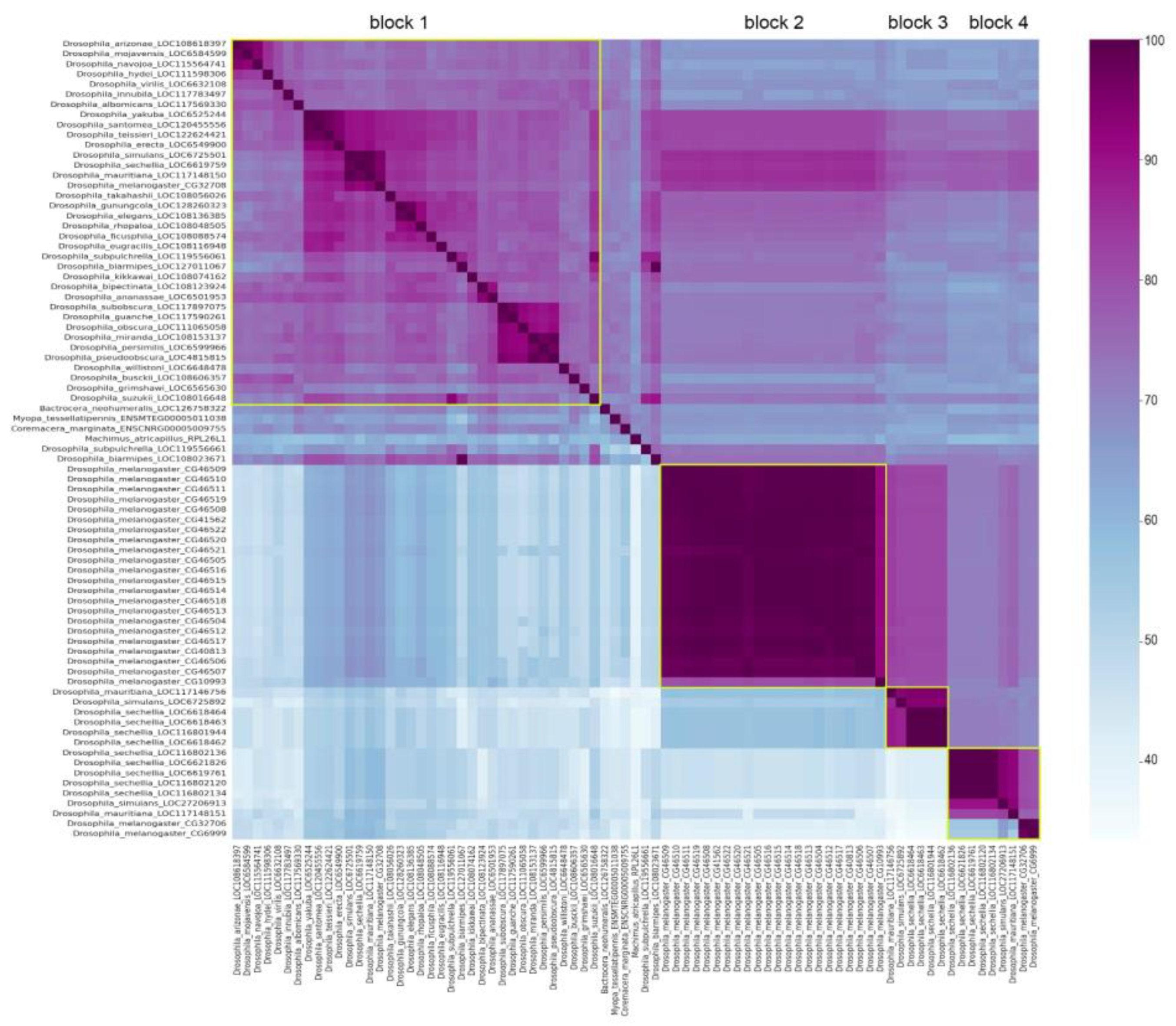
3.2. Phylogenetic analysis for homologous genes of melanogaster subgroup

To evaluate the evolutionary relationship between homologs, we performed the collecting, multiple alignment, and phylogenetic analysis of coding sequences for all Esf2/ABP1 family genes of D. melanogaster, D. simulans, D. mauritiana, and D. sechellia species. For generation of the phylogenetic tree, we used the Maximum Likelihood method and the Tamura-Nei model [24] with the orthologous D. yakuba gene LOC6525244 as the outgroup (Figure 4). We have built the tree with the highest log likelihood (-1 933.81) (Figure 4). Based both on tree characteristics and synteny analysis (Figure 2, Figure 4), we proposed that duplication events of Esf2/ABP1 genes in cluster 1 and cluster 2 regions of the X chromosome have occurred before the splitting of D. melanogaster from common ancestor species with the simulans clade. Orthologous genes from cluster 2 (embedded in the intron of InaE gene in all four analyzed genomes) turn out to be in two monophyletic groups: one group contains all genes from cluster 2 of the simulans clade species, whereas another group includes the homologous CG10993 gene from cluster 2 and all 21 tandem woodpecker genes of cluster 3 of D. melanogaster (Figure 4). The close location of the branches of the last group on the tree indicates a potential origin of cluster 3 duplications from the single copy CG10993 after speciation of D. melanogaster.
We evidently revealed a low level of divergence between the first upstream orthologous parental copies from cluster 1 for all four analyzed species. This information is consistent with our analysis of nucleotide and amino acid identity (Figure 1 block 1, Figure 2, Table S1). The branch distribution for the following duplicated genes from cluster 1 inferred from the tree topology confirms high evolutionary rates for these copies; moreover, most divergence events appeared to occur before the splitting of the species. Thus, duplicated copies from cluster 1 have diverged considerably from their parental copies; however, within individual species, D. melanogaster and D. seichellia, all duplicated paralogs in cluster 1 are found to be extremely close to each other (Figure 4, Figure 1 block 4). This branch forms a monophyletic group, indicating that the generation of the first duplicated copy occurred in a common ancestor of these species, whereas second gene duplications in these regions in D. melanogaster and D. sechellia appear to emerge from the first duplicated copies after the splitting of the species.
While most Drosophila species harbor a single Esf2/ABP1 gene copy, the D. melanogaster genome contains multiple paralogs according to our and previously published data [40]. To facilitate subsequent analysis, we introduced new designations for them (Table 1). We defined the parental paralog CG32708 as cuckoo because its daughter copies have been scattered at different locations of the X chromosome. Both duplicated copies in cluster 1, CG23706 and CG6999, were named as chaffinch 1 and chaffinch 2, correspondingly. We defined the single paralog CG10993 in cluster 2 as brambling. Tandem amplified copies in cluster 3 were named as woodpeckers (woodpecker 1, woodpecker 2,…, woodpecker 21), as mentioned above.
To detect signatures of sequence evolution in Esf2/ABP1 homologs of D. melanogaster, we performed the codon-based Z-test that estimates the ratios of synonymous and nonsynonymous divergence between analyzed genes and close orthologs of D. simulans (Table S3). We found that purifying selection maintains the parental copy, cuckoo. In relation to other duplicated copies in all three genomic locations, we did not find significant deviations from neutral evolution.

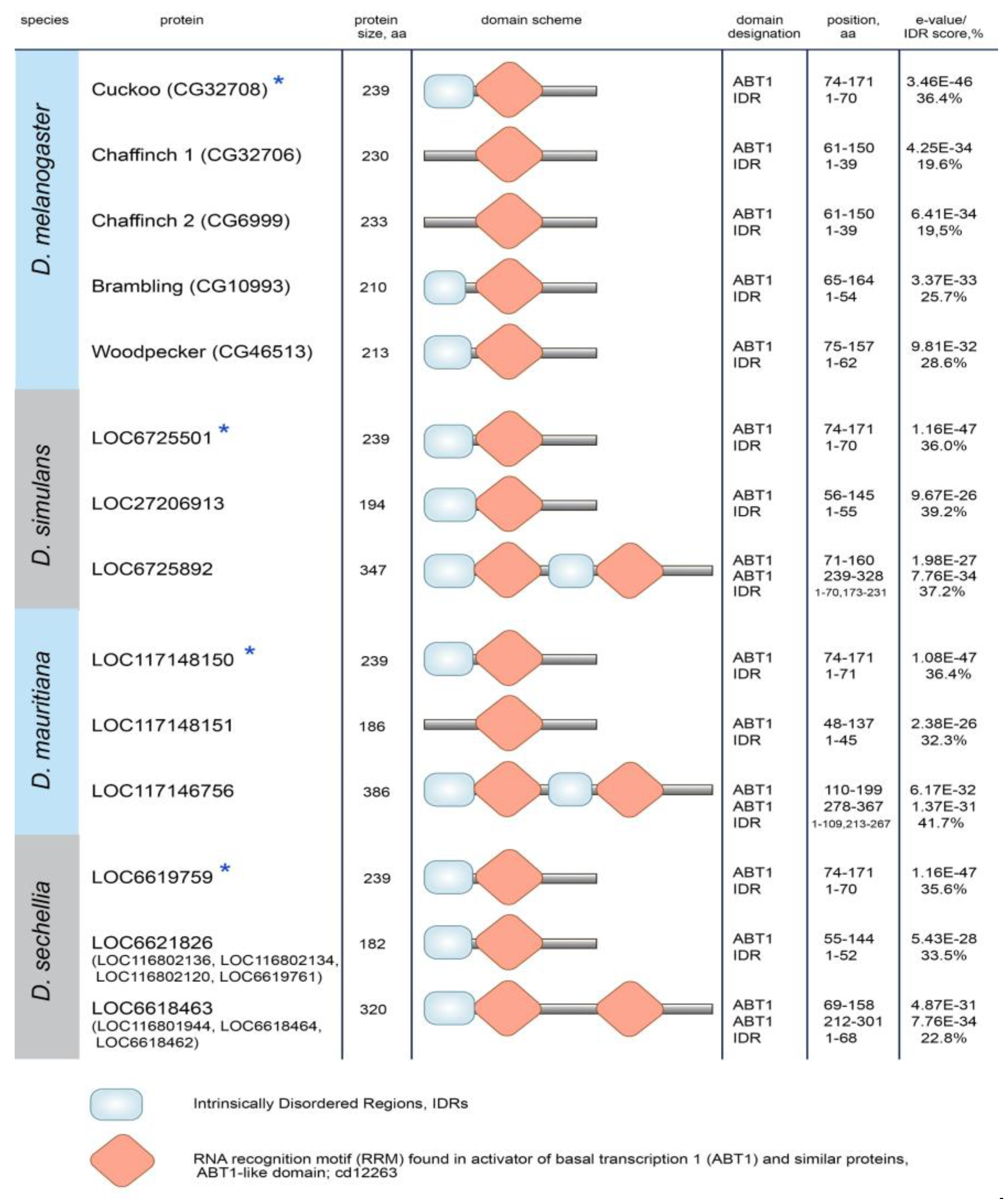
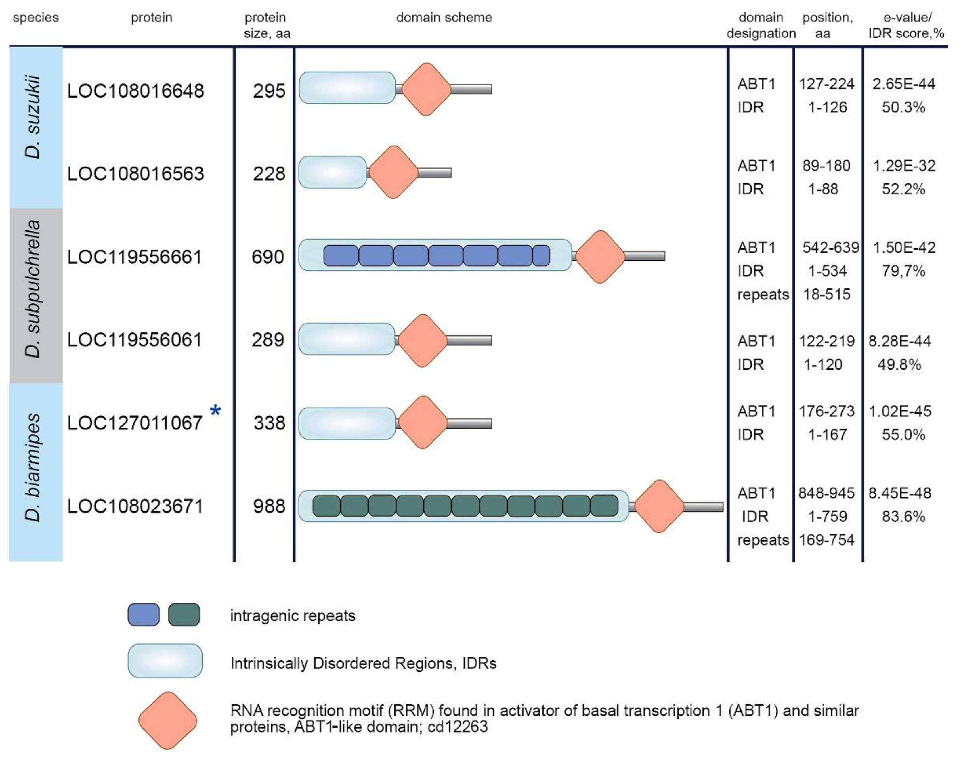
3.3. Protein domain search in Esf2/ABP1 proteins

To detect potential innovations and functional sequence divergence in duplicated copies, we performed a search and comparison of known protein domains across Esf2/ABP1 homologs. Domain prediction for Esf2/ABP1 proteins of the melanogaster subgroup revealed that they shared with high e-value the RRM (RNA recognition motif) ABT1-like domain (domain found in activator of basal transcription 1) (Figure 5, Figure S2) that has been firstly described for the Esf2/ABP1 family [7]. The most striking divergence occurred within the species of the simulans clade with duplicated copies embedded in the intron of InaE gene (cluster 2). The insertion of the duplicated copy into the intron of the unrelated gene was supplemented by duplication of extended internal fragment, providing a second RRM ABT1-like domain in these proteins (Figure 5, Figure S2). We did not observe an intragenic duplication for the orthologous Brambling protein of D. melanogaster, indicating that the emergence and fixation of this whole-domain duplication occurred after the splitting of D. melanogaster from their common ancestor. All proteins analyzed also contain predicted Intrinsically Disordered Regions (IDRs) (with length from 39 aa to 109 aa); however, the overall disorder score for these proteins does not reach a high level (less than 50% of total length, from 19.5% to 41.7%) (Figure 5).
Protein domain determination for Esf2/ABP1 proteins of the suzukii subgroup revealed that all Esf2/ABP1 homologs retain the RRM ABT1-like domain near their C-termini (Figure 6, Figure S2). In addition, they contain extended continuous (in the range of 88 aa to 759 aa) IDRs at their N-termini (Figure 6). All of them have the overall disorder score equal to or exceeding 50% of the total length. Two proteins, LOC119556661 of D. subpulchrella and LOC108023671 of D. biarmipes, achieved strongly increased relative size (up to 80% of the whole length) of their continuous IDRs due to multiple intragenic tandem duplications in these regions (Figure 6, Figure S3). Thus, the divergence pattern of duplicated Esf2/ABP1 genes in the suzukii subgroup is significantly different from that of the melanogaster subgroup duplicated orthologs. A gain of IDRs for duplicated copies is currently considered as a mechanism of the acquisition of new interaction partners and can reflect a putative new functional role [49,50].

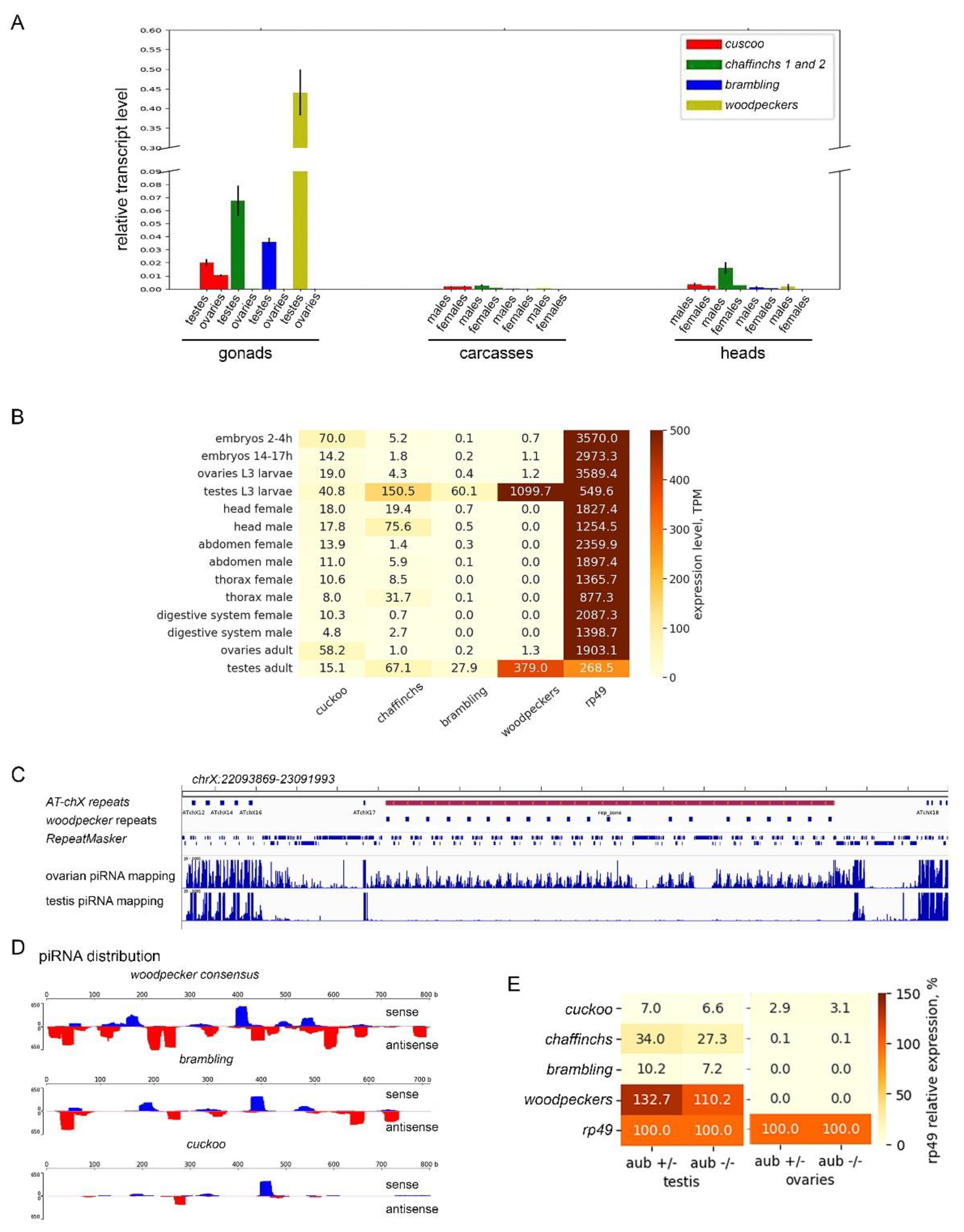
3.4. Diversification of the expression pattern of Esf2/ABP1 paralogs in D. melanogaster and D. sechellia

Using RT-qPCR and RNAseq data (Figure 7A,B), we revealed a sex-dimorphic pattern of Esf2/ABP1 paralog expression in selected tissues of D. melanogaster. For differential expression analysis we used PolyA + RNAseq libraries of embryos, third instar larval gonads, and adult tissues/body parts for each sex. All Esf2/ABP1 family genes are transcribed in the testes, with the highest transcript level found for the sum of woodpecker genes. In the ovaries we observed a noticeable level of transcripts only for cuckoo, whereas other paralogs were not expressed. Parental gene cuckoo was found to be expressed ubiquitously in all selected tissues (Figure 7B) with maximal transcript level in 2-4 h embryos, larval testes, and adult ovaries. Duplicated copies from cluster 1, chaffinch 1, and chaffinch 2, taken together, show maximal transcript level in larval and adult testes, heads, and thoraxes of adult males, demonstrating strong male-biased expression divergence from the parental gene. Brambling and woodpeckers, duplicated genes from clusters 2 and 3, correspondingly, exhibit a pronounced testis-specific expression pattern, likely associated with beneficial male germline activities (Figure 7B). Our RT-qPCR analysis of gonads, carcasses, and heads of adult flies of the wild-type Batumi strain of D. melanogaster generally confirmed the results obtained using RNAseq expression analysis (Figure 7A). Observed male germline enriched expression of young duplicated genes is interesting in the context of their acquisition and evolution. We perform the analogous RT-qPCR analysis of transcripts for Esf2/ABP1 genes in selected tissues of D. sechellia adult males and females. In whole we found a similar expression pattern to D. melanogaster: a ubiquitous expression of the older parental gene, but male-specific and testis-biased expression of more young duplicated copies (Figure S4).
As we have shown above (Figure 3A), woodpecker repeats have amplified in the D. melanogaster genome in the pericentromeric heterochromatin region, which is located within the expanded germline-specific AT-chX piRNA cluster, one of the specialized genomic regions providing long transcript precursors for producing a bulk of small piRNAs in gonads [38,47]. It is shown that the AT-chX is one of the major piRNA clusters both in the testes and ovaries of D. melanogaster [33,38]. However, woodpecker repeats are annotated as genes, containing one intron and possessing the highest nucleotide and amino acid homology among themselves (more than 99%) (Figure 1 block 2) that likely resulted from gene conversion homogenizing these gene duplicates or from very recent tandem duplication. Taking into account their testis-specific expression (Figure 7B), we asked whether the amplification of woodpecker genes within active piRNA cluster leads to potential piRNA production in the gonads.
To gain insights into peculiarities of woodpecker repeat expression, we analyzed profiles of small RNAs in the libraries from gonads of wild-type lines of D. melanogaster. Here we analyzed “locally unique” reads that map only to the region containing AT-chX and woodpecker repeats (see Materials and Methods for details). We found that multiple piRNA reads selected by size (23–29 nt) from the ovarian small RNA library were mapped to genome coordinates corresponding to the region. Surprisingly, piRNA reads mapped to woodpecker-containing region were not practically observed in the testis small RNA library (Figure 7C). Ovarian sense and antisense woodpecker-mapped piRNAs are distributed along the whole length of the woodpecker repeat consensus sequence with permitting 0–2 mismatches (Figure 7D), indicating that this genomic region is actually a part of the AT-chX piRNA cluster in the ovaries. Thus, woodpecker repeats function as a part of the double-stranded piRNA cluster in a sex-dependent manner only in the ovaries of D. melanogaster.
The repression of transposon activity is considered a main function of the piRNA pathway in Drosophila [46]; however, a cohort of piRNAs has been shown to take part in the repression of protein-coding genes [33,38,51]. Does the presence of antisense woodpecker-derived piRNAs indicate the possibility of silencing of Esf2/ABP1 family genes in the ovaries of D. melanogaster due to high complementarity to their transcripts? To evaluate the potential feasibility of piRNA-mediated silencing, we independently mapped ovarian piRNA reads to transcript sequences of woodpecker, cuckoo, brambling, and chaffinch genes with the indicated number of mismatches, from 0 to 2 (Table 2). Evidently, the most read numbers were found to be mapped to woodpecker repeats themselves (201.6 rpm for sense, 568.6 rpm for antisense). The calculated ping-pong signature value (z10 score) suggested that the ping-pong amplification mechanism [52] is active in the biogenesis of these piRNAs in the ovaries. We also found that sense and antisense piRNA reads can be mapped to brambling and cuckoo transcripts (Table 2; Figure 7). Only a small number of piRNA reads was perfectly mapped (with 0-1 mismatch) to these transcripts, indicating piRNA origin from the distinct region. For transcripts of both chaffinch genes, we did not observe perfectly mapped piRNAs above background level (Table 2). Considering that target silencing requires a significant number of antisense piRNAs with a high level of complementarity to the target [33,38], we assumed that these piRNAs could presumably repress expression of woodpecker and brambling genes in the ovaries. However, piRNA silencing potential for repression of cuckoo, which is the only Esf2/ABP1 paralog expressed in the ovaries, appears to be not enough, because the level of mapped antisense piRNAs is less than 100 rpm (Table 2). Our comparative analysis of polyA + RNAseq libraries of gonads of piRNA mutants (with disrupted piRNA biogenesis) and their heterozygous siblings revealed no significant change in the expression of all Esf2/ABP1 paralogs in the gonads, suggesting that they are not regulated by piRNAs (Figure 7E). Note that woodpecker repeats are transcribed as protein-coding genes only in the testes, whereas in the ovaries they are transcribed only as bidirectional piRNA precursors within the large piRNA cluster.

4. Discussion

Here we provide analysis of the evolutionary history of Esf2/ABP1 gene duplications in Drosophila species of the subgenus Sophophora. Using high-quality genome assemblies and synteny analysis, we identified 56 homologous Esf2/ABP1 genes in 16 species of the melanogaster group, separating approximately 25 MYA of evolution (Figure S1, Figure 1, Figure 2). Our analysis revealed that despite the single copy of the Esf2/ABP1 gene per genome harbored in the majority of Drosophila species, gene duplication events occurred in four species of the melanogaster subgroup and independently in three species of the suzukii subgroup (Figure S1, Figure 2, Table S1). We found that recurrent DNA-mediated duplication events occurred at the same cytolocation nearby the parental copy as well as with translocation and amplification of duplicated gene copies in the introns of unrelated genes or in heterochromatic piRNA cluster (Figure 2).
Our phylogenetic and functional domain analyses (Figure 4, Figure 5) allow us to reconstruct the evolutionary history of Esf2/ABP1 gene duplications in D. melanogaster and its sibling species of the simulans clade (Figure 8A). According to the proposed scenario, duplication events took place before speciation of D. simulans and D. mauritiana, whereas they occurred both before and after speciation in D. melanogaster and D. sechellia, providing the emergence and fixation of multiple new genes encoding proteins with RNA-binding ABT1-like domain (Figure 5). Duplicated paralogs in D. melanogaster do not exhibit any traces of pseudogenization and possess the characteristic features of functional copies. They contain an unbroken ORF, one conserved intron sequence, and an RNA-binding domain (ABT1-like) encoding region (Figure 5, Figure S2). We found that they acquired predominantly male-specific and testis-enriched expression pattern, unlike the parental gene, cuckoo (CG32708), which is expressed in different male and female tissues, with the highest expression in the ovaries and testes (Figure 7A,B). Among them, two duplicated copies in the same location with the parental gene, chaffinch 1 (CG32706) and chaffinch 2 (CG6999), have diverged considerably from cuckoo; however, they were found to maintain a high identity to each other (Figure 1 block 4, Figure 4).
The fate of most gene duplications is a rapid deletion or pseudogenization, but in some cases a duplicate copy can be fixed in the genome by drift or selection during a so-called fixation phase and subsequently maintained over time owing to acquiring a benefit function in the organism [6]. More than a dozen models were developed to explain the emergence, evolution, and maintenance of gene duplications in genomes [2,5,6]. Note that the parental gene cuckoo is found to be essential for fly development and cell proliferation in D. melanogaster [9,10,11,12]. cuckoo has a high nucleotide and protein identity with orthologous genes of other species of the melanogaster group in the syntenic location (Figure 1 block 1, Figure 2, Table S1) and is maintained under purifying selection in the genome (Table S3). Molecular functions of chaffinch 1 and chaffinch 2 genes are not understood to date; however, we determined the distinct pattern of chaffinch gene expression, i.e., male-biased expression with a maximal level in the testes, and the absence or very low expression level in female tissues (Figure 7A,B). It allows excluding subfunctionalization or duplication–degeneration–complementation models from consideration, because in these cases expression pattern must be shared between the subfunctionalized original and duplicated copies. According to the neofunctionalization model, duplicated copies occasionally accumulate substitutions to acquire a novel gene function that will be supported by selection, whereas a parental copy will not. Due to the conservation of the whole exon-intron structure and functional domain of chaffinch genes (Figure 5), we do not consider the neofunctionalization model as permissible. The Z-test for positive selection for both chaffinch genes showed no deviation from neutrality (Table S3). We proposed that the most suitable model for the evolution of chaffinch genes is the “beneficial increase in dosage”. As the heterogametic sex in Drosophila, males possess a gene-rich X chromosome and a strongly heterochromatic Y chromosome with few protein-coding genes [53]. To correct the potentially lethal imbalance of X-linked gene expression level in males, the mechanism of dosage compensation in somatic cells of D. melanogaster males provides an approximately two-fold increasing expression of most X-linked genes with the aid of attraction of the MSL (Male-Specific Lethal) protein complex to the male X chromosome for acetylation of histone H4 at K16 position (acetylH4K16), a mark of active chromatin [54,55]. However, the somatic dosage compensation system appears to be not applicable to male germline cells, where MSL complex is not assembled and the chromatin of the X chromosome loses a high level of acetylH4K16 modification [56,57]. It is shown that testis germ cells in Drosophila are characterized by a massive wave of transcription on the spermatogonial stage but undergo meiotic X-chromosome inactivation later on the stage of mature spermatocytes [58]. Taking all these circumstances into account, we propose that owing to the extra transcript level in the period of active transcription in the testes, these X-linked gene duplications have been fixed and retained by a dosage selection, according to the beneficial increase of gene dosage model.
The same logic appears applicable to the remaining duplicated Esf2/ABP1 genes in D. melanogaster, brambling and woodpeckers, since they all support a strong testis-restricted expression, preserve the exon-intron structure, and retain a functional ABT1-like domain (Figure 5, Figure 7A,B). woodpecker tandem gene amplification is unique to the D. melanogaster genome. According to our phylogenetic analysis, brambling and all woodpecker genes form the monophyletic group on the tree (Figure 4), confirming the origin of the last ones from the single-copy brambling gene after speciation of D. melanogaster (Figure 8A). We could assume that both brambling and woodpecker genes were fixed and preserved in the genome also owing to the beneficial increase of gene dosage. However, in the case of woodpecker genes, their genomic location leads to unexpected functional consequences that likely require a correction of the proposed model. The introduction of young duplicated copies into the piRNA cluster (Figure 2, Figure 3) resulted in their ovary-specific functional innovation. While in the testes these repeats are expressed as protein-coding genes, providing increased gene dosage; in the ovaries these repeats are subject to double-stranded non-canonical transcription as long precursors for the piRNA biogenesis (Figure 7C,D). Determination of the forces ensuring the preservation of highly identical woodpecker genes in this unusual genomic location appears to be complex. At the same time, the mechanism of gene conversion can impact their maintenance. We found that piRNA silencing potential of woodpecker-derived antisense piRNAs for repression of cuckoo, which is the only Esf2/ABP1 paralog with detected expression in the ovaries, is rather weak owing to a non-perfect piRNA complementarity to cuckoo transcripts (Table 2). A biological significance of the production of these piRNAs in the female germline is currently unclear and requires further research. In summary, here we uncovered a case of the functional adaptation of duplicated non-transposon genes with their sex-specific transformation into non-coding small RNAs. The acquisition of this potentially sex-antagonistic novel function took place owing to the insertion and amplification in a new genomic context without a significant divergence of woodpecker repeats from their parental brambling gene.
Note that a related history has been reconstructed earlier for the origin, evolution, and maintenance of the Stellate-Suppressor of Stellate (Ste-Su(Ste)) genetic system in the D. melanogaster genome [59,60]. Tandem amplification arrays of highly homologous genes, Stellate and Su(Ste), arose from a chimeric Y-chromosomal intermediate precursor on the X and Y chromosomes, respectively. The getting of Su(Ste) repeats into heterochromatin context as well as the insertion of transposon hoppel in the Su(Ste) promoter appear to facilitate Su(Ste) repeat pseudogenization and the acquisition of bidirectional transcription across them, providing abundant Su(Ste) piRNAs for the silencing of harmful Stellate genes. Su(Ste) repeats have lost protein-coding potential but evolved to form the major testis-specific piRNA cluster in D. melanogaster genome [38,51], ensuring Stellate gene repression and maintenance of correct spermatogenesis. Thus, woodpecker repeats, which are proposedly at an earlier stage of their evolution than the Ste-Su(Ste) system, can be considered as a valuable model for studying the evolutionary forces driving the formation of piRNA clusters.
In relation to the pattern of duplications in species of the simulans clade, we can note that according to our hypothesis (Figure 8A), in D. simulans and D. mauritiana genomes the parental gene and both duplicated copies were inherited from an immediate ancestor of the clade. In D. sechellia, additional amplification events supposedly happened after speciation. Considering a short divergence time between the simulans clade species (Figure 8B), we can suggest that amplified copies of D. sechellia located in cluster 1 and cluster 2 are very young. They maintain extremely high identity between each other within their genome locations (Figure 1 blocks 3 and 4). Analyzing the expression pattern of Esf2/ABP1 genes in the selected tissues of D. sechellia adult males and females, we revealed a picture strikingly similar to D. melanogaster, with male-specific and testis-biased expression of young duplicated copies and a ubiquitous expression of the parental gene (Figure S4). Taking into account intragenic duplication providing the acquisition of the second RNA-binding ABT1-like domain by the duplicated copies of cluster 2 (Figure 5), we can propose that testis-specific dosage selection also took place for their retention and maintenance.
We uncovered several duplication events for Esf2/ABP1 genes in the suzukii subgroup species (Figure 2). Despite that in both the melanogaster and suzukii subgroup species we observed distribution of Esf2/ABP1 family genes only on the X chromosome, the duplication events in these subgroups likely occurred through different mechanisms as independent processes (Figure 2, Table S1). A search of functional domains for Esf2/ABP1 proteins of the suzukii subgroup allows us to reveal an unexpected domain diversification. These Esf2/ABP1 proteins, in addition to ABT1-like domain preservation, contain long Intrinsically Disordered Regions, IDRs, composing 50 to 80% of the whole polypeptide length (Figure 6, Figure S2, Figure S3). They are significantly longer than those of melanogaster subgroup orthologs (Figure 5, Figure 6). IDRs are generally described as protein regions that do not adopt a defined three-dimensional structure under physiological conditions, but they are nevertheless functional. It is known that the majority of eukaryotic proteins contain both structured and disordered regions; however, only about 10% of proteins consist of IDRs representing half or more of their length, relating to the category of highly disordered proteins [50]. Among the Esf2/ABP1 family proteins of the suzukii subgroup species, we determined two, LOC119556661 of D. subpulchrella and LOC108023671 of D. biarmipes, containing multiple intragenic tandem repeat sequences that contribute to great extension of their continuous IDRs to near 80% of their whole length, consisting of 534 aa and 759 aa, respectively (Figure 6; Figure S3). Note that these tandem repeats are strongly enriched by acidic amino acid and serine residues, in sum consisting of more than 40% of their content (Figure S3). A common feature of long IDRs is their ability to form dynamic multivalent, flexible, and self-aggregating molecular networks, providing the assembly of liquid-liquid phase-separated droplets or hydrogels, commonly named biomolecular condensates [50,61]. The proteins with extended IDRs are mostly involved in the DNA, RNA, and protein binding to ensure molecular communication and regulation of cellular functions [50,62]. It is shown that expanded IDRs of certain RNA-binding proteins enable the formation in the cells of macromolecular granule-like assemblies, allowing localization, accumulation, and storage of RNAs [50,61]. Although the verification of molecular functions of expanded IDRs in fly tissues needs further investigations, we propose that Esf2/ABP1 proteins in the suzukii subgroup species evolved with the acquisition of novel functional features mediated by their IDRs for RNA accumulation and processing. The tandem expansion of internal repeats is one of the ways by which IDR-encoding genes arose and spread during evolution [63]. According to our findings, we assume that intragenic duplications and fragment shuffling by recombination were presumable selection instruments driving the evolution of Esf2/ABP1 family genes in a common ancestor of the suzukii subgroup flies. After species divergence, the expansion of IDRs owing to multiple internal repeats independently in Esf2/ABP1 copies of D. subpulchrella and D. biarmipes appears to contribute to following evolutionary adaptation. However, the evolutionary scenario for this subgroup can hardly be accurately reconstructed owing to the complicated pattern of arising and loss of Esf2/ABP1 genes (Figure 2).

Conclusions

To investigate the evolutionary history of Esf2/ABP1 family duplicates in Drosophila, we employed a multifaceted approach, combining comparative genomics, phylogenetic analysis, functional domain analysis, and tissue-specific transcriptomic analysis. This strategy allowed us to identify and characterize 47 duplicated gene copies across seven Drosophila species clustered at three distinct genomic locations on the X chromosome. Recurrent gene duplication events occurred in four species of the melanogaster subgroup and independently in three species of the suzukii subgroup. In both cases duplication provided the emergence and fixation of multiple new genes encoding proteins with RNA-binding ABT1-like domain. In three species of the suzukii subgroup, Esf2/ABP1 genes evolved with domain diversification: in addition to RNA-binding ABT1-like domain preservation, all homologous proteins acquired expanded Intrinsically Disordered Regions.
The first and the second duplication events in four species of the melanogaster subgroup probably occurred before the splitting of D. melanogaster from the species of the simulans clade, but the third one occurred after. Gene, named cuckoo, and its orthologs are parental, characterized by ubiquitous expression in different fly tissues, and found to be the essential gene for fly development and cell proliferation. The chaffinch 1 and chaffinch 2 genes, resulting from a second duplication event, have a distinct pattern of expression, i.e., male-biased expression with a maximal level in the testes. We proposed that the most suitable model for the evolution of chaffinch genes is the “beneficial increase in dosage.” Purifying selection maintains the parental copy, cuckoo. In relation to other duplicated copies in all three genomic locations, we did not find significant deviations from neutral evolution. The insertion and amplification of young duplicated woodpecker copies into the piRNA cluster lead to sex-specific functional innovation. Whereas in the testes these repeats are expressed as protein-coding genes; in the ovaries they undergo non-canonical transcription as long piRNA precursors.
This way, used complex approach permitted us to reconstruct duplicated gene evolutionary trajectories and revealing how their functions and expression patterns diverged over time.

Supplementary Materials

The following supporting information can be downloaded at the website of this paper posted on Preprints.org. Figure S1: Phylogenetic tree of Esf2/ABP1 family orthologs and paralogs in the genus Drosophila, Figure S2: Alignment of RRM ABT1 like domains predicted in Esf2/ABP1 family proteins of the melanogaster subgroup and suzukii subgroup, Figure S3: Representation of multiple tandem intragenic repeats for proteins LOC119556661 of D. subpulchrella (6.5 repeats) (A) and LOC108023671 of D. biarmipes (11 repeats) (B), Figure S4: RT-qPCR analysis of transcript levels of Esf2/ABP1 family genes in the gonads, carcasses, and heads of adult male and female flies of D. sechellia, Table S1: The table contains data for determination of orthologs and paralogs of Esf2/ABP1 gene family in genome assemblies of Drosophila species, Table S2: List of RNA library resources used for the analyses, Table S3: A codon-based test of neutrality, the Z-test, for selection type for the Esf2/ABP1 genes of D. melanogaster was performed using MEGA software.

Author Contributions

L.V.O.: conceptualization, methodology, writing—original draft preparation, visualization, project administration, and supervision. A.A.K.: methodology, data curation, formal analysis, writing—original draft preparation. E.D.D.: data acquisition, formal analysis, investigation, visualization, and writing—review and editing. E.Yu.Ya.: data acquisition, formal analysis, and writing—review and editing. A.V.Ch.: investigation, validation, formal analysis, and writing—review and editing. All authors have read and agreed to the published version of the manuscript.

Funding

This study was supported by the IDB RAS Government basic research program in 2025 № 0088-2024-0017.

Data Availability Statement

The open-source RNAseq data resources list used in this study is included in the Supplementary Materials, Table S2.

Acknowledgments

We thank S. Yu. Sorokina and A. M. Kulikov for helpful advices, I. A. Kombarov and S. S. Bazylev for help with experiments. We thank the Drosophila collection of Institute of Cytology and Genetics, SB RAS, Russia, for fly strains. The research was done using equipment of the Core Centrum of Institute of Developmental Biology RAS and the Collection of laboratory and natural strains of Drosophila of Institute of Developmental Biology RAS.

Conflicts of Interest

The authors declare no conflicts of interest.

References

  1. Long, M.; Betrán, E.; Thornton, K.; Wang, W. The origin of new genes: Glimpses from the young and old. Nat. Rev. Genet. 2003, 4, 865–875. [Google Scholar] [CrossRef]
  2. Conant, G.C.; Wolfe, K.H. Turning a hobby into a job: How duplicated genes find new functions. Nat. Rev. Genet. 2008, 9, 938–950. [Google Scholar] [CrossRef] [PubMed]
  3. Ohno, S. Evolution by Gene Duplication, Springer: New York, USA, 1970; pp. 1–160.
  4. Force, A.; Lynch, M.; Pickett, F.B.; Amores, A.; Yan, Y.L.; Postlethwait, J. Preservation of duplicate genes by complementary, degenerative mutations. Genetics 1999, 151, 1531–1545. [Google Scholar] [CrossRef] [PubMed]
  5. Assis, R.; Bachtrog, D. Neofunctionalization of young duplicate genes in Drosophila. Proc. Natl. Acad. Sci. USA 2013, 110, 17409–17414. [Google Scholar] [CrossRef] [PubMed]
  6. Innan, H.; Kondrashov, F. The evolution of gene duplications: Classifying and distinguishing between models. Nat. Rev. Genet. 2010, 11, 97–108. [Google Scholar] [CrossRef]
  7. Oda, T.; Kayukawa, K.; Hagiwara, H.; Yudate, H.T.; Masuho, Y.; Murakami, Y.; Tamura, T.A.; Muramatsu, M.A. A novel TATA-binding protein-binding protein, ABT1, activates basal transcription and has a yeast homolog that is essential for growth. Mol. Cell. Biol. 2000, 20, 1407–1418. [Google Scholar] [CrossRef]
  8. Hoang, T.; Peng, W.T.; Vanrobays, E.; Krogan, N.; Hiley, S.; Beyer, A.L.; Osheim, Y.N.; Greenblatt, J.; Hughes, T.R.; Lafontaine, D.L. Esf2p, a U3-associated factor required for small-subunit processome assembly and compaction. Mol. Cell. Biol. 2005, 25, 5523–5534. [Google Scholar] [CrossRef]
  9. Mummery-Widmer, J.L.; Yamazaki, M.; Stoeger, T.; Novatchkova, M.; Bhalerao, S.; Chen, D.; Dietzl, G.; Dickson, B.J.; Knoblich, J.A. Genome-wide analysis of Notch signaling in Drosophila by transgenic RNAi. Nature 2009, 458, 987–992. [Google Scholar] [CrossRef]
  10. Schnorrer, F.; Schönbauer, C.; Langer, C.C.; Dietzl, G.; Novatchkova, M.; Schernhuber, K.; Fellner, M.; Azaryan, A.; Radolf, M.; Stark, A.; Keleman, K.; Dickson, B.J. Systematic genetic analysis of muscle morphogenesis and function in Drosophila. Nature 2010, 464, 287–291. [Google Scholar] [CrossRef]
  11. Neumüller, R.A.; Richter, C.; Fischer, A.; Novatchkova, M.; Neumüller, K.G.; Knoblich, J.A. Genome-wide analysis of self-renewal in Drosophila neural stem cells by transgenic RNAi. Cell Stem Cell 2011, 8, 580–593. [Google Scholar] [CrossRef]
  12. Viswanatha, R.; Li, Z.; Hu, Y.; Perrimon, N. Pooled genome-wide CRISPR screening for basal and context-specific fitness gene essentiality in Drosophila cells. Elife 2018, 7, e36333. [Google Scholar] [CrossRef]
  13. Clifton, B.D.; Jimenez, J.; Kimura, A.; Chahine, Z.; Librado, P.; Sánchez-Gracia, A.; Abbassi, M.; Carranza, F.; Chan, C.; Marchetti, M.; Zhang, W.; Shi, M.; Vu, C.; Yeh, S.; Fanti, L.; Xia, X.Q.; Rozas, J.; Ranz, J.M. Understanding the Early Evolutionary Stages of a Tandem Drosophila melanogaster-Specific Gene Family: A Structural and Functional Population Study. Mol. Biol. Evol. 2020, 37, 2584–2600. [Google Scholar] [CrossRef] [PubMed]
  14. Chang, C.H.; Mejia Natividad, I.; Malik, H.S. Expansion and loss of sperm nuclear basic protein genes in Drosophila correspond with genetic conflicts between sex chromosomes. Elife 2023, 12, e85249. [Google Scholar] [CrossRef] [PubMed]
  15. Kuvaeva, E.E.; Cherezov, R.O.; Kulikova, D.A.; Mertsalov, I.B. The Drosophila toothrin Gene Related to the d4 Family Genes: An Evolutionary View on Origin and Function. Int. J. Mol. Sci. 2024, 25, 13394. [Google Scholar] [CrossRef] [PubMed]
  16. Brand, C.L.; Oliver, G.T.; Farkas, I.Z.; Buszczak, M.; Levine, M.T. Recurrent Duplication and Diversification of a Vital DNA Repair Gene Family Across Drosophila. Mol. Biol. Evol. 2024, 41, msae113. [Google Scholar] [CrossRef]
  17. Zakerzade, R.; Chang, C.H.; Chatla, K.; Krishnapura, A.; Appiah, S.P.; Zhang, J.; Unckless, R.L.; Blumenstiel, J.P.; Bachtrog, D.; Wei, K.H. Diversification and recurrent adaptation of the synaptonemal complex in Drosophila. PLoS Genet. 2025, 21, e1011549. [Google Scholar] [CrossRef]
  18. Clark, A.G.; et al. Drosophila 12 Genomes Consortium. Evolution of genes and genomes on the Drosophila phylogeny. Nature 2007, 450, 203–218. [Google Scholar] [CrossRef]
  19. Miller, D.E.; Staber, C.; Zeitlinger, J.; Hawley, R.S. Highly Contiguous Genome Assemblies of 15 Drosophila Species Generated Using Nanopore Sequencing. G3 (Bethesda) 2018, 8, 3131–3141. [Google Scholar] [CrossRef]
  20. Kim, B.Y.; Wang, J.R.; Miller, D.E.; Barmina, O.; Delaney, E.; Thompson, A.; Comeault, A.A.; Peede, D.; D'Agostino, E.R.R.; Pelaez, J.; Aguilar, J.M.; Haji, D.; Matsunaga, T.; Armstrong, E.E.; Zych, M.; Ogawa, Y.; Stamenković-Radak, M.; Jelić, M.; Veselinović, M.S.; Tanasković, M.; Erić, P.; Gao, J.J.; Katoh, T.K.; Toda, M.J.; Watabe, H.; Watada, M.; Davis, J.S.; Moyle, L.C.; Manoli, G.; Bertolini, E.; Košťál, V.; Hawley, R.S.; Takahashi, A.; Jones, C.D.; Price, D.K.; Whiteman, N.; Kopp, A.; Matute, D.R.; Petrov, D.A. Highly contiguous assemblies of 101 drosophilid genomes. Elife 2021, 10, e66405. [Google Scholar] [CrossRef]
  21. Edgar, R.C. MUSCLE: Multiple sequence alignment with high accuracy and high throughput. Nucleic Acids Res. 2004, 32, 1792–1797. [Google Scholar] [CrossRef]
  22. Kumar, S.; Stecher, G.; Suleski, M.; Sanderford, M.; Sharma, S.; Tamura, K. MEGA12: Molecular Evolutionary Genetic Analysis version 12 for adaptive and green computing. Mol. Biol. Evol. 2024, 41, msae263. [Google Scholar] [CrossRef] [PubMed]
  23. Lemoine, F.; Correia, D.; Lefort, V.; Doppelt-Azeroual, O.; Mareuil, F.; Cohen-Boulakia, S.; Gascuel, O. NGPhylogeny.fr: New generation phylogenetic services for non-specialists. Nucleic Acids Res. 2019, 47, W260–W265. [Google Scholar] [CrossRef] [PubMed]
  24. Tamura, K.; Nei, M. Estimation of the number of nucleotide substitutions in the control region of mitochondrial DNA in humans and chimpanzees. Mol. Biol. Evol. 1993, 10, 512–526. [Google Scholar] [CrossRef] [PubMed]
  25. Tamura, K.; Stecher, G.; Kumar, S. MEGA11: Molecular Evolutionary Genetics Analysis Version 11. Mol. Biol. Evol. 2021, 38, 3022–3027. [Google Scholar] [CrossRef]
  26. Nei, M.; Gojobori, T. Simple methods for estimating the numbers of synonymous and nonsynonymous nucleotide substitutions. Mol. Biol. Evol. 1986, 3, 418–426. [Google Scholar] [CrossRef]
  27. Wang, J.; Chitsaz, F.; Derbyshire, M.K.; Gonzales, N.R.; Gwadz, M.; Lu, S.; Marchler, G.H.; Song, J.S.; Thanki, N.; Yamashita, R.A.; Yang, M.; Zhang, D.; Zheng, C.; Lanczycki, C.J.; Marchler-Bauer, A. The conserved domain database in 2023. Nucleic Acids Res. 2023, 51, D384–D388. [Google Scholar] [CrossRef]
  28. Conte, A.D.; Mehdiabadi, M.; Bouhraoua, A.; Miguel Monzon, A.; Tosatto, S.C.E.; Piovesan, D. Critical assessment of protein intrinsic disorder prediction (CAID) - Results of round 2. Proteins 2023, 91, 1925–1934. [Google Scholar] [CrossRef]
  29. Bergman, C.M.; Haddrill, P.R. Strain-specific and pooled genome sequences for populations of Drosophila melanogaster from three continents. F1000Res. 2015, 4, 31. [Google Scholar] [CrossRef]
  30. Teixeira, F.K.; Okuniewska, M.; Malone, C.D.; Coux, R.X.; Rio, D.C.; Lehmann, R. piRNA-mediated regulation of transposon alternative splicing in the soma and germ line. Nature 2017, 552, 268–272. [Google Scholar] [CrossRef]
  31. Yang, H.; Jaime, M.; Polihronakis, M.; Kanegawa, K.; Markow, T.; Kaneshiro, K.; Oliver, B. Re-annotation of eight Drosophila genomes. Life Sci. Alliance 2018, 1, e201800156. [Google Scholar] [CrossRef]
  32. Maksimov, D.A.; Laktionov, P.P.; Posukh, O.V.; Belyakin, S.N.; Koryakov, D.E. Genome-wide analysis of SU(VAR)3-9 distribution in chromosomes of Drosophila melanogaster. Chromosoma 2018, 127, 85–102. [Google Scholar] [CrossRef] [PubMed]
  33. Chen, P.; Kotov, A.A.; Godneeva, B.K.; Bazylev, S.S.; Olenina, L.V.; Aravin, A.A. piRNA-mediated gene regulation and adaptation to sex-specific transposon expression in D. melanogaster male germline. Genes Dev. 2021, 35, 914–935. [Google Scholar] [CrossRef] [PubMed]
  34. Ramalingam, V.; Natarajan, M.; Johnston, J.; Zeitlinger, J. TATA and paused promoters active in differentiated tissues have distinct expression characteristics. Mol. Syst. Biol. 2021, 17, e9866. [Google Scholar] [CrossRef] [PubMed]
  35. Mahadevaraju, S.; Pal, S.; Bhaskar, P.; McDonald, B.D.; Benner, L.; Denti, L.; Cozzi, D.; Bonizzoni, P.; Przytycka, T.M.; Oliver, B. Diverse somatic Transformer and sex chromosome karyotype pathways regulate gene expression in Drosophila gonad development. bioRxiv [Preprint]. 2024 2024.08.12.607556.
  36. Bray, N.L.; Pimentel, H.; Melsted, P.; Pachter, L. Near-optimal probabilistic RNA-seq quantification. Nat. Biotechnol. 2016, 34, 525–527. [Google Scholar] [CrossRef]
  37. Srivastav, S.P.; Feschotte, C.; Clark, A.G. Rapid evolution of piRNA clusters in the Drosophila melanogaster ovary. Genome Res. 2024, 34, 711–724. [Google Scholar] [CrossRef]
  38. Kotov, A.A.; Adashev, V.E.; Godneeva, B.K.; Ninova, M.; Shatskikh, A.S.; Bazylev, S.S.; Aravin, A.A.; Olenina, L.V. piRNA silencing contributes to interspecies hybrid sterility and reproductive isolation in Drosophila melanogaster. Nucleic Acids Res. 2019, 47, 4255–4271. [Google Scholar] [CrossRef]
  39. Antoniewski, C. Computing siRNA and piRNA overlap signatures. Methods Mol. Biol. 2014, 1173, 135–146. [Google Scholar] [CrossRef]
  40. Fan, C.; Chen, Y.; Long, M. Recurrent tandem gene duplication gave rise to functionally divergent genes in Drosophila. Mol. Biol. Evol. 2008, 25, 1451–1458. [Google Scholar] [CrossRef]
  41. Lachaise, D.; Cariou, M.L.; David, J.R.; Lemeunier, F.; Tsacas, L.; Ashburner, M. Historical biogeography of the Drosophila-Melanogaster species subgroup. Evol. Biol. 1988, 22, 159–225. [Google Scholar] [CrossRef]
  42. Russo, C.A.; Takezaki, N.; Nei, M. Molecular phylogeny and divergence times of drosophilid species. Mol. Biol. Evol. 1995, 12, 391–404. [Google Scholar] [CrossRef]
  43. Tamura, K.; Subramanian, S.; Kumar, S. Temporal patterns of fruit fly (Drosophila) evolution revealed by mutation clocks. Mol. Biol. Evol. 2004, 21, 36–44. [Google Scholar] [CrossRef] [PubMed]
  44. Garrigan, D.; Kingan, S.B.; Geneva, A.J.; Andolfatto, P.; Clark, A.G.; Thornton, K.R.; Presgraves, D.C. Genome sequencing reveals complex speciation in the Drosophila simulans clade. Genome Res. 2012, 22, 1499–1511. [Google Scholar] [CrossRef] [PubMed]
  45. Suvorov, A.; Kim, B.Y.; Wang, J.; Armstrong, E.E.; Peede, D.; D'Agostino, E.R.R.; Price, D.K.; Waddell, P.; Lang, M.; Courtier-Orgogozo, V.; David, J.R.; Petrov, D.; Matute, D.R.; Schrider, D.R.; Comeault, A.A. Widespread introgression across a phylogeny of 155 Drosophila genomes. Curr. Biol. 2022, 32, 111–123.e5. [Google Scholar] [CrossRef] [PubMed]
  46. Malone, C.D.; Brennecke, J.; Dus, M.; Stark, A.; McCombie, W.R.; Sachidanandam, R.; Hannon, G.J. Specialized piRNA pathways act in germline and somatic tissues of the Drosophila ovary. Cell 2009, 137, 522–535. [Google Scholar] [CrossRef]
  47. Konstantinidou, P.; Loubalova, Z.; Ahrend, F.; Friman, A.; Almeida, M.V.; Poulet, A.; Horvat, F.; Wang, Y.; Losert, W.; Lorenzi, H.; Svoboda, P.; Miska, E.A.; van Wolfswinkel, J.C.; Haase, A.D. A comparative roadmap of PIWI-interacting RNAs across seven species reveals insights into de novo piRNA-precursor formation in mammals. Cell Rep. 2024, 43, 114777. [Google Scholar] [CrossRef]
  48. Chang, C.H.; Gregory, L.E.; Gordon, K.E.; Meiklejohn, C.D.; Larracuente, A.M. Unique structure and positive selection promote the rapid divergence of Drosophila Y chromosomes. Elife 2022, 11, e75795. [Google Scholar] [CrossRef]
  49. Montanari, F.; Shields, D.C.; Khaldi, N. Differences in the Number of Intrinsically Disordered Regions between Yeast Duplicated Proteins, and Their Relationship with Functional Divergence. PLoS ONE 2011, 6, e24989. [Google Scholar] [CrossRef]
  50. van der Lee, R.; Buljan, M.; Lang, B.; Weatheritt, R.J.; Daughdrill, G.W.; Dunker, A.K.; Fuxreiter, M.; Gough, J.; Gsponer, J.; Jones, D.T.; Kim, P.M.; Kriwacki, R.W.; Oldfield, C.J.; Pappu, R.V.; Tompa, P.; Uversky, V.N.; Wright, P.E.; Babu, M.M. Classification of intrinsically disordered regions and proteins. Chem. Rev. 2014, 114, 6589–6631. [Google Scholar] [CrossRef]
  51. Aravin, A.A.; Naumova, N.M.; Tulin, A.V.; Vagin, V.V.; Rozovsky, Y.M.; Gvozdev, V.A. Double-stranded RNA-mediated silencing of genomic tandem repeats and transposable elements in the D. melanogaster germline. Curr. Biol. 2001, 11, 1017–1027. [Google Scholar] [CrossRef]
  52. Brennecke, J.; Aravin, A.A.; Stark, A.; Dus, M.; Kellis, M.; Sachidanandam, R.; Hannon, G.J. Discrete small RNA-generating loci as master regulators of transposon activity in Drosophila. Cell 2007, 128, 1089–1103. [Google Scholar] [CrossRef]
  53. Kotov, A.A.; Bazylev, S.S.; Adashev, V.E.; Shatskikh, A.S.; Olenina, L.V. Drosophila as a Model System for Studying of the Evolution and Functional Specialization of the Y Chromosome. Int. J. Mol. Sci. 2022, 23, 4184. [Google Scholar] [CrossRef]
  54. Prestel, M.; Feller, C.; Straub, T.; Mitlöhner, H.; Becker, P.B. The activation potential of MOF is constrained for dosage compensation. Mol. Cell 2010, 38, 815–826. [Google Scholar] [CrossRef] [PubMed]
  55. Lucchesi, J.C.; Kuroda, M.I. Dosage compensation in Drosophila. Cold Spring Harb, Perspect Biol. 2015, 7, a019398. [Google Scholar] [CrossRef] [PubMed]
  56. Rastelli, L.; Kuroda, M.I. An analysis of maleless and histone H4 acetylation in Drosophila melanogaster spermatogenesis. Mech. Dev. 1998, 71, 107–117. [Google Scholar] [CrossRef]
  57. Meiklejohn, C.D.; Landeen, E.L.; Cook, J.M.; Kingan, S.B.; Presgraves, D.C. Sex chromosome-specific regulation in the Drosophila male germline but little evidence for chromosomal dosage compensation or meiotic inactivation. PLoS Biol. 2011, 9, e1001126. [Google Scholar] [CrossRef]
  58. Mahadevaraju, S.; Fear, J.M.; Akeju, M.; Galletta, B.J.; Pinheiro, M.M.L.S.; Avelino, C.C.; Cabral-de-Mello, D.C.; Conlon, K.; Dell'Orso, S.; Demere, Z.; Mansuria, K.; Mendonça, C.A.; Palacios-Gimenez, O.M.; Ross, E.; Savery, M.; Yu, K.; Smith, H.E.; Sartorelli, V.; Yang, H.; Rusan, N.M.; Vibranovski, M.D.; Matunis, E.; Oliver, B. Dynamic sex chromosome expression in Drosophila male germ cells. Nat. Commun. 2021, 12, 892. [Google Scholar] [CrossRef]
  59. Chang, C.H.; Larracuente, A.M. Heterochromatin-enriched assemblies reveal the sequence and organization of the Drosophila melanogaster Y chromosome. Genetics 2019, 211, 333–348. [Google Scholar] [CrossRef]
  60. Adashev, V.E.; Kotov, A.A.; Bazylev, S.S.; Shatskikh, A.S.; Aravin, A.A.; Olenina, L.V. Stellate Genes and the piRNA Pathway in Speciation and Reproductive Isolation of Drosophila melanogaster. Front. Genet. 2021, 11, 610665. [Google Scholar] [CrossRef]
  61. Banani, S.F.; Lee, H.O.; Hyman, A.A.; Rosen, M.K. Biomolecular condensates: Organizers of cellular biochemistry. Nat. Rev. Mol. Cell Biol. 2017, 18, 285–298. [Google Scholar] [CrossRef]
  62. Holehouse, A.S.; Kragelund, B.B. The molecular basis for cellular function of intrinsically disordered protein regions. Nat. Rev. Mol. Cell Biol. 2024, 25, 187–211. [Google Scholar] [CrossRef]
  63. Tompa, P. Intrinsically unstructured proteins evolve by repeat expansion. Bioessays 2003, 25, 847–855. [Google Scholar] [CrossRef]
Figure 1. Heatmap profile of pairwise alignment by MUSCLE v3.8.1551 of sequences of Esf2/ABP1 family homologs from species across the genus Drosophila (see Figure S1 for the phylogenetic tree). Data above the diagonal line show the nucleotide alignment identity of the CDS sequences. Data below the diagonal line show the protein alignment identity of the amino acid sequences. Identity percent for each pairwise alignment was calculated as the ratio of the number of matching bases/amino acids to the number of alignment columns, excluding gaps. Machimus atricapillis gene RPL26L1 was used as an unrelated control. Several data blocks are highlighted with rectangular yellow frames and designated on the top. Heatmap scale is represented on the right.
Preprints 172621 g001
Figure 2. Survey of duplication events and synteny analysis of Esf2/ABP1 homologs. The presence, copy number, and absence of homologous genes across the phylogeny of the melanogaster group are represented. The number of blue figures in each location indicates the copy number of Esf2/ABP1 homologs. Synteny analysis showed that a single Esf2/ABP1 copy is located on the X chromosome, mainly flanked by APC4 and CCT2 genes (see Table S1 for additional info). We found that four species of the melanogaster subgroup, D. melanogaster, D. simulans, D. mauritiana, and D. sechellia, and three species of the suzukii subgroup, D. suzukii, D. biarmipes, and D. subpulchrella, are subjected to gene duplication events. We subdivided the genomic location of duplicated copies into three clusters for each subgroup as indicated on the top of the figure. Note that, except for cluster 1, the genomic locations of the clusters do not coincide for these subgroups except for their distribution on the X chromosome.
Preprints 172621 g002
Figure 3. (A) Distribution of Esf2/ABP1 paralog repeats (woodpeckers) within the AT-chX piRNA cluster on the pericentromeric region of the D. melanogaster X chromosome. On the top: the scheme of the germline-specific AT-chX piRNA cluster is shown according to [38]. In the middle: Integrative Genomics Viewer (IGV) was used for repeat visualization in the D. melanogaster genome assembly. At least 21 woodpecker repeats are located between AT-chX 17 and AT-chX 18 repeats within a 109.58-kb region in cytolocation 20B. The view encompassing the inner part of the AT-chX piRNA cluster is shown. Several AT-chX repeats (upper track) upstream and downstream of the woodpecker repeat-containing region (middle track) accompanied by RepeatMasker mapping (bottom track) are indicated in the browser view. Using the RepeatMasker tool, we identified a repeating pattern of fused transposon fragments that separate individual woodpecker copies from each other (on the bottom square, one magnified unit of fused transposon fragments is presented). (B) woodpecker copy number estimation in four wild-type D. melanogaster strains (see Materials and Methods for details).
Preprints 172621 g003
Figure 4. Maximum Likelihood phylogenetic tree for resolving the evolutionary relationship of Esf2/ABP1 homologs in D. melanogaster and sibling species of the simulans clade with the highest log likelihood (-1933.81). The percentage of replicate trees in which the associated taxa clustered together (500 replicates) is shown near the branches. For the outgroup, LOC6525244 gene of Drosophila yakuba was used. For all genes, their genomic locations are indicated by the icons according to Figure 2. Deeply colored blue figures on the icons mark genes corresponding to certain nodes. In the grey box on the top right, the common scheme of duplicated copy distribution in the analyzed genomes is represented according to Figure 2.
Preprints 172621 g004
Figure 5. Protein domain determination for Esf2/ABP1 proteins of the melanogaster subgroup species. Blue asterisks mark proteins encoded by the parental gene copy in each species. All Esf2/ABP1 homologs, as predicted, retain the RRM ABT1-like domain with high e-value (last column). The overall disorder scores for the proteins are indicated as percentages in the last column. Only predicted Intrinsically Disordered Regions (IDRs) more than 50 aa in length are specified on the scheme. Note that we provided domain data for only Woodpecker 11 protein, CG46513, but the same properties were found for the other 20 Woodpecker proteins.
Preprints 172621 g005
Figure 6. Protein domain determination for Esf2/ABP1 homologous proteins of the suzukii subgroup. A blue asterisk marks the protein encoded by the parental gene copy. All Esf2/ABP1 homologs retain the RRM ABT1-like domain. In addition, all proteins contain extended Intrinsically Disordered Regions (IDRs) at the amino terminus as indicated on the scheme. The overall disorder scores for the whole proteins are indicated as percentages in the last column. We did not specify predicted continuous IDRs less than 50 aa on the scheme. We identified multiple intragenic duplications for LOC119556661 of D. subpulchrella (6.5 repeats of 83 aa) and LOC108023671 of D. biarmipes (11 repeats of 54 aa) that significantly impact IDR expansion in these proteins.
Preprints 172621 g006
Figure 7. (A) RT-qPCR analysis of transcript levels of Esf2/ABP1 family paralog in the gonads, carcasses, and heads of adult male and female flies of w1118 strain of D. melanogaster. The aggregated expression levels present for chaffinch and woodpecker genes. The expression levels of mRNAs were normalized to rp49 transcripts. Error bars represent standard errors of the mean. (B) Expression analysis of Esf2/ABP1 family genes from w1118 D. melanogaster in PolyA + RNA-seq libraries of embryos, third instar larval gonads, and adult tissues/body parts for each sex. The heatmap shows the expression of cuckoo, brambling, the collective gene expression of chaffinch 1 and chaffinch 2 genes, and woodpecker genes. RpL32 (rp49) was used as the endogenous control (last column). Data are represented in TPM (transcripts per million). (C) Distribution of “locally unique” reads mapped to genomic region containing woodpecker repeats. The inner fragment of the AT-chX piRNA cluster encompassing woodpecker repeats (marked as rep_zone on the top (red bar)) is visualized using the Integrative Genomics Viewer (IGV). On the bottom: piRNAs map on the woodpecker repeat-containing region only in wild-type D. melanogaster ovaries but not in testes. The y-axis at the left represents read coverage of mapped piRNAs (number of reads per nucleotide position). (D) piRNA distribution across woodpecker consensus, cuckoo, and brambling coding sequences was performed, permitting 0–2 mismatches. The y-axis shows read coverage (read number per nucleotide position). The sense read density is shown in blue; the antisense read density is shown in red (in relation to woodpecker, cuckoo, and brambling transcripts). (E) Comparative analysis of differential expression of Esf2/ABP1 family genes in gonads of D. melanogaster of piRNA mutants (aub -/-) and their heterozygous siblings (aub +/-) using RNA-seq library data. The expression levels were normalized to rp49 transcripts taken as 100%. Up-regulation of Esf2/ABP1 family genes in the case of disruption of the piRNA pathway is not observed.
Preprints 172621 g007
Figure 8. (A) Reconstruction of the basic events of the emergence of duplicated copies of Esf2/ABP1 genes in the melanogaster subgroup genomes based on phylogenetic and functional domain analysis. A single Esf2/ABP1 (parental) copy is located on the X chromosome flanked by APC4 and CCT2 genes in the genome of an intermediate ancestor fly. Duplication events indicated here by green arrows occurred both before and after speciation. Duplicated copies of the simulans clade species contain intragenic duplication, encoding ABT1-like domain. (B) The cladogram exhibits the relationship between D. melanogaster and the simulans clade species. D. melanogaster splits from the common ancestor with the simulans clade between 4.3 and 6.5 million years ago. D. simulans, D. mauritiana, and D. sechellia diverged from each other near 250 - 400 thousand years ago.
Preprints 172621 g008
Table 1. The list of new designations for Esf2/ABP1 family genes in D. melanogaster.
Gene number New gene designation
CG32708 cuckoo
CG32706 chaffinch 1
CG6999 chaffinch 2
CG10993 brambling
CG40813 woodpecker 1
CG46504 woodpecker 2
CG46505 woodpecker 3
CG46506 woodpecker 4
CG46507 woodpecker 5
CG46508 woodpecker 6
CG46509 woodpecker 7
CG46510 woodpecker 8
CG46511 woodpecker 9
CG46512 woodpecker 10
CG46513 woodpecker 11
CG46514 woodpecker 12
CG46515 woodpecker 13
CG46516 woodpecker 14
CG46517 woodpecker 15
CG46518 woodpecker 16
CG46519 woodpecker 17
CG46520 woodpecker 18
CG46521 woodpecker 19
CG46522 woodpecker 20
CG41562 woodpecker 21
Table 2. Result of independent mapping of piRNA reads from Oregon R ovarian library to woodpecker, cuckoo, brambling, and chaffinch genes with the indicated mismatch number. The number of 10-nt overlapping of sense and antisense piRNA pairs and the z10 score are indicated in the two bottom lines, respectively. NA – data not available.
gene woodpeckers brambling cuckoo chaffinch1 chaffinch2
piRNA
read type
sense piRNAs antisense piRNAs sense piRNAs antisense piRNAs sense piRNAs antisense piRNAs sense piRNAs antisense piRNAs sense piRNAs antisense piRNAs
Mapped reads (0-2 mm), rpm 201.6 568.6 115.8 203.8 68.8 47.2 0.6 0.2 0 0.2
Mapped reads (0 mm), rpm 150.3 422.7 1.1 55.2 15.9 0.9 0 0 0 0
Mapped reads (1 mm), rpm 40.5 129.5 25.2 51.4 10.1 3.2 0 0 0 0
Mapped reads (2 mm), rpm 10.7 16.4 89.5 97.1 45.7 43.2 0.6 0.2 0 0.2
Overlap_10, pairs 365 155 23 0 0
z10-score 2.86 3.08 1.01 NA NA
Disclaimer/Publisher’s Note: The statements, opinions and data contained in all publications are solely those of the individual author(s) and contributor(s) and not of MDPI and/or the editor(s). MDPI and/or the editor(s) disclaim responsibility for any injury to people or property resulting from any ideas, methods, instructions or products referred to in the content.
Copyright: This open access article is published under a Creative Commons CC BY 4.0 license, which permit the free download, distribution, and reuse, provided that the author and preprint are cited in any reuse.
Prerpints.org logo

Preprints.org is a free preprint server supported by MDPI in Basel, Switzerland.

Subscribe

Disclaimer

Terms of Use

Privacy Policy

Privacy Settings

© 2025 MDPI (Basel, Switzerland) unless otherwise stated