Preprint
Article

This version is not peer-reviewed.

Predictive Value of Apelin-36 for the No-Reflow Phenomenon in STEMI Patients

A peer-reviewed article of this preprint also exists.

Submitted:

18 December 2025

Posted:

19 December 2025

You are already at the latest version

Abstract

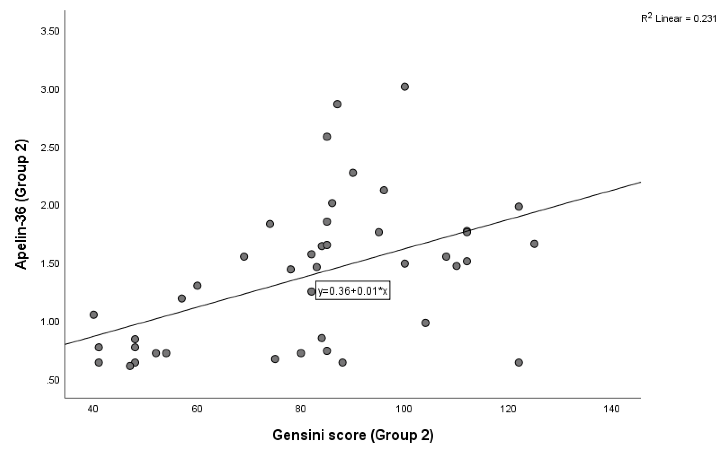
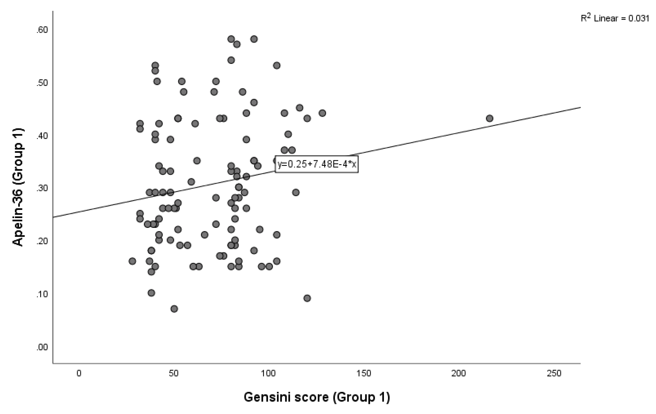
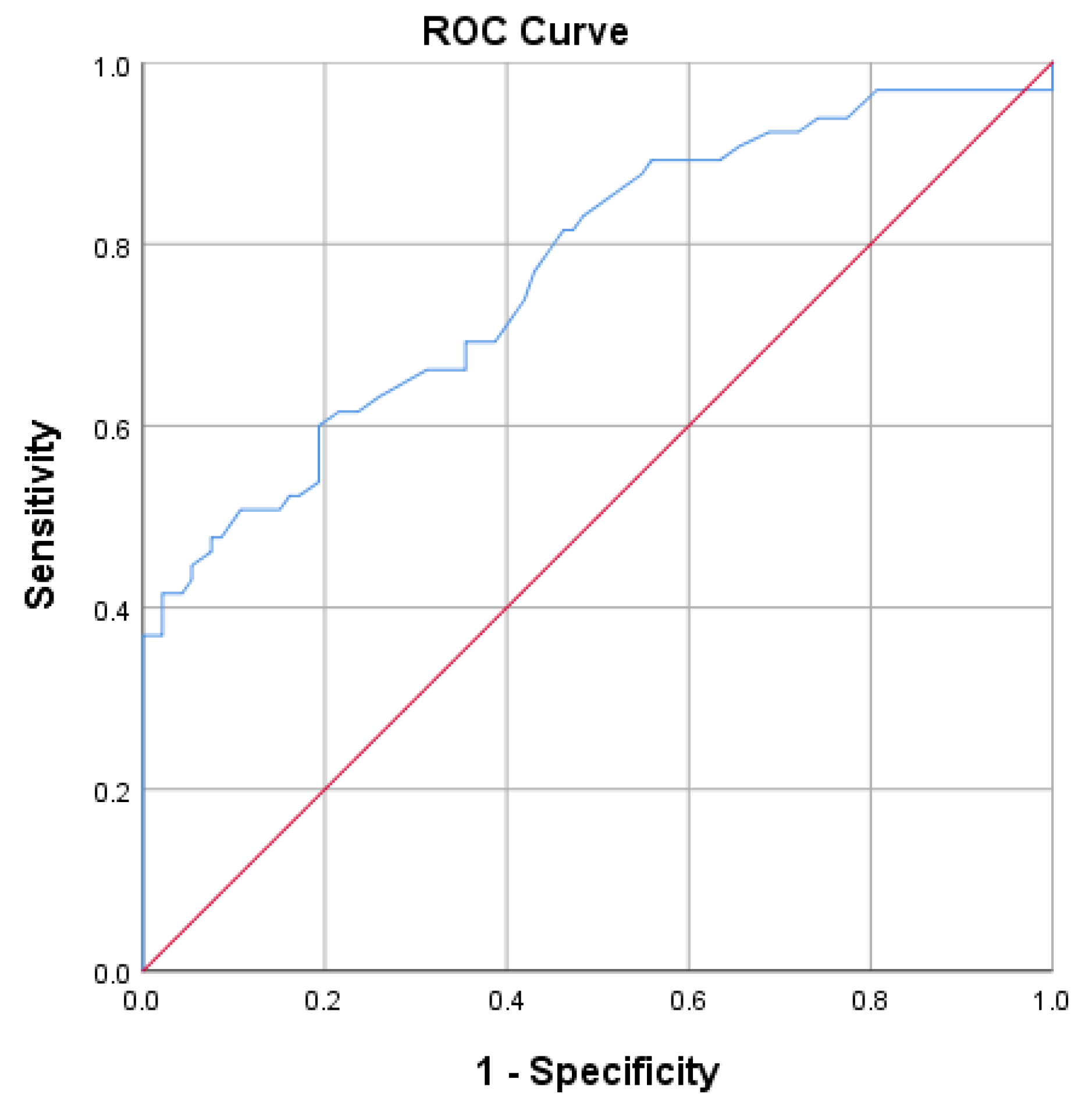
Background: Apelin-36 may be used to identify patients with ST-segment elevation myocardial infarction (STEMI) who are at risk for the no-reflow phenomenon. Patients presenting with STEMI were evaluated and stratified according to their apelin-36 levels. Methods: In this study, 161 patients presenting with STEMI within 12 hours of symptom onset and undergoing primary percutaneous coronary intervention (pPCI) were enrolled. Biochemical parameters, including apelin-36, troponin T, creatine kinase (CK), the MB fraction of creatine kinase (CK-MB), total cholesterol, triglycerides, and other routine laboratory parameters, were measured. Blood samples for apelin-36 measurement were collected prior to PCI, centrifuged to obtain serum, and preserved at -80⁰C until being assayed. Two-dimensional echocardiography was performed in all patients. Thereafter, patients were divided into two groups according to their level of Apelin-36. Results: Among the 161 consecutive STEMI patients, 115 (71.42%) had Apelin-36 levels ≤0.58ng/mL (group 1), whereas 46 (28.57%) had Apelin-36 levels >0.58ng/mL (group 2). In total, 51 (31.67%) STEMI patients experienced no-reflow phenomenon after PCI: 29 (18.01%) patients with apelin-36 ≤ 0.58ng/mL and 22 (13.66%) with a value > 0.58ng/mL (p < 0.001). In terms of Gensini score, the mean value in group 1 was (70.29 (±28.76), while in group 2, it was 81.95 (±23.82) (p=0.004). Overall, a positive correlation between apelin-36 and Gensini score was observed in both groups using Kendall’s correlation analysis (group 1: Figure 2, p=0.05; group 2: Figure 2, p<0.0001). Binary logistic regression analysis identified apelin-36 and diabetes mellitus as significant predictors at the 5% level, with p-values of 0.045 and 0.036, respectively. Patients with apelin-36 levels ≤ 0.58ng/mL had troponin T levels of 290.0 (8.5-9510.0), while those with a value > 0.58ng/mL had troponin T levels of 132.15 (9.4-5190.0) (p < 0.012). The receiver operating characteristics (ROC) curve of apelin-36 was used to plot the true positive rate against the false positive rate at different cut-off points, with AUC=0.67 (95% CI, 0.57-0.76), and the cut-off value for apelin-36 was 0.58ng/mL, with p=0.001. Conclusions: Significant associations were observed between apelin-36 and no-reflow phenomenon in patients with STEMI. An apelin-36 cut-off value of 0.58ng/mL, measured at admission, could be used to identify patients who were at increased risk of no-reflow phenomenon/reperfusion injury.

Keywords: 
;  ;  

1. Introduction

Apelin is encoded by the APLN gene, which is located on the arm of the X chromosome (Xq-25-q-26.1) [1]. Apelin peptides are formed by cleavage of the 77-amino acid precursor, pre-proapelin. Its biologically active forms are apelin-55, apelin-36, apelin-17, apelin-13, and apelin-12 [2]. Plasma kallikrein, neprilysin, and angiotensin-converting enzyme 2 (ACE2) degrade apelin peptides, generating fewer active peptides [3,4,5]. Apelin mRNA is found in all organs of the rat [6]. Human apelin mRNA is widely distributed across blood vessels, the central nervous system, and major organs like the heart, lungs, and kidneys. The predominant expression of apelin in endocardial and vascular endothelial cells implies that circulating apelin likely originates from these tissues [7,8].
Apelin peptides act primarily through the apelin receptor (APLNR, also known as APJ) and interact with G proteins. The receptor is broadly expressed in the heart, vasculature, kidneys, ovaries, and early human embryonic tissues [9]. Stimulation of the apelin/APJ system improves cardiac contractility by augmenting myofilament calcium sensitivity and activating the protein kinase C (PKC)–extracellular regulated kinase (ERK1/2) pathways, through which it regulates the degradation of troponins during ischemia/myocardial infarction [10,11,12,13].
The cardioprotective effect of apelin during myocardial ischemia–reperfusion is linked to the inhibition of the mitochondrial permeability transition pore, glycogen synthase kinase-3β, and adenylyl cyclase, while it activates several key pathways, such as PI3-kinase, Akt, ERK1/2, NOS, MMP, and the mitoKATP channel (Figure 1) [14].
Acute myocardial infarction (AMI) results from the complete occlusion of a coronary artery at the site of a ruptured plaque, which exposes its inner core and triggers thrombus formation. During reperfusion therapy, the abrupt return of blood flow leads to fatal damage to cardiac cells through the activation of multiple intracellular pathways, a phenomenon known as myocardial ischemia–reperfusion injury (MIRI) and encompassing myocardial stunning, no-reflow phenomenon (microvascular damage), reperfusion injury, and lethal reperfusion injury [15]. The pathophysiology of myocardial ischemia–reperfusion injuries is characterized by pH paradox, calcium overload, burst of reactive oxygen species (ROC), mitochondrial dysfunction, imbalance of protein phosphorylation, and inflammation [16,17,18,19,20].
Following the no-reflow phenomenon (microvascular damage), which is a type of MIRI, the expressions of apelin and apelin receptor mRNA and protein increase initially and then decline from 24 hours onward. In preclinical models, administration of apelin or elabela (a polypeptide that is also encoded by the APLN gene) at the time of reperfusion protects against ischemia–reperfusion injury, indicating potential for clinical use [21,22].
Apelin-36 peptide protects the heart against ischemia–reperfusion injury in vivo by activating the Reperfusion Injury Salvage Kinase (RISK) pathway (ERK and Akt-kinase) and delaying mPTP opening (downregulation of reactive oxygen species (ROS)) (Figure 1) [23,24]. In addition, Apelin (AP) and Elabela (Ela), components of the apelinergic system, are assumed to modulate endothelial function and contribute to atherosclerosis [25,26].
As far as we know, the role of Apelin-36 in the prediction of no-reflow phenomenon among STEMI patients has not previously been investigated.
The aim of this study was to evaluate the level of apelin-36 in the serum in patients with STEMI who underwent primary percutaneous coronary intervention (pPCI).

2. Methods

2.1. Study Design

Our study was performed as a prospective study, which included 161 consecutive patients who were diagnosed with ST-segment elevation myocardial infarction (STEMI). These patients were enrolled in our center from October 2024 to June 2025. Based on their serum apelin-36 levels, the patients were divided into two groups: group 1 (apelin-36 level ≤ 5.8ng/mL) and group 2 (apelin-36 level >5.8ng/mL). Patients were considered eligible for STEMI if they met all of the following criteria: (1) cardiac biomarker detection (evidence of elevated cardiac troponin (cTn), based on the ESC 0h/1h or 0h/2h diagnostic algorithms); (2) clinical symptoms (symptoms consistent with myocardial ischemia); (3) electrocardiographic (ECG) changes (new ST-segment elevation at the J-point in at least two contiguous leads, defined as ≥2.5mm in men <40 years, ≥2.5mm in men ≥ 40 years, or ≥1.5mm in women (regardless of age) in leads V2-V3 and/or ≥1 mm in other leads, as well as in the absence of left ventricle [LV] hypertrophy or left bundle branch block [LBBB]); and (4) patients who underwent primary percutaneous coronary intervention (pPCI) (persistent ST-segment elevation and symptoms of ischemia of ≤12h) [27]. The exclusion criteria were previous myocardial infarction, thyroid dysfunction, renal insufficiency, inflammatory disease, acute infectious disease, and malignancy.
A comprehensive clinical history was obtained, including assessment of established coronary risk factors (diabetes mellitus, dyslipidemia, arterial hypertension, and smoking), prior pharmacological treatments, and the time elapsed from symptom onset to hospital admission.
Laboratory evaluations comprised measurements of apelin-36, creatine kinase (CK), creatine kinase-MB (CK-MB), cardiac troponin T (cTnT), cholesterol, triglycerides, and standard biochemical parameters. Blood samples were collected approximately 30 minutes after hospital admission. To measure Apelin-36, serum was separated from the blood by centrifugation at 3.000rpm for 10 min and kept frozen at -80⁰ until analysis. Apelin-36 concentrations were measured using an enzyme-linked immunosorbent assay (ELISA) kit (Phoenix Pharmaceuticals, Inc., CA, USA), following the manufacturer’s protocol (Awareness Technology, ChemWell 2 Automated ELISA, University Clinical Center of Kosova (UCCK), Prishtina, Kosova).
Revascularization was performed in all patients, accompanied by periprocedural pharmacotherapy in accordance with current guidelines. Post-percutaneous coronary intervention (PCI) management followed standard therapeutic protocols: aspirin (100 mg), clopidogrel (75mg) or prasugrel (10mg), β-blockers, lipid-lowering agents, and either angiotensin-converting enzyme (ACE) inhibitors or angiotensin II receptor blockers (ARBs), in line with international recommendations [27,28].
Comprehensive transthoracic echocardiography was performed in all patients to quantify left ventricular ejection fraction (LVEF) and assess overall cardiac function.
The study was conducted in accordance with the Declaration of Helsinki and was approved by the Ethics Committees of Medical Faculty-University of Prishtina “Hasan Prishtina”, and the Kosovo Doctors Chamber (KDC), Prishtina.

2.2. Statistical Analysis

The principal aim of this study was to evaluate the relationship between Apelin-36 levels and the occurrence of the no-reflow phenomenon in patients presenting with ST-segment elevation myocardial infarction (STEMI). Continuous variables are reported as mean ± SD or median (IQR) for normally and non-normally distributed data. Categorical variables are presented as counts and percentages. Group comparisons were performed using Student’s t-test or the Mann–Whitney test for continuous variables and chi-square or Fisher’s exact test for categorical variables. Binary logistic regression was used to identify independent predictors of the outcome. Results are presented as odds ratios (ORs) with 95% confidence intervals (CIs). Model fit was evaluated using the Hosmer–Lemeshow test. Receiver operating characteristic (ROC) curve analysis was performed to assess the discriminative ability of the variables, plotting the sensitivity against the 1-specificity across a range of thresholds. The area under the curve (AUC) was calculated to quantify predictive accuracy for binary outcomes, and the optimal cut-off was determined based on the highest combination of sensitivity and specificity. A two-tailed p-value <0.05 was considered statistically significant. All statistical analyses were performed using SPSS statistical software, version 26.

3. Results

The baseline characteristics of study population are summarized in Table 1. In this study, we included 161 consecutive STEMI patients: 115 (71.42%) with Apelin-36 ≤0.58ng/mL (group 1) and 46 (28.57%) with a value >0.58ng/mL (group 2). In group 1, the mean age was 62.61 ±10.95 years, and 96 (59.62%) of the patients were male, while in group 2, the mean age was 59.37 (±11.30) years, and 36 (22.36%) of the patients were male. The comparison of diabetes mellitus, creatinine, and troponin T between groups with different levels of apelin-36 (≤0.58ng/mL and >0.58ng/mL) was statistically significant (the p-value was 0.006 for diabetes mellitus, 0.014 for creatinine, and 0.012 for troponin T). In terms of Gensini score, the mean value in group 1 was 70.29 (±28.76), while in group 2, it was 81.95 (±23.82) (p=0.004). A positive correlation between apelin-36 and Gensini score was observed in both groups using Kendall’s correlation analysis (group 1: Figure 2, p=0.05; group 2: Figure 2, p<0.0001). In total, 51 (31.67%) of the STEMI patients experienced no-reflow phenomenon after PCI: 29 (18.01%) patients with apelin-36 ≤0.58ng/mL and 22 (13.66%) with a value >0.58ng/mL (p=0.001).
Figure 3. Correlation between apelin-36 levels and Gensini score in group 2 (Kendall’s correlation coefficient r = 0.39; p <0.0001).
Preprints 190411 g003
Based on binary logistic regression, we analyzed the relationship between one dichotomous dependent variable (TIMI flow grade) and other variables (BMI, diabetes mellitus, smoking, apelin-36, creatine kinase-MB, creatine kinase, and troponin T). Apelin-36 and diabetes mellitus are significant predictors at the 5% level, with p-values of 0.045 and 0.036. Predictors such as BMI, smoking, creatine kinase, creatine kinase-MB, and troponin T, with p-values above 0.05, are not significant predictors of the TIMI flow grade at the 5% level.
Table 2. Binary logistic regression analysis of no-reflow phenomenon.
Parameter OR 95% CI P-value
BMI (kg/m2) 0.89 0.69-1.16 0.41
Diabetes mellitus (n/%) 0.07 0.007-0.85 0.036
Smoking (n/%) 4.96 0.76-32.10 0.09
Apelin-36 (ng/mL) 0.038 0.002-0.92 0.045
Creatine kinase-MB (U/L) 1.003 0.99-1.01 0.57
Creatine kinase (U/L) 0.99 0.99-1.00 0.15
Troponin T (pg/mL) 1.00 1.00-1.001 0.16
OR: odds ratio; BMI: body mass index.
A receiver operating characteristics (ROC) curve of Apelin-36 was used to plot the true positive rate against the false positive rate across varying cut-off points; the area under the curve (AUC) was 0.77 (95% CI, 0.69-0.84), while the cut- off value for apelin-36 was 0.58ng/mL, with p<0.001.
Table 3 presents the AUC values of biochemical parameters other than apelin-36.

4. Discussion

This study investigated the predictive value of Apelin-36 in relation to the occurrence of the no-reflow phenomenon in STEMI patients.
The role of apelinergic peptides in the pathogenesis of myocardial ischemia–reperfusion injury (MIRI) is yet to be elucidated. However, many studies have reported that apelin activity may exert a beneficial effect in STEMI patients due to its cardioprotective potential.
The rupture of an atherosclerotic plaque represents the first step in the sequence of events leading to thrombotic coronary occlusion and subsequent myocardial infarction. The oxygen supply to cells is reduced due to impaired blood flow, ultimately resulting in cardiac hypoxia or ischemia [29,30].
Intervention, such as primary PCI, to restore coronary blood flow is essential for myocardial salvage. Nevertheless, reperfusion can aggravate myocardial damage. This phenomenon, known as ischemia–reperfusion (I/R) injury, encompasses a series of events—including pH paradox, calcium overload, burst of reactive oxygen species (ROC), mitochondrial dysfunction, imbalance of protein phosphorylation, and inflammation—that ultimately lead to myocardial damage, such as myocardial stunning and no-reflow phenomenon (microvascular damage). Approximately 30% of patients are estimated to develop I/R injury, which significantly worsens their prognosis [31,32].
Regarding signaling mechanisms, as an APJ agonist, apelin protects against I/R injury, mainly by engaging the Akt/No and ERK ½ pathways and delaying mPTP opening (downregulation of reactive oxygen species (ROS)) (Figure 1). In our study, the use of ROC curve analysis of Apelin-36 for the prediction of no-reflow phenomenon (unsuccessful reperfusion, TIMI flow grade ≤2) was statistically significant, with AUC=0.77 (95% CI, 0.69-0.84) and p<0.001, confirming its influence on myocardial ischemia–reperfusion injury (MIRI) (Figure 4) [33,34,35,36,37,38,39]. Thus, in our study, patients with apelin-36 levels ≤0.58ng/mL showed a significantly higher incidence of the no-reflow phenomenon compared with those with apelin-36 levels >0.58ng/mL, with p=0.001 (Table 1). Based on binary logistic regression analysis, apelin-36 is a significant predictor of the TIMI flow grade at the 5% level, with OR=0.038, CI of 0.002-0.92, and p=0.045 (Table 2). This study indicates that lower Apelin-36 concentrations may aggravate MIRI, as has also been observed in preclinical research. Wang et al. demonstrated that the absence of apelin increases susceptibility to infarction and worsens cardiac function in both ex vivo and in vivo I/R models [40]. Apelin has recently gained attention as a potential therapy for myocardial I/R injury, owing to its ability to protect against various pathological mechanisms underlying the condition [41].
Apelin improves myocardial contractility by binding to the APJ receptor and activating downstream signaling pathways, including PI3K/Akt and ERK1/2, which augment calcium availability and sensitivity [10]. Myocardial infarction is characterized by decreased phosphorylation of cardiac troponin (cTn), reduced Ca2+ sensitivity, and diminished ATPase activity. Elevated cytosolic Ca2+ subsequently triggers protease I (calpain I) activation, leading to proteolytic degradation of troponins. Concurrently, apelin and APJ expression are upregulated, a process that is believed to exert cardioprotective effects by attenuating ischemic myocardial damage (degradation of troponin) [12,42,43,44,45]. In our study, patients with apelin-36 levels ≤0.58ng/mL showed significantly higher levels of troponin T than those with apelin-36 levels >0.58ng/mL (p=0.012), confirming the role of apelin in reducing troponin degradation (Table 1).
Apelin and APJ receptor expressions are increased in human coronary atherosclerotic plaques, co-localizing with inflammatory macrophages and vascular smooth muscle cells, which indicates their involvement in plaque progression and vascular remodeling [46]. Through ERK activation, apelin/APJ signaling elevates endothelial intercellular adhesion molecule-1 (ICAM-1) and vascular cell adhesion molecule-1 (VCAM-1) levels and stimulates monocyte chemoattractant protein-1 (MCP-1) release via the NF-κB/JNK pathway, thereby facilitating monocyte recruitment and adhesion—key events in early atherogenesis [47,48].
In patients with stable coronary artery disease (CAD), plasma apelin levels are reduced and inversely associated with the severity of coronary lesions, showing a negative correlation with the Gensini score [49,50]. Apelin upregulation during acute coronary syndrome (ACS) shows a positive correlation with the Gensini score, suggesting a compensatory cardioprotective role [51]. In our study, the Gensini score was significantly lower in group 1 (apelin-36 ≤ 0.58ng/mL) than in group 2 (apelin-36 > 0.58ng/mL) (p=0.004) (Table 1). Overall, apelin-36 levels were positively correlated with the Gensini score. Specifically, Kendall’s correlation analysis demonstrated a weak positive correlation in group 1 (r = 0.12; p =0.05) and a moderate positive correlation in group 2 (r = 0.39; p <0.0001) (Figure 2 and Figure 3).
This result suggests that apelin plays a cardioprotective role and that its upregulation following ACS-STEMI may have therapeutic potential, particularly in no-reflow phenomenon/myocardial ischemia–reperfusion injury.
One limitation of this study is that it only included patients with STEMI. Assessing apelin levels in control groups of patients without ST-segment elevation myocardial infarction (NSTEMI) and patients with stable angina would strengthen a comparative analysis.

5. Conclusions

Significant associations were observed between apelin-36 and no-reflow phenomenon in patients with STEMI. An apelin-36 cut-off value of 0.58ng/mL, measured at admission, was used to identify patients who were at increased risk of no-reflow phenomenon/reperfusion injury. Furthermore, apelin-36 levels were positively correlated with the Gensini score. Overall, these findings suggest a cardioprotective role and indicate its potential therapeutic relevance through upregulation following STEMI.

Disclosure statement

Authors declare that they have no conflicts of interest.

Author Contributions

Conceptualization, X.K., A.B., J.V., B.B., and X.J.; methodology, X.K., J.V., B.B., G.G., A.B., and A.B.; software, P.Ç., K.J., and L. S.; validation, X.J., J.V., and A.B.; investigation, G.G., X.K., A.B., K.J., L.S., and P.Ç.; writing—original draft preparation, X.K. and A.B.; writing—review and editing: A.B. and X.K.; supervision, A.B. and X.J. All authors have read and agreed to the published version of the manuscript.

Funding

This research was supported by the Ministry of Education, Science, Technology and Innovation, Republic of Kosovo.

Institutional Review Board Statement

This study was conducted in accordance with the Declaration of Helsinki and approved by the Ethics Committees of Medical Faculty-University of Prishtina, “Hasan Prishtina”, and the Kosovo Doctors Chamber (KDC), Prishtina.

Informed Consent Statement

Written informed consent was obtained from all subjects involved in this study, in accordance with ethical guidelines.

Data Availability Statement

The data presented in this study are available from the corresponding author upon reasonable request due to privacy and ethical considerations.

Conflicts of Interest

The authors declare no conflicts of interest. .

References

  1. Hu, G.; Wang, Z.; Zhang, R.; Sun, W.; Chen, X. The role of apelin/apelin receptor in energy metabolism and water homeostasis: a comprehensive narrative review. Front Physiol. 2021, 12, 632886. [Google Scholar] [CrossRef]
  2. Tatemoto, K.; Hosoya, M.; Habata, Y.; Fujii, R.; Kakegawa, T.; Zou, M.X.; Kawamata, Y.; Fukusumi, S.; Hinuma, S.; Kitada, C.; et al. Isolation and characterization of a novel endogenous peptide ligand for the human APJ receptor. Biochem Biophys Res Commun. 1998, 251, 471–476. [Google Scholar] [CrossRef]
  3. Wang, W.; Shen, M.; Fischer, C.; Basu, R; Hazra, S.; Couvineau, P.; Paul, M.; Wang, F.; Toth, S.; Mix, D.S.; et al. Apelin protects against abdominal aortic aneurysm and the therapeutic role of neutral endopeptidase resistant apelin analogs. Proc Natl Acad Sci. 2019, 116, 13006–13015. [Google Scholar] [CrossRef] [PubMed]
  4. McKinnie, S.M.; Fischer, C.; Tran, K.M.; Wang, W.; Mosquera, F.; Oudit, G.Y.; Vederas, J.C. The metalloprotease neprilysin degrades and inactivates apelin peptides. Chembiochem. 2016, 17, 1495–1498. [Google Scholar] [CrossRef] [PubMed]
  5. Wang, W.; McKinnie, S.M.; Farhan, M.; Paul, M.; McDonald, T.; McLean, B.; Llorens-Cortes, C.; Hazra, S.; Murray, A.G.; Vederas, J.C.; et al. Angiotensin-converting enzyme 2 metabolizes and partially inactivates pyr-apelin-13 and apelin-17: physiological effects in the cardiovascular system. Hypertension. 2016, 68, 365–377. [Google Scholar] [CrossRef]
  6. Kawamata, Y.; Habata, Y.; Fukusumi, S.; Hosoya, M.; Fujii, R.; Hinuma, S.; Nishizawa, N.; Kitada, C.; Onda, H.; Nishimura, O.; et al. Molecular properties of apelin: tissue distribution and receptor binding. Biochim Biophys Acta 2001, 1538, 162–171. [Google Scholar] [CrossRef] [PubMed]
  7. Read, C.; Nyimanu, D.; Williams, T.L.; Huggins, D.J.; Sulentic, P.; Macrae, R.G.; Yang, P; Glen, R.C.; Maguire, J.J.; Davenport, A.P. International union of basic and clinical pharmacology. CVII. Structure and pharmacology of the apelin receptor with a recommendation that elabela/toddler is a second endogenous peptide ligand. Pharmacol Rev. 2019, 71, 467–502. [Google Scholar] [CrossRef]
  8. Kleinz, M.J.; Davenport, A.P. Immunocytochemical localization of the endogenous vasoactive peptide apelin to human vascular and endocardial endothelial cells. Regul Pept. 2004, 118, 119–125. [Google Scholar] [CrossRef]
  9. Medhurst, A.D.; Jennings, C.A.; Robbins, M.J.; Davis, R.P.; Ellis, C.; Winborn, K.Y.; Lawrie, K.W.; Hervieu, G.; Riley, G.; Bolaky, J.E.; et al. Pharmacological and immunohistochemical characterization of the APJ receptor and its endogenous ligand apelin. J Neurochem. 2003, 84, 1162–1172. [Google Scholar] [CrossRef]
  10. Perjes, A.; Skoumal, R.; Tenhunen, O.; Konyi, A.; Simon, M.; Horvath, I.G.; Kerkelä, R.; Ruskoaho, H.; Szokodi, I. Apelin increases cardiac contractility via protein kinase Cε-and extracellular signal-regulated kinase-dependent mechanisms. PloS one 2014, 9, e93473. [Google Scholar] [CrossRef]
  11. Tang, L.; Qiu, H.; Xu, B.; Su, Y.; Nyarige, V.; Li, P.; Chen, H.; Killham, B.; Liao, J.; Adam, H.; et al. Microparticle mediated delivery of apelin improves heart function in post myocardial infarction mice. Circ Res. 2024, 135, 777–798. [Google Scholar] [PubMed]
  12. Krasniqi, X.; Berisha, B.; Gashi, M.; Koçinaj, D.; Jashari, F.; Vincelj, J. Influence of apelin-12 on troponin levels and the rate of MACE in STEMI patients. BMC cardiovasc Disord. 2017, 17, 195. [Google Scholar] [CrossRef] [PubMed]
  13. Pi, Y.; Zhang, D.; Kemnitz, K.R.; Wang, H.; Walker, J.W. Protein kinase C and A sites on troponin I regulate myofilament Ca2+ sensitivity and ATPase activity in the mouse myocardium. J Physiol. 2003, 552, 845–857. [Google Scholar] [CrossRef]
  14. Popov, S.V.; Maslov, L.N.; Mukhomedzyanov, A.V.; Kurbatov, B.K.; Gorbunov, A.S.; Kilin, M.; Azev, V.N.; Khlestkina, M.S.; Sufianova, G.Z. Apelin is a prototype of novel drugs for the treatment of acute myocardial infarction and adverse myocardial remodeling. Pharmaceutics 2023, 14 15, 1029. [Google Scholar] [CrossRef]
  15. Davidson, S.M.; Adameová, A.; Barile, L.; Cabrera-Fuentes, H.A.; Lazou, A.; Pagliaro, P.; Stensløkken, K.O.; Garcia-Dorado, D. EU-CARDIOPROTECTION COST Action (CA16225). Mitochondrial and mitochondrial-independent pathways of myocardial cell death during ischaemia and reperfusion injury. J Cell Mol Med 2020, 24, 3795–3806. [Google Scholar] [CrossRef]
  16. Kim, J.S.; Jin, Y.; Lemasters, J.J. Reactive oxygen species, but not Ca2+ overloading, trigger pH-and mitochondrial permeability transition-dependent death of adult rat myocytes after ischemia-reperfusion. Am J Physiol Heart Circ Physiol. 2006, 16 290, H2024–H2034. [Google Scholar] [CrossRef] [PubMed]
  17. Yellon, D.M.; Hausenloy, D.J. Myocardial reperfusion injury. N Engl J Med. 2007, 17 357, 1121–1135. [Google Scholar] [CrossRef]
  18. Bugger, H.; Pfeil, K. Mitochondrial ROS in myocardial ischemia reperfusion and remodeling. Biochim Biophys Acta Mol Basis Dis. 2020, 18 1866, 165768. [Google Scholar] [CrossRef]
  19. Timmers, L.; Pasterkamp, G.; de Hoog, V.C.; Arslan, F.; Appelman, Y.; de Kleijn, D.P. The innate immune response in reperfused myocardium. Cardiovasc Res. 2012, 94, 276–283. [Google Scholar] [CrossRef]
  20. García-Niño, W.R.; Zazueta, C.; Buelna-Chontal, M.; Silva-Palacios, A. Mitochondrial quality control in cardiac-conditioning strategies against ischemia-reperfusion injury. Life 2021, 11, 1123. [Google Scholar]
  21. Zeng, X.J.; Zhang, L.K.; Wang, H.X.; Lu, L.Q.; Ma, L.Q.; Tang, C.S. Apelin protects heart against ischemia/reperfusion injury in rat. Peptides 2009, 30, 1144–1152. [Google Scholar] [CrossRef] [PubMed]
  22. Wang, W.; McKinnie, S.M.; Patel, V.B.; Haddad, G.; Wang, Z.; Zhabyeyev, P.; Das, S.K.; Basu, R.; McLean, B.; Kandalam, V.; Penninger, J.M. Loss of Apelin exacerbates myocardial infarction adverse remodeling and ischemia-reperfusion injury: therapeutic potential of synthetic Apelin analogues. J Am Heart Assoc. 2013, 2, e000249. [Google Scholar] [PubMed]
  23. Simpkin, J.C.; Yellon, D.M.; Davidson, S.M.; Lim, S.Y; Wynne, A.M.; Smith, C.C. Apelin-13 and apelin-36 exhibit direct cardioprotective activity against ischemia reperfusion injury. Basic Res Cardiol. 2007, 23 102, 518–528. [Google Scholar]
  24. Fernandez Rico, C.; Konate, K.; Josse, E.; Nargeot, J.; Barrère-Lemaire, S.; Boisguérin, P. Therapeutic peptides to treat myocardial ischemia-reperfusion injury. Front Cardiovasc Med. 2022, 9, 792885. [Google Scholar] [CrossRef]
  25. Bäck, M.; Yurdagul, A.; Tabas, I.; Öörni, K.; Kovanen, P.T. Inflammation and its resolution in atherosclerosis: mediators and therapeutic opportunities. Nat Rev Cardiol. 2019, 16, 389–406. [Google Scholar] [PubMed]
  26. Chapman, F.A.; Maguire, J.J.; Newby, D.E.; Davenport, A.P.; Dhaun, N. Targeting the apelin system for the treatment of cardiovascular diseases. Cardiovasc Res. 2023, 119, 2683–2696. [Google Scholar]
  27. 27; Byrne, R.A.; Rossello, X.; Coughlan, J.; Barbato, E.; Berry, C.; Chieffo, A.; Claeys, M.J.; Dan, G.A.; Dweck, M.R.; Galbraith, M.; Gilard, M. 2023 ESC guidelines for the management of acute coronary syndromes: developed by the task force on the management of acute coronary syndromes of the European Society of Cardiology (ESC). Eur Heart J Acute Cardiovasc Care 2024, 13, 55–161. [Google Scholar]
  28. Sousa-Uva, M.; Neumann, F.J.; Ahlsson, A.; Alfonso, F.; Banning, A.P.; Benedetto, U.; Byrne, R.A.; Collet, J.P.; Falk, V.; Head, S.J.; Jüni, P. 2018 ESC/EACTS Guidelines on myocardial revascularization. Eur J Cardiothorac Surg. 2019, 55, 4–90. [Google Scholar]
  29. O’Connell, E.J.; Martinez, C.A.; Liang, Y.G.; Cistulli, P.A.; Cook, K.M. Out of breath, out of time: interactions between HIF and circadian rhythms. Am J Physiol Cell Physiol. 2020, 319, C533–C540. [Google Scholar]
  30. Abe, H.; Semba, H.; Takeda, N. The roles of hypoxia signaling in the pathogenesis of cardiovascular diseases. J Atheroscler Thromb. 2017, 24, 884–894. [Google Scholar]
  31. Yu, P.; Ma, S.; Dai, X.; Cao, F. Elabela alleviates myocardial ischemia reperfusion-induced apoptosis, fibrosis and mitochondrial dysfunction through PI3K/AKT signaling. Am J Transl Res. 2020, 12, 4467. [Google Scholar]
  32. Deng, J. Advanced research on the regulated necrosis mechanism in myocardial ischemia-reperfusion injury. Int J Cardiol. 2021, 32 334, 97–101. [Google Scholar]
  33. Welt, F.G.; Batchelor, W.; Spears, J.R.; Penna, C.; Pagliaro, P.; Ibanez, B.; Drakos, S.G.; Dangas, G.; Kapur, N.K. Reperfusion injury in patients with acute myocardial infarction: JACC scientific statement. J Am Coll Cardiol. 2024, 33 83, 2196–2213. [Google Scholar]
  34. Sagris, M.; Apostolos, A.; Theofilis, P.; Ktenopoulos, N.; Katsaros, O.; Tsalamandris, S.; Tsioufis, K; Toutouzas, K.; Tousoulis, D. Myocardial Ischemia–Reperfusion injury: Unraveling pathophysiology, clinical manifestations, and emerging prevention strategies. Biomedicines 2024, 12, 802. [Google Scholar] [CrossRef]
  35. Welt, F.G.; Batchelor, W.; Spears, J.R.; Penna, C.; Pagliaro, P.; Ibanez, B.; Drakos, S.G.; Dangas, G; Kapur, N.K. Reperfusion injury in patients with acute myocardial infarction: JACC scientific statement. J Am Coll Cardiol. 2024, 83, 2196–2213. [Google Scholar] [CrossRef]
  36. Pisarenko, O.I.; Shulzhenko, V.S.; Studneva, I.M.; Serebryakova, L.I.; Pelogeykina, Y.A.; Veselova, O.M. Signaling pathways of a structural analogue of apelin-12 involved in myocardial protection against ischemia/reperfusion injury. Peptides. 2015, 73, 67–76. [Google Scholar] [CrossRef] [PubMed]
  37. 37; An, S.; Wang, X.; Zhang, X.; Meng, H.; Li, W.; Chen, D.; Ge, J. Apelin protects against ischemia-reperfusion injury in diabetic myocardium via inhibiting apoptosis and oxidative stress through PI3K and p38-MAPK signaling pathways. Aging (Albany NY). 2020, 12, 25120. [Google Scholar]
  38. 38; Wang, X.; Zhang, L.; Feng, M.; Xu, Z.; Cheng, Z.; Qian, L. ELA-11 protects the heart against oxidative stress injury induced apoptosis through ERK/MAPK and PI3K/AKT signaling pathways. Front Pharmacol. 2022, 13, 873614. [Google Scholar]
  39. 39; Rakhshan, K.; Sharifi, M.; Ramezani, F.; Azizi, Y.; Aboutaleb, N. ERK/HIF-1α/VEGF pathway: a molecular target of ELABELA (ELA) peptide for attenuating cardiac ischemia–reperfusion injury in rats by promoting angiogenesis. Mol Biol Rep. 2022, 49, 10509–10519. [Google Scholar]
  40. Wang, W.; McKinnie, S.M.; Patel, V.B.; Haddad, G.; Wang, Z.; Zhabyeyev, P.; Das, S.K.; Basu, R.; McLean, B.; Kandalam, V.; Penninger, J.M. Loss of Apelin exacerbates myocardial infarction adverse remodeling and ischemia-reperfusion injury: therapeutic potential of synthetic Apelin analogues. J Am Heart Assoc. 2013, 2, e000249. [Google Scholar]
  41. Rossin, D.; Vanni, R.; Lo Iacono, M.; Cristallini, C.; Giachino, C.; Rastaldo, R. APJ as promising therapeutic target of peptide analogues in myocardial infarction-and hypertension-induced heart failure. Pharmaceutics 2023, 41 15, 1408. [Google Scholar] [CrossRef]
  42. Chandrasekaran, B.; Dar, O.; McDonagh, T. The role of apelin in cardiovascular function and heart failure. Eur J Heart Fail. 2008, 42 10, 725–732. [Google Scholar] [CrossRef]
  43. Khatami, S.; Faghihi, M.; Zarrin, A.; Sarveazad, A.; Yousefifard, M.; Ghorbani, A. Cardioprotective Effects of Apelin in Myocardial Ischemia/Reperfusion Injury: A Systematic Review and Meta-Analysis. J Cardiovasc Pharmacol. 2022, 43, 10–97. [Google Scholar] [CrossRef]
  44. Wyderka, R.; Osuch, Ł.; Ołpińska, B.; Łoboz-Rudnicka, M.; Diakowska, D.; Leśków, A.; Jaroch, J. The Impact of the Apelinergic System on the Cardiovascular System. Int J Mol Sci. 2025, 26, 10087. [Google Scholar] [CrossRef] [PubMed]
  45. Szokodi, I.; Tavi, P.; Földes, G.; Voutilainen-Myllylä, S.; Ilves, M.; Tokola, H.; Pikkarainen, S.; Piuhola, J.; Rysä, J.; Tóth, M.; Ruskoaho, H. Apelin, the novel endogenous ligand of the orphan receptor APJ, regulates cardiac contractility. Circ Res. 2002, 91, 434–440. [Google Scholar] [CrossRef]
  46. Namazi, G.; Salami, R.; Pourfarzam, M.; Asa, P.; Mafi, A.; Raygan, F. Association of the serum apelin, but not ghrelin, with the presence and severity of coronary artery disease. Indian Heart J. 2021, 73, 214–217. [Google Scholar] [CrossRef] [PubMed]
  47. 47; Lu, Y.; Zhu, X.; Liang, G.X.; Cui, R.R.; Liu, Y.; Wu, S.S.; Liang, Q.H.; Liu, G.Y.; Jiang, Y.; Liao, X.B.; Xie, H. Apelin–APJ induces ICAM-1, VCAM-1 and MCP-1 expression via NF-κB/JNK signal pathway in human umbilical vein endothelial cells. Amino Acids 2012, 43, 2125–2136. [Google Scholar]
  48. 48; Liu, L.Q.; Zhang, P.; Qi, Y.Z.; Li, H.; Jiang, Y.H.; Yang, C.H. Quercetin attenuates atherosclerosis via modulating apelin signaling pathway based on plasma metabolomics. Chin J Integr Med. 2023, 29, 1121–1132. [Google Scholar]
  49. Hendrianus; Adiarto, S.; Prakoso, R.; Firdaus, I.; Indriani, S.; Rudiktyo, E.; Widyantoro, B.; Taofan; Ambari, A.M.; Sukmawan, R. A novel peptide elabela is associated with hypertension-related subclinical atherosclerosis. High Blood Press Cardiovasc Prev. 2023, 30, 37–44. [Google Scholar] [CrossRef] [PubMed]
  50. 50; Li, Z.; Bai, Y.; Hu, J. Reduced apelin levels in stable angina. Intern Med. 2008, 47, 1951–1955. [Google Scholar]
  51. Güzelburç, Ö.; Demirtunç, R.; Altay, S.; Kemaloğlu Öz, T.; Tayyareci, G. Plasma apelin level in acute myocardial infarction and its relation with prognosis: a prospective study. JRSM Cardiovasc Dis. 2021, 10, 2048004020963970. [Google Scholar] [PubMed]
Figure 1. The cardioprotective effects of apelin. APJ: apelin receptor; GSK-3β: glycogen synthase kinase 3β; mPTP: mitochondrial permeability transition pore; PLC: phospholipase C; PKC: protein kinase C; ERK1/2: extracellular regulated kinase; MLC: myosin light-chain; IP3: inositol triphosphate; MMP: matrix metalloproteinase; Akt: protein kinase B; NOS: NO-synthase; ROS: reactive oxygen species.
Preprints 190411 g001
Figure 2. Correlation between apelin-36 levels and Gensini score in group 1 (Kendall’s correlation coefficient r = 0.12; p =0.05).
Preprints 190411 g002
Figure 4. ROC curve analysis of Apelin-36 for the prediction of no-reflow phenomenon in STEMI patients. AUC=0.77 (95% CI, 0.69-0.84); p<0.001.
Preprints 190411 g004
Table 1. Baseline characteristics according to Apelin-36 level.
Characteristics Apelin-36 ≤ 0.58ng/mL
(n=115)
Apelin-36 > 0.58ng/mL
(n=46)
P-value
Age (year) 62.61 (±10.95) 59.37 (±11.30) 0.22
Gender (male) (n/%) 96 (59.62) 36 (22.36) 0.80
BMI (kg/m2) 27.28 (±5.32) 26.24 (±5.64) 0.96
Medical history
Hypertension (n/%) 66 (40.99) 24 (14.90) 0.61
Diabetes mellitus (n/%) 35 (21.73) 10 (6.21) 0.006
Smoking (n/%) 69 (42.85) 27 (16.77) 0.54
Laboratory values
Hemoglobin (mg/dL) 138.55 (±23.68) 141.0 (±15.49) 0.99
Cholesterol (mmol/L) 5.17 (1.16) 5.02 (1.49) 0.33
Triglyceride (mmol/L) 1.75 (±1.12) 1.52 (±0.75) 0.43
Glucose (mmol/L) 10.27 (±5.77) 8.48 (±4.21) 0.02
Creatinine (umol/L) 105.73 (31.27) 92.95 (26.97) 0.014
Creatine kinase-MB (U/L) 124.0 (15.0-466.0) 90 (16.0-520.0) 0.17
Creatine kinase (U/L) 431.0 (210-7224.0) 752.50 (350.0-5057.0) 0.61
Troponin T (pg/mL) 290.0 (8.5-9510.0) 132.15 (9.4-5190.0) 0.012
Ejection fraction (%) 49.43 (±6.55) 50.30 (±7.14) 0.44
Gensini score (Mean ±s.d) 70.29 (±28.76)
81.95 (±23.82)
0.004
Final TIMI grade flow ≤ 2 (n/%) 29 (18.01) 22 (13.66) 0.001
BMI: body mass index; TIMI: thrombolysis in myocardial infarction.
Table 3. Area under the curve values for biochemical analysis.
Parameter AUC (95% CI) P-value
Apelin-36 0.77 (0.69-0.84) <0.001
Creatine kinase 0.57 (0.34-0.80) 0.49
Creatine kinase-MB 0.59 (0.37-0.80) 0.40
Troponin T 0.57 (0.36-0.78) 0.49
Na+ 0.41 (0.20-0.62) 0.41
K+ 0.55 (0.33-0.77) 0.61
Ca++ 0.51 (0.31-0.72) 0.88
Cholesterol 0.70 (0.51-0.58) 0.07
Triglyceride 0.42 (0.20-0.64) 0.47
Hemoglobin 0.59 (0.37-0.81) 0.37
Glucose 0.67 (0.48-0.86) 0.10
BUN 0.60 (0.40-0.80) 0.33
Creatinine 0.59 (0.36-0.81) 0.40
Uric acid 0.65 (0.45-0.85) 0.15
AUC: area under the curve.
Disclaimer/Publisher’s Note: The statements, opinions and data contained in all publications are solely those of the individual author(s) and contributor(s) and not of MDPI and/or the editor(s). MDPI and/or the editor(s) disclaim responsibility for any injury to people or property resulting from any ideas, methods, instructions or products referred to in the content.
Copyright: This open access article is published under a Creative Commons CC BY 4.0 license, which permit the free download, distribution, and reuse, provided that the author and preprint are cited in any reuse.
Prerpints.org logo

Preprints.org is a free preprint server supported by MDPI in Basel, Switzerland.

Subscribe

Disclaimer

Terms of Use

Privacy Policy

Privacy Settings

© 2026 MDPI (Basel, Switzerland) unless otherwise stated