
Submitted:
15 December 2025
Posted:
17 December 2025
You are already at the latest version
Abstract
Food allergy is an exaggerated immune response, mediated by Immunoglobulin E (IgE) or by cells, to food antigens. Dogs and cats may present with both dermatological and gastrointestinal manifestations, although non-seasonal pruritus is the most com-mon clinical sign. Despite advances in understanding the immunopathogenesis of this condition, the elimination–provocation trial remains the gold standard for diagnosis. However, new diagnostic approaches, like molecular allergen macroarrays and lym-phocyte proliferation assays, may complement traditional strategies, opening new perspectives for accurate diagnosis. For long-term management, strict avoidance of of-fending allergens is essential, but emerging therapeutic interventions, including im-munotherapy using food components and targeted modulation of the gut–skin axis, are promising for improving clinical outcomes. This review summarizes current knowledge and highlights innovative approaches that can transform the diagnosis and manage-ment of food allergy in companion animals.
Keywords:
food allergy
; dog
; cat
; elimination-provocation test
; delayed food reactions
; molecular allergen macroarray
; lymphocyte proliferation test
; immunotherapy
; microbiota modulation
1. Introduction
Food allergy is an exaggerated immune response to food antigens [1,2] and may occur by both a type I (IgE-mediated) or a type IV (cell-mediated) hypersensitivity reaction [1,3]. However, some controversy regarding the nomenclature for this condition may arise as some authors refer to “food allergy” as an “adverse food reaction”. In fact, the latter term encompasses food allergy, an immunological event, but also food intolerance, a toxic/pharmacological/idiosyncratic reaction to food [3]. Hence, considering that cutaneous adverse food reactions described in dogs and cats appear to have an essentially immunological basis, we have chosen to use the term "food allergy" in this review.
The prevalence of food allergy in dogs and cats is less than 1% but represents 10 to 25% of allergic skin diseases in companion animals [4] and affects all ages and genders, in both canine and feline species [5]. There is no racial predisposition, but more than 1/3 of the dogs with food allergy is represented by four breeds: German Shepherd, Labrador, Golden Retriever and West Highland White Terrier [5].
In dogs, the most common allergen sources involved in food allergy are beef, dairy products, chicken, wheat and lamb. In cats, beef, fish and chicken are the most important. [6,7] The most common allergenic food proteins range between 15 and 40 kDa [3].
In food allergy, intolerance to food antigens may occur due to genetic predisposition, intestinal dysbiosis, alterations in gastrointestinal permeability or in the local immune system [3]. In fact, it is currently accepted that gut and skin microbiome can influence susceptibility to food allergy in children by various mechanisms of immune system modulation [8]. Some authors suggest that the type of acquired hypersensitivity could be explained by the allergenicity of food components at the gastrointestinal tract level [9].
Diagnosing food allergies in dogs and cats can be challenging but, as a general rule, once a food allergy diagnosis is confirmed and the causative ingredients are identified, the prognosis is excellent [10]. This review aims to summarize the most reliable diagnostic techniques and long-term management options, currently available for dogs and cats with food allergies.
2. Clinical Signs
An animal with food allergy may present with dermatological and/or gastrointestinal manifestations [1].
Non-seasonal pruritus is the most common manifestation of food allergy in dogs and cats, which may be generalized, especially in dogs, or localized [1,5,10]. Face, ears, ventrum and feet are areas commonly affected in both species [1,5,10]. Perianal pruritus and otitis externa may also be observed [1,5,10].
In cats, food allergy can manifest as one of four known skin reaction patterns (miliary dermatitis, head and neck pruritus, symmetrical self-induced alopecia, and feline eosinophilic complex) [5,11] but, according to Bryan and Frank (2010), head and neck are the most affected areas in this species [11]. Plasma cell or erythematous pododermatitis can also be a clinical sign of food allergy in cats [11].
Patients with food allergies are predisposed to secondary fungal and bacterial infections, which can increase the level of pruritus [1,5,10]. Secondary lesions such as lichenification, hyperpigmentation, excoriations and alopecia may also occur [1,10].
Nearly 40% of food allergic dogs present with cutaneous signs before one year of age, while in cats the onset of clinical signs appears to occur later [5]. Furthermore, in dogs, skin lesions may show quite similar to those of environmental-related atopic dermatitis, but clinical signs associated with food allergy generally occur earlier in life than in environmental allergy [10,12].
In more than 20% of dogs and cats with food allergy, the most common non-cutaneous signs are vomiting and diarrhea, but other gastrointestinal signs, like abdominal discomfort, flatulence, borborygmi and frequent defecations can be seen concurrently with the dermatological signs [1,13]. In dogs, some cases of anaphylaxis, conjunctivitis, increased frequency of bowel movements, symmetrical lupoid onychomycosis and sneezing have been described [13]. In cats, conjunctivitis, salivating, flatulence, hyperactive behavior and respiratory signs were reported less commonly [13].
3. Diagnosis
3.1. Elimination-Provocation Test
The diagnosis of food allergy should be based on an elimination-provocation test, in all animals with non-seasonal pruritus [1,11,14].
Before performing an elimination test, it is recommended to eliminate ectoparasites, such as sarcoptic mites and fleas, and control any secondary infections [10,11].
The elimination diet should last for at least eight weeks in dogs and cats, to achieve a diagnostic sensitivity greater than 90% [1,15]. If there is a clinical improvement during the elimination trial, a challenge test with the original diet should last up to 10 to 14 days [1,11]. A diagnosis of food allergy could be achieved if the previous diet leads to clinical deterioration and if the animal improves again by returning to the elimination diet [1,16].
The elimination diet should be performed by using a homecooked diet, based on a novel protein source. Although, usually, this is not possible because it is laborious and expensive, mainly for large breeds [3,16]. Commercial diets with hydrolyzed proteins or, eventually, a novel protein commercial diet stand as good alternatives. This decision should be based on diet history and dietary preferences of the animal, and the financial possibilities of the owner [16].
Commercial diets require less preparation time but present a higher risk of contamination with other protein sources during manufacturing. Homemade diets are safer in terms of contamination risk, but may be nutritionally unbalanced, especially in young, rapidly growing, large breed dogs [1,10]. Theoretically, homecooked diets also have the advantage of preventing adverse reactions to any food additives, but more studies are needed to prove this hypothesis in relation to commercial pet food [3].
The main concern regarding the use of commercial novel protein diets during elimination trials is the possibility of contamination of these diets with other protein sources [17]. Pagani et al. (2018) analyzed 11 antigen-limited wet diets, by Polymerase Chain Reaction (PCR), and demonstrated the presence of animal proteins not mentioned on the label, with a cross-contamination of 54.5% [18]. Ricci et al. (2018) studied 38 pet diets, used during the elimination diet protocol, and found that 75% were contaminated with other animal species not listed on the label [19]. So, only with the adoption of general best practices by the pet food industry, similar to what occurs in the baby food industry, it is possible to assure that these antigen-limited diets can be a good option for diagnosing food allergies in dogs and cats [18]. For these reasons, monoproteic diets are more useful as maintenance diets, when we already have a food allergy diagnosis, than as elimination diets [1].
Insect-based diets have been suggested as a novel protein diet, useful in elimination trials [3,20]. Nevertheless, these proteins are very similar to those of house dust mites and, for this reason, are not a good alternative for dogs sensitized to allergens from these mites [3]. This phenomenon, called cross-reactivity, occurs when antibodies are produced against a primary sensitizing allergen, but also react with other proteins of similar sequence. This is a major concern even with homecooked diets, because nowadays pet owners typically feed their animals a variety of pet foods, treats, and human food, making it more difficult to select a protein source that the animal has never eaten before [21]. Still, we know that taxonomically closer protein sources may present a greater risk of cross-reactivity [10].
In cases of IgE-mediated food allergy, an extensively hydrolyzed diet is a good option for an elimination diet, since the low molecular weight of those hydrolyzed proteins prevents allergen IgE cross-linking and thus mast cell activation [3,22]. In fact, many hydrolyzed diets, such as a hydrolyzed salmon diet or a hydrolyzed poultry feather diet, have been used and have proven useful for diagnosing food allergies [1,23]. Moreover, diets with hydrolyzed protein showed absence or less contamination than novel protein diets [19,22] besides the lower allergenicity of hydrolyzed proteins [1]. Nevertheless, feeding with hydrolyzed diets could not be sufficient to prevent clinical signs associated with food allergy, and one possible explanation is related to the fact that these diets may contain proteins that stimulate helper T-lymphocytes [24].
During the elimination diet period, owners should be advised not to feed their pet any oral flavored medicines or antiparasitic products, food supplements, treats, chew toys, snacks or human food [1,10,11,16,21]. It is important to monitor coprophagic dogs to prevent the ingestion of undigested material [10] and, due to their hunting behavior, cats should be kept indoors [10,21]. In multi-pet households, all animals should be fed the same diet or kept in separate rooms [21]. Cats tend to be more of a challenge to get into the diet and, because of the risk of developing hepatic lipidosis, it is recommended to have an alternative exclusion diet at hand if the cat stops eating [1]. Since cats are obligate carnivores, a homecooked diet only with a meat source is a good option and increases the likelihood of acceptance by the cats [1].
In fact, the most important factor and one of the biggest challenges in ensuring a good elimination protocol is related to owners’ compliance [10,21]. Communication with the owner during the diet trial is crucial to ensure compliance and identify difficulties with the protocol [1,21].
If no improvement in clinical signs is observed, but there is strong suspicion of food allergy, a second elimination trial with a different protein source should be considered [1,10]. When facing food allergy and a concomitant environmental allergy, interpreting the elimination tests becomes more difficult as clinical signs may not disappear completely [3].
In the initial phase of an elimination trial, anti-inflammatory drugs may be needed to control the signs [10]. Concomitant administration of an anti-inflammatory dose of prednisolone during the first two weeks of the elimination diet can reduce itching and inflammation to a normal level. This protocol, besides useful in reducing the duration of the diet trial to four to six weeks, may also improve the owner’s compliance to the elimination diet, and reduce the likelihood of secondary infections during this period [25]. Furthermore, oral administrations of glucocorticoids may also have the benefit of increasing animal compliance due to the side effect of polyphagia [21]. Alternatively, oclacitinib may also be used in the initial phase of the elimination diet, to reduce the duration of the protocol [26].
To confirm a diagnosis of food allergy, a provocation test is absolutely necessary to rule out seasonal environmental allergy, since an improvement observed during an elimination diet may be due to seasonal causes [3,10,16]. On the other and, if treatment for ectoparasites or control of a possible secondary infection is carried out simultaneously with the start of the elimination diet, this provocation test can help differentiate the causes of pruritus [10].
The challenge test consists of refeeding the animal its previous diet, including all snacks, treats, and any dietary supplements [3]. However, offering different types of food can result in a more specific diagnosis if each food is introduced one at a time. Typically, clinical signs resulting from an IgE-mediated reaction appear within a few hours after provocation, while a cell-mediated reaction may show signs one to two weeks after the initial challenge [3,21,27]. Currently, cell-mediated reactions are believed to be the most frequent, as most dogs with food allergies show late clinical signs after the provocation test [27,28,29]. Shimakura and Kawano (2021) concluded that in dogs, limbs and face are the main areas affected after a provocation test [28].
Tinsley et al. (2023) revealed that, for a challenge test, one teaspoon of the offending ingredient is enough for the reappearance of food allergy signs [30]. However, more studies are needed to know how much food is needed to cause a flare [21]. When clinical signs reappear, the new diet should be discontinued, returning to the elimination diet, to avoid worsening of clinical signs [21]. Once stable, it is important to introduce a new ingredient every 10-14 days, based on the patient's dietary history [10,11,21].
3.2. Other Diagnostic Tests
Intradermal tests with food antigens are not recommended for diagnosis of food allergy [1]. But, although more studies are needed, Martins et al. (2019) suggested combining in vivo (intradermal tests) and in vitro (immunodot blot) tests for a more reliable diagnosis of food allergy. One of the big advantages of combining tests can be a faster diagnosis than a 2-month diet trial [31].
In turn, the patch tests are not useful for diagnosing food allergy in dogs but can be a good tool for selecting a protein source for a diet trial [32]. Combining these tests with skin prick tests appears to be an even more effective protocol for this selection [33].
Like patch tests, western blot may be useful for selecting an elimination diet, but not for diagnosing food allergies [34,35].
Food-specific IgE determinations in feces, hair and saliva are not recommended for the diagnosis of food allergy. Serum IgE testing for food allergens, while a frequently used diagnostic tool, is also unreliable in dogs and cats [1,3,36,37]. Moreover, overdiagnosis of food allergy, using these tests, may lead owners to feed their animals a diet containing more exotic protein sources, which may be useful for performing a diet trial if the clinical case truly indicates such diet [37].
It has been suggested that the low utility of IgE tests to identify food allergens that cause reactions in dogs could be explained by the increased IgE levels against components related to beef, resulting from canine vaccination with products containing fetal bovine serum [9]. On the other hand, the cutoff value can vary, depending on the laboratory, and this factor may explain the difference in the number of food allergens identified by each IgE test. Furthermore, there are individual differences, leading to different clinical triggering cutoff values between patients [9].
Similarly, serum Immunoglobulin G (IgG) determination by Enzyme-Linked Immunosorbent Assay (ELISA) is not reliable for the diagnosis of food allergy or for the identification of the allergens responsible for the clinical signs of food allergy in dogs [38].
In 2024, Olivry and his team developed and validated a quantitative macroarray for the determination of IgE sensitizations in dogs and cats – Pet Allergy Xplorer (PAX®) from Nextmune (Stockholm, Sweden), including molecular environmental and food allergens, which allows differentiation between cross-reactivity and primary sensitization [39]. However, only occasionally the PAX-test detects serum specific IgE associated with the offending food allergens [29].
Compared to healthy dogs, the lymphocyte provocation test proved to be more accurate in detecting the association between test results and clinical signs than the common IgE-based tests (serological, intradermal or prick tests), probably because most food allergies in dogs could be explained by a lymphocyte-mediated reaction, and not by an IgE-mediated reaction [9,29]. Suto et al. (2015) suggested that atopic-like dermatitis might actually be caused by a type IV hypersensitivity-mediated food-allergy. By studying 54 dogs with history and clinical signs compatible with atopic dermatitis, but with negative IgE test results, they verified that 90.7% of the studied animals had positive lymphocyte reactions against one or more food allergens [40].
Recently, Fernandez-Lozano et al. (2024) developed a lymphocyte proliferation test (LPT), using co-cultures of canine peripheral blood mononuclear cells (PBMCs) with food allergens and were able to detect dogs with delayed food allergies. The study was conducted only on dogs with delayed reactions after the food challenge test, as those were suspected of having cell-mediated food allergies, and, in this way, it was possible to achieve 100% sensitivity and 100% specificity for this test. The authors also pointed out as an advantage of this technique, the possibility of direct determination of cell count, without the need for radioactive isotopes or an expensive flow cytometer. In a practical way, this test makes it possible to identify the food components that cause flares in dogs with delayed food allergies [29].
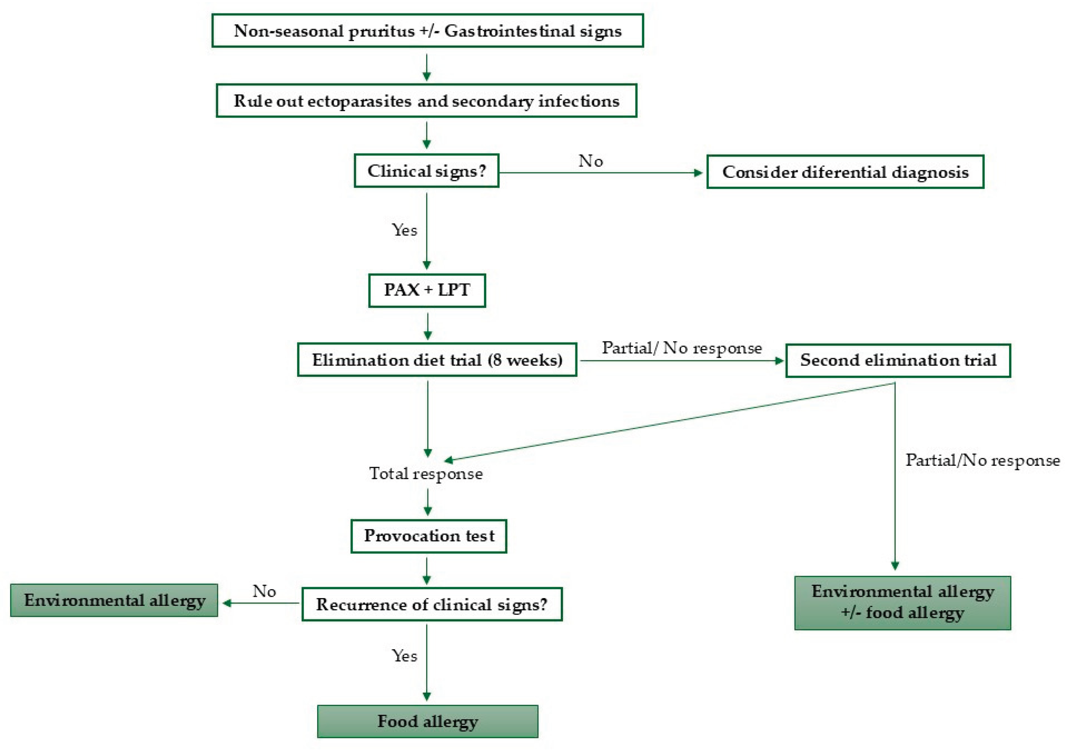
The diagnostic steps proposed in this review are summarized in Figure 1.
4. Therapeutic Approaches
The main goal of food allergy treatment is to avoid the allergens responsible for the clinical signs [16,21].
For long-term management of a food-allergic animal, this can be fed with a novel protein diet, a hydrolyzed diet or another commercial diet lacking offending-allergens [3,16,21]. A homecooked diet can be an option, but it is recommended to be formulated by a veterinary nutritionist, especially regarding cats [11,16,21]. For cats, palatability is also important when choosing a lifelong diet [11].
In case of accidental ingestion of an offending-allergen, a short-term of antipruritic/anti-inflammatory therapy can be used for faster control of the clinical signs [3,21].
Sublingual immunotherapy with food antigens appears to be safe and well tolerated by dogs [41,42]. In a small, comparative, double-blind, randomized, placebo-controlled study, immunotherapy for canine food allergy significantly reduced itching and skin lesions, but further studies are needed to study efficacy and safety in dogs with food allergy [41,42].
The gut-skin axis has been the subject of numerous studies, and dysregulation of the gut microbiota has been associated with a higher prevalence of skin diseases, including allergic skin diseases. Interventions such as dietary changes, probiotics, prebiotics, postbiotics, and fecal microbiota transplantation are promising strategies for modulating the microbiota and reducing the use of antimicrobials, helping to decrease antimicrobial resistance [43]. An example of this are the results of the study by Noli et al. (2023), in which forty allergic dogs with dermatological signs were studied before and after feeding a hypoallergenic diet based on hydrolyzed fish/rice starch, for eight weeks. Fecal samples were collected pre- and pos-diet, and the authors concluded that the rice in this diet modified the microbiota, promoting the development of bacteria responsible to produce short-chain fatty acids, essential for intestinal homeostasis. Dogs fed this diet showed a reduction in the prevalence of Bacteroidetes, a characteristic of dysbiotic dogs [44].
5. Conclusions
Food allergy remains a diagnostic and therapeutic challenge in dogs and cats, mainly due to the absence of reliable biomarkers and the need for time-consuming elimination-provocation trials. However, recent advances are reshaping the future of diagnosing this condition. The development of molecular allergen macroarrays such as the Pet Allergy Xplorer (PAX®) and the lymphocyte proliferation assays will allow a better understanding of the immunopathogenesis of delayed-type reactions, opening new perspectives for precision diagnostics.
From a therapeutic point of view, immunotherapy using food antigens, modulation of the gut–skin axis through pre-, pro- and postbiotics, and the refinement of hydrolyzed protein diets represent promising strategies to improve long-term management. These approaches can also reduce the need of anti-inflammatory drugs and antibiotics, as well as promote the balance of the gut microbiota.
The future of food allergy management in veterinary medicine will depend on the integration of classic clinical approaches with molecular, immunological, and microbiome-based tools. Larger controlled studies are needed to validate these innovative diagnostic and therapeutic options, aiming at a more personalized, efficient, and well-being-focused care for allergic dogs and cats.
Funding
This research was funded by National Funds through FCT - Foundation for Science and Technology under the Project UIDB/05183.
Acknowledgments
The authors wish to thank MED (https://doi.org/10.54499/UIDP/05183/20 20) & CHANGE (https://doi.org/10.54499/LA/P/0121/2020).
Conflicts of Interest
The authors declare no conflicts of interest.
Abbreviations
The following abbreviations are used in this manuscript:
| IgE | Immunoglobulin E |
| PCR | Polymerase Chain Reaction |
| IgG | Immunoglobulin G |
| ELISA | Enzyme-Linked Immunosorbent Assay |
| PAX | Pet Allergy Xplorer |
| LPT | Lymphocyte Proliferation Test |
| PBMCs | Peripheral Blood Mononuclear Cells |
References
- Mueller, R.S.; Unterer, S. Adverse Food Reactions: Pathogenesis, Clinical Signs, Diagnosis and Alternatives to Elimination Diets. Vet. J. 2018, 236, 89–95. [Google Scholar] [CrossRef]
- Halliwell, R.; Pucheu-Haston, C.M.; Olivry, T.; Prost, C.; Jackson, H.; Banovic, F.; Nuttall, T.; Santoro, D.; Bizikova, P.; Mueller, R.S. Feline Allergic Diseases: Introduction and Proposed Nomenclature. Vet. Dermatol. 2021, 32, 8–e2. [Google Scholar] [CrossRef]
- Jackson, H.A. Food Allergy in Dogs and Cats; Current Perspectives on Etiology, Diagnosis, and Management. J Am Vet Med Assoc. 2023. [Google Scholar] [CrossRef]
- Olivry, T.; Mueller, R. Critically Appraised Topic on Adverse Food Reactions of Companion Animals (3): Prevalence of Cutaneous Adverse Food Reactions in Dogs and Cats. BMC Vet. Res. 2017, 4 13. [Google Scholar] [CrossRef]
- Olivry, T.; Mueller, R.S. Critically Appraised Topic on Adverse Food Reactions of Companion Animals (7): Signalment and Cutaneous Manifestations of Dogs and Cats with Adverse Food Reactions. BMC Vet. Res. 2019, 15, 140. [Google Scholar] [CrossRef] [PubMed]
- Roudebush, P. Ingredients and Foods Associated with Adverse Reactions in Dogs and Cats. Vet. Dermatol. 2013, 24, 293–294. [Google Scholar] [CrossRef]
- Mueller, R.; Olivry, T.; Prélaud, P. Critically Appraised Topic on Adverse Food Reactions of Companion Animals (2): Common Food Allergen Sources in Dogs and Cats. BMC Vet. Res. 2016, 7 12. [Google Scholar] [CrossRef] [PubMed]
- Łoś-Rycharska, E.; Gołębiewski, M.; Grzybowski, T.; Rogalla-Ładniak, U.; Krogulska, A. The Microbiome and Its Impact on Food Allergy and Atopic Dermatitis in Children. Postepy Dermatol. Alergol. 2020, 37, 641–650. [Google Scholar] [CrossRef]
- Fujimura, M.; Masuda, K.; Hayashiya, M.; Okayama, T. Flow Cytometric Analysis of Lymphocyte Proliferative Responses to Food Allergens in Dogs with Food Allergy. J. Vet. Med. Sci. Jpn. Soc. Vet. Sci. 2011, 73, 1309–1317. [Google Scholar] [CrossRef] [PubMed]
- Gaschen, F.P.; Merchant, S.R. Adverse Food Reactions in Dogs and Cats. Vet. Clin. North Am. Small Anim. Pract. 2011, 41, 361–379. [Google Scholar] [CrossRef]
- Bryan, J.; Frank, L.A. Food Allergy in the Cat: A Diagnosis by Elimination. J. Feline Med. Surg. 2010, 12, 861–866. [Google Scholar] [CrossRef]
- Picco, F.; Zini, E.; Nett, C.; Naegeli, C.; Bigler, B.; Rüfenacht, S.; Roosje, P.; Gutzwiller, M.E.R.; Wilhelm, S.; Pfister, J.; et al. A Prospective Study on Canine Atopic Dermatitis and Food-Induced Allergic Dermatitis in Switzerland. Vet. Dermatol. 2008, 19, 150–155. [Google Scholar] [CrossRef]
- Mueller, R.S.; Olivry, T. Critically Appraised Topic on Adverse Food Reactions of Companion Animals (6): Prevalence of Noncutaneous Manifestations of Adverse Food Reactions in Dogs and Cats. BMC Vet. Res. 2018, 13 14, 341. [Google Scholar] [CrossRef]
- Hobi, S.; Linek, M.; Marignac, G.; Olivry, T.; Beco, L.; Nett, C.; Fontaine, J.; Roosje, P.; Bergvall, K.; Belova, S.; et al. Clinical Characteristics and Causes of Pruritus in Cats: A Multicentre Study on Feline Hypersensitivity-Associated Dermatoses. Vet. Dermatol. 2011, 22, 406–413. [Google Scholar] [CrossRef] [PubMed]
- Olivry, T.; Mueller, R.; Prélaud, P. Critically Appraised Topic on Adverse Food Reactions of Companion Animals (1): Duration of Elimination Diets. BMC Vet. Res. 2015, 15 11, 225. [Google Scholar] [CrossRef] [PubMed]
- Miller, J.; Simpson, A.; Bloom, P.; Diesel, A.; Friedeck, A.; Paterson, T.; Wisecup, M.; Yu, C.-M. 2023 AAHA Management of Allergic Skin Diseases in Dogs and Cats Guidelines. J. Am. Anim. Hosp. Assoc. 2023, 16 59, 255–284. [Google Scholar] [CrossRef]
- Olivry, T.; Mueller, R. Critically Appraised Topic on Adverse Food Reactions of Companion Animals (5): Discrepancies between Ingredients and Labeling in Commercial Pet Foods. BMC Vet. Res. 2018, 17 14. [Google Scholar] [CrossRef] [PubMed]
- Pagani, E.; Soto Del Rio, M. de L.D.; Dalmasso, A.; Bottero, M.T.; Schiavone, A.; Prola, L. Cross-Contamination in Canine and Feline Dietetic Limited-Antigen Wet Diets. BMC Vet. Res. 2018, 18 14, 283. [Google Scholar] [CrossRef]
- Ricci, R.; Conficoni, D.; Morelli, G.; Losasso, C.; Alberghini, L.; Giaccone, V.; Ricci, A.; Andrighetto, I. Undeclared Animal Species in Dry and Wet Novel and Hydrolyzed Protein Diets for Dogs and Cats Detected by Microarray Analysis. BMC Vet. Res. 2018, 14, 209. [Google Scholar] [CrossRef]
- Cesar, C.G.L.; Marchi, P.H.; Amaral, A.R.; Príncipe, L. de A.; do Carmo, A.A.; Zafalon, R.V.A.; Miyamoto, N.N.; Garcia, N.A.C.R.; Balieiro, J.C. de C.; Vendramini, T.H.A. An Assessment of the Impact of Insect Meal in Dry Food on a Dog with a Food Allergy: A Case Report. Anim. Open Access J. MDPI 2024, 14, 2859. [Google Scholar] [CrossRef]
- Jackson, H.A.; Dembele, V. Conducting a Successful Diet Trial for the Diagnosis of Food Allergy in Dogs and Cats. Vet. Dermatol. 2024, 35, 586–592. [Google Scholar] [CrossRef]
- Lesponne, I.; Naar, J.; Planchon, S.; Serchi, T.; Montano, M. DNA and Protein Analyses to Confirm the Absence of Cross-Contamination and Support the Clinical Reliability of Extensively Hydrolysed Diets for Adverse Food Reaction-Pets. Vet. Sci. 2018, 5, 63. [Google Scholar] [CrossRef]
- Lewis, T.; Moore, G.; Laporte, C.; Daristotle, L.; Frantz, N. Evaluation of Hydrolyzed Salmon and Hydrolyzed Poultry Feather Diets in Restrictive Diet Trials for Diagnosis of Food Allergies in Pruritic Dogs. Front. Vet. Sci. 2025, 23 12. [Google Scholar] [CrossRef] [PubMed]
- Masuda, K.; Sato, A.; Tanaka, A.; Kumagai, A. Hydrolyzed Diets May Stimulate Food-Reactive Lymphocytes in Dogs. J. Vet. Med. Sci. 2020, 82, 177–183. [Google Scholar] [CrossRef]
- Favrot, C.; Bizikova, P.; Fischer, N.; Rostaher, A.; Olivry, T. The Usefulness of Short-course Prednisolone during the Initial Phase of an Elimination Diet Trial in Dogs with Food-induced Atopic Dermatitis. Vet. Dermatol. 2019, 25 30. [Google Scholar] [CrossRef] [PubMed]
- Fischer, N.; Spielhofer, L.; Martini, F.; Rostaher, A.; Favrot, C. Sensitivity and Specificity of a Shortened Elimination Diet Protocol for the Diagnosis of Food-induced Atopic Dermatitis (FIAD). Vet. Dermatol. 2021, 32. [Google Scholar] [CrossRef]
- 27; Olivry, T.; Mueller, R.S. Critically Appraised Topic on Adverse Food Reactions of Companion Animals (9): Time to Flare of Cutaneous Signs after a Dietary Challenge in Dogs and Cats with Food Allergies. BMC Vet. Res. 2020, 16, 158. [Google Scholar] [CrossRef]
- Shimakura, H.; Kawano, K. Results of Food Challenge in Dogs with Cutaneous Adverse Food Reactions. Vet. Dermatol. 2021, 28 32, 293–e80. [Google Scholar] [CrossRef]
- Fernandez-Lozano, C.; Mas-Fontao, A.; Auxilia, S.; Welters, M.; Olivrī, A.; Mueller, R.; Olivry, T. Evaluation of a Direct Lymphocyte Proliferation Test for the Diagnosis of Canine Food Allergies with Delayed Reactions after Oral Food Challenge. Vet. Dermatol. 2024, 36, 433–442. [Google Scholar] [CrossRef]
- Tinsley, J.; Griffin, C.; Sheinberg, G.; Griffin, J.; Cross, E.; Gagné, J.; Romero, A. An Open-label Clinical Trial to Evaluate the Efficacy of an Elemental Diet for the Diagnosis of Adverse Food Reactions in Dogs. Vet. Dermatol. 2023, 30 35. [Google Scholar] [CrossRef]
- Martins, L.M.; Campos, I.; Antunes, C.; Costa, A.; Valdevira, A.; Bento, O. Testes intradérmicos e imunodots podem ser úteis no diagnostico de alergia canina a carne – Intradermal testing and immunodot may be useful in diagnosing dog allergy to meat. Rev Port Imunoalergologia 2019, 27(1), 21–27. [Google Scholar] [CrossRef]
- Johansen, C.; Mariani, C.; Mueller, R.S. Evaluation of Canine Adverse Food Reactions by Patch Testing with Single Proteins, Single Carbohydrates and Commercial Foods. Vet. Dermatol. 2017, 32 28, 473–e109. [Google Scholar] [CrossRef]
- Possebom, J.; Cruz, A.; Gmyterco, V.C.; de Farias, M.R. Combined Prick and Patch Tests for Diagnosis of Food Hypersensitivity in Dogs with Chronic Pruritus. Vet. Dermatol. 2022, 33 33, 124–e36. [Google Scholar] [CrossRef] [PubMed]
- Maina, E.; Matricoti, I.; Noli, C. An Assessment of a Western Blot Method for the Investigation of Canine Cutaneous Adverse Food Reactions. Vet. Dermatol. 2018, 29, 217–e78. [Google Scholar] [CrossRef]
- Favrot, C.; Linek, M.; Fontaine, J.; Beco, L.; Rostaher, A.; Fischer, N.; Couturier, N.; Jacquenet, S.; Bihain, B.E. Western Blot Analysis of Sera from Dogs with Suspected Food Allergy. Vet. Dermatol. 2017, 28, 189. [Google Scholar] [CrossRef] [PubMed]
- Mueller, R.; Olivry, T. Critically Appraised Topic on Adverse Food Reactions of Companion Animals (4): Can We Diagnose Adverse Food Reactions in Dogs and Cats with in Vivo or in Vitro Tests? BMC Vet. Res. 2017, 36 13, 275. [Google Scholar] [CrossRef]
- Lam, A.T.H.; Johnson, L.N.; Heinze, C.R. Assessment of the Clinical Accuracy of Serum and Saliva Assays for Identification of Adverse Food Reaction in Dogs without Clinical Signs of Disease. J Am Vet Med Assoc. 2019, 37. [Google Scholar] [CrossRef]
- Plantinga, E.; Leistra, M.H.G.; Sinke, J.; Vroom, M.W.; Savelkoul, H.; Hendriks, W.H. Measurement of Allergen-Specific IgG in Serum Is of Limited Value for the Management of Dogs Diagnosed with Cutaneous Adverse Food Reactions. Vet. J. 2017, 220. [Google Scholar] [CrossRef]
- Olivry, T.; Fontao, A.; Aumayr, M.; Ivanovova, N.; Mitterer, G.; Harwanegg, C. Validation of a Multiplex Molecular Macroarray for the Determination of Allergen-Specific IgE Sensitizations in Dogs. Vet. Sci. 2024, 11, 482. [Google Scholar] [CrossRef]
- Suto, A.; Suto, Y.; Onohara, N.; Tomizawa, Y.; Yamamoto-Sugawara, Y.; Okayama, T.; Masuda, K. Food Allergens Inducing a Lymphocyte-Mediated Immunological Reaction in Canine Atopic-like Dermatitis. J. Vet. Med. Sci. 2015, 77, 251–254. [Google Scholar] [CrossRef]
- Maina, E.; Cox, E. A Double Blind, Randomized, Placebo Controlled Trial of the Efficacy, Quality of Life and Safety of Food Allergen-Specific Sublingual Immunotherapy in Client Owned Dogs with Adverse Food Reactions: A Small Pilot Study. Vet. Dermatol. 2016, 41 27, 361–e91. [Google Scholar] [CrossRef] [PubMed]
- Maina, E.; Pelst, M.; Hesta, M.; Cox, E. Food-Specific Sublingual Immunotherapy Is Well Tolerated and Safe in Healthy Dogs: A Blind, Randomized, Placebo-Controlled Study. BMC Vet. Res. 2017, 42 13, 25. [Google Scholar] [CrossRef] [PubMed]
- Lagoa, T.; Martins, L.; Queiroga, M.C. Microbiota Modulation as an Approach to Prevent the Use of Antimicrobials Associated with Canine Atopic Dermatitis. Biomedicines. 2025, 43 13, 2372. [Google Scholar] [CrossRef] [PubMed]
- Noli, C.; Varina, A.; Barbieri, C.; Pirola, A.; Olivero, D. Analysis of Intestinal Microbiota and Metabolic Pathways before and after a 2-Month-Long Hydrolyzed Fish and Rice Starch Hypoallergenic Diet Trial in Pruritic Dogs. Vet. Sci. 2023, 10, 478. [Google Scholar] [CrossRef]
Figure 1.
Proposed diagnostic algorithm for food allergy in dogs and cats.
Disclaimer/Publisher’s Note: The statements, opinions and data contained in all publications are solely those of the individual author(s) and contributor(s) and not of MDPI and/or the editor(s). MDPI and/or the editor(s) disclaim responsibility for any injury to people or property resulting from any ideas, methods, instructions or products referred to in the content. |
© 2025 by the authors. Licensee MDPI, Basel, Switzerland. This article is an open access article distributed under the terms and conditions of the Creative Commons Attribution (CC BY) license (http://creativecommons.org/licenses/by/4.0/).
Copyright: This open access article is published under a Creative Commons CC BY 4.0 license, which permit the free download, distribution, and reuse, provided that the author and preprint are cited in any reuse.


