Preprint
Article

This version is not peer-reviewed.

Identification of Fungal Pathogens of Chinese Chestnut Fruit Rot and Analysis of Resistance Differences among Major Cultivars

Submitted:

01 December 2025

Posted:

02 December 2025

You are already at the latest version

Abstract

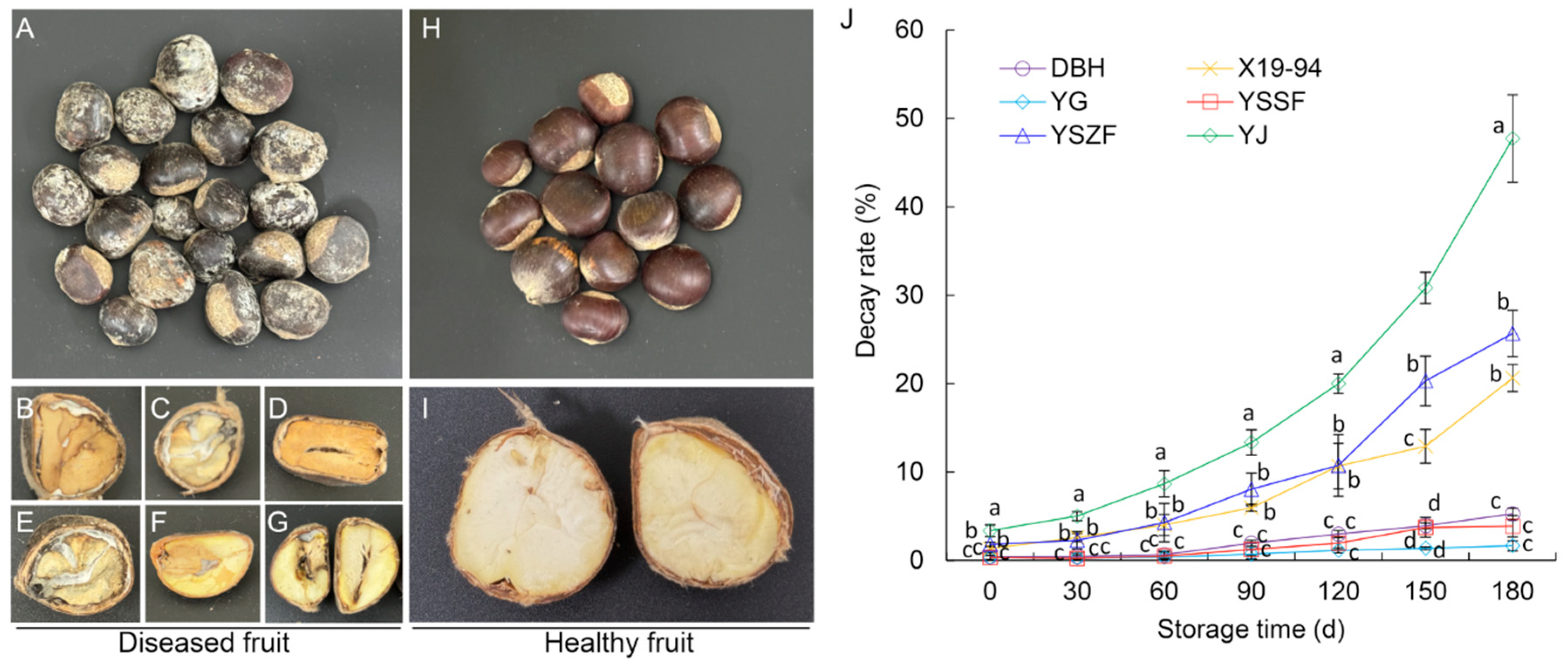
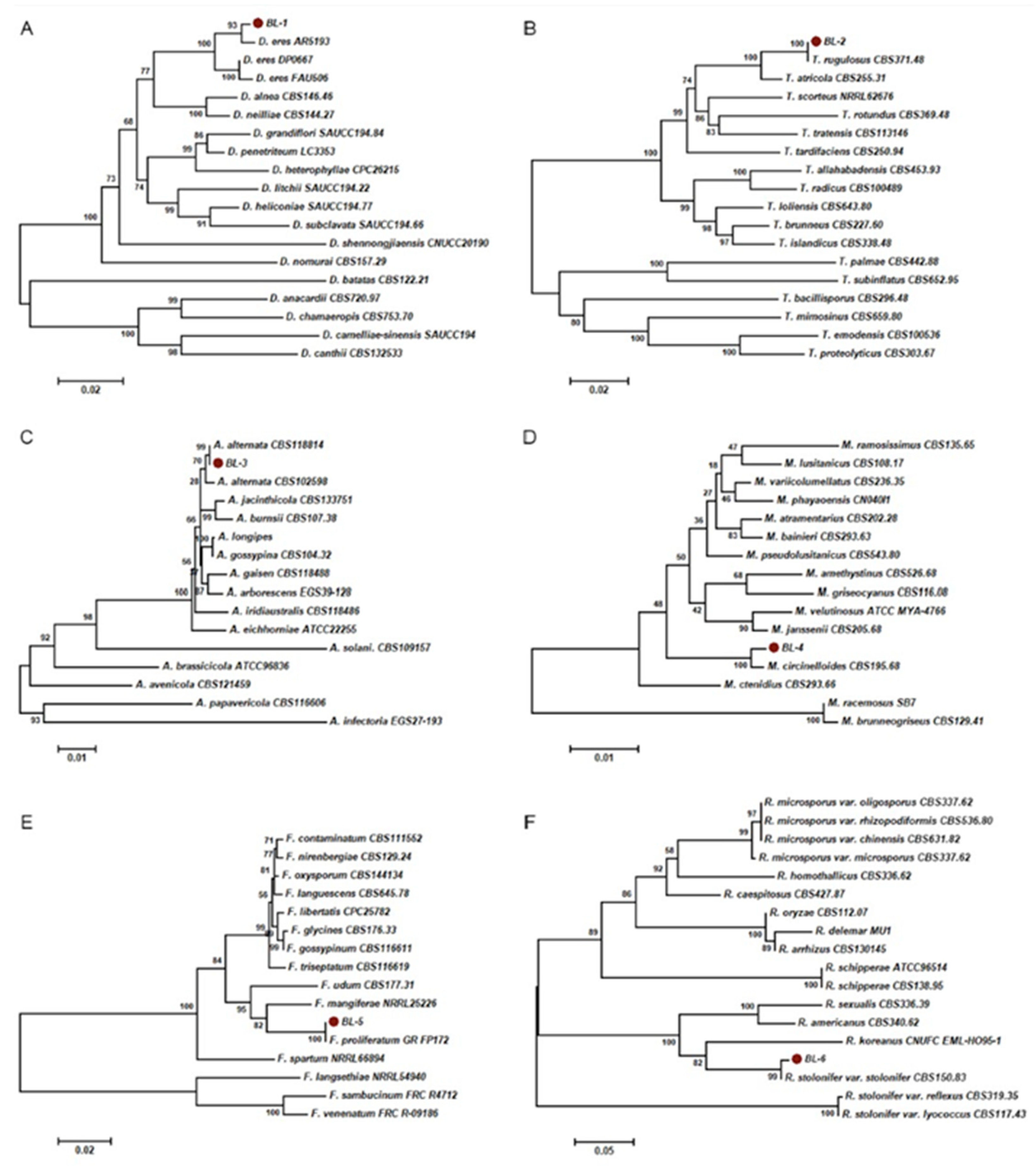
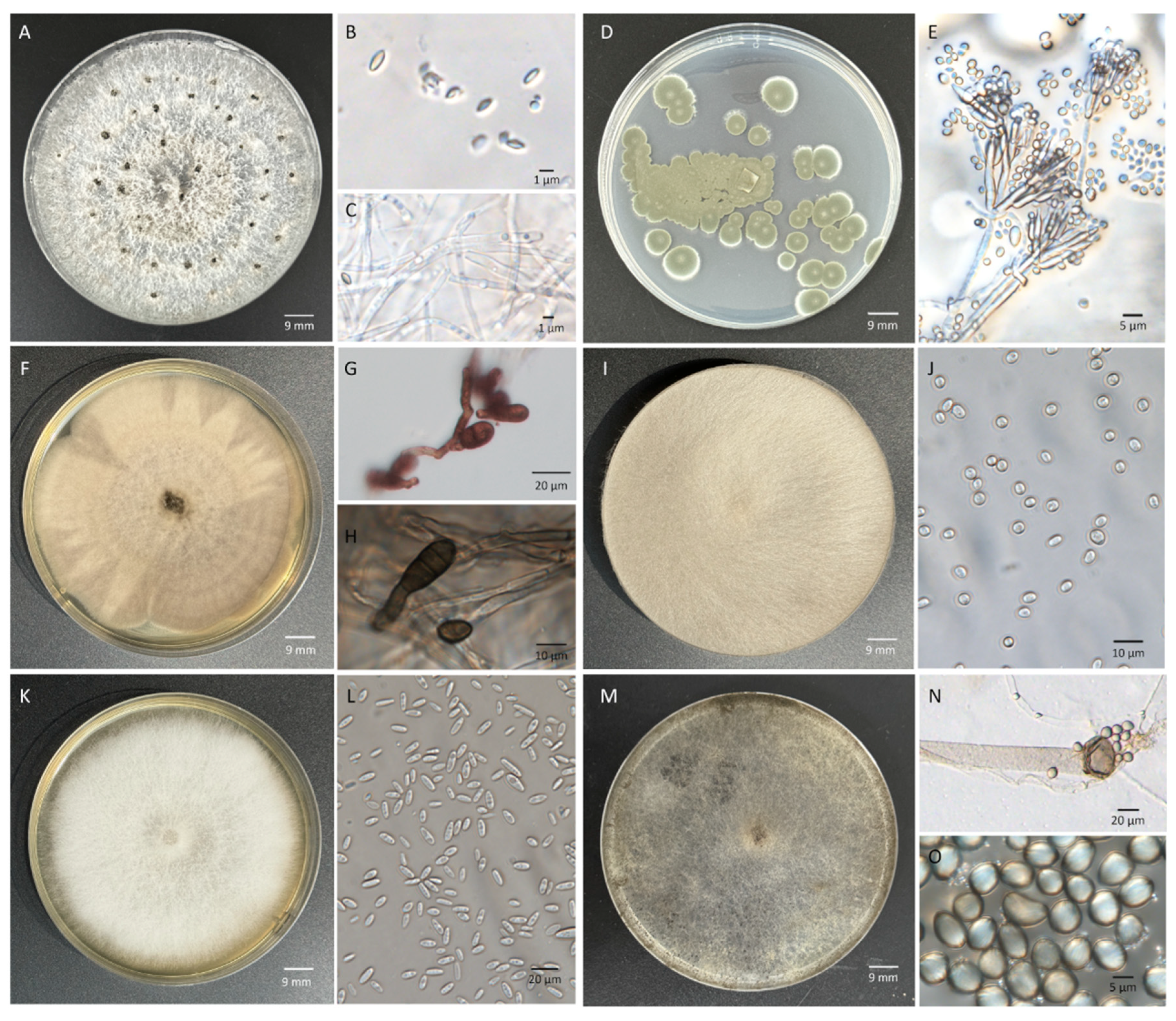
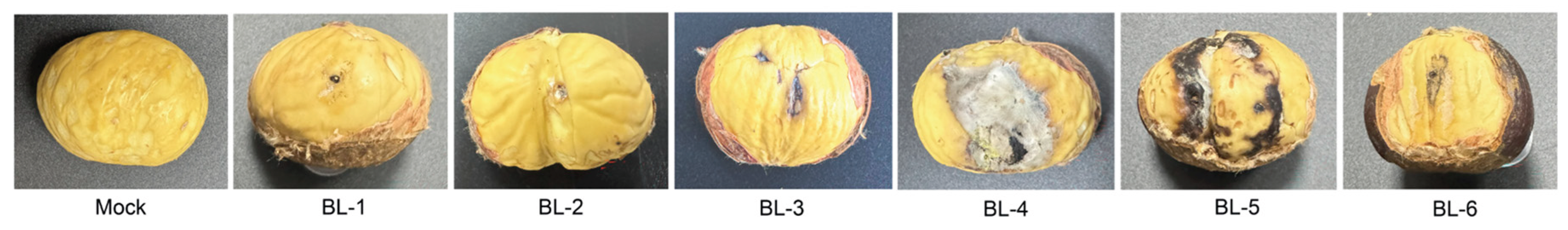
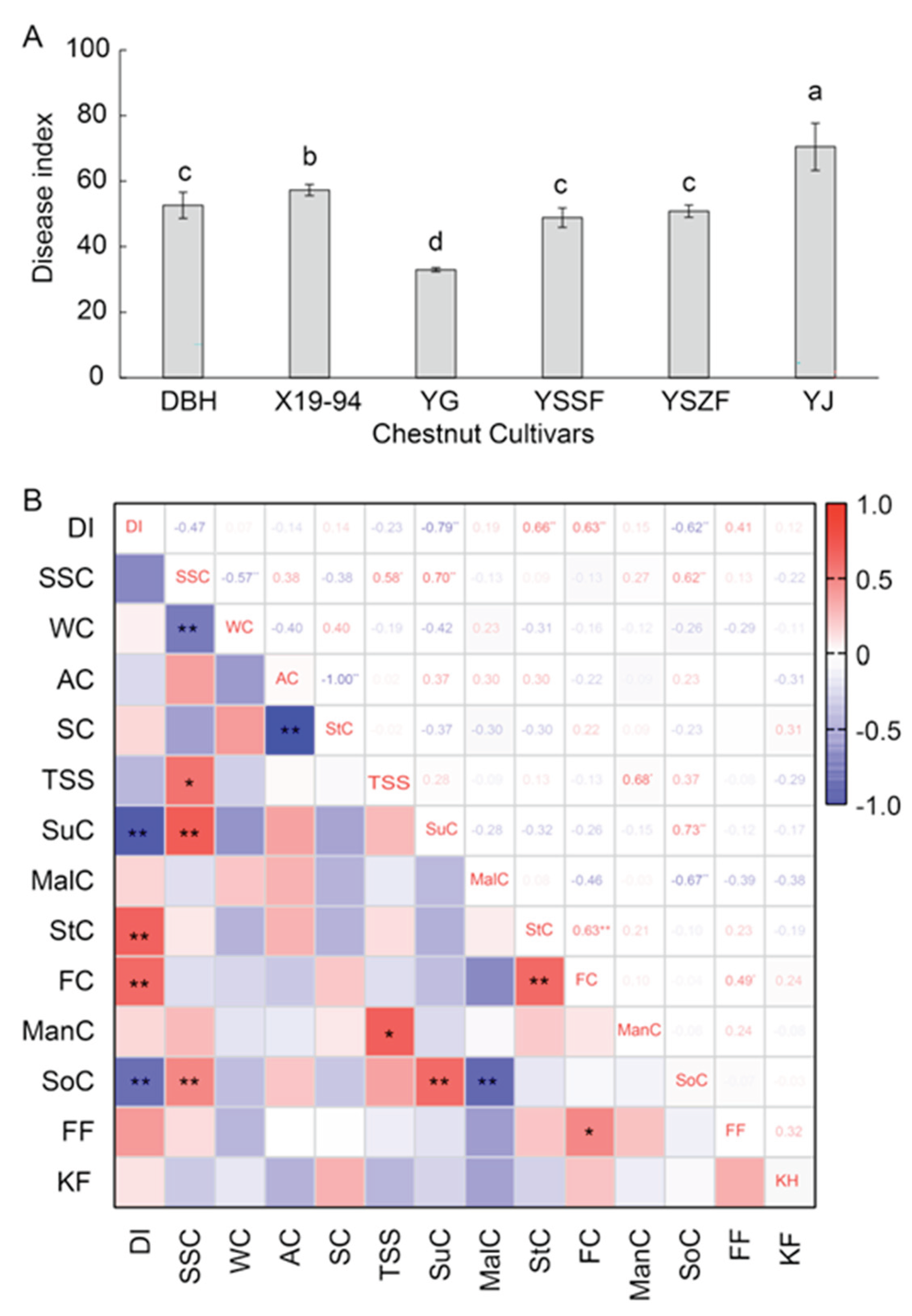
This study aimed to identify fungal species causing fruit rot of chestnut (Castanea mollissima) in Hebei Province, China and analyze the resistance differences among major cultivars. A total of 220 fungal isolates were obtained from healthy and diseased kernels, which were classified into six distinct genera. Based on both morphological and molecular analyses, these isolates were identified as Diaporthe eres (48.6% isolation frequency), Talaromyces rugulosus (22.3%), Alternaria alternata (10.5%), Mucor circinelloides (9.5%), Fusarium proliferatum (5.5%), and Rhizopus stolonifer var. stolonifer (3.6%). Among these, D. eres was firstly reported to cause fruit rot on C. mollissima in China. Moreover, disease resistance evaluation of major cultivars showed significant differences: YG, YSSF, and DBH exhibited strong resistance under both natural conditions (with 1.7% to 5.3% DI after 180 days storage) and artificial inoculation (with 33.0±0.6 to 52.6±4.0 DI); while YJ was highly susceptible (with 47.7% decay incidence and 70.5±7.2 DI). Correlation analysis revealed that the disease index was negatively correlated with sucrose and sorbitol contents, but positively correlated with stachyose and fructose contents. This study advances the understanding of postharvest chestnut fruit rot, and provide a theoretical basis for breeding resistant cultivars and developing control strategies to mitigate losses and ensure food safety.

Keywords: 
;  ;  ;  ;  ;  ;  

1. Introduction

The Chinese chestnut (Castanea mollissima Blume), a key economic forest species, is widely cultivated throughout China and was introduced to Europe in the 19th century [1]. The nuts are rich in starch, protein, functional polysaccharides, essential fatty acids, vitamins, and minerals, which impart various health benefits, such as lowering cholesterol, preventing obesity and diabetes, combating tumors, and enhancing immunity [2]. This importance is reflected in China's status as the world's largest producer, with an annual yield exceeding 1.52 million tons since 2023 (FAOSTAT, http://faostat.fao.org). However, chestnuts are prone to rot and deteriorate due to microbial infection during the harvest and postharvest storage periods, leading to significant quality and production losses. In China, improper storage of chestnuts results in annual losses amounting to 20–30% of the total production, and even exceeds 50% in several production areas by the late storage [3]. Moreover, pathogens also produce mycotoxins (e.g., aflatoxins, ochratoxins, fumonisins, and T-2/HT-2 toxins), posing a serious food safety threat [4]. Hence, it is crucial to clarify the incidence of rot disease and the species of pathogens in different chestnut cultivars for developing targeted control strategies.
Various pathogens have been reported to cause chestnut fruit rot during preharvest and postharvest storages [2]. Apart from two bacterial species, Pseudomonas vesicularis [5] and Pse. syringae [6], the disease primarily results from complex infections of multiple pathogenic fungi, many of which exhibit latent colonization. Reported fungal genera include Alternaria, Botryosphaeria, Colletotrichum, Diaporthe, Fusarium, Penicillium, Rhizopus, Trichothecium, Sclerotinia, Gnomoniopsis, Phomopsis, Acrospeira, Aspergillus, Botrytis, Diplodia and Parvum [4,7,8]. A total of 308 fungal operational taxonomic units were detected during chestnuts postharvest storage in Italy chestnut fruits, with new contaminants emerging in specific phases such as the ‘cold bath’ and ‘storage’ [4]. Nevertheless, the identity and pathogenicity of the predominant fungi among them have not been identified.
Furthermore, pathogen types and community structure are strongly influenced by geographic origin and cultivar differences [9]. For instance, G. smithogilvyi was first reported as a nut rot pathogen in Australian C. sativa [10]; whereas, G. clavulata and G. paraclavulata were documented only in the USA, and G. daii was described as a novel pathogen affecting C. mollissima in China [7]. Additionally, cultivar characteristics (e.g., skin structure, fruit firmness, and sugar content) also significantly influence pathogen infection and spread [11]. However, current studies on the diversity of chestnut fruit pathogens remains narrowly focused on single cultivars and specific production regions, neglecting the resistance differences among various chestnut cultivars and the mechanisms of pathogens complex-infections, thereby complicating the development of effective control strategies.
Chestnuts from the Yanshan area of Hebei Province, as the largest contiguous cultivation belt in China, hold a prestigious position in the global market [12]. During the surveys of chestnut rot conducted in Hubei provinces in China, typical fruit rot symptoms were observed. Nonetheless, the major pathogenic fungal species in this area remain unknown. Our aim in this study was to identify the pathogenic species of chestnut fruit rot and to analyze the disease resistance differences among major cultivars in this region. These findings will offer a theoretical basis to inform the breeding of rot-resistant chestnut cultivars and the creation of sustainable postharvest strategies, which are crucial for minimizing losses and ensuring food safety.

2. Materials and Methods

2.1. Disease Surveys and Isolation of Fungi

Chestnuts cv. Dabanhong (DBH), Yanguang (YG), Yanshan Shuofeng (YSSF), Yanshan Zaofeng (YSZF), Yanjing (YJ), and Yanli1 (X19-94) are the main cultivars in the Yanshan area. All these cultivars were grafted onto three-year-old YSZF seedling rootstocks in 2004, with five trees per cultivar planted at a 3 m × 3 m spacing and managed under standard horticultural practices. The fruits were separately harvested at the mature stage based on uniform size and free of defects in Chestnut Germplasm Repository of the Changli Institute of Pomology, Hebei Academy of Agriculture and Forestry Sciences (119°15’ E, 39°72’ N) [13]. The collected fruits were packaged in PE bags and stored at 4℃ for further experiment.
Chestnut rot development was assessed at 30-day intervals over a 180-day cold storage (4℃) period. The chestnut decay incidence (equation 1) was recorded as the percentage of decayed nuts among 200 randomly selected fruits per cultivar, with three replicates. Chestnuts exhibiting typical symptoms were collected and photographed. Additionally, five healthy fruits were randomly sampled at each time point for tissue isolation.
D e c a y   i n c i d e n c e = N u m b e r s   o f   d e c a y   c h e s t n u t s T o t a l   n u m b e r s   o f   c h e s t n u t s × 100 %
Fungi were isolated following Chen et al. (2023) [14] with minor modifications. Tissues segments (5 mm × 5 mm) were excised from both healthy and decayed chestnuts at each sampling time. The tissues were surface-sterilized in 70 % ethanol for 1 min, followed by 1 % sodium hypochlorite for 5 min, rinsed thoroughly with sterile distilled water, and placed on potato dextrose agar (PDA). After incubated at 25℃ in the dark, isolates were purified by transferring single hyphal tips to new PDA plates. The resulting cultures were incubated were incubated under the same conditions for approximately 7 days and subsequently stored at 4°C [15].

2.2. Morphological Identification and Characterization

Morphological characterization of the isolates was performed on sporulating cultures grown on PDA at 25℃ under a 12-h light regime for 7 days. Cultural characteristics, including colony color and texture, were documented. Colony diameters were measured daily to determine growth rates. Micromorphological features were observed using an Olympus BX53 optical microscope equipped with an Olympus DP74 color camera (Olympus, Tokyo, Japan), and over 200 conidia were randomly measured.

2.3. DNA Extraction and Phylogenetic Analysis

Genomic DNA was extracted from 7-day-old mycelium grown on PDA using a Super Plant Genomic DNA DP360 Kit (Tiangen Biotech, Beijing, China) according to the manufacturer’s instructions. The rDNA-ITS (ITS) region was amplified with primers ITS1/ITS4 for preliminary identification [16]. For precise species identification, multiple gene regions (Table S1) were amplified and sequenced [16,17,18,19]. The PCR mixture and conditions for all gene regions followed established methods [7,14]. Amplification products were verified by 1% agarose gel electrophoresis and sequenced by a commercial provider (Beijing Tsingke Biotech Co., Ltd., Beijing, China).
Sequence queries were conducted using the NCBI BLASTn algorithm (http://www.ncbi.nlm.nih.gov/genbank). Phylogenetic analyses incorporated additional sequences from GenBank (Tables S2–S6). Sequences for each locus were aligned with ClustalX [20], manually refined in MEGA7.0 [21], then concatenated in BioEdit [22]. Phylogenetic trees were reconstructed using Neighbor-joining (NJ) method in MEGA7.0.

2.4. Pathogenicity Study

Conidia were prepared according to Jia et al. (2003) [23] with minor modifications. After incubation on PDA at 25 °C for 14 days, conidia were harvested by scraping into sterile 0.02% (v/v) Tween 80 solution. The suspension was filtered through three layers of mirror cleaning paper to remove mycelial and agar. The conidial concentration was determined and adjusted to 2×10⁶ conidia/mL using a hemocytometer. Fresh conidial suspensions were prepared individually for each fungal species.
For pathogenicity assays [2], healthy and uniformly sized chestnuts (cv. YSZF) were surface-sterilized by immersion in 1% sodium hypochlorite for 10 min, rinsed thoroughly with sterile distilled water, and air-dried on sterile filter paper. A single wound (0.5 mm diameter, 5 mm depth) was made on each chestnut. Each wound was inoculated with 20 μL of the conidial suspension of a single isolate, with equal volume of sterile 0.02% (v/v) Tween 80 solution serving as the control. All fruits were placed in a moist chamber and kept at 25℃ for 15 days under a 12-h-light/12-h-dark photoperiod. The experiment was conducted twice with three replicates (10 chestnuts each) per fungi species.

2.5. Assessing of Disease Severity on Chestnut Cultivars

A combined inoculum was prepared by mixing equal volumes of fresh conidial suspensions (2×10⁶ conidia/mL) from representative strains of each pathogenic species. The six chestnut cultivars, DBH, YG, YSSF, YSZF, YJ, and X19-94 stored at 4℃ for 40 days after harvest, were surface-sterilized and inoculated with 20 μL of the combined inoculum, following the incubation conditions as described above.
Disease severity was quantified 15 days after inoculation by measuring lesion diameters. Each chestnut was assessed severity on a 0-9 scale, in which 0, no lesion; 1, lesion≤1.5 mm; 3, 1.5 mm<lesion≤5 mm; 5, 5 mm<lesion≤10 mm; 7, 10 mm<lesion≤20 mm; and 9, lesion>20 mm or complete rot. The disease index (DI) was calculated using the following equation (Equation (2)):
D I = X 0 × 0 + X 1 × 1 + X 3 × 3 + X 5 × 5 + X 7 × 7 + X 9 × 9 X 0 + X 1 + X 3 + X 5 + X 7 + X 9 × 100
in which X0, X1, X3, X5, X7, and X9 represent the number of chestnuts with severity scores of 0, 1, 3, 5, 7, and 9, respectively. Each treatment investigated 50 fruits, and the experiment was conducted twice with three replicates per treatment.

2.6. Statistical Analysis

All data were expressed as means ± standard deviation and analyzed with three replicates per treatment at each time point. For each time, statistical analysis was performed using one-way analysis of variance (ANOVA) following Duncan’s multiple range test at a 95% confidence interval for mean comparisons. The correlation was evaluated with the Pearson correlation coefficient (r) following the quality data of chestnuts in our previous studies [24]. All analyses were conducted using SPSS software (version 22.0, IBM Corp., Armonk, NY, USA).

3. Results

3.1. Disease Symptom and Development During Postharvest Storage

During cold storage, disease chestnuts exhibiting a range of typical symptoms were observed across all cultivars. Superficially, the outer and inner seed coats generally remained intact, though the inner coats occasionally darkened or brownish-black. Internally, dark brown to black necrotic lesions developed on the kernel surface, often penetrating deeply into the tissue. Cross-sections frequently revealed grayish-white to dark gray, streaked cavities. In severe cases, the shells lost their luster and were covered with conspicuous grayish-white or olive-green mold. The kernels became shriveled and exhibited either dry or soft rot, with internal tissues turning yellowish-brown or grayish-black (Figure 1A–G). In contrast, healthy chestnuts displayed plump, glossy shells and firm kernels of uniform milky-white to pale-yellow color (Figure 1H,I).
Significant differences in decay development were observed among the chestnut cultivars over 180-day storage period. At the initiation of storage (0 d), cultivar YJ showed the highest decay incidence (3.4%), followed by YSZF (1.8%) and X19-94 (1.3%), while DBH, YSSF, and YG were all below 0.5%. As storage progressed, decay incidence increased in all cultivars, though the rate of increase and final severity varied markedly. By day 180, YJ reached the highest incidence (47.7%), whereas YSZF and X19-94 showed moderate levels of 25.7% and 20.6%, respectively. In contrast, DBH, YSSF, and YG consistently maintained the lowest decay incidence throughout storage, with final incidence ranging only from 1.7% to 5.3% at 180 days (Figure 1J). These results indicate that YG, YSSF, and DBH are relatively resistant to chestnuts decay, whereas YSZF and X19-94 exhibit intermediate susceptibility, and YJ is highly susceptible.

3.2. Fungal Isolation and Identification

A total of 220 fungal isolates (23 from healthy kernels and 197 from diseased kernels) were obtained from 35 healthy and 55 diseased samples collected during cold storage. Preliminary identification was based on colony morphology, conidial characteristics on PDA, and ITS sequence analysis. As a result, all isolates were classified into six genera: Diaporthe (48.6%), Talaromyces (22.3%), Alternaria (10.5%), Mucor (9.5%), Fusarium (5.5%), and Rhizopus (3.6%). All genera were isolated from both healthy and diseased tissues. Six representative isolates (BL-1 to BL-6), representing each of the six genera, were selected for further study.

3.3. Molecular Identification and Phylogenetic Analysis

The six representative isolates were sequenced and subjected to phylogenetic analysis using the Neighbor-joining (NJ) method with either combined muti-locus sequences or the ITS region alone. As shown in Figure 2A, a multi-locus phylogenetic analysis of BL-1 isolate and related Diaporthe species was processed based on ITS, TUB, TEF, CAL, and HIS gene regions, with a total alignment length of 2312 characters including gaps. BL-1 clustered with reference strains of Diaporthe eres (including AR5193) with 93% bootstrap-support, confirming its identification as D. eres. For BL-2, multi-locus analysis of ITS, TUB, and RPB2 (1852 characters) showed clustering with Talaromyces rugulosus CBS371.48 with 100% bootstrap-support, identifying it as T. rugulosus (Figure 2B). Similarly, BL-3 (2110 characters from ITS, GAPDH TEF, and RPB2) clustered with Alternaria alternata CBS118814 under 99% bootstrap-support value, confirming its identification as A. alternata (Figure 2C). In the case of BL-5, BLAST analysis of the ITS region showed 99.79% sequence similarity to Fusarium proliferatum (isolate MT-S-1, IGPEB-SH11, DHHJYK2 et al.), F. oxysporum (A221), and F. fujikuroi (FPM27); phylogenetic analysis based on combined RPB2 and TEF sequences (1426 characters) further placed the isolate within the F. proliferatum clade with 100% bootstrap support relative to the reference stain GR_FP172, establishing its identity as F. proliferatum (Figure 2E). For BL-4 and BL-6, ITS sequences showed up to 97% identity with reference sequences of Mucor circinelloides and Rhizopus stolonifer in the NCBI database, respectively. Phylogenetic analysis of ITS region confirmed that BL-4 formed a well-supported clade with M. circinelloides CBS526.68 (Figure 2D), while BL-6 clustered with R. stolonifer var. stolonifera CBS150.83 under 99% NJ bootstrap-support (Figure 2F). These results support the identification of BL-4 as M. circinelloides and BL-6 as R. stolonifer var. stolonifera.

3.4. Morphology and Taxonomy

Diaporthe eres Nitschke (1870) [14].
Index Fungorum No.: 802934.
Description: Colonies on PDA at 25 °C under a 12-h light regime exhibited a growth rate of 14.8±0.6 mm/d. Aerial mycelium initially was white and fluffy, later developed white-cream to light brown, cottony, mycelial mats, and eventually produced gray to black pycnidia. Alpha conidia were hyaline, aseptate, ellipsoidal to fusiform, measuring 3.3-8.5 μm × 0.9-3.1 μm (mean±SD = 5.2±1.7 μm × 2.1±0.8 μm, n=200). Beta conidia were not observed. Vegetative hyphae were hyaline, smooth-walled, septate, and branched (Figure 3A–C).
Material examined: China, Hebei Province, Qinhuangdao city, Changli county, from healthy and rot diseased fruits of Chinese chestnuts (Castanea mollissima), H.J. Xu, culture: BL-1.
Talaromyces rugulosus (Thom) Samson, N. Yilmaz, Frisvad & Seifert (2011) [25]
Index Fungorum No.: 560672.
Description: Colonies on PDA were plane with white margin, regular of edge, green to dull green conidia pile, exudates and soluble pigment absent at 25 °C. Hyphae appeared septate, smooth to coarse, and branched. Conidiophores were biverticillate with smooth-walled stipes and unswollen tops, 3-6 metulae (6.5-13.5 μm × 2.3-4.0 μm), 3-8 acerose phialides (7.7-14.0 μm × 1.3-2.5 μm) per metulae. Conidia were gray green in color, smooth walled, ellipsoidal or round, 1.5-5.5 µm × 1.5-3.0 µm (3.0±0.9 µm × 2.4±0.4 µm) (n = 200). Ascomata were not observed (Figure 3D,E).
Material examined: China, Hebei Province, Qinhuangdao city, Changli county, from healthy and rot diseased fruits of Castanea mollissima, representative isolate culture: BL-2.
Alternaria alternata (Fr.) Keissl. (1912) [26]
Index Fungorum No.: 119834.
Description: Colonies on PDA at 25 °C were initially grayish-white, later developing a pale gray to gray-brown base, occasionally with brownish center zones. Abundant aerial mycelium extended from the center, forming distinct radial surface patterns. The growth rate was 10.2±1.5 mm/d. Conidiophores were pale to brown, smooth, septate, solitary or clustered, straight or flexuous, and 32.6-85.3 µm × 1.8-4.2 µm (59.9.2±13.8 µm × 3.1±0.7 µm, n=50). Conidia were ellipsoid to ovoid or obclavate to obpyriform, brown, smooth, measuring 8.3-27.5 µm × 4.2 to 14.5 µm (18.7±5.6 µm × 11.4±2.6 µm, n = 200), with 0-2 longitudinal and 1-5 transverse septa, beakless or bearing a light-brown, columnar to tapered beak (Figure 3F–H).
Material examined: China, Hebei Province, Qinhuangdao city, Changli county, from healthy and rot diseased fruits of Castanea mollissima, culture: BL-3.
Mucor circinelloides Tiegh. (1875) [27]
Index Fungorum No.: 198947
Description: Colonies grew rapidly and reach a height of up to 21.1±1.6 mm/d on PDA at 25 °C. Abundant aerial mycelium exhibited velvety texture and light gray, most frequently reaching the lid of the petri dish. Sporangiospores were globose or globose columellae, hyaline, smooth, small, 2.6-5.8 µm × 2.2-5.5 µm (4.0±0.8 µm × 3.3±0.7 µm, n = 200) (Figure I–J).
Material examined: China, Hebei Province, Qinhuangdao city, Changli county, from healthy and rot diseased fruits of Castanea mollissima, culture: BL-4.
Fusarium proliferatum (Matsush.) Nirenberg (1976) [28]
Index Fungorum No.: 362256
Description: Colonies produced abundant, white, villous aerial mycelium, with regular margins and no zonation or violet pigmentation. The growth rate was 11.1±0.9 mm/d on PDA at 25 °C. Conidia were usually formed in chains, 0-septate, and club shaped with a flattened base. Microconidia were abundant, 0-septate, oval, elliptical or club, 7.4-15.4 µm × 2.3-7.4 µm (11.5±2.0 µm × 4.8±1.0 µm, n = 200). Macroconidia were slender, sickle or straight, 1-3 septate, 25.7-48.5 µm × 1.8-3.9 μm (38.8±4.4 µm × 2.6±0.4 µm) (Figure 3K,L).
Material examined: China, Hebei Province, Qinhuangdao city, Changli county, from healthy and rot diseased fruits of Castanea mollissima, culture: BL-5.
Rhizopus stolonifer var. stolonifer (Ehrenb.) Vuill. (1902) [29]
Index Fungorum No.: 417250
Description: Colonies formed abundant aerial mycelium, dense and fluffy, initially white, and then gray-white to black-brown on PDA at 25 °C. The growth rate was rapid, 37.7±2.5 mm/d, and reaching the lid of the petri dish after cultured 24 h. Hyphae were broad, ribbon-like, branched, coenocytic, and aseptate. Sporangiophores were unbranched, erect, and terminating in sporangia. Sporangia were spherical, initially white, and then black, contained a large number of sporangiospores and columella. Sporangium spores were non-motile, light to dark brown, in various forms (spherical, ellipsoidal and angular), non-septum, with continuous and obvious ridges on the surface along the spores, 6.4-9.6 µm × 5.8-8.4 μm (8.3±1.0 µm × 6.7±0.7 µm, n=200) (Figure 3M–O).
Material examined: China, Hebei Province, Qinhuangdao city, Changli county, from healthy and rot diseased fruits of Castanea mollissima, culture: BL-6.

3.5. Pathogenicity Study

After 15 days of inoculation on wounded nuts (cv. YSSF), all six isolates, namely D. eres (BL-1), T. rugulosus (BL-2), A. alternata (BL-3), M. circinelloides (BL-4), F. proliferatum (BL-5), and R. stolonifer (BL-6), caused brown to black rot symptoms, with the artificially infected incidence of 100%. None of the control nuts showed any disease or symptoms (Figure 4). The pathogens were re-isolated from the inoculated chestnuts after showing typical symptoms, satisfying Koch’s postulates.

3.6. Disease Severity of Virous Chestnut Cultivars

To evaluate the resistance of various chestnut cultivars to fungal infection, disease indices were assessed following inoculation with the mixed fungi. As shown in Figure 5A, significant differences in disease severity were observed among the chestnut cultivars at 15 days post-inoculation (Figure 5A). The YG cultivar exhibited the strongest resistance, with the lowest disease index of 33.0±0.6. For cultivar YSSF, YSZF and DBH, the disease index had slightly increased (48.9±3.0, 50.8±1.9, and 52.6±4.0, respectively), followed by X19-94 (57.3±1.7). In contrast, the disease index of cultivar YJ was highest (70.5±7.2), indicating it was a relative susceptible cultivar.
To better analyze relative factors of quality that influence chestnut rot disease, correlation analysis was analyzed between the disease index of chestnuts infected with mixed fungi and soluble sugars, and firmness (Table S8) [24]. As shown in Figure 5, the disease index of chestnuts showed positive correlations with stachyose content (StC) and fructose content (FC) (P < 0.01 and r > 0.5). However, it had negative correlations (P < 0.01 and r < -0.5) with sucrose content (SuC) and sorbitol content (SoC), and showed no significant correlation with respiratory intensity.

4. Discussion

The accurate identification of fungal pathogens has shifted from morphology-based approaches toward an integrated taxonomic approach centered on multi-locus phylogenetic analysis [30]. This transition addresses the limitations of relying solely on the ITS region as a universal barcode [31]. In this study, ITS variability is insufficient to distinguish closely related species with distinct ecological or pathogenic traits in genera such as Diaporthe, Talaromyces, Alternaria, and Fusarium. By combining gene loci with different evolutionary rates—e.g., ITS, GAPDH, TEF1, and RPB2—the multi-locus phylogenetic analysis significantly enhances both the resolution and statistical support of the phylogenetic tree, thereby enabling more precise species delineation [32]. It should be emphasized that molecular data do not replace morphological examination; rather, detailed morphological characterization remains essential for validating phylogenetically defined species boundaries [33]. By adopting this combined approach, we successfully identified these pathogens and overcame the constraints of relying solely on the ITS region, thereby ensuring accurate species identification.
This study systematically revealed the diversity of pathogenic fungi causing postharvest fruit rot in chestnuts during cold storage and identified the resistance differences among various cultivars. Our results indicate that chestnut fruit rot is primarily caused by six pathogenic fungi: Diaporthe eres, Talaromyces rugulosus, Alternaria alternata, Mucor circinelloides, Fusarium proliferatum, and Rhizopus stolonifer var. stolonifer. Among these, the fungi D. eres had the highest isolation frequency (48.6%), suggesting it may play a dominant role in postharvest chestnut rot. Interestingly, previous studies showed that D. eres can infect more than 280 hosts, such as walnuts (Juglans regia) [34], cherry (Prunus avium) [15], pear (Pyrus bretschneideri) [35], persimmon (Diospyros kaki) [36], and so on. To our knowledge, this is the first report of D. eres causing fruit rot on chestnut (Castanea mollissima) in China. Notably, all six fungi were isolated from both symptomatic and apparently healthy kernels, indicating that the pathogens may have established latent infection prior to harvest or colonized through wounds or natural openings after harvest [37]. These latent infections likely developed into symptomatic disease during storage, as the physiological state of the fruit changed (e.g., physiological changes and nutritional composition alterations) [2]. This finding highlights the importance of integrating pre-harvest health management and timely postharvest measures for controlling chestnut rot [37].
Evaluation of natural decay incidence across the storage period and disease severity after artificial inoculation revealed significant resistance differences among the chestnut cultivars, indicating significant genetic differentiation existed in disease resistance among them [2]. Cultivar YJ consistently exhibited the highest disease severity in natural and artificial conditions, classifying it as a highly susceptible cultivar. Conversely, YG, YSSF, and DBH maintained the lowest decay incidence throughout storage, with YG showing the lowest disease index in the inoculation assay, indicating it possesses the strongest resistance. These resistance differences provide a direct theoretical basis for the selection and promotion of resistant cultivars. Furthermore, differences were observed in the degree of disease occurrence between natural and artificially inoculated nuts among several chestnut cultivars, suggesting there are distinct underlying resistance mechanisms or the effect of complex environmental conditions [38,39]. Natural decay incidence reflects the comprehensive resistance level of cultivar to multiple pathogens, whereas the artificial inoculation accurately characterizes the ability of resisting specific pathogens expansion [40]. YG showed the relative lowest natural decay incidence and the lowest inoculation disease index, suggesting that it presents advantageous physical structures and physiological or biochemical resistance mechanisms to possess both broad-spectrum and specific resistance [41,42].
Physical barriers, such as fruit firmness and waxy layer structure, limit pathogens infection, whereas nutrients like sucrose, sorbitol and fructose directly affect pathogens colonization and expansion [43,44]. In this study, correlation analysis revealed a significant negative correlation between the disease index and the contents of sucrose (SuC) and sorbitol (SoC), and a significant positive correlation with stachyose (StC) and fructose (FC). These findings provide an insight into the relationship between chestnut quality traits and their disease resistance. Sucrose and sorbitol, as key osmoregulatory substances, energy sources and metabolic substrates, help maintain cellular vitality and enhance plant immunity when present at higher concentrations [45], thereby restricting pathogen spread. For instance, sucrose transporters SWEETs positively modulate sucrose synthesis and defense responses to enhance plant immunity [46]; while the transcription factor MdWRKY79 regulates MdNLR16 to promote sorbitol-modulated resistance against A. alternata [47]. Conversely, the metabolic shift of sucrose and sorbitol toward fructose, stachyose and other soluble sugars by invertases—a process enhanced in pathogen-infected tissues [47,48]—may provide more suitable carbon sources for pathogen or mark a decline in fruit resistance [49]. Fruit firmness showed no significant correlation with the disease index, suggesting that physical barriers might not be a key resistance factor of chestnuts against these pathogens, which primarily invade through wounds. These findings suggest that regulating specific sugar compositions in chestnut kernels through breeding or postharvest treatments represents a promising strategy to enhance the disease resistance.

5. Conclusions

In this study, we identified six major pathogenic species—Diaporthe eres, Talaromyces rugulosus, Alternaria alternata, Mucor circinelloides, Fusarium proliferatum, and Rhizopus stolonifer var. stolonifera—causing Chinese chestnut fruit rot in Hubei provinces in China. Further, we evaluated the disease resistance of six major cultivars and preliminarily revealed the relationship between soluble sugars and disease resistance in the nuts. These findings provide practical guidance for designing control strategies against chestnut fruit rot and for screening and breeding resistant cultivars. Future studies could focus on clarifying the infection mechanisms of these pathogens, uncovering physiological and biochemical resistance mechanisms across cultivars, and developing postharvest preservation technologies utilizing resistance-inducing substances.

Supplementary Materials

The following supporting information can be downloaded at the website of this paper posted on Preprints.org, Table S1: Gene regions and primers used in this study; Tables S2–S6: Isolates and GenBank accession numbers used in this study. Table S7: The data of disease index, soluble sugars content and firmness used for correlation analysis.

Author Contributions

Conceptualization, H.X., L.H. and G.W.; methodology, W.Z., Y.G. and J.C.; software, S.Z., Y.L. and J.C.; validation, H.X., G.N. and W.Z.; formal analysis, L.L. and R.J.; investigation, Y.G., G.N. and Y.L.; resources, S.Z., Y.G. and L.L.; data curation, J.C., L.L. and R.J.; writing—original draft preparation, H.X. and X.M.; writing—review and editing, H.X., G.W. and L.H.; visualization, W.S. and X.M.; supervision, H.X. and L.H.; project administration, H.X. and G.W.; funding acquisition, H.X. All authors have read and agreed to the published version of the manuscript.

Funding

This research was funded by Key Program of Basic Research Funds of Hebei Academy of Agriculture and Forestry Sciences, China, grant number XXX” and Construction of Scientific and Technological Innovation Talents in Hebei Academy of Agriculture and Forestry Sciences (No. C25R0601).

Institutional Review Board Statement

Not applicable.

Informed Consent Statement

Not applicable.

Data Availability Statement

The original contributions presented in this study are included in the article and supplementary material. Further inquiries can be directed to the corresponding authors.

Acknowledgments

We would like to thank the Key Program of Basic Research Funds of Hebei Academy of Agriculture and Forestry Sciences, China and Construction of Scientific and Technological Innovation Talents in Hebei Academy of Agriculture and Forestry Sciences (No. C25R0601) for financial support.

Conflicts of Interest

The authors declare no conflicts of interest. The funders had no role in the design of the study; in the collection, analyses, or interpretation of data; in the writing of the manuscript; or in the decision to publish the results.

References

  1. Goodell, E. Castanea mollissima: a Chinese chestnut for the Northeast. Arnoldia 1983, 43, 17–27. [Google Scholar] [CrossRef]
  2. Wen, A.; Zhu, Y.; Geng, Y.; Qin, L. Physiological response of chestnuts (Castanea mollissima Blume) infected by pathogenic fungi and their correlation with fruit decay. Food Chemistry 2024, 22, 101450. [Google Scholar] [CrossRef]
  3. Vettraino, A.M.; Bianchini, L.; Caradonna, V.; Forniti, R.; Goffi, V.; Zambelli, M.; Testa, A.; Vinciguerra, V.; Botondi, R. Ozone gas as a storage treatment to control Gnomoniopsis castanea, preserving chestnut quality. Journal of the Science of Food and Agriculture 2019, 99, 6060–6065. [Google Scholar] [CrossRef]
  4. Bastianelli, G.; Morales-Rodriguez, C.; Thomidis, T.; Vannini, A. Fungal community and toxigenic taxa in chestnut fruits in postharvest conditioning process and storage. Journal of the Science of Food and Agriculture 2024, 104, 8953–8964. [Google Scholar] [CrossRef] [PubMed]
  5. Zhang, N.N.; Li, S.M.; Wen, X.L.; Feng, L.N.; Wang, J.F.; Yang, W.J.; Huo, J.H.; Lan, S.H.; Sun, W.M.; Qi, H.X. Identification and biological characteristics of the pathogen causing pink disease of chestnut. Journal of Agricultural Science and Technology 2021, 23, 145–152. [Google Scholar]
  6. Laue, B.; Steele, H.; Green, S. Survival, cold tolerance and seasonality of infection of European horse chestnut (Aesculus hippocastanum) by Pseudomonas syringae pv. aesculi. Plant Pathology 2014, 63, 1417–1425. [Google Scholar] [CrossRef]
  7. Jiang, N.; Tian, C. An emerging pathogen from rotted chestnut in China: Gnomoniopsis daii sp. nov. Forests 2019, 10, 1016. [Google Scholar] [CrossRef]
  8. Li, M.; Yang, S.; Peng, L.; Zeng, K.; Feng, B.; Jingjing, Y. Compositional shifts in fungal community of chestnuts during storage and their correlation with fruit quality. Postharvest Biology and Technology 2022, 191, 111983. [Google Scholar] [CrossRef]
  9. Rodrigues, P.; Venâncio, A.; Lima, N. Mycobiota and mycotoxins of almonds and chestnuts with special reference to aflatoxins. Food Research International 2012, 48, 76–90. [Google Scholar] [CrossRef]
  10. Shuttleworth, L.A.; Guest, D.I. The infection process of chestnut rot, an important disease caused by Gnomoniopsis smithogilvyi (Gnomoniaceae, Diaporthales) in Oceania and Europe. Australasian Plant Pathology 2017, 46, 397–405. [Google Scholar] [CrossRef]
  11. El Kayal, W.; Chamas, Z.; El-Sharkawy, I.; Subramanian, J. Comparative anatomical responses of tolerant and susceptible European plum varieties to black knot disease. Plant Disease 2021, 105, 3244–3249. [Google Scholar] [CrossRef]
  12. Wang, Z.; Chen, Y.; Fang, H.; Jia, Y.; Yun, Z.; Wang, L.; Wang, X. Comparative evaluation of physical and chemical properties, nutritional components and volatile substances of 24 chestnut varieties in Yanshan area. Food Chemistry 2025, 477, 143624. [Google Scholar] [CrossRef]
  13. Zhang, X.; Li, Y.; Zhang, S.; Guo, Y.; Liu, J.; Fan, L.; Wang, G. Analysis of nut phenotypic traits and comprehensive evaluation of kernel yield traits of chestnut varieties (strains) in the Beijing-Tianjin-Hebei Region. Forest Research 2025, 38, 83–92. [Google Scholar]
  14. Chen, P.; Abeywickrama, P.D.; Ji, S.; Zhou, Y.; Li, X.; Zhang, W.; Yan, J. Molecular identification and pathogenicity of Diaporthe eres and D. hongkongensis (Diaporthales, Ascomycota) associated with cherry trunk diseases in China. Microorganisms 2023, 11, 2400. [Google Scholar] [CrossRef]
  15. Xu, H.; Zhou, R.; Fu, J.; Yuan, Y.; Li, H.; Hao, N. Characterization of Sclerotinia nivalis causing Sclerotinia rot of Pulsatilla koreana in China. European Journal of Plant Pathology 2015, 143, 1–9. [Google Scholar] [CrossRef]
  16. Gao, Y.; Zhang, Z.; Ji, M.; Ze, S.; Wang, H.; Yang, B.; Hu, L.; Zhao, N. Identification and pathogenicity of Fusarium species from herbaceous plants on grassland in Qiaojia County, China. Microorganisms 2025, 13, 113. [Google Scholar] [CrossRef]
  17. Glass, N.L.; Donaldson, G.C. Development of primer sets designed for use with the PCR to amplify conserved genes from filamentous ascomycetes. Applied and environmental microbiology 1995, 61, 1323–1330. [Google Scholar] [CrossRef]
  18. Quaedvlieg, W.; Kema, G.; Groenewald, J.; Verkley, G.; Seifbarghi, S.; Razavi, M.; Gohari, A.M.; Mehrabi, R.; Crous, P.W. Zymoseptoria gen. nov.: a new genus to accommodate Septoria-like species occurring on graminicolous hosts. Persoonia-Molecular Phylogeny and Evolution of Fungi 2011, 26, 57–69. [Google Scholar] [CrossRef] [PubMed]
  19. Park, J.; Kim, S.; Jo, M.; An, S.; Kim, Y.; Yoon, J.; Jeong, M.-H.; Kim, E.Y.; Choi, J.; Kim, Y. Isolation and identification of Alternaria alternata from potato plants affected by leaf spot disease in Korea: selection of effective fungicides. Journal of Fungi 2024, 10, 53. [Google Scholar] [CrossRef] [PubMed]
  20. Larkin, M.A.; Blackshields, G.; Brown, N.P.; Chenna, R.; McGettigan, P.A.; McWilliam, H.; Valentin, F.; Wallace, I.M.; Wilm, A.; Lopez, R. Clustal W and Clustal X version 2.0. Bioinformatics 2007, 23, 2947–2948. [Google Scholar] [CrossRef]
  21. Kumar, S.; Stecher, G.; Tamura, K. MEGA7: molecular evolutionary genetics analysis version 7.0 for bigger datasets. Molecular biology and evolution 2016, 33, 1870–1874. [Google Scholar] [CrossRef]
  22. Hall, T.; Biosciences, I.; Carlsbad, C. BioEdit: an important software for molecular biology. GERF Bulletin of Biosciences 2011, 2, 60–61. [Google Scholar]
  23. Jia, Y.; Valent, B.; Lee, F. Determination of host responses to Magnaporthe grisea on detached rice leaves using a spot inoculation method. Plant Disease 2003, 87, 129–133. [Google Scholar] [CrossRef] [PubMed]
  24. Ying, L.; Shuhang, Z.; Yan, G.; Xinfang, Z.; Guangpeng, W. Diversity analysis of soluble sugar related traits in Chinese chestnut. Journal of Plant Genetic Resources 2023, 24, 493–504. [Google Scholar]
  25. Yilmaz, N.; Visagie, C.M.; Houbraken, J.; Frisvad, J.C.; Samson, R.A. Polyphasic taxonomy of the genus Talaromyces. Studies in mycology 2014, 78, 175–341. [Google Scholar] [CrossRef]
  26. Li, L.; Pan, H.; Liu, W.; Chen, M.; Zhong, C. First report of Alternaria alternata causing postharvest rot of kiwifruit in China. Plant Disease 2017, 101, 1046. [Google Scholar] [CrossRef]
  27. Wagner, L.; Stielow, J.B.; de Hoog, G.S.; Bensch, K.; Schwartze, V.U.; Voigt, K.; Alastruey-Izquierdo, A.; Kurzai, O.; Walther, G. A new species concept for the clinically relevant Mucor circinelloides complex. Persoonia 2019, 44, 67. [Google Scholar] [CrossRef]
  28. López-Moral, A.; Antón-Domínguez, B.I.; Lovera, M.; Arquero, O.; Trapero, A.; Agustí-Brisach, C. Identification and pathogenicity of Fusarium species associated with wilting and crown rot in almond (Prunus dulcis). Scientific Reports 2024, 14, 5720. [Google Scholar] [CrossRef]
  29. Liou, G.Y.; Chen, S.R.; Wei, Y.H.; Lee, F.L.; Fu, H.M.; Yuan, G.F.; Stalpers, J.A. Polyphasic approach to the taxonomy of the Rhizopus stolonifer group. Mycological Research 2007, 111, 196–203. [Google Scholar] [CrossRef] [PubMed]
  30. Lücking, R.; Aime, M.C.; Robbertse, B.; Miller, A.N.; Ariyawansa, H.A.; Aoki, T.; Cardinali, G.; Crous, P.W.; Druzhinina, I.S.; Geiser, D.M.; et al. Unambiguous identification of fungi: where do we stand and how accurate and precise is fungal DNA barcoding? IMA Fungus 2020, 11, 14. [Google Scholar] [CrossRef]
  31. Schoch, C.L.; Seifert, K.A.; Huhndorf, S.; Robert, V.; Spouge, J.L.; Levesque, C.A.; Chen, W.; Consortium, F.B.; List, F.B.C.A.; Bolchacova, E.; et al. Nuclear ribosomal internal transcribed spacer (ITS) region as a universal DNA barcode marker for Fungi. Proceedings of the national academy of Sciences 2012, 109, 6241–6246. [Google Scholar] [CrossRef]
  32. Li, J.; Jiang, H.B.; Jeewon, R.; Hongsanan, S.; Bhat, D.; Tang, S.M.; Lumyong, S.; Mortimer, P.; Xu, J.C.; Erio, C.; et al. Alternaria: update on species limits, evolution, multi-locus phylogeny, and classification. Studies in Fungi 2023, 8, 1–61. [Google Scholar] [CrossRef]
  33. Yang, B.; Zhang, Z.; Yang, C.Q.; Wang, Y.; Orr, M.C.; Wang, H.; Zhang, A.B. Identification of species by combining molecular and morphological data using convolutional neural networks. Systematic biology 2022, 71, 690–705. [Google Scholar] [CrossRef] [PubMed]
  34. Bai, Y.; Lin, L.; Pan, M.; Fan, X. Studies of Diaporthe (Diaporthaceae, Diaporthales) species associated with plant cankers in Beijing, China, with three new species described. MycoKeys 2023, 98, 59–86. [Google Scholar] [CrossRef]
  35. Zhong, Q.; Xie, Y.; Huo, G.; Cui, C. Postharvest fruit rot of pear caused by Diaporthe eres in China. Journal of Plant Pathology 2023, 105, 1153–1153. [Google Scholar] [CrossRef]
  36. Geng, M.; Wang, J. First report of black rot on persimmon fruits caused by Diaporthe eres in China. Plant Disease 2023, 107, 3292. [Google Scholar] [CrossRef]
  37. Alegbeleye, O.O.; Singleton, I.; Sant'Ana, A.S. Sources and contamination routes of microbial pathogens to fresh produce during field cultivation: A review. Food Microbiology 2018, 73, 177–208. [Google Scholar] [CrossRef]
  38. Collinge, D.; Jørgensen, H.J.L.; Latz, M.; Manzotti, A.; Ntana, F.; Rojas, E.; Jensen, B. Searching for novel fungal biological control agents for plant disease control among endophytes. Endophytes for a growing world 2019, 31, 25. [Google Scholar]
  39. Danso Ofori, A.; Zheng, T.; Titriku, J.K.; Appiah, C.; Xiang, X.; Kandhro, A.G.; Ahmed, M.I.; Zheng, A. The role of genetic resistance in rice disease management. International Journal of Molecular Sciences 2025, 26, 956. [Google Scholar] [CrossRef]
  40. Huang, C.; Cui, D.; Li, Y.; Zhuang, Y.; Sui, X.; Fan, Q. Identification and assessment of resistance to Fusarium head blight and mycotoxin accumulation among 99 wheat varieties. Agronomy 2025, 15, 1542. [Google Scholar] [CrossRef]
  41. Molina, A.; Jordá, L.; Torres, M.Á.; Martín-Dacal, M.; Berlanga, D.J.; Fernández-Calvo, P.; Gómez-Rubio, E.; Martín-Santamaría, S. Plant cell wall-mediated disease resistance: Current understanding and future perspectives. Molecular Plant 2024, 17, 699–724. [Google Scholar] [CrossRef]
  42. Jones, J.D.G.; Dangl, J.L. The plant immune system. Nature 2006, 444, 323–329. [Google Scholar] [CrossRef]
  43. van Schie, C.C.; Takken, F.L. Susceptibility genes 101: how to be a good host. Annual review of phytopathology 2014, 52, 551–581. [Google Scholar] [CrossRef]
  44. Shiade, S.R.G.; Zand-Silakhoor, A.; Fathi, A.; Rahimi, R.; Minkina, T.; Rajput, V.D.; Zulfiqar, U.; Chaudhary, T. Plant metabolites and signaling pathways in response to biotic and abiotic stresses: Exploring bio stimulant applications. Plant Stress 2024, 12, 100454. [Google Scholar] [CrossRef]
  45. Hajný, J.; Trávníčková, T.; Špundová, M.; Roenspies, M.; Rony, R.M.I.K.; Sacharowski, S.; Krzyszton, M.; Zalabák, D.; Hardtke, C.S.; Pečinka, A.; et al. Sucrose-responsive osmoregulation of plant cell size by a long non-coding RNA. Molecular Plant 2024, 17, 1719–1732. [Google Scholar] [CrossRef]
  46. Singh, J.; James, D.; Das, S.; Patel, M.K.; Sutar, R.R.; Achary, V.M.M.; Goel, N.; Gupta, K.J.; Reddy, M.K.; Jha, G.; et al. Co-overexpression of SWEET sucrose transporters modulates sucrose synthesis and defence responses to enhance immunity against bacterial blight in rice. Plant, Cell & Environment 2024, 47, 2576–2594. [Google Scholar]
  47. Meng, D.; Li, C.; Park, H.-J.; González, J.; Wang, J.; Dandekar, A.M.; Turgeon, B.G.; Cheng, L. Sorbitol modulates resistance to Alternaria alternata by regulating the expression of an NLR resistance gene in apple. The Plant Cell 2018, 30, 1562–1581. [Google Scholar] [CrossRef] [PubMed]
  48. Chang, Q.; Liu, J.; Lin, X.; Hu, S.; Yang, Y.; Li, D.; Chen, L.; Huai, B.; Huang, L.; Voegele, R.T.; et al. A unique invertase is important for sugar absorption of an obligate biotrophic pathogen during infection. New Phytologist 2017, 215, 1548–1561. [Google Scholar] [CrossRef] [PubMed]
  49. Asad, M.A.U.; Yan, Z.; Zhou, L.; Guan, X.; Cheng, F. How abiotic stresses trigger sugar signaling to modulate leaf senescence? Plant Physiology and Biochemistry 2024, 210, 108650. [Google Scholar] [CrossRef]
Figure 1. Disease symptoms and decay incidence of Chinese chestnut rot. (A-I) Naturally rotted chestnuts (A-G) and healthy chestnuts (H-I) were compared with representative symptoms on external surface (A, H) and internal cross-section (B-G, I). (J) Decay incidence in six chestnut cultivars during cold storage. Data are expressed as the mean ± SD. Different letters in each storage period indicate significant differences at p < 0.05.
Preprints 187657 g001
Figure 2. Phylogenetic analysis of chestnut fruit rot pathogens based on NJ method. (A) Multi-locus phylogeny of BL-1 and related Diaporthe species inferred from combined ITS, TUB, TEF, CAL, and HIS sequences. Aligned gene boundaries spanned 1-551 bp, 552-998 bp, 999-1377 bp, 1378-1806 bp, and 1807-2312 bp, respectively. Scale bar indicates 0.02 substitutions per nucleotide position. (B) Multi-locus phylogeny of BL-2 and Talaromyces species (ITS: 1-601, TUB: 602-1049, RPB2: 1050-1852). Scale bar: 0.02. (C) Multi-locus phylogeny of BL-3 and Alternaria species (ITS: 1-555, GAPDH: 556-1138, TEF:1139-1386, RPB2: 1387-2110). Scale bar: 0.01. (D) ITS-based phylogeny of BL-4 and related Mucor species. Scale bar: 0.01. (E) Multi-locus phylogeny of BL-5 and Fusarium species (RPB2: 1-787, TEF: 788-1426). Scale bar: 0.02. (F) ITS-based phylogeny of BL-6 and Rhizopus species. Scale bar: 0.05. Bootstrap support values (1000 repetitions) are indicated at nodes. The six isolates analyzed in this study (BL-1 to BL-6) are emphasized with red dots.
Preprints 187657 g002
Figure 3. Morphological characters of pathogens isolated from disease chestnuts. (A-C) D. eres BL-1: colony (A), conidia (B) and hyphae (C). (D, E) T. rugulosus BL-2: colony (D), conidiophores and conidia (E). (F-H) A. alternata BL-3: colony (F), conidiophores (G), conidia and hyphae (G, H). (I-J) M. circinelloides BL-4: colony (I) and conidia (J). (K, L) F. proliferatum BL-5: colony (K) and conidia (L). (M-O) R. stolonifer BL-6: colony (M), hyphae, sporangiophores and sporangia (N), and sporangium spores (O). All colonies were cultured on PDA for 7 days at 25 °C under a 12-h photoperiod. Micromorphological observations were performed at 40× magnification.
Preprints 187657 g003
Figure 4. Pathogenicity of the six isolates to Castanea mollissima cv. YSSF. Chestnut fruits were wounding inoculated without (Mock) and with 2×10⁶ conidia/mL of the fungal isolates (BL-1 to BL-6), respectively. Inoculated nuts were incubated in a moist chamber and kept at 25℃ for 15 days with a 12-h photoperiod. Representative nuts were photographed at 15 d after inoculation. The experiment was repeated twice and similar results were observed.
Preprints 187657 g004
Figure 5. Disease index of chestnut inoculated with mixed fungi and correlation analysis between the disease index and relevant contents in chestnut samples storage for 40 days. Following 40 days of postharvest storage at 4 °C, nuts of the six chestnut cultivars (DBH, YG, YSSF, YSZF, YJ, and X19-94) were inoculated with the combined inoculum. The disease index was determined at 15 d after inoculation by measuring the lesion diameter of 50 nuts each cultivar with three replicates. The content of soluble sugars and firmness data of the chestnut cultivars, following 40 days of postharvest storage at 4 °C, were listed in Table S8. DI: disease index, SSC: soluble solid content, WC: water content, AC: amylose content, SC: starch content, TSS: total soluble sugar content, SuC: sucrose content, MalC: maltose content, StC: Stachyose content, FC: fructose content, ManC: mannitol content, SoC: sorbitol content, FF: fruit firmness, KF, Kernel firmness.
Preprints 187657 g005
Disclaimer/Publisher’s Note: The statements, opinions and data contained in all publications are solely those of the individual author(s) and contributor(s) and not of MDPI and/or the editor(s). MDPI and/or the editor(s) disclaim responsibility for any injury to people or property resulting from any ideas, methods, instructions or products referred to in the content.
Copyright: This open access article is published under a Creative Commons CC BY 4.0 license, which permit the free download, distribution, and reuse, provided that the author and preprint are cited in any reuse.
Prerpints.org logo

Preprints.org is a free preprint server supported by MDPI in Basel, Switzerland.

Subscribe

Disclaimer

Terms of Use

Privacy Policy

Privacy Settings

© 2025 MDPI (Basel, Switzerland) unless otherwise stated