Preprint
Article

This version is not peer-reviewed.

Antimicrobial Profile of Moldovan Cynara scolymus L.: Insights into Its Natural Antibiotic Potential

Submitted:

03 November 2025

Posted:

04 November 2025

You are already at the latest version

Abstract

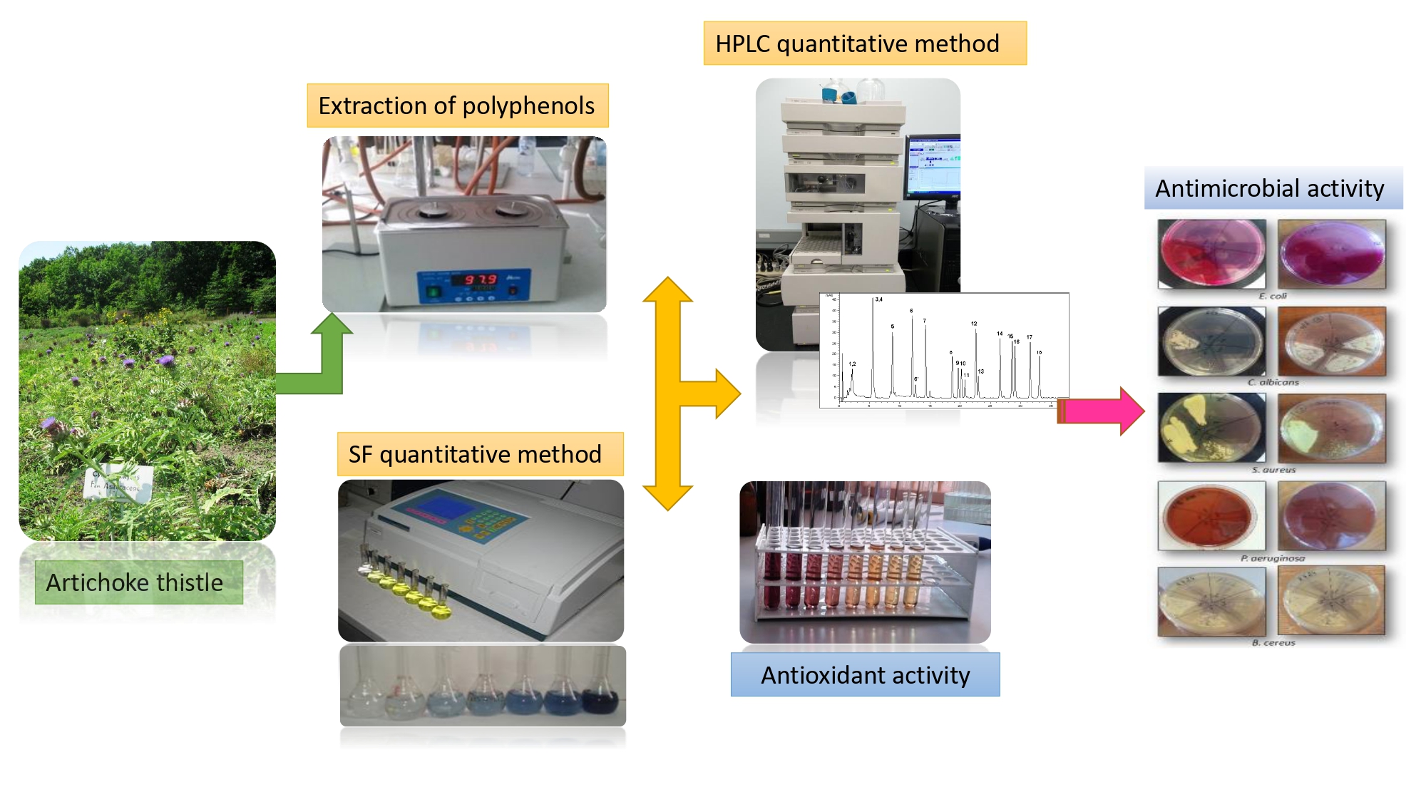
Artichoke, a medicinal plant with various therapeutical uses, is widely cultivated in many world geographical areas. The aim of this study was to establish the antimicrobial profile by means of comparative evaluation of the phytochemical constituents , antioxidant, anti-lipid peroxidation and antimicrobial activities of the basal and cauline leaves, as well as the by-products: stems, bracts, inflorescences from Cynara scolymus L. cultivated in the Republic of Moldova. Qualitative and quantitative characterization of the main phenolic compounds from ethanolic extracts was carried out by the HPLC-UV-MS method. The in vitro antioxidant activity was evaluated using DPPH˙, ABTS˙+, FRAP and NO˙ scavenging methods. Lipid lowering effect was establish with malonic dialdehyde complex and thiobarbituric acid. Antimicrobial properties were screened using diffusion method. The HPLC UV-MS analysis highlighted that green aerial parts of C. scolymus are characterized by the presence of five phenolic acids (kaempferol, gentisic, chlorogenic, p-coumaric, ferulic and caffeic) and four flavonoid heterosides and aglycones (isoquercitrin, quercitrin, luteolin and apigenin). Correlation between total polyphenolic content and antioxidant activity was found to be statistically significant (p<0.01). The extracts of C. scolymus aerial parts exhibited significant antibacterial and antifungal activities, (p<0.05) against all tested microorganisms, while no inhibitory effect for inflorescences was observed. Artichoke leaves and by-products may be considered important and promising sources of bioactive compounds for herbal medicinal products, functional foods and nutraceuticals, due to their antimicrobial properties.

Keywords: 
;  ;  ;  

1. Introduction

Medicinal plants have been used for thousands of years in health maintenance and remain a primary source of healthcare. Currently, the research of plant extracts with compounds with potential antimicrobial therapeutic application is an increasingly explored direction in the medical field. This approach constitutes a promising strategy for combating the phenomenon of antibiotic resistance, which numerous retrospective studies have highlighted the significant increase in the number of bacterial species that are capable of developing resistance mechanisms to the action of classical antimicrobial agents [1,2]. In this context, many extracts and constituents of plant origin are analyzed to be exploited in the development of new chemotherapeutic applications, with the ability to prevent and treat infections, especially those caused by multidrug-resistant bacteria [3,4,5]. Of particular interest is the species Cynara scolymus, which constitutes a gold mine in traditional medicine [6], which gives the Cynara species a special importance in research aimed at identifying effective natural alternatives to antibiotics.
Artichoke thistle Cynara scolymus L., (Cynara cardunculus var. scolymus L.), a species that belongs to the Asteraceae family, originally from Ethiopia, spread throughout the Mediterranean basin [7,8], was introduced into culture in temperate areas of Europe, as well within the experimental collection of the Scientifical Practical Center in the Domain of Medicinal Plants (SPCDMP) (46°56'08.6"N 28°41'43.4"E) of National Institute for Health and Medical Research of Nicolae Testemiţanu State University of Medicine and Pharmacy (Nicolae Testemiţanu SUMPh), from Chisinau, Republic of Moldova. C. scolymus is a robust, vivacious plant, perennial in the humid subtropical climate. Temperature is the most important factor in artichoke cultivation; thus, in the temperate areas of Europe, with a mild climate, the plant is grown only by annual cultivation from seeds [9,10]. Besides temperature, directly proportional to exposure to ultraviolet B rays is the accumulation of active principles, which are mainly carried out in the cuticle, epidermis and trichomes [11,12,13].
As mentioned in the literature [14,15] the phytochemical complex of artichoke is formed by groups of substances of secondary metabolism: polyphenols (caffeoylquinic acids - chlorogenic and caffeic acids, cynarin), flavonoids (luteolin, apigenin; flavonosides - rutoside, cynarozide, scolimoside), sesquiterpene lactones (cynaropicrin), sterol compounds (taraxasterol, pseudotaraxasterol), tannins and anthocyanins. The diversity of the chemical composition, which possesses a broad spectrum of pharmacological actions such as: antioxidant, anti-inflammatory, antibacterial, anti-proliferative, anti-HIV, hepatoprotective and hypocholesterolemic [16], allows the use of artichoke as a cholagogue and choleretic [17], hepatoprotective, hypolipidemic, antioxidant, diuretic, hypoglycemic [18] and antimicrobial remedy [19,20,21]. Moreover, artichoke leaf infusion is well-known in folk medicine, traditionally used as a cholagogue and fat metabolism enhancer in the treatment of fever, liver disorders, bile stones, blood cholesterol, urticaria, asthma, and eczema [22,23,24].
Leaf of C. scolymus - Cynarae folium is recognized as a medicinal plant product in the European Pharmacopeia [25]. Moreover, the Romanian Pharmacopoeia specifies the type of leaves used as basal leaves of the plant [26]. Nevertheless, many phenolic compounds with high antioxidant capacity were found in different parts of artichoke by-products (bract, stem and inflorescence) [27]. Additionally, El-Nashar et al. demonstrated that the extract obtained from artichoke bract waste exhibits both antioxidant activity and anti-Alzheimer's potential [28]. Furthermore, Cioni et al. demonstrated that pretreated extracts from artichoke’s stem and bract discards exhibited efficacy against S. aureus, B. cereus bacteria and the HSV-2 virus due to metabolites such as cynarine, chlorogenic acid, caffeic acid, luteolin, and apigenin [29]. These bioactive molecules exhibit multiple, often synergistic mechanisms that compromise microbial viability, affecting both Gram-positive and Gram-negative bacteria as well as certain fungi, can interact with lipid bilayers and membrane proteins through hydrophobic and hydrogen bonding interactions, leading to increased permeability, leakage of ions and cellular contents, and eventual loss of membrane integrity. Pereira et al. demonstrated that phenolic-rich extracts from artichoke leaves caused significant leakage of intracellular nucleic acids and proteins from Escherichia coli and Staphylococcus aureus, indicating membrane damage as a primary antimicrobial mechanism [30]. Aerial parts of C. scolymus are used in the production of nanoparticles through green synthesis [31,32,33]. Sampaio et al. used flower heads to produce silver nanoparticles with antibacterial actions [34]. Khedr et al. compared flower stems and bracts of C. scolymus extracts in the green synthesis of silver nanoparticles with apoptotic effect [35]. Thus, basal and cauline leaves, as well the by-products: stems, bracts, and inflorescences from C. scolymus, cultivated in the collection of SPCDMP could be promising sources of natural products due to the phenolic profile with antioxidant and antimicrobial actions of the species.

2. Results

2.1. Spectrophotometrical Assays for the quantification of Total Phenolic Compounds

The results obtained by applying the spectrophotometric methodology allowed the quantitative estimation of the main groups of biologically active compounds from extracts of aerial parts of C. scolymus (basal and cauline leaves, stems, bracts and inflorescences) cultivated in the collection of SPCDMP of Nicolae Testemițanu SUMPh. The highest concentrations of total phenolic content were established in basal and cauline leaves. The total polyphenolic amounts in extracts of aerial parts ranged from 15.47 to 0.94 mg GAE/g dry weight recalculated in gallic acid equivalent. The total flavonoid content ranged from 7.47 to 0.11 mg/g expressed as mg of rutin equivalent per g dry weight. The amounts of phenolic compounds detected in the samples are shown in Table 1.

2.2. HPLC-MS Analysis of the Extracts

For a more precise technique, liquid chromatography, with detection by mass spectrometry, was used. C. scolymus aerial parts extracts were characterized by the presence of 10 compounds, including five phenolic acids (kaempferol, gentisic, chlorogenic, p-coumaric and ferulic acids) and five flavonoid glycosides and aglycones (isoquercitrin, myricetin, quercitrin, luteolin, apigenin), as shown in Table 2.
Values are the mean ± SD (n = 3). BLQ - below limit of quantification (0,1 µg/ml); ND: not detected compound.
The analysis highlighted that the compound with the highest concentration proved to be chlorogenic acid in all analyzed extracts, determined maximum in basal leaf extract (515.93 µg/ml) and lower in bracts extract (3.98 µg/ml), suggesting that C. scolymus plants could serve as a significant source of chlorogenic acid, known for its notable therapeutic benefits.

2.3. Antioxidant properties of C. scolymus aerial parts extracts

The antioxidant properties of extracts obtained from leaves, stems, bracts and inflorescences were determined by applying several specific and non-specific in vitro methods to determine the capacity to capture and neutralize free radicals. The scavenging effect of C. scolymus extracts, determined by the DPPH˙ method, was measured as the IC50 value based on the obtained linear regression graph. The results of the DPPH˙ scavenging test demonstrated that basal leaves have strong antioxidant activity (IC50 96.14 µg/mL), cauline leaves - moderate antioxidant activity (IC50 125.82 µg/mL) and stem possess weak antioxidant activity (IC50 412.89 µg/mL), as shown in Table 3. There was a significant correlation between total polyphenolic content with DPPH˙ scavenging activity of C. scolymus aerial parts extracts (R2=0.999, 0.998, 0.998, 0.997 and 0.998, where p<0.01, respectively).
The antioxidant capacity of extracts carried out with ABTS˙+, measured as Trolox equivalents revealed the highest activity for the extract obtained from basal leaves (IC50 32.9 µg/mL) and for the extract obtained from cauline leaves (IC50 29.1 µg/mL). The correlation between total polyphenolic content and the antioxidant test values (ABTS˙+ inhibition %) were considered good (R2 = 0.9532, 0.9598, 0.916, 0.981, 0.947, where p<0.01, respectively). Furthermore, the FRAP assay estimated the electron-donating capacity of C. scolymus aerial parts extracts. In this study, the highest FRAP activity of the basal leaf extracts (67.7 μM EDTAE/g dw) it was found. Stems and bracts exhibited a lower reduction capacity than C. scolymus basal and cauline leaves (p < 0.001). With NO˙ radical scavenging assay the highest significant (p<0.05) NO˙ inhibitory activity among the extracts was obtained for basal leaves extract with a percentage inhibition of 67.7%. The lowest NO˙ radical scavenging percentage was detected in stems, bracts and inflorescence extracts, with no significant difference (p>0.05).
The results of the antioxidant test for the suppression of LDL oxidation established that 17.5% of lipoproteins are oxidized in the absence of copper sulfate, the negative control sample (Cu-) under conditions of 37°C for 24 hours. The highest activity in counteracting LDL oxidation was established for the samples with the application of leaf extracts (60.8-61.2% inhibition), with no significant difference (p>0.05). For the extracts obtained from artichoke bracts and stems, the malonic dialdehyde test values show 57.82% and 54.13%, respectively (p<0.05). The inflorescence extract did not show antioxidant effect under conditions of induced lipoprotein oxidation. Ascorbic acid, in the 1 mg/ml concentration, maintained 58.2% of the experimental lipoproteins in the non-oxidized form. The antioxidant activity of 1 mg of C. scolymus extract, which determines its ability to suppress lipoprotein oxidation, is similar to the activity of 1 mg of ascorbic acid. Therefore, the antioxidant compounds contained in the C. scolymus aerial parts extracts possess the ability to counteract the oxidation of low-density lipoproteins, and the antioxidant effect is achieved based on the mechanism of proton and electron transfer.

2.4. Antimicrobial Activity of C. scolymus aerial parts extracts

The antimicrobial properties of C. scolymus extracts in study against gram-positive bacteria (Staphylococcus aureus ATCC 25923, Corynebacterium diphtheriae ATCC 13812, Bacillus cereus ATCC 11778, Enterococcus faecalis ATCC 19433), gram-negative bacteria (Escherichia coli ATCC 25922, Pseudomonas aeruginosa ATCC 27853), and yeast (Candida albicans ATCC 10231), have been assessed in this study. The results shown in Table 4 indicate that the green aerial plant extracts of C. scolymus efficiently suppress the growth of microorganisms, with variable efficacy, although with significantly lower potency (p < 0.05) compared to the positive controls (tetracycline and miconazole).
Most bacteria were susceptible to C. scolymus basal and cauline leaf extracts, whereas S. aureus, E. coli, C. diphtheriae, and B. cereus were most sensitive, as demonstrated by low minimum inhibitor concentration (MIC) values. Results of antimicrobial activity of the four aerial parts extracts (basal and cauline leaves, stems and bracts) suggested that P. aeruginosa was the most resistant strain to analyzed plant extracts, followed by E. faecalis. Furthermore, the extract obtained from the C. scolymus inflorescence did not show antibacterial or antifungal actions in the investigated concentrations. The antifungal activity of basal leaves extract against C. albicans started at 1.466 mg/mL with an inhibition zone of 8.1 mm, cauline leaves at 1.532 mg/mL with an inhibition zone of 7.7 mm and bracts extract at 1.635 mg/mL with an inhibition zone of 6.2 mm, stems extract suppressed yeast grow at a concentration of 3.435 mg/mL with an inhibition zone of 7.25 mm respectively.

3. Discussion

The present study brings novelty by researching the phytochemical content and determining the antioxidant, LDL peroxidative and antimicrobial actions of both artichoke leaves and artichoke by-products (bracts, stems and inflorescences), which demonstrate the possibility to encompass in use the entire aerial part of the plant in natural products processing. The phenolic compounds in C. scolymus have been widely studied due to the varied therapeutic potential [36,37]. Their presence and abundance are related to metabolic reactions, which are influenced by: the analyzed botanical part, ontomorphogenetic phase of plant development, and climatic growing conditions, including the complex of abiotic stressors [38]. Phenolic compounds are polar compounds [39]; thus, for their extraction, ethanol of 70% was used, based on previous research [40]. The spectrophotometric assay regarding the amount of secondary metabolites of phenolic nature, in artichoke leaf, showed that the total polyphenolic content is higher than the total flavonoid content, similar to other authors [41,42]. In Romanian artichoke leaf extract, obtained through maceration with water, the total phenolic content (TPC) was quantified as 15.2 mg/g [43]. Studies analyzing artichoke leaves from Poland reported TPC values of 33.5 mg/g and total flavonoid content (TFC) of 17.9 mg/g for methanolic extracts [44], and TPC of 27 mg/g for ethanolic extracts [45]. The variation in total phenolic content across the literature could be explained by both extrinsic and intrinsic factors, including the solubility of the compounds in the solvents used. This solubility is influenced by the structure of the hydroxyl groups and the molecular size and length of the hydrocarbon chains of the bioactive compounds [46]. Artichoke by-products, generated during agricultural procedures and the processing industry, represent a significant amount of discarded material. Complementary studies conducted in other cultivation areas on C. scolymus agro-industrial discards, such as stems and bracts and inflorescence, show variability in the content of total polyphenols and flavonoids. The TPC of Moldovan artichoke discards, compared to the optimized ethanolic extract of bracts and stems from Portugal [47] (where TPC was 21.6 mg/g) and to Italian artichoke bract extracts (5 mg/g) [48,49], is lower. Nevertheless, the content is higher compared to Turkish methanolic extracts (2.4 mg/g) [50].
Further, through HPLC-MS investigation, we revealed that leaves, stem, bracts, and inflorescences contain hydroxycinnamic acid derivatives such as chlorogenic and caffeic acids and flavones such as luteolin and apigenin, which are the main compounds responsible for health effects, i.e., antioxidant, antimicrobial, hypolipidemic, antiatherogenic, anti-inflammatory and anticancer activities, recorded in literature data [19,29,51]. The results obtained for quantifying phenolic metabolites by HPLC-UV-MS analysis indicated chlorogenic acid (3-O-caffeoylquinic acid) as a major component. Caffeic acid was also found in high amounts in our studied aerial parts samples, followed by the aforementioned flavonoids, apigenin, luteolin, and hydroxycinnamic acid derivatives p-cumaric and ferulic acids. Our investigation reports and confirms the presence of the previously reported metabolites [52,53,54], along with distinctions [55], contributing to the growing evidence on the phytochemical diversity and the therapeutic potential of aerial parts extracts.
The antioxidant potential of C. scolymus aerial parts was thoroughly assessed using DPPH˙, ABTS˙+, FRAP and NO˙ in vitro methods, affirming the promising role of green aerial parts of the plant as natural antioxidants. Basal and cauline leaves of C. scolymus grown in the steppe climate conditions of the Republic of Moldova, highlighted remarkable DPPH˙ and ABTS˙+ radical scavenging activity, an effect attributed to their high content of chlorogenic acid. Therefore, the results of the positive correlation between the TPC, TFC, and the in vitro DPPH˙ and ABTS˙+ antioxidant methods, suggest that phenolic compounds act as reducing agents, hydrogen donors, and singlet oxygen scavengers and may exert an important antioxidant capacity of C. scolymus green aerial parts, recorded by other authors as well [56,57].
Moreover, the electron donating capacity of C. scolymus aerial parts extracts was monitored by FRAP assay. The principle of this method is based on the reduction of ferric 2,4,6-tripyridyl-s-triazine complex (Fe3+ - TPTZ) to its ferrous colored form (Fe2+ -TPTZ) in the presence of antioxidants [58]. The absorbance of this resultant blue-green colored solution of samples was measured at 700 nm which was related to the Fe2+ amount in the mixture. The ability to reduce the ferric ions (Fe3+) was recorded for basal leaves, cauline leaves, stems and bracts. The FRAP assay confirmed the reducing power in the limits of 67.7 μMEDTAE/g dw to 22.45 μMEDTAE/g dw.
Aerial parts of C. scolymus were examined for their possible scavenging ability of NO˙. The principle of the method consists in determining the production of the nitric oxide radical generated by sodium nitroprusside. Nitric oxide interacts with oxygen and forms nitrites, which are determined spectrophotometrically using the Greiss reagent. The chromophore formation occurs due to the diazotization of nitrite with sulfanilamide and its coupling with naphthyl ethylenediamine [59]. The highest inhibitory activity among the extracts was obtained for basal leaves extract with a percentage inhibition of 67.7%, the lowest NO˙ radical scavenging activity was detected in stems, bracts and inflorescence extracts. The results of the antioxidant activity of aerial parts of C. scolymus assessed by these in vitro methods provide important information on their intrinsic antioxidant potential with minimal environmental interference.
It is well-known that polyphenolic compounds exhibit various pharmacological activities, including hypolipidemic and antiatherogenic effects [60]. Mocelin et al. revealed a significant decrease in oxidized-LDL concentration, and antioxidized-LDL in rats treated with leaf extract of C. scolymus [51]. Furthermore, Mokhtari et al. demonstrated a significant improvement in plasma lipid profiles by reducing total cholesterol, triglycerides, and LDL–cholesterol while increasing HDL–-cholesterol during administration in mice of artichoke bract extract [61]. In our study, we showed that the ability to suppress human low-density lipoprotein oxidation in vitro had the green aerial parts of C. scolymus. The LDL was greatly reduced by basal and cauline leaf extracts (61.2% and 60.8%), followed by bracts extracts (57.82%). The lowest percentage of inhibition LDL oxidation was observed for artichoke stem extract (54.13%). The inflorescence did not exhibit an antioxidant effect under conditions of induced lipoprotein oxidation. Our results suggest that the LDL oxidation capacity is possible due to myricetin, quercetin, isoquercitrin and kaempferol, reported in data literature as exhibiting favorable hypolipidemic effect [62] and identified in C. scolymus basal and cauline leaves, stems and bracts but not in inflorescences.
To assess the antimicrobial profile of the aerial parts of C. scolymus cultivated in the Republic of Moldova, we determined the antimicrobial activity of the extracts in the study. The antimicrobial properties of C. scolymus extracts, cultivated worldwide and reported throughout the years, have been commonly associated with secondary metabolites such as: flavonoids, tannins, essential oils, glycosides and phenols [63,64,65]. There is a wide range of results, mostly varying depending on the area from where the artichoke was harvested and the extraction method, though not only [66]. The antimicrobial assay carried out by Scavo et al. showed that the C. cardunculus L. var. altilis ethanolic extract was found to be the most active and effective in inhibiting the growth of Gram-positive species [67]. Zhu et al. revealed that leaf extract was found to be most effective against all of the tested organisms, followed by the artichoke head and stem extracts, and the ethanol fraction showed the most significant antimicrobial activity compared to other extraction solvents [68]. The antimicrobial activity of the plant discards shows that Moldovan artichoke inflorescence was ineffective against strains used in the experiments, unlike Mejri et al. inflorescence extract, which did show antimicrobial activity against S. aureus. But the lack of inhibitory effect against E. coli, and C. albicans, confirms the attribution of antibacterial activity mainly to hydroxycinnamic acids and flavones [69]. Our results of the antimicrobial assay showed important antimicrobial potential for basal and cauline leaves, stems and bracts ethanolic extracts, against Gram-positive bacteria (Staphylococcus aureus Corynebacterium diphtheriae, Bacillus cereus, Enterococcus faecalis), Gram-negative bacteria (Escherichia coli, Pseudomonas aeruginosa), and yeast (Candida albicans), outcomes that can be connected with the ones obtained for the antioxidant assays, as all of them may be related to the phenolic composition of these samples. These data are of paramount importance for medicine and health care in order to diminish the burden of antimicrobial resistance by subsequently using C. scolymus standardized extracts as alternative antimicrobial drugs.

4. Materials and Methods

4.1. Chemicals

The following 18 standards used in the phytochemical analysis of HPLC-UV-MS were purchased from Sigma-Aldrich (Schnelldorf, Germany): caftaric acid (>97%), chlorogenic acid (>95%), gentisic acid (>95%), isoquercitrin (quercetin 3-d-glucoside) (≥98%), quercitrin (quercetin 3-rhamnoside) (≥78%), luteolin (≥98%), sinapic acid (≥98%), fisetin (≥98%), patuletin (≥98%), apigenin (>95%), caffeic acid (≥95%), myricetin (≥97%), vanillic acid (≥97%), hyperoside (≥95%), p-coumaric acid (≥98%), ferulic acid (≥99%), kaempferol (≥97.0%), rutin (≥94%), with chromatographically pure reagents. From Sigma-Aldrich (Schnelldorf, Germany) were obtained galic acid (≥98.0%), DPPH˙, FRAP, Folic-Ciocalteau and Greiss reagents, sodium nitroprusside dihydrate and Trolox (>97%), as well. Ascorbic acid, potassium persulfate were obtained from Merck (Darmstadt, Germany). ABTS˙+ from Alfa Aesar GmbH & KG. EDTA and TPTZ were purchased from HiMedia Laboratories (India). All solvents and chemical reagents used were of analytical grade or higher.

4.2. Plant Materials

Specimens of Cynara scolymus L. aerial parts: basal leaves, cauline leaves, stems, bracts and inflorescences were collected from the Scientifical Practical Center in the Domain of Medicinal Plants of Nicolae Testemițanu SUMPh from Chisinau, Republic of Moldova, during the flowering period. The taxonomic affiliation of artichoke thistle to the C. scolymus species was determined and confirmed by macro- and microscopic studies. Labeled, natural dried, samples of C. scolymus species collected in the experimental collection of SPCDMP were kept in the Herbar at Pharmacognosy and Pharmaceutical Botany Department of the Faculty of Pharmacy of Nicolae Testemițanu SUMPh, with the voucher code (CS).2004.2.24. The harvested aerial parts of C. scolymus (Figure 1) were ground into powder using a RETSCH laboratory knife mill at 5000 rpm and passed through a 0.8 mm sieve.

4.2. Extract Preparation

To obtain the plant extracts, Soxhlet extraction method assisted by ultrasound was used. Ten grams of powdered plant material were extracted with 70% ethanol, ratio between the vegetal material and the solvent was 5:100. The extraction was performed for 4 hours at the boiling temperature of the solvent. After the extraction was completed, the extracts were filtered and evaporated to dryness on Laborota 1011 evaporator. The extraction yield of C. scolymus basal leaves, cauline leaves, stems, bracts, and inflorescences was determined using relation: Y (%) = (ME/MP) × 100, where Y: Yield of extraction (%); ME: Mass of the dry extract obtained (g); MP: Mass of plant powder used (g).

4.3. Total Phenolic Content Assessment

The evaluation of the total phenolic content (TPC) for each extract was carried out using the Folin–Ciocalteu described by Singleton and Rossi [70], with some modifications [71]. 150 μL Folin Ciocalteu reagent (1/10) is added to 300 μL of extract sample. After incubation for 10 min at room temperature, 1.2 ml of 10% sodium bicarbonate solution and 1.35 mL of purified water are added. The samples were stored for 45 minutes in the dark. The absorbance was read at a wavelength of 765 nm at Specord 200 Plus Spectrofotometer (Germany) against a blank solution. A calibration curve was stablished employing the standard gallic acid (Sigma Aldrich), encompassing concentrations ranging from 0 to 115 μg/mL (y=0.1119x-0.0025; R2=0.9993). The outcomes were expressed in milligrams of gallic acid equivalents (GAE) per gram of dry weight (dw).

4.4. Total Flavonoid Content Assessment

The total flavonoid content was determined spectrophotometrically according to aluminium chloride colorimetric method [71]. Each plant extracts were dissolved in methanol and mixed with 0.1 mL of 10% aluminium chloride hexahydrate, 0.1 mL of 1 M potassium acetate and 2.8 mL of deionized water. After 40 minutes incubation at room temperature, the absorbance of the reaction mixture was determined spectrophotometrically at 415 nm. Rutin was chosen as a standard in the concentration range of 0.05 to 0.1 mg/mL (y=29.116x-0.0181; R2=0.9992). The total flavonoid content was expressed in milligram of rutin equivalents (RE) per gram of dry weight (dw).

4.5. Analyses using High-Performance Liquid Chromatography

HPLC coupled with mass spectrometer, HP 1100 autosampler, HP 1100 thermostat, HP 1100 UV detector, Agilent Ion Trap 1100 VL mass spectrometer was used. The working conditions were as follow: analytical column - Zorbax SB-C18 100 mm x 3.0 mm, 3.5 µm; Zorbax SB-C18 precolumn; mobile phase: methanol mixture: acetic acid solution 0.1% (V/V), gradient elution (start 5% methanol, up to 35 minutes 42% methanol, up to 38 min 42% methanol, up to 45 minutes 5% methanol - reequilibration); flow rate: 1 ml/min, temperature: 48ºC; detection: ultraviolet, 330 nm up to 17 minutes, 370 nm up to 38 minutes/MS; injection volume: 5 µl. MS working conditions: ion source: ESI (electrospray); ionization mode: negative; nebulizer: nitrogen, pressure 70 psi; drying gas: nitrogen, flow rate 12 L/min, temperature 360ºC; capillary potential: +3000 V; analysis mode: specific ion monitoring (polyphenolcarboxylic acids) or Auto MS (flavonoids and their aglycones). Each class of compounds was detected at the wavelength corresponding to the maximum absorption of the UV spectrum. For quantitative determination, a calibration graph was made for each compound in the concentration range 0.5-5.0 µg/ml. Two types of samples of each aerial parts extracts were analyzed in parallel, one as such, and the other hydrolyzed. The reason for which hydrolysis was performed is that usually some flavone aglycones or some polyphenol-carboxylic acids are not in a free state, but bound as glycosides, esters, etc [73]. The hydrolysis was performed according to the following protocol: one part of the extract was diluted with one part of 2 N hydrochloric acid solution and maintained on a water bath at 80ºC for 60 minutes.

4.6. Antioxidant Activities

4.6.1. DPPH Assay

The free radical scavenging activity of the fractions was measured in vitro by 2,2′- diphenyl-1-picrylhydrazyl (DPPH˙) assay. The stock solution was prepared by dissolving 24 mg DPPH˙ with 100 mL methanol and stored at 20°C until required. The working solution was obtained by diluting DPPH˙ solution with methanol to obtain an absorbance of about 0.98±0.02 at 517 nm using UV-Vis Jasco V530 spectrophotometer (Jasco, Japan). A 3 mL aliquot of this solution was mixed with 100 μL of the sample at various concentrations (10 - 500 μg/mL). The reaction mixture was shaken well and incubated in the dark for 15 minutes at room temperature. Then the absorbance was taken at 517 nm. The control was prepared as above without any sample. The scavenging activity was estimated based on the percentage of DPPH˙ radical scavenged as as follows: AA DPPH˙ (%) = A control – A Sample / A Control * 100; where, A control is the optical density of the control (containing all reagents except for the extract) and A sample is the optical density in the presence of the extract. The extract concentration providing 50% of free radical scavenging activity (IC50) was calculated from the graph of the radical scavenging activity percentage against extract concentration.

4.6.2. ABTS Assay

The 2,2′-azinobis (3-ethylbenzthiazoline-6-sulphonic acid), commonly called ABTS˙+ cation scavenging activity was performed [74]. To generate ABTS•+ radical cation, 7 mM of ABTS and 2.45 mM of potassium persulfate were mixed and incubated in the dark at room temperature for at least 16 h. The resulting working solution was diluted with 50% methanol for an initial absorbance of about 0.70±0.02 at 745 nm, with temperature control set at 30°C. Free radical scavenging activity was assessed by mixing 300 μL of test sample with 3.0 ml of ABTS˙+ working standard in a microcuvette. The antioxidant capacity of test samples was expressed as IC50 (anti-radical activity) which is the concentration necessary for 50% reduction of ABTS˙+.

4.6.3. Ferric Reducing Antioxidant Potential Assay

Ferric reducing antioxidant potential (FRAP) of the extracts was evaluated according to the method proposed by Benzie and Strain [75]. Briefly, FRAP reagent was prepared by mixing in 25 mL acetate buffer (30 mM; pH 3.6), 2.5 mL TPTZ solution (10 mM) and 2.5 mL ferric chloride solution (20 mM). The mixture was incubated for 15 min at 37 °C before use. EDTA was employed as a standard in this assay, its calibration curve concentrations ranged from 50 mg/L to 500 mg/L in water. The results were reported as μg of EDTA equivalents (EDTAE) per g dry weight. A higher inhibition value indicates a higher antioxidant activity.

4.6.4. Nitric oxide reducing Assay

The sodium nitroprusside solution was prepared immediately before the test by dissolving 10 mmol of sodium nitroprusside in 20 mmol of phosphate buffer solution (pH 7.4). The reagent mixture contains 0.5 ml of sample and 0.5 ml of sodium nitroprusside solution and is incubated at 25°C for 150 minutes. After incubation, 2 ml of Greiss reagent (1% sulfanilamide solution, 2% phosphoric acid solution and 0.1% naphthylethylenediamine dihydrochloride solution) is added to the reagent mixture and the absorbance is measured at a wavelength of 542 nm. Ascorbic acid is applied as a positive control at a concentration of 0.1 mg/ml.

4.6.5. In vitro determination of the capacity to inhibit low-density lipoprotein oxidation

Before using human LDL for this assay, this experiment was approved by the ethics committee of Institute of Microbiology and Biotechnology from the Republic of Moldova. Low-density lipoproteins were obtained from blood serum by the heparin sedimentation method [74,75,76]. To 2 mL of serum, 400 EU heparin (pharmacopoeial solution 5000 EU/ml) and 150 μL of the 1 M manganese chloride solution, are added. The sample is incubated for 30 minutes at 0oC, then centrifuged for 30 minutes at 0oC. The sediment is washed with 0.9% sodium chloride solution and the centrifugation procedure is repeated. The sediment obtained represents the lipoproteins, which are quantitatively transferred to 1 M sodium chloride solution, so that the protein content is 2 g/l. To 0.1 mL of LDL, 10 μL of antioxidant solution is added. After 5 minutes of incubation, LDL oxidation is induced by adding 33.3 μL of 50 μM copper sulfate solution. The samples are incubated for 24 hours at 37oC. After incubation, the oxidative process is interrupted with EDTA solution (final concentration of 27 mM).
The value of the degree of oxidation of LDL is determined by the concentration of thiobarbituric acid reactive substances (malonic dialdehyde) [77,78]. 1 ml of 0.67% thiobarbituric acid and 1 mL of 15% trichloroacetic acid are added to the samples with oxidized LDL, after which the samples are incubated for 1 hour at 95oC. Next, the samples are cooled on ice for 5 minutes and centrifuged for 15 minutes at 3000 g. The absorbance of the malonic dialdehyde complex with thiobarbituric acid is measured at a wavelength of 535 nm at T80+ UV/VIS Spectrometer, PG Instruments Ltd, UK. The calculation is made based on the LDL protein concentration, using the molar extinction coefficient of the malonic dialdehyde complex or in % inhibition compared to the positive control sample. The positive control sample contains the LDL solution, in which lipid oxidation is induced with copper sulfate in the absence of the antioxidant. As a positive control, ascorbic acid is applied at a concentration of 0.1 mg/mL. The value of the results is expressed as ascorbic acid equivalent. To exclude lipid autoxidation, the negative control sample is introduced in which lipid oxidation occurs in the absence of copper sulfate.

4.7. Antimicrobial Activity

For the bioassay six bacterial strains Staphylococcus aureus (ATCC 25923), Escherichia coli (ATCC 25922), Pseudomonas aeruginosa (ATCC 27853), Bacillus cereus (ATCC 11778), Corynebacterium diphtheriae (ATCC 13812), Enterococcus faecalis (ATCC 19433) and one fungal strain of Candida albicans (ATCC 10231), were taken into account for this study.
To determine the antimicrobial effect of the extracts, the screening by the agar well diffusion method, described previously [79,80], was carried out. Wells were made in Mueller Hinton agar plates using a sterile metal punch (6 mm in diameter). The plates were inoculated with a sterile swab moistened with microbial suspension according to the 0.5 Mac Farland turbidity standard. Then, 100 µL of plant extract was added to each well. The plates were introduced in the refrigerator for 30 min to allow the extracts to diffuse well into the agar, then incubated at 37°C for 18 h. Antimicrobial activity was detected by measuring the zone of inhibition (including the diameter of the wells) after the incubation period.
The Minimal Inhibitory Concentration (MIC) and Minimum Bactericidal/Fungicidal Concentration (MBC/MFC) of aerial parts extracts were determined by the dilution method in liquid media according to CLSI (Clinical and Laboratory Standards Institute of the United States of America) [81].
Serial two-fold dilutions of plant extracts with adjusted bacterial concentration (108 CFU/mL, 0.5 McFarland’s standard) were used to determine MIC in broth medium. The control contained only inoculated broth with microorganisms and was incubated at 37 °C for 24 h. The lowest concentrations of test samples which did not show any visible growth of test organisms after macroscopic evaluation were determined as MICs, expressed in mg/mL. MBC/MFC is considered the lowest concentration of plant extract that killed at least 99.9% of the initial inoculums. Similar tests were performed simultaneously for growth control (broth + inoculum) and sterility control (broth + test sample). Tetracycline (10 µg/mL) and ketoconazole (10 µg/mL), purchased from Sigma Aldrich (Germany) were used as the positive controls (standard drugs) for bacteria and fungi, respectively. All assays were performed in triplicate.

4.8. Statistical Analysis

The correlations among different measured and derived traits were estimated by calculating Pearson correlation coefficients using the statistical tool in Excel 2022. Data were subjected to analysis of variance (ANOVA) using SPSS software version 20.0 (IBM Corporation, Chicago, IL, USA). Statistical significance was determined for p values below 0.05, and the results were expressed as mean values ± standard deviation (SD).

5. Conclusions

This study focused on the chemical composition and biological properties of basal and cauline leaves of C. scolymus as well as on its stems, bracts and inflorescence by-products. According to the HPLC-UV-MS analysis, the investigated ethanolic extracts, have the key compounds chlorogenic and caffeic acids, luteolin-7-O-glucoside and apigenin, with organ-specific variations in concentration. Overall antioxidant capacity, assessed in vitro through DPPH˙, ABTS˙+, FRAP, NO˙ assays, demonstrated high scavenging potential of green aerial parts of the species. Specifically, we reveal strong positive correlations between TPC and antioxidant capacities, further validating the contribution of these compounds to the biological activity of C. scolymus. The results of the antimicrobial assay showed important antimicrobial potential for the leaves and green by-products of C. scolymus exhibited with varying degrees of potency. The most notable effect was observed against Gram-positive bacteria, including Staphylococcus aureus, Corynebacterium diphtheriae, Bacillus cereus, and Enterococcus faecalis, as well as antifungal activity. Thus, C. scolymus leaves and by-products extracts, may represent a more sustainable pathway toward achieving the goals of One Health in an age increasingly threatened by antibiotic resistance.

Author Contributions

Conceptualization, C.C.; methodology, C.C.; L.R., L.V., G.B., D.B.; formal analysis, C.C., L.R., L.V., G.B., D.B.; investigation, C.C., L.R., L.V., G.B., D.B.; validation, G.B., L.V., C.T.; resources, C.C., L.R., L.V., G.B.; software, C.C., L.R., L.V., G.B., D.B.; writing - original draft preparation, C.C.; review and editing, C.C., L.R., D.B., T.C.; supervision, T.C.; project administration, C.C.All authors have read and agreed to the published version of the manuscript.

Funding

The study was funded by the national project number 25.80012.8007.06TC, of the National Agency for Development and Research, Republic of Moldova.

Acknowledgments

National Agency for Development and Research from the Republic of Moldova.

Conflicts of Interest

The authors declare no conflicts of interest.

References

  1. Koma, P.L.; Matotoka, M.M.; Mazimba, O.; Masoko, P. Isolation and In Vitro Pharmacological Evaluation of Phytochemicals from Medicinal Plants Traditionally Used for Respiratory Infections in Limpopo Province. Antibiotics 2025, 14, 965. [CrossRef]
  2. Mirković, S.; Martinović, M.; Tadić, V.M.; Nešić, I.; Jovanović, A.S.; Žugić, A. Antimicrobial and Antioxidant Activity of Essential Oils from Selected Pinus Species from Bosnia and Herzegovina. Antibiotics 2025, 14, 677. [CrossRef]
  3. Matlala, M.P.; Matotoka, M.M.; Shekwa, W.; Masoko, P. Antioxidant: Antimycobacterial and Antibiofilm Activities of Acetone Extract and Subfraction Artemisia afra Jacq. ex Willd. Against Mycobacterium smegmatis. Antibiotics 2024, 13, 1027. [CrossRef]
  4. Šovljanski, O.; Aćimović, M.; Tomić, A.; Lončar, B.; Miljković, A.; Čabarkapa, I.; Pezo, L. Antibacterial and Antifungal Potential of Helichrysum italicum (Roth) G. Don Essential Oil. Antibiotics 2024, 13, 722. [CrossRef]
  5. Kurćubić, V.S.; Đurović, V.; Stajić, S.B.; Dmitrić, M.; Živković, S.; Kurćubić, L.V.; Mašković, P.Z.; Mašković, J.; Mitić, M.; Živković, V.; et al. Multitarget Phytocomplex: Focus on Antibacterial Profiles of Grape Pomace and Sambucus ebulus L. Lyophilisates Against Extensively Drug-Resistant (XDR) Bacteria and In Vitro Antioxidative Power. Antibiotics 2024, 13, 980. [CrossRef]
  6. Alessandroni, L.; Bellabarba, L.; Corsetti, S.; Sagratini, G. Valorization of Cynara cardunculus L. var. scolymus Processing By-Products of Typical Landrace “Carciofo Di Montelupone” from Marche Region (Italy). Gastronomy 2024, 2, 129-140. [CrossRef]
  7. Ciobanu, N., Cojocaru-Toma, M., Pompuș, I., Chiru, T., Ciobanu, C., Benea, A. Plante din colecția Centrului Științific de cultivare a Plantelor medicinale. Compendium. IP Univ. de stat de Medicină şi Farmacie Nicolae Testemiţanu, Chișinău: Print Caro, Moldova, 2019; pp. 29-30. ISBN 978-9975-56-660-5.
  8. Se˻kara, A.; Kalisz, A.; Gruszecki, R.; Grabowska, A.; Kunicki, E. Globe artichoke – a vegetable, herb and ornamental of value in central Europe. The Journal of Horticultural Science and Biotechnology 2015, 90(4), 365–374. [CrossRef]
  9. Sałata, A.; Lombardo, S.; Pandino, G.; Mauromicale, G.; Buczkowska, H.; Nurzyńska-Wierdak, R. Biomass yield and polyphenol compounds profile in globe artichoke as affected by irrigation frequency and drying temperature. Industrial Crops and Products 2022, 176, 114375. ISSN 0926-6690. [CrossRef]
  10. Zagoskina, N.V.; Zubova, M.Y.; Nechaeva, T.L.; Kazantseva, V.V.; Goncharuk, E.A.; Katanskaya, V.M.; Baranova, E.N.; Aksenova, M.A. Polyphenols in Plants: Structure, Biosynthesis, Abiotic Stress Regulation, and Practical Applications (Review). International Journal of Molecular Sciences 2023, 9, 24(18), 13874. [CrossRef]
  11. Lombardo, S.; Pandino, G.; Ierna, A.; Mauromicale, G. Variation of polyphenols in a germplasm collection of globe artichoke, Food Research International 2012, 46, 2, 544-551. ISSN 0963-9969. [CrossRef]
  12. Crews, T.E.; Carton, W.; Olsson, L. Is the future of agriculture perennial? Imperatives and opportunities to reinvent agriculture by shifting from annual monocultures to perennial polycultures. Global Sustainability 2018, 1, 11. [CrossRef]
  13. Vico, G.; Brunsell, N. Tradeoffs between water requirements and yield stability in annual vs. perennial crops. Advances in Water Resources 2018, 112, 189-202. ISSN 0309-1708. [CrossRef]
  14. Zhang, Y.; Li, Y.; Jiang, L.; Tian, Li, J.; Xiao, Z. Potential of Perennial Crop on Environmental Sustainability of Agriculture. Procedia Environmental Sciences 2011, 10, 1141-1147. ISSN 1878-0296, . [CrossRef]
  15. Gominho, J.; Dolores Curt, M.; Lourenço, A.; Fernández, J.; Pereira, H. Cynara cardunculus L. as a biomass and multi-purpose crop: A review of 30 years of research. Biomass and Bioenergy, 2018, 109, 257-275. ISSN 0961-9534. [CrossRef]
  16. Służały, P.; Paśko, P.; Galanty, A. Natural Products as Hepatoprotective Agents—A Comprehensive Review of Clinical Trials. Plants 2024, 13, 1985. [CrossRef]
  17. Ayuso, P.; Quizhpe, J.; Rosell, M.d.l.Á.; Peñalver, R.; Nieto, G. Bioactive Compounds, Health Benefits and Food Applications of Artichoke (Cynara scolymus L.) and Artichoke By-Products: A Review. Journal of Applied Sciences 2024, 14, 4940. [CrossRef]
  18. Jalili, C.; Moradi, S.; Babaei, A.; Boozari, B.; Asbaghi, O.; Lazaridi, A.V.; Hojjati Kermani, M.A.; Miraghajani, M. Effects of Cynara scolymus L. on glycemic indices: A systematic review and meta-analysis of randomized clinical trials. Complementary Therapies in Medicine 2020, 52, 102496, ISSN 0965-2299. [CrossRef]
  19. Colombo, R.; Moretto, G.; Pellicorio, V.; Papetti, A. Globe Artichoke (Cynara scolymus L.) By-Products in Food Applications: Functional and Biological Properties. Foods 2024, 13, 1427. [CrossRef]
  20. Al Masalmeh, A.M.; Mallah, E.; Mansoor, K.; Abu-Qatouseh, L.; El-Hajji, F.D.; Idkaidek, N.; Al-Bashiti, I.; Issa, I.H.; Al Meslamani, A.Z.; Aws, S. Pharmacokinetic interaction of rosuvastatin with artichoke (Cynara scolymus L.) leaf extract in rats. Journal of Applied Pharmaceutical Science 2023, 13(06), 179–192. [CrossRef]
  21. El Sohafy, S.M.; Shams Eldin, S.M.; Sallam, S.M.; Bakry, R.; Nassra, R.A.; Dawood, H.M Exploring the ethnopharmacological significance of Cynara scolymus bracts: Integrating metabolomics, in-Vitro cytotoxic studies and network pharmacology for liver and breast anticancer activity assessment. Journal of Ethnopharmacology 2024, 334, 118583. ISSN 0378-8741. [CrossRef]
  22. Ramos, P.A.B.; Guerra, Â.R.; Guerreiro, O.; Santos, S.A.O.; Oliveira, H.; Freire, C.S.R.; Silvestre, A.J.D.; Duarte, M.F. Antiproliferative Effects of Cynara cardunculus L. var. altilis (DC) Lipophilic Extracts. International Journal of Molecular Sciences 2017, 18, 63. [CrossRef]
  23. Mohaddese, M. Cynara scolymus (artichoke) and its efficacy in management of obesity. Bulletin of Faculty of Pharmacy Cairo University 2018, 56, 2, 115-120. ISSN 1110-0931. [CrossRef]
  24. Lattanzio, V.; Paul A. Kroon, Vito Linsalata, Angela Cardinali, Globe artichoke: A functional food and source of nutraceutical ingredients. Journal of Functional Foods 2009, 1, 2, 131-144. ISSN 1756-4646. [CrossRef]
  25. .
  26. Romanian Ministry of Health. Romanian Pharmacopoeia. 10th ed. Bucharest: Romanian. 1993; pp. 334–336.
  27. Zazzali, I.; Gabilondo, J.; Mallmann, L.P.; Rodrigues, E.; Perullini, M.; Santagapita, P.R. Overall evaluation of artichoke leftovers: Agricultural measurement and bioactive properties assessed after green and low-cost extraction methods. Food Bioscience 2021, 41, 100963. [CrossRef]
  28. El-Nashar, H.A.S.; Abbas, H.; Zewail, M.; Noureldin, M.H.; Ali, M.M.; Shamaa, M.M.; Khattab, M.A.; Ibrahim, N. Neuroprotective Effect of Artichoke-Based Nanoformulation in Sporadic Alzheimer’s Disease Mouse Model: Focus on Antioxidant, Anti-Inflammatory, and Amyloidogenic Pathways. Pharmaceuticals 2022, 15, 1202. [CrossRef]
  29. Cioni, E.; Di Stasi, M.; Iacono, E.; Lai, M.; Quaranta, P.; Luminare, A.G.; Gambineri, F.; De Leo, M.; Pistello, M.; Braca, A. Enhancing antimicrobial and antiviral properties of Cynara scolymus L. waste through enzymatic pretreatment and lactic fermentation. Food Bioscience 2024, 57, 103441. ISSN 2212-4292. [CrossRef]
  30. Pereira, C.; Barros, L.; José Alves M.; Santos-Buelga, C.; Ferreira I.C. Artichoke and milk thistle pills and syrups as sources of phenolic compounds with antimicrobial activity. Food Funct 2016, Jul 13;7(7):3083-90. PMID: 27273551. [CrossRef]
  31. Baran, A.; Keskin, C.; Baran, M.F.; Huseynova, I.; Khalilov, R.; Eftekhari, A.; Irtegun-Kandemir, S.; Kavak, D.E. Ecofriendly Synthesis of Silver Nanoparticles Using Ananas comosus Fruit Peels: Anticancer and Antimicrobial Activities. Journal of Biological Inorganic Chemistry 2021, 30, 2058149. [CrossRef]
  32. Erdogan, O.; Abbak, M.; Demirbolat, G.M.; Birtekocak, F.; Aksel, M.; Pasa, S. Green synthesis of silver nanoparticles via Cynara scolymus leaf extracts: The characterization, anticancer potential with photodynamic therapy in MCF7 cells. PLoS ONE 2019, 14(6), e0216496. https:// doi.org/10.1371/journal.pone.0216496.
  33. Balasubramanian, B.; Gangwar, J.; James, N.; Pappuswamy, M.; Anand, A.V.; Al-Dhabi, N.A.; Valan Arasu, M.; Liu, W.-C.; Sebastian, J.K. Green Synthesis of Bioinspired Nanoparticles Mediated from Plant Extracts of Asteraceae Family for Potential Biological Applications. Antibiotics 2023, 12, 543. [CrossRef]
  34. Sampaio, S.; Viana, J. Production of Silver Nanoparticles by Green Synthesis Using Artichoke (Cynara Scolymus L.) Aqueous Extract and Measurement of Their Electrical Conductivity. Advances in Natural Sciences: Nanoscience and Nanotechnology 2018, 9 (4): 045002. [CrossRef]
  35. Khedr, A.I.M.; Farrag, A.F.S.; Nasr, A.M.; Swidan, S.A.; Nafie, M.S.; Abdel-Kader, M.S.; Goda, M.S.; Badr, J.M.; Abdelhameed, R.F.A. Comparative Estimation of the Cytotoxic Activity of Different Parts of Cynara scolymus L.: Crude Extracts versus Green Synthesized Silver Nanoparticles with Apoptotic Investigation. Pharmaceutics 2022, 14, 2185. [CrossRef]
  36. Grabowska, A.; Caruso, G.; Mehrafarin, A.; Kalisz, A.; Gruszecki, R.; Kunicki, E.; Sękara, A. Application of modern agronomic and biotechnological strategies to valorise worldwide globe artichoke (Cynara cardunculus L.) potential - an analytical overview. Italian Journal of Agronomy 2018, 13, 4, 1252. ISSN 1125-4718. [CrossRef]
  37. Buzzanca, C.; Di Stefano, V.; D’Amico, A.; Gallina, A.; Grazia Melilli, M. A systematic review on Cynara cardunculus L.: bioactive compounds, nutritional properties and food-industry applications of a sustainable food. Natural Product Research 2024. ISSN 1478-6419. [CrossRef]
  38. Mandim, F.; Petropoulos, S.A.; Giannoulis, K.D.; Santos-Buelga, C.; Ferreira, I.C.F.R.; Barros, L. Chemical Composition of Cynara cardunculus L. var. altilis Bracts Cultivated in Central Greece: The Impact of Harvesting Time. Agronomy 2020, 10, 1976. [CrossRef]
  39. Shi, L.; Zhao, W.; Yang, Z.; Subbiah, V.; Suleria, H.A.R. Extraction and characterization of phenolic compounds and their potential antioxidant activities. Environmental Science and Pollution Research 2022, 29(54), 81112-81129. [CrossRef]
  40. Ciobanu, C.; Diug, E.; Calalb, T.; Tomuta, I.; Achim, M. Optimisation of ultrasound-assisted extraction method of biologically active compounds from Cynara scolymus L. Curierul medical, 2015, 58, 2, 23-28. ISSN 1857-0666. [Google Scholar].
  41. Riadh, I.; Tlili, I.; R’him, T.; Rached, Z.; Arfaoui, K.; Pék, Z.; Lenucci, M. S.; Daood, H.; Helyes, L. Assessment of The Phenolic and Flavonoid Content in Certain Globe Artichoke (Cynara scolymus L.) Cultivars Grown in Northern Tunisia. Turkish Journal of Agriculture - Food Science and Technology 2022, 10(6), 1125–1129. [CrossRef]
  42. Costea, L.; Chițescu, C.L.; Boscencu, R.; Ghica, M.; Lupuliasa, D.; Mihai, D.P.; Deculescu-Ioniță, T.; Duțu, L.E.; Popescu, M.L.; Luță, E.-A.; et al. The Polyphenolic Profile and Antioxidant Activity of Five Vegetal Extracts with Hepatoprotective Potential. Plants 2022, 11, 1680. [CrossRef]
  43. Vamanu, E.; Vamanu, A.; Niţă, S.; Colceriu, S. Antioxidant and Antimicrobial Activities of Ethanol Extracts of Cynara Scolymus (Cynarae folium, Asteraceae Family). Tropical Journal of Pharmaceutical Research 2011; 10 (6). 777-783. [CrossRef]
  44. Sałata, A.; Nurzyńska-Wierdak, R.; Lombardo, S.; Pandino, G.; Mauromicale, G.; Ibáñez-Asensio, S.; Moreno-Ramón, H.; Kalisz, A. Polyphenol Profile, Antioxidant Activity and Yield of Cynara cardunculus altilis in Response to Nitrogen Fertilisation. Agronomy 2024, 14, 739. [CrossRef]
  45. Biel, W.; Witkowicz, R.; Piątkowska, E. et al. Proximate Composition, Minerals and Antioxidant Activity of Artichoke Leaf Extracts. Biological Trace Element Research 2020, 194, 589–595. [CrossRef]
  46. Barbosa, C.H.; Duarte, M.P.; Andrade, M.A.; Mateus, A.R.; Vilarinho, F.; Fernando, A.L.; Silva, A.S. Exploring Cynara cardunculus L. by-products potential: Antioxidant and antimicrobial properties. Industrial Crops and Products 2024, 222, 1, 119559. ISSN 0926-6690. [CrossRef]
  47. Noriega-Rodríguez, D.; Soto-Maldonado, C.; Torres-Alarcón, C.; Pastrana-Castro, L.; Weinstein-Oppenheimer, C.; Zúñiga-Hansen, M.E. Valorization of Globe Artichoke (Cynara scolymus) Agro-Industrial Discards, Obtaining an Extract with a Selective Effect on Viability of Cancer Cell Lines. Processes 2020, 8, 715. [CrossRef]
  48. Pagano, I.; Piccinelli, A.L.; Celano, R.; Campone, L.; Gazzerro, P.; De Falco, E.; Rastrelli, L. Chemical profile and cellular antioxidant activity of artichoke by-products. Food & Function 2016, 7(12), 4841-4850. 10.1039/C6FO01443G.
  49. Lavecchia, R.; Maffei, G.; Paccassoni, F. et al. Artichoke Waste as a Source of Phenolic Antioxidants and Bioenergy. Waste and Biomass Valorization 2019, 10, 2975–2984. [CrossRef]
  50. Kayahan, S.; Saloglu, D. Comparison of Phenolic Compounds and Antioxidant Activities of Raw and Cooked Turkish Artichoke Cultivars. Frontiers in Sustainable Food Systems 2021, 5, 761145. [CrossRef]
  51. Mocelin, R.; Marcon, M.; Santo, G.D.; Zanatta, L.; Sachett, A.; Schönell, A.P.; Bevilaqua, F.; Giachini, M.; Chitolina, R.; Wildner, S.M.; Duarte, M.; Conterato, G.; Piato, A.L..; Gomes, D.; Roman Junior, W. Hypolipidemic and antiatherogenic effects of Cynara scolymus in cholesterol-fed rats. Revista Brasileira de Farmacognosia 2016, 26, 2, 233-239. ISSN 0102-695X. [CrossRef]
  52. Zayed, A.; Serag, A.; Farag, M.A. Cynara cardunculus L.: Outgoing and potential trends of phytochemical, industrial, nutritive and medicinal merits. Journal of Functional Foods 2020, 69, 103937. ISSN 1756-4646. [CrossRef]
  53. Luca, S. V.; Kulinowski, L.; Ciobanu, C.; Zengin, G.; Czerwinska, M.E.; Granica, S.; Xiao, J.; Skalicka-Wozniak, K.; Trifan, A. Phytochemical and multi-biological characterization of two Cynara scolymus L. varieties: A glance into their potential large scale cultivation and valorization as bio-functional ingredients. Industrial Crops and Products 2022, 178. ISSN 0926-6690. [CrossRef]
  54. Samah, M.; El Sohafy, Safa M.; Shams Eldin, S. M.; Bakry, S.R.; Nassra, R.; Dawood, H. Exploring the ethnopharmacological significance of Cynara scolymus bracts: Integrating metabolomics, in-Vitro cytotoxic studies and network pharmacology for liver and breast anticancer activity assessment. Journal of Ethnopharmacology 2024, 334, 118583. ISSN 0378-8741. [CrossRef]
  55. Magdy, A.; Mohamed, A.; Meshrf, W.; Marrez, A.D. In vitro antimicrobial, antioxidant and anticancer activities of globe artichoke (Cynara cardunculus var. scolymus L.) bracts and receptacles ethanolic extract. Biocatalysis and Agricultural Biotechnology 2020, 29, 101774. ISSN 1878-8181. [CrossRef]
  56. Ben Salem, M.; Affes, H.; Athmouni, K.; Ksouda, K.; Dhouibi, R.; Sahnoun, Z.; Hammami, S.; Zeghal, K.M. Chemicals Compositions, Antioxidant and Anti-Inflammatory Activity of Cynara scolymus Leaves Extracts, and Analysis of Major Bioactive Polyphenols by HPLC. Evidence-Based Complementary and Alternative Medicine 2017, 4951937. [CrossRef]
  57. Kollia, E.; Markaki, P.; Zoumpoulakis, P.; Proestos, C. Antioxidant activity of Cynara scolymus L. and Cynara cardunculus L. extracts obtained by different extraction techniques. Natural Product Research 2017, 31(10), 1163-1167. [CrossRef]
  58. Christodoulou, M.C.; Orellana Palacios, J.C.; Hesami, G.; Jafarzadeh, S.; Lorenzo, J.M.; Domínguez, R.; Moreno, A.; Hadidi, M. Spectrophotometric Methods for Measurement of Antioxidant Activity in Food and Pharmaceuticals. Antioxidants 2022, 11, 2213. [CrossRef]
  59. Farnad, N.; Heidari, R.; Aslanipour, B. Phenolic composition and comparison of antioxidant activity of alcoholic extracts of Peppermint (Mentha piperita). Food Measure 2014, 8, 113–121. [CrossRef]
  60. Porro, C.; Benameur, T.; Cianciulli, A.; Vacca, M.; Chiarini, M.; De Angelis, M.; Panaro, M.A. Functional and Therapeutic Potential of Cynara scolymus in Health Benefits. Nutrients 2024, 16, 872. [CrossRef]
  61. Mokhtari, I.; Shahat, A.A.; Noman, O.M.; Milenkovic, D.; Amrani, S.; Harnafi, H. Effects of Cynara scolymus L. Bract Extract on Lipid Metabolism Disorders Through Modulation of HMG-CoA Reductase, Apo A-1, PCSK-9, p-AMPK, SREBP-2, and CYP2E1 Expression. Metabolites 2024, 14(12), 728. [CrossRef]
  62. Sun, P.; Zhao, L.; Zhang, N.; Zhou, J.; Zhang, L.; Wu, W.; Ji, B.; Zhou, F.. Bioactivity of Dietary Polyphenols: The Role in LDL-C Lowering. Foods 2021, 10(11), 2666. [CrossRef]
  63. Mandim, F.; Petropoulos, S.A.; Giannoulis, K.D; Dias, M.I.; Fernandes, A.; Pinela, J.; Kostic, M.; Soković, M.; Barros, L.; Santos-Buelga, C.; Ferreira, I. Seasonal variation of bioactive properties and phenolic composition of Cynara cardunculus var. altilis, Food Research International 2020, 134, 109281. ISSN 0963-9969. [CrossRef]
  64. Ali, M.A.; Shallan, M.A.; Meshrf, W.A.; Marrez, D.A. Phenolic Constituents, Antioxidant and Antimicrobial Activities of Globe Artichoke (Cynara scolymus L.) Aqueous Extracts. Tropical Journal of Natural Product Research 2021, 5(11), 1986-1994. doi.org/10.26538/tjnpr/v5i11.16.
  65. Alsubaiei, S.R.M.; Alfawaz, H.A.; Amina, M.; Al Musayeib, N.M.; El-Ansary, A.; Ahamad, S.R.; Noman, O.M.; Maini, J.A. Comparative Chemical Profiling and Biological Potential of Essential Oils of Petal, Choke, and Heart Parts of Cynara scolymus L. Head. Journal of Chemistry 2022, 2355004. [CrossRef]
  66. Shallan, M.A.; Ali, M.A.; Meshrf, W.A.; Marrez, D.A. In vitro antimicrobial, antioxidant and anticancer activities of globe artichoke (Cynara cardunculus var. scolymus L.) bracts and receptacles ethanolic extract. Biocatalysis and Agricultural Biotechnology 2020, 29, 101774. [CrossRef]
  67. Scavo, A.; Pandino, G.; Restuccia, C.; Parafati, L.; Cirvilleri, G.; Mauromicale, G. Antimicrobial activity of cultivated cardoon (Cynara cardunculus L. var. altilis DC.) leaf extracts against bacterial species of agricultural and food interest. Industrial Crops and Products 2019, 129, 206-211. ISSN 0926-6690. [CrossRef]
  68. Zhu, X.; Zhang, H.; Lo R.; Lu, Y. Antimicrobial Activities of Cynara scolymus L. Leaf, Head, and Stem Extracts. Journal of Food Science 2005, 70, p. 45. https://api.semanticscholar.org/CorpusID:96734013}.
  69. Mejri, F.; Baati, T.; Martins, A.; Selmi, S.; Serralheiro, M.L.; Falé, P.L.; Rauter, A.; Casabianca, H.; Hosni, K. Phytochemical analysis and in vitro and in vivo evaluation of biological activities of artichoke (Cynara scolymus L.) floral stems: Towards the valorization of food by-products. Food Chemistry 2020, 333, 127506. [CrossRef]
  70. Singleton, V.L.; Orthofer, R.; Lamuela-Raventos, R.M. Analysis of total phenols and other oxidation substrates and antioxidants by means of Folin-Ciocalteu reagent. Methods in Enzymology 1999, 299, 152–178. [CrossRef]
  71. Benedec, D.; Oniga, I.; Hanganu, D.; Tiperciuc, B.; Nistor, A.; Vlase, A.-M.; Vlase, L.; Pușcaș, C.; Duma, M.; Login, C.C.; et al. Stachys Species: Comparative Evaluation of Phenolic Profile and Antimicrobial and Antioxidant Potential. Antibiotics 2023, 12, 1644. [CrossRef]
  72. Benedec, D.; Oniga, I.; Hanganu, D.; Vlase, A.-M.; Ielciu, I.; Crișan, G.; Fiţ, N.; Niculae, M.; Bab, T.; Pall, E.; et al. Revealing the Phenolic Composition and the Antioxidant, Antimicrobial and Antiproliferative Activities of Two Euphrasia sp. Extracts. Plants 2024, 13, 1790. [CrossRef]
  73. Vlase, L.; Benedec, D.; Hanganu, D.; Damian, G.; Csillag, I.; Sevastre, B.; Mot, A.C.; Silaghi-Dumitrescu, R.; Tilea, I. Evaluation of Antioxidant and Antimicrobial Activities and Phenolic Profile for Hyssopus officinalis, Ocimum basilicum and Teucrium chamaedrys. Molecules 2014, 19, 5490-5507. [CrossRef]
  74. Cano, A.; Maestre, A.B.; Hernández-Ruiz, J.; Arnao, M.B. ABTS/TAC Methodology: Main Milestones and Recent Applications. Processes 2023, 11, 185. [CrossRef]
  75. Benzie, I.F.; Strain, J.J. The ferric reducing ability of plasma (FRAP) as a measure of "antioxidant power": the FRAP assay. Anal. Journal of Biochemistry 1996, 239 (1), 70-76. ISSN 0003-2697. [CrossRef]
  76. Abeyrathne, E.D.N.S.; Nam, K.; Ahn, D.U. Analytical Methods for Lipid Oxidation and Antioxidant Capacity in Food Systems. Antioxidants 2021, 10, 1587. [CrossRef]
  77. Ghani, A.; Barril, C.; Bedgood, D.R.; Prenzler, P.D. Measurement of antioxidant activity with the thiobarbituric acid reactive substances assay. Food Chemistry 2017, 230, 195-207. ISSN 0308-8146. [CrossRef]
  78. Janero, D.R. Malondialdehyde and thiobarbituric acid-reactivity as diagnostic indices of lipid peroxidation and peroxidative tissue injury. Free Radical Biology and Medicine 1990, 9(6), 515-40. [CrossRef]
  79. Popescu, L.; Cojocari, D.; Ghendov-Mosanu, A.; Lung, I.; Soran, M.-L.; Opriş, O.; Kacso, I.; Ciorîţă, A.; Balan, G.; Pintea, A.; et al. The Effect of Aromatic Plant Extracts Encapsulated in Alginate on the Bioactivity, Textural Characteristics and Shelf Life of Yogurt. Antioxidants 2023, 12, 893. [CrossRef]
  80. Macari, A.; Sturza, R.; Lung, I.; Soran, M.-L.; Opriş, O.; Balan, G.; Ghendov-Mosanu, A.; Vodnar, D.C.; Cojocari, D. Antimicrobial Effects of Basil, Summer Savory and Tarragon Lyophilized Extracts in Cold Storage Sausages. Molecules 2021, 26, 6678. [CrossRef]
  81. M100; Performance Standars for Antimicrobial Susceptibility Testing, 29th ed. Clinical and Laboratory Standards Institute: Wayne, PA, USA, 2019.
Figure 1. Grinded aerial parts of C. scolymus used in the experimental analyses: (a) – basal leaves; (b) – cauline leaves; (c) – Stems; (d) – Bracts; (e) – Inflorescences.
Preprints 183442 g001
Table 1. Extraction yield, total polyhenolic, and flavonoidic values of the aerial parts extracts of C. scolymus.
Samples Yield (%) TPC (mg/ g dw GAE ) TFC (mg/ g dw RE )
Basal leaves 17.24 15.47 ± 0.86 7.47 ± 1.32
Cauline leaves 17.14 13.18 ± 0.73 5.84 ± 0.66
Stems 13.12 6.62 ± 0.39 1.95 ± 0.92
Bracts 14.96 2.56 ± 0.40 1.39 ± 0.37
Inflorescenes 3.88 0.94 ± 0.44 0.11 ± 0.08
Values are expressed as the mean of 3 determinations ± SD, (p < 0.01).
Table 2. Phenolic compounds identified in C. scolymus extracts by HPLC-UV-MS.
Polyphenolic Compounds RT ± SD (min) [M-H]-
exp.
(m/z)
Basal leaves (µg/ml) Cauline leaves
(µg/ml)
Stems
(µg/ml)
Bracts
(µg/ml)
Inflorescenes
(µg/ml)
Gentisic acid 2.15+ 0.07 179 BLQ BLQ BLQ BLQ ND
Caffeic acid 5.60+ 0.04 173 138.944±0.79 123.469± 0.654 11.031±0.253 4.202±1.085 0.190+0.216
Myricetin 21.13 + 0.06 179 BLQ BLQ BLQ BLQ ND
Quercitrin 23.00 + 0.13 447 BLQ BLQ BLQ BLQ ND
Luteolin-7-O-glucoside 29.10 + 0.19 285 74.981±0.184 24.411±0.356 2.289±0.332 1.897±0.036 0.673±0.077
Kaempferol 31.60 + 0.17 595 BLQ BLQ BLQ BLQ BLQ
Apigenin 33.10 + 0.15 269 13.791±0.723 23.179±1.73 2.201±0.22 3.991±0.2 4.740±0.24
Chlorogenic acid 5.62+ 0.05 353 515.93±8.966 485.74±9.097 115.07±6.679 3.98±0.301 12.25±0.488
p-coumaric acid 8.7+ 0.08 163 1.397±0.019 1.255±0.07 0,292±0,07 0,419±0,024 ND
Ferulic acid 12.2 + 0.10 193 1.495±0.028 0.789±0.028 0.313±0.04 0.749±0.035 ND
Izoquercitrin 19.60 + 0.10 463 BLQ BLQ BLQ BLQ ND
Table 3. Antioxidant capacity of vegetative aerial parts of C. scolymus.
Samples DPPH˙
IC50(µg/mL)
ABTS˙+
IC50(µg/mL)
FRAP
(μM/gdw)
NO˙
I %
LDL oxidation
I %
Basal leaves 96.14±0.17 29.1±0.37 67.7±0.7 60.1±0.12 61.2±0.40
Cauline leaves 125.82±0.22 32.9±0.23 56.97±1.31 57.52±0.13 60.8±0.38
Stems 412.89±0.48 80.03±1.17 33.58±0.39 50.27±0.06 54.13±0.87
Bracts 2182.68±0.65 1446±1.55 22.45±0.32 50.18±0.003 57.82±0.39
Inflorescences 6960.92±0.21 1011.39±1.07 N/E 50.45±0.05 N/E
Trolox 12.08±0.03 2.55±0.08 - - -
EDTA - - 99.58±0.01 - -
Ascorbic acid - - - 85.7±0.05 58.2±0.01
Each value is the mean ± SD of three independent measurements. N/E—no effect.
Table 4. Antimicrobial and antifungal activity of C. scolymus aerial parts against bacteria and yeast strains.
Test Strains Zone of Inhibition, (mm) MIC, (mg/mL) MBC/MFC, (mg/mL)
BL CL ST BC IF TC MC BL CL ST BC IF TC MC BL CL ST BC IF TC MC
B. cereus 10.2
± 0.20
9.3
± 0.58
9.2
± 0.7
8.1
± 0.10
N/E 21.0 ± 1.00 N/A 0.301
±0.03
0.259
±0.05
0.344
±0.02
0.448
±0.03
N/
E
0.001±0.00 N/A 0.301
±0.03
0.592
±0.06
0.793
±0.01
0.879
±0.06
N/E 0.001±0.00 N/A
C. diphtheriae 12.4
± 0.47
11.1
± 0.40
6.2± 0.20 7.2
±
0.20
N/E 22.0 ± 0.00 N/A 0.301
±0.03
0.592
±0.06
0.793
±0.01
1.649
±0.03
N/
E
0.005±0.00 N/A 1.489
±0.02
1.545
±0.01
3.430
±0.01
N/E N/E 0.016±0.00 N/A
E. coli 8.5
± 0.30
7.3
± 0.25
4.5
± 0.18
5.7
± 0.25
N/E 18.0 ± 0.57 N/A 0.301
±0,03
0.592
±0.06
1.366
±0.16
1.649
±0.03
N/
E
0.005±0.00 N/A 1,489
±0.02
1,545
±0.01
3,430
±0.01
3,430
±0.01
N/E 0.005±0.00 N/A
E. faecalis
9.6
± 0.32
9.2
± 0.20
5.7
± 0.17
6.9
± 0.10
N/E 22.0 ± 0.00 N/A 0.762 ±0.02 0.592
±0.06
1.366
±0.16
1.649
±0,03
N/
E
0.005±0.00 N/A 1.489
±0.02
1.545
±0.01
3.430
±0.01
N/E N/E 0.008±0.00 N/A
P. aeruginosa 6.2
± 0.29
5.9
± 0.20
4.1
±
0.10
N/E N/E 24.0 ± 1.12 N/A 1.489
±0.02
1.545
±0.01
1.366
±0.16
N/E N/
E
0.005±0.00 N/A 3.505
±0.01
3.642
±0.04
3.430
±0.01
N/E N/E 0.012±0.00 N/A
S. aureus 10.7 ± 0.30
10.2
± 0.29
8.6
± 0.21
7.5
± 0.10
N/E 19.0 ± 1.22 N/A 0.301
±0.03
0.592
±0.06
0.793
±0.01
0.448
±0.03
N/
E
0.001±0.00 N/A 0.762
±0.02
1.545
±0.01
3.430
±0.01
1.649
±0.03
N/E 0.001±0.00 N/A
C. albicans 8.1
± 0.10
7.7
± 0.12
7.2
± 0.12
6.2
± 0.25
N/E N/A 22.0 ± 0.00 1.466
±0.02
1.532
±0.01
3.435
±0.01
1.635
±0.03
N/
E
N/A 0.012±0.00
3.517
±0.01
3.624
±0.04
3.435
±0.01
N/E N/E N/A 0.016±0.00
MIC—minimum inhibitory concentration; MBC—minimum bactericidal concentration; MFC—minimum fungicidal concentration; N/E- no effect; N/A – not appliable. BL- basal leaves; CL- cauline leaves; ST- stems; BC- bracts; IF-inflorescences; TC- Tetracycline; MC- Miconazole. Values represent means of triplicate determinations (n = 3) ± standard deviations (p ≤ 0.05).
Disclaimer/Publisher’s Note: The statements, opinions and data contained in all publications are solely those of the individual author(s) and contributor(s) and not of MDPI and/or the editor(s). MDPI and/or the editor(s) disclaim responsibility for any injury to people or property resulting from any ideas, methods, instructions or products referred to in the content.
Copyright: This open access article is published under a Creative Commons CC BY 4.0 license, which permit the free download, distribution, and reuse, provided that the author and preprint are cited in any reuse.
Prerpints.org logo

Preprints.org is a free preprint server supported by MDPI in Basel, Switzerland.

Subscribe

Disclaimer

Terms of Use

Privacy Policy

Privacy Settings

© 2025 MDPI (Basel, Switzerland) unless otherwise stated