
Submitted:
11 July 2025
Posted:
14 July 2025
You are already at the latest version
Abstract
In folk medicine, species of the genus Melampyrum (Orobanchaceae) are traditionally used for treating dermatological conditions, neuralgia, rheumatism, and wounds. M. nemorosum L. possesses a diverse chemical profile that supports its therapeutic potential. This study aimed to investigate the principal biologically active compounds and evaluate the acute toxicity, antimicrobial, anti-inflammatory, haemostatic, and wound-healing activities of aqueous-ethanolic extracts (40% ethanol (MN40) and 70% ethanol (MN70)) of M. nemorosum herb. Nineteen phenolic compounds were identified in the extracts, including phenolic acids, hydroxycinnamic acids, flavonoids, and tannin metabolites. At a dose of 100 mg/kg, the extracts exhibited anti-inflammatory activity in the formalin-induced paw oedema model. Haemostatic effects were demonstrated by a reduction in bleeding time by 38.5% (MN40) and 45.5% (MN70). Both extracts significantly accelerated wound healing, with MN70 showing the most pronounced effect: achieving 97.8% closure by day 11 and complete healing by day 13. Additionally, both extracts exhibited antimicrobial activity. The extract MN70 was the most effective across all tested parameters. These findings reported here for the first time for this plant and support the potential of M. nemorosum herb extracts for further preclinical and clinical development as a multifunctional phytotherapeutic agent.
Keywords:
Melampyrum nemorosum
; wood cow-wheat
; phenolic compounds
; anti-inflammatory activity
; hemostatic effect
; antimicrobial activity
; wound-healing activity
1. Introduction
Plants of the genus Melampyrum L. of the family Orobanchaceae Ventare traditionally used in folk medicine across various countries. This genus includes 37 species, 10 of which are native to Ukraine. They are commonly found in mountainous regions of both the Eastern and Western Mediterranean, as well as in the temperate zones of Europe, North Africa, and Asia [1,2,3]. The considerable diversity in the chemical composition of these species accounts for a broad spectrum of pharmacological properties and biological activity [4]. Our analysis of the available literature indicates that plants of the genus Melampyrum are valuable medicinal species widely distributed throughout Ukraine. Considering their geographic distribution, the most promising species in terms of raw material availability include M. cristatum, M. nemorosum, M. pratense, and M. arvense. To date, the plant raw materials of Melampyrum species have been insufficiently studied. Consequently, further investigation of their raw materials and extracts is warranted, with the aim of developing and introducing novel plant-based medicinal products into clinical practice and pharmaceutical application. Based on these considerations, M. nemorosum (Wood cow-wheat) (Figure 1) was selected as the target species for this study.
The medicinal raw materials derived from Melampyrum plants are rich in various biologically active compounds (BAC), including carbohydrates (notably sucrose), iridoids (aucubin, catalpol, isocatapol, 8-epiloganin, melampyroside, methyl ester of gardoside, musainoside) [5,6], flavonoids (derivatives of apigenin, luteolin, and quercetin) [6,7], cardenolides, steroid saponins [8], alkaloids, carboxylic acids (particularly caffeic acid), fatty oils, ascorbic acid, carotene, and pectins [4,9,10]. This complex phytochemical profile underpins the high pharmacological potential of these plants for medicinal use.
M. nemorosum L. exhibits a diverse chemical profile encompassing several classes of BAC. The plant contains iridoids (catalpol, isocatapol, aucubin acetate, agnuside), flavonoids (apigenin 7-glucoside, luteolin 7-glucoside, derivatives of apigenin, luteolin, and quercetin) [10,11], saponins, the carbohydrate melampyrite, alkaloids, and tannins (5.8%), along with free amino acids (0.1–0.9%) such as L-glutamic acid, L-proline, and tyrosine. Organ-specific distribution includes vitamin C and starch in the stem; aucubin, carotenoids (0.14%), starch, vitamin C, and various flavonoids—such as rutin, luteolin, and glycosides of apigenin, luteolin, and quercetin—in the leaves; vitamin C in the flowers; and aucubin, catalpol, dextrin, and starch in the seeds. This phytochemical complexity supports the therapeutic potential of M. nemorosum and provides a compelling rationale for further pharmacological investigation [4,12].
In folk medicine of European countries, Melampyrum species are employed in the treatment of various dermatological conditions [8], dizziness, gastrointestinal disorders, neuralgia, rheumatism, wound healing [13], and as sedatives, among other uses [4,6,14,15]. However, the current body of scientific literature contains limited validated data on the pharmacological activity of M. nemorosum, indicating that this species and its extracts remain largely unexplored within this context. Consequently, research into the anti-inflammatory, antimicrobial, hemostatic, and wound-healing activities of M. nemorosum extracts represents a significant and novel contribution to the field.
Currently, Melampyrum species are not included in any pharmacopeias and are not utilized in conventional clinical practice [16,17]. Nevertheless, their widespread distribution in Europe, along with the presence of diverse groups of BAC, underscores their promise for further scientific investigation. These compounds may serve as a foundation for the development of novel plant-based medicinal products.
The aim of this study was to investigate the principal biologically active compounds, as well as assess the acute toxicity, antimicrobial, anti-inflammatory, hemostatic, and wound-healing activities of aqueous-ethanolic extracts of Melampyrum nemorosum herb, with the objective of expanding scientific knowledge and substantiating the potential application of this plant in modern medical and pharmaceutical practice.
2. Materials and Methods
2.1. Plant Raw Materials
The raw materials (herb) of M. nemorosum were harvested in the Ivano-Frankivsk region (Mykytintsi village, Kosiv district, 48°23′44″ N, 25°03′43″ E) in phases of peak (mass) flowering. During collection, standard protocols for harvesting medicinal plant materials were strictly followed, with careful attention paid to the preservation of the surrounding flora [18]. Prior to harvesting, plant species were accurately identified. The plant’s identity was confirmed based on the botanical catalogue [19] with the consulting assistance of professor A.R. Grytsyk from Ivano-Frankivsk National Medical University (IFNMU) Voucher specimens No. 578–580 of M. nemorosum were deposited at the Department of Pharmaceutical Management, Drug Technology, and Pharmacognosy at IFNMU. The aerial parts (tops 20-30 cm) were collected by cutting with a knife under dry weather conditions, ensuring an absence of rainfall for 3–5 days prior. Immediately after harvesting, the raw materials were dried (14 days) in a shaded, well-ventilated area, avoiding direct exposure to sunlight. The plant material was spread in a thin layer approximately 2–3 cm thick and was periodically turned to ensure uniform drying. The raw material was stored in a well-ventilated room in paper bags and was used for extract preparation within three months of collection.
2.2. Extracts Preparation
Liquid extracts from M. nemorosum herb (approximately 1000 g) were obtained using the percolation method with 40% and 70% ethanol solutions as extractants. The plant material was crushed and sieved to obtain fractions with a particle size of 1–3 mm. Based on preliminary calculations, the required volumes of 40% and 70% ethanol solution were prepared. The processed raw material was loaded into a laboratory percolator and moistened with the ethanol solution through the upper nozzle until a liquid “mirror” formed above the plant layer. The raw material was macerated for 24 hours. Subsequently, percolation was carried out using a fresh portion of the extractant at a flow rate of 0.31 mL/min. The initial portion of the extract, corresponding to 85% of the raw material mass, was collected and set aside. Extraction continued until a liquid extract with a drug-to-extract ratio (DER) of 1:7 (for 40% ethanol) or 1:5 (for 70% ethanol) was obtained. The collected extract was concentrated using a laboratory rotary vacuum evaporator Buchi B-300 (Buchi AG, Flawil, Switzerland), at a temperature of (55 ± 5) °C until the volume was reduced to 15% of the original raw material mass. The concentrated residue was then dissolved in the previously set-aside first portion of the extract and stored at 10 °C for 48 hours. The final product yield was 1:1 relative to the mass of the raw material. The finished extracts were filtered, dispensed into containers, and appropriately labelled. The liquid extracts from M. nemorosum herb, obtained using 40% and 70% ethanol solutions, were designated with the conventional names MN40 and MN70, respectively.
2.3. Phytochemical Research
Flavonoid content was analysed using high-performance liquid chromatography (HPLC) on an Agilent Technologies 1200 system (Santa Clara, California, USA). The mobile phase consisted of acetonitrile (solvent A) and 0.1% aqueous formic acid (solvent B), applied in a gradient mode: 0 min – 30% A / 70% B; 20 min – 70% A / 30% B; 22 min – 100% A; 30 min – 100% A. Separation was performed on a Zorbax SB-C18 column (3.5 µm, 150 × 4.6 mm; Agilent Technologies, Santa Clara, California, USA) at a flow rate of 0.25 mL/min. The injection volume was 100 µL, and the column temperature was maintained at 25 °C. Detection was carried out using a diode-array detector (DAD) at 280 and 365 nm, with spectral data collected in the 210–270 nm range [20,21].
Hydroxycinnamic acids were examined using the same HPLC system. Methanol (A) and 0.1% formic acid in water (B) served as the mobile phase, with gradient elution as follows: 0 min – 25% A / 75% B; 25 min – 75% A / 25% B; 27 min – 100% A; 35 min – 100% A. The separation was carried out on a Zorbax SB-Aq column (4.6 mm × 150 mm, 3.5 µm; Agilent Technologies, Santa Clara, California, USA) at a flow rate of 0.5 mL/min, with the column maintained at 30 °C and an injection volume of 4 µL. Detection was performed at 250 and 275 nm, with absorption spectra recorded between 210 and 270 nm [22].
Tannin-related compounds were also analysed using the Agilent 1200 HPLC system also. The mobile phase consisted of methanol (A) and 0.1% aqueous formic acid (B), with the following gradient: 0 min – 20% A / 80% B; 25 min – 75% A / 25% B; 27 min – 100% A; 35 min – 100% A. Separation was conducted on a Zorbax SB-C18 column (3.5 µm, 150 × 4.6 mm) at a flow rate of 0.25 mL/min, with the column temperature set to 35 °C and an injection volume of 4 µL. Detection was carried out at 250 and 275 nm, with spectra recorded in the 210–270 nm range [23].
The identification and quantification of compounds were performed by comparing retention times and UV spectra with those of certified reference standards, including quinic, gallic, benzoic, syringic, caffeic, p-coumaric, trans-ferulic, sinapic, and trans-cinnamic acids, as well as rutin, quercetin-3-glucoside, naringin, neohesperidin, quercetin, luteolin, apigenin, naringenin, pyrocatechin, epicatechin, epicatechin gallate, and gallocatechin (Sigma-Aldrich, Burlington, MA, USA). All measurements were performed in triplicate for statistical reliability.
The content of total phenolic compounds in the extracts was quantified using a spectrophotometric method in accordance with the State Pharmacopoeia of Ukraine, edition 2.0, with results expressed in pyrogallol equivalents [24]. Total flavonoid concentration was determined spectrophotometrically following the same pharmacopeial guidelines, with rutin used as the reference standard [16,24]. The same methods have been successfully used for analysing other plant extracts [25,26,27]. All analyses were performed in triplicate to ensure statistical reliability.
2.4. Pharmacological Research of the Extracts
The experiments were conducted in accordance with international and national standards for the humane treatment of animals. The study adhered to the provisions of the Law of Ukraine No. 3447-IV “On the Protection of Animals from Cruelty” [28], the “European Convention for the Protection of Vertebrate Animals Used for Experimental and Other Scientific Purposes” [29] as well as the generally accepted ethical principles for animal experimentation approved by the First National Congress of Ukraine on Bioethics and adopted in Strasbourg on March 18, 1986 [28,29]. The research was approved by the Bioethics Commission of the IFNMU (protocol 151/25 dated 10 April 2025).
The animals were bred in the vivarium of IFNMU and standardized according to physiological and biochemical parameters. They were housed under standard sanitary and hygienic conditions in plastic cages and fed a standard diet.
2.4.1. Anti-Inflammatory Activity
The study of the extracts anti-inflammatory activity was conducted using the method developed by O.V. Stefanov [30], employing male white rats weighing 130–240 g. The animals were divided into six experimental groups, each consisting of six individuals. Experimental procedures and data analysis were performed in comparison with two reference anti-inflammatory agents: the non-steroidal drug diclofenac sodium (administered at 8 mg/kg) and quercetin (administered at 5 mg/kg) [31,32,33]. To evaluate the effects of M. nemorosum extracts on the exudative phase of inflammation, the rat paw oedema model was used. In this model, 0.1 mL of a 2% aqueous formalin solution was injected subplantarly under the aponeurosis of the hind paw. Rats in groups I and II received oral administration of the respective extracts MN40 and MN70 at a dose of 100 mg/kg body weight, two hours before and immediately after the injection of the phlogogenic agent. Group III received diclofenac sodium at 8 mg/kg (using the injectable solution “Diclofenac-Darnitsa,” 25 mg/mL, 3 mL ampoules, manufactured by PrJSC “Darnitsa,” Kyiv, Ukraine). Group IV was treated with quercetin at 5 mg/kg (granules “Quercetin,” manufactured by PJSC “Borshchahivskiy Chemical-Pharmaceutical Plant,” Kyiv, Ukraine). Group V (control) received only 0.1 mL of 2% formalin without any treatment. Group VI consisted of intact animals that were not subjected to any intervention. The volume of the rat hind paw was measured using an oncometric method before the experiment and at 1, 3, and 5 hours after formalin injection, corresponding to the peak phase of oedema.
The anti-inflammatory efficacy of the M. nemorosum extracts was assessed by the degree of inhibition of formalin-induced oedema compared to the untreated control group. The inflammation inhibition index was calculated using the following formula:
where: - A – percentage of anti-inflammatory activity; Vk – increase in paw volume in the control group (arbitrary units); Vd – increase in paw volume in the experimental group (arbitrary units).
2.4.2. Wound Healing and Haemostatic Activity
The wound healing and hemostatic effects of M. nemorosum herb extracts were evaluated in sexually mature guinea pigs weighing over 300 g, bred at the vivarium of the Clinical and Biological Experimental Base of IFNMU. The animals were pre-standardised based on physiological and biochemical parameters.
Hemostatic activity was assessed by measuring bleeding time using the Duke method, which involved making a linear skin incision and recording the duration of bleeding with a stopwatch [34,35,36]. A standardised linear incised wound was created by making a scalpel incision through all layers of depilated skin on the lateral surface of the hind limb thigh. The wound dimensions were 1.5 × 0.3 cm. Prior to incision, local anaesthesia was administered using a 2% solution of novocaine. The animals were divided into four groups, each consisting of six individuals. After wound induction: Group I and II received MN40 and MN70 respectively. Group III was treated with a reference preparation, liquid extract of Polygonum hydropiper (manufacturer: Ternopharm LLC, Ternopil, Ukraine); Group IV served as the untreated control and received no therapeutic intervention.
The wound healing activity of the liquid extracts from M. nemorosum herb was studied using an aseptic incised wound model. Under local anaesthesia, a linear incision measuring 15 mm in length and 3 mm in depth was made on the depilated skin of the lateral surface of the hind limb thigh [37,38].
The animals were divided into four groups, each consisting of six guinea pigs. After wound induction, Group I and II received MN40 and MN70, respectively; Group III was treated with the reference preparation “Rotokan” (manufacturer: JSC “Lubnypharm,” Lubny, Ukraine); Group IV served as the untreated control. Throughout the experimental period, planimetric analysis of the wounds was performed by precisely measuring the wound surface area. In addition, the general physiological condition of the animals was regularly assessed, including activity, appetite, behavioural responses, and external appearance. Special attention was given to the rate of wound closure, observing changes in wound area and stages of tissue regeneration. This comprehensive approach allowed for a detailed evaluation of the therapeutic efficacy of the tested extracts.
2.4.3. Antimicrobial and Antifungal Activity
Experimental studies on the antimicrobial activity of M. nemorosum herb extracts against pure cultures of Gram-positive bacteria and yeasts were conducted at the Department of Microbiology, Virology, and Immunology of IFNMU.
The antimicrobial activity of the extracts was evaluated using clinical isolates of both antibiotic-sensitive and antibiotic-resistant microorganisms. Bacterial cultures were identified using biochemical microtests: ENTEROtest 24, STAPHYtest 16, STREPTOtest 16, and NEFERMENTtest 24 (manufacturer: Lachema, Brno, South Moravia, Czech Republic), taking into account morphological and cultural characteristics, in accordance with the 9th edition of Bergey’s Manual of Determinative Bacteriology [39]. Yeast-like fungi were identified using 40 biochemical tests and the VITEK 2 system with the VITEK 2 YST ID card (manufacturer: bioMérieux, Marcy-l’Étoile, Rhône, France) [40,41,42].
Screening of antimicrobial activity was performed using the agar well diffusion method, known for its high sensitivity and ability to clearly distinguish active plant extracts from inactive ones. Petri dishes were filled with 30 mL of agar medium and placed on a flat horizontal surface. After solidification, wells with a diameter of 4.0 mm were made using a sterile punch. The agar surface was evenly inoculated with a suspension of test cultures at a concentration of 1×10⁸ CFU/mL.
Wells in the experimental group were filled with 20 µL of plant extracts (concentration: 10 mg/mL), while control wells received 20 µL of the corresponding extractants (40% and 70% aqueous ethanol). After 24 hours of incubation, the diameters of the zones where bacterial growth was inhibited were measured. These zones of inhibition indicated the effectiveness of the tested substances in suppressing bacterial development. Measurements were taken using a ruler or a specialised calliper, recording the size of the microbial growth-free areas around the application sites.
The fungistatic activity was assessed after 2 days of incubation, and fungicidal activity after 4 days. All culture plates were digitally documented, and image analysis was performed using the UTHSCSA ImageTool 3.0 software (The University of Texas Health Science Centre at San Antonio, ©1995–2002, USA).
2.5. Statistical Analysis
The results are expressed as the mean ± standard deviation (SD). In the phytochemical analysis, each value represents the average of no fewer than three independent measurements, while in the pharmacological experiments, at least six replicates were used. Confidence intervals were determined based on the critical values of Student’s t-distribution [16,43,44].
3. Results
The obtained liquid extracts of M. nemorosum herb were transparent, light green to green in colour, odourless, and free from visible mechanical impurities.
3.1. Phytochemical Research
Phenolic compounds in the liquid extracts of M. nemorosum herb were determined by HPLC and spectrophotometry (Table 1).
A total of 19 phenolic compounds were identified, including 2 phenolic acids, 5 hydroxycinnamic acids, 9 flavonoids, and 3 tannin metabolites.
3.2. Preliminary Standardisation of the Extracts
The quality assessment of the M. nemorosum herb liquid extracts (MN40 and MN70) was carried out according to the methods of European pharmacopeia [16] and the following parameters: identification A (description), identification B (main phenolic compounds by thin-layer chromatography (TLC) method), quantitative content of flavonoids (not less than 0.8% (for MN40) or 0,9 % (for MN70)), heavy metals (not more than 0.002%), dry residue (not less than 12%), ethanol content (38-42 or 68-72%, respectively) and microbiological purity (microbiological quality criteria: TAMC (total aerobic microbial count): acceptance criterion: ≤105 CFU/g or CFU/mL, maximum allowable limit: 500,000 CFU/g or CFU/mL; TYMC (Total yeast and mold count): acceptance criterion: ≤104 CFU/g or CFU/mL, maximum allowable limit: 50,000 CFU/g or CFU/mL; Bile-tolerant Gram-negative bacteria: acceptance criterion: ≤104 CFU/g or CFU/mL; Escherichia coli: absence in 1 g or 1 mL; Salmonella spp.: absence in 25 g or 25 mL).
Flavonoids and hydroxycinnamic acids (caffeic and chlorogenic acids) were identified using TLC. Chromatographic analysis was carried out using a solvent system consisting of glacial acetic acid R: water R: ethyl acetate R (20: 20: 60). Two reagents were used to detect components on the chromatographic plate: the first — a solution of aminoethyl ester of diphenylboronic acid R with a concentration of 10 g/L in methanol R, which promotes the fluorescence of certain compounds, and the second — a solution of macrogol 400 R with a concentration of 50 g/L in methanol R, which was used to enhance the signal. Identification of substances was carried out under ultraviolet light with a wavelength of 365 nm using a special UV lamp, which allowed for clear visualisation of chromatographic zones and determination of the presence of target compounds. As a result of comparison with authentic samples, the presence of hydroxycinnamic acids (caffeic and chlorogenic) and flavonoids (quercetin, rutin) was confirmed.
The studied extracts of M. nemorosum herb met the specified numerical parameters and microbiological purity requirements outlined in the proposed draft quality control methods.
3.3. Anti-Inflammatory Activity
The obtained M. nemorosum herb extracts demonstrated the absence of toxic effects and were attributed to Category V according to the Globally Harmonized System (GHS) of Classification and Labelling of Chemicals [45], which corresponds to substances considered practically non-toxic (LD50 ≥ 5000 mg/kg) [46].
From the literature data, it is known that BAС of species of the genus Melampyrum exhibit anti-inflammatory properties [9,13]. The study of the anti-inflammatory activity of the M. nemorosum herb extracts was carried out on white linear male rats (130-240 g) in accordance with the methodological recommendations of the State Centre of the Ministry of Health of Ukraine [30]. Data characterising the anti-inflammatory activity of the extracts are presented in Table 2.
The results presented in Table 2 indicate that, in animals of the control group, the inflammatory process in the paw was accompanied by a sustained increase in its volume, which persisted until the end of the experiment. In contrast, administration of the herb extracts led to suppression of the inflammatory response of varying intensity compared to the control group, with measurable effects observed as early as 1 hour after treatment initiation.
3.5. Wound Healing and Hemostatic Activity
The study was conducted on 24 guinea pigs grown in the vivarium of the IFNMU in order to evaluate the wound healing and hemostatic activity. “Rotokan” - a combined herbal preparation was chosen as a drug for comparison [17]. Its effectiveness is due to the presence of phytoactive substances that have anti-inflammatory, antimicrobial and antispasmodic effects. In addition, “Rotokan” strengthens blood vessels, accelerates the healing of mucous membranes and has weak hemostatic properties. To simulate a wound in guinea pigs, small incisions were made on the skin and abdominal muscles with a scalpel, having previously cut off the hair, treated with a 5% iodine solution and anaesthetised with barbamil anaesthesia (1% aqueous barbamil solution, 0.8 ml per 100 mg of mass). The wound size was 1.5 x 0.3 cm. Bleeding time was determined using the Duke method [36]. This method allows for determining how long bleeding lasts after the tissues have been cut. The results of the study of bleeding time (M±m, s) are given in Table 3.
The dynamics of the wound healing process are presented in Table 4.
The data presented in Table 4 show that in animals of the control group, the wound healing process progressed relatively slowly: by day 9, the wound closure area reached only 51.11%, and complete healing was observed only on day 16. In contrast, animals in the experimental groups exhibited a faster healing process.
3.6. Antimicrobial and Antifungal Activity
Data on antimicrobial and antifungal activity of the M. nemorosum herb extracts are given in Table 5.
4. Discussion
In the liquid extracts of M. nemorosum herb, nineteen phenolic compounds were identified, including two phenolic acids, five hydroxycinnamic acids, nine flavonoids, and three tannin metabolites. Among the phenolic acids, benzoic acid was predominant. The main hydroxycinnamic acids were p-coumaric acid and trans-cinnamic acid. Rutin and quercetin were identified as the dominant flavonoids. Epicatechin was the major tannin metabolite. The most potent anti-inflammatory compounds among the identified compounds are chlorogenic acid, caffeic acid, quercetin, rutin, naringin, naringenin, and apigenin. These substances act via modulation of signalling pathways like NF-kB, inhibition of cytokines, and reduction of inflammatory enzyme expression. Chlorogenic acid exhibits notable anti-inflammatory properties by inhibiting the activation of nuclear factor kappa B (NF- kB), a key transcription factor involved in the expression of pro-inflammatory cytokines; reducing the production of inflammatory mediators such as TNF-α, IL-6, and IL-1β; modulating oxidative stress and suppressing the expression of COX-2 and iNOS, enzymes involved in inflammation [47]. Caffeic acid also exhibits strong anti-inflammatory effects by inhibiting pro-inflammatory cytokines (e.g., TNF-α, IL-6) and enzymes such as COX-2 and iNOS. Its mechanism of action involves the suppression of NF- kB activation and oxidative stress [48,49].
Several identified flavonoids in these liquid extracts also exhibit pronounced anti-inflammatory activity through diverse molecular mechanisms. Quercetin has been shown to inhibit key pro-inflammatory cytokines such as IL-1β, IL-6, and TNF-α, as well as enzymes like COX-2, primarily by suppressing the JAK1/STAT3/HIF-1α and NF-kB signaling pathways [50,51]. Rutin exerts similar effects by downregulating TNF-α, IL-1β, COX-2, and iNOS expression, and modulating NF-kB, MAPK, and Nrf2/HO-1 pathways [52,53]. Naringin has demonstrated the ability to suppress IL-6 and TNF-α production and inhibit NF-kB and MAPK/ERK signalling, contributing to its anti-inflammatory and antioxidant effects [54,55]. Naringenin, a related flavanone, reduces the expression of inflammatory mediators and oxidative stress by activating Nrf2 and inhibiting NF-kB, making it a promising candidate for managing chronic inflammation [56,57]. Lastly, apigenin has been reported to inhibit IL-1β, IL-6, TNF-α, and COX-2, while modulating NF-kB, MAPK, and Nrf2 pathways, thereby exhibiting both anti-inflammatory and neuroprotective properties [58,59].
Many of the identified phenolic acids and flavonoids also exhibit significant wound-healing and hemostatic activities. Chlorogenic acid, caffeic acid, p-coumaric acid, and trans-ferulic acid are known for their antioxidant and anti-inflammatory properties, which are critical in promoting tissue repair and reducing inflammation during wound healing [60,61,62]. Flavonoids such as quercetin, rutin, and kaempferol have demonstrated accelerative effects on wound closure, primarily through modulation of oxidative stress and enhancement of collagen synthesis [63,64,65]. Quercetin and its derivatives, including quercetin-3-O-glucoside and rhamnetin, also exhibit hemostatic effects by promoting platelet aggregation and vasoconstriction [66,67]. Additionally, catechins and epicatechin have been reported to improve wound healing by enhancing angiogenesis and modulating inflammatory responses [68,69,70]. Overall, these compounds contribute to wound repair via antioxidant, anti-inflammatory, and hemostatic mechanisms, supporting their potential therapeutic use in wound management.
The MN40 extract demonstrated moderate anti-inflammatory activity (Table 2), with inhibition increasing progressively from 15.89% at 1 hour to 35.36% at 5 hours, approaching the efficacy of quercetin. In contrast, the MN70 extract exhibited higher activity at all time points compared to MN40. The maximum anti-inflammatory effect of MN70 (46.24%) was observed 5 hours after administration of the phlogogenic agent, surpassing the activity of quercetin (37.30%) and approaching that of diclofenac sodium (60.69%). These findings indicate that liquid extracts of M. nemorosum herb, administered at a dose of 100 mg/kg body weight, exert anti-inflammatory effects of varying intensity in the formalin-induced paw oedema model. The extract MN70 (70% ethanol as extractant) showed greater efficacy than MN40 (40% ethanol), with MN70 outperforming quercetin and approaching the activity level of diclofenac sodium by the fifth hour of the experiment. These results are consistent with previous studies demonstrating the anti-inflammatory potential of ethanol-based plant extracts in formalin-induced paw oedema models; however, extracts of M. nemorosum herb were investigated in this context for the first time. For example, tannin-rich and flavonoid-containing preparations have shown significant oedema inhibition and modulation of myeloperoxidase activity in similar experimental models [71]. The enhanced activity of MN70 may be attributed to the greater solubility and extraction efficiency of bioactive polyphenols in 70% ethanol, as reported in comparative studies of ethanolic extracts from Bougainvillea buttiana and Sanghuang species [72,73]. The superior performance of MN70 compared to quercetin aligns with findings that whole-plant extracts often exert synergistic effects due to the combined action of multiple bioactive constituents such as rutin, apigenin, and caffeic acid, which are known to modulate key inflammatory pathways, including NF-kB and COX-2 [50,74].
The results of the study (Table 3) demonstrate that topical application of liquid extracts MN40 and MN70 from M. nemorosum herb led to a reduction in bleeding time by 38.49% and 45.48%, respectively, compared to the control group. However, when compared to the reference drug, Water pepper liquid extract, the hemostatic activity of the M. nemorosum herb extracts was less pronounced, as evidenced by an increase in bleeding time by 44.75% (MN40) and 28.30% (MN70) relative to the water pepper group. Water Pepper liquid extract reduced bleeding time by 57.51% compared to the control group. Thus, the liquid extracts of M. nemorosum herb exhibit hemostatic activity when applied topically to a wound, reducing bleeding time by 1.62-fold (MN40) and 1.83-fold (MN70) compared to the control. The MN70 extract demonstrated the most pronounced effect, exceeding the activity of MN40 by 11.36% in terms of bleeding time reduction. These findings are in line with previous studies on the hemostatic potential of ethanol-based plant extracts, however, extracts of M. nemorosum herb were investigated in this context for the first time. For instance, ethanol extracts of Euonymus fortunei significantly shortened bleeding and clotting times in animal models, likely due to increased intracellular calcium levels in platelets and enhanced coagulation factor activity [75]. Similarly, Chromolaena odorata ethanol extracts demonstrated potent hemostatic effects in vivo, reducing bleeding time in rabbit ear artery models and promoting wound healing [76]. The superior performance of MN70 may be attributed to the higher extraction efficiency of active polyphenolic compounds in 70% ethanol, which is consistent with findings from other plant-based hemostatic agents [77]. Furthermore, the presence of flavonoids and tannins, known to enhance platelet aggregation and vascular constriction, likely contributes to the observed activity [78].
The results of the study (Table 4) confirm that topical application of the liquid extracts from M. nemorosum herb (MN40 and MN70) significantly accelerates wound surface healing compared to the control group. The most pronounced effect was observed with the MN70 extract: by day 7, the wound healing area reached 71.85%, by day 11 it increased to 97.78%, and complete wound closure was recorded by day 13. The MN40 extract also promoted tissue regeneration, though its efficacy was slightly lower, with complete healing observed on day 15. The reference drug, Rotokan, demonstrated better wound healing activity than the control but was less effective than both the M. nemorosum herb extracts. In group II (MN70), complete healing occurred by day 13, while in group I (MN40), the healing rate reached 98.89%, and in group III (Rotokan), 95.93%. By day 15, all experimental groups achieved 100% wound closure. Thus, the liquid extracts of M. nemorosum herb exhibit a pronounced wound-healing effect when applied topically, surpassing the efficacy of the reference drug Rotokan. The MN70 extract demonstrated the highest activity.
These findings are consistent with previous studies on the wound-healing potential of ethanol-based plant extracts, however, extracts of M. nemorosum herb were investigated in this context for the first time. For example, ethanol extracts of Costus speciosus leaves and seeds significantly enhanced wound contraction and epithelialization in excision wound models, with leaf extracts showing faster granulation and reepithelialization than seed extracts [79]. Similarly, Madhuca longifolia leaf and bark ethanol extracts demonstrated wound closure rates comparable to standard treatments like Betadine, supporting the role of polyphenolic compounds in tissue regeneration [80]. The superior performance of MN70 may be attributed to the higher extraction efficiency of flavonoids and phenolic acids in 70% ethanol, which are known to promote angiogenesis, collagen synthesis, and modulation of inflammatory mediators involved in wound repair [81,82]. Furthermore, the synergistic action of compounds such as rutin, apigenin, and caffeic acid, identified in M. nemorosum, likely contributes to the enhanced healing response observed in the MN70 group.
Both M. nemorosum herb extracts MN40 and MN70 demonstrated antimicrobial properties. The MN40 extract inhibited the growth of Staphylococcus aureus and Enterococcus faecalis. The MN70 extract showed an exceptionally broad spectrum of activity, demonstrating the ability to inhibit the growth of numerous clinically significant microorganisms isolated from various biological materials. Overall, the extract was more effective against Gram-positive bacteria, including: Staphylococcus aureus, S. epidermidis, S. haemolyticus, Enterococcus faecalis, Streptococcus pyogenes (β-hemolytic group A), S. dysgalactiae ssp. equisimilis (β-hemolytic group G), S. agalactiae (β-hemolytic group B), S. anginosus, S. gordonii, S. oralis, S. sanguinis, S. pneumoniae, and Bacillus subtilis. Among Gram-negative bacteria, the extract inhibited Escherichia coli, including hemolytic E. coli hly+, and Acinetobacter baumannii. It also demonstrated antifungal activity against species of the genus Candida (C. albicans, C. tropicalis, C. glabrata, C. lusitaniae, C. lipolytica, C. kefyr), as well as Aspergillus fumigatus, Rhodotorula mucilaginosa, and Geotrichum candidum. Importantly, the extracts retained antimicrobial activity in many cases against strains of microorganisms resistant to commonly used antibiotics, highlighting their potential in addressing antibiotic resistance.
5. Conclusions
Melampyrum nemorosum herb extracts are promising substances with significant hemostatic and wound-healing potential for medical applications. A total of 19 major phenolic compounds were identified and quantified in the liquid extracts, forming the basis for their standardisation. The liquid extracts of M. nemorosum herb were found to exhibit pronounced anti-inflammatory, antimicrobial, hemostatic, and wound-healing activities, reported here for the first time for this plant. The most promising preparation was the liquid extract obtained using 70% aqueous ethanol. Further preclinical and clinical studies may support its potential implementation in clinical practice.
Author Contributions
Conceptualization, A.G., R.K, A.R. and O.K.; methodology, A.G., R.K., O. Y., A.K., A.R. and O.K.; software, V.R., R.H., and O.K.; validation, V.R., A.G., R.K., O. Y., R.H., A.K., A.R. and O.K.; formal analysis, V.R., O. Y., and R.H.; investigation, V.R., O. Y., and R.H.; resources, A.G.; data curation, V.R., R.K., O. Y., R.H., A.K., A.G., and O.K.; writing—original draft preparation, V.R., A.G., O. Y., R.H., A.R. and O.K.; writing—review and editing, A.G., R.K., A.K., A.R. and O.K.; visualization, V.R., O. Y., R.H., and O.K.; supervision, A.G. and A.R.; project administration, A.G. All authors have read and agreed to the published version of the manuscript.
Funding
This research received no external funding.
Institutional Review Board Statement
The pharmacological properties of M. nemorosum extracts were studied according to the “European Convention for the Protection of Vertebrate Animals Used for Experimental and Other Scientific Purposes” (Strasbourg, 1986). The research was approved by the Bioethics Commission of the Ivano-Frankivsk National Medical University (protocol 151/25 dated 10 April 2025).
Informed Consent Statement
Not applicable.
Data Availability Statement
The data supporting the results of this study can be obtained from the corresponding authors upon reasonable request.
Conflicts of Interest
The authors declare no conflicts of interest.
References
- Melampyrum L. Sp. Pl.: 605 (1753). Plants of the World Online 2025.
- Melampyrum L. 2: 605 (1753). International Plant Names Index 2025.
- Zerova, D. Plant Identification Guide to Ukraine; Urozhay: Kyiv, 1965;
- Grodzinsky, A.M. Medicinal Plants: Encyclopedic Guide; Ukrainian encyclopedia named after M. P. Bazhana.; 1990;
- Munteanu, M.F.; Vlase, L. The Determination of the Iridoids from the Melampyrum Species by Modern Chromatographic Methods. Not Bot Hort Agrobot Cluj 2011, 39, 79. [CrossRef]
- Fási, L.; Wéber, E.; Czigle, S.; Sztojkov-Ivanov, A.; Liktor-Busa, E.; Blunden, G.; Hohmann, J.; Háznagy-Radnai, E. Isolation of Iridoids and Flavones from the Anti-Inflammatory, Antioxidative and Antimicrobial Extract of Melampyrum Barbatum. Planta Med 2015, 81, s-0035-1565684. [CrossRef]
- Korkotian, E.; Botalova, A.; Odegova, T.; Galishevskaya, E.; Skryabina, E.; Segal, M. Complex Effects of Aqueous Extract of Melampyrum Pratense and of Its Flavonoids on Activity of Primary Cultured Hippocampal Neurons. Journal of Ethnopharmacology 2015, 163, 220–228. [CrossRef]
- Háznagy-Radnai, E.; Wéber, E.; Czigle, S.; Berkecz, R.; Csedő, K.; Hohmann, J. Identification of Iridoids, Flavonoids and Triterpenes from the Methanolic Extract of Melampyrum Bihariense A. Kern. and the Antioxidant Activity of the Extract. Chromatographia 2014, 77, 1153–1159. [CrossRef]
- Vogl, S.; Atanasov, A.G.; Binder, M.; Bulusu, M.; Zehl, M.; Fakhrudin, N.; Heiss, E.H.; Picker, P.; Wawrosch, C.; Saukel, J.; et al. The Herbal Drug Melampyrum Pratense L. (Koch): Isolation and Identification of Its Bioactive Compounds Targeting Mediators of Inflammation. Evidence-Based Complementary and Alternative Medicine 2013, 2013, 1–10. [CrossRef]
- Galishevskaya, E.E.; Petrichenko, V.M. Phenolic Compounds from Two Melampyrum Species. Pharm Chem J 2010, 44, 497–500. [CrossRef]
- Pagani, F.; Romussi, G. [Flavonoids of Melampyrum nemorosum L. (Scrophulariaceae)]. Boll Chim Farm 1971, 110, 695–703.
- Kotov, M. Genus Melampyrum L. Flora URSR; AN URSR: Kyiv, 1960; Vol. 9;
- Háznagy-Radnaia, E.; Fásia, L.; Wéberb, E.; Pinkec, G.; Királyd, G.; Sztojkov-Ivanove, A.; Gáspáre, R.; Hohmanna, J. Anti-Inflammatory Activity of Melampyrum Barbatum and Isolation of Iridoid and Flavonoid Compounds. Natural Product Communications 2018, 13, 235–236. [CrossRef]
- Munteanu, F. Antimicrobial Activity of Melampyrum Cristatum, Melampyrum Bihariense and Melampyrum Arvense Tinctures. Afr. J. Pharm. Pharmacol. 2012, 6, 2808–2812. [CrossRef]
- Štajner, D.; Popović, B.M.; Boža, P.; Kapor, A. Antioxidant Capacity of Melampyrum Barbatum – Weed and Medicinal Plant. Phytotherapy Research 2009, 23, 1006–1010. [CrossRef]
- European Pharmacopoeia; 11th ed.; Council of Europe: Strasbourg, 2022;
- Kovalenko, V.N. Compendium 2024. Medicines; MORION: Kyiv, Ukraine, 2024;
- European Medicines Agency. Guideline on Good Agricultural and Collection Practice (GACP) for Starting Materials of Herbal Origin.; EMEA/HMPC/246816/2005; 2006.
- Dobrochaeva, D.N.; Kotov, M.I.; Prokudin, Y.N.; Barbarich, A.I. Key to Higher Plants of Ukraine; Naukova Dumka: Kyiv, Ukraine, 1999;
- Maliuvanchuk, S.; Grytsyk, A.; Popadynets, O.; Kotyk, T.; Raal, A.; Koshovyi, O. Ajuga Reptans L. Herb Extracts: Phytochemical Composition and Pharmacological Activity Screening. Plants 2025, 14, 219. [CrossRef]
- Hordiei, K.; Gontova, T.; Trumbeckaite, S.; Yaremenko, M.; Raudone, L. Phenolic Composition and Antioxidant Activity of Tanacetum Parthenium Cultivated in Different Regions of Ukraine: Insights into the Flavonoids and Hydroxycinnamic Acids Profile. Plants 2023, 12, 2940. [CrossRef]
- Vronska, LV. Chromatographic Profile of Hydroxycinnamic Acids of Dry Extract of Blueberry Shoots. Pharmaceutical journal 2019, 5–18.
- Matiusha, K.; Grytsyk, A.; Hrytsyk, R.; Raal, A.; Koshovyi, O. Phytochemical Research and Screening of Pharmacological Activity in Eryngium Planum L. Herb Extracts. Applied Sciences 2025, 15, 1433. [CrossRef]
- State Pharmacopoeia of Ukraine; 2nd ed.; Ukrainian Scientific Pharmacopoeial Center of Drugs Quality: Kharkiv, Ukraine, 2015;
- Krivoruchko, E.; Markin, A.; Samoilova, V.A.; Ilina, T.; Koshovyi, O. Research in the Chemical Composition of the Bark of Sorbus Aucuparia. Ceska a Slovenska Farmacie 2018, 67, 113–115.
- Sepp, J.; Koshovyi, O.; Jakstas, V.; Žvikas, V.; Botsula, I.; Kireyev, I.; Tsemenko, K.; Kukhtenko, O.; Kogermann, K.; Heinämäki, J.; et al. Phytochemical, Technological, and Pharmacological Study on the Galenic Dry Extracts Prepared from German Chamomile (Matricaria Chamomilla L.) Flowers. Plants 2024, 13, 350. [CrossRef]
- Koshovyi, O.; Sepp, J.; Jakštas, V.; Žvikas, V.; Kireyev, I.; Karpun, Y.; Odyntsova, V.; Heinämäki, J.; Raal, A. German Chamomile (Matricaria Chamomilla L.) Flower Extract, Its Amino Acid Preparations and 3D-Printed Dosage Forms: Phytochemical, Pharmacological, Technological, and Molecular Docking Study. IJMS 2024, 25, 8292. [CrossRef]
- Law of Ukraine No. 3447-IV of 21.02.2006 “On the Protection of Animals from Cruelty” (as amended and supplemented). 2006.
- European Convention for the Protection of Vertebrate Animals Used for Experimental and Other Scientific Purposes 1999.
- Stefanov, O.V. Preclinical Studies of Drugs; Avicenna: Kyiv, Ukraine, 2001;
- Herrera-Ru, M.; Rojas-Bibr, M.G.; Nunez, Z.; Dominguez-, B.E.; Aviles-Fol, M.; Fuentes-Ma, M.; Tortoriell, J.; Zamilpa, A. Anti-Inflammatory Extracts and Coumaroyl Ursolic Acid Derivatives from Distictis Buccinatoria. International J. of Pharmacology 2015, 11, 852–857. [CrossRef]
- Hrytsyk, Y.; Koshovyi, O.; Lepiku, M.; Jakštas, V.; Žvikas, V.; Matus, T.; Melnyk, M.; Grytsyk, L.; Raal, A. Phytochemical and Pharmacological Research in Galenic Remedies of Solidago Canadensis L. Herb. Phyton 2024, 93, 2303–2315. [CrossRef]
- Huzio, N.; Grytsyk, A.; Raal, A.; Grytsyk, L.; Koshovyi, O. Phytochemical and Pharmacological Research in Agrimonia Eupatoria L. Herb Extract with Anti-Inflammatory and Hepatoprotective Properties. Plants 2022, 11, 2371. [CrossRef]
- Yakovleva, L.V.; Tkacheva, O.V.; Butko, Ya.O.; Laryanovska, Yu.B. Experimental Study of New Drugs for Local Treatment of Wounds; Publishing House of the National University of Pharmacy: Kharkiv, 2013;
- Kickler, T.S. Dr William W. Duke: Pioneer in Platelet Research. JAMA 2009, 301, 2267. [CrossRef]
- Brinkhous, K.M. W. W. Duke and His Bleeding Time Test: A Commentary on Platelet Function. JAMA 1983, 250, 1210. [CrossRef]
- Watson, S.I.; Gkini, E.; Bishop, J.; Scandrett, K.; Napit, I.; Lilford, R.J. Modelling Wound Area in Studies of Wound Healing Interventions. BMC Med Res Methodol 2024, 24. [CrossRef]
- Danko, G.V. PROVING CHARACTERISTICS OF A MEDICINAL PREPARATION BASED ON MINT THISTLE WITH PLANT SOURCES USED IN THE TREATMENT OF ANIMALS WITH WOUNDS. Scientific Bulletin of the Lviv National University of Veterinary Medicine and Biotechnology named after S.Z. Gzhytsky 2010, 12, 48–53.
- Bergey, D.H. Bergey’s Manual of Determinative Bacteriology; Baltimore, Williams & Wilkins Co, 1957;
- Kutsyk, R. Study of Antimicrobial Activity of Medicinal Plants of the Carpathian Region against Antibiotic-Resistant Clinical Strains of Staphylococci. Galician Medical Herald 2005, 12, 52–58.
- Hrytsyk, R.; Kutsyk, R.; Yurchyshyn, O.; Struk, О.; Kireev, I.; Grytsyk, A. The Investigation of Antimicrobial and Antifungal Activity of Some Artemisia L. Species. Pharmacia 2021, 68, 93–100. [CrossRef]
- Koshovyi, O.; Hrytsyk, Y.; Perekhoda, L.; Suleiman, M.; Jakštas, V.; Žvikas, V.; Grytsyk, L.; Yurchyshyn, O.; Heinämäki, J.; Raal, A. Solidago Canadensis L. Herb Extract, Its Amino Acids Preparations and 3D-Printed Dosage Forms: Phytochemical, Technological, Molecular Docking and Pharmacological Research. Pharmaceutics 2025, 17, 407. [CrossRef]
- Lapach, S.N.; Chubenko, A.V.; Babich, P.N. Statistical Methods in Biomedical Research Using Excel; MORION: Kyiv, 2000;
- Lee Rodgers, J.; Nicewander, W.A. Thirteen Ways to Look at the Correlation Coefficient. The American Statistician 1988, 42, 59–66. [CrossRef]
- Globally Harmonized System of Classification and Labelling of Chemicals (GHS), Ninth Revised Edition (Rev. 9). 2021.
- Reznik, V.V.; Grytsyk, A.R. STUDY OF ACUTE TOXICITY OF EXTRACTS FROM WOOD COW-WHEAT (MELAMPYRUM NEMOROSUM L.). Pharmaceutical Review 2025, 2.
- Liang, N.; Kitts, D. Role of Chlorogenic Acids in Controlling Oxidative and Inflammatory Stress Conditions. Nutrients 2015, 8, 16. [CrossRef]
- Chao, P.; Hsu, C.; Yin, M. Anti-Inflammatory and Anti-Coagulatory Activities of Caffeic Acid and Ellagic Acid in Cardiac Tissue of Diabetic Mice. Nutr Metab (Lond) 2009, 6. [CrossRef]
- Alam, M.; Ahmed, S.; Elasbali, A.M.; Adnan, M.; Alam, S.; Hassan, Md.I.; Pasupuleti, V.R. Therapeutic Implications of Caffeic Acid in Cancer and Neurological Diseases. Front. Oncol. 2022, 12. [CrossRef]
- Li, Y.; Yao, J.; Han, C.; Yang, J.; Chaudhry, M.; Wang, S.; Liu, H.; Yin, Y. Quercetin, Inflammation and Immunity. Nutrients 2016, 8, 167. [CrossRef]
- Zhang, F.; Zhang, Y.; Zhou, J.; Cai, Y.; Li, Z.; Sun, J.; Xie, Z.; Hao, G. Metabolic Effects of Quercetin on Inflammatory and Autoimmune Responses in Rheumatoid Arthritis Are Mediated through the Inhibition of JAK1/STAT3/HIF-1α Signaling. Mol Med 2024, 30. [CrossRef]
- Forouzanfar, F.; Sahranavard, T.; Tsatsakis, A.; Iranshahi, M.; Rezaee, R. Rutin: A Pain-Relieving Flavonoid. Inflammopharmacol 2025, 33, 1289–1301. [CrossRef]
- Lee, G.B.; Kim, Y.; Lee, K.E.; Vinayagam, R.; Singh, M.; Kang, S.G. Anti-Inflammatory Effects of Quercetin, Rutin, and Troxerutin Result From the Inhibition of NO Production and the Reduction of COX-2 Levels in RAW 264.7 Cells Treated with LPS. Appl Biochem Biotechnol 2024, 196, 8431–8452. [CrossRef]
- Stabrauskiene, J.; Kopustinskiene, D.M.; Lazauskas, R.; Bernatoniene, J. Naringin and Naringenin: Their Mechanisms of Action and the Potential Anticancer Activities. Biomedicines 2022, 10, 1686. [CrossRef]
- Elshahid, Z.A.; Salama, A.; Gouhar, S.A. Assessment of the Synergistic Anti-Inflammatory Effect of Naringin/Sulindac for the Treatment of Osteoarthritis: In Vitro and in Vivo. ADV TRADIT MED (ADTM) 2024, 24, 265–283. [CrossRef]
- Solanki, S.; Vig, H.; Khatri, N.; Singh, B.P.; Khan, M.S.; Devgun, M.; Wal, P.; Wal, A. Naringenin: A Promising Immunomodulator for Anti-Inflammatory, Neuroprotective and Anti-Cancer Applications. AIAAMC 2025, 24, 1–25. [CrossRef]
- Salehi, B.; Fokou, P.V.T.; Sharifi-Rad, M.; Zucca, P.; Pezzani, R.; Martins, N.; Sharifi-Rad, J. The Therapeutic Potential of Naringenin: A Review of Clinical Trials. Pharmaceuticals 2019, 12, 11. [CrossRef]
- Ginwala, R.; Bhavsar, R.; Chigbu, D.G.I.; Jain, P.; Khan, Z.K. Potential Role of Flavonoids in Treating Chronic Inflammatory Diseases with a Special Focus on the Anti-Inflammatory Activity of Apigenin. Antioxidants 2019, 8, 35. [CrossRef]
- Siddiquee, R.; Mahmood, T.; Ansari, V.A.; Ahsan, F.; Bano, S.; Ahmad, S. Apigenin Unveiled: An Encyclopedic Review of Its Preclinical and Clinical Insights. Discov. Plants 2025, 2. [CrossRef]
- Bagdas, D.; Gul, N.Y.; Topal, A.; Tas, S.; Ozyigit, M.O.; Cinkilic, N.; Gul, Z.; Etoz, B.C.; Ziyanok, S.; Inan, S.; et al. Pharmacologic Overview of Systemic Chlorogenic Acid Therapy on Experimental Wound Healing. Naunyn-Schmiedeberg’s Arch Pharmacol 2014, 387, 1101–1116. [CrossRef]
- Romana-Souza, B.; Dos Santos, J.S.; Monte-Alto-Costa, A. Caffeic Acid Phenethyl Ester Promotes Wound Healing of Mice Pressure Ulcers Affecting NF-κB, NOS2 and NRF2 Expression. Life Sciences 2018, 207, 158–165. [CrossRef]
- Song, H.S.; Park, T.W.; Sohn, U.D.; Shin, Y.K.; Choi, B.C.; Kim, C.J.; Sim, S.S. The Effect of Caffeic Acid on Wound Healing in Skin-Incised Mice. Korean J Physiol Pharmacol 2008, 12, 343. [CrossRef]
- Huang, H.; Chen, Y.; Hu, J.; Guo, X.; Zhou, S.; Yang, Q.; Du, Y.; Jin, Y.; Liu, G.; Peng, Y. Quercetin and Its Derivatives for Wound Healing in Rats/Mice: Evidence from Animal Studies and Insight into Molecular Mechanisms. International Wound Journal 2024, 21. [CrossRef]
- Gopalakrishnan, A.; Ram, M.; Kumawat, S.; Tandan, S.; Kumar, D. Quercetin Accelerated Cutaneous Wound Healing in Rats by Increasing Levels of VEGF and TGF-Β1. Indian J Exp Biol 2016, 54, 187–195.
- Carvalho, M.T.B.; Araújo-Filho, H.G.; Barreto, A.S.; Quintans-Júnior, L.J.; Quintans, J.S.S.; Barreto, R.S.S. Wound Healing Properties of Flavonoids: A Systematic Review Highlighting the Mechanisms of Action. Phytomedicine 2021, 90, 153636. [CrossRef]
- Yang, D.; Wang, T.; Long, M.; Li, P. Quercetin: Its Main Pharmacological Activity and Potential Application in Clinical Medicine. Oxidative Medicine and Cellular Longevity 2020, 2020, 1–13. [CrossRef]
- Frenț, O.-D.; Stefan, L.; Morgovan, C.M.; Duteanu, N.; Dejeu, I.L.; Marian, E.; Vicaș, L.; Manole, F. A Systematic Review: Quercetin—Secondary Metabolite of the Flavonol Class, with Multiple Health Benefits and Low Bioavailability. IJMS 2024, 25, 12091. [CrossRef]
- Zawani, M.; Fauzi, M. Epigallocatechin Gallate: The Emerging Wound Healing Potential of Multifunctional Biomaterials for Future Precision Medicine Treatment Strategies. Polymers 2021, 13, 3656. [CrossRef]
- Zheng, X.-Q.; Zhang, X.-H.; Gao, H.-Q.; Huang, L.-Y.; Ye, J.-J.; Ye, J.-H.; Lu, J.-L.; Ma, S.-C.; Liang, Y.-R. Green Tea Catechins and Skin Health. Antioxidants 2024, 13, 1506. [CrossRef]
- Tajammal, S.A.; Coffey, A.; Tan, S.P. Green Tea Polyphenols in Wound Healing: Therapeutic Mechanisms, Potential Applications and Challenges in Commercial Use for Diabetic Wound Healing. Processes 2025, 13, 653. [CrossRef]
- Soyocak, A.; Kurt, H.; Cosan, D.T.; Saydam, F.; Calis, I.; Kolac, U.; Koroglu, Z.O.; Degirmenci, I.; Mutlu, F.S.; Gunes, H. Tannic Acid Exhibits Anti-Inflammatory Effects on Formalin-Induced Paw Edema Model of Inflammation in Rats. Hum Exp Toxicol 2019, 38, 1296–1301. [CrossRef]
- Castañeda-Corral, G.; Cedillo-Cortezano, M.; Petricevich, V.L. Parallel In Vitro and In Silico Studies of the Anti-Inflammatory Activity of Bioactive Compounds Found in Different Ethanolic Extracts of Bracts from B. x Buttiana (Var. Rose): A Comparative Analysis. Pharmaceuticals 2025, 18, 821. [CrossRef]
- Lin, W.-C.; Deng, J.-S.; Huang, S.-S.; Wu, S.-H.; Lin, H.-Y.; Huang, G.-J. Evaluation of Antioxidant, Anti-Inflammatory and Anti-Proliferative Activities of Ethanol Extracts from Different Varieties of Sanghuang Species. RSC Adv. 2017, 7, 7780–7788. [CrossRef]
- Xu, J.; Hu, H.; Jiang, H.; Wei, Q.; Zhang, H.; Lu, Q. The Therapeutic Mechanisms of Quercetin on Inflammatory Diseases: An Update. Inflammopharmacol 2025, 33, 3015–3049. [CrossRef]
- Ouyang, X.-L.; Shu, L.-Y.; Qin, X.-Y.; Yang, L. Evaluations of Hemostatic Activity of Ethanol Extract of Euonymus Fortunei Aerial Parts. JPRI 2021, 8–15. [CrossRef]
- Li, H.-F.; Feng, H.; Wang, Y.; Pan, Z.-C.; Yin, L.; Qiu, H.-L.; Qiao, H.; Zhao, J.-Q.; Xia, X.-Y.; Hou, J.-C.; et al. Evaluation of Hemostatic, Anti-Inflammatory, Wound Healing, Skin Irritation and Allergy, and Antimicrobial Properties of Active Fraction from the Ethanol Extract of Chromolaena Odorata (L.) R.M. King & H. Rob. Journal of Ethnopharmacology 2024, 331, 118330. [CrossRef]
- Ghosh, S.; Sarkhel, S.; Ghosh, K.; Dhar, S.; Karar, S.; Roychowdhury, V. Plant-Derived Hemostats. Rev. Bras. Farmacogn. 2023, 33, 259–271. [CrossRef]
- Ebrahimi, F.; Torbati, M.; Mahmoudi, J.; Valizadeh, H. Medicinal Plants as Potential Hemostatic Agents. J Pharm Pharm Sci 2020, 23, 10–23. [CrossRef]
- Zishan, S.A.; Uddin, Md.M.; Mohammad, M.; Asadul Karim Azad, S.M.; Naima, J.; Ibban, S.S.; Saiful Islam Arman, Md. Costus Speciosus Leaf and Seed Extracts for Wound Healing: A Comparative Evaluation Using Mice Excision Wound Models. Clin Phytosci 2024, 10. [CrossRef]
- Sharma, S.; Sharmaa, M.C.; Kohlib, D.V. WOUND HEALING ACTIVITY AND FORMULATION OF ETHER-BENZENE 95% ETHANOL EXTRACT OF HERBAL DRUG MADHUCA LONGIFOLIA LEAVES IN ALBINO RATS. Journal of Optoelectronics and Biomedical Materials 2010, 1, 13–15.
- Sharma, A.; Khanna, S.; Kaur, G.; Singh, I. Medicinal Plants and Their Components for Wound Healing Applications. Futur J Pharm Sci 2021, 7. [CrossRef]
- Vitale, S.; Colanero, S.; Placidi, M.; Di Emidio, G.; Tatone, C.; Amicarelli, F.; D’Alessandro, A.M. Phytochemistry and Biological Activity of Medicinal Plants in Wound Healing: An Overview of Current Research. Molecules 2022, 27, 3566. [CrossRef]
Figure 1.
Melampyrum nemorosum L. in natural conditions. Flowering phase: A – onset of flowering; B – peak (mass) flowering. Photo: V. Reznik.
Figure 1.
Melampyrum nemorosum L. in natural conditions. Flowering phase: A – onset of flowering; B – peak (mass) flowering. Photo: V. Reznik.
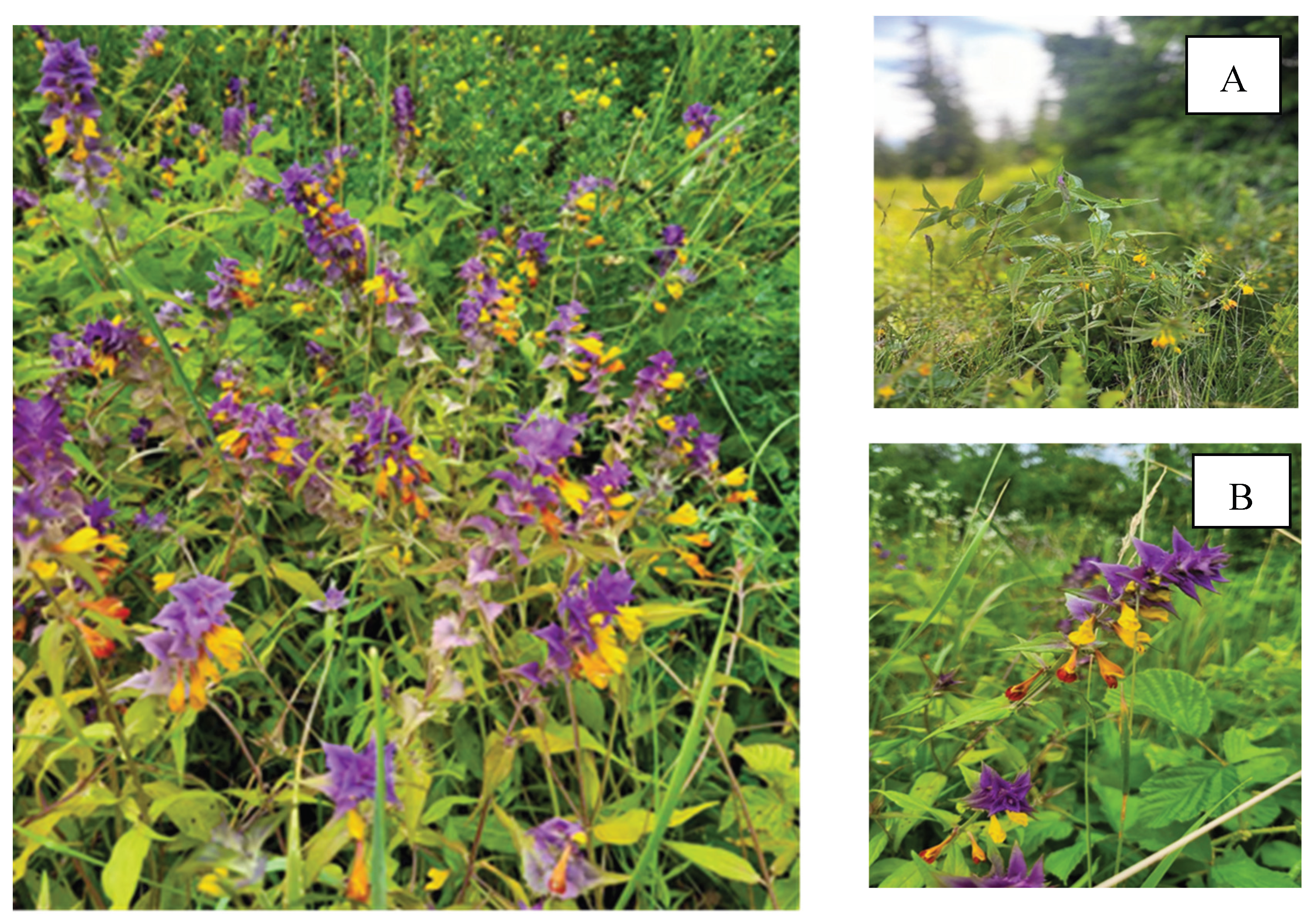

Table 1.
Content of phenolic compounds in the liquid extracts of Melampyrum nemorosum herb.
| № | Compound | Content in the dry residue of the extract, µg/g | |
|---|---|---|---|
| MN40 | MN70 | ||
| Phenolic acids | |||
| 1 | Hydroxyphenylacetic acid | 399 ± 28 | 439 ± 31 |
| 2 | Benzoic acid | 9483 ± 558 | 10431 ± 813 |
| Hydroxycinnamic acids | |||
| 3 | Chlorogenic acid | 1492 ± 59 | 1790 ± 121 |
| 4 | Caffeic acid | 1156 ± 54 | 1364 ± 64 |
| 5 | p-Coumaric acid | 5102 ± 336 | 6633 ± 437 |
| 6 | trans-Ferulic acid | 188 ± 16 | 263 ± 22 |
| 7 | trans-Cinnamic acid | 2926 ± 139 | 4291 ± 254 |
| Flavonoids | |||
| 8 | Rutin | 33885 ± 1343 | 40992 ± 1416 |
| 9 | Quercetin-3-O-glucoside | 3899 ± 16 | 4237 ± 26 |
| 10 | Naringin | 6793 ± 294 | 8530 ± 456 |
| 11 | Neohesperidin | 6183 ± 252 | 8 129 ± 254 |
| 12 | Quercetin | 8916 ± 332 | 11 591 ± 431 |
| 13 | Naringenin | 3485 ± 132 | 4879 ± 185 |
| 14 | Apigenin | 1068 ± 56 | 1548 ± 81 |
| 15 | Rhamnetin | 6723 ± 284 | 8403 ± 355 |
| 16 | Kaempferol | 2531 ± 111 | 3392 ± 149 |
| Tannin metabolites | |||
| 17 | Catechin | 1956 ± 103 | 1779 ± 94 |
| 18 | Epicatechin | 4158 ± 209 | 4491 ± 227 |
| 19 | Gallocatechin | 1214 ± 118 | 1578 ± 164 |
| Content of compound groups in the liquid extract, % (spectrophotometry) | |||
| Total polyphenols (%) | 4.73 ± 0.20 | 6.77 ± 0.21 | |
| Flavonoids (%) | 0.86 ± 0.07 | 1,01 ± 0.06 | |
Notes: MN40 – extract obtained with 40% aqueous-ethanol solution, MN70 – extract obtained with 70% aqueous-ethanol solution.
Table 2.
The effect of Melampyrum nemorosum herb extracts on the development of limb oedema in rats.
Table 2.
The effect of Melampyrum nemorosum herb extracts on the development of limb oedema in rats.
| Group of animals | Dose, mg/kg | Inflammatory response suppression index, % | |||||
|---|---|---|---|---|---|---|---|
| In 1 hour | In 3 hours | In 5 hours | In 1 hour | In 3 hours | In 5 hours | ||
| Group І (MN40) | 100 | 29.45±1.57** | 32.13±3.33* | 29.31±5.92 | 15.89 | 33.47 | 35.36 |
| Group ІІ (MN70) | 100 | 24.95±3.12 | 27.88±1.98* | 24.38±1.46* | 28.82 | 42.25 | 46.24 |
| Group ІІІ (Diclofenac sodium) |
8 | 21.29±2.23* | 20.18±4.51* | 17.82±4.29* | 39.25 | 58.21 | 60.69 |
| Group ІV (Quercetin) | 5 | 24.51±2.20* | 29.15±1.46* | 28.43±2.3* | 30.07 | 39.65 | 37.30 |
| Group V (control animals) |
- | 35.041±4.14 | 48.30±5.86 | 45.35±6.62 | - | - | - |
Notes: * — statistically significant difference compared to the control group (p ≤ 0.05); ** — statistically significant difference compared to the group of animals treated with diclofenac sodium (p ≤ 0.05); MN40 – extract obtained with 40% aqueous-ethanol solution, MN70 – extract obtained with 70% aqueous-ethanol solution.
Table 3.
The effect of the liquid extracts from Melampyrum nemorosum herb and water pepper on bleeding duration.
Table 3.
The effect of the liquid extracts from Melampyrum nemorosum herb and water pepper on bleeding duration.
| Group of animals | Bleeding duration, s, n = 6 | Reduction in bleeding time, relative to the control group, % |
|---|---|---|
| Group І (MN40) | 89.50±13.48* | 38.49 |
| Group ІІ (MN70) | 79.33±9.28* | 45.48 |
| Group ІІІ (Liquid extract of Polygonum hydropiper) | 61.83±3.38* | 57.51 |
| Group IV (control animals) | 145.5±12.24 | - |
Notes: * — statistically significant difference compared to the control group, P ≤ 0.05. MN40 – extract obtained with 40% aqueous-ethanol solution, MN70 – extract obtained with 70% aqueous-ethanol solution.
Table 4.
Dynamics of the wound healing process with the use of the liquid extracts from Melampyrum nemorosum herb.
Table 4.
Dynamics of the wound healing process with the use of the liquid extracts from Melampyrum nemorosum herb.
| Group of animals | Wound healing area in dynamics (days), % | |||||||
|---|---|---|---|---|---|---|---|---|
| 2 | 3 | 5 | 7 | 9 | 11 | 13 | 15 | |
| Group І (MN40) | 5.93 | 17.78 | 34.44 | 57.04 | 77.04 | 92.96 | 98.89 | 100 |
| Group ІІ (MN70) | 4.81 | 19.26 | 40.37 | 71.85 | 89.63 | 97.78 | 100 | 100 |
| Group ІІ (Rotokan) | 5.19 | 18.52 | 30.37 | 53.17 | 75.93 | 87.78 | 95.93 | 100 |
| Group IV (control animals) | 2.59 | 7.78 | 28.52 | 37.41 | 51.11 | 73.33 | 85.93 | 96.30 |
Notes: MN40 – extract obtained with 40% aqueous-ethanol solution, MN70 – extract obtained with 70% aqueous-ethanol solution.
Table 5.
Antimicrobial and antifungal activity of Melampyrum nemorosum herb extracts (at a concentration of 10 mg/mL).
Table 5.
Antimicrobial and antifungal activity of Melampyrum nemorosum herb extracts (at a concentration of 10 mg/mL).
| Types of microorganisms | Clinical material | Resistance | Diameters of growth inhibition zones, mm | |||
| Ethanol 40% | Ethanol 70% | MN40 | MN70 | |||
| Staphylococci | ||||||
| Staphylococcus аureus | ATCC 25923 | S | 13.00±0.54 | 18.10±1.50 | growth | 22.17±1.15* |
| Staphylococcus aureus | Pharynx | BSSA | growth | 20.35±1.05 | 11.63±0.08* | 32.02±1.60* |
| Staphylococcus аureus | Skin | S | growth | 16.68±0.49 | 10.32±0.60* | 24.71±1.55* |
| Staphylococcus aureus | Wound | BSSA, MLS | growth | 22.06±0.82 | 12.59±0.57* | 33.00±3.67* |
| Staphylococcus aureus | Wound | MRSA | growth | 16.62±0.94 | 10.57±0.50* | 23.99±1.34* |
| Staphylococcus epidermidis | Skin | MLS, Ind+ | growth | 22.75±1.01 | growth | 22.96±0.57 |
| Staphylococcus haemolyticus | Sputum | MRSH | growth | 14.25±0.49 | growth | 22.99±2.17* |
| Enterococci | ||||||
| Enterococcus faecalis | Urethra | Tet, FQin | growth | 11.73 ±0.43 | 11.92±0.64* | 21.12±1.29* |
| Enterococcus faecalis | Urethra | Tet, FQin | growth | growth | 13.99±1.33* | 16.80±0.54* |
| β-Hemolytic streptococci | ||||||
| Streptococcus pyogenes (β Str A) | Pharynx | S | 14.60±2.28 | 16.39±0.82 | growth | 14.73±2.08 |
| Streptococcus pyogenes (β Str A) | Pharynx | S | growth | 10.43±0.42 | growth | 13.45±1.30* |
| Streptococcus dysgalactiae (β Str G) | Pharynx | S | growth | growth | growth | 12.18±1.17* |
| Streptococcus agalactiae (β Str B) | Vaginal mucus | S | growth | 17.45±1.29 | growth | 18.89±2.32* |
| α-Hemolytic streptococci | ||||||
| Streptococcus anginosus | Pharynx | AMO, Tet, MLS | 15.51±1.28 | 13.63±1.20 | growth | 14.59±0.76 |
| Streptococcus gordonii | Oral cavity | S | 11.53±0.98 | 13.04±0.21 | growth | 13.07±0.67 |
| Streptococcus oralis | Oral cavity | S | growth | 14.67±0.56 | 9.67±0.63* | 20.44±1.07* |
| Streptococcus sanguinis | Oral cavity | S | growth | 19.54±0.87 | growth | 20.65±1.02 |
| Streptococcus pneumonia | Sputum | S | growth | growth | growth | growth |
| Streptococcus pneumonia | Sputum | β-Lac, Tet | growth | growth | growth | 17.40±0.21* |
| Enterobacteria | ||||||
| Escherichia coli | Wound | S | growth | 8.70±0.52 | growth | growth |
| Escherichia coli | Wound | S | growth | growth | growth | 12.89±0.56* |
| Escherichia coli | Wound | AMO Tet, FQin | growth | growth | growth | 11.79±0.69* |
| Escherichia coli hly+ | Fecal sample | AMO, MLs | growth | growth | growth | 13.20±0.95* |
| Escherichia fergusonii | Fecal sample | ESbL | growth | growth | growth | growth |
| Providencia rettgeri | Urine sample | ESbL | growth | growth | growth | growth |
| Morganella morganii | Urine sample | ESbL | growth | growth | growth | growth |
| Non-fermenting bacteria | ||||||
| Pseudomonas aureginosa | Wound | ESbL | growth | growth | growth | growth |
| Pseudomonas aureginosa | Pus from the wound | ESbL | growth | growth | growth | growth |
| Acinetobacter baumani | Sputum | ESbL | growth | growth | growth | 10.61±1.03* |
| Bacilli | ||||||
| Bacilus subtilus | АТСС 6051 | S | growth | 11.89±0.40 | growth | 17.75±1.37* |
| Fungi | ||||||
| Candida albicans | Oral cavity | FCZ-R | growth | 16.35±1.3 | growth | 22.22±1.25* |
| Candida albicans | Sputum | FCZ-R | growth | 21.86±1.22 | growth | 31.64±2.24* |
| Candida albicans | Urine sample | FCZ-R | growth | 10.82±0.34 | growth | 15.53±0.64* |
| Candida albicans | Oral cavity | FCZ-S | growth | 11.15±0.58 | growth | 15.24±0.29* |
| Candida tropicalis | Sputum | FCZ-R | growth | 10.02±0.44 | growth | 16.34±0.80* |
| Candida glabrata | FRS 585 | FCZ-S | growth | 19.59±1.89 | growth | 21.29±1.20 |
| Candida lusitaniae | Oral cavity | FCZ-R | growth | 10.13±1.47 | growth | 19.95±0.97* |
| Candida lusitaniae | Oral cavity | FCZ-S | growth | 20.96±1.22 | growth | 26.88±0.55* |
| Candida lipolytica | Oral cavity | FCZ-R | growth | 10.29±0.81 | growth | 15.16±0.60* |
| Candida kefyr | Oral cavity | FCZ-S | 10.59±0.99 | 15.60±0.43 | growth | 26.55±2.30* |
| Aspergillus fumigatus | Sputum | FCZ-R | growth | growth | growth | 17.04±3.20* |
| Rhodotorula mucilaginosa | Pus from the wound | FCZ-S | growth | 25.68±0.99 | growth | 32.32±1.25* |
| Geotrichum candidum | Fecal sample | FCZ-S | growth | 10.73±0.59 | growth | 20.22±0.87* |
Notes: 1. MRSA – methicillin-resistant S. aureus; BSSA – borderline methicillin-resistant S. aureus (β-lactamase hyperproduction); ESβL – extended-spectrum β-lactamases; MLS resistance – resistance to macrolides and lincosamides; Tet – tetracycline; FQin – fluoroquinolones; S – antibiotic-sensitive strains; FCZ-R – fluconazole-resistant; FCZ-S – fluconazole-sensitive. 2. p < 0.05 compared to the control (solvent).
Disclaimer/Publisher’s Note: The statements, opinions and data contained in all publications are solely those of the individual author(s) and contributor(s) and not of MDPI and/or the editor(s). MDPI and/or the editor(s) disclaim responsibility for any injury to people or property resulting from any ideas, methods, instructions or products referred to in the content. |
© 2025 by the authors. Licensee MDPI, Basel, Switzerland. This article is an open access article distributed under the terms and conditions of the Creative Commons Attribution (CC BY) license (http://creativecommons.org/licenses/by/4.0/).
Copyright: This open access article is published under a Creative Commons CC BY 4.0 license, which permit the free download, distribution, and reuse, provided that the author and preprint are cited in any reuse.


