Preprint
Article

This version is not peer-reviewed.

Lactic Acid Bacteria from Traditional Fermented Milk: Antimicrobial Potential Against Foodborne Pathogens

Submitted:

28 October 2025

Posted:

30 October 2025

You are already at the latest version

Abstract

Lactic acid bacteria (LAB) are increasingly recognized for their role in food biopreservation due to their ability to synthesize antimicrobial compounds. Milk naturally harbors a wide variety of lactic acid bacteria, offering a promising source for identifying strains with biopreservative potential. This study investigated the antagonistic effects, safety characteristics, and technological properties of LAB strains isolated from traditionally fermented milk. Thirty-two dairy samples were analyzed, and the resulting LAB isolates were screened for inhibitory activity against Listeria monocytogenes CECT 4032 and Staphylococcus aureus CECT 976 using agar spot and well diffusion assays. All tested strains exhibited strong antimicrobial effects, with particularly notable inhibition of L. monocytogenes. After phenotypic screening, five representative isolates were selected for molecular identification and further assessment of safety-related attributes, functional capabilities, auto- and co-aggregation properties 16S rRNA gene sequencing revealed that four strains belonged to the genus Enterococcus, specifically, one E. faecium and three E. durans, while one was classified as a Lactococcus species. Moreover, none of the strains showed proteolytic or lipolytic activities which highlights their potential use in dairy fermentation processes.

Keywords: 
;  ;  ;  ;  

1. Introduction

Lactic acid bacteria (LAB) have attracted considerable scientific interest due to their capacity to produce a broad spectrum of antimicrobial substances. These metabolites contribute to the inhibition of undesirable microorganisms, such as spoilage bacteria and foodborne pathogens, thereby enhancing the microbial safety and extending the shelf life of a wide range of food products. Their natural occurrence in fermented foods and recognized safety status further support their application as bio-preservative agents in food systems [1]. These Gram-positive, fermentative bacteria are classified within the phylum Firmicutes, which includes a variety of genera important in food fermentation [2]. The incorporation of LAB into food systems contributes significantly to improving sensory qualities, including flavor, consistency, and nutritional content. Beyond their well-known technological contributions to fermentation, LAB also play a pivotal role in improving food safety. This is largely due to their capacity to inhibit the growth of spoilage microorganisms and pathogenic bacteria through the synthesis of various antimicrobial compounds. By producing such bioactive metabolites, LAB help create an inhospitable environment for undesirable microbes, thereby contributing to the microbiological stability and safety of food products [3,4]. Lactic acid bacteria exhibit antimicrobial activity through the synthesis of various bioactive metabolites. Among the antimicrobial compounds produced by LAB, organic acids, particularly lactic and acetic acid, play a fundamental role in pathogen inhibition. These acids reduce the pH of the surrounding environment, thereby creating conditions that are hostile to the survival and proliferation of many spoilage and pathogenic microorganisms. In addition to organic acids, LAB produce a variety of other antimicrobial substances that contribute to their protective role in food systems. These include diacetyl, which interferes with bacterial metabolism; carbon dioxide (CO₂), which exerts an inhibitory effect by reducing oxygen availability; and hydrogen peroxide, which induces oxidative stress in microbial cells. Moreover, LAB are known for their ability to synthesize bacteriocins, ribosomally produced antimicrobial peptides that exhibit targeted activity against closely related bacteria, including several foodborne pathogens. These compounds often act synergistically, enhancing the overall inhibitory effect against a broad spectrum of spoilage and pathogenic microorganisms. The combined action of these compounds enhances the overall inhibitory potential of LAB in fermented food systems [5,6]. Among the various antimicrobial substances produced by LAB, bacteriocins are considered especially promising. These peptides exhibit strong inhibitory effects against a wide range of pathogenic bacteria associated with foodborne illnesses. Their potent antimicrobial properties make them promising agents for improving food safety and prolonging product shelf life [7,8]. Lactococcus and Enterococcus species are commonly found in raw and fermented milk, reflecting their strong adaptation to dairy ecosystems [9,10]. Species such as L. garvieae and various subspecies of L. lactis have been reported to produce a broad spectrum of bacteriocins [11]. Notably, L. lactis strains exhibiting antimicrobial activity have gained considerable interest as promising biopreservatives in the dairy industry [12]. Enterococcus species likewise produce enterocins, a group of bacteriocins that exhibit antimicrobial activity against Gram-positive bacteria, including important foodborne pathogens like Listeria spp. [13]. Among Enterococcus species, E. faecalis and E. faecium are the main producers of enterocins. These strains have been extensively studied owning to their reliable ability to generate various types of enterocins with strong antimicrobial effects [14]. Numerous studies have focused on isolating LAB with antimicrobial activity from dairy matrices. For instance, Achemchem et al. [15] reported the isolation of the bacteriocin-producing Enterococcus faecium F58 strain from traditional soft goat cheese, while Ghrairi et al. [16] reported LAB with anti-Listeria activity from Rigouta, a traditional Tunisian cheese. Additionally, Perin, and Nero [17] identified Lactococcus and Enterococcus as predominant genera in the microbial community of raw goat’s milk. They also highlighted the ability of these bacteria to inhibit the growth of Listeria monocytogenes ATCC 7644. Elotmani et al. [18] also reported the presence of LAB with antilisterial activity in Raïb, a traditional Moroccan fermented milk. In light of these findings, the present study aimed to isolate LAB strains from traditionally fermented milk and assess their antimicrobial activity against key foodborne pathogens. Special emphasis was placed on evaluating the ability of these isolates to produce bacteriocins, antimicrobial peptides known for their targeted inhibitory effects against pathogenic and spoilage microorganisms. Additionally, their safety characteristics and technological functionalities were examined to determine their suitability for application in cheese biopreservation.

2. Materials and Methods

2.1. Isolation of LAB from Milk

A total of 32 samples of spontaneously fermented milk were analyzed to isolate LAB. Samples were serially diluted in sterile saline to obtain appropriate concentrations for microbial plating and then applied onto two selective culture media, Man, Rogosa, and Sharpe (MRS) agar (Biokar Diagnostics, France) and M17 agar (Biokar Diagnostics, France) supplemented with 0.5% glucose (GM17). The inoculated plates were incubated at 30 °C for 24 to 48 hours. After incubation, colonies showing typical LAB morphology were isolated and purified. The bacterial isolates obtained were subsequently evaluated for their ability to inhibit the growth of specific pathogenic bacteria.

2.2. Screening for Bacteriocinogenic LAB

To screen for bacteriocin production, several colonies were randomly picked and transferred onto two sets of agar plates for further analysis [15]. These plates were then incubated anaerobically at 30 °C for 16 to 24 hours to allow sufficient bacterial growth. After the initial incubation period, the plates were overlaid with a layer of soft agar (6ml, 0.75% agar) that had been inoculated with approximately 10⁸ CFU/mL of an overnight culture of the indicator organisms, either Listeria monocytogenes CECT 4032 or Staphylococcus aureus 976. These indicator strains were previously cultivated in Tryptic Soy Broth (TSB) (Biokar Diagnostics, France) under optimal conditions to ensure sufficient growth and viability before use in the overlay assay. The formation of clear inhibition zones indicated antimicrobial activity.

2.3. Indicator Pathogens and Antimicrobial Spectrum

The inhibitory potential of the isolated strains was assessed using both the agar spot and well diffusion methods, following the methods described by Achemchem et al. [19]. In the agar spot test, 5 µL of overnight LAB cultures grown in either MRS or GM17 broth were carefully spotted onto the surface of solidified agar plates of the corresponding medium. After incubation at 30 °C for 24 hours to allow bacterial growth, The plates were overlaid with soft agar previously inoculated with an overnight culture of the indicator organisms. The overlay was poured gently to ensure even distribution across the plate surface. Following a second incubation, inhibition zones around the colonies were observed as indicators of antimicrobial activity.
For the well diffusion assay, LAB strains were cultured overnight at 30 °C in MRS or GM17 broth. The cultures were then centrifuged, and the resulting supernatants were collected to obtain cell-free supernatants (CFS). Wells were created in Mueller Hinton Agar (MHA) plates (Biokar Diagnostics, France) using sterile stainless-steel cylinders. After wells were prepared in the agar medium, each plate was overlaid with soft agar previously inoculated with an overnight culture of the designated indicator microorganism. Once the overlay solidified at room temperature, 100 µL of the prepared CFS was carefully added to each well. To ensure uniform diffusion of antimicrobial compounds into the agar, the plates were first incubated at 4 °C for 2 hours. This was followed by incubation at 37 °C for 16 hours to allow the growth of the indicator strain and expression of any inhibitory activity. Antimicrobial effects were assessed by measuring the diameter of the clear inhibition zones around the wells.
Ten pathogenic bacterial strains were chosen as indicator organisms to assess the antimicrobial activity of the tested isolates. The selected Gram-positive strains included L. monocytogenes CECT 4032, CECT 7467, CECT 5725, and CECT 935; S. aureus CECT 976; and Bacillus subtilis DSMZ 6633. The Gram-negative group consisted of Pseudomonas aeruginosa CECT 118, Escherichia coli ATCC 25922 and CECT 4076, and Salmonella enterica CECT 704. All indicator strains were cultivated in TSB at 37 °C for 18 hours before use in the assays. As a positive control, Enterococcus faecium F58, a well-characterized bacteriocin-producing strain, was grown on MRS agar under standard incubation conditions.

2.4. Stability of Bacteriocin-like Activity After Exposure to Proteolytic Enzymes

To evaluate whether the antibacterial compounds produced by the isolated strains were proteinaceous, their sensitivity to proteolytic enzymes was tested. Cell-free supernatants (CFS) obtained from cultures at the early stationary phase were incubated at 37 °C for 2 hours with proteinase K (1 mg/mL), prepared in the buffer recommended by the manufacturer (Qiagen, Germany). L. monocytogenes CECT 4032 was used as the indicator strain, while untreated CFS served as negative controls. Residual antibacterial activity in both treated and untreated samples was assessed through the well diffusion assay.

2.5. Identification of Antagonistic LAB

2.5.1. Phenotypic and Biochemical Identification

Strains producing bacteriocins were first examined using phenotypic methods, including Gram staining, catalase activity testing, and microscopic observation of cellular morphology. Their biochemical characteristics were also evaluated, such as their ability to grow at 10 °C and 45 °C, tolerate 4% and 6.5% NaCl, and grow on Bile Esculin Azide (BEA) agar (Biokar Diagnostics, France). To achieve more precise taxonomic identification, the isolates were analyzed using the API 20 Strep identification system (bioMérieux, France), according to the manufacturer’s guidelines.

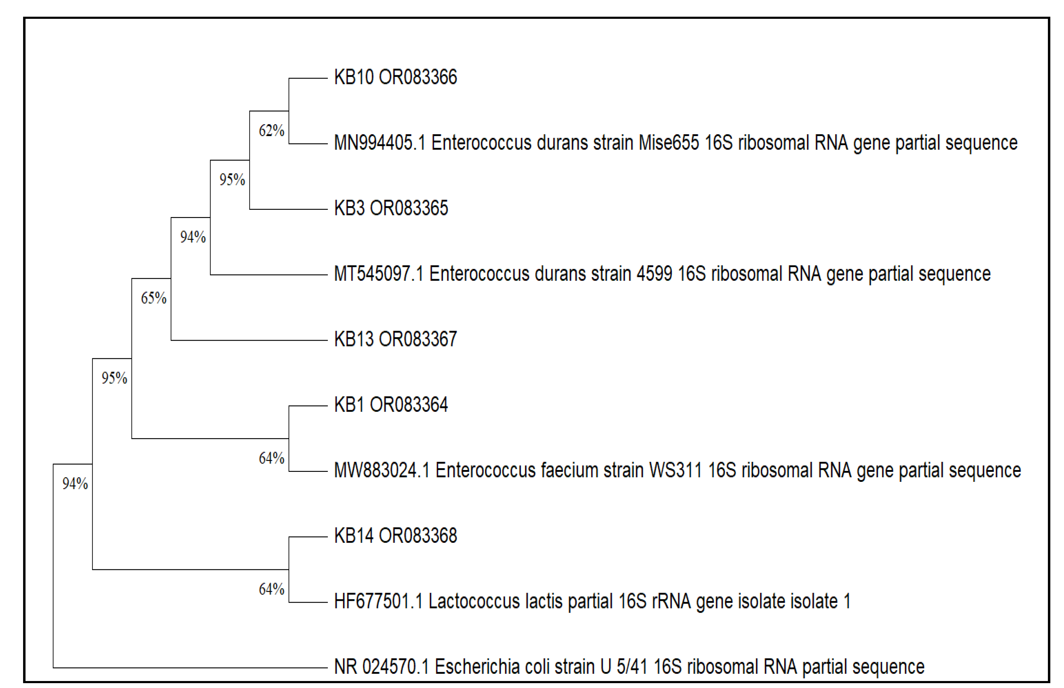
2.5.2. Molecular Identification (16S rRNA)

Following the procedure described by Elidrissi et al. [20], genomic DNA was extracted from bacterial cultures and quantified. A fragment of about 1500 base pairs from the 16S rRNA gene was amplified using universal primers in a 25 µL PCR reaction, with standard thermal cycling including an initial denaturation, 35 cycles of denaturation, annealing, extension, and a final elongation step. The PCR products were checked by agarose gel electrophoresis. Purified amplicons were then sequenced using specific primers. The sequencing products were further purified, analyzed by capillary electrophoresis, and the resulting sequences were identified by BLAST comparison against the NCBI database.
These procedures were performed at the Technical Support Units for Scientific Research (UATRS), affiliated with the National Center for Scientific and Technical Research (CNRST), Morocco.

2.6. Technological Assessment of LAB Strains

2.6.1. Proteolytic and Lipolytic Activities

Proteolytic activity was evaluated by inoculating the isolates onto Trypticase Soy Agar (TSA) (Biokar Diagnostics, France) containing 1.5% (w/v) skim milk, following the protocol of Achemchem et al. [19]. The presence of clear halos around the colonies was used as an indicator of proteolytic activity.
The lipolytic activity of the isolates was examined by testing them on nutrient agar enriched with 10 g/L Tween 20 and 0.1 g/L calcium chloride, following the protocol established by Albayrak, and Duran [21]. Ten microliters of each culture were placed onto the prepared medium and incubated under anaerobic conditions at 37 °C for 72 hours. The detection of lipase production was based on the visual observation of precipitate formation due to the interaction between calcium ions and fatty acids.

2.6.2. Aggregation Abilities

The aggregation properties of the selected LAB strains were evaluated to determine their potential for adhesion and interaction with pathogenic microorganisms, following the procedures outlined by Elidrissi et al. [20] and Zanzan et al. [22].
Auto-aggregation tests were conducted to evaluate the ability of LAB strains to adhere to their own cells. The degree of auto-aggregation was calculated as a percentage using the formula: Auto-aggregation (%): 1 - (At/A0) * 100, where A0 and At represent the initial and final absorbance at 600 nm, respectively.
To determine the co-aggregation capacity between LAB strains and the foodborne pathogen L. monocytogenes CECT 4032, equal volumes of each bacterial culture were combined and incubated at room temperature without agitation for 4 and 24 hours. Absorbance measurements were taken for the individual cultures as well as their mixture. Co-aggregation percentage was calculated using: Coaggregation % = ((Alab + Apat) – Amix)/ (Alab + Apat) × 100, where Alab + Apat are the initial absorbances of the LAB and pathogen suspensions, respectively, and Amix is the absorbance of the mixture at the specified time point.

2.7. Evaluation of Safety-Related Traits

2.7.1. Hemolytic Activity

The bacterial isolates were tested for hemolytic activity by inoculating them onto blood agar containing 5% sheep blood, followed by incubation at 37 °C for 48 hours. After incubation, the presence of hemolysis was evaluated [23]. S. aureus CECT 976 served as the positive control.

2.7.2. Antibiotic Susceptibility

The antibiotic susceptibility of the bacterial isolates was evaluated using the disc diffusion method, following the procedure described by Doménech-Sánchez et al. [24]. All isolates were tested against a range of antibiotics.
The antibiotics tested in this research comprised streptomycin (25 µg), vancomycin (30 µg), netilmicin (30 µg), ampicillin (20 µg), ciprofloxacin (5 µg), penicillin G (10 units), erythromycin (15 µg), chloramphenicol (30 µg), fosfomycin (200 µg), fusidic acid (10 µg), tetracycline (30 µg), gentamicin (10 µg), and kanamycin (30 µg). The inhibition zone diameters around the antibiotic discs were measured, and the results were interpreted based on the guidelines provided by CLSI [25] and Mazlumi et al. [26]. Based on the inhibition zone diameters, isolates were classified as susceptible, intermediate, or resistant.

2.7.3. Gelatinase Activity and Biogenic Amine Production

Gelatinase activity was assessed by inoculating 1 µL of an overnight culture onto Brain Heart Infusion (BHI) agar (Biokar Diagnostics, France) supplemented with 3% (v/v) gelatin, following the protocol described by Amidi-Fazli, and Hanifian [27]. The inoculated plates were incubated at 37 °C for 48 to 72 hours, then transferred to 4 °C for 4 hours. The appearance of opaque halos surrounding the colonies was interpreted as a positive indication of gelatinase activity.
The ability of the LAB isolates to produce biogenic amines was assessed using a decarboxylase assay, following the method described Maijala [28]. The test was carried out on Maijala agar medium supplemented with 20 g/L histidine, used as the amino acid pre-cursor. The formation of a purple coloration in and around the bacterial colonies served as an indicator of histamine production.

2.9. Statistical Analysis

Statistical analyses were performed using one-way analysis of variance (ANOVA), followed by Tukey’s post hoc test, with Statistica version 6.0.

3. Results

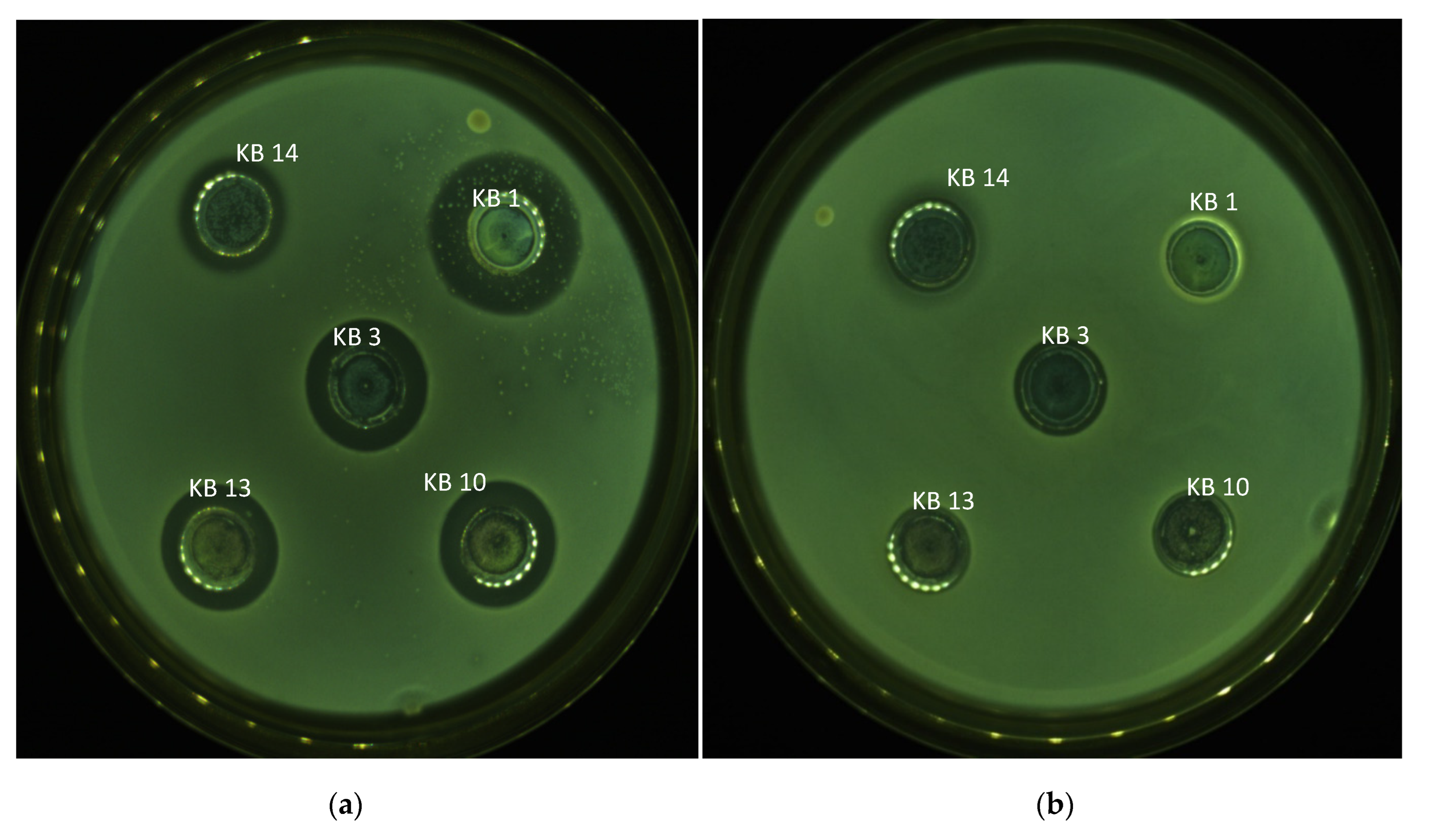
3.1. Bacteriocinogenic Potential of LAB Strains

Lactic acid bacteria isolated from dairy-derived samples were subjected to initial screening to assess their antimicrobial potential. This preliminary evaluation employed the double-layer agar technique against two common foodborne pathogens: L. monocytogenes CECT 4032 and S. aureus CECT 976. Isolates that demonstrated inhibitory effects against at least one of these indicator strains were subsequently selected for further analysis. Their capacity to produce antimicrobial metabolites was then investigated using the agar well diffusion method. To confirm these findings, the antagonistic activity of the isolates was also tested in liquid culture against both L. monocytogenes and S. aureus (Table 1).
Out of all the isolates tested, 18 LAB strains demonstrated significant antimicrobial activity, producing distinct inhibition zones ranging from 11 to 19 mm in diameter against one or both target pathogens. These strains were subjected to basic phenotypic characterization, including Gram staining, catalase testing, and assessment of growth under various environmental conditions (Table 1). All selected isolates were Gram-positive, catalase-negative, and exhibited coccoid morphology under microscopic observation, features characteristic of typical LAB.
To further characterize these antimicrobial strains, the API 20 STREP identification system was used. From this group, five representative isolates were selected for molecular identification by 16S rRNA gene sequencing. Sequence alignment revealed a high degree of similarity to known LAB reference strains available in the GenBank database (Table 2). The phylogenetic relationships of these isolates are depicted in Figure 1, highlighting their clustering with closely related species. Based on the 16S rRNA sequence data, the strains were identified as Lactococcus lactis, Enterococcus faecium, and Enterococcus durans.
Eighteen strains were assessed for their ability to inhibit different pathogenic microorganisms. Using the spot method, all tested LAB isolates demonstrated clear antimicrobial activity against Listeria spp., indicating a strong antagonistic potential. In addition to this consistent antilisterial effect, several strains exhibited inhibitory activity against a broader panel of pathogens, including S. aureus CECT 976, Bacillus subtilis DSMZ 6633, Escherichia coli ATCC 25922, E. coli CECT 4076, Salmonella enterica CECT 704, and Pseudomonas aeruginosa CECT 118 (Table A1 and Table A2).
In the agar well diffusion assay, all selected LAB strains exhibited clear inhibitory effects against L. monocytogenes, confirming their ability to produce antimicrobial substances capable of diffusing through the agar medium. No inhibition zones were detected against either of the E. coli strains or S. enterica CECT 704, indicating the absence of measurable activity against these Gram-negative bacteria under the tested conditions.
Strain-specific antimicrobial patterns were observed against other indicator organisms. E. faecium KB1 showed inhibitory activity against P. aeruginosa CECT 118. E. durans strains KB3, KB10, and KB13 inhibited B. subtilis DSMZ 6633, while L. lactis KB14 exhibited activity against S. aureus CECT 976. These results highlight the variability in antimicrobial performance among the tested LAB strains, with some demonstrating broader spectra of activity beyond L. monocytogenes.
The cell-free supernatants from the five selected strains were treated with proteinase K to assess the nature of the antimicrobial compounds. The enzymatic digestion neutralized the inhibitory activity, indicating that the active substances were proteinaceous, likely corresponding to bacteriocins (Figure 2).
None of the five tested strains exhibited gelatinase or hemolytic activity, indicating the absence of these potential virulence factors. Table 3 summarizes the antibiotic susceptibility results for the five isolates evaluated in this research. All strains were susceptible to fosfomycin and tetracycline. However, strain KB1 exhibited resistance to penicillin G, streptomycin, gentamicin, netilmicin, and kanamycin. Strain KB3 showed resistance only to streptomycin, while KB13 was the only isolate resistant to chloramphenicol. In contrast, strains KB14 and KB10 exhibited favorable susceptibility profiles, being either sensitive or intermediately susceptible to all 13 antibiotics tested. Additionally, none of the isolates showed proteolytic or lipolytic activity, further supporting their safety potential for use in food-related applications.

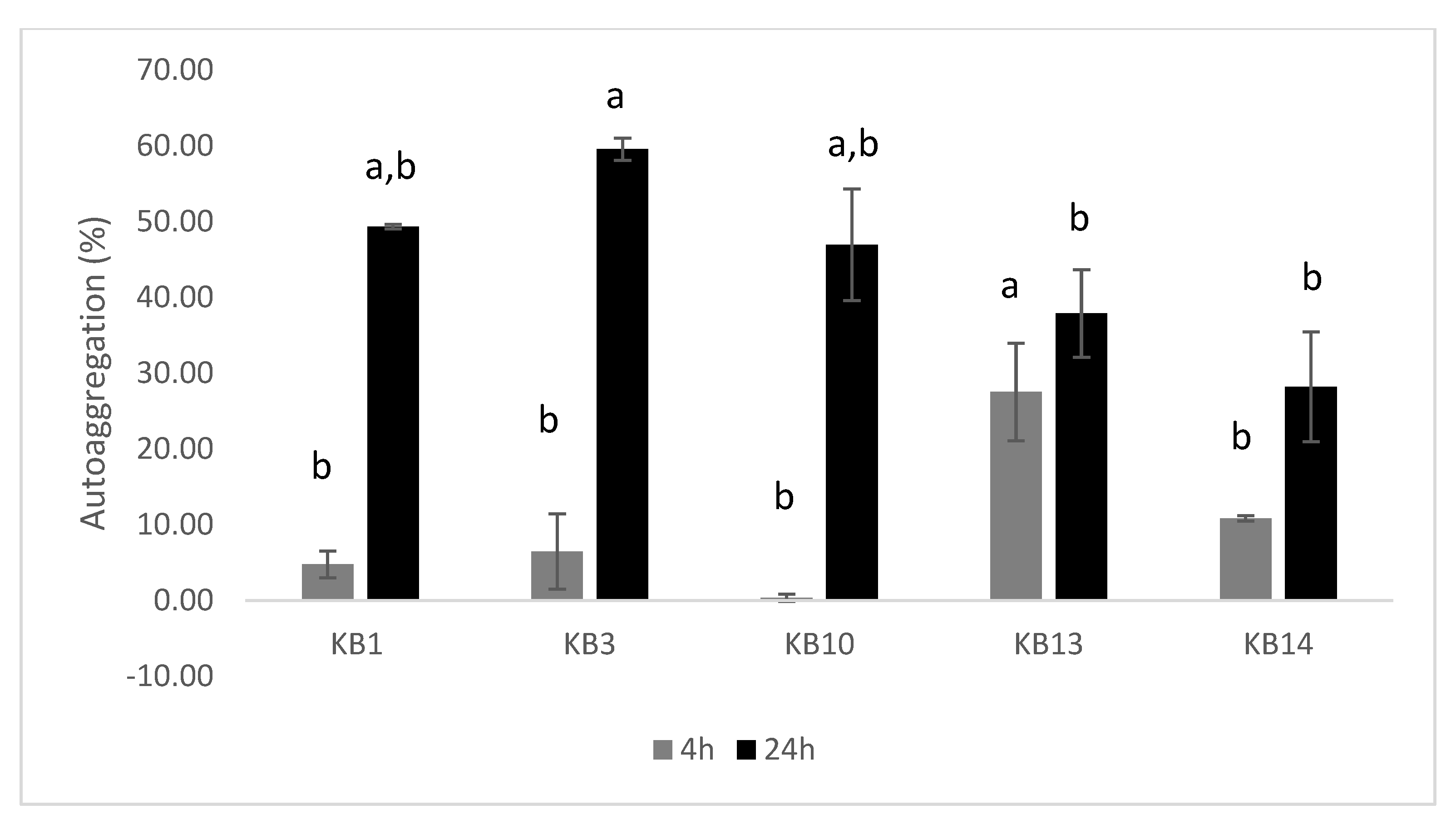
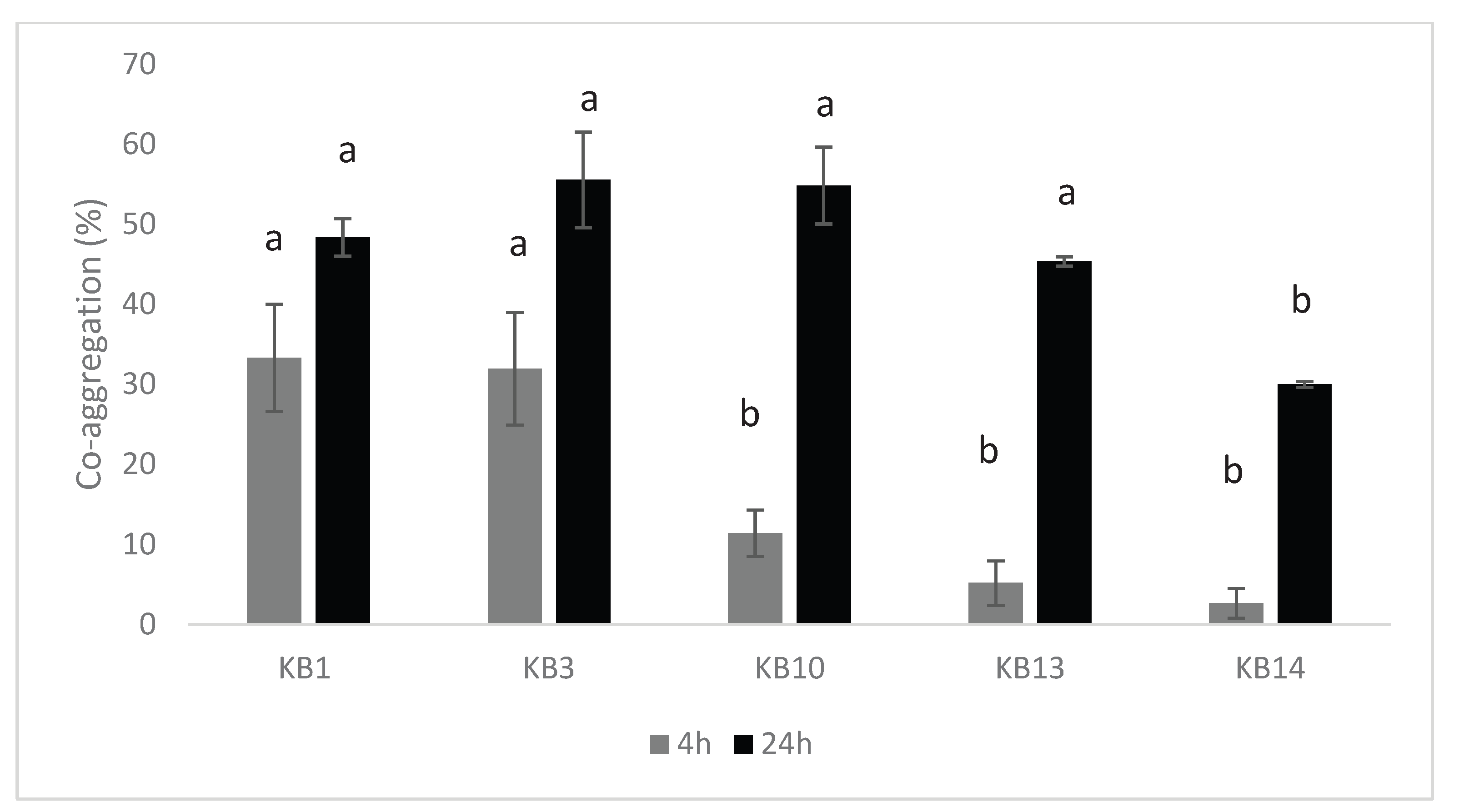
3.2. Aggregation Capacity of LAB Isolates

The ability of the five selected LAB isolates to auto-aggregate was evaluated over time. After 4 hours of incubation, auto-aggregation levels varied considerably among the strains, ranging from 0.35 ± 0.50% to 27.53 ± 6.46% (Figure 3). A substantial increase was observed after 24 hours, with values reaching between 28.21 ± 7.25% and 59.56 ± 1.48%, indicating a time-dependent enhancement in cell-to-cell aggregation capacity. This characteristic is often associated with improved colonization potential and stability in microbial communities.
Co-aggregation with L. monocytogenes CECT 4032 also revealed strain-specific differences (Figure 4). Among the tested isolates, L. lactis KB14 showed the lowest level of co-aggregation, while E. durans KB3 exhibited the highest interaction with the pathogen. After 24 hours, co-aggregation values ranged from 30.00 ± 0.38% to 55.52 ± 5.96%, with statistically significant differences between strains (p < 0.05). These findings suggest varying potential among isolates for pathogen exclusion through co-aggregation, which may contribute to their probiotic or bioprotective roles.

4. Discussion

4.1. Antimicrobial Potential and Safety Assessment of LAB Strains

In the present study, LAB isolated from raw goat’s milk exhibited pronounced antimicrobial activity against L. monocytogenes, corroborating previous findings that emphasize the inhibitory potential of dairy-derived LAB [6,15,17,29,30,31,32,33]. Prior research has particularly highlighted the antimicrobial capabilities of E. faecium, E. durans, and L. lactis, especially those isolated from traditional artisanal cheeses. These strains have demonstrated the ability to inhibit not only L. monocytogenes, but also a range of other foodborne pathogens, including Bacillus cereus and P. aeruginosa [15,34,35,36,37,38,39,40].
Antibiotic susceptibility testing revealed that none of the isolates exhibited resistance to vancomycin, in agreement with previously published data [21,40,41]. Notably, L. lactis KB14, isolated in this study, was susceptible to ampicillin, chloramphenicol, and erythromycin, in line with the findings reported by Kazancıgil et al. [42].
Collectively, these findings support the potential application of the studied LAB strains as natural biopreservatives in cheese production. Their strong antimicrobial activity and favorable antibiotic resistance profiles suggest that they could contribute to enhancing food safety and extending shelf life in fermented dairy products.

4.2. Proteolytic and Lipolytic Activities

Our results are consistent with those of Albayrak, and Duran [21], who reported that Enterococcus strains isolated from dairy sources lacked proteolytic activity. This absence of proteolytic function suggests that certain Enterococcus spp. may have limited capacity to degrade milk proteins, potentially preserving the structural integrity and sensory attributes of fermented dairy products. However, proteolytic activity is not universally absent in this genus. Several other studies have reported the presence of proteolytic enzymes in Enterococcus species, highlighting strain-dependent variability. For instance, Achemchem et al. [19], de Sousa et al. [38], Mercha et al. [43], and Islam et al. [44] observed proteolytic activity in different Enterococcus strains, suggesting that some isolates may contribute to flavor development or textural changes.
Regarding Lactococcus strains, similar variability in proteolytic capabilities has been noted. With regard to Lactococcus strains, Cheng et al. [45], reported that the isolates tested exhibited proteolytic activity. Conversely, Allam et al. [46] identified two Lactococcus strains that lacked this enzymatic function, reinforcing the notion that proteolytic capacity is strain-specific and must be evaluated individually when selecting candidates for starter or adjunct cultures.
In our study, none of the tested strains displayed lipolytic activity, as no visible hydrolysis of lipids was observed. This finding aligns with previous reports indicating that nisin-producing L. lactis strains generally lack lipolytic enzymes [12]. Similarly, multifunctional LAB strains isolated from artisanal cheeses have also been shown to be non-lipolytic [21]. The absence of lipolytic activity is particularly advantageous in dairy fermentation, as excessive lipid hydrolysis can lead to off-flavors or rancid notes, compromising product quality.
Taken together, these results suggest that the selected LAB strains, due to their lack of both proteolytic and lipolytic activities, may be suitable for applications in dairy products where minimal enzymatic degradation is desired. Their neutral enzymatic profile makes them excellent candidates for preserving the sensory integrity of fresh cheeses or for use as protective cultures in biopreservation strategies without negatively influencing flavor or texture.

4.3. Auto-Aggregation and Co-Aggregation Capabilities of LAB Strains

The capacity of LAB strains to auto-aggregate and co-aggregate is widely recognized as a key functional characteristic that underpins their ability to persist and colonize host environments, such as the gastrointestinal tract. These aggregation properties contribute not only to microbial adhesion and biofilm formation but also to the exclusion of potential pathogens through competitive mechanisms. In the present study, the evaluated LAB strains exhibited varying levels of auto-aggregation after 4 hours of incubation, with some isolates demonstrating significantly stronger aggregation capabilities than others. These results align with those reported in the literature [42,47,48]. A longer incubation period (24 hours) led to a noticeable enhancement in auto-aggregation capacity for all tested strains, particularly for E. durans KB3, which exhibited the highest level of auto-aggregation. This increase over time may reflect enhanced expression of surface proteins or exopolysaccharides that promote cell–cell adhesion during prolonged growth phases.
In terms of co-aggregation, the tested strains also displayed a time-dependent increase in their ability to aggregate with L. monocytogenes CECT 4032, in agreement with patterns previously observed in the literature [49,50]. Notably, the co-aggregation ability of the Lactococcus strain was significantly lower than that of Enterococcus strains after 24 hours of incubation, suggesting species-specific differences in surface structures or in the composition of extracellular polymeric substances involved in cell interactions.
Both auto- and co-aggregation are considered beneficial traits for probiotic and bioprotective functions. Auto-aggregation is often associated with enhanced biofilm-forming ability, which facilitates stronger adhesion to intestinal epithelial surfaces and promotes colonization. Co-aggregation, on the other hand, plays a crucial role in the exclusion of enteric pathogens by forming physical barriers or interfering with pathogen adhesion, thus contributing to microbial equilibrium and host protection [33,51]. Collectively, these characteristics underline the promising potential of the selected LAB strains not only as probiotics capable of colonizing and persisting in the gastrointestinal environment but also as effective functional cultures for application in food systems, particularly for improving microbial safety and shelf life.

4.4. Protein-Based Features of Antimicrobial Substances from LAB

The LAB strains evaluated exhibited antimicrobial activity suggestive of bacteriocin production. These inhibitory effects were eliminated following treatment with a proteolytic enzyme (Figure 2), confirming the proteinaceous nature of the active compounds. Bacteriocin-producing LAB are increasingly recognized as promising natural biopreservatives in the food industry, particularly in the dairy sector, owing to their ability to enhance microbial safety and extend shelf life without compromising product quality. The LAB demonstrated strong potential for use in cheese production through the secretion of bacteriocins effective against foodborne pathogens such as L. monocytogenes [52,53]. Their incorporation in cheese formulations not only improves microbiological stability by inhibiting spoilage and pathogenic microorganisms but also helps maintain sensory attributes. This preservation of organoleptic properties is especially crucial in artisanal and traditional cheeses, where flavor and texture are key factors influencing consumer preference.

5. Conclusions

In this research, LAB were isolated and identified from raw goat milk and assessed for their antimicrobial potential against selected foodborne pathogens. Sequencing of the 16S rRNA gene revealed five isolates belonging to E. faecium, E. durans, and L. lactis. These strains exhibited significant antimicrobial activity against L. monocytogenes, a major concern in dairy safety due to its ability to survive refrigeration and cause severe illness. Additionally, some isolates demonstrated inhibitory effects against other important spoilage and pathogenic microorganisms, including P. aeruginosa, B. subtilis, and S. aureus, suggesting a broad-spectrum antimicrobial capacity. Importantly, none of the isolates showed hemolytic or gelatinase activity, nor did they produce histamine, which collectively indicates a favorable safety profile suitable for food applications.
These findings represent an important step toward identifying effective LAB strains for biopreservation. To fully harness their application potential, future research should focus on the purification and comprehensive biochemical characterization of the bacteriocins and other antimicrobial metabolites produced by these strains. Understanding their molecular structure, mechanisms of action, and stability under various food processing and storage conditions will be essential for their effective integration into industrial processes.
Moreover, it is crucial to evaluate the effectiveness of these strains in real food matrices under realistic storage and production conditions. Such trials should assess not only microbial inhibition, but also the impact of LAB on sensory attributes (e.g., flavor, texture, aroma) and product shelf life. Investigating their interactions with the native microbiota and technological parameters will be vital for successful incorporation into commercial cheese production.
In conclusion, the selected LAB strains demonstrate both safety and functionality, supporting their application as natural preservatives in the dairy industry. Their future integration into sustainable food preservation strategies may contribute to cleaner-label products, enhanced food safety, and extended shelf life of both artisanal and industrial fermented cheeses.

Author Contributions

Conceptualization, K.B., Ah.E. and F.A.; methodology, K.B., Ah.E. and F.A; formal analysis, K.B.; investigation, K.B., Ab.E., Y.E., M.Z.; resources, F.A.; writing—original draft preparation, K.B.; writing—review and editing, A.E., Ab.E., Y.E., M.Z. and F.A.; supervision, F.A.; funding acquisition, F.A. All authors have read and agreed to the published version of the manuscript.

Funding

The authors express their gratitude to the EU PRIMA programme and the Moroccan Ministry of Higher Education, Scientific Research and Innovation (MESRSI) for their support of the Pas-Agro-Pas project [Grant number PRIMA/0016/2022].

Data Availability Statement

Data sharing is not applicable to this article as no new data were created or analyzed in this study.

Acknowledgments

Kaoutar Boussif would like to express gratitude to CNRST for the Ph.D. scholarship support (grant number: 25UIZ2020).

Conflicts of Interest

The authors declare that they have no conflict of interest.

Abbreviations

The following abbreviations are used in this manuscript:
LAB Lactic Acid Bacteria
BLAST Basic Local Alignment Search Tool
MRS De Man, Rogosa and Sharpe
BEA Bile Esculin Agar
BHI Brain Heart Infusion
MHA Mueller-Hinton Agar
TSA Tryptic Soy Agar

Appendix A

Table A1. Antimicrobial Activity of Lactic Acid Bacteria Isolates Against Indicated Strains using the Agar Spot Test.
Strains KB1 KB2 KB3 KB4 KB5 KB6 KB7 KB8 KB9 KB10 KB11 KB12 KB13 KB14 KB15 KB16 KB17 KB18
L. monocytogenes CECT 7467 +++ +++ +++ +++ +++ +++ +++ + + + + + + +++ +++ +++ +++ +++
L. monocytogenes CECT 4032 + +++ ++ ++ +++ ++ +++ + + + + + + ++ ++ ++ ++ ++
L. monocytogenes CECT 5725 +++ +++ +++ +++ +++ +++ +++ + + + + + + ++ +++ +++ ++ ++
L. monocytogenes CECT 935 + +++ ++ ++ ++ ++ +++ + + + + + + ++ ++ ++ ++ ++
S. aureus CECT 976 +++ ++ +++ +++ ++ +++ ++ + + + + + + ++ ++ ++ ++ ++
E. coli CECT 4076 + + ++ + + + + + + ++ + + + ++ + ++ ++ ++
E. coli ATCC 25922 + + + + + ++ ++ * * * * * * ++ ++ ++ ++ ++
B. subtilis DSMZ 6633 ++ + + + + + + * * * * * * ++ ++ ++ ++ ++
S. enterica CECT 704 ++ ++ ++ ++ ++ ++ ++ * * * * * * ++ +++ ++ ++ ++
P. aeruginosa CECT 118 +++ + ++ ++ + ++ ++ * * * * * * +++ ++ ++ ++ ++
Sensitivity was expressed as the size of inhibition zones: *: unclear halo, -: No inhibition, + < 15mm, ++ between 15 and 20 mm, +++ > 20mm. DSMZ, German Collection of Microorganisms and Cell Cultures; CECT, Spanish Type Culture Collection; ATCC: American Type Culture Collection.
Table A2. Inhibition Effect of Selected LAB against Foodborne Pathogens using the Agar Well Diffusion Test.
Strains KB1 KB2 KB3 KB4 KB5 KB6 KB7 KB8 KB9 KB10 KB11 KB12 KB13 KB14 KB15 KB16 KB17 KB18
L. monocytogenes CECT 7467 ++ ++ ++ ++ ++ ++ ++ ++ ++ ++ ++ ++ ++ ++ ++ + ++ ++
L. monocytogenes CECT 4032 ++ ++ ++ ++ ++ ++ ++ ++ ++ + + + + + ++ ++ ++ +
L. monocytogenes CECT 5725 ++ ++ ++ ++ ++ ++ ++ ++ ++ + + + + ++ ++ ++ + +
L. monocytogenes CECT 935 ++ ++ ++ ++ ++ ++ ++ ++ ++ + + + + + + + + +
S. aureus CECT 976 - - - - - - - - - - - - - + + + + +
E. coli CECT 4076 - - - - - - - - - - - - - - - - - -
E. coli ATCC 25922 - - - - - - - - - - - - - - - - - -
B. subtilis DSMZ 6633 - + + + + + + + + + + + + - - - - -
S. enterica CECT 704 - - - - - - - - - - - - - - - - - -
P. aeruginosa CECT 118 ++ - - - - - - - - - - - - - - - - -
Sensitivity was expressed as the size of inhibition zones: (–) no inhibition; (+) <15 mm; (++) between 15 and 20 mm.

References

  1. Maky, M.A., Ishibashi, N., Gong, X., Sonomoto, K., Zendo, T. Distribution of Bacteriocin-like Substance-Producing Lactic Acid Bacteria in Egyptian Sources. Appl Microbiol 2025, 5. [CrossRef]
  2. Alvarez-Sieiro, P., Montalbán-López, M., Mu, D., Kuipers, O.P. Bacteriocins of lactic acid bacteria: extending the family. Appl Microbiol Biotechnol 2016, 100, 2939-2951. [CrossRef]
  3. Karapetyan, K., Huseynova, N., Arutjunyan, R., Tkhruni, F., Haertle, T. Perspective of using new strains of lactic acid bacteria for biopreservation. Biotechnol Biotechnol Equip 2010, 24, 460-464. [CrossRef]
  4. Zanzan, M., Achemchem, F., Hamadi, F., Latrache, H., Elmoslih, A., Mimouni, R. Anti-adherence activity of monomicrobial and polymicrobial food-derived Enterococcus spp. biofilms against pathogenic bacteria. Curr Microbiol 2023, 80, 216. [CrossRef]
  5. De Martinis, E.C.P., Freitas, F.Z. Screening of lactic acid bacteria from Brazilian meats for bacteriocin formation. Food Control 2003, 14, 197-200. [CrossRef]
  6. Ortolani, M.B.T., Moraes, P.M., Perin, L.M., Viçosa, G.N., Carvalho, K.G., Silva Júnior, A., Nero, L.A. Molecular identification of naturally occurring bacteriocinogenic and bacteriocinogenic-like lactic acid bacteria in raw milk and soft cheese. J Dairy Sci 2010, 93, 2880-2886. [CrossRef]
  7. Gálvez, A., Abriouel, H., López, R.L., Omar, N.B. Bacteriocin-based strategies for food biopreservation. Int J Food Microbiol 2007, 120, 51-70. [CrossRef]
  8. Perez, R.H., Zendo, T., Sonomoto, K. Novel bacteriocins from lactic acid bacteria (LAB): various structures and applications. Microb Cell Fact 2014, 13, S3. [CrossRef]
  9. Bintsis, T. Yeasts in different types of cheese. AIMS microbiol 2021, 7, 447-470. [CrossRef]
  10. Giraffa, G. Lactic Acid Bacteria: Enterococcus in Milk and Dairy Products☆. In Encyclopedia of Dairy Sciences (Third Edition), P.L.H. McSweeney and J.P. McNamara, Editors, Academic Press: Oxford, Country, 2022, p. 151-159. [CrossRef]
  11. Sadiq, S., Imran, M., Hassan, M.N., Iqbal, M., Zafar, Y., Hafeez, F.Y. Potential of bacteriocinogenic Lactococcus lactis subsp. lactis inhabiting low pH vegetables to produce nisin variants. LWT - Food Science and Technology 2014, 59, 204-210. [CrossRef]
  12. Kondrotiene, K., Kasnauskyte, N., Serniene, L., Gölz, G., Alter, T., Kaskoniene, V., Maruska, A.S., Malakauskas, M. Characterization and application of newly isolated nisin producing Lactococcus lactis strains for control of Listeria monocytogenes growth in fresh cheese. LWT 2018, 87, 507-514. [CrossRef]
  13. Foulquié Moreno, M.R., Sarantinopoulos, P., Tsakalidou, E., De Vuyst, L. The role and application of enterococci in food and health. Int J Food Microbiol 2006, 106, 1-24. [CrossRef]
  14. Mancini, L., Carbone, S., Calabrese, F.M., Celano, G., De Angelis, M. Isolation and characterization from raw milk of Enterocin B-producing Enterococcus faecium: A potential dairy bio-preservative agent. Appl Food Res 2025, 5, 100857. [CrossRef]
  15. Achemchem, F., Martínez-Bueno, M., Abrini, J., Valdivia, E., Maqueda, M. Enterococcus faecium F58, a bacteriocinogenic strain naturally occurring in Jben, a soft, farmhouse goat’s cheese made in Morocco. J Appl Microbiol 2005, 99, 141-150. [CrossRef]
  16. Ghrairi, T., Manai, M., Berjeaud, J.M., Frère, J. Antilisterial activity of lactic acid bacteria isolated from rigouta, a traditional Tunisian cheese. J Appl Microbiol 2004, 97, 621-628. [CrossRef]
  17. Perin, L.M., Nero, L.A. Antagonistic lactic acid bacteria isolated from goat milk and identification of a novel nisin variant Lactococcus lactis. BMC Microbiol 2014, 14, 36. [CrossRef]
  18. Elotmani, F., Revol-Junelles, A.-M., Assobhei, O., Millière, J.-B. Characterization of anti-Listeria monocytogenes bacteriocins from Enterococcus faecalis, Enterococcus faecium, and Lactococcus lactis strains isolated from Raïb, a moroccan traditional fermented milk. Curr Microbiol 2002, 44, 10-17. [CrossRef]
  19. Achemchem, F., Cebrián, R., Abrini, J., Martínez-Bueno, M., Valdivia, E., Maqueda, M. Antimicrobial characterization and safety aspects of the bacteriocinogenic Enterococcus hirae F420 isolated from Moroccan raw goat milk. Can J Microbiol 2012, 58, 596-604. [CrossRef]
  20. Elidrissi, A., Ezzaky, Y., Boussif, K., Achemchem, F. Isolation and characterization of bioprotective lactic acid bacteria from Moroccan fish and seafood. Braz J Microbiol 2023, 54, 2117-2127. [CrossRef]
  21. Albayrak, Ç.B., Duran, M. Isolation and characterization of aroma producing lactic acid bacteria from artisanal white cheese for multifunctional properties. LWT 2021, 150, 112053. [CrossRef]
  22. Zanzan, M., Ezzaky, Y., Achemchem, F., Hamadi, F., Amzil, K., Latrache, H. Bacterial biofilm formation on stainless steel: exploring the capacity of Enterococcus spp. isolated from dairy products on AISI 316 L and AISI 304 L surfaces. Biologia 2024, 79, 1539-1551. [CrossRef]
  23. Angmo, K., Kumari, A., Savitri, Bhalla, T.C. Probiotic characterization of lactic acid bacteria isolated from fermented foods and beverage of Ladakh. LWT - Food Science and Technology 2016, 66, 428-435. [CrossRef]
  24. Doménech-Sánchez, A., Laso, E., Pérez, M.J., Berrocal, C.I. Microbiological levels of randomly selected food contact surfaces in hotels located in Spain during 2007–2009. Foodborne Pathog Dis 2011, 8, 1025-1029. [CrossRef]
  25. CLSI, Performance Standards for Antimicrobial Susceptibility Testing, in CLSI supplement M100. 2020: Wayne, PA.
  26. Mazlumi, A., Panahi, B., Hejazi, M.A., Nami, Y. Probiotic potential characterization and clustering using unsupervised algorithms of lactic acid bacteria from saltwater fish samples. Sci. Rep 2022, 12, 11952. [CrossRef]
  27. Amidi-Fazli, N., Hanifian, S. Biodiversity, antibiotic resistance and virulence traits of Enterococcus species in artisanal dairy products. Int Dairy J 2022, 129, 105287. [CrossRef]
  28. Maijala, R.L. Formation of histamine and tyramine by some lactic acid bacteria in MRS-broth and modified decarboxylation agar. Lett Appl Microbiol 1993, 17, 40-43. [CrossRef]
  29. Benkerroum, N., Oubel, H., Zahar, M., Dlia, S., Filali-Maltouf, A. Isolation of a bacteriocin-producing Lactococcus lactis subsp. lactis and application to control Listeria monocytogenes in Moroccan jben. J Appl Microbiol 2000, 89, 960-968. [CrossRef]
  30. Simova, E.D., Beshkova, D.B., Dimitrov Zh, P. Characterization and antimicrobial spectrum of bacteriocins produced by lactic acid bacteria isolated from traditional Bulgarian dairy products. J Appl Microbiol 2009, 106, 692-701. [CrossRef]
  31. Cavicchioli, V., Dornellas, W., Perin, L., Pieri, F., Franco, B., Todorov, S., Nero, L. Genetic diversity and some aspects of antimicrobial activity of lactic acid bacteria isolated from goat milk. Appl Biochem Biotechnol 2015. [CrossRef]
  32. Schittler, L., Perin, L.M., de Lima Marques, J., Lando, V., Todorov, S.D., Nero, L.A., da Silva, W.P. Isolation of Enterococcus faecium, characterization of its antimicrobial metabolites and viability in probiotic Minas Frescal cheese. J Food Sci Technol 2019, 56, 5128-5137. [CrossRef]
  33. Kouhi, F., Mirzaei, H., Nami, Y., Khandaghi, J., Javadi, A. Potential probiotic and safety characterisation of enterococcus bacteria isolated from indigenous fermented motal cheese. Int Dairy J 2022, 126, 105247. [CrossRef]
  34. Rodríguez, E., González, B., Gaya, P., Nuñez, M., Medina, M. Diversity of bacteriocins produced by lactic acid bacteria isolated from raw milk. Int Dairy J 2000, 10, 7-15. [CrossRef]
  35. González, L., Sandoval, H., Sacristán, N., Castro, J.M., Fresno, J.M., Tornadijo, M.E. Identification of lactic acid bacteria isolated from Genestoso cheese throughout ripening and study of their antimicrobial activity. Food Control 2007, 18, 716-722. [CrossRef]
  36. Chahad, O.B., El Bour, M., Calo-Mata, P., Boudabous, A., Barros-Velàzquez, J. Discovery of novel biopreservation agents with inhibitory effects on growth of food-borne pathogens and their application to seafood products. Res Microbiol 2012, 163, 44-54. [CrossRef]
  37. Akabanda, F., Owusu-Kwarteng, J., Tano-Debrah, K., Parkouda, C., Jespersen, L. The use of lactic acid bacteria starter culture in the production of Nunu, a spontaneously fermented milk product in Ghana. Int J Food Sci 2014, 2014, 721067. [CrossRef]
  38. de Sousa, M.A., Muller, M.P., Berghahn, E., de Souza, C.F.V., Granada, C.E. New enterococci isolated from cheese whey derived from different animal sources: High biotechnological potential as starter cultures. LWT 2020, 131, 109808. [CrossRef]
  39. Fugaban, J.I.I., Holzapfel, W.H., Todorov, S.D. Probiotic potential and safety assessment of bacteriocinogenic Enterococcus faecium strains with antibacterial activity against Listeria and vancomycin-resistant enterococci. Curr Res Microb Sci 2021, 2, 100070. [CrossRef]
  40. Slyvka, I., Tsisaryk, O., Musii, L., Kushnir, I., Koziorowski, M., Koziorowska, A. Identification and Investigation of properties of strains Enterococcus spp. Isolated from artisanal Carpathian cheese. Biocatal Agric Biotechnol 2022, 39, 102259. [CrossRef]
  41. Sonei, A., Edalatian Dovom, M.R., Yavarmanesh, M. Evaluation of probiotic, safety, and technological properties of bacteriocinogenic Enterococcus faecium and Enterococcus faecalis strains isolated from lighvan and koozeh cheeses. Int Dairy J 2024, 148, 105807. [CrossRef]
  42. Kazancıgil, E., Demirci, T., Öztürk-Negiş, H.İ., Akın, N. Isolation, technological characterization and in vitro probiotic evaluation of Lactococcus strains from traditional Turkish skin bag Tulum cheeses. Ann Microbiol 2019, 69, 1275-1287. [CrossRef]
  43. Mercha, I., Lakram, N., Kabbour, M., Bouksaim, M., Zkhiri, F., El Maadoudi, E.H. Probiotic and technological features of Enterococcus and Weissella isolates from camel milk characterised by an Argane feeding regimen. Arch Microbiol 2020, 202. [CrossRef]
  44. Islam, M.Z., Uddin, M.E., Rahman, M.T., Islam, M.A., Harun-ur-Rashid, M. Isolation and characterization of dominant lactic acid bacteria from raw goat milk: Assessment of probiotic potential and technological properties. Small Rumin Res 2021, 205, 106532. [CrossRef]
  45. Cheng, T., Wang, L., Guo, Z., Li, B. Technological characterization and antibacterial activity of Lactococcus lactis subsp. cremoris strains for potential use as starter culture for cheddar cheese manufacture. Food Sci Technol 2022, 42, e13022. [CrossRef]
  46. Allam, M.G.M., Darwish, A.M.G., Ayad, E.H.E., Shokery, E.S., Darwish, S.M. Lactococcus species for conventional Karish cheese conservation. LWT - Food Science and Technology 2017, 79, 625-631. [CrossRef]
  47. Azat, R., Liu, Y., Li, W., Kayir, A., Lin, D., Zhou, W.-W., Zheng, X. Probiotic properties of lactic acid bacteria isolated from traditionally fermented Xinjiang cheese. J Zhejiang Univ Sci B 2016, 17. [CrossRef]
  48. Ayyash, M., Abushelaibi, A., Al-Mahadin, S., Enan, M., El-Tarabily, K., Shah, N. In-vitro investigation into probiotic characterisation of Streptococcus and Enterococcus isolated from camel milk. LWT 2018, 87, 478-487. [CrossRef]
  49. Ben Braïek, O., Morandi, S., Cremonesi, P., Smaoui, S., Hani, K., Ghrairi, T. Safety, potential biotechnological and probiotic properties of bacteriocinogenic Enterococcus lactis strains isolated from raw shrimps. Microb Pathog 2018, 117, 109-117. [CrossRef]
  50. Nascimento, L.C.S., Casarotti, S.N., Todorov, S.D., Penna, A.L.B. Probiotic potential and safety of enterococci strains. Ann Microbiol 2019, 69, 241-252. [CrossRef]
  51. Krausova, G., Hyrslova, I., Hynstova, I. In vitro evaluation of adhesion capacity, hydrophobicity, and auto-aggregation of newly isolated potential probiotic strains. Fermentation 2019, 5, 100. [CrossRef]
  52. Furtado, D.N., Todorov, S.D., Landgraf, M., Destro, M.T., Franco, B.D. Bacteriocinogenic Lactococcus lactis subsp. lactis DF04Mi isolated from goat milk: Application in the control of Listeria monocytogenes in fresh Minas-type goat cheese. Braz J Microbiol 2015, 46, 201-6. [CrossRef]
  53. Beshkova, D., Frengova, G. Bacteriocins from lactic acid bacteria: Microorganisms of potential biotechnological importance for the dairy industry. Engineering in Life Sciences 2012, 12, 419-432. [CrossRef]
Figure 1. Phylogenetic Tree of Selected LAB Strains.
Preprints 182648 g001
Figure 2. Impact of Proteinase K Treatment on the Antibacterial Activity of Selected Strains: (a) Untreated Control; (b) Treated with Proteinase K.
Preprints 182648 g002
Figure 3. Auto-aggregation Ability (%) of LAB Isolated from Fermented Goat’s Milk.
Preprints 182648 g003
Figure 4. Co-aggregation Ability (%) of LAB Isolated from Fermented Goat’s Milk.
Preprints 182648 g004
Table 1. Phenotypic Identification of Selected Strains of LAB.
Strains Gram Staining Catalase Activity Growth Ability
at 10 °C
Growth Ability
at 45 °C
Growth Ability
in 4% NaCl
Growth Ability
in 6,5%NaCl
Growth in Bile-Esculin-Azide Agar
KB1 + - + + + + +
KB2 + - + + + + +
KB3 + - + + + + +
KB4 + - - + + + +
KB5 + - + + + + +
KB6 + - + + + + +
KB7 + - + + + + +
KB8 + - + + + + +
KB9 + - + + + + +
KB10 + - + + + + +
KB11 + - + + + + +
KB12 + - + + + + +
KB13 + - + + + + +
KB14 + - + + + + +
KB15 + - + + + + +
KB16 + - + + + + +
KB17 + - + + + + +
KB18 + - + + + + +
+: Positive reaction or Growth; -: Negative reaction or Lack of growth.
Table 2. Genetic Identification of Selected Antibacterial LAB Strains.
Selected strains Origin of isolate Identification Number of accessions
KB1 Goat’s
milk
E. faecium OR083364
KB3 E. durans OR083365
KB10 E. durans OR083366
KB13 E. durans OR083367
KB14 L. lactis OR083368
Table 3. Antibiotic Susceptibility of Selected LAB Strains.
Antibiotic KB1 KB3 KB10 KB13 KB14
Vancomycin I S S I S
Fosfomycin S S S S S
Penicillin G R S S S S
Ampicillin I S S S S
Ciprofloxacin I S I I I
Fusidic Acid I S S S I
Streptomycin R R I I I
Gentamicin R S I I I
Chloramphenicol S S S R S
Netilmicin R S I I I
Erythromycin R S S S S
Tetracycline S S S S S
Kanamycin R S I I I
Disclaimer/Publisher’s Note: The statements, opinions and data contained in all publications are solely those of the individual author(s) and contributor(s) and not of MDPI and/or the editor(s). MDPI and/or the editor(s) disclaim responsibility for any injury to people or property resulting from any ideas, methods, instructions or products referred to in the content.
Copyright: This open access article is published under a Creative Commons CC BY 4.0 license, which permit the free download, distribution, and reuse, provided that the author and preprint are cited in any reuse.
Prerpints.org logo

Preprints.org is a free preprint server supported by MDPI in Basel, Switzerland.

Subscribe

Disclaimer

Terms of Use

Privacy Policy

Privacy Settings

© 2025 MDPI (Basel, Switzerland) unless otherwise stated