Preprint
Article

This version is not peer-reviewed.

Temperature-Dependent Luminescence and Defect Recombination Pathways in As-Grown Gd3Ga5O12 Single Crystals

Submitted:

27 October 2025

Posted:

29 October 2025

You are already at the latest version

Abstract

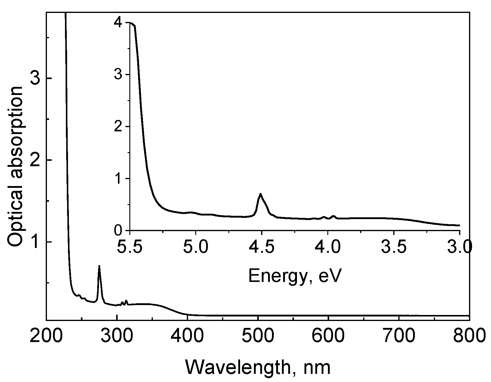
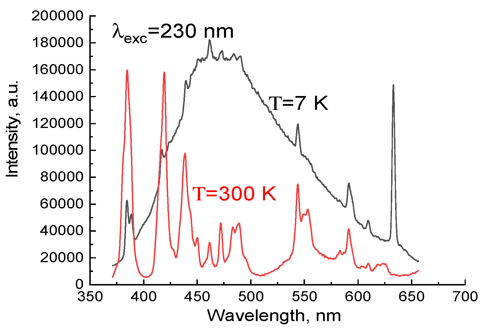
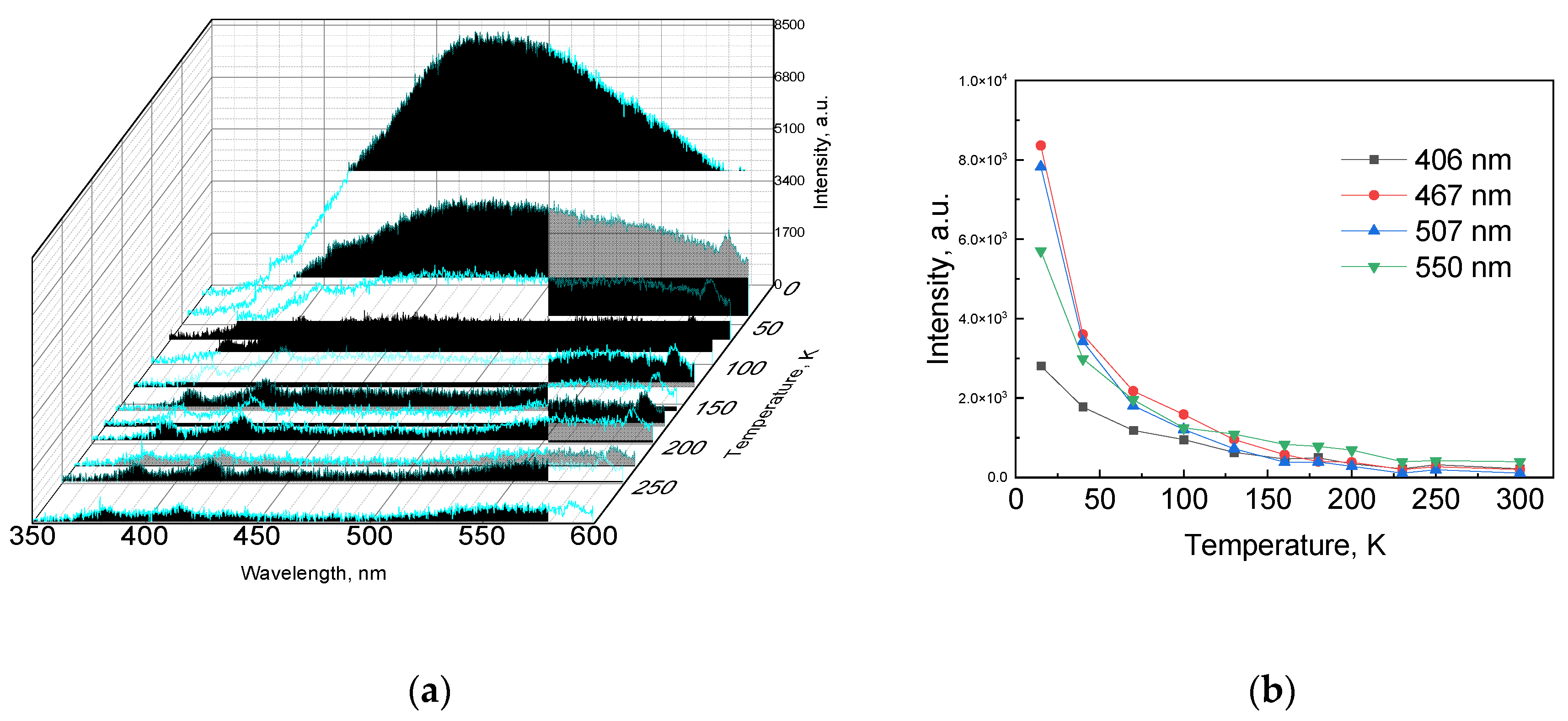
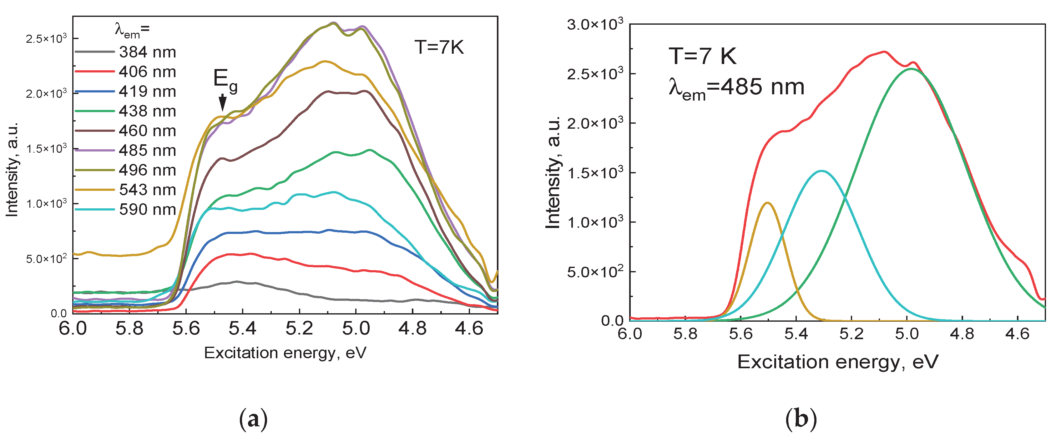
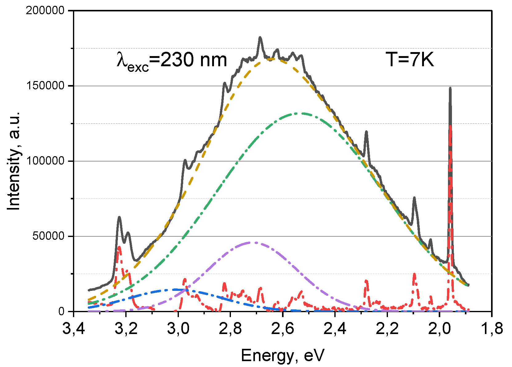
The intrinsic defect-related luminescence of as-grown Gd3Ga5O12 (GGG) single crystals was investigated using synchrotron excitation in the vacuum-ultraviolet region. The photoluminescence spectrum at 7 K exhibits a broad emission band centered near ~2.5 eV (≈500 nm), accompanied by narrow 4f–4f transitions of uncontrolled Tb3+ impurity ions. Gaussian decomposition of the broad band reveals three components at approximately 3.05, 2.70, and 2.50 eV, attributed to self-trapped excitons (STE), antisite-associated recombination, and F/F+-type oxygen-vacancy centers, respectively. Temperature-dependent photoluminescence measurements show that the defect-related luminescence is governed by two thermally activated nonradiative channels with activation energies of ~5–8 meV and 42–56 meV. The shallow channel dominates at 20–50 K, while the main quenching occurs between 100 and 150 K, leading to nearly complete suppression of the broad emission at room temperature. The present results therefore characterize the emission behavior of intrinsic, shallow defect centers in as-grown GGG and contribute to understanding defect recombination pathways that are critical for optimizing garnet-based scintillation and photonic materials.

Keywords: 
;  ;  ;  ;  ;  ;  

1. Introduction

Gadolinium gallium garnet (Gd3Ga5O12, GGG) is a cubic oxide (Ia3d), typically obtained as high-quality single crystals and widely used as a substrate for the epitaxial growth of optical and magneto-optical films [1,2,3,4]. Its wide band gap (Eg ≈ 5.6 eV) and high transparency from the infrared to the ultraviolet range determine its value for photonic applications [3]. When doped with rare-earth ions, GGG exhibits efficient phosphor and scintillation behavior: in Ce:GGG, both emission spectra and decay kinetics are governed by the local crystal environment and the chemical nature of the host [5,6,7], whereas Er3+-, Pr3+-, and Dy3+-doped systems show characteristic f–f transitions [8,9]. For low-temperature phosphor synthesis, sol–gel methods and isomorphic substitutions have been developed [10]. Surface mechanical properties crucial for optical quality have been examined using nanoscratch and nanoindentation techniques [11,12,13], and Co diffusion has been identified as a factor influencing color-center formation [14].
In nano- and thin-film forms, the functionality of GGG expands further: GGG:Tb3+ and GGG:Eu3+ nanoparticles exhibit stable luminescence, including pressure-dependent behavior [15,16,17], while thin films grown by liquid-phase epitaxy are used in fast scintillators [18,19]. GGG has also been proposed as a wide-band-gap ceramic electrolyte for low-temperature fuel cells [20]. Cr3+ doping provides reproducible thermometric characteristics, and Ca/Si cation substitutions in Cr:GGG allow adjustment of emission wavelength and decay time [21,22]. In Ce:GGG, the position of the Ce3+ 5d1 level relative to the conduction band and shallow traps has been established, which is essential for understanding competition between radiative and trapping channels [23].
GGG is also recognized as a frustrated magnet that exhibits field-induced phase transitions and an enhanced magnetocaloric effect upon Al substitution [24,25,26,27]. The crystal demonstrates high radiation resistance: reversible amorphization/recrystallization processes [28], radiation-induced absorption and Raman changes under neutron and heavy-ion irradiation [29,30,31,32], and track-formation behavior described by the inelastic thermal spike model have been reported [33,34,35,36]. Similarity of radiation response to the garnet matrix in YIG confirms the universality of structural disordering and recovery mechanisms [37,38]. Exceptional mechanical strength and thermal stability of GGG have been verified under dynamic compression up to 2.6 TPa [39].
The intrinsic defect-related luminescence of garnets arises primarily from oxygen-vacancy centers F (VO with two electrons) and F⁺ (VO with one electron), together with self-trapped and defect-localized excitons (STE/LE). In YAG [43] and LuAG [44], a fast violet emission band at ~3.1–3.2 eV (τ ≈ 2–3 ns), excited at ~3.3/5.3/6.5 eV, is attributed to F⁺ centers, whereas a blue-green band at ~2.6–2.7 eV corresponds to F centers [43,44,45]. Antisite disorder (YAl/LuAl) promotes the formation of F+–antisite complexes that shift and broaden these bands and partially suppress STE emission; the F/F⁺ centers can also be excited via the excitonic continuum (~6.5–7.3 eV) [43,44,45].
In GGG-based mixed garnets, such as GAGG:Ce, and in YAG, swift heavy-ion irradiation induces stable photo-induced absorption in the UV–visible range and strongly suppresses VUV-excitonic emission, reflecting increased F-type defect concentrations and competition with Ce3+ 5d–4f emission channels, including possible Ce3+/C4+ charge conversion [46,47]. For sintered garnet crystals, oxygen partial pressure, grain boundaries, and impurity–defect complexes enhance F/F+ emission and suppress STE, whereas oxidative annealing reduces color-center populations, which is crucial for optical and scintillation performance. Systematic low-temperature studies of such intrinsic defect-related luminescence in sintered crystals remain scarce, representing a significant open research direction.
However, despite extensive studies of radiation and dopant effects, the native defect structure of as-grown GGG remains insufficiently characterized. Addressing this issue is essential for targeted control of trapping processes, suppression of nonradiative channels, and the rational design of radiation-resistant luminescent materials based on GGG. To fill this gap, in this work we investigate the luminescence and the temperature dependence of luminescence in as-grown GGG.

2. Materials and Methods

Gd₃Ga₅O₁₂ (GGG) single crystals were grown by the Czochralski method using an iridium crucible in a weakly oxidizing atmosphere at the SRC “Electron-Carat” (Lviv, Ukraine) [33]. The growth atmosphere consisted of a mixture of 98% Ar and 2% O₂. High-purity oxides Gd₂O₃ and Ga₂O₃ (99.99 wt%) were used as starting materials. It is known that the presence of tetravalent impurity ions (such as Si⁴⁺ and Zr⁴⁺) in the starting oxides can lead to the formation of cation vacancies and promote a spiral growth mechanism in rare-earth gallium garnet crystals. To suppress these effects, a small amount (10⁻²–10⁻³ wt.%) of calcium oxide (CaO) was added to the melt.
It should also be noted that GGG crystals usually contain a small amount (10⁻³–10⁻⁴ wt.%) of uncontrolled impurity ions, which is practically unavoidable during the growth process. The samples were prepared in the form of flat plates with a thickness of 0.48 mm, oriented along the (111) plane, and polished on both sides to optical quality.
Luminescence spectroscopy measurements were performed under synchrotron radiation excitation. The excitation and emission spectra were recorded at the FinEstBeAMS undulator beamline [48,49,50,51,52,53,54], located at the 1.5 GeV storage ring of the MAX IV synchrotron facility (Lund, Sweden). The excitation photon energy range extended from 4.5 to 11.0 eV. Emission in the UV–visible region (200–700 nm) was analyzed using an Andor Shamrock SR-303i (0.3 m) monochromator coupled to a Newton DU970P-BVF CCD detector, providing a total detection range of 200–900 nm. The GGG samples were mounted on the cold finger of a closed-cycle cryostat (Advanced Research Systems) and positioned inside an ultra-high-vacuum chamber maintained at a pressure of ~10⁻⁹ mbar.
Luminescence was studied at 9 K using synchrotron radiation at the Superlumi P66 beamline (PETRA III, DESY) [55]. Excitation was selected by a 2 m monochromator with a spectral resolution of 4 Å. The emitted luminescence was analyzed using an ANDOR Kymera monochromator (2 Å resolution) equipped with a CCD camera and a Hamamatsu R6358 photomultiplier. The excitation spectra were corrected using a sodium salicylate standard to ensure data accuracy.

3. Results and Discussion

The absorption spectrum exhibits a steep increase in absorption at λ ≲ 225 nm (≈5.6–5.7 eV), corresponding to the fundamental band-gap edge of GGG [3]. In the UV–visible region, three weak bands are observed at 254, 275, and 313 nm, assigned to intra-configurational transitions of Gd3+: 8S7/26Dj (254 nm), 8S7/26Ij (275 nm), and 8S7/26Pj (313 nm). The presence of Ca2+ (introduced as a charge-compensating additive) manifests as an additional weak band near ≈350 nm (≈3.55 eV) [30,33].
The photoluminescence spectra under pulsed-laser excitation at λex = 230 nm (≈5.4 eV), i.e., near the Gd–O charge-transfer edge and effectively pumping both host and defect centers, were measured at T = 7 K. The spectrum spans 3.3–1.8 eV (375–688 nm) and consists of (i) a broad defect-related band with a maximum at ≈475 nm, attributed to recombination involving oxygen-vacancy-related centers, including F-type centers and antisite-associated defects, and (ii) narrow Tb³⁺ lines at 382, 420, 440, 474, 543, 583, and 630 nm, corresponding to 5D₃/5D47Fj transitions (see Table 1) [16, 87].
Figure 1. Absorption spectra of GGG.
Preprints 182587 g001
Figure 2. Luminescence spectra of GGG at 7 and 300 K.
Preprints 182587 g002
Gaussian decomposition of the broad band reveals three subcomponents centered at 3.05, 2.70, and 2.50 eV. By analogy with related garnets such as YAG and LuAG [43,44,56], the 2.50 eV component is associated with F-center recombination, whereas the 3.05 and 2.70 eV components are likely due to STE emission and radiative recombination near GdGa antisite environments. At 300 K, the defect-related band is almost entirely quenched, while the narrow Tb3+ emission persists, with the 5D47F5 transition at 543 nm being the most thermally stable. The fact that the defect emission disappears at approximately 300 K suggests that at room temperature, non-radiative processes (probably thermally activated release of charge carriers from traps or transition to the non-radiative recombination pathway) predominate over radiative recombination for these defect centers.
Figure 3a presents the VUV excitation spectrum of the GGG crystal measured at 7 K (monitoring the broad emission band across the full range). The spectrum is dominated by a very intense, broad excitation band spanning approximately 4.5–5.8 eV. The presence and strength of this feature indicate a high concentration of defect-related states introduced during crystal growth.
Within this broad 4.5–5.8 eV band, at least three sub-bands can be distinguished. Multi-peak Gaussian decomposition (Figure 3b) reveals maxima at approximately 4.9, 5.3, and 5.5 eV. Each component may be attributed to a specific electronic transition. The highest-energy component (~5.5 eV) corresponds to the intrinsic charge-transfer band of the GGG lattice. In rare-earth garnets, charge-transfer (CT) transitions typically involve electron transfer from O 2p states either into the conduction band or directly to a rare-earth ion [41]. The fundamental absorption edge of GGG lies at ~5.6–5.7 eV [3], therefore the 5.5 eV band likely corresponds to excitation across the band gap or to a bound exciton just below the conduction band. This feature reflects host-lattice excitation, which subsequently relaxes and emits via defect- or matrix-related channels. Its broad character, rather than a sharp line, is consistent with an extended, delocalized excitation.
The intermediate sub-band (~5.3 eV) is associated with oxygen-vacancy-related states and/or self-trapped excitons (STE). Oxygen vacancies in GGG (F-centers when capturing an electron) produce in-gap states that can be excited in this energy range. In the related mixed garnet Gd3Al2Ga3O12:Ce (GAGG:Ce), an excitation band at 5.2–5.3 eV under VUV irradiation has been assigned to STE localized at anion vacancies, eventually leading to F-center formation [40,46]. Thus, we attribute the 5.3 eV band in GGG to excitation of STE or vacancy-associated intermediate states, which may either decay radiatively or convert into stable F-centers.
The lowest-energy excitation component (~4.9 eV, with a shoulder down to ~4.7 eV) corresponds to deeper defect states. These may involve antisite defects or more complex vacancy clusters. Gd/Ga antisite defects (Gd³⁺ on a Ga site) introduce strongly localized electronic states due to the altered crystal field and charge-compensation environment. Such antisites can capture excitons or charge carriers. Therefore, the 4.9 eV band likely corresponds to excitons bound to GdGa antisites or vacancy–defect complexes. The lower excitation energy indicates that these states lie relatively deep within the band gap.
Notably, pristine GGG already exhibits a rich defect-excitation structure. Previous studies of GGG irradiated with heavy ions or neutrons also reported the formation of color centers producing new absorption and luminescence bands [30,32,33]. Potera et al. showed that electron-irradiated GGG develops transient absorption bands near 5.0 eV and 4.3 eV, attributed to F+ and F centers, respectively [31]. These absorptions correspond to excitation of the same defect centers observed here in luminescence.
Thus, the excitation spectrum (Figure 4) provides the first direct insight into the nature and energetic hierarchy of luminescent defect states in GGG.
The luminescence spectrum at 7 K was further analyzed using a Gaussian decomposition to resolve overlapping components (Figure 4). In this figure, the broad 477 nm band (excited at 230 nm) was decomposed into several Gaussian subbands, excluding the narrow Tb^3+ lines, which were selected separately or removed for clarity. The analysis revealed three main broadband components with peak positions approximately at 400 nm, 467 nm, and 507 nm (in terms of photon energy, this corresponds to ~3.1 eV, ~2.65 eV, and ~2.45 eV, respectively). These values ​​closely match the subband energies deduced from the excitation spectrum analysis earlier (3.05 eV, 2.70 eV, 2.50 eV). Each subband likely corresponds to a different type of defect center recombination. The 507 nm component (2.45–2.50 eV) is attributed to F-centers. In the classical model, an F-center can trap a hole (forming an excited state F) and then emit a photon upon relaxation to the ground state. Andriychuk et al. observed broad luminescence with a wavelength of 2.5 eV in GGG and attributed it to such recombination of radiation-induced color centers [56]. Our results are consistent with this: the 507 nm band appears in the unirradiated crystal (which likely contains some intrinsic oxygen vacancies from growth). Our data convincingly show that the key effect of defective optics of GGG is the broad band at ~500 nm (≈2.5 eV): its amplitude is maximum at 7 K, and at 300 K it almost completely disappears while narrow Tb³⁺ lines are preserved, confirming its matrix-defect nature and excluding the contribution of 4f–4f transitions. Gaussian deconvolution isolates the contribution at 2.50 eV as a signal of F/F⁺ centers (oxygen vacancies), while the neighboring subbands at 2.70 and 3.05 eV are associated with STE and antisites; together with edge pumping at 230 nm (≈5.4 eV) [33,56]. In support of this, we present the results of a study where, in epitaxial GGG films grown from a Bi2O3–B2O3 solution-melt, the authors attribute a broad luminescence band with a maximum at about 500 nm to Bi3+ ions [57]. This emission, observed with selective ultraviolet synchrotron excitation, manifests itself as a broad band in the range of 380–750 nm. It is important to note that the authors report strong luminescence at room temperature (300 K), the intensity of which decreases by only 20% upon heating from 10 K. This distinguishes their results from our samples, in which the broad glow at room temperature is quenched.
The temperature dependence of luminescence intensity for GGG crystal (Figure 4) was investigated. Thermal quenching of luminescence bands associated with defect centers is often analyzed using the empirical expression (Table 1):
I = I 0 1 + i B i   e E i k B T
Where I₀ is the photoluminescence intensity at very low temperature, Bᵢ are constants, and Eᵢ are the activation energies of nonradiative levels. The physical meaning of Bᵢ depends on the adopted model of thermal quenching. The calculated activation energies are summarized in Table 2.
For all four emission bands centered at 406, 467, 507, and 550 nm, an initial decrease in intensity is observed with increasing temperature. This behavior is governed by a shallow activation channel with E1 = 5–8 meV and B1 = 3.65–11.5. The characteristic onset temperature T on E 1 / k B ln B 1 is found to be 31–35 K for the 467 and 507 nm bands and ~46–54 K for the 406 and 550 nm bands. This regime corresponds to thermal release of carriers from very shallow localized states within the defect complex, leading to a redistribution between radiative and nonradiative pathways even at cryogenic temperatures.
The dominant contribution originates from a second channel with activation energies E2 = 41.7–55.6 meV and large pre-exponential factors B2 = 50–615. The temperature of half-quenching, defined by I T 1 / 2 = I 0 / 2 , is estimated as T 1 / 2 E 2 / k B ln B 2 ). For the 507 and 467 nm bands, T 1 / 2 100 103 K; for the 406 and 550 nm bands, T 1 / 2 123 124 K. These values are in full agreement with the experimental trends shown in Figure 4b, where the intensity decays by approximately one order of magnitude between 80 and 150 K and approaches the noise level above 150 K.
The 467 and 507 nm bands exhibit nearly identical activation parameters ( E 2 55.6 meV, large B2), indicating a common origin associated with recombination involving oxygen vacancies in F/F+ configurations. In contrast, the 406 and 550 nm bands, characterized by slightly lower activation energies E 2 41.7 41.8 meV and significantly smaller B2, reflect contributions from defects which possess a weaker temperature sensitivity and persist to slightly higher temperatures.
Unlike the defect-related broad bands, the sharp 4f–4f transitions of Tb3+ remain observable at 300 K due to strong shielding of the 4f electrons, confirming that the broad emission is not impurity-derived but arises from matrix intrinsic defects.
The temperature dependence of photoluminescence quantitatively demonstrates that defect-related emission in GGG is governed by the competition between a shallow activation channel (5–8 meV) dominating at 20–50 K and a main nonradiative channel with activation energy of 42–56 meV, which controls the quenching between 100 and 150 K. The derived activation energies and intensity trends are consistent with recombination processes involving oxygen-vacancy-related centers (F/F+) and associated defect complexes [31,56], explaining the disappearance of the broad emission band near 500 nm at room temperature while the Tb³⁺ lines remain unaffected.
In contrast to our non-irradiated GGG crystal, where the defect-related emission band near 2.5 eV (≈500 nm) is fully quenched by ~300 K due to shallow activation energies (~42–56 meV), strongly X-ray–irradiated GGG (GGG-V) reported by Andriichuk et al. [56] retains the same emission to temperatures above 500 K. In those irradiated samples, the excitation spectrum contains two pronounced bands at 4.5 and 5.44 eV, and distinct TSL and thermally stimulated electron emission peaks appear in the range 145–285 K, indicating the formation of deeper and thermally stable oxygen-vacancy–related trap complexes. Thus, our results represent the behavior of as sintered, shallow defect centers, whereas the literature data reflect radiation-induced stabilization of deeper vacancy aggregates.
Figure 4. Temperature dependence of the luminescence spectra GGG single crystal (a) and temperature dependence on several bands (b).
Preprints 182587 g005

4. Conclusions

In this work, we have established the optical signatures and thermal stability of defect-related luminescence in as-grown GGG single crystals. Low-temperature photoluminescence spectra reveal a broad emission band centered near ~2.5 eV (~500 nm), accompanied by characteristic 4f–4f transitions of residual Tb3+ ions. Spectral deconvolution demonstrated that the broad band consists of three contributions located at ~3.05, 2.70, and 2.50 eV, which were assigned to self-trapped exciton recombination, antisite-defect–assisted emission, and oxygen-vacancy centers (F/F+), respectively.
The temperature dependence of the luminescence intensity showed that the defect-related emission is controlled by two thermally activated nonradiative channels with activation energies of ~5–8 meV and 42–56 meV. The main quenching occurs between 100 and 150 K, leading to the near-complete disappearance of the broad emission at room temperature. This behavior confirms that shallow vacancy-related centers dominate the recombination processes in the as-grown material.
Comparison with previously reported X-ray–irradiated GGG demonstrated a notable difference in trap depth and thermal stability: while irradiated crystals retain 2.5 eV emission up to >500 K, the emission in our samples is fully quenched by ~300 K. Thus, the present study characterizes the luminescence behavior of native defect complexes and provides quantitative insight into their recombination and thermal deactivation mechanisms, relevant for the optimization of garnet-based scintillators and optical ceramics.

Author Contributions

Conceptualization, G.M.A., Z.T.K., A.Ki. and S.U.; methodology, Z.T.K. and A.Ki.; software, G.E.S. and E.P.; validation, Z.T.A., E.P., S.U. and A.Sa.; formal analysis, Z.T.A., A.Ka., A.Ki., G.E.S. and Y.Sm.; investigation, Z.T.A., A.Ka., A.M.Zh., G.E.S., S.U. and A.Sa.; resources, A.Ka., A.M.Zh., S.U., K.Ch., Y.Sm. and A.I.P.; data curation, G.M.A., Z.T.K., Z.T.A., A.Ka., A.M.Zh., E.P., K.Ch., Y.Sm. and A.Sa.; writing—original draft preparation, Z.T.K.; writing—review and editing, G.M.A., Z.T.K. and A.I.P.; visualization, Z.T.A., A.M.Zh., G.E.S., E.P., K.Ch. and Y.Sm.; supervision, A.Ki. and A.I.P.; project administration, G.M.A. and A.I.P.; funding acquisition, G.M.A.

Funding

This research was funded by the Science Committee of the Ministry of Science and Higher Education of the Republic of Kazakhstan (grant number AP19680626). In addition, Anatoli I. Popov was supported by Latvian research project lzp-2023/1-0453 “Prediction of long-term stability of functional materials under extreme radiation conditions” and the Latvian State Research Program No. VPP-IZM-CERN-2022/1-0001.

Data Availability Statement

The raw data supporting the conclusions of this article will be made available by the authors upon reasonable request.

Acknowledgments

The authors MAX IV Laboratory for time on FinEstBeAMS Beamline under Proposal 20231736. The authors acknowledge DESY (Hamburg, Germany), a member of the Helmholtz Association HGF, for providing experimental facilities. The work was carried out at the P66 beamline of PETRA III. Beamtime was allocated under proposal I-20231138.

Conflicts of Interest

The authors declare no conflict of interest.

References

  1. O’Kane, D.F.; Sadagopan, V.; Geiss, E.A.; Mandel, E. Crystal Growth and Characterization of Gadolinium Gallium Garnet. J. Electrochem. Soc. 1973, 120, 1272–1278. [Google Scholar] [CrossRef]
  2. Xu, Y.-N.; Ching, W.Y.; Brickeen, B.K. Electronic structure and bonding in garnet crystals Electronic structure and bonding in garnet crystals Gd3Sc2Ga3O12, Gd3Sc2Al3O12, and Gd3Ga3O12 compared to Y3Al3O12. Phys. Rev. B 2000, 61, 1817. [Google Scholar] [CrossRef]
  3. Ghimire, K.; Haneef, H.F.; Collins, R.W.; Podraza, N.J. Optical Properties of Single-Crystal Gd₃Ga₅O₁₂ from the Infrared to the Ultraviolet. Phys. Status Solidi B 2015, 252, 2191–2198. [Google Scholar] [CrossRef]
  4. Zhao, G.; Li, T.; Xu, J. Growth of epitaxial substrate Gd3Ga3O12 (GGG) single crystal through pure GGG phase polycrystalline material. J. Cryst. Growth 2002, 237-239, 720–724. [CrossRef]
  5. Syvorotka, I.I.; Sugak, D.Y.; Wierzbicka, A.; Wittlin, A.; Przybylińska, H.; Barzowska, J.; Barcz, A.J.; Berkowski, M.; Domagała, J.Z.; Mahlik, S.; et al. Optical Properties of Pure and Ce³⁺-Doped Gadolinium Gallium Garnet Crystals and Epitaxial Layers. J. Lumin. 2015, 164, 31–37. [Google Scholar] [CrossRef]
  6. Mikhailov, M.M.; Neshchimenko, V.V.; Shavlyuk, V.V. Binding-Type Effects in LED Phosphor GGG:Ce³⁺. Opt. Mater. 2014, 38, 33–36. [Google Scholar] [CrossRef]
  7. Thirumalaisamy, T.K.; Saravanakumar, S.; Butkute, S.; Kareiva, A.; Saravanan, R. Structure and Charge Density of Ce-Doped GGG. J. Mater. Sci. Mater. Electron. 2016, 27, 1920–1928. [Google Scholar] [CrossRef]
  8. Wang, Y.; You, Z.; Li, J.; Zhu, Z.; Ma, E.; Tu, C. Spectroscopy of Highly Doped Er³⁺:GGG and Er³⁺/Pr³⁺:GGG. J. Phys. D: Appl. Phys. 2009, 42, 215406. [Google Scholar] [CrossRef]
  9. Wang, Y.; You, Z.; Li, J.; Zhu, Z.; Tu, C. Optical Properties of Dy³⁺ in GGG. J. Phys. D: Appl. Phys. 2010, 43, 075402. [Google Scholar] [CrossRef]
  10. Butkute, S.; Zabiliute, A.; Skaudzius, R.; Zukauskas, A.; Kareiva, A. Sol–Gel Synthesis and Substitution in Ga-Garnets. J. Sol-Gel Sci. Technol. 2015, 76, 210–219. [Google Scholar] [CrossRef]
  11. Li, C.; Zhang, F.; Meng, B.; Rao, X.; Zhou, Y. Varied-Depth Nanoscratch on GGG. Mater. Des. 2017, 125, 180–188. [Google Scholar] [CrossRef]
  12. Li, C.; Wu, Y.; Li, X.; Ma, L.; Zhang, F.; Huang, H. Nanoindentation of GGG Single Crystal. Appl. Opt. 2018, 57, 3661–3668. [Google Scholar] [CrossRef]
  13. Li, C.; Zhang, F.; Wang, X.; Rao, X. Crack-Free Grinding of GGG. J. Mater. Process. Technol. 2020, 279, 116577. [Google Scholar] [CrossRef]
  14. Sugak, D.; Syvorotka, I.I.; Yakhnevych, U.; Buryy, O.; Vakiv, M.; Ubizskii, S.; Włodarczyk, D.; Зhydachevskyy, Ya.; Pieniążek, A.; Jakiela, R.; Suchocki, A. Investigation of Co Ions Diffusion in Gd₃Ga₅O₁₂ Single Crystals. Acta Phys. Pol. A 2018, 133(4), 959–964. [Google Scholar] [CrossRef]
  15. Reshmi, C.P.; Ramesh, A.R.; Suresh, A.; Jaiswal-Nagar, D. Magnetic, magnetocaloric and optical properties of nano Gd₃Ga₅O₁₂garnet synthesized by citrate sol-gel method. Int. J. Nano Dimens. 2025, 16, 190–201. [Google Scholar]
  16. Luchechko, A.; Kostyk, L.; Varvarenko, S.; Tsvetkova, O.; Kravets, O. Green-Emitting Gd₃Ga₅O₁₂:Tb³⁺ Nanoparticles Phosphor: Synthesis, Structure, and Luminescence. Nanoscale Res. Lett. 2017, 12, 263. [Google Scholar] [CrossRef] [PubMed]
  17. Jovanić, B.; Bettinelli, M.; Radenković, B.; Despotović-Zrakić, M.; Bogdanović, Z. Optical spectroscopy of nanocrystalline Gd₃Ga₅O₁₂ doped with Eu3+ and high pressures. Mater. Chem. Phys. 2012, 132, 273–277. [Google Scholar] [CrossRef]
  18. Baillard, A.; Douissard, P.-A.; Loiko, P.; Martin, T.; Mathieu, E.; Camy, P. Terbium-doped gadolinium garnet thin films grown by liquid phase epitaxy for scintillation detectors. RSC Adv. 2025, 15, 20560–20568. [Google Scholar] [CrossRef]
  19. Wollesen, L.; Douissard, P.-A.; Pauwels, K.; Baillard, A.; Loiko, P.; Brasse, G.; Mathieu, J.; Simeth, S.J.; Kratz, M.; Dujardin, C.; et al. Microstructured growth by liquid phase epitaxy of scintillating Gd₃Ga₅O₁₂(GGG) doped with Eu3+. J. Alloys Compd. 2025, 1010, 177267. [Google Scholar] [CrossRef]
  20. Wang, K.; Xu, J.; Murakami, H.; Yamahara, H.; Seki, M.; Tabata, H.; Tonouchi, M. Terahertz Time-Domain Spectroscopy of Substituted Gadolinium Gallium Garnet. Condens. Matter 2025, 10, 1. [Google Scholar] [CrossRef]
  21. Li, J.; Yousaf, M.; Ahmed, J.; Bibi, B.; Noor, A.; Alhokbany, N.; Sajid, M.; Nazar, A.; Shah, M.A.K.Y.; Guo, X.; Ding, X.; Mushtaq, N.; Lu, Y. Gd₃Ga₅O₁₂: A wide bandgap semiconductor electrolyte for ceramic fuel cells, effective at temperatures below 500 C. J. Rare Earths 2025, 43, 2248–2256. [Google Scholar] [CrossRef]
  22. Elzbieciak-Piecka, K.; Sójka, M.; Tian, F.; Li, J.; Zych, E.; Marciniak, L. The comparison of the thermometric performance of optical ceramics, microcrystals and nanocrystals of Cr3+-doped Gd₃Ga₅O₁₂ garnets. J. Alloys Compd. 2023, 963, 171284. [Google Scholar] [CrossRef]
  23. Shimoyama, N.; Kodama, S.; Bartosiewicz, K.; Fujiwara, C.; Yanase, I.; Takeda, H. Modification of Photoluminescence Wavelength and Decay Constant of Cr:Gd₃Ga₅O₁₂ Substituted by Ca/Si Cation Pair. J. Ceram. Soc. Jpn. 2024, 132(7), 364–368. [Google Scholar] [CrossRef]
  24. Kitaura, M.; Zen, H.; Watanabe, S.; Masai, H.; Kamada, K.; Kim, K.J.; Ueda, J. Relationship between Ce³⁺ 5d₁ Level, Conduction-Band Bottom, and Shallow Electron Trap Level in Gd₃Ga₅O₁₂:Ce and Gd₃Al₁Ga₄O₁₂:Ce Crystals Studied via Pump–Probe Absorption Spectroscopy. Opt. Mater. X 2025, 25, 100398. [Google Scholar] [CrossRef]
  25. Petrenko, O.A.; Balakrishnan, G.; Paul, D.McK.; Yethiraj, M.; McIntyre, G.J.; Wills, A.S. Field Induced Magnetic Order in the Frustrated Magnet Gadolinium Gallium Garnet. J. Phys. Conf. Ser. 2009, 145, 012026. [CrossRef]
  26. Deen, P.P.; Florea, O.; Lhotel, E.; Jacobsen, H. Updating the Phase Diagram of the Archetypal Frustrated Magnet Gd₃Ga₅O₁₂. Phys. Rev. B 2015, 91, 014419. [Google Scholar] [CrossRef]
  27. Sackville Hamilton, A.C.; Lampronti, G.I.; Rowley, S.E.; Dutton, S.E. Enhancement of the Magnetocaloric Effect Driven by Changes in the Crystal Structure of Al-Doped GGG, Gd₃Ga₅−ₓAlₓO₁₂ (0 ≤ x ≤ 5). J. Phys. Condens. Matter 2014, 26(11), 116001. [Google Scholar] [CrossRef]
  28. Jin, X.; Zhao, J.; Gao, L.; Ma, H.; Oimod, H.; Zhu, H.; Li, Q.; Su, T.; Zhu, H.; Tegus, O.; Zhao, J. Influence of High-Pressure Heat Treatment on Magnetocaloric Effects and Magnetic Phase Transition in Single Crystal Gd₃Ga₅O₁₂. J. Rare Earths 2024, 42(11), 2112–2118. [Google Scholar] [CrossRef]
  29. Chukalkin, Y.G.; Shtirts, V.R. Radiation Amorphization and Recovery of Crystal Structure in Gd₃Ga₅O₁₂ Single Crystals. Phys. Status Solidi A 1994, 144(1), 9–14. [Google Scholar] [CrossRef]
  30. Mironova-Ulmane, N.; Sildos, I.; Vasil’chenko, E.; Chikvaidze, G.; Skvortsova, V.; Kareiva, A.; Muñoz-Santiuste, J.E.; Pareja, R.; Elsts, E.; Popov, A.I. Optical Absorption and Raman Studies of Neutron-Irradiated Gd₃Ga₅O₁₂ Single Crystals. Nucl. Instrum. Methods Phys. Res. B 2018, 435, 306–312. [Google Scholar] [CrossRef]
  31. Potera, P.; Matkovskii, A.; Sugak, D.; Grigorjeva, L.; Millers, D.; Pankratov, V. Transient Color Centers in GGG Crystals. Radiat. Eff. Defects Solids 2002, 157(6–12), 709–713. [CrossRef]
  32. Potera, P.; Ubizskii, S.; Sugak, D.; Schwartz, K. Induced Absorption in Gadolinium Gallium Garnet Irradiated by High-Energy ²³⁵U Ions. Acta Phys. Pol. A 2010, 117, 181–183. [Google Scholar] [CrossRef]
  33. Karipbayev, Z.T.; Kumarbekov, K.; Manika, I.; Dauletbekova, A.; Kozлoвskiy, A.L.; Sugak, D.; Ubizskii, S.B.; Akilbekov, A.; Suchikova, Y.; Popov, A.I. Optical, Structural, and Mechanical Properties of Gd₃Ga₅O₁₂ Single Crystals Irradiated with 84Kr⁺ Ions. Phys. Status Solidi B 2022, 259(8), 2100415. [Google Scholar] [CrossRef]
  34. Meftah, A.; Costantini, J.M.; Khalfaoui, N.; Boudjadar, S.; Stoquert, J.P.; Studer, F.; Toulemonde, M. Experimental Determination of Track Cross-Section in Gd₃Ga₅O₁₂ and Comparison to the Inelastic Thermal Spike Model Applied to Several Materials. Nucl. Instrum. Methods Phys. Res. B 2005, 237(3–4), 563–574. [CrossRef]
  35. oulemonde, M.; Meftah, A.; Costantini, J.M.; Schwartz, K.; Trautmann, C. Out-of-Plane Swelling of Gadolinium Gallium Garnet Induced by Swift Heavy Ions. Nucl. Instrum. Methods Phys. Res. B 1998, 146(1–4), 426–430. [CrossRef]
  36. Meftah, A.; Assmann, W.; Khalfaoui, N.; Stoquert, J.P.; Studer, F.; Toulemonde, M.; Trautmann, C.; Voss, K.-O. Electronic Sputtering of Gd₃Ga₅O₁₂ and Y₃Fe₅O₁₂ Garnets: Yield, Stoichiometry and Comparison to Track Formation. Nucl. Instrum. Methods Phys. Res. B 2011, 269(9), 955–958. [Google Scholar] [CrossRef]
  37. Costantini, J.-M.; et al. Swift Heavy Ion-Beam Induced Amorphization and Recrystallization of Yttrium Iron Garnet. J. Phys.: Condens. Matter 2015, 27, 496001. [Google Scholar] [CrossRef]
  38. Costantini, J.-M.; Miro, S.; Lelong, G.; Guillaumet, M.; Toulemonde, M. Damage Induced in Garnets by Heavy-Ion Irradiations: A Study by Optical Spectroscopies. Philos. Mag. 2018, 98(4), 312–328. [Google Scholar] [CrossRef]
  39. Ozaki, N.; Nellis, W.J.; Mashimo, T.; Ramzan, M.; Ahuja, R.; Kaewmaraya, T.; Kimura, T.; Knudson, M.; Miyanishi, K.; Sakawa, Y.; Sano, T.; Kodama, R. Dynamic Compression of Dense Oxide (Gd₃Ga₅O₁₂) from 0. 4 to 2.6 TPa: Universal Hugoniot of Fluid Metals. Sci. Rep. 2016, 6, 26000. [Google Scholar] [CrossRef]
  40. Khanin, V.M.; Rodnyi, P.A.; Wieczorek, H.; Ronda, C.R. Electron Traps in Gd3Ga3Al2O12:Ce Garnets Doped with Rare-Earth Ions. Tech. Phys. Lett. 2017, 43, 439–442. [Google Scholar] [CrossRef]
  41. Lisiecki, R.; Komar, J.; Macalik, B.; Strzęp, A.; Berkowski, M.; Ryba-Romanowski, W. Exploring the Impact of Structure-Sensitive Factors on Luminescence and Absorption of Trivalent Rare Earth Embedded in Disordered Crystal Structures of Gd₃Ga₅O₁₂–Gd₃Al₅O₁₂ and Lu₂SiO₅–Gd₂SiO₅ Solid Solution Crystals. Low Temp. Phys. 2023, 49, 310–315. [Google Scholar] [CrossRef]
  42. Aralbayeva, G.M.; Manika, I.; Karipbayev, Zh.; Suchikova, Y.; Kovachov, S.; Sugak, D.; Popov, A.I. Micromechanical Properties of Gd₃Ga₅O₁₂ Crystals Irradiated with Swift Heavy Ions. J. Nano-Electron. Phys. 2023, 15(5), 05020. [CrossRef]
  43. Zorenko, Y.; Zorenko, T.; Voznyak, T.; Mandowski, A.; Xia, Q.; Batentschuk, M.; Friedrich, J. Luminescence of F⁺ and F centers in Al₂O₃-Y₂O₃ oxide compounds. IOP Conf. Ser. Mater. Sci. Eng. 2010, 15, 012060. [Google Scholar] [CrossRef]
  44. Babin, V.; Laguta, V.V.; Maaroos, A.; Makhov, A.; Nikl, M.; Zazubovich, S. Luminescence of F⁺-type centers in undoped Lu₃Al₅O₁₂ single crystals. Phys. Status Solidi B 2011, 248, 845–852. [Google Scholar] [CrossRef]
  45. Varney, C.R.; Selim, F.A. Color centers in YAG. AIMS Mater. Sci. 2015, 2, 560–572. [Google Scholar] [CrossRef]
  46. Pankratova, V.; Skuratov, V.A.; Buzanov, O.A.; Mololkin, A.A.; Kozlova, A.P.; Kotlov, A.; Popov, A.I.; Pankratov, V. Radiation effects in Gd₃(Al,Ga)₅O₁₂:Ce³⁺ single crystals induced by swift heavy ions. Opt. Mater. X 2022, 16, 100217. [Google Scholar] [CrossRef]
  47. Pankratova, V.; Butikova, J.; Kotlov, A.; Popov, A.I.; Pankratov, V. Influence of swift heavy ions irradiation on optical and luminescence properties of Y₃Al₅O₁₂ single crystals. Opt. Mater. X 2024, 23, 100341. [Google Scholar] [CrossRef]
  48. Maji, S.B.; Vanetsev, A.; Mändar, H.; Nagirnyi, V.; Chernenko, K.; Kirm, M. Investigation of Luminescence Properties of Hydrothermally Synthesized Pr³⁺ Doped BaLuF₅ Nanoparticles under Excitation by VUV Photons. Opt. Mater. 2024, 154, 115781. [Google Scholar] [CrossRef]
  49. Krutyak, N.; Nagirnyi, V.; Romet, I.; Deyneko, D.; Spassky, D. Energy Transfer Processes in NASICON-Type Phosphates under Synchrotron Radiation Excitation. Symmetry 2023, 15, 749. [Google Scholar] [CrossRef]
  50. Spassky, D.; Voznyak-Levushkina, V.; Arapova, A.; Zadneprovski, B.; Chernenko, K.; Nagirnyi, V. Enhancement of Light Output in ScxY1−xPO4:Eu3+ Solid Solutions. Symmetry 2020, 12, 946. [Google Scholar] [CrossRef]
  51. Romet, I.; Tichy-Rács, É.; Lengyel, K.; Feldbach, E.; Kovács, L.; Corradi, G.; Chernenko, K.; Kirm, M.; Spassky, D.; Nagirnyi, V. Interconfigurational d–f Luminescence of Pr³⁺ Ions in Praseodymium Doped Li₆Y(BO₃)₃ Single Crystals. J. Luminescence 2024, 265, 120216. [Google Scholar] [CrossRef]
  52. Bartosiewicz, K.; Szysiak, A.; Tomala, R.; Gołębiewski, P.; Węglarz, H.; Nagirnyi, V.; Kirm, M.; Romet, I.; Buryi, M.; Jary, V.; et al. Energy-Transfer Processes in Nonstoichiometric and Stoichiometric Er3+, Ho3+, Nd3+, Pr3+, and Cr3+-Codoped Ce:YAG Transparent Ceramics: Toward High-Power and Warm-White Laser Diodes and LEDs. Phys. Rev. Appl. 2023, 20, 014047. [Google Scholar] [CrossRef]
  53. Gundacker, S.; Pots, R. H.; Nepomnyashchikh, A.; Radzhabov, E.; Shendrik, R.; Omelkov, S.; Kirm, M.; Acerbi, F.; Capasso, M.; Paternoster, G.; Mazzi, A.; Gola, A.; Chen, J.; Auffray, E. Vacuum Ultraviolet Silicon Photomultipliers Applied to BaF₂ Cross Luminescence Detection for High Rate Ultrafast Timing Applications. Phys. Med. Biol. 2021, 66 (11), 114002. [CrossRef]
  54. Saaring, J.; Feldbach, E.; Nagirnyi, V.; Omelkov, S.; Vanetsev, A.; Kirm, M. Ultrafast Radiative Relaxation Processes in Multication Cross Luminescence Materials. IEEE Trans. Nucl. Sci. 2020, 67(6), 1009–1013. [Google Scholar] [CrossRef]
  55. Smortsova, Y.; Chukova, O.; Kirm, M.; Nagirnyi, V.; Pankratov, V.; Kataev, A.; Kotlov, A. The P66 time-resolved VUV spectroscopy beamline at PETRA III storage ring of DESY. J. Synchrotron Rad. 2025, 32. [Google Scholar] [CrossRef]
  56. Andriichuk, V.A.; Volzhenskaya, L.G.; Zakharko, Ya.M.; Zorenko, Yu.V. Influence of structure defects upon the luminescence and thermostimulated effects in Gd3Ga5O12 crystals. J. Appl. Spectrosc. 1987, 47, 902–905. [Google Scholar] [CrossRef]
  57. Randoshkin, V.V.; Vasil’eva, N.V.; Kolobanov, V.N.; Mikhailin, V.V.; Petrovnin, N.N.; Spasskii, D.A.; Sysoev, N.N. Effect of Gd₂O₃ concentration in Bi-containing high-temperature solutions on the luminescence of epitaxial Gd₃Ga₅O₁₂ films. Inorg. Mater. 2009, 45, 418–422. [Google Scholar] [CrossRef]
Figure 3. Excitation spectrum of GGG luminescence at 7 K (a) and Gaussian deconvolution of the spectrum at 485 nm (b).
Preprints 182587 g003
Figure 4. Luminescence spectrum of GGG at 7 K with Gaussian deconvolution (λₑₓ = 230 nm).
Preprints 182587 g004
Table 1. Main luminescence peaks of Tb³⁺ ions and transitions in GGG single crystal
Emission Band, (nm) Excitation, (nm) Possible Transitions
382 230 5D37F6
420 230 5D37F5
440 230 5D37F4
474 230 5D47F6
543 230 5D47F5
583 230 5D47F4
630 230 5D47F3
Table 2. Obtained by Eq(1) activation energy values of defect centers in GGG single crystal.
Λem, nm B1 E1, meV B2 E2, meV
406 3,65 6 50,3 41,7
467 7.9 5.6 521 55.6
507 11.5 7.3 614.9 55.6
550 5,95 6,2 51,7 41,8
Disclaimer/Publisher’s Note: The statements, opinions and data contained in all publications are solely those of the individual author(s) and contributor(s) and not of MDPI and/or the editor(s). MDPI and/or the editor(s) disclaim responsibility for any injury to people or property resulting from any ideas, methods, instructions or products referred to in the content.
Copyright: This open access article is published under a Creative Commons CC BY 4.0 license, which permit the free download, distribution, and reuse, provided that the author and preprint are cited in any reuse.
Prerpints.org logo

Preprints.org is a free preprint server supported by MDPI in Basel, Switzerland.

Subscribe

Disclaimer

Terms of Use

Privacy Policy

Privacy Settings

© 2025 MDPI (Basel, Switzerland) unless otherwise stated