Preprint
Article

This version is not peer-reviewed.

Bighorn Sheep Cloned Embryos Produced by ISCNT via HMC from Domestic Sheep Oocytes Treated with Resveratrol During IVM

A peer-reviewed article of this preprint also exists.

Submitted:

26 August 2025

Posted:

26 August 2025

You are already at the latest version

Abstract

The bighorn sheep is listed on the IUCN Red List of Threatened Species. By interspecies somatic cell nuclear transfer (ISCNT), other endangered species are cloned using somatic cells as nuclear donors, fusing them with enucleated oocytes from heterologous domestic species. On the other hand, resveratrol added during in vitro maturation (IVM) of domestic sheep oocytes favors the development of embryos produced in vitro. The aim of this study was to treat O. aries oocytes with resveratrol during IVM using them as cytoplasts in ISCNT via handmade cloning (HMC), evaluating its effect on the in vitro development of Mexican bighorn sheep (O. c. mexicana) cloned embryos. Post-mortem skin fibroblasts from an adult male specimen from the Chapultepec Zoo were frozen for 8 years, thawed, and reseeded for 8 cell passages. For IVM, O. aries oocytes were treated with 0, 0.5, or 1.0 µM resveratrol. Matured oocytes were manually enucleated, and triplets (O. aries cytoplast-O. c. mexicana karyoplast-O. aries cytoplast) were formed and electrically fused. The reconstructed embryos were chemically activated and cultured until they developed into blastocysts. For IVM, no differences were found between treatments, yet at 0.5 µM, resveratrol significantly increased (p<0.05) the blastocyst rate and decreased the fragmentation rate.

Keywords: 
;  ;  ;  

1. Introduction

Gene banks preserve the greatest genetic diversity of species, including agricultural, forestry, livestock, and microbial species [1]. They are essential for the conservation of sperm, eggs, and embryos, as well as the cells and tissues of various mammal species at some level of risk. This is the case for the bighorn sheep Ovis canadensis, which is listed on the IUCN Red List of Threatened Species, albeit in the category of "least concern" [2]. It is important to conserve as much of its population as possible to promote the conservation of current biological diversity [3].
One way to achieve this goal is through the application of animal reproduction technologies, such as embryo production through in vitro fertilization (IVF) or somatic cell nuclear transfer (SCNT), embryo freezing, and embryo transfer (ET). The combination of these technologies represents an alternative for the conservation and reproductive improvement of endangered species of economic and ecological importance, and even for humans with infertility problems [4].
Somatic cell nuclear transfer (SCNT) allows the production of genetically identical individuals originating from a single nuclear donor. The donor cell is a somatic cell derived in culture from various tissues of the animal to be cloned and is known as the “karyoplast”. This type of cell is fused with an activated, nucleus-free oocyte, called a “cytoplast”, or with a fertilized egg from which the pronuclei have been removed, to give rise to a cloned embryo of the species from which the karyoplast was originated [5]. Both the cytoplast that will receive the karyoplast and the karyoplast itself must have specific physiological characteristics for successful SCNT, the subsequent development of the reconstructed embryo, and the new cloned individual.
One branch of SCNT is ISCNT (Interspecies Somatic Cell Nuclear Transfer), in which animals of ecological interest (generally species at risk of extinction or even already extinct) are cloned from the somatic cells of these animals as karyoplasts but fuse them with enucleated oocytes from phylogenetically homologous or heterologous domestic species, that is, non-homologous [6].
A well-known case of ISCNT was the Pyrenean Ibex (Capra pyrenaica pyrenaica). Skin fibroblasts cryopreserved for 10 years from the last surviving goat until 2000 were thawed and fused with enucleated oocytes from a domestic goat (Capra hircus). The cloned embryos were transferred into hybrid and purebred female Spanish Ibex (C. p. hispanicus), resulting in the birth of one live offspring. This sets a good precedent for obtaining offspring from endangered or extinct species due to ISCNT [7].
Other authors generated a clone of a mouflon sheep (O. orientalis musimon) via ISCNT using granulosa cells from females found dead in the desert, which were fused with domestic sheep (O. aries) oocytes, resulting in offspring [8]. Additionally, it was possible to clone the gaur (Bos gaurus), an Asian buffalo, by ISCNT from gaur somatic cells and domestic cattle oocytes, getting offspring [9].
In México, we obtained the first Mexican bighorn sheep (O. canadensis mexicana) embryos at the blastocyst stage, which were cloned by ISCNT through handmade cloning (HMC) from ear skin fibroblasts of the species that were fused with enucleated oocytes of domestic sheep (O. aries) [10].
One of the challenges facing SCNT is the reprogramming of the karyoplast, which bears the cellular identity of the tissue from which it originates; that is, it has already differentiated and possesses the morphophysiological characteristics and genetic imprint of the cell type in question. It is through certain treatments and, upon fusion with the cytoplast, that the karyoplast must regain its totipotentiality and state of cellular undifferentiation, thus ensuring its nuclear reprogramming [11,12,13].
On the other hand, considering that the gene banks of animal species store tissues, somatic cells, gametes or embryos of the organisms that are to be conserved or preserved, and that this implies the application of technologies such as slow or rapid freezing (vitrification), this represents another challenge owing to the damage caused to the cryopreserved cell, which affects the success rates in the development of the embryos obtained from them.
In works carried out in the ovine species [14,15], we observed that the use of an antioxidant (resveratrol) supplemented during in vitro maturation (IVM) of oocytes, favors the development of compact morulae in embryos produced by SCNT, as well as blastocysts produced by vitrified IVF, after thawing.
Resveratrol is a phytoalexin present in some plants and their fruits, which acts as a defense mechanism against pathogens that affect them [16].
Among the first studies reporting the use of this antioxidant as a supplement in oocyte cultures to produce mammalian embryos is that of Kwak et al. [17], who demonstrated its positive effect on embryonic development when it was used at 0.5 to 1 µM in porcine embryos produced by IVF.
Its use has been shown to reduce the levels of reactive oxygen species (ROS), which are overproduced in in vitro cultures because of incubation conditions and the handling to which the embryos are subjected. One possible explanation is that resveratrol increases the level of reduced glutathione (GSH). Reactive oxygen species, although necessary for certain cellular processes, when produced in excess, cause damage to the cell (cytoplasmic and nuclear membranes), which results in alterations in embryonic development such as cell blockage or death [15].
In the present study, resveratrol was used for its antioxidant activity and because it has been documented that it also functions as a trigger for genetic signals in embryonic development. The oocyte is known to provide an epigenetic environment for genes necessary for embryonic development, such as NANOG, Oct3/4, and SOX2, genes that maintain pluripotency [18]. This could be a good precedent that enables it to reprogram the karyoplast nucleus with which it will fuse in the SCNT.
Therefore, the objective of this study was to evaluate the effect of resveratrol supplementation in the IVM of domestic sheep oocytes (O. aries) used as cytoplasts in ISCNT, on the in vitro development rate (IVD) of the reconstructed bighorn sheep (O. canadensis mexicana) cloned embryos, from ear skin fibroblasts of a post-mortem adult male of this wild species that had been cryopreserved for 8 years.

2. Materials and Methods

The Reagents were obtained from Sigma-Aldrich Chemical Co., unless otherwise indicated. Incubation conditions were 38.5°C, 5% CO2, and saturated humidity.

2.1. In vitro Culture of Fibroblasts from Bighorn Sheep (O. c. mexicana)

In 2016, an 8 h post-mortem (death because of natural causes) adult male (5 years old) Mexican bighorn sheep (O. c. mexicana) was sampled at the Chapultepec Zoo in Mexico City (Collection permit SGPA/DGVS/07250/15 and approval from the Ethics Commission of the Biological and Health Sciences Division, Universidad Autónoma Metropolitana Iztapalapa or UAM-I). Approximately 1 cm³ of its ear skin was removed and transported on ice to the Assisted Animal Reproduction Laboratory at the UAM-I within 5 h. Once in the laboratory, the tissue was kept refrigerated for 24 h before processing
Following the methodology described by Navarro-Maldonado et al. [19], the tissue was subjected to an enzymatic disaggregation process with collagenases type I and II (0.2%/0.2%, Gibco (Waltham, USA), in phosphate-buffered saline without calcium or magnesium (DPBS, Dulbecco´s PBS, In Vitro, S.A., CDMX, México) and incubated at 38.5 °C for 2 h under constant oscillation. The cells in suspension were washed by centrifugation and seeded in Dulbecco's Modified Eagle Medium (DMEM, In Vitro, S.A., CDMX, México), supplemented with 10% fetal bovine serum (SFB, Microlab, S.A. de C.V., CDMX, México) and 2% antibiotic-antifungal (In Vitro, S.A., CDMX, México). For 4 weeks, the cultures with a confluence of≥ 90% underwent cell passages every 7 days.
The cell passage consisted of removing the culture medium and washing three times with 1 mL of DPBS (Dulbecco's PBS, In Vitro, S.A., CDMX, México), and then, 1 mL of trypsin (0.05%/0.05% Trypsin-Verseno, In Vitro, S.A., CDMX, México) was added and incubated for 5 min or until the cells were completely detached from the base of the culture dish.
Then, 1 mL of DMEM (In Vitro, S.A., CDMX, México) supplemented with 10% FBS (Microlab, S.A. de C.V., CDMX, México) and 2% antibiotic-antifungal (In Vitro, S.A., CDMX, México) was added to inactivate the action of trypsin. The medium with the cells in suspension was placed in 1.5 mL microcentrifuge tubes (Sorenson, Biociences, Inc., Salt Lake, USA) and centrifuged at 500 x g for 5 min. The supernatant was decanted and 1 mL of DMEM was added to the cell pellet, which was reseeded in Petri dishes (Nunc, Walham, USA), adding 2 mL until completing 3 mL of supplemented DMEM and incubated under the described conditions. The procedure was repeated until the cell passages were completed (5th to 9th), reseeding half of the cell population from each passage, and the other half was cryopreserved. For cryopreservation, the cells (fibroblasts) were stored at -80°C for 24 h and subsequently at -196°C for 8 years in a DMSO-based freezing medium (In Vitro, S.A., CDMX, México).
After 8 years of cryopreservation and before cloning by ISCNT, the fibroblasts were thawed and reseeded. To thaw them, the cryotubes were removed from the LN2 tank and thawed at 30 °C in a water bath, immediately after they were centrifuged at 500 x g for 5 min, the supernatant was discarded, and 1 mL of supplemented DMEM was added. The samples were subsequently reseeded in 2 mL of additional medium and incubated under the conditions described. The fibroblasts were synchronized in the G0/G1 stages of their cell cycle by contact inhibition, after which they were cultured until they reached confluence (7 days). They were subsequently detached with trypsin as previously described and maintained in TCM-199 with HEPES (In Vitro S.A., CDMX, México) supplemented with 20% NBCS (Biowest, Nuaillé, France) (T20) to use them as karyoplasts for ISCNT via HMC [20].

2.2. In Vitro Maturation of Domestic Sheep (O. aries) Oocytes

Following the methodology described by Hernández-Martínez et al. [10], with some modifications, ovaries from O. aries at a local slaughterhouse were collected and transported to the laboratory (transfer time of 1 h) in saline solution (0.9% NaCl and 1% antibiotic-antifungal) at 30-35 °C. Complex-oocyte-cumulus (COC) were aspirated from ovarian follicles (2-5 mm in diameter) in TCM-199 with HEPES (In Vitro, S.A., CDMX, México) supplemented with 100 IU/ml of heparin sodium salt. The recovered COC were selected based on their morphology and number of granulosa cell layers [21].
The samples were incubated for 22 h in in vitro maturation medium (IVM) TCM-199 supplemented with cysteine [0.57 mM], D-glucose [3.05 mM], polyvinyl alcohol [PVA] [0.001 g/mL], sodium pyruvate [0.91 mM], 10% NBCS (Biowest, Nuaillé, France), 0.1 IU of FSH-LH (Pluset, Calier, Italy), gentamicin [50 µg/mL], and epidermal growth factor [EGF, 10 ng/mL]. Three treatment groups of O. aries COC were formed during IVM: the control group CG (without resveratrol), experimental group 1 EG1 (0.5 µM resveratrol), and experimental group 2 EG2 (1.0 µM resveratrol).
The criteria for the main signs of IVM evaluation were the expansion of cumulus cells and the presence of the first polar body, which indicated that the oocytes were in metaphase II (MII) [20].

2.3. Production of O. c. mexicana Cloned Embryos by ISCNT Via HMC

Once IVM was completed, the COC with expanded cumulus cells was incubated in 500 μL of hyaluronidase (0.5 mg mL-1 in TCM-199 with HEPES) for 8 min, and the oocytes were denuded of cumulus cells by gentle pipetting with a micropipette (200 μL). Zona pellucida-free oocytes were placed in T2 medium (TCM-199 with 2% NBCS), and oocytes with a first polar body (PB) were selected under a stereomicroscope, indicating that they were MII. Oocytes with PB were incubated in IVM medium supplemented with demecolcine (0.5 μg mL-1) for 1 h under the same conditions.
For the preparation of the cytoplasts, 30 µL drops corresponding to the following solutions were placed on a 60 x 15 mm Petri dish: T2, pronase (2 mg/mL in T10), T10 (TCM-199 supplemented with 10% NBCS and 0.5 µg/mL cytochalasin B) and T20 (TCM-199 supplemented with 20% NBCS). The drops were covered with mineral oil, and cytoplasts were prepared in this dish.
Once the demecolcine incubation period was complete, the oocytes were transferred to the T2 drop of the cytoplast preparation dish. Groups of 20 to 30 oocytes were then transferred to the pronase drop for 3 min or until the zona pellucida (ZP) was completely dispersed. Immediately afterward, the ZP-free oocytes were placed in the T20 drop to inactivate the action of pronase. This procedure was repeated for all available oocytes.
Groups of six ZP-free oocytes were distributed in each T10 droplet, and handmade cloning (HMC) was performed via manual enucleation using a microblade (Shearer Precision Products, USA). To do this, the portion of the oocyte cytoplasm closest to the membrane containing the genetic material (metaphase plate and first polar body) was excised with a microblade. The enucleated oocytes (cytoplasts) were collected in the T20 droplets to restore their spherical shape as a sign of their viability.
Fifteen µL drops of: T20, phytohemagglutinin (5 mg/mL in TCM-199 with HEPES), T2, and fusion media (0.3 M D-mannitol and 1 mg/mL polyvinyl alcohol) were placed on the lid of a 33 mm Petri dish. The drops were covered with mineral oil. Enucleated O. aries oocytes (cytoplasts) were immersed in phytohemagglutinin for 4 seconds. A single O. c. mexicana skin fibroblast (karyoplast) was subsequently placed between two cytoplasts, forming cell triplets.
For triplet fusion, a cell electrofusion device (Instrument BLS Budapest, Hungary) connected to a fusion chamber (BTX microslide, model 450 Holliston, USA) was used, which covered the cells with fusion medium. Cell triplets (O. aries cytoplast–O. c. mexicana karyoplast–O. aries cytoplast) were fused using a 0.2 kV/cm pulse for 9 µsec.
The reconstructed bighorn sheep cloned embryos were activated by incubating them in calcium ionophore A23187 (8 µg/mL) for 5 min and in 6-dimethylaminopurine (6-DMAP 2 mM) for 4 h under the described conditions. Then, the embryos were cultured in a WOW (Well of Well) system, based on Vajta et al. [22] and improved by VitaVitro (Shenzhen, China), in 50 µL of BO-IVC (IVF Biosciences Cornwall United Kingdom) covered with mineral oil, for 7 days (168 h) and under the described conditions [10]. At the end of the culture, the development rate for each treatment was evaluated.

2.4. Statistical Analysis

The data were expressed as the means ± SDs; an arccosine transformation was performed on the data obtained for embryonic development. Comparisons between means for both the IVM and the different IVD stages were analyzed via ANOVA with a significance level of p < 0.05.

3. Results

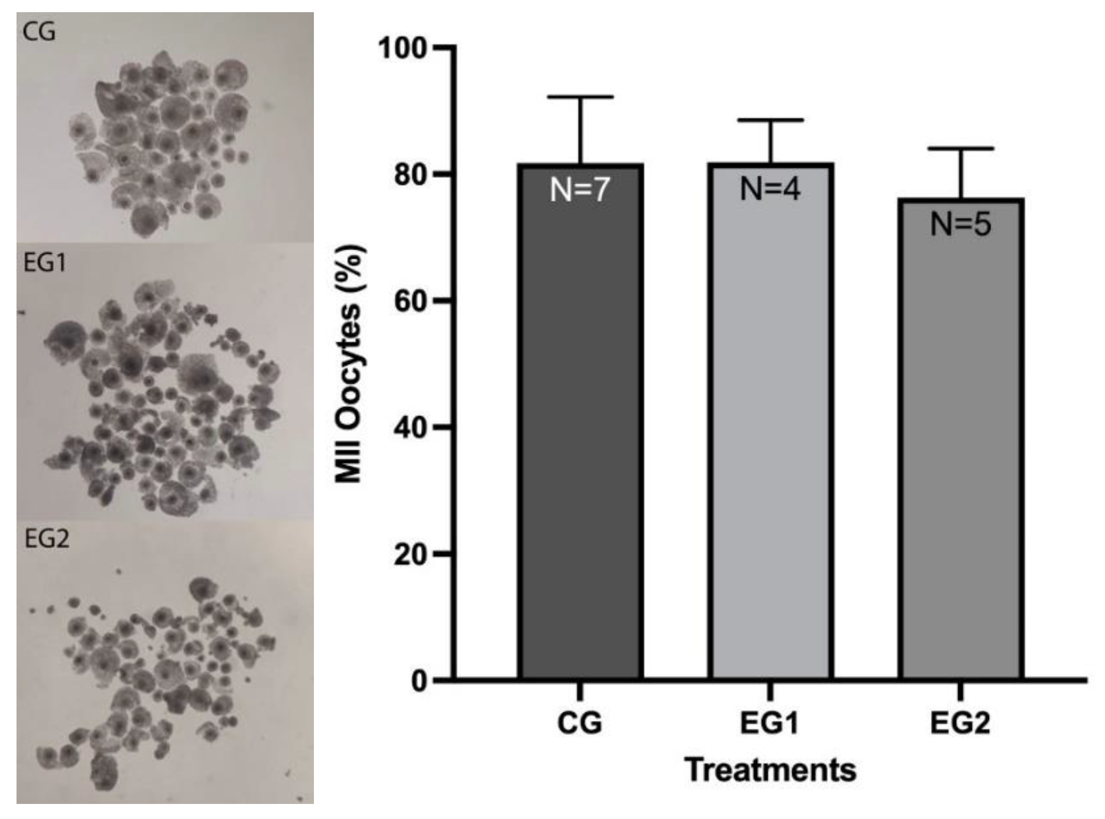
3.1. Effect of Resveratrol on the IVM Rate of O. aries Oocytes

A total of 357 COC were placed in CG, 237 in EG1, and 280 in EG2. Of these, 197 in the CG (81.8±10.4%), 156 in the EG1 (81.9±6.7%), and 129 in the EG2 (76.3±7.7%) reached IVM, with no significant differences between the groups (p>0.05) (Figure 1).

3.2. Production of O. c. mexicana Cloned Embryos by ISCNT Via HMC

Five replicates were performed, resulting in 40, 69, and 32 bighorn sheep cloned embryos for groups CG, EG1, and EG2, respectively. Compared with the CG, EG1 presented a statistically significant increase (p<0.05) in the percentage of blastocysts, and a statistically significant decrease in the percentage of fragmented embryos when compared with the other groups (Table 1 and Figure 2).

4. Discussion

Although the resveratrol treatments used did not show significant differences in the IVM rates of O. aries oocytes, at 0.5 µM, this antioxidant had a positive effect on the blastocyst rate of O. c. mexicana cloned embryos produced by ISCNT. This confirms the hypothesis that the oocyte, although not homologous, provides the epigenetic environment necessary for embryonic development, since it is responsible for the reprogramming of the somatic cell by providing the transcription factors NANOG, Oct3/4, and SOX2, genes that maintain pluripotency [18]. In addition, the oocyte has DNA repair factors [23].
In general, embryonic genomic activation in the SCNT depends on the ability of the recipient oocyte to block transcription of the donor cell's DNA and the corresponding translation of mRNA [6]. This allows it to reprogram the donor cell to return it to its undifferentiated, totipotential state.
To promote reprogramming of the donor somatic cell nucleus by the recipient oocyte, the cell cycles must be synchronized between the first and second stages. The somatic cell must be arrested in the G0 or G1 phase of its cell cycle, whereas the oocyte must be in MII. Arresting somatic cells in these phases involves reducing the amount of serum in the culture media; contact inhibition through cell confluence; and the use of cycloheximide, roscovitine, or dimethyl sulfoxide (DMSO) [11,12].
Another important factor is the cell passage of the donor cell, with early passages (<6–8) being preferable, since the probability of karyotype abnormalities or epigenetic alterations that may accumulate in prolonged in vitro culture is reduced [13].
In the present study, somatic cells from bighorn sheep corresponding to passages 6 to 8 were brought to confluence and exposed to DMSO during freezing and remained frozen at -196°C for 8 years. This likely allowed them to remain in the G0 and G1 phases of their cycle, facilitating reprogramming. In addition, oocytes in the early stages of life are used, since they contain high levels of the maturation-promoting factor (MPF), which prematurely condenses chromatin, silencing the transcription of the donor cell genome after its transfer into the recipient oocyte [6].
On the other hand, resveratrol is known to act as an antioxidant by reducing the levels of ROS, a product of cellular metabolism, and increasing the level of intracellular reduced glutathione (GSH). Reactive oxygen species damage oocytes and embryos by decreasing ATP, blocking development, altering DNA methylation, and modifying histones [15].
Additionally, resveratrol is a trigger of gene signals required for embryonic development, such as sirtuin 1 (SIRT1), a member of the sirtuin deacetylase family, which regulates the acetylation of several transcription factors and regulates cell cycle progression, in addition to promoting the cellular response to metabolic stress [24]. This SIRT1 gene participates in the regulation of mitochondrial biogenesis, ATP generation, and AMPK regulation, increasing β-oxidation and fatty acid consumption and thus improving embryonic development [25].
Resveratrol also contributes to embryonic compaction by increasing the expression of E-cadherin (uvomorulin), promoting its transcriptional activity. E-cadherin is a product of oocyte origin, a calcium-dependent molecule, and the main component of cell-cell adherents’ junctions. It is associated with embryonic compaction and blastocyst formation, as it plays an important role in trophectoderm differentiation by inducing a polarization of the epithelial phenotype [14,26,27].
Therefore, in the present study, by adding resveratrol to the O. aries oocytes used as cytoplasts in ISCNT, the aim was to provide them with additional tools that would enable them to reprogram the O. c. mexicana cell nucleus. This possibly favored the increased rate of bighorn sheep cloned blastocysts observed when using oocytes matured in 0.5 µM resveratrol.
In previous work, resveratrol at this concentration favored the rate of compact morulae in O. aries cloned embryos produced by HMC [14] and blastocysts produced by IVF in this species [15].
Regarding the cloning technique used, in 2013 Stroud et al. [28] cloned Rocky Mountain bighorn sheep (Ovis canadensis canadensis) embryos by ISCNT via traditional cloning (TC) using micromanipulators, from post-mortem skin fibroblasts of an adult male fused with enucleated O. aries oocytes, resulting in 16% blastocysts. These results are like those obtained in previous studies, in which we obtained 16.1% cloned blastocysts of female O. c. mexicana [20] and 14.3% of male O. c. mexicana, using ear skin fibroblasts of live specimens as karyoplasts that were fused with manually encucleated for HMC O. aries oocytes, but without being subjected to resveratrol [10]. In the present study, by incorporating resveratrol during the IVM of O. aries oocytes used to clone Mexican bighorn sheep embryos, the blastocyst rate increased to 31%, surpassing the studies. This finding is important considering that the fibroblasts also came from a post-mortem specimen and from fibroblasts cryopreserved 8 years.
Loi et al. [8] reported the successful cloning of O. orientalis musimon by TC using O. aries oocytes. The authors used granulosa cells from two female mouflons found dead in the pasture. The cloned embryos reached the blastocyst stage and were transferred to O. aries surrogate mothers, resulting in the birth of an apparently normal mouflon clone calf.
Folch et al. [7] cloned an extinct goat, the bucardo or Pyrenean Ibex (Capra pyrenaica pyrenaica), from skin fibroblasts of the last specimen of the species that were fused with oocytes of a domestic goat (Capra hircus). The cloned embryos of the bucardo were transferred into female Spanish Ibex or hybrids (Spanish Ibex x domestic goat), obtaining offspring.
Since Vajta et al. [29] developed HMC, which involves manually enucleating the oocyte without micromanipulators, some domestic species have been cloned using this technique, such as pigs [30,31], sheep [32], cattle [33,34,35] and horses [36] as well as wild species such as buffalo [37,38,39].
Both TC and HMC have the same objective, but the instruments used to enucleate the oocytes vary. These variables include the use of micromanipulators for TC, as well as the need to use ZP-free oocytes for HMC. Furthermore, the latter requires two cytoplasts to fuse with a karyoplast, whereas the former requires only one cytoplast per karyoplast. One advantage of HMC over TC lies primarily in cost [12].
There are a few studies of ISCNT carried out in wild sheep, with cleavage rates of 84.6% for Ovis ammon [40] and 87.4% for Ovis orientalis isphahanica [41], results that are below those obtained in this study (94-100%). Regarding the blastocyst rate, most studies report rates of 7.6–15%, while Loi et al. [8] reported 30.4% in Ovis orientalis musimon, like the data obtained in this study.
The present study reports for the first time, 31% of cloned embryos at the blastocyst stage produced by ISCNT via HMC, from resveratrol-treated O. aries oocytes (as cytoplasts) and fibroblasts (as karyoplasts) that were thawed after 8 years of cryopreservation, from a post-mortem adult male bighorn sheep (O. c. mexicana), an endemic species of México that is listed on the IUCN Red List. This reinforces the feasibility of bringing non-existent species back to life, as well as the ability of somatic cells to survive cryopreserved for long periods.
The possibility of performing ET of bighorn sheep cloned embryos in surrogate females of a homologous species remains to be studied, to determine pregnancy and birth rates, as well as to evaluate the health of cloned offspring of this species.

5. Conclusions

It is concluded that by supplementing O. aries oocytes with resveratrol during IVM and using them as cytoplasts in ISCNT and then fusing them with post-mortem adult male O. c. mexicana fibroblasts cryopreserved for 8 years, better rates of bighorn sheep cloned blastocysts are obtained.

6. Patents

Patent Title No. 394003 referred to Preservation of Bighorn Sheep.

Author Contributions

“Conceptualization, Demetrio Alonso Ambríz-García and María del Carmen Navarro-Maldonado; Data curation, José Roberto Vazquez-Avendaño and Demetrio Alonso Ambríz-García; Formal analysis, José Roberto Vazquez-Avendaño and Demetrio Alonso Ambríz-García; Funding acquisition, María del Carmen Navarro-Maldonado; Investigation, José Roberto Vazquez-Avendaño, Alfredo Trejo-Córdova and María del Carmen Navarro-Maldonado; Methodology, José Roberto Vazquez-Avendaño, José Antonio Sandoval-Zárate, Fernando Gual-Sill, Miriam Fahiel Casillas Avalos and Jessica Elivier Nuñez-Macias; Project administration, María del Carmen Navarro-Maldonado; Resources, Demetrio Alonso Ambríz-García, Alfredo Trejo-Córdova and María del Carmen Navarro-Maldonado; Supervision, María del Carmen Navarro-Maldonado; Validation, José Roberto Vazquez-Avendaño and María del Carmen Navarro-Maldonado; Visualization, José Roberto Vazquez-Avendaño and María del Carmen Navarro-Maldonado; Writing – original draft, José Roberto Vazquez-Avendaño, Jessica Elivier Nuñez-Macias and María del Carmen Navarro-Maldonado; Writing – review & editing, José Roberto Vazquez-Avendaño, Demetrio Alonso Ambríz-García, Alfredo Trejo-Córdova, José Antonio Sandoval-Zárate, Miriam Fahiel Casillas Avalos and María del Carmen Navarro-Maldonado.. All authors have read and agreed to the published version of the manuscript.”

Funding

“This research was funded by the National Council of Humanities, Science and Technology (CONAHCyT), now Secretariat of Science, Humanities, Technology and Innovation (SECIHTI) through the Basic Frontier Science Project Agreement (Convenio del Proyecto de Ciencia Básica de Frontera) C-459/2024”.

Institutional Review Board Statement

The animal study protocol was approved by the Institutional Ethics Committee, “Ethics Commission of the Biological and Health Sciences Division of the Universidad Autónoma Metropolitana Iztapalapa,” and approval of the “Collection Permit SGPA/DGVS/07250/15” for studies involving animals.

Informed Consent Statement

“Not applicable.”

Data Availability Statement

The data are available from the first author, José Roberto Vazquez- Avendaño (jrva@xanum.uam.mx), upon request.

Acknowledgments

The Authors would like to thank CONAHCyT (now SECITHI) for funding through the Basic Frontier Science Project Agreement C-459/2024.

Conflicts of Interest

“The authors declare no conflict of interest.”

References

  1. Gobierno de México. (17 de noviembre 2023). Bancos de germoplasma, protectores de la soberanía nacional. https://www.gob.mx.
  2. Festa-Bianchet, M. (2020). Ovis canadensis. The IUCN Red List of Threatened Species 2020: e.T15735A22146699. [CrossRef]
  3. Gobierno de México. (5 marzo 2015). En franja de recuperación, borrego cimarrón en México. Secretaria de Medio Ambiente y Recursos Naturales. https://www.gob.mx.
  4. Rosete, F, J, V., Álvarez, G. H., Urbán, D. D., Fragoso, I. A., Asprón, P. M. A., Ríos, U. A., Pérez, R. S., & De La Torre, S. J. F. (2021). Biotecnologías reproductivas en el ganado bovino: cinco décadas de investigación en México. Revista Mexicana de Ciencias Pecuarias, 12 (Supl 3):39-78. [CrossRef]
  5. Navarro-Maldonado, M. C., García, A. D., & Serrrano, H. (2003). Técnicas de clonación de embriones. Ciencia Veterinaria, 9-2003-4.
  6. Lagutina, I., Fulka, H., Lazzari, G., & Galli, C. (2013). Interspecies somatic cell nuclear transfer: advancements and problems. Cellular reprogramming, 15(5), 374–384. [CrossRef]
  7. Folch, J., Cocero, M. J., Chesné, P., Alabart, J. L., Domínguez, V., Cognié, Y., Roche, A., Fernández-Arias, A., Martí, J. I., Sánchez, P., Echegoyen, E., Beckers, J. F., Bonastre, A. S., & Vignon, X. (2009). First birth of an animal from an extinct subspecies (Capra pyrenaica pyrenaica) by cloning. Theriogenology, 71(6), 1026–1034. [CrossRef]
  8. Loi, P., Ptak, G., Barboni, B., Fulka, J., Jr, Cappai, P., & Clinton, M. (2001). Genetic rescue of an endangered mammal by cross-species nuclear transfer using post-mortem somatic cells. Nature biotechnology, 19(10), 962–964. [CrossRef]
  9. Lanza, R. P., Cibelli, J. B., Diaz, F., Moraes, C. T., Farin, P. W., Farin, C. E., Hammer, C. J., West, M. D., & Damiani, P. (2000). Cloning of an endangered species (Bos gaurus) using interspecies nuclear transfer. Cloning, 2(2), 79–90. [CrossRef]
  10. Hernández-Martínez, S., Hernández-Pichardo, J.E., Vazquez-Avendaño, J.R., Ambríz-García D.A. & Navarro-Maldonado M.d.C. (2020). Developmental dynamics of cloned Mexican bighorn sheep embryos using morphological quality standards. Veterinary Medicine and Science, 6, 382–392. [CrossRef]
  11. Wilmut, I., Schnieke, A. E., McWhir, J., Kind, A. J., & Campbell, K. H. (1997). Viable offspring derived from fetal and adult mammalian cells. Nature, 385(6619), 810–813. [CrossRef]
  12. Verma, G., Arora, J. S., Sethi, R. S., Mukhopadhyay, C. S., & Verma, R. (2015). Handmade cloning: recent advances, potential and pitfalls. Journal of animal science and biotechnology, 6, 43. [CrossRef]
  13. Galli, C., & Lazzari, G. (2021). Current applications of SCNT in advanced breeding and genome editing in livestock. Reproduction. [CrossRef]
  14. Martínez-Ibarra, J.L., Espinoza-Mendoza, E.A., Rangel-Santos, R., Ambriz-García, D.A., & Navarro-Maldonado, M.D.C. (2018). Effect of resveratrol on the in vitro maturation of ovine (Ovis aries) oocytes and the subsequent development of handmade cloned embryos. Veterinaria México, 5(4). [CrossRef]
  15. González-Garzón, A. C., Ramón-Ugalde, J. P., Ambríz-García, D. A., Vazquez-Avendaño, J. R., Hernández-Pichardo, J. E., Rodríguez-Suastegui, J. L., Cortez-Romero, C., & Del Carmen Navarro-Maldonado, M. (2023). Resveratrol Reduces ROS by Increasing GSH in Vitrified Sheep Embryos. Animals, 13(23), 3602. [CrossRef]
  16. Gambini, J., López-Grueso, R., Olaso-González, G., Inglés, M., Abdelazid, K., Alami, M. E., Bonet-Costa, V., Borrás, C., & Viña, J. (2013). Resveratrol: distribución, propiedades y perspectivas. Revista Española de Geriatría y Gerontología, 48(2), 79-88. [CrossRef]
  17. Kwak, S. S., Cheong, S. A., Jeon, Y., Lee, E., Choi, K. C., Jeung, E. B., & Hyun, S. H. (2012). The effects of resveratrol on porcine oocyte in vitro maturation and subsequent embryonic development after parthenogenetic activation and in vitro fertilization. Theriogenology, 78(1), 86–101. [CrossRef]
  18. Takahashi, K., & Yamanaka, S. (2006). Induction of pluripotent stem cells from mouse embryonic and adult fibroblast cultures by defined factors. Cell, 126(4), 663–676. [CrossRef]
  19. Navarro-Maldonado M. C., Hernández-Martínez S., Vázquez-Avendaño J. R., Martínez-Ibarra J. L., Zavala-Vega N. L.,Vargas-Miranda B., Rivera-Rebolledo J. A. & Ambríz-García D. A. (2015). Deriva de células epiteliales de tejido de piel descongelado de Ovis canadensis mexicana para la formación de un banco de germoplasma. Acta Zoológica Mexicana (n. s.), 31(2): 275-282.
  20. Vazquez-Avendaño, J. R., Hernández-Martínez, S., Hernández- Pichardo, J. E., Rivera-Rebolledo, J. A., Ambriz-García, D. A., & Navarro-Maldonado, M. C. (2017). Efecto del uso de medio secuencial humano en la producción de blastocistos de hembra Ovis canadensis mexicana por clonación manual interespecies. Acta Zoológica Mexicana (n.s.), 33(2), 328-338.
  21. Asociación para el Estudio de la Biología Reproductiva (ASEBIR). (2015). Criterios ASEBIR de valoración morfológica de ovocitos, embriones tempranos y blastocistos humanos. Cuadernos de embriología clínica (3th ed., pp. 9–75). Madrid: ASEBIR.
  22. Vajta, G., Korösi, T., Du, Y., Nakata, K., Ieda, S., Kuwayama, M., & Nagy, Z. P. (2008). The Well-of-the-Well system: an efficient approach to improve embryo development. Reproductive biomedicine online, 17(1), 73–81. [CrossRef]
  23. Wakayama, S., Ito, D., Hayashi, E., Ishiuchi, T., & Wakayama, T. (2022). Healthy cloned offspring derived from freeze-dried somatic cells. Nature Communications, 13:3666. [CrossRef]
  24. Jeong, J. K., Kang, M. H., Gurunathan, S., Cho, S. G., Park, C., Park, J. K., & Kim, J. H. (2015). Pifithrin-α ameliorates resveratrol-induced two-cell block in mouse preimplantation embryos in vitro. Theriogenology, 83(5), 862–873. [CrossRef]
  25. Itami, N., Shirasuna, K., Kuwayama, T., & Iwata, H. (2015). Resveratrol improves the quality of pig oocytes derived from early antral follicles through sirtuin 1 activation. Theriogenology, 83(8), 1360–1367. [CrossRef]
  26. Nagafuchi, A., Shirayoshi, Y., Okazaki, K., Yasuda, K. & Takeichi, M. (1987) Transformation of cell adhesión properties by exogenously introduced E-cadherin cDNA. Nature, 29, 341–3.
  27. Watson, A. J. & Barcroft, L.C. 2001. Regulation of blastocyst formation. Frontiers in Bioscience 6, d708-730, May 1, 2001.
  28. Stroud, T.K., Xiang, T., Romo, S., & Kjelland, M.E. (2014). Rocky Mountain bighorn sheep (Ovis canadensis canadensis) embryos produced using somatic cell nuclear transfer. Reproduction, Fertility and Development, 26(1), 133-133. [CrossRef]
  29. Vajta, G., Lewis, I. M., Hyttel, P., Thouas, G. A., & Trounson, A. O. (2001). Somatic cell cloning without micromanipulators. Cloning, 3(2), 89–95. [CrossRef]
  30. Du, Y., Kragh, P., Zhang, X., Purup, S., Yang, H., Bolund, L., & Vajta, G. (2005). High Overall In Vitro Efficiency of Porcine Handmade Cloning (HMC) Combining Partial Zona Digestion and Oocyte Trisection with Sequential Culture. Cloning And Stem Cells, 7(3), 199-205. [CrossRef]
  31. Kumbha, R., Hosny, N., Matson, A., Steinhoff, M., Hering, B. J., & Burlak, C. (2020). Efficient production of GGTA1 knockout porcine embryos using a modified handmade cloning (HMC) method. Research in Veterinary Science, 128, 59–68. [CrossRef]
  32. Zhang, P., Liu, P., Dou, H., Chen, L., Chen, L., Lin, L., Tan, P., Vajta, G., Gao, J., Du, Y., & Ma, R. Z. (2013). Handmade cloned transgenic sheep rich in omega-3 Fatty acids. PloS one, 8(2), e55941. [CrossRef]
  33. Tecirlioglu, R. T., Cooney, M. A., Lewis, I. M., Korfiatis, N. A., Hodgson, R., Ruddock, N. T., Vajta, G., Downie, S., Trounson, A. O., Holland, M. K., & French, A. J. (2005). Comparison of two approaches to nuclear transfer in the bovine: hand-made cloning with modifications and the conventional nuclear transfer technique. Reproduction, fertility, and development, 17(5), 573–585. [CrossRef]
  34. Vajta, G., Lewis, I. M., Trounson, A. O., Purup, S., Maddox-Hyttel, P., Schmidt, M., Pedersen, H. G., Greve, T., & Callesen, H. (2003). Handmade somatic cell cloning in cattle: analysis of factors contributing to high efficiency in vitro. Biology of reproduction, 68(2), 571–578. [CrossRef]
  35. Cortez, J., Murga, N., Segura, G., Rodríguez, L., Vásquez, H., & Maicelo-Quintana, J. (2017). Capacidad de Dos Líneas Celulares para la Producción de Embriones Clonados mediante Transferencia Nuclear de Células Somáticas. Revista de Investigaciones Veterinarias del Perú, 28(4), 928-938. [CrossRef]
  36. Lagutina, I., Lazzari, G., Duchi, R., Colleoni, S., Ponderato, N., Turini, P., Crotti, G., & Galli, C. (2005). Somatic cell nuclear transfer in horses: effect of oocyte morphology, embryo reconstruction method, and donor cell type. Reproduction (Cambridge, England), 130(4), 559–567. [CrossRef]
  37. Selokar, N. L., George, A., Saha, A. P., Sharma, R., Muzaffer, M., Shah, R. A., Palta, P., Chauhan, M. S., Manik, R. S., & Singla, S. K. (2011). Production of interspecies handmade cloned embryos by nuclear transfer of cattle, goat, and rat fibroblasts to buffalo (Bubalus bubalis) oocytes. Animal reproduction science, 123(3-4), 279–282. [CrossRef]
  38. Priya, D., Selokar, N. L., Raja, A. K., Saini, M., Sahare, A. A., Nala, N., Palta, P., Chauhan, M. S., Manik, R. S., & Singla, S. K. (2014). Production of wild buffalo (Bubalus arnee) embryos by interspecies somatic cell nuclear transfer using domestic buffalo (Bubalus bubalis) oocytes. Reproduction in domestic animals = Zuchthygiene, 49(2), 343–351. [CrossRef]
  39. Duah, E.K., Mohapatra, S.K., Sood, T.J., Sandhu, A., Singla, S.K., Chauhan, M.S., Manik, R.S., & Palta, P. (2016). Production of hand-made cloned buffalo (Bubalus bubalis) embryos from non-viable somatic cells. In Vitro Cell Development Biology Animal. Dec;52(10):983-988.
  40. Pan, X., Zhang, Y., Guo, Z., & Wang, F. (2014). Development of interspecies nuclear transfer embryos reconstructed with argali (Ovis ammon) somatic cells and sheep ooplasm. Cell biology international, 38(2), 211–218. [CrossRef]
  41. Hajian, M., Hosseini, S.M., Forouzanfar, M., Abedi, P., Ostadhosseini, S., Hosseini, L., Moulavi, F., Gourabi, H., Shahverdi, A.H., Vosough Taghi Dizaj, A., Kalantari, S.A., Fotouhi, Z., Iranpour, R., Mahyar, H., Amiri-Yekta, A., Nasr-Esfahani, M.H., 2011. “Conservation cloning” of vulnerable Esfahan mouflon (Ovis orientalis isphahanica): in vitro and in vivo studies. European Journal of Wildlife Research. 57, 959–969. [CrossRef]
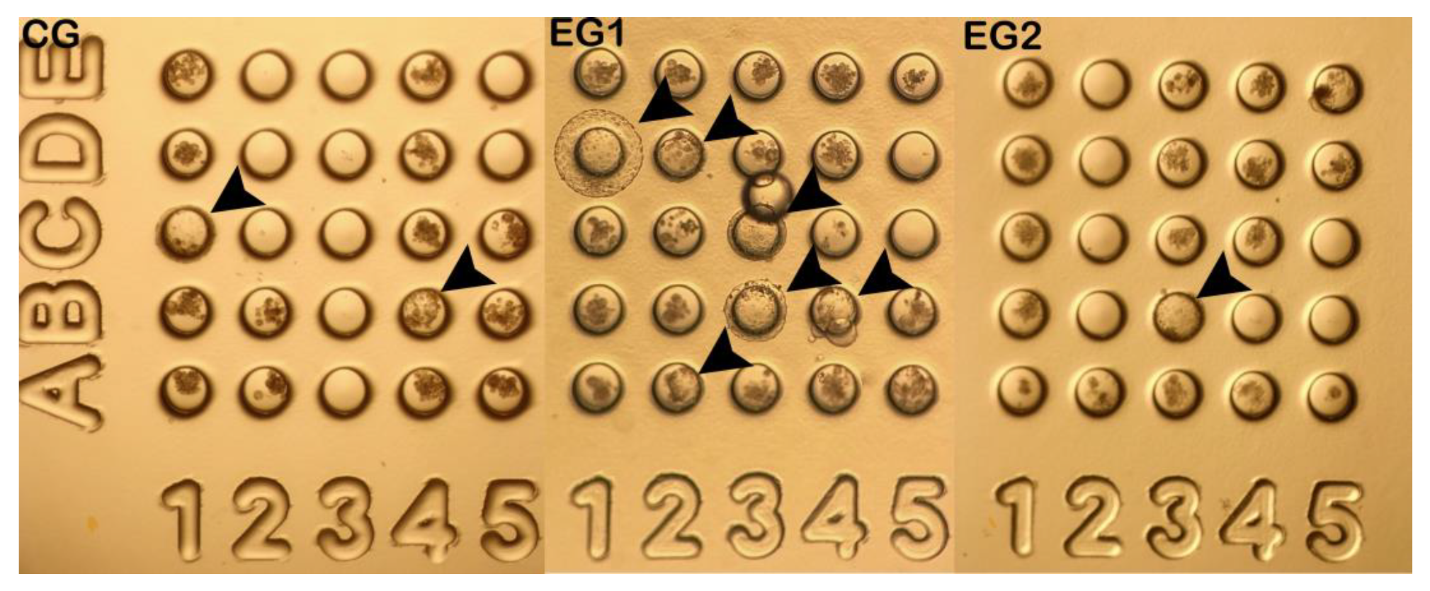
Figure 1. Effect of resveratrol on the IVM of sheep oocytes. The effect of 0.0 (CG), 0.5 (EG1), and 1.0 (EG2) μM resveratrol concentrations during IVM was analyzed. Bars represent mean ± SD. Significant differences (p>0.05).
Preprints 173925 g001
Figure 2. Ovis canadensis mexicana cloned embryos in the WOW system on day 7 of culture, obtained from O. aries oocytes treated with 0.0 (CG), 0.5 (EG1), and 1.0 μM (EG2) of resveratrol during IVM. Arrows indicate bighorn sheep cloned embryos at different blastocyst stages: Coordinates 1C (One blastocyst in CG); 1D, 2A, 2D, 3B, 3C, 4B (Six blastocysts in EG1, three of them expanded); and 3B (One blastocyst in EG2). Magnification 40X.
Preprints 173925 g002
Table 1. In vitro development rate of O. c. mexicana cloned embryos produced by ISCNT using HMC from O. aries oocytes treated with resveratrol.
Group No. Cleavage
N (%)
4-16 cells
N (%)
Morula
N (%)
Blastocysts
N (%)
Fragmented
N (%)
CG 40 38 (94±8.0) 6 (16±18.5) 9 (21±15.4) 6 (16±3.2)a 17 (46±8.8)a
EG1 69 69 (100) 12 (20±22.9) 16 (24±6.5) 22 (31±12.0)b 19 (25±10.4)b
EG2 32 32 (100) 8 (20±19.9) 8 (26±17.3) 2 (6±6.3)a 14 (42±6.4)a
Disclaimer/Publisher’s Note: The statements, opinions and data contained in all publications are solely those of the individual author(s) and contributor(s) and not of MDPI and/or the editor(s). MDPI and/or the editor(s) disclaim responsibility for any injury to people or property resulting from any ideas, methods, instructions or products referred to in the content.
Copyright: This open access article is published under a Creative Commons CC BY 4.0 license, which permit the free download, distribution, and reuse, provided that the author and preprint are cited in any reuse.
Prerpints.org logo

Preprints.org is a free preprint server supported by MDPI in Basel, Switzerland.

Subscribe

Disclaimer

Terms of Use

Privacy Policy

Privacy Settings

© 2025 MDPI (Basel, Switzerland) unless otherwise stated