
Submitted:
06 August 2025
Posted:
08 August 2025
You are already at the latest version
Abstract
Cockroaches are widely recognized as significant vectors for various infectious and parasitic diseases affecting both animals and humans. Cockroaches are more than just common pests; they are among the most medically important insects due to their frequent presence in human habitats. These resilient creatures are major reservoirs and vectors for numerous pathogens, including bacteria, viruses, parasites, and fungi. Recognizing the role of cockroaches in the ecosystem of disease is crucial, and implementing effective control measures is necessary. This study focuses on identifying bacterial contaminants present on the surfaces of Oriental cockroaches (Blatta orientalis) collected from pig farms. Using the Vitek 2 Compact system, a modern platform for rapid and automated phenotypic identification, several bacterial species were isolated. The predominant microorganism identified was E. coli, followed by Staphylococcus spp., Streptococcus spp., and Enterobacter spp. These findings highlight the potential role of cockroaches in the transmission of harmful infectious diseases to both animal populations and humans.
Keywords:
cockroaches
; Blatta orientalis
; Vitek
; bacterial pathogens
; pig farms
1. Introduction
Cockroaches are widely distributed insects of considerable economic, veterinary, and public health importance. The relationship between insects and humans has evolved over a long history, profoundly affecting human health [1]. Their extensive evolution has equipped them with a remarkable ability to adapt and survive in a wide range of environments, including extreme conditions. Many species are exceptionally well-suited to a synanthropic lifestyle, thriving in human settlements, livestock farms, and food industry facilities [2]. This synanthropic behavior raises critical concerns regarding their role in the transmission of infectious and parasitic pathogens that may affect both humans and animals [3]. Cockroaches are among the most significant sanitary pests that can directly and indirectly harm animals and humans. They are among the few insects that play the role of important vectors in the spread of some of the most devastating diseases affecting humans – dysentery in a maternity ward in the USA in 1950, leprosy in Mexico in 1954 [4,5].
Research has demonstrated that cockroaches are capable of carrying microorganisms and parasites on their surfaces, which positions them as significant vectors in the transmission of various infectious and parasitic diseases affecting both animals and humans [6,7,8,9,10,11,12]. Numerous studies indicate that synanthropic species of cockroaches are the primary carriers of food-borne pathogenic microorganisms [13,14,15]. Although cockroaches do not bite, they contribute to pathogen transmission through surface contamination and fecal excretion [16]. In light of the complex epidemic scenario related to the spread of African swine fever, an increasing number of researchers are raising concerns regarding the potential role of cockroaches and other insects in the dissemination of the causative agent of this disease [17,18,19].
Pathogens can survive on cockroaches or in their digestive systems for over a month after exposure to contaminated environments or food and water. These pathogens may be released into the environment through their chitinous exoskeletons or feces [20,21]. Given their potential to contribute to the spread of disease, assessing microbial contamination levels in cockroach populations cohabiting with humans and animals is of paramount importance [22]. To evaluate the epidemic risk, specialized methods have been developed for the cultivation and identification of microbial contaminants present on the surface of cockroaches [20; 23-27]. Among contemporary techniques enabling rapid and automated phenotypic identification of microorganisms through colorimetric analysis, the Vitek 2 Compact system (BioMérieux, France) is increasingly utilized in clinical practice [28]. This system typically employs Vitek 2 GP ID and Vitek 2 GN ID cards for the identification of Gram-positive and Gram-negative bacteria, respectively, facilitating the precise differentiation of over 150 distinct pathogenic species. This method is increasingly used as a complementary analytical approach for identifying microbes in various samples from humans and animals, including cockroaches [29,30,31,32,33,34]. The data collected by many researchers justify and clarify the role of cockroaches in transmitting infectious disease agents, functioning mechanically and serving as one of the three main links in the epidemiological chain, specifically as a spreading factor [23; 27; 35,36,37,38,39,40].
The aim of this study was to investigate surface bacterial carriage in field populations of cockroaches inhabiting pig farms in the Republic of Bulgaria, with a view to identifying health and epidemic risks.
2. Materials and Methods
A total of 350 Oriental cockroaches, collected from four field populations, were examined in this study, following the methodology of Solomon [24] and Davari [27]. Specimens were collected between June and July 2023 from four industrial-scale pig farms in Bulgaria using monitoring traps. Following euthanasia by supercooling, groups of ten cockroaches were placed in sterile plastic containers containing 5 mL of sterile saline to create pooled samples. Surface contamination was extracted from each pool by vigorous vortexing at low speed (600-800 rpm) for 120 seconds, following the protocols of Alikhani et al. [25] and Haile et al. [26]. From the supernatant, 1 mL was pipetted into two sterile Eppendorf tubes and transported under refrigeration to a specialized microbiological laboratory for densitometric identification of Gram-positive and Gram-negative microorganisms using the VITEK 2 Compact System (BioMérieux, France). Samples were inoculated onto blood agar (Blood Agar Base, HiMedia Laboratories, India) and MacConkey agar (HiMedia Laboratories, India), and incubated aerobically at 37 ℃ for 24 hours, following a similar procedure described by Mariam [41].
Data Analysis
The statistical package IBM® SPSS® Statistics 26.0 was used to process the data.
3. Results
Microbiological analysis aimed at evaluating the epidemiological potential of synanthropic cockroaches as mechanical vectors of pathogens of sanitary-hygienic and veterinary relevance revealed a substantial bacterial load on their external surfaces (Table 1).
The quantitative distribution of Gram-negative and Gram-positive microorganisms isolated from the surface of synanthropic cockroaches collected from the surveyed pig farms is shown in Figure 1 and Figure 2. The Cramér’s V values, ranging from 0.092 to 0.482, indicate a weak to moderate association between bacterial species variability and farm location.
As shown in the summarized data in Figure 3, Escherichia coli was the predominant microbial isolate recovered from synanthropic cockroaches across all pig farms, followed by Staphylococcus spp., Streptococcus spp., and Enterobacter spp.
4. Discussion
Cockroaches are recognized as sanitary and hygienic pests of high epidemiological importance, acting as vectors for numerous pathogenic agents. When inhabiting contaminated environments or consuming infected food and water, various viruses, bacteria, fungi, and protozoa may persist on their exoskeletons or within their digestive tracts for extended periods and be subsequently released into the environment [20,21].
Fang et al. [22] emphasize the critical need to assess microbial contamination in cockroach populations, particularly those cohabiting with humans and animals. Globally, most studies in this area have focused on German cockroaches and other synanthropic or exotic species. In Bulgaria, one of the few studies was conducted by Popova et al. [42] also on an exotic cockroach species. However, there remains a substantial knowledge gap regarding the vectorial role of the black oriental cockroach, especially considering its frequent and severe infestations in pig farming facilities documented in our field observations.
In our investigation, significant surface bacterial contamination was detected in oriental cockroaches from pig farms (Table 1). The isolates included nine microbial taxa: Escherichia coli, Staphylococcus spp., Streptococcus spp., Enterobacter spp., Klebsiella pneumoniae, Pseudomonas spp., Enterococcus spp., Serratia marcescens, and Proteus mirabilis. These microorganisms are considered conditionally pathogenic, forming part of the normal or transient flora of the skin, mucous membranes, and intestines of mammals and birds, yet capable of causing disease under specific conditions in humans and animals.
Our findings are consistent with previous studies, such as those by Zarchi et al. [43], who identified 19 bacterial species from cockroach surfaces and digestive tracts, including E. coli, Group D Streptococcus, Bacillus spp., Klebsiella pneumoniae, and Proteus vulgaris. Similarly, Holakouei et al. [44] reported a dominant presence of Klebsiella spp., Pseudomonas spp., Proteus spp., Citrobacter spp., Enterobacter spp., and Serratia spp. in German cockroaches.
A pronounced prevalence of Gram-negative bacteria, particularly from the Enterobacteriaceae family, was observed in our samples. This aligns with studies by Fotedar et al. [23], Cloarec et al. [35], Rivault et al. [45], and Vythilingam et al. [46], which reported similar bacterial profiles in German and American cockroach species. These bacteria pose serious health threats, including urinary tract infections, sepsis, and gastroenteritis [47].
Controlling synanthropic cockroach populations is essential, particularly in livestock environments, where their presence increases the risk of zoonotic transmission. Our findings support previous research indicating the role of American cockroaches in spreading enteropathogens [45; 48-51].
Experimental studies by Zurek & Schal [52] demonstrated that cockroaches shed large amounts of viable, virulent E. coli in their feces after initial exposure to contaminated material. Similarly, our study identified E. coli as the predominant contaminant on the surface of oriental cockroaches across all sampled farms. Pathogenic strains of E. coli are among the most frequently isolated pathogens in swine in veterinary practice [53].
Enterotoxigenic and verotoxigenic strains of E. coli are the main cause of colienteritis and colienterotoxicosis in newborn and weaned pigs, as well as edematous disease in postweaned pigs, often with high mortality [54,55,56].
Waldvogel et al. [57] reported the persistence of viable E. coli F18 strains in cockroach feces for up to 8 days post-exposure, with high bacterial loads (4.4 x 10⁵ CFU g-1) comparable to those found in infected pigs (1.9 x 10⁶ CFU g-1). This underscores the vector potential of cockroaches for these pathogens. Multiple studies have confirmed the transmission capacity of German cockroaches for verotoxigenic E. coli F18, an important pathogen of pigs [57,58]. Therefore, targeted cockroach density control is recommended as an integral part of disease prevention and control programs in pig farming [57].
The demonstrated vector competence of cockroaches for E. coli F18, along with the high concentrations of fecal coliforms in their excreta, represents a significant epidemiological concern [57]. The ability of cockroaches to migrate into infected farms poses a considerable risk for pathogen dissemination, potentially undermining disinfection protocols and compromising biosecurity measures. This threat is particularly pronounced during disease outbreaks, when control interventions—such as animal removal, cleaning of feeders and drinkers, and intensive disinfection using strong-smelling agents—often provoke mass migration of cockroaches to adjacent, unaffected facilities. Given that E. coli F18 cells remain viable and virulent after passage through the cockroach digestive tract and can be excreted in large numbers for over a week, the potential for disease spread via migrating cockroaches is both evident and alarming [52].
Staphylococcus spp. and Streptococcus spp. were the next most frequently isolated genera in our study. Numerous investigations have highlighted the potential role of cockroaches as vectors of bacterial pathogens, particularly these two genera. Their frequent isolation from cockroach populations suggests a possible route for transmission to humans and animals. Studies by Fotedar et al. [23] in Taiwan and India, Salehzadeh et al. [59] in Iran, and Vazirianzadeh et al. [60] have reported significant associations between cockroach infestations and the presence of pathogenic bacteria. Additionally, Schauer et al. [61,62] and Lampert et al. [63] identified high concentrations of staphylococci in various cockroach species, especially in the hindgut of Shelfordella lateralis. Of particular concern is Staphylococcus aureus, a bacterium colonizing the skin and mucosal surfaces of approximately 30% of the human population, known for its ability to cause serious infections [64]. The increasing detection of antibiotic-resistant S. aureus strains in cockroaches, as documented by Menasria et al. [38], Islam et al. [65] and Abdolmaleki et al. [66], underscores their growing importance as a public health threat.
Our investigation revealed concerning levels of Klebsiella pneumoniae contamination across all surveyed pig farms. These findings are consistent with those of Cotton et al. [67], who reported a high prevalence of Klebsiella in German cockroaches. Their work highlights the critical role of cockroaches as vectors in the transmission of nosocomial infections, emphasizing the urgent need for comprehensive pest control strategies in both agricultural and healthcare environments.
In one of the studied farms, Proteus mirabilis was detected on the surfaces of examined cockroaches. This bacterium has also been identified in the gastrointestinal tracts of German and American cockroaches [68,69,70], as well as in exotic species [42]. Additionally, the presence of Pseudomonas aeruginosa was confirmed—an opportunistic pathogen frequently isolated from various cockroach species, including those implicated in hospital infestations [71]. P. aeruginosa is particularly notable for its high resistance to disinfectants and its prominent role in healthcare-associated infections.
5. Conclusions
The findings of this study highlight significant sanitary and epidemiological risks associated with cockroach infestations in livestock production and food processing environments. In particular, the detection of Blatta orientalis in swine facilities was strongly associated with high levels of bacterial surface contamination. Escherichia coli was identified as the predominant microbial isolate, followed by Staphylococcus spp., Streptococcus spp., Enterobacter spp., Klebsiella pneumoniae, Pseudomonas spp., Enterococcus spp., Serratia marcescens, and Proteus mirabilis, reflecting a diverse and complex microbial profile. These results underscore the necessity of implementing strict hygiene and biosecurity measures to mitigate the risk of pathogen transmission in such settings.
Author Contributions
Conceptualization, B.B-M., P.M. and G.Z.; methodology, B.B-M., P.M. and G.Z.; resources, B.B-M., P.M. and G.Z; writing—original draft preparation, B.B-M., P.M. and G.Z.; writing—review and editing, B.B-M., P.M. and G.Z.; visualization, B.B-M., P.M. and G.Z. All authors have read and agreed to the published version of the manuscript.
Informed Consent Statement
Not applicable.
Funding
This research received no external funding.
Data Availability Statement
The original contributions presented in this manuscript are included in the article. Further inquiries can be directed to the corresponding author.
Conflicts of Interest
The authors declare no conflicts of interest.
Abbreviations
The following abbreviations are used in this manuscript:
| GP ID | Gramm positive identification card |
| GN ID | Gramm negative identification card |
| PS | Pulled samples |
| n | number |
References
- Dent D.; Binks, R. H. Insect pest management. 2020, Cabi, pp 1-11.
- Boneva-Marutsova, B. Study on the Distribution and Evaluation of the Efficiency of the Control Methods of Synanthropic Cockroach Species in Animal Farms and Food Processing Plants in Bulgaria; Ph.D. Thesis, Trakia University, Stara Zagora, Bulgaria, 2024.
- Alesho N. A. Synanthropic cockroaches of Russia. Proceedings of the International Colloquia on Social Insects. 1997, Volume 3-4., 45-50 ref. 37.
- Roth L. M.; Willis, E. R. The medical and veterinary importance of cockroaches. Smirhsonian Miscellaneous Collections. 1957, pp. 134 : 137.
- Czajka E. ; Pancer K,; Kochman , M.; Gliniewicz , Al.; Sawicka, B.; Rabczenko, D.; Stypułkowska-Misiurewicz, H. Characteristics of bacteria isolated from body surface of German cockroaches caught in hospitals. Przegl Epidemiol. 2003., 57 (4), 655-62.
- Odinets, A.A.; Seradzhi, V.E.; Degtyareva, L.A.; Odinets, O.L. The level of social organization of synanthropic cockroaches. Proceedings of the Colloquia on Social Insects, St. Petersburg. 1993, Vol. 2, pp. 221–222. Available online: http://pestkiller.ru/sochrorganizachiya.shtml (accessed on 1 August 2025) (RU).
- Donets, A.V. Problematic issues in cockroach control in medical and preventive institutions. Bulletin of Hygiene and Epidemiology. 2004, 8(1), 116–120 (RU).
- Vatev, N.T.; Kevorkyan, A.K.; Rakadzhieva, T.A.; Stoilova, Y.D. Manual for Practical Exercises in the Epidemiology of Infectious Diseases. Raikov Publishing, 2006. pp. 26–27 (BG).
- Vahabi A.; Rafinejad J.; Mohammadi, P.; Biglarian, F. Regional evaluation of bacterial contamination in hospital environment cockroaches. J Environ Health Sci Eng. 2007, 4, 57–60.
- Patel A.; Jenkins M.; Rhoden K.; Barnes, A. N. A systematic review of zoonotic enteric parasites carried by flies, cockroaches, and dung beetles. Pathogens. 2022, 11. [CrossRef]
- Merad Y.; Belkacemi, M.; Merad, Z.; Bassaid, A.; Benmansour, Z.; Matmour D.; Belmokhtar Z. Fungal carriage of hospital trapped cockroaches: A prospective study. New Microbes New Infect. 2023, 52, 101086. [CrossRef]
- Geng, D.; Yu, H.; Zhao, T.; Li, C. The Medical Importance of Cockroaches as Vectors of Pathogens: Implications for Public Health. Zoonoses. 2025, 5(1), 982. [CrossRef]
- Pai H. H.; Chen W. C.; Peng C. F. Isolation of bacteria with antibiotic resistance from household cockroaches (Periplaneta americana and Blattella germanica). Acta Trop. 2005, 93 (3), 259–265. [CrossRef]
- Blazar J.M.; Lienau E.K.; Allard, M.W. Insects as vectors of foodborne pathogenic bacteria. Terrestrial Arthropod Reviews. 2011, 4 (1), 5-16. [CrossRef]
- Fila M.; Woźniakowski, G. African swine fever virus – the possible role of flies and other insects in virus transmission. Journal of Veterinary Research. 2020, 64 (1-7). [CrossRef]
- Yoon H.; Hong, S.-K; Lee Il.; Lee, E.-S. Insects as potential vectors of African swine fever virus in the Republic of Korea. Authorea Preprints. 2020. [CrossRef]
- Medrano M. A literature review to gather the scientific evidence for an African Swine Fever virus (ASFV) exposure assessment of US domestic pigs raised in total confinement and/or with outdoor access to ASFV-infected feral swine. 2023, https://conservancy.umn.edu/items/4c8f4b8a-c992-4d4f-a911-53096ccabf77.
- Moges F.; Eshetie, S.; Endris M. Cockroaches as a source of high bacterial pathogens with multidrug resistant strains in Gondar Town, Ethiopia. Biomed Res Int. 2016, 2825056. [CrossRef]
- Liu J.; Yuan, Y.; Feng, L.; Lin, C.; Ye, C.; Liu J.; Liu H. Intestinal pathogens detected in cockroach species within different food-related environment in Pudong, China. Scientific Reports. 2024, 14 (1), 1-10. [CrossRef]
- Fang W.; Fang, Z.; Liu, Z.; Yuan, J.; Zhang X.; Peng, H.; Xiao, Y. Phylogenetic analysis of bacterial community in the gut of American cockroach (Periplaneta americana). Wei Sheng wu xue bao=Acta Microbiologica Sinica. 2013, 53 (9), 984-994.
- Fotedar R.; Shriniwas U. B.; Verma, A. Cockroaches (Blattella germanica) as carriers of microorganisms of medical importance in hospitals. Epidemiol Infect. 1991, 107 (1), 181–187. [CrossRef]
- Solomon F.; Belayneh F.; Kibru G.; Ali, S. Vector potential of Blattella germanica (L.) (Dictyoptera: Blattidae) for medically important bacteria at food handling establishments in Jimma town, Southwest Ethiopia. BioMed Research International. 2016, (1), 3490906. [CrossRef]
- Alikhani M. Y.; Parsavash S.; Arabestani M. R.; Hosseini , S. M. Prevalence of antibiotic resistance and class 1 integrons in clinical and environmental isolates of Pseudomonas aeruginosa. Avicenna J Clin Microbiol Infect. 2017, 4 (4): 12086. [CrossRef]
- Haile T.; Mariam, A.T.; Kiros S.; Teffera, Z. Cockroaches as carriers of human gastrointestinal parasites in Wolkite Town, southwestern Ethiopia. J Parasitol Vector Biol. 2018, 10 (2), 33‒38. [CrossRef]
- Davari B.; Hassanvand, A. E.; Salehzadeh, A.; Alikhani, M. Y.; Hosseini, S. M. Bacterial Contamination of Collected Cockroaches and Determination their Antibiotic Susceptibility in Khorramabad City, Iran. Journal of Arthropod-Borne Diseases. 2023, 17 (1), 63-71. [CrossRef]
- Alyas S. S.; Ibrahim R. K.; Kareem, A. A. Study the pathogenicity of the bacteria associated with Periplanta americana cockroach on Gelleria mollonella. L worm larva. In Journal of Physics: Conference Series. 2021. (Vol. 1999, No. 1, p. 012032). IOP Publishing.
- Bizzini A.; Jaton, K.; Romo, D.; Bille, J.; Prod’hom G.; Greub, G. Matrix-assisted laser desorption ionization–time of flight mass spectrometry as an alternative to 16S rRNA gene sequencing for identification of difficult-to-identify bacterial strains. J Clin Microbiol. 2011, 49, 693–696. [CrossRef]
- Rettinger A.; Krupka, I.; Grunwald, K.; Dyachenko, V.; Fingerle, V.; Konrad, R.; Heribert, R.; Busch U. et al. Leptospira spp. strain identification by MALDI TOF MS is an equivalent tool to 16S rRNA gene sequencing and multi locus sequence typing (MLST). BMC Microbiol. 2012, 12, 185. [CrossRef]
- Perez-Sancho M.; Vela, A.I.; Garcıa-Seco, T.; Gottschalk, M.; Domınguez L.; Fernandez-Garayzabal, J.F. Assessment of MALDI-TOF MS as alternative tool for Streptococcus suis identification. Front Public Health. 2015. 3, 202. [CrossRef]
- Singhal N.; Kumar M.; Virdi, J.S. MALDI-TOF MS in clinical parasitology: applications, constraints and prospects. Parasitology. 2016, 143, 1491–1500. [CrossRef]
- Manukumar H.M.; Umesha, S. MALDI-TOF-MS based identification and molecular characterization of food associated methicillin-resistant Staphylococcus aureus. Sci Rep. 2017, 7, 1–16. [CrossRef]
- Mehainaoui A.; Menasria, T.; Benouagueni, S.; Benhadj, M.; Lalaoui R.; Gacemi-Kirane, D. Rapid screening and characterization of bacteria associated with hospital cockroaches (Blattella germanica L.) using MALDI-TOF mass spectrometry. Journal of applied microbiology. 2021, 130 (3), 960-970. [CrossRef]
- Cloarec A., Rivault, C.; Fontaine F.; Le Guyader, A. Cockroaches as carriers of bacteria in multi-family dwellings. Epidemiology & Infection. 1992, 109 (3), 483-490. [CrossRef]
- Al-bayati N. Y.; Al-Ubaidi A. S.; Al-Ubaidi, I. K. Risks associated with cockroach Periplaneta americana as a transmitter of pathogen agents. Diyala Journal of Medicine. 2011, 1 (1), 91-97.
- Menasria T.; Moussa, F.; El-Hamza, S.; Tine, S.; Megri R.; Chenchouni H. Bacterial load of German cockroach (Blattella germanica) found in hospital environment. Pathog Glob Health. 2014a, 108 (3), 141– 147. [CrossRef]
- Menasria T.; Tine, S.; Souad, E.; Mahcene, D.; Moussa, F.; Benammar L.; Mekahlia, M. N. A survey of the possible role of German cockroaches as a source for bacterial pathogens. J Adv Sci Appl Eng. 2014b, 1 (1), 67–70.
- Molewa M. L.; Barnard T.; Naicker, N. A potential role of cockroaches in the transmission of pathogenic bacteria with antibiotic resistance: A scoping review. The Journal of Infection in Developing Countries. 2022, 16 (11), 1671-1678. [CrossRef]
- Alhajeri N.; Alharbi J. S.; El-Azazy, O. M. The Potential Role of the American Cockroach (Periplaneta americana) as a Vector of Enterobacteriaceae in Kuwait. Egyptian Academic Journal of Biological Sciences E Medical Entomology & Parasitology. 2023, 15 (1). [CrossRef]
- Mariam S. H. Isolation and Characterization of Gram-Negative Bacterial Species from Pasteurized Dairy Products: Potential Risk to Consumer Health. J Food Qual. 2021, 17, 1‒10. [CrossRef]
- Popova T.; Trencheva K.; Tomov, R. Investigation on the microflora of the Brazilian cockroach Blaberus giganteus (L.)(Blattodea: Blaberidae) with a view to assess its epizootiological significance. Ecology and Future-Journal of Agricultural Science and Forest Science. 2010, 9 (3), 30-33.
- Zarchi A. A. K.; Vatani H. A survey on species and prevalence rate of bacterial agents isolated from cockroaches in three hospitals. Vector-Borne and Zoonotic Diseases. 2009, 9 (2), 197-200. [CrossRef]
- Holakuie Naieni, K.; Ladonni, H.; Asle Soleimani, H.; Afhami, Sh.; Shayeghi, M. The role of German cockroach in hospital infection. J Sch Public Health. 2004, 2, 43–54.
- Rivault C.; Cloarec A.; Le Guyader A. Bacterial load of cockroaches in relation to urban environment. Epidemiology & Infection. 1993, 110 (2), 317-325. [CrossRef]
- Vythilingam I.; Jeffery, J.; Oothuman, P.; AR A. R.; Sulaiman, A. Cockroaches from urban human dwellings: isolation of bacterial pathogens and control. The Southeast Asian Journal of Tropical Medicine and Public Health. 1997, 28 (1), 218-222.
- Schapheer, C.; González, L. M.; Villagra, C. Microorganism Diversity Found in Blatta orientalis L.(Blattodea: Blattidae) Cuticle and Gut Collected in Urban Environments. Insects. 2024, 15 (11), 903. [CrossRef]
- Fotedar R.; Banerjee, U.; Samantray, J. C. Vector potential of hospital houseflies with special reference to Klebsiella species. Epidemiology and infection. 1992a, 109 (1), 143.
- Grübel P.; Hoffman, J. S.; Chong, F. K.; Burstein, N. A.; MePani C.; Cave, D. R. Vector potential of houseflies (Musca domestica) for Helicobacter pylori. Journal of Clinical Microbiology. 1997, 35 (6), 1300-1303. [CrossRef]
- Kobayashi M.; Sasaki, T.; Saito, N.; Tamura, K.; Suzuki, K.; Watanabe H.; Agui, N. Houseflies: not simple mechanical vectors of enterohemorrhagic Escherichia coli O157: H7. The American journal of tropical medicine and hygiene. 1999, 61 (4), 625-629. [CrossRef]
- Pai H.H.; Chen W. C.; Peng, C. F. Isolation of non-tuberculous mycobacteria from hospital cockroaches (Periplaneta americana). J Hosp Infect. 2003, 53 (3), 224–228. [CrossRef]
- Zurek L.; Schal, C. Evaluation of the German cockroach (Blattella germanica) as a vector for verotoxigenic Escherichia coli F18 in confined swine production. Veterinary Microbiology. 2004, 101 (4), 263-267. [CrossRef]
- Fairbrother J. M. Neonatal Escherichia coli diarrhea. Diseases of swine. 1999, 8, 433-441.
- Bosworth B.T.; Casey, T. Procedure for multiplex PCR for porcine E. coli. In: Proceedings of the 97th ASM General Meeting, Miami Beach, FL. 1997, May 4–8.
- Bertschinger H.U. Postweaning Escherichia coli diarrhea and edema disease. In: Straw, B.E., D’Allaire, S., Mengeling, W.L., Taylor, D.J. (Eds.), Disease of Swine. Iowa University Press, Ames, IA. 1999, pp. 441–454.
- Moon H.W.; Hoffman, L.J.; Cornick, N.A.; Booher S.L.; Bosworth, B.T. Prevalence of some virulence genes among Escherichia coli isolates from swine presented to a diagnostic laboratory in Iowa. J. Vet. Diagn. Invest. 1999, 11, 557-560. [CrossRef]
- Waldvogel M. G.; Moore, C. B.; Nalyanya, G. W.; Stringham, S. M., Watson, D. W.; Schal, C. O. B. Y. Integrated cockroach (Dictyoptera: Blattellidae) management in confined swine production. In Proceedings of the 3rd international conference of urban pests. Prague (Czech Republic): Graficke Zavody Hronov, 1999, July, pp. 183-188.
- Zurek L.; Gore, J.C.; Stringham, S.M.; Watson, D.W.; Waldvogel M.G.; Schal, C. Boric acid dust as a component of an integrated cockroach management program in confined swine production. J. Econ. Entomol. 2003, 96, 1362–1366. [CrossRef]
- Salehzadeh A.; Tavacol, P.; Mahjub, H. Bacterial, fungal and parasitic contamination of cockroaches in public hospitals of Hamadan, Iran. J Vector Borne Dis 2007, 44, 105-110.
- Vazirianzadeh B.; Mehdinejad, M.; Dehghani, R. Identification of bacteria which possible transmitted by Polyphaga aegyptica (Blattodea: Blattidae) in the region of Ahvaz, SW Iran. Jundishapur Journal of Microbiology. 2009, 2 (1).
- Schauer C.; Thompson C. L.; Brune, A. The bacterial community in the gut of the cockroach Shelfordella lateralis reflects the close evolutionary relatedness of cockroaches and termites. Applied and environmental microbiology. 2012, 78 (8), 2758-2767. [CrossRef]
- Schauer C.; Thompson C.; Brune, A. Pyrotag sequencing of the gut microbiota of the cockroach Shelfordella lateralis reveals a highly dynamic core but only limited effects of diet on community structure. PLoS One. 2014, 9 (1), e85861. [CrossRef]
- Lampert N.; Mikaelyan A.; Brune, A. Diet is not the primary driver of bacterial community structure in the gut of litter-feeding cockroaches. BMC Microbiol. 2019, 19 (1), 238. [CrossRef]
- Gorwitz R.J.; Kruszon-Moran, D.; McAllister, S.K.; McQuillan, G.; McDougal, L.K.; Fosheim, G.E.; Jensen, B.J.; Killgore, G.; Tenover F. C.; Kuehnert, M. J. Changes in the prevalence of nasal colonization with Staphylococcus aureus in the United States, 2001–2004. J Infect Dis. 2008, 197 (9):1226–1234. [CrossRef]
- Islam A.; Nath, A. D.; Islam, K.; Islam, S.; Chakma, S.; Hossain, M. B.; Al-Faruq A.; Hassan, M. M. Isolation, identification and antimicrobial resistance profile of Staphylococcus aureus in cockroaches (Periplaneta americana). J Adv Vet Anim Res. 2016, 3 (3), 221–228. [CrossRef]
- Abdolmaleki Z.; Mashak, Z.; Safarpoor Dehkordi, F. Phenotypic and genotypic characterization of antibiotic resistance in the methicillin-resistant Staphylococcus aureus strains isolated from hospital cockroaches. Antimicrobial Resistance & Infection Control. 2019, 8 (1), 1-14. [CrossRef]
- Cotton M. F.; Wasserman, E.; Pieper, C. H.; Theron, D. C.; Van Tubbergh, D.; Campbell G.; Barnes, J. Invasive disease due to extended spectrum beta-lactamase-producing Klebsiella pneumoniae in a neonatal unit: the possible role of cockroaches. Journal of Hospital Infection. 2000, 44 (1), 13-17. [CrossRef]
- Robertson A. R. The Isolation and Characterization of the Microbial Flora in the Alimentary Canal of Gromphadorhina portentosa Based on rDNA Sequences. Electronic Theses and Dissertations. 2007, Paper 2069. https://dc.etsu.edu/etd/2069.
- Haghi F. M.; Nikookar, H.; Hajati, H.; Harati, M. R.; Shafaroudi, M. M.; Yazdani-Charati J.; Ahanjan, M. Evaluation of bacterial infection and antibiotic susceptibility of the bacteria isolated from cockroaches in educational hospitals of Mazandaran University of medical sciences. Bull Env Pharmacol Life Sci. 2014, 3, 25-28.
- Kundera I. N.; Sapu E. H.; Bialangi, M. Identification of Bacteria on Cockroach Feet (Periplaneta americana) in Resident Bay of Palu Permai and Sensitivity Test Against Antibiotics. Techno Jurnal Penelitian. 2020, 9 (1), 353-362. [CrossRef]
- Saitou K.; Furuhata, K. Kawakami, Y.; Fukuyama M. Biofilm formation abilities and disinfectant-resistance of Pseudomonas aeruginosa isolated from cockroaches captured in hospitals. Biocontrol science. 2009, 14 (2), 65-68. [CrossRef]
Figure 1.
Quantitative distribution of Gram-negative microorganisms isolated from Blatta orientalis across different pig farms.
Figure 1.
Quantitative distribution of Gram-negative microorganisms isolated from Blatta orientalis across different pig farms.
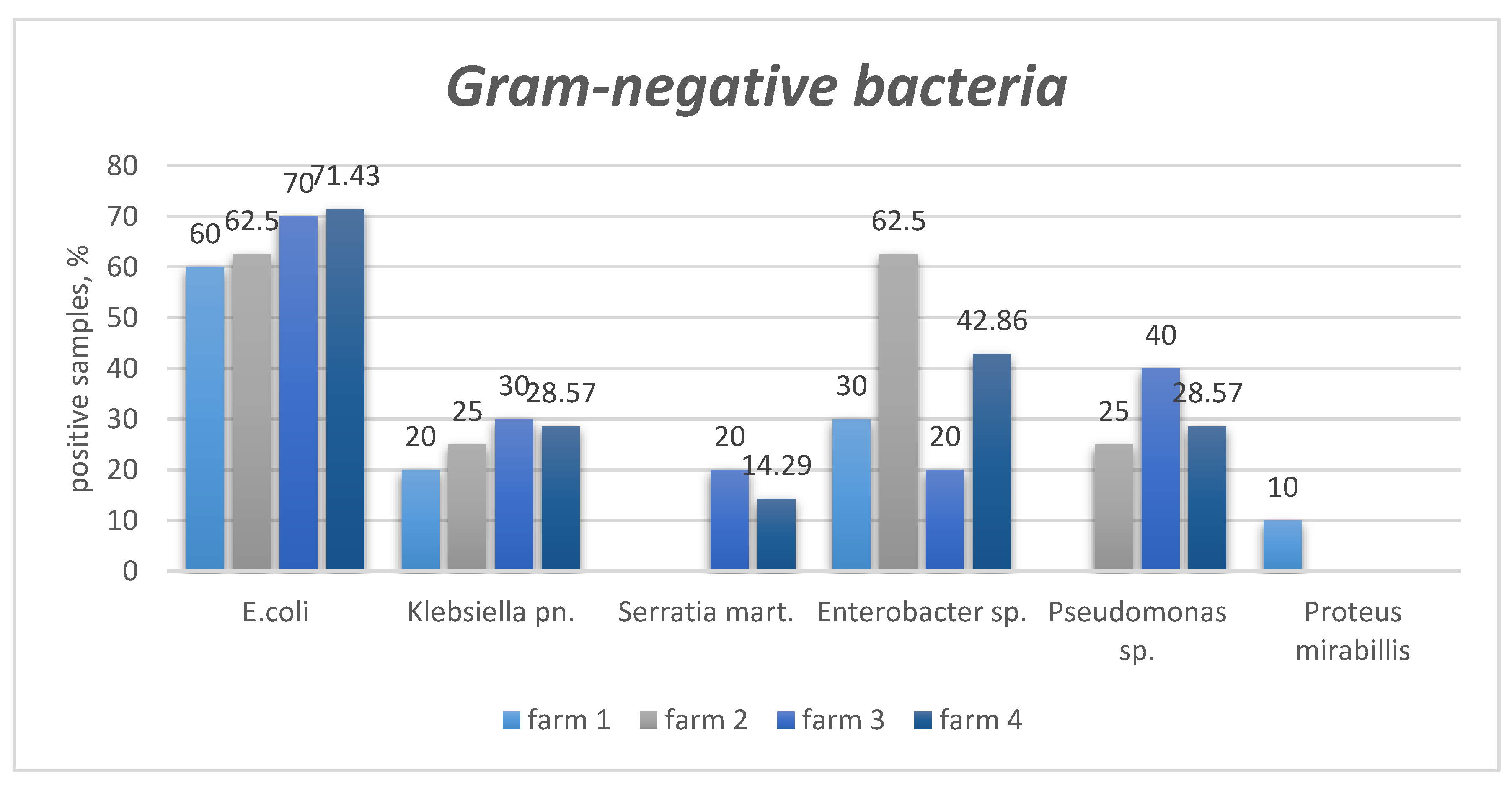
Figure 2.
Quantitative distribution of Gram-positive microorganisms isolated from Blatta orientalis across different pig farms.
Figure 2.
Quantitative distribution of Gram-positive microorganisms isolated from Blatta orientalis across different pig farms.
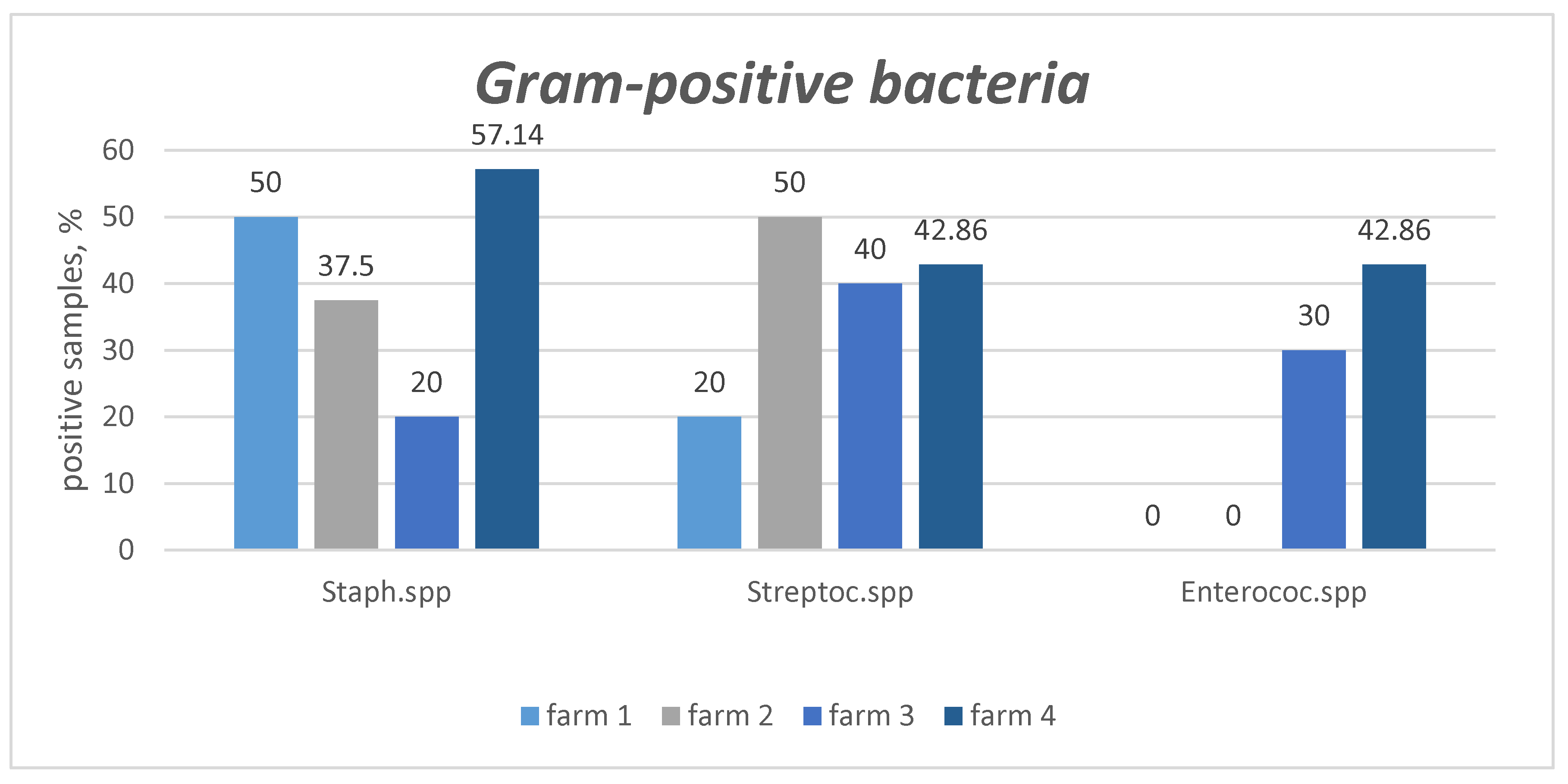
Figure 3.
Summary of the quantitative distribution of microorganisms isolated from Blatta orientalis across all pig farms.
Figure 3.
Summary of the quantitative distribution of microorganisms isolated from Blatta orientalis across all pig farms.
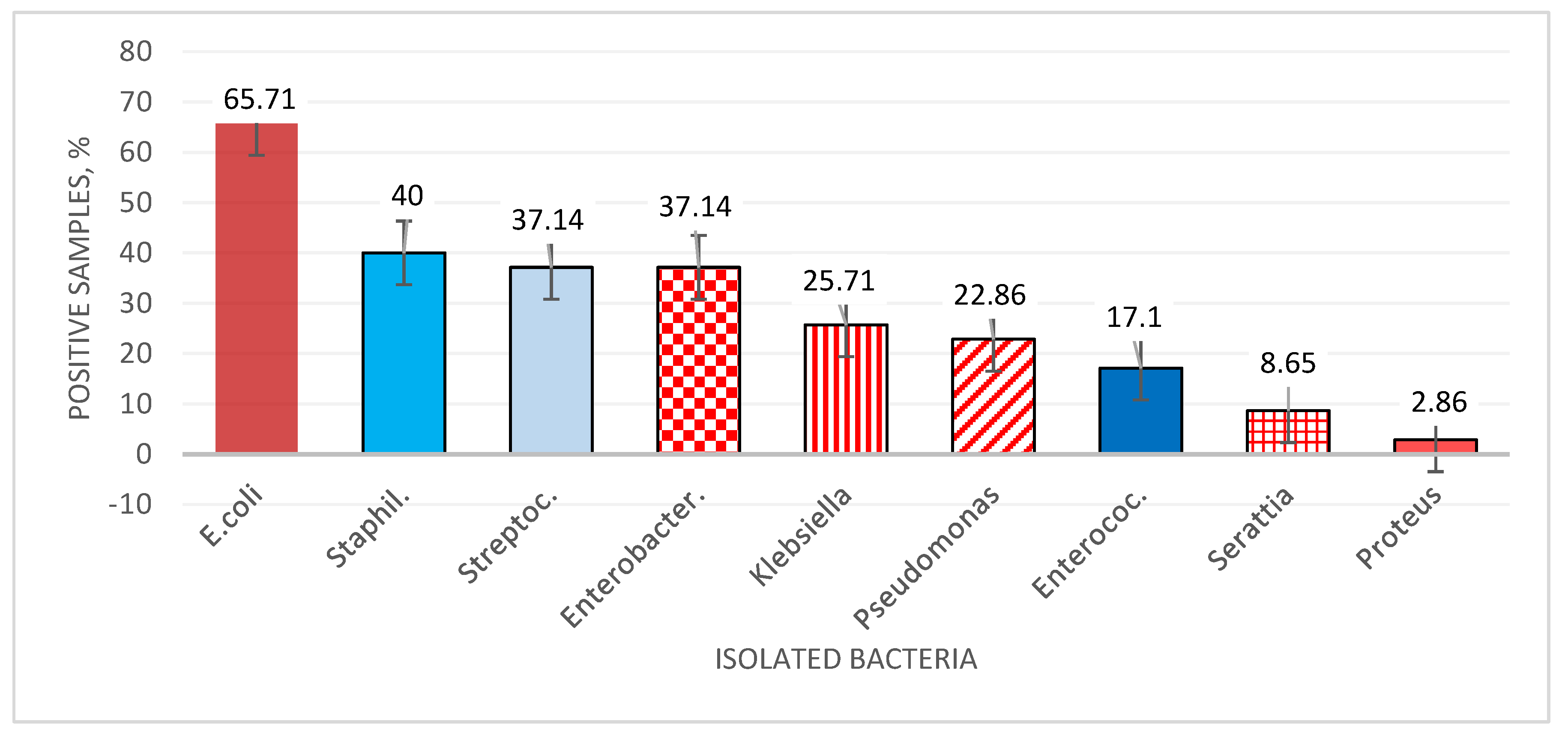

Table 1.
Surface bacterial contamination in Blatta orientalis collected from pig farms.
| Pathogen | Criteria | Farm 1 (n of PS = 10) |
Farm 2 (n of PS = 8) |
Farm 3 (n of PS = 10) |
Farm 4 (n of PS = 7) |
Total (n of PS = 35) |
|---|---|---|---|---|---|---|
| Gram-negative bacteria | ||||||
|
E. coli |
presens | (6) 60% | (5) 62.5% | (7) 70% | (5) 71.4% | (23) 65.7% |
| absence | (4) 40% | (3) 37.5% | (3) 30% | (2) 28.6% | (12) 34.3% | |
| Cramer’s V = 0.102; Sig. (p) = 0.047 | ||||||
| Klebsiella pneumoniae | presens | (2) 20% | (2) 25% | (3) 30% | (2) 28.6% | (9) 25.7% |
| absence | (8) 80% | (6) 75% | (7) 70% | (5) 71.4% | (26) 74.3% | |
| Cramer’s V = 0.092; Sig. (p) = 0.046 | ||||||
| Serattia marcescens | presens | (0) 0% | (0) 0% | (2) 20% | (1) 14.3% | (3) 8.6% |
| absence | (10) 100% | (8) 100% | (8) 80% | (6) 85.7% | (32) 91.4% | |
| Cramer’s V = 0.323; Sig. (p) = 0.030 | ||||||
| Enterobacter spp. | presens | (3) 30% | (5) 62.5% | (2) 20% | (3) 42.9% | (13) 37.1% |
| absence | (7) 70% | (3) 37.5% | (8) 80% | (4) 57.1% | (22) 62.9% | |
| Cramer’s V = 0.329; Sig. (p) = 0.028 | ||||||
| Pseudomonas spp. | presens | (0) 0% | (2) 25% | (4) 40% | (2) 28.6% | (8) 22.9% |
| absence | (10) 100% | (6) 75% | (6) 60% | (5) 71.4% | (27) 77.1% | |
| Cramer’s V = 0.370; Sig. (p) = 0.018 | ||||||
| Proteus mirabillis | presens | (1) 10% | (0) 0% | (0) 0% | (0) 0% | (1) 2.9% |
| absence | (9) 90% | (8) 100% | (10) 100% | (7) 100% | (34) 97.1% | |
| Cramer’s V = 0.271; Sig. (p) = 0.046 | ||||||
| Gram-positive bacteria | ||||||
| Staphylococcus spp. | presens | (5) 50% | (3) 37.5% | (2) 20% | (4) 57.1% | (14) 40% |
| absence | (5) 50% | (5) 62.5% | (8) 80% | (3) 42.9% | (21) 60% | |
| Cramer’s V = 0.291; Sig. (p) = 0.039 | ||||||
| Streptococcus spp. | presens | (2) 20% | (4) 50% | (4) 40% | (3) 42.9% | (13) 37.1% |
| absence | (8) 80% | (4) 50% | (6) 60% | (4) 57.1% | (22) 62.9% | |
| Cramer’s V = 0.237; Sig. (p) = 0.051 | ||||||
|
Enterococcus spp. |
presens | (0) 0% | (0) 0% | (3) 30% | (3) 42.9% | (6) 17.1% |
| absence | (10) 100% | (8) 100% | (7) 70% | (4) 57.1% | (29) 82.9% | |
| Cramer’s V = 0.482; Sig. (p) = 0.043 | ||||||
Legend: n – number; PS - pulled sample of ten Blatta orientalis individuals; Cramer’s V – measure of association strength between categorical variables (range: 0 = no association to 1 = perfect association); Sig. (p) – p-value indicating the probability that the observed association occurred by chance; values less than 0.05 were considered statistically significant. All statistical analyses were performed using IBM® SPSS® Statistics version 26.0.
Disclaimer/Publisher’s Note: The statements, opinions and data contained in all publications are solely those of the individual author(s) and contributor(s) and not of MDPI and/or the editor(s). MDPI and/or the editor(s) disclaim responsibility for any injury to people or property resulting from any ideas, methods, instructions or products referred to in the content. |
© 2025 by the authors. Licensee MDPI, Basel, Switzerland. This article is an open access article distributed under the terms and conditions of the Creative Commons Attribution (CC BY) license (http://creativecommons.org/licenses/by/4.0/).
Copyright: This open access article is published under a Creative Commons CC BY 4.0 license, which permit the free download, distribution, and reuse, provided that the author and preprint are cited in any reuse.


