
Submitted:
22 September 2024
Posted:
23 September 2024
You are already at the latest version
Abstract
An outbreak of esophagitis and ingluvitis caused by Salmonella Typhimurium in passerines seized from illegal wildlife trafficking is described. This illegal activity causes stress and leads to lowered immunity in the birds. Additionally, inadequate hygiene conditions predispose the birds to diseases such as salmonellosis. Few studies report the occurrence of Salmonella-induced lesions in the esophagus and crop of wild birds; thus, this study aimed to describe the disease in these species, investigate its presence in the environment, and determine the antimicrobial resistance profile. Three birds of the genus Sporophila were necropsied. In the esophagus and crop, yellowish plaques corresponding to necrosis with bacterial aggregates were observed. Salmonella spp. was isolated from these lesions, with genus confirmation via MALDI-TOF. Environmental samples were collected from the enclosures and cages where the animals were quarantined, and the same bacterium was isolated. In two birds, the serotype S. Typhimurium was identified. Antibiograms performed on the strains from the birds and the environment revealed resistance to antibiotics in the classes of Penicillins, Sulfonamides, Aminoglycosides, Monobactams, Tetracyclines, and first and third-generation Cephalosporins. To the authors' knowledge, this is the first report of this agent causing death in Sporophila due to esophagitis and ingluvitis. It is also the first report of salmonellosis in three species of passerines in Brazil. The study underscores the importance of understanding the pathogens circulating in wild animals, especially within the context of One Health.
Keywords:
avian pathology
; histopatology
; microbiology
; MALDI-TOF
; molecular serotyping
; One Health
; salmonellosis
1. Introduction
Most wildlife trafficking victims in Brazil are birds, with this illegal activity impacting around 20% of native bird species, including some that are endangered [1]. Birds of the genus Sporophila are significant victims of illegal breeding and trafficking, with many of these seized animals being sent to rehabilitation and screening centers for eventual release back into the wild [2]. Trafficked birds often suffer from poor physical conditions and exhibit liver and biochemical issues due to inadequate nutrition [3]. Furthermore, the stress from trafficking, combined with poor hygiene, leads to weakened immunity, making these birds more susceptible to opportunistic infections and diseases such as Salmonellosis [4].
Salmonella are Gram-negative bacilli belonging to the Enterobacteriaceae family [5,6]. They are divided into two species, S. bongori and S. enterica [7]. S. enterica is further classified into six subspecies: S. enterica subsp. enterica, S. enterica subsp. salamae, S. enterica subsp. arizonae, S. enterica subsp. diarizonae, S. enterica subsp. houtenae, and S. enterica subsp. indica [5,8]. Based on surface antigen characteristics and biochemical properties, Salmonella are subdivided into serovars [6]. Currently, over 2,600 serovars have been identified, with 99% of them belonging to S. enterica subsp. enterica [7,9].
This bacterium can infect humans, domestic animals, wildlife, and even insects, contaminating the environment, water, or food through sick individuals or carriers [4,10,11]. Contamination through the consumption of contaminated eggs and chicken meat makes domestic poultry significant agents in the transmission of Salmonellosis to humans [12]. However, recent studies show that wild birds can harbor serovars relevant to the One Health context, acting as vectors for this pathogen to both humans and domestic animals [13,14,15]. Additionally, the circulation of the pathogen in the wild, whether zoonotic or not, can directly impact wild species populations by causing the death of infected individuals [16].
In passeriformes, the circulation of the bacteria is associated with group feeding practices, especially when humans encourage the congregation of species by providing food in urban and peri-urban environments, or when birds are kept under inadequate conditions in captivity [17]. In these birds, the Typhimurium serovar has been most frequently reported as the cause of outbreaks in Norway [18,19], Sweden [16,20], Switzerland [14], England [21,22,23], Wales [22,23], the United States [24,25,26], Brazil [27], Austria [28], Canada [29], New Zealand [30], Japan [31,32], and Poland [33].
Despite some descriptions, few studies report the occurrence of salmonellosis in clinically healthy free-living passeriformes or in those seized from trafficking and reintroduced into the wild. Thus, the presence of the pathogen in wild birds may be underdiagnosed, as testing is rarely performed on these birds [17]. Birds that become ill show acute progression and die within 24 hours, with reluctance to fly, apathy, and difficulty feeding due to characteristic lesions; necropsy reveals yellowish plaques in the esophagus and crop [14,17]. The aim of this study was to describe the occurrence of salmonellosis in passeriformes of the Sporophila genus, seized from wildlife trafficking and kept in a rehabilitation center, describe the presence of the pathogen in the environment, and examine the antimicrobial resistance profile of the isolated strains.
2. Materials and Methods
2.1. Birds
A wildlife screening and rehabilitation center in the state of Paraíba, Brazil had been experiencing an outbreak of mortality in passerine birds for several weeks. Three birds from the genus Sporophila, all confiscated and housed at the same wildlife rehabilitation center, died spontaneously and were sent for necropsy: Bird 1 was a male, adult caboclinho (Sporophila bouvreuil), seized by the environmental police from a residence in the city of João Pessoa; Bird 2 was a male, adult papa-capim (Sporophila nigricollis), seized from a residence in the city of Campina Grande; and Bird 3 was a male, adult golado (Sporophila albogularis), seized from a residence in João Pessoa, along with six other passeriformes. All birds exhibited symptoms of regurgitation and progressive weight loss, ultimately leading to death within two to four weeks after the onset of clinical signs.
2.2. Necropsy and Histological Evaluation
Necropsies were performed, and tissue samples were collected from the thoracic and abdominal organs, as well as the brain, skin, and bones. All samples were fixed in 10% buffered formalin for subsequent preparation of histological slides, stained with standard hematoxylin and eosin (H&E) staining.
2.3. Microbiology
2.3.1. Bird Samples
During necropsy, aseptic samples were collected from lesions in the esophagus and crop of each bird. For microbiological analysis, the samples were cultured on blood agar base enriched with 5% sheep blood and MacConkey agar, then incubated under aerobic conditions at 37°C. After bacterial growth, identification was performed based on morpho-tinctorial characteristics. For confirmation of the isolate, colonies from the MacConkey agar were subcultured on selective media for the differentiation of Salmonella and Shigella, using Hektoen Enteric (HE) agar and Salmonella-Shigella (SS) agar, allowing the observation of black colonies that produce hydrogen sulfide (H₂S) in both media. After identifying the bacterial isolates, they were selected for antimicrobial susceptibility testing using the disk diffusion method on Mueller-Hinton agar. Disks containing standardized concentrations of antimicrobials were applied, followed by incubation for 24 hours. After incubation, the inhibition zones were measured to assess resistance to the tested antimicrobials.
2.3.2. Microbiological Environmental Samples
Microbiological samples were collected from three enclosures (enclosure 1, enclosure 2, and enclosure 3) and three cages (cage 1, cage 2, and the cage where passeriform birds were quarantined at the wildlife screening and rehabilitation center). To obtain Salmonella spp. samples from the enclosures, three sterile drag swabs were prepared, one for each enclosure. Gauze pads were attached to a 70 cm cotton string, packaged, and autoclaved. The drag swab was run across the entire floor of the enclosure, then placed in a glass tube containing Rappaport–Vassiliadis selective enrichment broth. In the cages, Stuart-type swabs were used, one for each cage, and swabbed across the floor and perches.
The drag swabs were incubated at 37°C for 24 hours, after which the samples were cultured on Hektoen agar and Salmonella-Shigella agar and incubated aerobically at 37°C, with a reading taken after 24 hours. Cultures and isolation from the Stuart-type swabs from the enclosures followed the same protocol used for the bird samples. In samples where Salmonella spp. was isolated, another antibiogram was performed in partnership with the Enterobacteria Laboratory at FIOCRUZ.
2.4. MALDI-TOF
After being isolated in culture media, the colonies from the samples of the three birds were sent in Brain Heart Infusion (BHI) agar with glycerol and subjected to biochemical confirmation through Matrix-Assisted Laser Desorption Ionization – Time Of Flight (MALDI-TOF) analysis, using the ribosomal protein extraction method directly on plate according to Barcelos et al. [34].
2.5. Serotyping
Samples in Mueller-Hinton agar were sent to the Enterobacteria Laboratory at FIOCRUZ, Rio de Janeiro, Brazil, where the identification of Salmonella spp. serotypes was performed using the slide agglutination method, which evaluates antigenic differences in the cell capsule (capsular antigens "Vi"), the cell wall (somatic antigens "O"), and the flagella (flagellar antigens "H"), as proposed by White - Kauffmann - Le Minor [35].
3. Results
3.1. Necropsy and Histological Evaluation
Upon external examination, all birds were found to be underweight, with body scores ranging from 2 to 3 on a scale of 1 to 5. Bird 12 also exhibited amputation of the carpus, metacarpus, and phalanges of the right wing. In the esophagus of all birds, yellowish plaques measuring 0.1 to 0.4 cm in diameter were observed, involving both the serosal and mucosal layers (Figure 1). These plaques extended into the crop in birds 1 and 3. The plaques had an irregular and shiny surface. Upon sectioning, they were soft, compact, and yellowish.
Histopathological evaluation revealed multifocal to coalescent ulcers of the mucosal surface in the esophagus and crop. Multifocal to coalescent necrosis was observed in the submucosa (Figure 2) or extensive necrosis (Figure 3), accompanied by the presence of heterophils, macrophages, fibrin, and a large number of short rod-shaped bacteria. Numerous blood vessels were occluded by fibrin thrombi, along with a small occasional presence of short rod-shaped bacteria. These lesions extended to the serosa of both organs but were more severe in the crop.
3.2. Microbiology
Bacterial cultures from the birds and the environment, which were plated on Hektoen Enteric Agar (HE) and Salmonella-Shigella Agar (SS), exhibited hydrogen sulfide production, characterized by a dark color at the center of the colonies. On MacConkey agar, the Salmonella colonies appeared colorless due to the lack of lactose fermentation. Birds 1, 2, and 3; Enclosures 1 and 2; and Cage 2 showed a dark center in the colonies on SS and HE due to hydrogen sulfide production. On MacConkey agar, they were lactose non-fermenters, appearing colorless (Table 1).
In the antibiogram, samples from Enclosures 1 and 2 and Cage 2 showed resistance to ampicillin and amoxicillin-clavulanate; Bird 3 was resistant to Sulfazotrim; Birds 1, 2, and 3, Enclosure 2, and Cage 2 were resistant to streptomycin; Bird 1 was resistant to aztreonam; Birds 1, 2, and 3, Enclosure 2, and Cage 2 were resistant to tetracycline; Enclosures 1 and 2 and Cage 2 were resistant to cefazolin; Birds 1 and 2 were resistant to cephalexin, and Bird 1 was resistant to ceftriaxone (Table 2).
3.3. MALDI-TOF
In the mass spectrometry technique, the genus of the strains isolated from the esophageal and ingluvial lesions of birds 1, 2, and 3 was confirmed (Table 3).
3.4. Serotyping
All strains were identified through serotyping as belonging to the genus Salmonella, species enterica, and subspecies enterica; except in Enclosure 1, where no sample growth occurred. Bird 1 and Enclosure 2 had the somatic antigens O:4 and O:5 identified. In Birds 1 and 2, it was possible to identify all antigens, allowing for the classification of the Typhimurium serotype in samples from these individuals (Table 4).
4. Discussion
The confirmation of mortality in passerine birds with lesions in the upper digestive tract associated with Salmonella infection in this study was possible through a set of diagnostic techniques. This enabled the elucidation of the pathogenesis of the disease characterized by regurgitation, weight loss, and death in three bird species of the genus Sporophila. To the best of the authors' knowledge, this is the first confirmation of esophagitis and ingluvitis in this bird species. These birds are among the most trafficked wildlife species in Brazil [36], and thus they could be involved in the spread of salmonellosis to other animals or humans.
Our study identified the infection of Salmonella enterica subsp. Enterica serovar Typhimurium in both the birds and the environment where they were being rehabilitated. Birds held in rehabilitation screening centers in Brazil, after a quarantine period, may later be released back into the wild [2]. These birds returning to the wild can contaminate the environment and have impacts on species conservation. Many cases of infection by Salmonella enterica subsp. Enterica serovar Typhimurium have been described in humans associated with the contamination of animal-origin foods, mainly eggs and chicken meat [37,38]. On the other hand, recent studies show that wild birds can harbor serovars relevant to One Health, as they can be vectors of this agent both to people and to domestic animals [13,14,15,39,40,41].
The history of trafficking and interaction with other species may have contributed to the development of fatal disease in the birds of this study. This differs from reports of isolation of the Typhimurium serovar in passerines in Brazil, where the agent was isolated from cloacal swabs of asymptomatic carrier birds [27]. We still do not know the main serovar found in Brazilian birds, but studies of avian salmonellosis outbreaks in different countries have proven that Salmonella Typhimurium is the most prevalent serovar in passerines [42]. Infection in these cases is usually related to the aggregation of species at feeding sites provided by people [13,19], adaptation of birds in urban areas [14], or contact with livestock that may harbor the pathogen [17]. These factors are also observed in trafficked birds in Brazil. In addition to contact with people due to trafficking, birds of the genus Sporophila have the habit of feeding on grass seeds and thus may have urban or rural habits [43], which further favors contact with humans and other animals. These factors increase the risk of both contamination and transmission of salmonellosis, since the Typhimurium serovar is considered one of the main causes of disease outbreaks in people, as well as in other animals, such as domestic cats [14,20] due to hunting habits [44]. Additionally, these birds make seasonal long-distance movements (>1000 km) [43], which can result in the dispersion of the agent to other regions.
The isolation of the bacterium in two enclosures and one cage at the location where the birds were housed indicates the circulation of the pathogen and its persistence in the environment. This characteristic is due to Salmonella's ability to form biofilms through microbial clusters that can resist on inert or living surfaces [45]. This pathogen has been isolated in enclosures, cages, and waterers at wildlife screening centers, associated with the high demand of animals that can be carriers of this agent [46,47]. This highlights the importance of monitoring salmonellosis in animals at wildlife screening centers that will be reintroduced into the wild.
Although it was not possible to detect all somatic and flagellar antigens of bird 1 and enclosure 2, the presence of O:4 and O:5 in these bacteria indicates they belong to serovars of group B, which may or may not be the Typhimurium serovar found in birds 2 and 3. In cage 2, it was possible to identify the presence of Salmonella enterica subsp. enterica without determination of the serovar. Salmonella serotypes are defined from an antigenic formula that considers somatic, flagellar, and capsular antigens. The "O" somatic antigens are also called major antigens and divide the bacteria into several serogroups; serogroup B includes strains possessing the O:1, O:5, O:12, and O:27 antigens, among which are the Paratyphi B and Typhimurium serovars [48]. Multi-drug resistance was observed in the Salmonella isolates from the three birds, two enclosures, and the cage. Streptomycin, an aminoglycoside antibiotic, is not typically used for treating salmonellosis but is commonly used as a growth promoter in animals, potentially leading to the emergence of resistant isolates throughout the production chain. This resistance not only contaminates meat but also soil and vegetation that could be ingested by other species [49]. Resistance mechanisms to tetracycline have also been linked to its use in livestock and its environmental discharge [49]. All samples in this study, except those from Enclosure 1, which did not show bacterial growth, were resistant to both streptomycin and tetracycline.
Resistance to beta-lactams in this study was noted in penicillin derivatives, cephalosporins, carbapenems, and monobactams, due to the ability of Enterobacteriaceae to produce beta-lactamase enzymes that inhibit the action of these antibiotics [50]. Sulfonamides act by inhibiting dihydropteroate synthetase, an enzyme involved in bacterial DNA and RNA synthesis, with resistance often linked to a gene that produces an enzyme insensitive to the drug’s effects [51].
Generally, the isolates from both the birds and the environment exhibited a resistance profile similar to that reported for S. Typhimurium. An evaluation of 11,447 strains showed the tetra-resistant ASSuT pattern (ampicillin, streptomycin, sulfonamides, tetracycline) and the penta-resistant ACSSuT pattern (ampicillin, chloramphenicol, streptomycin, sulfonamides, tetracycline) [37], similar to the resistance patterns found in this study on a smaller scale.
In addition to the microbiological findings, the esophageal nodules observed in the crop and esophagus of the three birds are considered pathognomonic lesions of salmonellosis in passerines associated with the Typhimurium serovar [17;13]. Similar lesions have been observed in outbreaks of salmonellosis in birds in Austria [28], Switzerland [14], Norway [19], Japan [31], and Canada [29]. These organs are the initial site of infection. After the development of these masses, sepsis occurs without intestinal lesions [28]. The reason for the development of lesions in this region is not yet known, but it is believed that there is an affinity for this tissue by the bacteria, associated with the way of contamination through the ingestion of food contaminated by feces [21]. Microscopic lesions are characterized by transmural ulcerative ingluvitis and esophagitis to varying degrees, infiltration of viable and degenerated histiocytes and heterophils, with the presence of intra and extracellular bacterial rods surrounding the necrotic areas and expanding the esophageal wall [14,19].
Death occurs due to septic shock favored by wasting and difficulty of the bird in feeding due to esophageal obstruction caused by the masses. The strains of this serovar seem to be adapted to the groups of birds they can affect, thus they can cause disease with high mortality rates in some groups and others being only asymptomatic carriers [52], which may explain the occurrence of death in animals of the same order in this description. To the authors' knowledge, until now, there had been no report of this esophageal form of salmonellosis in passerines in Brazil.
5. Conclusions
The reported cases in this study highlight the harmful potential of Salmonella Typhimurium in passerines of the Sporophila genus and underscore the importance of curbing wildlife trafficking, as it was a significant factor in the development of the disease in these species. Furthermore, knowledge of the pathogens circulating in wild animals enables the development of mitigative measures to prevent the loss of individuals and protect against zoonoses, since wild animals can act as vectors or reservoirs of diseases for humans and other animals.
Author Contributions
For research articles with several authors, a short paragraph specifying their individual contributions must be provided. The following statements should be used “Conceptualization, K.L.S. and R.B.L.; methodology, K.L.S.; E.S.L; E.M.R-S. and R.B.L.; validation, K.L.S. and R.B.L.; formal analysis, E.M.R-S. and R.B.L.; investigation, K.L.S.; M.O.F.; E.S.L; R.A.F.S; M.S.S; E.M.R-S.; I.V.S.; W.D.Q.S. and R.B.L.; resources, R.B.L.; data curation, K.L.S.; I.V.S.; W.D.Q.S.; A.C.C.F.; E.M.R-S. and R.B.L.; writing—original draft preparation, K.L.S; writing—review and editing, K.L.S.; M.O.F.; L.R.C.E.; E.M.R-S. and R.B.L.; visualization, K.L.S.; E.M.R-S. and R.B.L.; supervision, E.M.R-S. and R.B.L.; project administration, K.L.S and R.B.L.; funding acquisition, R.B.L. All authors have read and agreed to the published version of the manuscript.
Funding
This research was funded by PROPESQ/PRPG/UFPB, grant “Pró-Publicação Edital - Investigation of the Involvement of Wildlife in the Occurrence of Endemic, Epidemic, and Pandemic Diseases in Northeast Brazil, PROPESQ/PRPG/UFPB, 2021”; The National Council for Scientific and Technological Development (CNPq, Brazil) grant 314413/2020-0; and CONCYTEC grant (PE501087383-2024 PROCIENCIA).
Institutional Review Board Statement
The study was conducted in accordance with the Declaration of Instituto Chico Mendes de Conservação da Biodiversidade - ICMBio (protocol code 77213-1 and date of approval 23/12/2020).
Acknowledgments
We express our gratitude to the Enterobacteria Laboratory at the Oswaldo Cruz Foundation, Rio de Janeiro, Brazil (IOC/FIOCRUZ), for conducting the antibiogram, molecular tests, and serological analyses. We also thank the Milk Quality Research Laboratory (Qualileite) at the Faculty of Veterinary Medicine and Animal Science (FMVZ) of the University of São Paulo (USP), São Paulo, Brazil, for performing the MALDI-TOF analysis.
Conflicts of Interest
The authors declare no conflicts of interest.
References
- Regueira, R.F.S.; Bernard, E. Wildlife sinks: Quantifying the impact of illegal bird trade in street markets in Brazil. Biological Conservation. 2012, 149, 16–22. [Google Scholar] [CrossRef]
- Nascimento, C.J.; Oliveira, A.M.; Santos, W.M.; Araújo, J.L.; Rola, L.D.; Guerra, R.R. Body condition, external morphology, parasitology, and histological and biometrical study of the gastro-intestinal tract of Sporophila nigricollis and Sporophila caerulescens seized from trafficking in Northeastern Brazil. Pesq. Vet. Bras. 2024, 44, e07358–1. [Google Scholar] [CrossRef]
- Siqueira, R.A.S.; de Lucena, R.B.; Cavalcanti, T.A.; de Lima Luna, A.C.; de Oliveira Firmino, M.; Guerra, R.R. Aspectos clínico-patológicos em papagaios-verdadeiros (Amazona aestiva, L. 1758) oriundos de apreensões do tráfico no estado da Paraíba, Brasil. Brazil. J. Vet. Med. 2016, 38, 439–444.
- Campos, J.A.; Guizelini, C.C.; Pavarini, S.P.; Azuaga, L.; Souza, M.L.; Leal, C.R.B.; Ramos, C.A.N.; Gomes, D.C. Pathological Aspects of a Septicemic Salmonellosis Outbreak Caused by Salmonella Serotype Typhimurium in Captive Blue-Fronted Amazon Parrots (Amazona aestiva). Pesq. Vet. Bras. 2024, 44, e07428. [Google Scholar] [CrossRef]
- Tindall, B.J.; Grimont, P.A.D.; Garrity, G.M.; Euzeby, J.P. Nomenclature and Taxonomy of the Genus Salmonella. Int. J. Syst. Evol. Microbiol. 2005, 55, 521–524. [Google Scholar] [CrossRef] [PubMed]
- Trabulsi, L.R.; Alterthum, F. Microbiologia, 6ª ed.; Editora Atheneu: São Paulo, Brazil, 2015; Salmonella, pp. 351-360. [Google Scholar]
- Issenhuth-Jeanjean, S.; Roggentin, P. , Mikoleit, M.; Guibourdenche, M.; De Pinna, E.; Nair, S; Fields, P. I.; Weill, F. Supplement 2008–2010 (no. 48) to the White-Kauffmann-Le Minor Scheme. Res. Microbiol. 2014, 165, 526–530. [Google Scholar] [CrossRef]
- Guibourdenche, M.; Roggentin, P. , Mikoleit, M.; Fields, P.I.; Bockemuhl, J.; Grimont, P.A.D.; Weill, F.X. Issenhuth-Jeanjean, S. Supplement 2003–2007 (No. 47) to the White-Kauffmann-Le Minor Scheme. Res. Microbiol. 2010, 161, 26–29. [Google Scholar] [CrossRef] [PubMed]
- Hassan, A.H.A.; Salam, H.S.H.; Abdel-Latef, G.K. Serological Identification and Antimicrobial Resistance of Salmonella Isolates from Broiler Carcasses and Human Stools in Beni-Suef, Egypt. Beni-Suef Univ. J. Basic Appl. Sci. 2016, 5, 202–207. [CrossRef]
- Braga, P.R.C.; Basso, R.M.; Martins, L.S.A. Occurrence of Salmonella spp. in Fecal Samples from Foals with and without Diarrhea in the State of São Paulo: Microbiological Diagnosis, Antimicrobial Susceptibility Profile, and Molecular Detection. Pesq. Vet. Bras. 2023, 43, e07194. [Google Scholar] [CrossRef]
- Pinto, P.N.; Torres, A.C.D.; Rodrigues, M.P.; Oliveira, L.B.; Costa, C.S.; Ecco, R.; Freitas Neto, O.C.; Martins, N.R.S. An Outbreak of Fatal Pullorum Disease (Salmonella Pullorum) in Guinea Fowl Keets (Numida meleagris). Pesq. Vet. Bras. 2023, 43, e07088. [Google Scholar] [CrossRef]
- Forsythe, S.J. Microbiologia Segurança Alimentos, 2nd ed.; Artmed: Porto Alegre, Brazil, 2013. [Google Scholar]
- Thomas, N.J.; Hunter, D.B.; Atkinson, C.T. Infectious Diseases of Wild Birds; Blackwell Publishing: Iowa, USA, 2007; Salmonellosis pp. 270–288. [Google Scholar]
- Giovannini, S.; Pewsner, M.; Hüssy, D.; Hächler, H.; Degiorgis, M-P. R.; Hirschheydt, J.; Origgi, F.C. Epidemic of Salmonellosis in Passerine Birds in Switzerland with Spillover to Domestic Cats. Vet. Pathol. 2013, 50, 597–606. [Google Scholar] [CrossRef]
- Silva, C.; Calva, E.; Maloy, S. One Health and Food-Borne Disease: Salmonella Transmission between Humans, Animals, and Plants. Microbiol. Spectr. 2014, 2, 1. [Google Scholar] [CrossRef]
- Söderlund, R.; Jernberg, C. ; Trönnberg, L; Pääjärvi, A; Agren, E.; Lahti, E.. Linked Seasonal Outbreaks of Salmonella Typhimurium among Passerine Birds, Domestic Cats, and Humans, Sweden, 2009 to 2016. Eurosurveillance, 2019; 24, 1–10. [Google Scholar] [CrossRef]
- Tizard, I.R. Salmonellosis in Wild Birds. Semin. Avian Exot. Pet Med. 2004, 13, 50–66. [Google Scholar] [CrossRef]
- Kapperud, G.; Stenwig, H.; Lassen, J. Epidemiology of Salmonella Typhimurium 0:4-12 Infection in Norway. Am. J. Epidemiol. 1998, 147, 774–782. [Google Scholar] [CrossRef] [PubMed]
- Refsump, T.; Vikoren, T.; Handeland, K.; Kapperud, G.; Holstad, G. Epidemiologic and Pathologic Aspects of Salmonella Typhimurium Infection in Passerine Birds in Norway. J. Wildl. Dis. 2003, 39, 64–72. [Google Scholar] [CrossRef]
- Tauni, M.A.; Österlund, A. Outbreak of Salmonella Typhimurium in Cats and Humans Associated with Infection in Wild Birds. J. Small Anim. Pract. 2000, 41, 339–341. [Google Scholar] [CrossRef] [PubMed]
- Hughes, L.A.; Shopland, S.; Wigley, P.; Bradon, H.; Leatherbarrow, A.H.; Williams, N.J.; Bennett, M.; Pinna, E.; Lawson, B.; Cunningham, A.A.; Chantrey, J. Characterisation of Salmonella enterica Serotype Typhimurium Isolates from Wild Birds in Northern England from 2005–2006. BMC Vet. Res. 2008, 4, 1–10. [Google Scholar] [CrossRef]
- Lawson, B.; Howard, T.; Kirkwood, J.K.; Macgregor, S.K.; Perkins, M.; Robinson, R.A.; Ward, L.R.; Cunningham, A.A. Epidemiology of Salmonellosis in Garden Birds in England and Wales, 1993 to 2003. EcoHealth 2010, 7, 294–306. [Google Scholar] [CrossRef]
- Mather, A.E.; Lawson, B.; Pinna, E.; Wigley, P.; Parkhill, J.; Thomson, N.R.; Page, A.J.; Holmes, M.A.; Paterson, G.K. Genomic Analysis of Salmonella enterica Serovar Typhimurium from Wild Passerines in England and Wales. Appl. Environ. Microbiol. 2016, 82, 6728–6735. [Google Scholar] [CrossRef]
- Locke, L.N.; Shillinger, R.B.; Jareed, T. Salmonellosis in Passerine Birds in Maryland and West Virginia. J. Wildl. Dis. 1973, 9, 144–145. [Google Scholar] [CrossRef]
- Hall, A.J.; Saito, E.K. Avian Wildlife Mortality Events Due to Salmonellosis in the United States, 1985–2004. J. Wildl. Dis. 2008, 44, 585–593. [Google Scholar] [CrossRef]
- Hernandez, S.M.; Keel, K.; Sanchez, S.; Trees, E.; Gerner-Smidt, P.; Adams, J.K.; Cheng, Y.; Ray, A.; Martin, G.; Presotto, A.; et al. Epidemiology of a Salmonella enterica subsp. enterica Serovar Typhimurium Strain Associated with a Songbird Outbreak. Appl. Environ. Microbiol. 2012, 78, 7290–7298. [Google Scholar] [CrossRef]
- Matias, C.A.R.; Pereira, I.A.; Araújo, M.S.; Santos, A.F.M.; Lopes, R.P.; Christakis, S.; Rodrigues, D.P.; Siciliano, S. Characteristics of Salmonella spp. Isolated from Wild Birds Confiscated in Illegal Trade Markets, Rio de Janeiro, Brazil. BioMed Res. Int. 2016. [Google Scholar] [CrossRef]
- Brunthaler, R.; Spergser, J.; Weissenböck, H. Multiple Epidemics in Austrian Fringillidae Caused by a Single Variant of Salmonella Typhimurium. J. Wildl. Dis. 2021, 57, 891–899. [Google Scholar] [CrossRef] [PubMed]
- Daoust, P.Y.; Busby, D.G.; Ferns, L.; Goltz, J.; McBurney, S.; Poppe, C.; Whitney, H. Salmonellosis in Songbirds in the Canadian Atlantic Provinces During Winter-Summer 1997–98. Can. Vet. J. 2000, 41, 54–59. [Google Scholar] [PubMed]
- Alley, M.R.; Connolly, J.H.; Fenwick, S.G.; Mackereth, G.F.; Leyland, M.J.; Rogers, L.E.; Haycock, M.; Nicol, C.; Reed, C.E. M. An Epidemic of Salmonellosis Caused by Salmonella Typhimurium DT160 in Wild Birds and Humans in New Zealand. N. Z. Vet. J. 2002, 50, 170–176. [Google Scholar] [CrossRef] [PubMed]
- Une, Y.; Sanbe, A.; Suzuki, S.; Niwa, T.; Kawakami, K.; Kurosawa, R.; Izumiya, H.; Watanabe, H.; Kato, Y. Salmonella enterica Serotype Typhimurium Infection Causing Mortality in Eurasian Tree Sparrows (Passer montanus) in Hokkaido. Jpn. J. Infect. Dis. 2008, 61, 166–167. [Google Scholar] [CrossRef]
- Fukui, D.; Takahashi, K.; Kubo, M.; Une, Y.; Kato, Y.; Izumiya, H.; Teraoka, H.; Asakawa, M.; Yanagida, K.; Bando, G. Mass Mortality of Eurasian Tree Sparrows (Passer montanus) from Salmonella typhimurium DT40 in Japan, Winter 2008–09. J. Wildl. Dis. 2014, 50, 484–495. [Google Scholar] [CrossRef]
- Krawiec, M.; Pietkiewicz, M.; Wieliczko, A. Salmonella spp. as a Cause of Mortality and Clinical Symptoms in Free-Living Garden Bird Species in Poland. Polish J. Vet. Sci. 2014, 17, 729–731. [Google Scholar] [CrossRef]
- Barcelos, M.M.; Martins, L.; Grenfell, R.C.; Juliano, L.; Anderson, K.L.; Santos, M.V.; Gonçalves, J.L. Comparison of Standard and On-Plate Extraction Protocols for Identification of Mastitis-Causing Bacteria by MALDI-TOF MS. Braz. J. Microbiol. 2019, 50, 849–857. [Google Scholar] [CrossRef]
- Salfinger, Y.; Tortorello, M.L. Compendium of Methods for the Microbiological Examination of Foods, 15th ed.; APHA Press: Washington, DC, USA, 2015; Salmonella, pp. 445–475. [Google Scholar]
- Costa, F.J.V.; Ribeiro, R. E.; Souza, C. A.; Navarro, R. D. Birds Species Trafficked in Brazil: A Meta-Analysis with Emphasis on Threatened Species. Fronteiras. 2018, 7, 324–346. [Google Scholar] [CrossRef]
- Wang, X.; Biswas, S.; Paudyal, N.; Pan, H.; Li, X.; Fang, W.; Yue, M. Antibiotic Resistance in Salmonella Typhimurium Isolates Recovered from the Food Chain through National Antimicrobial Resistance Monitoring System Between 1996 and 2016. Front. Microbiol. 2019, 10, 1–12. [Google Scholar] [CrossRef]
- Seribelli, A.A.; Cruz, M.F.; Vilela, F.P.; Frazão, M.R.; Paziani, M.H.; Almeida, F.; Medeiros, M.I.C.; Rodrigues, D.P.; Kress, M.R.Z.; Allard, M.W.; Falcão, J.P. Phenotypic and Genotypic Characterization of Salmonella Typhimurium Isolates from Humans and Foods in Brazil. PLoS ONE. 2020, 15, e0236868. [Google Scholar] [CrossRef]
- Fischer, E.F.; Müller, R.; Todte, M.; Taubert, A.; Hermosilla, C. Role of Free-Ranging Synanthropic Egyptian Geese (Alopochen aegyptiaca) as Natural Host Reservoirs for Salmonella spp. in Germany. Animals. 2023, 13, 3403. [Google Scholar] [CrossRef] [PubMed]
- Corradini, C.; De Bene, A.F.; Russini, V.; Carfora, V.; Alba, P.; Cordaro, G.; Senese, M.; Terracciano, G.; Fabbri, I.; Di Sirio, A.; et al. Detection of Salmonella Reservoirs in Birds of Prey Hosted in an Italian Wildlife Centre: Molecular and Antimicrobial Resistance Characterisation. Microorganisms 2024, 12, 1169. [Google Scholar] [CrossRef]
- Kobuszewska, A.; Wysok, B. Pathogenic Bacteria in Free-Living Birds, and Its Public Health Significance. Animals. 2024, 14, 968. [Google Scholar] [CrossRef] [PubMed]
- Beleza, A.J.F.; Maciel, W.C.; Lopes, E.S.; Albuquerque, A.H.; Carreira, A.S; Nogueira, C.H.G.; Bandeira, J.M.; Vasconcelos, R.H.; Teixeira, R.S.C. Evidence of the Role of Free-Living Birds as Disseminators of Salmonella spp. Arq. Inst. Biol. 2020, 87, 1–14. [Google Scholar] [CrossRef]
- Silva, J.M.C. Seasonal Movements and Conservation of Seedeaters of the Genus Sporophila in South America. Studies in Avian Biology 1999, 19, 272–280. [Google Scholar]
- Trouwborst, A.; McCormack, P.C.; Camacho, E.M. Domestic Cats and Their Impacts on Biodiversity: A Blind Spot in the Application of Nature Conservation Law. People and Nature. 2020, 2, 235–250. [Google Scholar] [CrossRef]
- Steenackers, H.; Hermans, K.; Vanderleyden, J.; Keersmaecker, S.C.J. Salmonella Biofilms: An Overview on Occurrence, Structure, Regulation and Eradication. Food Research International. 2012, 45, 502–531. [Google Scholar] [CrossRef]
- Bueno, D.J.; Rodríguez, F.I.; Machado, L.C.; Soria, M.A.; Procura, F.; Gómez, S.C.; Hoffmann, T.M.; Alcain, A.; Caffer, M.I.; Latorre, J.D.; et al. Study of Salmonella spp. from Cage Papers Belonging to Pet Birds in an Argentinean Canary Breeder Championship. Animals. 2024, 14, 1207. [Google Scholar] [CrossRef]
- Morais, R. .; Costa, C.; Bravo, E.; Santana, A.; Félix, S.; Sant´Ana, G.; Baptista, L.; Sant´Ana, C. Avaliação Sanitária do Setor de Quarentena do Centro de Triagem de Animais Silvestres de Goiânia (Goiás). Rev. Processos Químicos. 2013, 7, 73–80. [Google Scholar] [CrossRef]
- Silva, N.; Junqueira, V.C.A.; Silveira, N.F. A.; Taniwaki, M.H.; Santos, R.F.S.; Gomes, R.A.R. Manual de Métodos de Análise Microbiológica de Alimentos e Água, 4th ed.; Livraria Varela: São Paulo, Brazil, 2010. [Google Scholar]
- McDermott, P.F.; Tyson, G.H.; Kabera, C.; Chen, Y.; Li, C.; Folster, J.P.; Ayers, S.L.; Lam, C.; Tate, H.P.; Zhao, S. Whole-Genome Sequencing for Detecting Antimicrobial Resistance in Nontyphoidal Salmonella. Antimicrob. Agents Chemother. 2016, 60. [Google Scholar] [CrossRef]
- Queenan, A.M.; Bush, K. Carbapenemases: The Versatile β-Lactamases. Clin. Microbiol. Rev. 2007, 20, 440–458. [Google Scholar] [CrossRef] [PubMed]
- Antunes, P.; Machado, J.; Sousa, J.C.; Peixe, L. Dissemination of Sulfonamide Resistance Genes (sul1, sul2, and sul3) in Portuguese Salmonella enterica Strains and Relation with Integrons. Antimicrob. Agents Chemother. 2005, 49, 836–839. [Google Scholar] [CrossRef] [PubMed]
- Berchieri Júnior, A.; Silva, A.V.; Gomes, A.P.; et al. Doenças das Aves, 2nd ed.; Fundação APINCO de Ciência e Tecnologia Avícola: Campinas, Brazil, 2009. [Google Scholar]
Figure 1.
Body score of Sporophila genus birds and macroscopic appearance of esophagitis and ingluvitis caused by Salmonella Typhimurium. (A) Bird 1, male adult caboclinho (Sporophila bouvreuil), body score of 2 (on a scale of 1-5). (B) Bird 1, yellowish plaque 0.4 cm in diameter extending from the serosa to the mucosa of the esophagus and crop. (C) Bird 2, male adult papa-capim (Sporophila nigricollis), body score of 2 (on a scale of 1-5). (D) Bird 2, multifocal to coalescent yellowish plaques ranging from 0.1 cm to 0.3 cm in diameter on the mucosa and serosa of esophagus and crop. (E) Bird 3, male adult golado (Sporophila albogularis), body score of 3 (on a scale of 1-5). (F) Bird 3, multifocal to coalescent yellowish plaques ranging from 0.2 cm to 0.3 cm in diameter on the mucosa and serosa of the esophagus and crop.
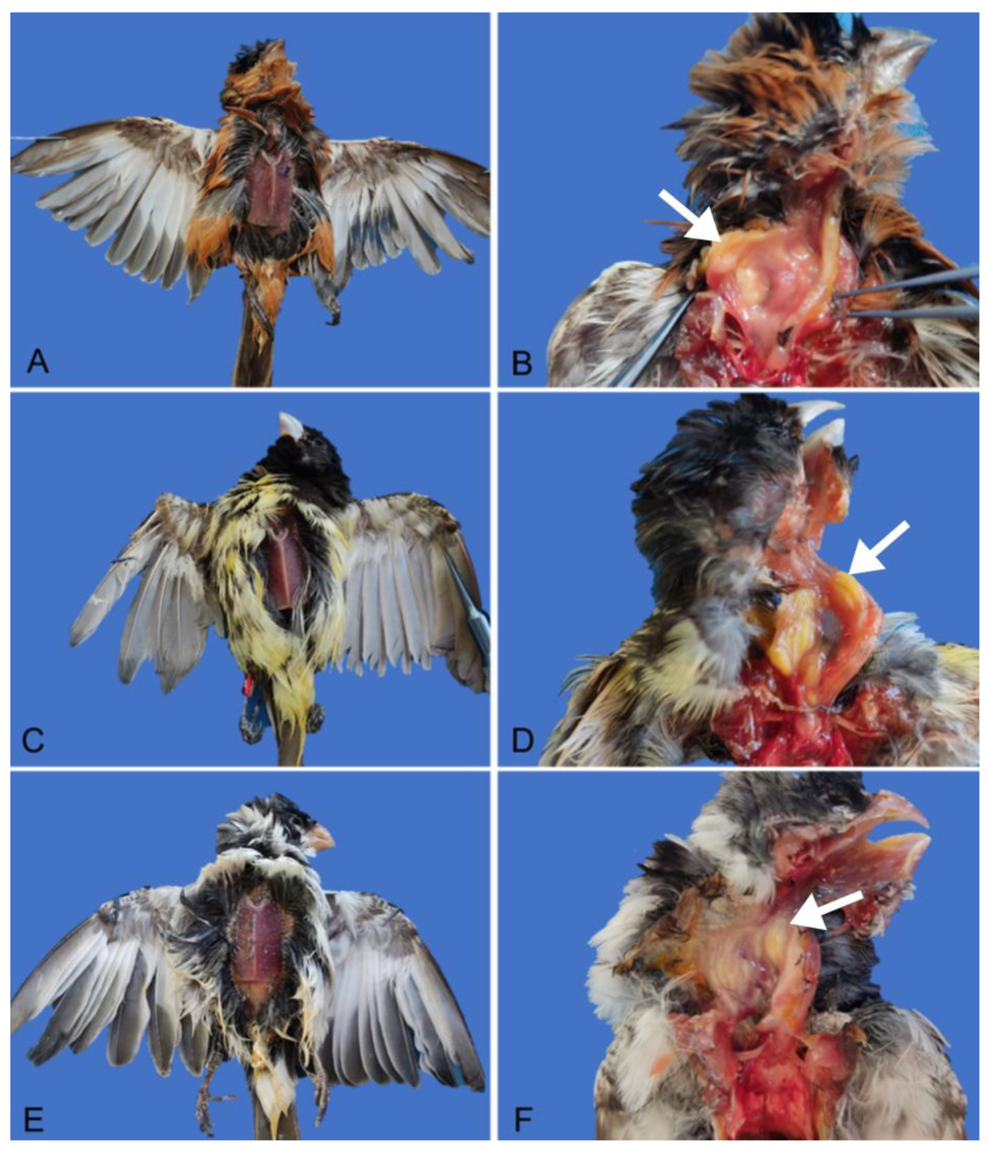
Figure 2.
Histopathology of esophagitis caused by Salmonella Typhimurium in a Caboclinho (Sporophila bouvreuil). (A, B, and C) The crop exhibits multifocal areas of ulceration on the mucosal surface and necrosis in the submucosa (arrow), associated with heterophils, fibrin, and bacterial aggregates (asterisk). Stained with hematoxylin and eosin. Observed under a 4x objective (A), 10x objective (B), and 40x objective (C). All images stained with hematoxylin and eosin.
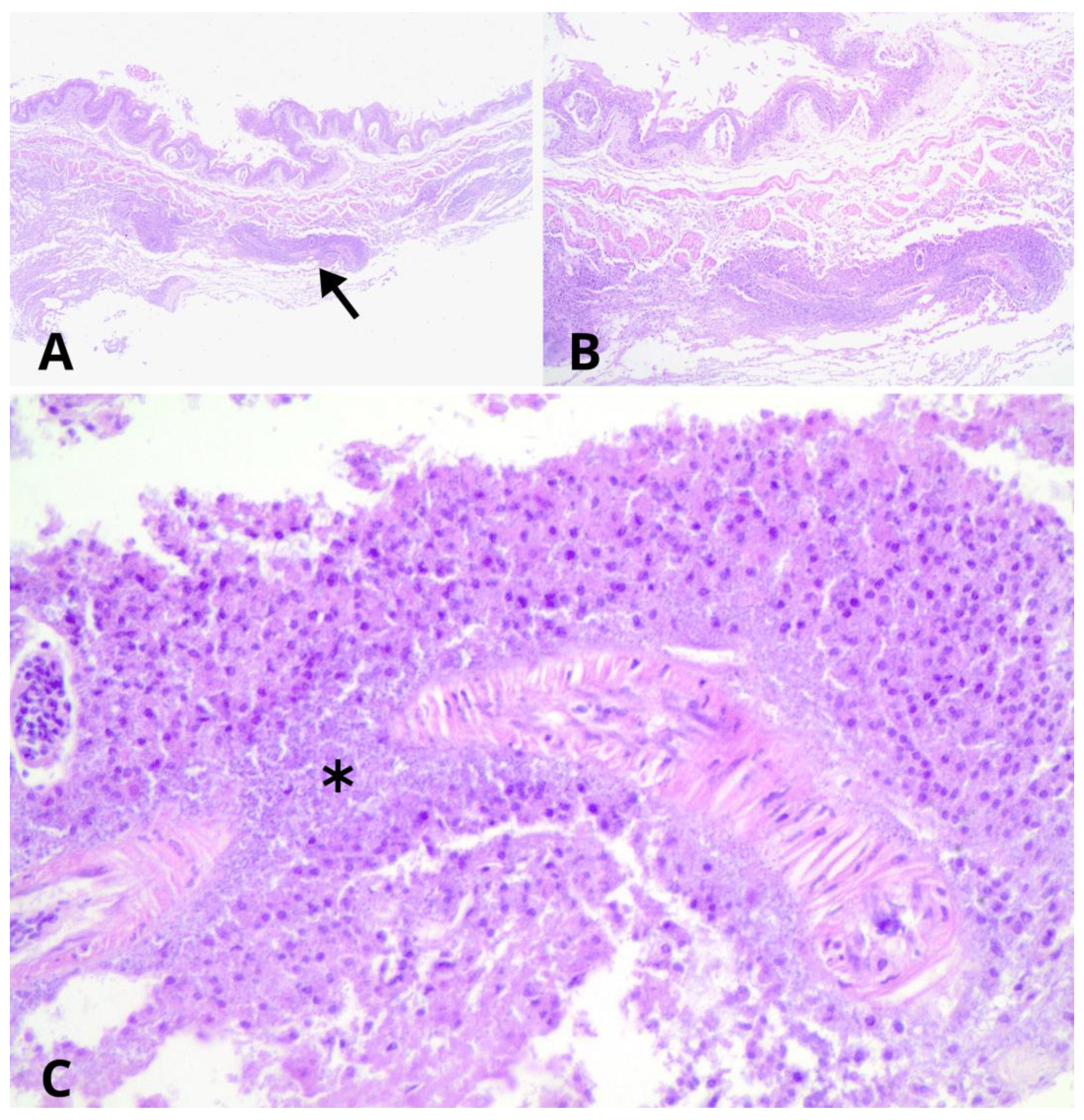
Figure 3.
Histopathology of ingluvitis caused by Salmonella Typhimurium in a Papa-capim (Sporophila nigricollis). (A, B, and C) The crop exhibits an extensive area of ulceration with adjacent necrosis, associated with heterophils, macrophages, fibrin, and bacterial aggregates. Stained with hematoxylin and eosin. Observed under a 4x objective (A), 10x objective (B), and 40x objective (C). All images stained with hematoxylin and eosin.
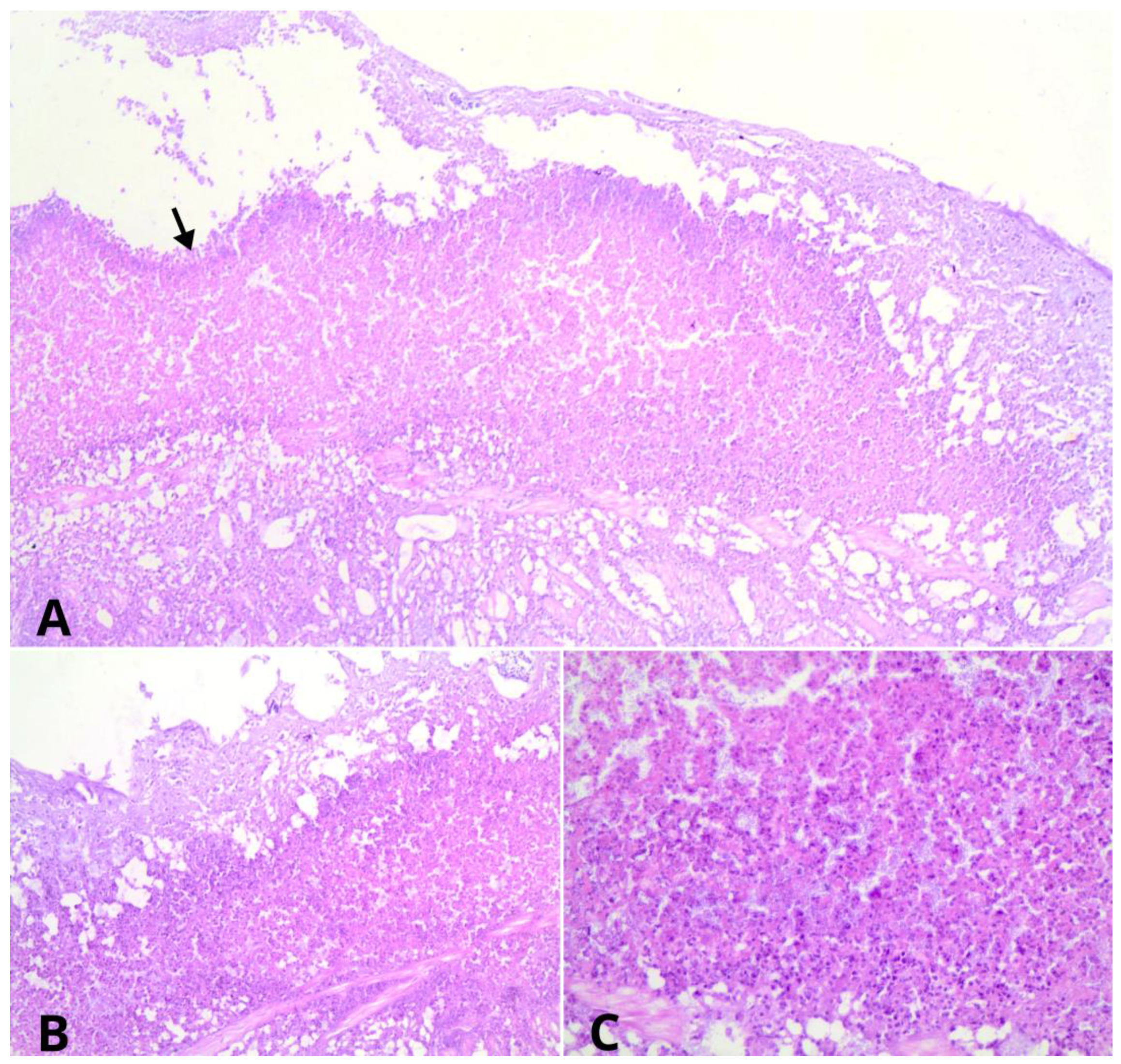

Table 1.
Bacterial cultures from birds of the genus Sporophila, necropsied with lesions of esophagitis and ingluvitis caused by Salmonella Typhimurium, as well as cultures from the enclosures and cages where these birds were quarantined.
Table 1.
Bacterial cultures from birds of the genus Sporophila, necropsied with lesions of esophagitis and ingluvitis caused by Salmonella Typhimurium, as well as cultures from the enclosures and cages where these birds were quarantined.
| Bacterial Culture Media | |||
| Sample | HE | SS | MacConkey Agar |
| Bird 1 | + | + | Lac- |
| Bird 2 | + | + | Lac- |
| Bird 3 | + | + | Lac- |
| Enclosure 1 | + | + | Lac- |
| Enclosure 2 | + | + | Lac- |
| Enclosure 3 | - | - | Lac+ |
| Cage 1 | - | - | Lac+ |
| Cage 2 | + | + | Lac- |
| Cage 3 | - | - | Lac+ |
| + : Production of hydrogen sulfide; - : No production of hydrogen sulfide; Lac + : Lactose-fermenting bacteria; Lac - : Non-lactose fermenting bacteria. | |||
Table 2.
Antibiogram of bacterial cultures from birds of the genus Sporophila, necropsied with esophagitis and ingluvitis lesions caused by Salmonella Typhimurium, as well as cultures from the enclosures and cages where these birds were quarantined.
Table 2.
Antibiogram of bacterial cultures from birds of the genus Sporophila, necropsied with esophagitis and ingluvitis lesions caused by Salmonella Typhimurium, as well as cultures from the enclosures and cages where these birds were quarantined.
| Class Antibiotic | Bird | Bird | Bird | Encl. | Encl. | Cag. |
|---|---|---|---|---|---|---|
| 1 | 2 | 3 | 1 | 2 | 2 | |
| Ampicilin* | - | - | - | + | + | + |
| Amoxcicilin + | - | - | - | + | + | + |
| Clavulonate* Amoxicilin* |
- |
- |
- |
- |
- |
- |
| Sulfazotrim* | - | - | + | - | - | - |
|
Sulfonamides Trimethoprim Sulfamethoxazole** |
- | - | - | x | - | - |
| Phenicoles Chloranfenicol** | - | - | - | x | - | - |
| Gentamicin*/** | - | - | - | x | - | - |
| Streptomycin** | + | + | + | x | + | + |
| Carbapenems Meropenem** | - | - | - | x | - | - |
| Nalidixic Acid** | - | - | - | x | - | - |
| Ciprofloxacin** | - | - | - | x | - | - |
| Monobactams Aztreonam* | + | - | - | - | - | - |
| Tetracycline Tetracyclin** | + | + | + | x | + | + |
| Cephazolin*/** Cephalosporins 1st Cephalexin* |
- */** +* |
- */** +* |
- */** - |
+*/x** - |
+*/-** - |
+*/-** - |
| Cephalosporins 2st Cefoxitin** | - | - | - | x | - | - |
| Ceftriaxone* | - | - | - | - | - | - |
| Cefotaxime* | + | - | - | - | - | - |
| Encl. 1 (Enclosure 1) and Encl. 2 (Enclosure 2): quarantine environment for passerines; Cag. 2 (Cage 2): quarantine cage for passerines within Enclosure 2; - : no antibiotic resistance; + : antibiotic resistance present; x : no growth in the sample. | ||||||
Table 3.
Results of the matrix-assisted laser desorption ionization-time of flight (MALDI-TOF) technique on bacterial cultures from birds of the genus Sporophila, necropsied with esophagitis and ingluvitis lesions caused by Salmonella Typhimurium.
Table 3.
Results of the matrix-assisted laser desorption ionization-time of flight (MALDI-TOF) technique on bacterial cultures from birds of the genus Sporophila, necropsied with esophagitis and ingluvitis lesions caused by Salmonella Typhimurium.
| Bird 1 | Bird 2 | Bird 3 | |
| Identified Bacterium |
Salmonella sp. Enterococcus faecalis |
Salmonella sp. | Salmonella sp. |
| Bird 1: Sporophila bouvreuil; Bird 2: Sporophila nigricollis; Bird 3: Sporophila albogularis. | |||
Table 4.
Serotyping of bacterial cultures from birds of the genus Sporophila, necropsied with lesions of esophagitis and ingluvitis caused by Salmonella Typhimurium, as well as cultures from the enclosures and cages where these birds were quarantined.
Table 4.
Serotyping of bacterial cultures from birds of the genus Sporophila, necropsied with lesions of esophagitis and ingluvitis caused by Salmonella Typhimurium, as well as cultures from the enclosures and cages where these birds were quarantined.
| Sample | Serotyping |
| Bird 1 | Salmonella enterica subesp. enterica (O:4,5) * |
| Bird 2 | Salmonella ser. Typhimurium |
| Bird 3 | Salmonella ser. Typhimurium |
| Cage 2 | Salmonella enterica subesp. enterica * |
| Enclosure 1 | Sample showed no bacterial growth in 24/48 hours. |
| Enclosure 2 | Salmonella enterica subesp. enterica (O:4,5) * |
| *: Flagellar structure not identifiable. | |
Disclaimer/Publisher’s Note: The statements, opinions and data contained in all publications are solely those of the individual author(s) and contributor(s) and not of MDPI and/or the editor(s). MDPI and/or the editor(s) disclaim responsibility for any injury to people or property resulting from any ideas, methods, instructions or products referred to in the content. |
© 2024 by the authors. Licensee MDPI, Basel, Switzerland. This article is an open access article distributed under the terms and conditions of the Creative Commons Attribution (CC BY) license (https://creativecommons.org/licenses/by/4.0/).
Copyright: This open access article is published under a Creative Commons CC BY 4.0 license, which permit the free download, distribution, and reuse, provided that the author and preprint are cited in any reuse.


