
Submitted:
29 July 2025
Posted:
30 July 2025
You are already at the latest version
Abstract
This study investigates the impact of olive tree variety and environmental stress on the production of bioactive compounds in olive leaves, focusing on phenolic content and antioxidant capacity. Three olive varieties—Kalamata, Koroneiki, and wild olive (Olea europaea var. Silvestris)—from two climatically distinct regions in Greece (Stamna and mountainous Paravola) were analyzed. Using three sustainable extraction methods, Ultrasound-Assisted Extraction (UAE), Enzyme-Assisted Extraction (EAE), and Microwave-Assisted Extraction (MAE), the total polyphenolic content (TPC) and antioxidant activity (AA%) of the olive leaf extracts were quantified. Results revealed significant variation in polyphenolic yield depending on both extraction technique and environmental conditions. EAE consistently outperformed other methods in polyphenol recovery, especially from wild olive leaves in the higher-stress environment of Paravola, which showed the highest TPC. This work underscores the synergistic role of varietal and climatic factors in secondary metabolite accumulation and highlights the potential of green extraction technologies in valorizing olive by-products.
Keywords:
plant by-product
; antioxidants
; enzymatic assisted extraction
; microwave assisted extraction
; sonicate assisted extraction
; cultivation conditions
; polyphenols
1. Introduction
Plants produce secondary metabolites that serve as distinctive and valuable resources for industries such as pharmaceuticals, food additives, fragrances, and various other commercial sectors. These compounds, although not directly involved in primary biological functions like growth or reproduction, are typically synthesized at later stages of development. They often have unique chemical structures, are restricted to specific taxonomic groups, and are produced in small quantities as complex mixtures within chemical families.
Unlike primary metabolites, which are essential for an organism's survival, the absence of secondary metabolites may not lead to immediate death but can compromise long-term health, fertility, and adaptability. These substances frequently occur in a limited range of species within a given lineage and are crucial in ecological interactions, especially in defending plants from herbivores and other interspecies threats. Historically, humans have harnessed these compounds for use including medicine, cosmetics, and recreational substances [1].
The olive tree (Olea europaea L.) is emblematic of Mediterranean ecosystems, thriving in evergreen sclerophyllous habitats. It has evolved to endure two distinct and intense environmental stress periods: the hot, dry, high-light conditions of summer and the freezing temperatures of winter. Studies have extensively documented the anatomical and physiological traits, environmental adaptations, and economic significance of the olive tree. Its fruit and oil are central to the Mediterranean diet, with well-known health benefits [2].
Adaptation to summer stress involves complex physiological responses [3]. Under harsh conditions, such as extreme heat and limited water, photolysis is disrupted, leading to an overproduction of reactive oxygen species (ROS), which causes oxidative damage and hampers photosynthesis. To cope, the plant shifts metabolic focus toward secondary metabolic pathways, a strategy that mitigates oxidative stress [4].
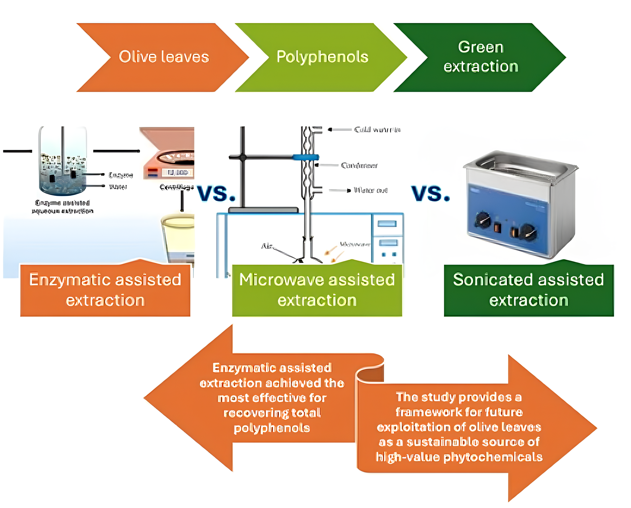
Given the widespread distribution, ecological resilience, and economic value of the olive tree, this study aimed to explore how secondary metabolite production, particularly phenolics, responds to environmental stress typical of Mediterranean summer. By using three sustainable extraction methods—Ultrasound assisted extraction (UAE), Enzymatic assisted extraction (EAE), and Microwave assisted extraction (MAE)—the study focused on measuring the total polyphenolic content in olive leaves through a time-sensitive lens, reflecting the dynamic nature of metabolite biosynthesis in relation to climatic stressors.
Olive leaves (OLs) are a rich natural source of bioactive compounds, notably polyphenols, which exhibit potent antioxidant, antimicrobial, anti-inflammatory, and anti-cancer activities [5-6]. The principal categories of polyphenols in olive leaves include simple phenols—primarily hydroxytyrosol (HT), flavonoids such as apigenin and luteolin glycosides, and secoiridoids, with oleuropein being the most abundant [7-8]. These secondary metabolites possess chemical structures highly suited for neutralizing free radicals, contributing to their strong antioxidant capabilities [9-10].
However, the concentration, composition, and bioavailability of polyphenols in olive leaves are influenced by a variety of genetic, environmental, and agronomic factors [11]. The olive cultivar is one of the most significant determinants; for example, cultivars such as Koroneiki, Kalamata, and wild olive (Olea europaea var. sylvestris) differ in their phenolic profiles and accumulation capacities [12]. Environmental conditions such as temperature extremes, water stress, and solar radiation—characteristics typical of Mediterranean ecosystems—can sig nificantly modulate phenolic biosynthesis, often enhancing it as a stress-response mechanism [13]. Additionally, leaf maturity plays a role, with younger leaves generally containing higher polyphenol concentrations [14]. Other contributing factors include soil composition, altitude, and cultivation practices, including irrigation, pruning, and the use of organic versus conventional methods [15]. Post-harvest conditions, such as drying methods and storage parameters, also affect phenolic compound stability and preservation [16].
Given the health-promoting properties of olive leaf polyphenols, there has been growing interest in utilizing them for dietary supplements, functional foods, and cosmetics. Accordingly, several green extraction techniques have been developed to optimize the recovery of these compounds, including ultrasound-assisted extraction (UAE), microwave-assisted extraction (MAE), and enzyme-assisted extraction (EAE) [17-18]. Studies have demonstrated that UAE enhances the extraction efficiency of oleuropein and hydroxytyrosol by improving cell wall disruption and solvent penetration [19]. MAE accelerates extraction kinetics, resulting in shorter processing times, increased extraction yields, and reduced solvent usage [20]. EAE employs enzymes such as cellulase and pectinase to degrade plant cell walls, thereby facilitating the release of bound polyphenols [21]. Furthermore, parameters such as solvent composition, pH, extraction time, temperature, and the solid-to-liquid ratio significantly influence the final phenolic profile of the extract [22].
The aim of the present study is to compare three eco-friendly extraction methods while considering cultivation parameters and the effects of climate variability between cultivated groves and wild olive fields. Specifically, this study evaluates the total polyphenol content in leaves from four olive varieties collected from two distinct regions in Western Greece, applying different extraction protocols to assess their impact on yield and composition.
2. Materials and Methods
2.1. Plant Material
Leaves from the olive trees were collected during September 2024. Three varieties from two different areas are collected. Koroneiki, Kalamata and a wild olive tree (Olea europaea var. Silvestris) from Stamna, Greece and a wild tree (Olea europaea var. Silvestris) from mountainous Paravola, Greece. The leaves are stored in freezer (-18 oC) until drying for 5 h. at 50 oC [23]. The residual humidity was determined using a moisture analyzer, OHAUS MB35 Halogen (Switzerland), as presented in Table 1. After that the dried leaves are pulverized (<700 μm) and stored in glass jars until used at room temperature in dark and dry place.
2.2. Climate Data
For the present study cultivation conditions were also analyzed. The climate data collection came from the site meteoblue.com, emy.gr and for the cultivation trees from Stamna the data collected from the digital counting system that the producer “Arxaia Oleneia” have on their olive groves, for the period of May to September 2024. The two areas analyzed (Figure 1) were Stamna Aitoloakarnanias, Greece (160 m above sea) and the mountainous area of Paravola Aitoloakarnanias, Greece (400 m above sea) and the distance between them is approximately 33 km.
2.3 Chemicals and Reagents
For analytical purposes, the following reagents were used: 2,2-Diphenyl-1-picrylhydrazyl (DPPH·) (Sigma-Aldrich, Germany). Absolute ethanol (CH3CH2OH), sodium carbonate (Na2CO3), and acetate buffer (CH3COONa × 3H2O) (Merck Darmstadt, Germany). Folin Ciocalteu reagent from Sigma-Aldrich. Gallic acid (3,4,5-trihydrobenzoic acid) 99% isolated from Rhus chinensis Mill. (JNK Tech. Co., Republic of Korea). For HPLC-MS/DAD analyses all solvents were HPLC grade; acetonitrile (99.9 % purity), water (≥99.9 % purity), methanol (>99.8 % purity), were obtained from ChemLab (Zeldegem, Belgium). All the other reagents and solvents used were of analytical grade.
2.4. Phytochemical Analyses
All measurements were performed with a SHIMADJU UV/VIS spectrophotometer (UV-1900, Kyoto, Japan) using 1 cm pathlength cuvettes. The content of total polyphenols (TPP) was determined using the method of Karabagias [24] in the following modification: in a 5 mL volumetric flask, 0.20 mL of the leaves extract followed by 2.50 mL of distilled water and 0.25 mL Folin-Ciocalteu reagent were added. After 3 min, 0.50 mL of saturated sodium carbonate (Na2CO3, 30% w/v) were also added into the mixture. Finally, the obtained solution was brought to 5 mL with distilled water. This solution was left for 2 h in the dark at room temperature. The results were presented as equivalents of gallic acid (GAE).
The total antioxidant capacity was determined using the DPPH (free radical scavenging activity). DPPH assay was based on the method of Karabagias et al. (2016) modified as follows: 1.9 mL of absolute ethanol solution of DPPH·(1.34 × 10⁻⁴ mol/L) and 1 mL of acetate buffer 100 mmol/L (100 Mm) (pH = 7.10 ± 0.01) were placed in a cuvette, and the absorbance of the DPPH· was measured at t = 0 (A0). Subsequently, 0.1 mL of each extract studied was added to the above medium and the absorbance was measured at regular time periods, until the absorbance value reached a plateau (steady state, At). The reaction in all cases was completed in 15 min. Each sample was measured in triplicate (n = 3). For this antioxidant test ethanol and acetate buffer (2:1, v/v) were used as the blank sample.
2.5. Enzyme Preparations
The following commercial enzyme preparations were used: pectinolytic preparation Pectinex XXL and Viscozyme L. (Cellulase, Hemicellulase, Xylanase), all from Novozymes A/S, Bagsvaerd, Denmark.
2.6. Extraction Processes
The MAE was performed in a Juro-Pro microwave oven (20MX82-L, made in PRC). The microwave unit operated at a frequency of 2.45 GHz with a power of 700 W. Finely grounded (particle size < 700 μm) of olive leaves powder (OLP) (2 g) were added in distilled water (100 mL) and rehydrated for 60 min at 25 °C. After rehydration the extract produced at 86 °C temperature, with power 265 W and a duration of 5 min [25].
The UAE was performed in an ultrasound system (Digital ultrasonic bath, Nahita) with a working frequency of 50 kHz and a power output of 120 W (Miliot Science, Finland). Finely grounded (particle size < 700 μm) of OLP samples (3 g) were mixed in distilled water (100 mL) for 60 min. 25 °C and treated in ultrasonic temperatures (65 °C) for 15 min. [26]
EAE was performed according to Vardakas [38]. Finely grounded (particle size < 700 μm) olive leaves (10 g) were mixed with water (100 mL), acidified (pH 4.0) with citric acid, and left for 1 h for rehydration at 25 °C. After pH adjustment (pH 4.0), the suspension (100.0 g) was placed in a 50 °C water bath (Memmert Schutzart DIN 40,050-IP 20, Germany) for 20 min before enzymes were added. After incubation at 50 °C for 30 min., the sample was placed in a boiling water bath for 10 min to inactivate the enzymes, then immediately cooled and finally filtered through a paper filter under vacuum.
All the extracts were stored in freezer ( -18 oC) until use.
2.7. HPLC – DAD and HPLC – MSQ Analysis
A High-Performance Liquid Chromatography method combined with Diode Array Detection (HPLC–DAD) was developed to determine the main phenolic compounds in the olive leaves extracts. Specifically, the investigation of the phenolic composition of extracts was performed on a HPLC system (Thermo Scientific, Mississauga, ON, Canada) equipped with a SpectraSystem 1000 degasser, a SpectraSystem P4000 pump, a SpectraSystem AS3000 autosampler, and a UV SpectraSystem UV8000 Photo Diode Array (PDA) detector, by applying the IOC-proposed analytical method with some modifications. The IOC method was performed according to analytical conditions referred to in the IOC/T.20/Doc No. 29 method (International Olive Council, 2009) [21], and they have been described in our previous work [17]. Briefly, the separation of the components of the extracts was achieved on a reversed-phase Discovery HS C18 column (250 × 4.6 mm, 5 µm) using a mobile phase consisting of 0.2% aqueous orthophosphoric acid (A) and MeOH/ACN (50:50 v/v) (B), at flow rate of 1.0 mL/min and ambient temperature. The injection volume was held constant at 20 μL. The applied gradient elution was as follows: 0 min, 96% A and 4% B; 40 min, 50% A and 50% B; 45 min, 40% A and 60% B; 60 min, 0% A and 100% B; 70 min, 0% A and 100% B; 72 min, 96% A and 4% B; 82 min, 96% A and 4% B. Chromatograms were monitored at 280 nm.
To identify additional phenolic compounds and obtain further data on the bioactive content of olive leaf extracts, a High-Performance Liquid Chromatography system coupled with a single quadrupole Mass Spectrometer was employed. Specifically, the LC-MSQ analyses were performed on an LC–MS system consisting of an Agilent 1260 infinity II HPLC system coupled to a MSQ single quadrupole mass spectrometer detector with electrospray ionization interface. Chromatographic separation was achieved on a reversed-phase Discovery HS C18 column (250 × 4.6 mm, 5 µm) using a mobile phase consisting of 0.2% aqueous formic acid (A) and MeOH/ACN (50:50 v/v) (B), at a flow rate of 0.6 mL/min and ambient temperature. The injection volume was held constant at 20 μL. The applied gradient elution was as follows: 0 min, 96% A and 4% B; 40 min, 50% A and 50% B; 45 min, 40% A and 60% B; 60 min, 0% A and 100% B; 70 min, 0% A and 100% B; 72 min, 96% A and 4% B; 82 min, 96% A and 4% B. Mass spectra were obtained in negative ion mode with an Electrospray ionization (ESI) source. The ESI conditions were as follows: Gas temperature 350 oC; Capillary Voltage 3000 V; Gas flow 11 L/min; Nebulizer pressure 40 psi, using as nebulizer gas nitrogen. The proposed elemental composition (EC) and identification of phenolic compounds were determined using spectrometric data from standard compounds and existing “in-house” databases
2.8. Statistical Analysis
The results reported in the present study are the mean values of at least three analytical determinations and the coefficients of variation expressed as the percentage ratios between the standard deviations and the mean values were found to be <5% in all cases. The means were compared using one-way ANOVA and Tukey’s test at a 95% confidence level.
3. Results
3.1. Climatological Analysis
Table 2 shows the average air temperatures and there are some differences between the two areas. In Stamna area in every month the temperature is 2 – 3 Celsius degrees higher than the mountainous area of Paravola. In Figure 2 the rain quantity can be seen at the same period in Aitoloakarnania in general while in the Stamna olive grove Table 3 shows the condition on the cultivated olive trees using smart irrigation system. According to Samec [27] these abiotic stressors like high temperatures and drought is a first sign that the polyphenolic production should be higher in Paravola’s wild tree leaves comparison with all other trees from Stamna.
Comparison the wild trees with the cultivated can be assumed that the abiotic stress on the wilds is higher than the Kalamata and Koroneiki according to the tables’ data.
3.2. Phytochemical Analyses
In Table 4 and Table 5presented the total polyphenol content (TPC) and the antioxidant activity (AA%) of the studied leaves using three different extraction methods.
As can be seen for Kalamata variety the most effective extraction method is the UAE, having approximately 45% and 34% more polyphenols than EAE and MAE.
For Stamna wild tree the most effective method was EAE. EAE has 83% and 90% more polyphenols, respectively. For mountainous Paravola wild tree the most effective method was again EAE having approximately 91% and 87% in comparison with the other varieties. Finally, for Koroneiki EAE is again the most functional method in comparison to the two others giving 68% and 50% higher total polyphenols content.
Paravola’s wild tree has the higher TPC using the EAE which is in the most cases the most effective method for polyphenolic extraction from olive leaves.
Antioxidant analysis shows that between all methods and olive leaves varieties there are not significant differences. EAE has still the highest AA% comparison with the others.
Several studies have reported that the mountainous Paravorla wild olive tree exhibits significantly higher total phenolic content (TPC) and, in some cases, antioxidant activity (AA%) when compared to similar extraction methods, including those utilizing organic solvents, through enzyme-assisted extraction (EAE). For instance, Karamazakcadik [28], employing microwave-assisted extraction (MAE), reported a TPC of 113.33 mg gallic acid equivalents per gram (mg GAE/g). Similarly [29] achieved optimal results using MAE, with a TPC of 10.45 mg GAE/g and an AA% of 96.34%. Zandona [30] also applied MAE and recorded a TPC of 35.25 mg GAE/g and an antioxidant activity of 38.96 mg Trolox equivalents per gram (mg TE/g).
Comparatively, studies employing ultrasound-assisted extraction (UAE) generally reported lower TPC and, in some cases, diminished AA%. Uysal [31] observed a TPC of 89.05 mg GAE/g and an AA of 195.12 mg TE/g, while Rodríguez-Fernández [32] reported a substantially lower TPC of 0.89 mg GAE/g. Notably, Arfaoui [33] recorded the highest TPC among reviewed studies, with a value of 269 mg GAE/g. For EAE method there are not many references. Chanioti [34] extracted olive leaves combined MAE and EAE methods. The results are still lower (29.52 mg GAE/g) than the Paravola's wild tree leave as referred in this study (924 mg GAE/L).
3.3. HPLC – DAD and HPLC – MSQ analysis
The below figures 3 – 5 show the identification of the compounds for MAE, UAE and EAE extractions respectively using HPLC-MSQ for the 4 studied olive tree varieties.
Figure 3.
HPLC-MSQ Total Ion Chromatograms (TIC) of the analyzed OLs extracts Paravola, Kalamata, Stamna and Koroneiki after EAE. The number above each peak represents the peak numbers, corresponding to the peak numbers in Table 6.
Figure 3.
HPLC-MSQ Total Ion Chromatograms (TIC) of the analyzed OLs extracts Paravola, Kalamata, Stamna and Koroneiki after EAE. The number above each peak represents the peak numbers, corresponding to the peak numbers in Table 6.
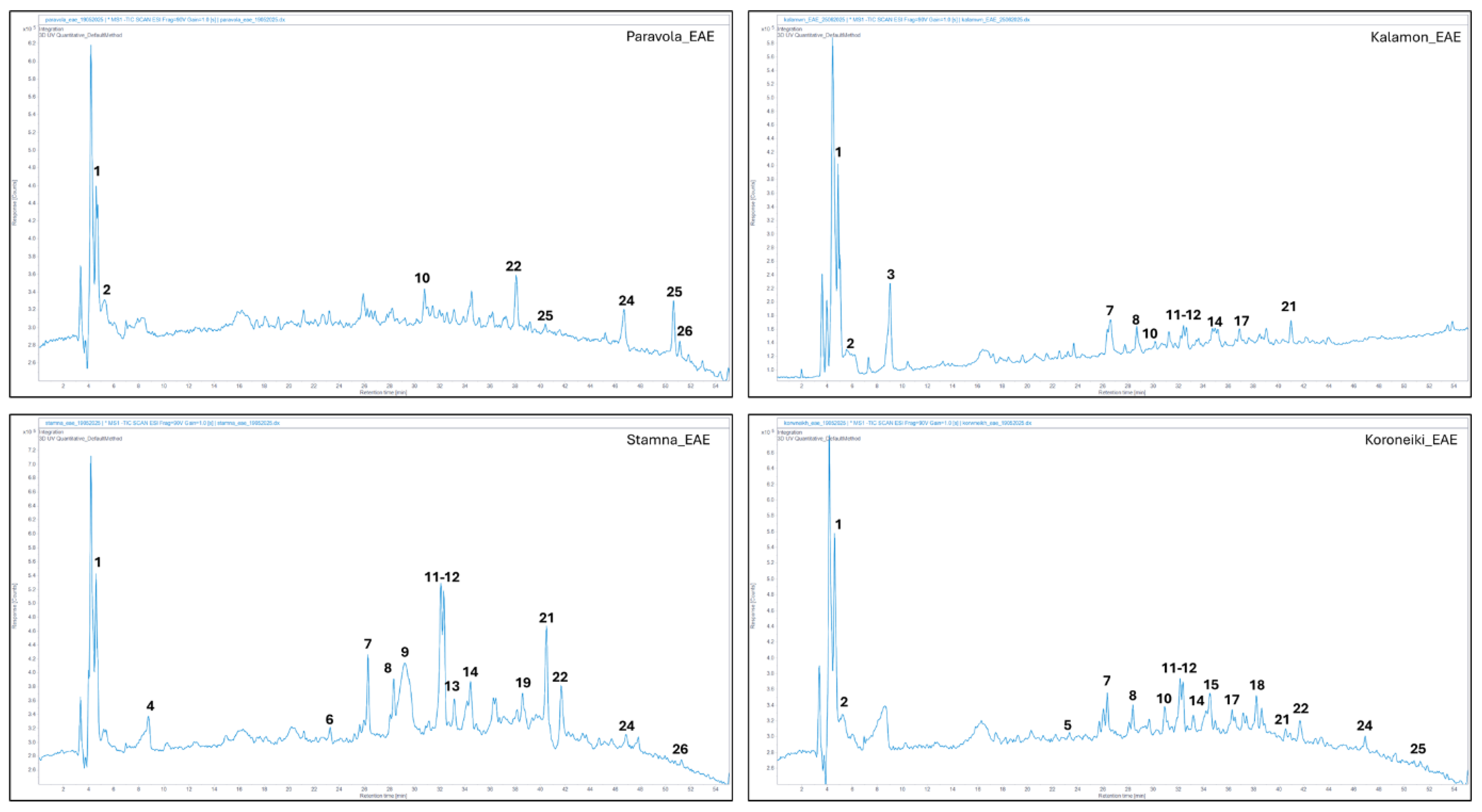
Figure 4.
HPLC-MSQ Total Ion Chromatograms (TIC) of the analyzed OLs extracts Paravola, Kalamata, Stamna and Koroneiki after UAE. The number above each peak represents the peak numbers, corresponding to the peak numbers in Table 6.
Figure 4.
HPLC-MSQ Total Ion Chromatograms (TIC) of the analyzed OLs extracts Paravola, Kalamata, Stamna and Koroneiki after UAE. The number above each peak represents the peak numbers, corresponding to the peak numbers in Table 6.
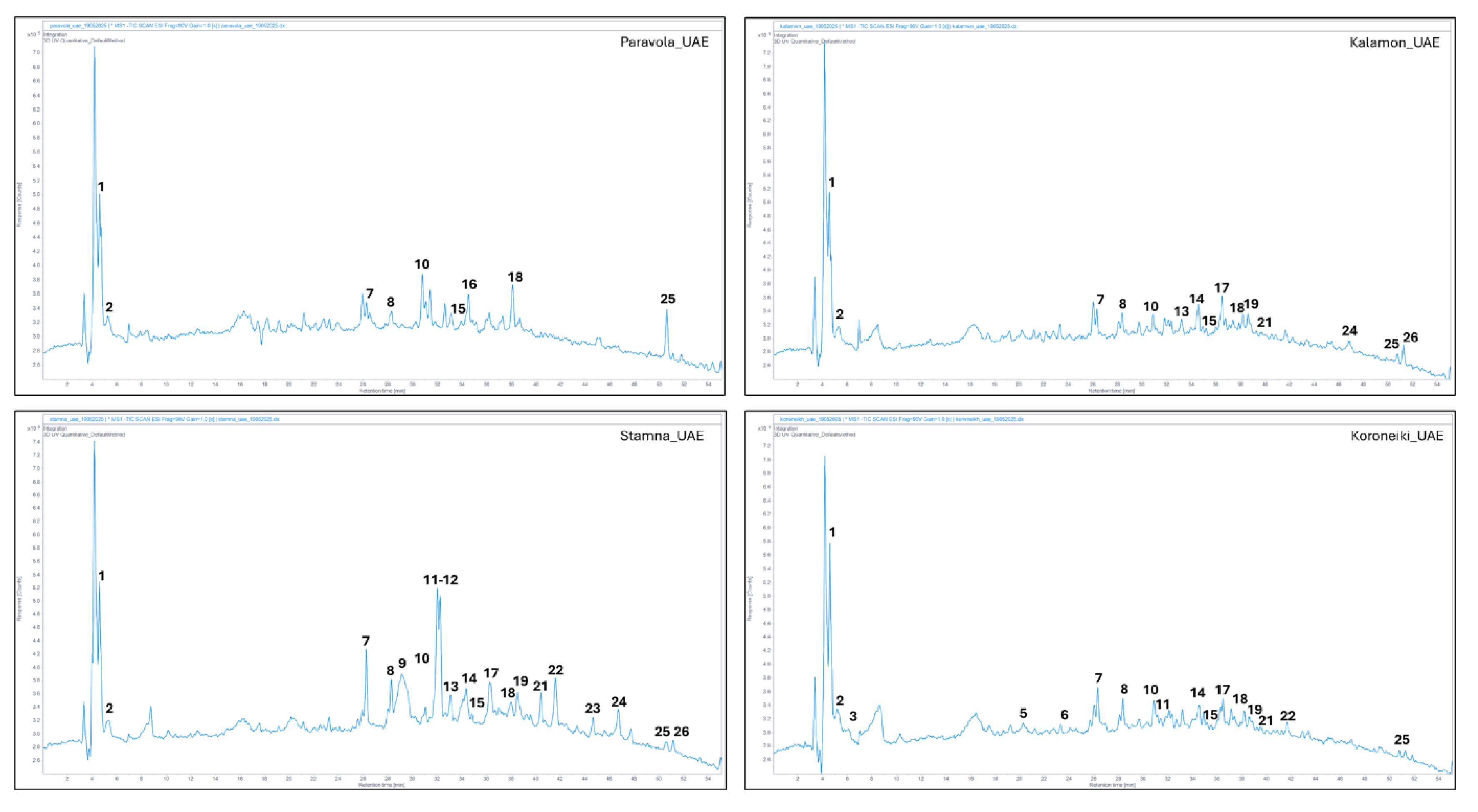
Figure 5.
HPLC-MSQ Total Ion Chromatograms (TIC) of the analyzed OLs extracts Paravola, Kalamata, Stamna and Koroneiki after MAE. The number above each peak represents the peak numbers, corresponding to the peak numbers in Table 6.
Figure 5.
HPLC-MSQ Total Ion Chromatograms (TIC) of the analyzed OLs extracts Paravola, Kalamata, Stamna and Koroneiki after MAE. The number above each peak represents the peak numbers, corresponding to the peak numbers in Table 6.
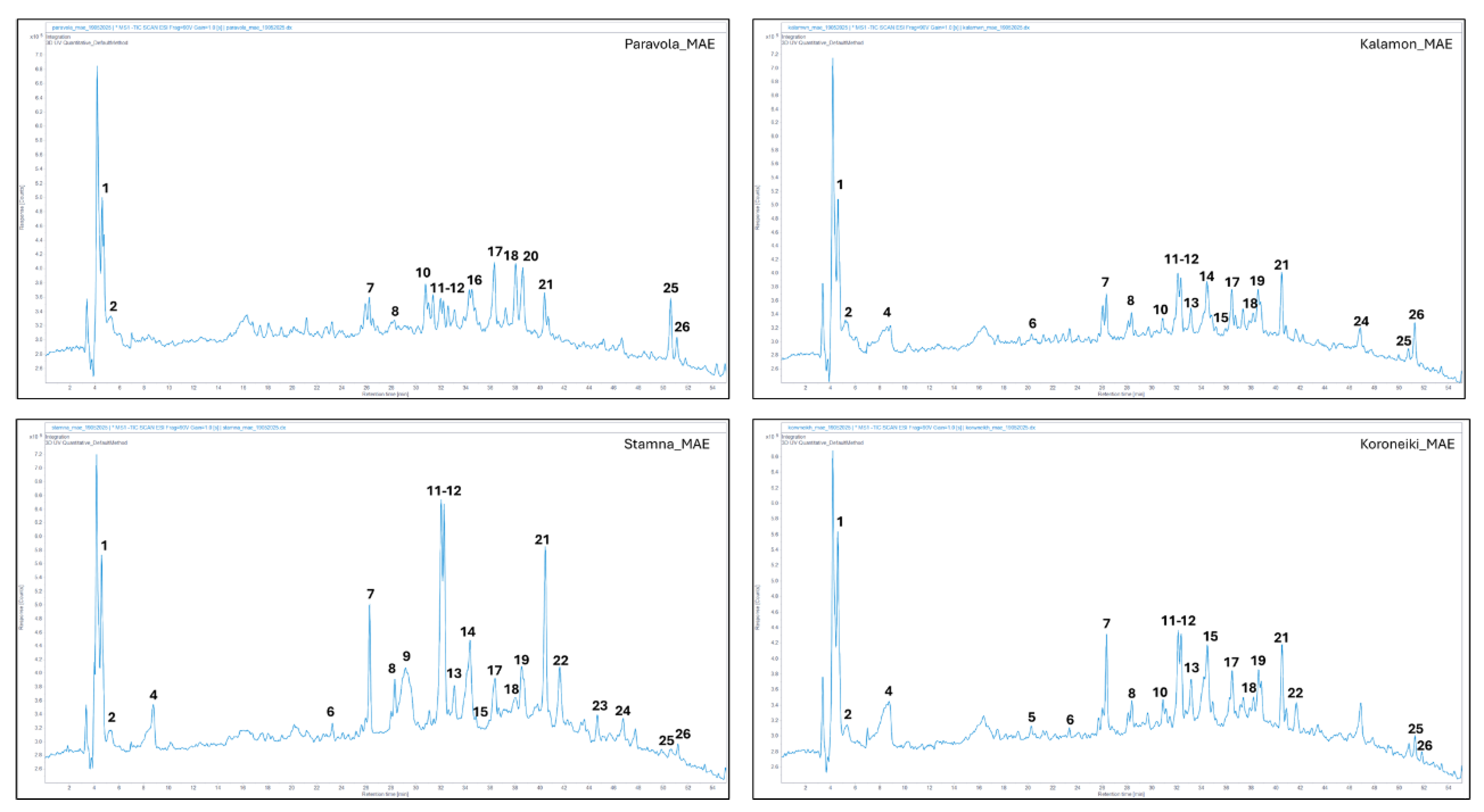
Table 6 presents the qualitative identification of bioactive phytochemicals across four distinct olive leaf cultivars. A total of 26 metabolites were detected, of which 12 were classified as secoiridoids and 9 as flavonoids. Among the detected compounds, several are considered rare or infrequently reported in Olea europaea leaves based on current phytochemical literature. Notably, Decaffeoyl Verbascoside (compounds 10) have been identified as uncommon derivatives within olive matrices, as reported by Amancio de Jesus [35]. Additionally, Lucidumoside C (compound 22), a secoiridoid glycoside previously characterized in Ligustrum lucidum, was detected and is considered a rare constituent in olive leaves [36]. Furthermore, two Luteolin glucoside isomers (compounds 14 and 20), differing in retention time and possibly in glycosylation position or sugar linkage, were also identified in Paravola's wild olive tree, using MAE method. These isomers have been documented at low abundance in certain olive cultivars [37], and their detection here highlights the metabolic diversity among the analyzed varieties.
4. Conclusions
The findings demonstrate that both environmental conditions and olive tree variety significantly influence the phenolic composition and antioxidant activity of olive leaves. The mountainous wild olive tree from Paravola, exposed to greater abiotic stress, yielded notably higher levels of polyphenols, particularly when extracted using enzyme-assisted method. Among the three green extraction techniques tested, EAE emerged as the most effective for recovering total polyphenols across all varieties. These results support the hypothesis that environmental stress enhances secondary metabolite biosynthesis in olive trees and suggest that targeted application of sustainable extraction technologies can optimize the recovery of bioactive compounds from agricultural by-products. The study also provides a framework for future exploitation of olive leaves as a sustainable source of high-value phytochemicals in the nutraceutical and pharmaceutical sectors.
Author Contributions
For research articles with several authors, a short paragraph specifying their individual contributions must be provided. The following statements should be used “Conceptualization, A.V. and A.E.G; methodology, A.V.; validation, A.E.G., P.S. and C.P.; formal analysis, M.X.; investigation, M-A.K.; writing—original draft preparation, A.V.; writing—review and editing, A.V., P.S. and M.X.; supervision, A.E.G. and C.P. All authors have read and agreed to the published version of the manuscript.” Please turn to the CRediT taxonomy for the term explanation. Authorship must be limited to those who have contributed substantially to the work reported.
Funding
Please add: “This research received no external funding”
Acknowledgments
The authors thank Dr. Ioannis Karabagias (University of Patras, Department of Food Science and Technology, Agrinio) for providing them with the ultra-sound bath. Special thanks are expressed to Prof. Skaltsounis Alexios-Leandros, Director of the Laboratory of Valorization of Bioactive Natural Product, Faculty of Pharmacy, University of Athens, for his insightful comments and valuable contribution to this article.
Conflicts of Interest
Declare conflicts of interest or state “The authors declare no conflicts of interest.”
Abbreviations
The following abbreviations are used in this manuscript:
| UAE | Ultra-sound assisted extraction |
| MAEA | Microwave assisted extraction |
| EAE | Enzymatic assisted extraction |
| TPC | Total polyphenolic content |
| AA% | Antioxidant activity |
| Ols | Olive leaves |
| HT | Hydroxytyrosol |
| DPPH | 2,2-Diphenyl-1-(2,4,6-trinitrophenyl)hydrazin-1-yl |
| HPLC-DAD | High-Performance Liquid Chromatography with Diode-Array Detection |
| HPLC-MSQ | High-Performance Liquid Chromatography mass spectrometry and quantification |
| TIC | Total Ion Chromatograms |
| GAE | Gallic acid equivalent |
References
- Ruby Tiwari, C.S.Rana. Plant secondary metabolites: a review. International Journal of Engineering Research and General Science 2015, Volume 3, Issue 5.
- Kostelenos G, Kiritsakis A. Olive tree history and evolution. In: Kiritsakis A, Shahidi F (eds) Olives and olive oil as functional foods: bioactivity, chemistry and processing. Wiley, New Jersey 2017, pp 1–12.
- Brito C, Dinis L-T, Moutinho-Pereira J-M, Correia CM. Drought stress effects and olive tree acclimation under a changing climate. Plants 2019, 8(7):232. [CrossRef]
- Trabelsi L, Gargouir K, Ben Hassena A, Mbadra C, Ghrab M, Ncube B, Van Staden J, Gargouri R. Impact of drought and salinity on olive water status and physiological performance in an arid climate. Agric Water Manag 2019, 213:749–759. [CrossRef]
- Benavente-Garcı́a, Castillo J., Lorente J., Ortuño A., Del Rio J. Antioxidant activity of phenolics extracted from Olea europaea L. Leaves. Food Chemistry 2000, 68(4), 457–462. [CrossRef]
- Obied, H. K., Allen, M. S., Bedgood, D. R., Prenzler, P. D., Robards, K., & Stockmann, R. Bioactivity and analysis of biophenols recovered from olive mill waste. Journal of Agricultural and Food Chemistry 2005, 53(4), 823–837. [CrossRef]
- Japón-Luján, R., Luque de Castro, M. D. Superheated liquid extraction of oleuropein and related biophenols from olive leaves. Journal of Chromatography A 2006, 1136(1), 185-191. [CrossRef]
- Omar, S. H. (2010). Oleuropein in olive and its pharmacological effects. Scientia Pharmaceutica 2005, 78(2), 133–154. [CrossRef]
- Visioli, F., & Galli, C. Biological properties of olive oil phytochemicals. Food Science and Nutrition 2002, 42(3), 209-221. [CrossRef]
- Cicerale, S., Lucas, L., & Keast, R. Antimicrobial, antioxidant and anti-inflammatory phenolic activities in extra virgin olive oil. Current Opinion in Biotechnology 2012, 23(2), 129-135. Current Opinion in Biotechnology. [CrossRef]
- Brahmi, F., Khodir, M., Mohamed, C., & Pierre, D. Chemical composition and biological activities of essential oils from the leaves of Olea europaea. Industrial Crops and Products 2017, 97, 212-219.
- Servili, M., Baldioli, M., Selvaggini, R., Macchioni, A., & Montedoro, G. Phenolic compounds of olive fruit: One- and two-dimensional nuclear magnetic resonance characterization of nüzhenide and its hydrolysis products. Journal of Agricultural and Food Chemistry 2004, 47(1), 12–18.
- Mousavi, S., Javanmard, M., & Rafiee, M. Effects of environmental and agronomic conditions on the phenolic compounds in olive leaves. Journal of Agricultural Science and Technology 2021, 23, 1-14.
- Romani, A., Pinelli, P., & Vincieri, F. F. Polyphenols and tannins in olive tree leaves. Journal of Pharmaceutical and Biomedical Analysis 2006, 29(6), 1291-1296.
- Malik, N. S. A., & Bradford, J. M. Changes in oleuropein levels during differentiation and development of floral buds in ‘Arbequina’ olives. Scientia Horticulturae 2008, 117(3), 203–208.
- Lafka, T. I., Lazou, A. E., Sinanoglou, V. J., & Lazos, E. S. Phenolic and antioxidant potential of olive oil mill wastes. Food Chemistry 2013, 125(1), 92-98.
- Zhang, Q. A., Fan, X. H., Li, Y., & Li, M. Effect of ultrasound on the extraction and antioxidant activity of phenolic compounds from olive leaves. Ultrasonics Sonochemistry 2018, 41, 346-352.
- Herrero, M., Cifuentes, A., & Ibáñez, E. Sub- and supercritical fluid extraction of functional ingredients from different natural sources: Plants, food-by-products, algae and microalgae: A review. Food Chemistry 2010, 98(1), 136-148.
- Medina-Torres, N., Ayora-Talavera, T., Espinosa-Andrews, H., Sánchez-Contreras, A., & Pacheco, N. Ultrasound assisted extraction for the recovery of phenolic compounds from vegetable sources. Agricultural Sciences 2017, 8, 746-769. [CrossRef]
- Ballard, T. S., Mallikarjunan, P., Zhou, K., & O'Keefe, S. F. Microwave-assisted extraction of phenolic antioxidant compounds from peanut skins. Food Chemistry 2010, 120(4), 1185-1192. [CrossRef]
- Wang, L., Yang, B., Du, X., & Yi, C. Optimization of supercritical fluid extraction of flavonoids from Pueraria lobata. Food Chemistry 2008, 108(2), 737–741.
- Dabbou, S., Gharbi, I., Brahmi, F., & Mechri, B. Olive leaves extract as a source of bioactive molecules: Extraction and qualitative and quantitative characterization. International Journal of Food Science & Technology 2017, 52(2), 325-332.
- Erbay, Z.; Icier, F. Optimization of Drying of Olive Leaves in a Pilot-Scale Heat Pump Dryer. Dry Technol. 2009, 27, 416–427. [CrossRef]
- Karabagias IK, Dimitriou E, Kontakos S, Kontominas MG. Phenolic profile, colour intensity, and radical scavenging activity of Greek unifloral honeys. Eur Food Res Technol 2016, 242:1201–1210. [CrossRef]
- Gabriela Silveira da Rosa, Thamiris Renata Martiny, Guilherme Luiz Dotto, Sai Kranthi Vanga, Débora Parrine, Yvan Gariepy, Mark Lefsrud, Vijaya Raghavan. Eco-friendly extraction for the recovery of bioactive compounds from Brazilian olive leaves. Sustainable Materials and Technologies 2021, 28. [CrossRef]
- Habib Shirzad, Vahid Niknam, Mehdi Taheri, Hassan Ebrahimzadeh. Ultrasound-assisted extraction process of phenolic antioxidants from Olive leaves: a nutraceutical study using RSM and LC–ESI–DAD–MS. J Food Sci Technol 2017, 54(8):2361–2371. [CrossRef]
- Dunja Šamec, Erna Karalija, Ivana Šola, Valerija Vujčić Bok and Branka Salopek-Sondi. The Role of Polyphenols in Abiotic Stress Response: The Influence of Molecular Structure. Plants 2021, 10(1), 118. [CrossRef]
- Deniz Karamazakcadik, Betόl Kilincli, Ceren Ilgaz, Pınar Kadiroglu. Anti-quorum sensing activity of olive leaf microwave-assisted extract. Discover Food 2025, 5:114. [CrossRef]
- Şah İsmail Kırbaşlar & Selin Şahin. Recovery of bioactive ingredients from biowaste of olive tree (Olea europaea) using microwave-assisted extraction: a comparative study. Biomass Conversion and Biorefinery 2023, 13:2849–2861. [CrossRef]
- Elizabeta Zandona, Maja Vukelic, Karla Hanousek Cica, Antonio Zandona, Jasna Mrvcic, Maja Katalinic, Ines Cindric, Almir Abdurramani and Irena Barukcic Jurina. Bioactive Properties of the Microwave-Assisted Olive Leaf Extract and Its Incorporation into a Whey Protein Isolate Coating of Semi-Hard Cheese. Foods 2025, 14, 1496. [CrossRef]
- Sengul Uysal, Biljana Lonˇcar, Aleksandra Cvetanovi´c Kljaki´c, Gokhan Zengin. Optimization of ultrasound-assisted extraction from Olea europaea leaves and analysis of their antioxidant and enzyme inhibition activities. Food Bioscience 2025, 63, 105798. [CrossRef]
- Raquel Rodríguez-Fernández, Ángela Fernández-Gómez, Juan C. Mejuto and Gonzalo Astray. Modelling Polyphenol Extraction through Ultrasound-Assisted Extraction by Machine Learning in Olea europaea Leaves. Foods 2023, 12, 4483. [CrossRef]
- Mariem Arfaoui, Mouna Boulares, Asma Bezzezi, Souha Ayachi, Mahmoud Ghrab, Nour Elhouda Jouini, Mnasser Hassouna, Sonia Boudiche. Effect of the enrichment with natural antioxidants obtained by maceration or ultrasound-assisted extraction from olive leaves on organic extra virgin olive oil. Rivista Italiana Delle Sostanze Grasse 2022, 99.
- Sofia Chanioti, Paraskevi Siamandoura, Constantina Tzia. Evaluation of Extracts Prepared from Olive Oil By-Products Using Microwave-Assisted Enzymatic Extraction: Effect of Encapsulation on the Stability of Final Products. Waste Biomass Valor 2016. [CrossRef]
- Raphael Amancio de Jesus, Wenes Ramos da Silva, Alberto Wisniewski Jr, Luís Fernando de Andrade Nascimento, Arie Fitzgerald Blank, Daniel Alves de Souza, Elma Regina Silva de Andrade Wartha, Paulo Cesar de Lima Nogueira, Valéria Regina de Souza Moraes. Microwave and ultrasound extraction of antioxidant phenolic compounds from Lantana camara Linn. leaves: Optimization, comparative study, and FT-Orbitrap MS analysis. Phytochemical analysis 2024, 34:4. [CrossRef]
- Jessica Paié-Ribeiro, Filipa Baptista, Maria José Gomes, Alfredo Teixeira, Victor Pinheiro, Divanildo Outor-Monteiro, Ana Novo Barros. Exploring the Variability in Phenolic Compounds and Antioxidant Capacity in Olive Oil By-Products: A Path to Sustainable Valorization. Antioxidants 2024, Nov 29;13(12):1470. [CrossRef]
- Lucía Olmo-García, Aadil Bajoub, Sara Benlamaalam, Elena Hurtado-Fernández, María Gracia Bagur-González, Mohammed Chigr, Mohamed Mbarki, Alberto Fernández-Gutiérrez and Alegría Carrasco-Pancorbo. Establishing the Phenolic Composition of Olea europaea L. Leaves from Cultivars Grown in Morocco as a Crucial Step Towards Their Subsequent Exploitation. Molecules 2018, 23(10), 2524. [CrossRef]
- Vardakas Alexios, Kechagias Achilleas, Penov Nikolay and Giannakas E. Aris. Optimization of Enzymatic Assisted Extraction of Bioactive Compounds from Olea europaea Leaves. Biomass 2024, 4(3), 647-657. [CrossRef]
Figure 1.
Map of the two areas studied
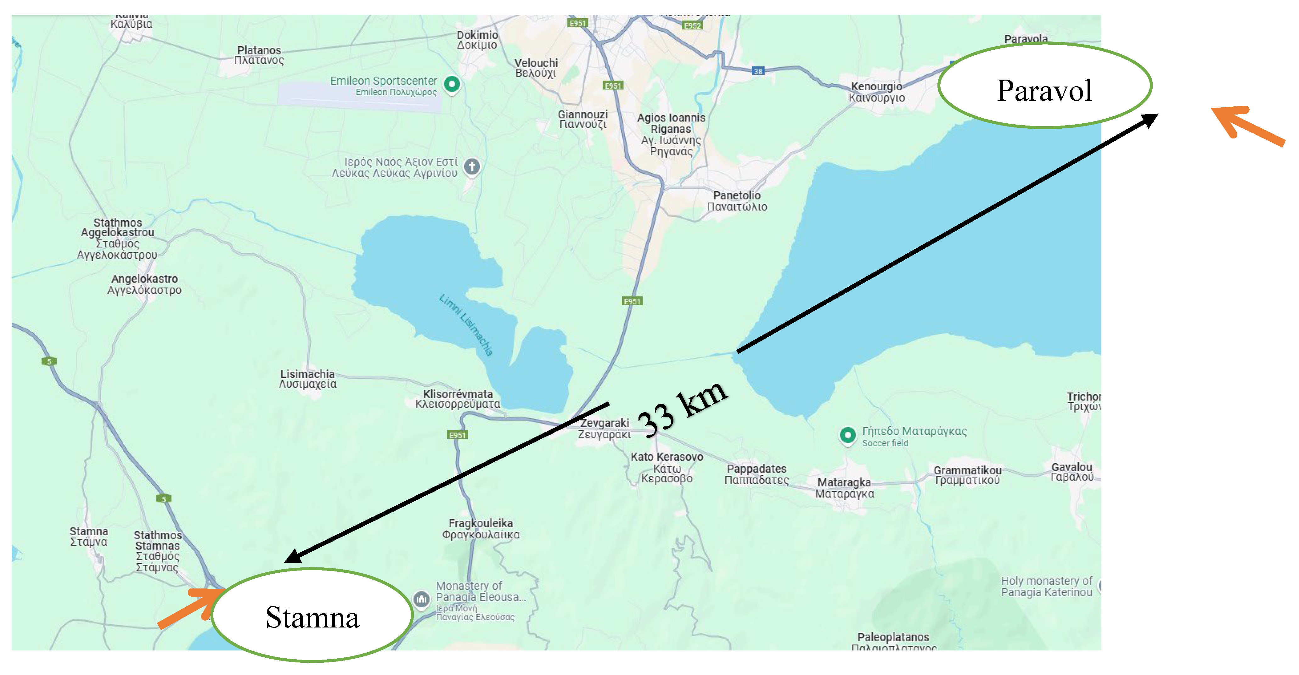
Figure 2.
Precipitation quantity per month.
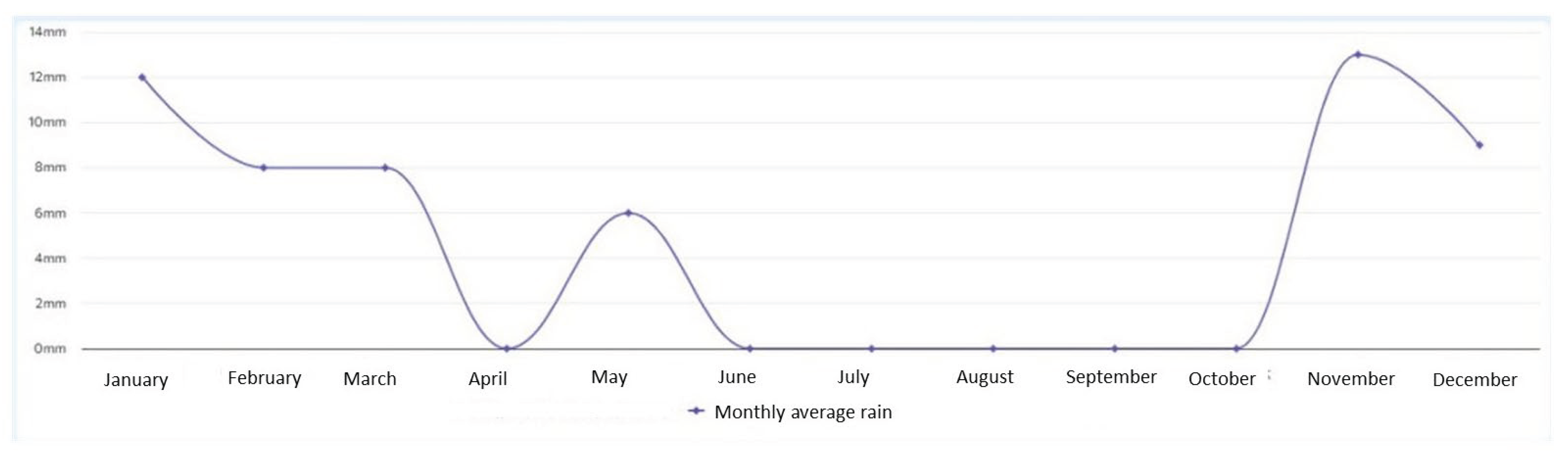

Table 1.
Moisture content1 of olive leaves powder.
| Olive tree variety | Wild tree from Paravola | Koroneiki | Wild tree from Stamna | Kalamata | |
| Residual humidity | 5.63 | 5.33 | 4.36 | 5.72 | |
1 residual humidity of olive leaves presented as %.
Table 2.
Monthly maximum, minimum and average temperatures of the two analyzing areas Paravola and Stamna.
Table 2.
Monthly maximum, minimum and average temperatures of the two analyzing areas Paravola and Stamna.
| Area / Olive variety | Month | Maximun air temperature (°C) | Minimum air temperature (°C) | Average air temperature (°C) |
|---|---|---|---|---|
| Paravola/ wild tree | May | 25.00 | 14.00 | 19.50 |
| June | 32.00 | 22.00 | 27.00 | |
| July | 33.00 | 23.00 | 28.00 | |
| August | 31.00 | 24.00 | 27.50 | |
| September | 26.00 | 17.00 | 21.50 | |
| Stamna/ Kalamata, Koroneiki and wild tree | May | 26.08 | 16,06 | 21.13 |
| June | 34.22 | 25.20 | 29.07 | |
| July | 34.86 | 25.83 | 31.46 | |
| August | 34.39 | 26.13 | 30.43 | |
| September | 29.99 | 20.97 | 24.65 |
Table 3.
Soil moisture in Stamna olive grove.
| Months | Maximum soil moisture (%) | Minimum soil moisture (%) | Average soil moisture (%) |
|---|---|---|---|
| May | 34.07 | 23.12 | 28.60 |
| June | 36.04 | 23.91 | 29.98 |
| July | 36.67 | 23.09 | 29.88 |
| August | 35.15 | 21.98 | 28.57 |
| September | 22.29 | 21.55 | 21.92 |
Table 4.
Contents of total polyphenols (g GAE/kg) from olive leaves using different extractions.
| Extraction methods | TPC (mg GAE/L) * | |||
|---|---|---|---|---|
| Kalamata | Stamna | Paravola | Koroneiki | |
| EAE | 144.75±7.24a | 375.00±18.75b | 924.00±46.20c | 466.00±23.30d |
| MAE | 174.50±8.73a | 63.50±3.18b | 81.50±4.08c | 149.00±7.45d |
| UAE | 266.50±13.33a | 38.00±1.90b | 116.00±5.80c | 231.00±11.55d |
*Values are presented as means ± standard deviations (n = 3). Different letters within a column indicate significant differences (Tukey’s test, P < 0.05).
Table 5.
Contents of antioxidant activity (AA%) from olive leaves using different extraction methods.
Table 5.
Contents of antioxidant activity (AA%) from olive leaves using different extraction methods.
| Extraction methods | DPPH (AA%) * | |||
|---|---|---|---|---|
| Kalamata | Stamna | Paravola | Koroneiki | |
| EAE | 61.00±3.05a | 64.99±3.25a | 63.42±3.17a | 61.00±3.05a |
| MAE | 38.68±1.94a | 40.62±2.03a | 40.70±2.04a | 44.43±2.22a |
| UAE | 35.86±1.79a | 37.60±1.88a | 33.25±1.66a | 35.73±1.79a |
*Values are presented as means ± standard deviations (n = 3). Different letters within a column indicate significant differences (Tukey’s test, P < 0.05).
Table 6.
List of Compounds Identified in Olive Leaf Extracts by LC-MSQ Analysis.
| Νο | [M-H]- (m/z) | Molecular Formula | Proposed Compound | Classification |
|---|---|---|---|---|
| 191.0 | C7H12O6 | Quinic acid | Cyclitol | |
| 133.0 | C4H6O5 | Malic acid | Dicarboxylic acid | |
| 190.8 | C6H7O7 | Citric acid | Carboxylic acid | |
| 151.0 | C8H8O3 | Oxydized Hydroxytyrosol | Phenol | |
| 241.1 | C11H14O6 | Εlenolic acid | Secoiridoid | |
| 389.1 | C16H21O11 | Oleoside | Secoiridoid | |
| 403.0 | C17H23O11 | Oleoside methyl ester | Secoiridoid | |
| 377.0 | C19H21O8 | Decarboxyl elenolic acid derivative | Secoiridoid | |
| 335.1 | C17H20O7 | Undendified | Secoiridoid | |
| 461.1 | C20H30O12 | Decaffeoyl Verbascoside | Polyphenol glycoside | |
| 555.1 | C25H32O14 | Hydroxyoleuropein isomer 1 | Secoiridoid | |
| 555.1 | C25H32O14 | Hydroxyoleuropein isomer 2 | Secoiridoid | |
| 593.0 | C27H30O15 | Luteolin-7-O-rutinoside | Flavonoid | |
| 447.0 | C21H19O11 | Luteolin-7-O-glucoside | Flavonoid | |
| 551.3 | C25H28O14 | Cafselogoside | Secoiridoid | |
| 403.0 | C17H23O11 | Secoxyloganin | Secoiridoid | |
| 577.2 | C27H29O14 | Apigenin 7-O-rutinoside | Flavonoid | |
| 431.1 | C21H20O10 | Apigenin-4-O-glucoside | Flavonoid | |
| 461.0 | C20H30O12 | Chryseriol-7-O- glucoside | Flavonoid | |
| 447.1 | C21H19O11 | Luteolin glucoside isomer | Flavonoid | |
| 539.2 | C25H32O13 | Oleuropein | Secoiridoid | |
| 583.1 | C27H36O14 | Lucidumoside C | Secoiridoid | |
| 523.2 | C25H31O12 | Ligstroside | Secoiridoid | |
| 285.0 | C15H10O6 | Luteolin | Flavonoid | |
| 269.1 | C15H10O5 | Apigenin | Flavonoid | |
| 299.1 | C16H12O6 | Diosmetin | Flavonoid |
Disclaimer/Publisher’s Note: The statements, opinions and data contained in all publications are solely those of the individual author(s) and contributor(s) and not of MDPI and/or the editor(s). MDPI and/or the editor(s) disclaim responsibility for any injury to people or property resulting from any ideas, methods, instructions or products referred to in the content. |
© 2025 by the authors. Licensee MDPI, Basel, Switzerland. This article is an open access article distributed under the terms and conditions of the Creative Commons Attribution (CC BY) license (http://creativecommons.org/licenses/by/4.0/).
Copyright: This open access article is published under a Creative Commons CC BY 4.0 license, which permit the free download, distribution, and reuse, provided that the author and preprint are cited in any reuse.


