
Submitted:
16 July 2025
Posted:
17 July 2025
You are already at the latest version
Abstract
Eperua oleifera Ducke (Fabaceae), commonly known as copaíba-jacaré, is traditionally used for therapeutic purposes, like Copaifera oleoresins. Previous GC-MS studies reported its chemical composition as mainly composed of diterpenic acids, consistent with species of the same genus. Although GC-MS remains widely used for comparing compound retention times and fragmentation patterns, its application to diterpenic acids requires a derivatization step to form methyl esters due to the poor chromatographic performance of carboxylic acids on methyl silicone stationary phases. This step may lead to misinterpretations, especially considering recent findings of naturally occurring methyl esters in oleoresins that may co-elute with derivatized acids. This study aimed to apply more sensitive analytical techniques to identify both target and untargeted compounds. The resin of E. oleifera was analyzed by GC-MS to assess the presence of volatile components. Additionally, UHPLC-HRMS was employed using full-scan MS, data-dependent acquisition (DDA), and parallel reaction monitoring (PRM) in both positive and negative ESI modes. GC-MS confirmed the absence of volatile sesquiterpenes, classifying E. oleifera as a resin. Targeted UHPLC-HRMS detected natural methyl esters of diterpenic acids, while untargeted analysis using Compound Discoverer software revealed flavonoids and phenolic compounds not previously reported. These findings support the application of UHPLC-HRMS as a powerful tool in phytochemical studies.
Keywords:
Eperua oleifera Ducke
; UHPLC-HRMS
; target and untargeted approach
; diterpene acids
; methyl esters of acid diterpenes
1. Introduction
Eperua oleifera Ducke (Fabaceae) is commonly known as “copaíba-jacaré” and is distributed in the Central Amazon, from Ecuador and Brazil to Guyana, and Venezuela [1]. Trees of the Eperua genus are known to share properties similar to those of another Fabaceae-Caesalpinoideae genus: Copaifera. Both exude a viscous oleoresin from the trunks of the trees, which is used for therapeutic purposes as a healing, antifungal, and antibacterial material [2]. However, despite the similarity in the therapeutic and morphological properties of these oleoresins, there are few reports focused specifically on Eperua species. Nevertheless, among the studied species, several classes of compounds were identified, including phenolic acids, flavonoids, sesquiterpenes, triterpenes, and, notably, diterpenes, which appear to be the most abundant [3,4].
Previously, our studies on the oleoresin of Eperua oleifera using Gas Chromatography coupled with Mass Spectrometry (GC-MS), as a standard analytical tool for analyzing terpene mixtures, after derivatization, led to the identification of three diterpene alcohols and nine diterpene acids [5, in press]. Typically, GC-MS analysis is performed following a derivatization reaction to produce the corresponding esters, due to the poor resolution of carboxylic acids on methyl silicone stationary phases. This introduces an additional analytical step that may introduce systematic errors and lead to the misinterpretation of results. As a result, carboxylic acids are commonly identified as their methyl esters, although naturally occurring methyl esters had not previously been reported in this type of oleoresin. Additionally, at the same previous study, phytochemical isolation using silica open column chromatography and traditional tools to natural products identification, such as multiple experiments using nuclear magnetic resonance (NMR), infrared and ultraviolet spectroscopy and direct insertion on high resolution mass spectrometry resulted on the unexpected description of methyl hardwickiate, a natural methyl ester not previously described in Amazon oleoresins [5, in press]. This stimulates further studies aimed at expanding the chemical knowledge of this oleoresin. Several questions arise from these findings, including the apparent absence of sesquiterpenoids or even monoterpenoids, which comprise the volatile and oily fraction of oleoresins; the potential presence of other chemical classes beyond traditional terpenoids; and the need to evaluate whether “auto”-esterification may occur during chromatographic processes, as well as to confirm the presence of additional diterpene esters alongside their corresponding diterpene carboxylic acids.
The usual approach for identifying and determining specialized substances from more polar chemical classes, such as flavonoids, phenolic acids, and alkaloids, involves High-Performance Liquid Chromatography (HPLC) with ultraviolet or Diode Array Detection (HPLC-DAD), or Gas Chromatography coupled with a Flame Ionization Detector (GC-FID) or GC-MS [6,7,8].The use of ultra-high-performance liquid chromatography coupled with high-resolution mass spectrometry (UHPLC-HRMS) analysis allows an entirely different perspective. While traditional methods can be slower and less sensitive, UHPLC-HRMS combines the fast and efficient separation of UHPLC with the high sensitivity and specificity of mass spectrometry, enabling faster, more accurate, and reliable analysis, especially in complex samples. Advances in liquid chromatography coupled with high-resolution mass spectrometry (LC-HRMS) have enabled both targeted and untargeted analyses across a wide range of complex matrices [9,10,11]. Several types of mass spectrometers—such as Time-Of-Flight (TOF), ion trap TOF, hybrid quadrupole TOF, and Orbitrap systems—routinely deliver high mass accuracy [10,12,13], allowing the determination of molecular formulas based on exact mass measurements. However, despite these technological advancements, compound identification remains a challenge. This is primarily due to the absence of many metabolites in reference databases, the wide dynamic range of metabolite concentrations, and limitations in the acquisition speed of mass spectrometry data. As a result, a significant number of detected peaks remain unidentified [10].
Software tools are available for compound detection, offered either as online platforms or as packages developed in programming languages such as R and Python [14,15,16,17]. Additionally, each mass spectrometer manufacturer provides its data processing software. The analysis of plant matrices produces complex results due to the vast diversity of naturally occurring compounds. In this context, Compound Discoverer has been utilized in untargeted metabolomics studies for the identification of compounds. This software is compatible with files generated by Orbitrap mass spectrometers and enables automated compound annotation through integration with the mzCloud database [18,19].
In this study, UHPLC-HRMS was employed using multiple data acquisition modes, including full mass spectrometry (Full MS), data-dependent acquisition (DDA), and parallel reaction monitoring (PRM), in both positive and negative ionization modes. Both targeted and untargeted analyses were applied as complementary approaches to advance the chemical characterization of the oleoresin from Eperua oleifera Ducke. The targeted analysis focused on detecting previously identified compounds from Eperua and Copaifera, comparing experimental data with reference parameters such as exact m/z values, retention times, and characteristic fragmentation patterns. In addition, the presence of sesquiterpenes was investigated over a broad range of detection limits using GC-MS. Meanwhile, the untargeted approach employed a metabolomics workflow to explore the potential of automated compound annotation using the Compound Discoverer software exclusively. Finally, experiments were also conducted to evaluate the possibility of Fischer esterification occurring within the chromatographic system and whether this could lead to the formation of esters from diterpenic acids.
2. Materials and Methods
2.1. Plant Material
The oilresin from Eperua oleifera Ducke was collected on June 6, 2023, in Manicoré, Amazonas, Brazil. The access was registered under code A9F18E3 in the SISGEN system. The oilresin from Copaiba multijuga Ducke was collected in Manaus, Amazonas, Brazil. The access was registered under code AAB3AA1 in the SISGEN system. The sample preparation for UHPLC-HRMS was performed with approximately 1 mg of each oilresin weighed into a vial and solubilized with 1 mL of Methanol.
2.2. GC-MS Analysis and Instrument Conditions
A Thermo Scientific 1300 Trace Gas Chromatograph coupled with an ISQ LT single quadrupole MS in a DB-5HT column of 30 m x 0.250 mm ID and 0.10 µm film thickness, 5%-phenyl-methylpolysiloxane. Pulsed Split injection mode was selected to inject 3 µL of sample into the inlet liner Ultra Inert, splitless, single taper, glass wool (Agilent Part number: 5190-3171, 4 mm x 900 µL) at 270 °C, using helium as the carrier gas at flow rate of 1.0 mL/min and split ratio of 20:1. The injection pulse pressure was set to 50.0 psi (3.960 mL/min) for 0.30 min, rate 1 ml/min, whereas the purge flow was set to 0.800 mL/min (injection mode: pulsed split). The initial temperature ramp of the oven started at 110.0 °C for 2 minutes and increased to 130.0 °C (for 5 min) at a rate of 3.0 °C/min, followed by a rise to 310.0 °C at 8.5 °C/min, and then held for 5 minutes. The total running time was 39.84 min. The MS transfer line temperature was set at 320 °C, and the ion source temperature was kept at 300 °C. The system was operated in EI mode at an energy level of 70 eV. The chromatogram was scanned in SCAN mode, with a mass range of m/z 50 to m/z 700. The identification of compounds was confirmed using the National Institute of Standards and Technology Library, 2017 (NIST17).
2.3. UHPLC-HRMS Analysis and Instrument Conditions
A Dionex Ultimate 3000 ultra-high performance liquid chromatography (UHPLC) system coupled to a QExactive Plus hybrid quadrupole Orbitrap mass spectrometer (Thermo Fisher Scientific, Bremen, Germany) equipped with an electrospray ionization (ESI) source was used. Separation was performed in a reversed-phase column (kinetex 2.6 µm PS C18, 100 Å, 100 mm x 2.1 mm; 2.6 µm) at 40 °C, with a constant flow rate of 300 μL/min and injection volume of 5 µL. A gradient chromatographic run started at 5% of mobile phase B (methanol with 0.1% formic acid) and 95% of mobile phase A (water with 5 mM ammonium formate and 0.1% formic acid). Mobile phase B increased to 10% at 1.0 minutes, 25% at 2 minutes, and 90% at 10 minutes. After reaching 100% of B at 14 minutes and maintaining this ratio until 16 minutes, the initial chromatographic condition was restored from 16.1 to 20.0 minutes.
The LC effluent was pumped to the mass spectrometer operating in a negative ESI mode, calibrated daily with a manufacturer’s calibration solution (Thermo Fisher Scientific, Bremen, Germany). ESI parameters were further optimized with the final setup: spray voltage of 2.9 kV, S-lens voltage of 80 V, the capillary temperature of 380 °C, auxiliary gas heater temperature of 350 °C, nitrogen sheath, auxiliary, and sweep gas were set at 30, 10, and 1 arbitrary unit, respectively. The strategy of acquisition were Full-scan and Data Dependent Analysis (DDA), at the same time, in a range of m/z 70 – m/z 1050 at a resolution of 70,000 full widths at half maximum (FWHM), automatic gain control (AGC) of 1 x 10⁶, and maximum injection time (IT) of 100 ms.
For the target compound identification study, a full MS scan approach was employed. The exact mass-to-charge (m/z) values of the detected targets were as follows: m/z 315.1966 ([M - H]-) to hardwickiic acid (C20H28O3), m/z 331.1915 ([M - H]-) to patagonic acid (C20H28O4), m/z 303.2330 ([M - H]-) to copalic acid (C20H32O2), m/z 333.2071 ([M - H]-) to agathic acid (C20H30O4), m/z 319.2278 ([M - H]-) to β-hydroxy-copalic acid (C21H34O3), m/z 335.2227 ([M - H]-) to dihydroagathic (pinifolic) acid (C20H32O4), m/z 305.2486 ([M - H]-) to eperuic acid (C20H34O2), m/z 303.2329 ([M - H]-) to kovalenic acid (C20H32O2), m/z 335.2227 ([M - H]-) to clerod-3-3n-15,18-dioic acid (C20H32O4), m/z 293.1758 ([M - H]-) to 14,15,16-trinor-hardwikiic acid (C17H26O4), m/z 317.2122 ([M - H]-) to 2-oxokolavenic acid (C20H30O3), m/z 329.2122 ([M - H]-) to methyl ester of hardwickiic acid or methyl hardwickiate (C21H30O3), m/z 317.2486 ([M - H]-) to methyl ester of copalic acid or methyl copalate (C21H34O2), m/z 375.2541 ([M - H]-) to acetoxy copalic acid methyl ester (C23H36O4) m/z 345.2071 ([M - H]-) to methyl ester of patagonic acid or methyl patagonate (C21H30O4), m/z 371.2227 ([M - H]-) to mono methyl ester of agathic acid or methyl agathate (C21H34O4), and m/z 319.2642 ([M - H]-) to methyl ester of eperuic acid (C21H36O2).
The second set of experiments was conducted using the Full MS and DDA approach, with the same gradient chromatographic run, employing methanol as mobile phase B and water as mobile phase A, without the addition of any additives.
Targeted mass spectrometry-based approaches were performed using the parallel reaction monitoring technique (PRM), the precursor ions were fixed at a resolution of 17,500 full width at half maximum (FWHM), automatic gain control (AGC) of 1 x 10⁶, maximum injection time (IT) of 100 ms and quadrupole isolation window of m/z 2. In the PRM approach, the precursor ions were fragmented in a higher energy collisional dissociation (HCD) cell with (N)CE of 40%, as described in Table 1.
Data were acquired and processed using Thermo Scientific TraceFinder 4.1 software (Thermo Fisher Scientific, Austin, TX, USA), with a mass tolerance of ±5 ppm.
2.3.1. Evaluation of the Kinetics of Methyl Ester Formation by UHPLC-HRMS
To evaluate the kinetics of methyl ester formation of acidic diterpenes, in the first experiment, 1 mg of each resin was weighed and dissolved in methanol containing 0.1% formic acid. In the second trial, the sample was dissolved in acetonitrile. The samples were analyzed with approximately 20-day intervals between the first and fourth analyses, and the coefficient of variation was calculated.
2.3.2. Data Processing and Analysis
The chemical composition was compiled using online databases (mzCloud and ChemSpider) and imported into the Compound Discoverer 3.3 analysis platform to identify chromatographic peaks. The binary sample model was employed for comparative analysis, such as that between blank solvent samples and Quality control samples. Additionally, [M + H]+, [M + Na]+, and [M + NH4 + H]+ were selected as the primary adduct ion modes in positive ionization mode, while [M - H]− and [M - H - H2O]− were chosen as the primary adduct ion modes in negative ionization mode. The upper and lower limits of molecular weight deviation were set to 5 ppm.
3. Results and Discussion
Oleoresins are characterized as complex mixtures composed of terpenoids from various classes. They typically consist of volatile liquid terpenoids—such as monoterpenes and sesquiterpenes—which act as solvents for heavier resinous terpenoids, including diterpenes and triterpenes, giving the material its characteristic viscous oil appearance [20]. Terpenes exhibit well-defined chemical and chromatographic characteristics as described in the literature. In GC-MS analyses, for example, they elute within specific temperature ranges depending on their class—sesquiterpenes typically elute between 120–200 °C [21]. These elution patterns are closely related to the molecular weights of the compounds and their corresponding temperature intervals. Such correlations enable a preliminary analysis of oleoresins by linking terpene classes to their characteristic elution temperatures. The first question addressed became evident. In the absence of monoterpenes and sesquiterpenes—the volatile liquid fraction— Eperua oleifera should no longer be considered to produce oleoresins and would be instead classified only as a resin.
The previous study with Eperua oleifera oleoresin using GC-MS, after derivatization of the oleoresin, detected diterpene acids commonly found in Copaifera and Eperua oleoresins and also isolated a natural methyl ester, identified by [5, in press]. A derivatization step is commonly employed to analyze oleoresins by GC-MS using a 5%-phenyl-methylpolysiloxane column, since the resolution of acid substances is not adequate. This procedure can lead to compound misidentification since it would not differentiate between the natural esters and their respective esters derivatized from diterpene acids. These findings support a targeted search for other diterpene acids and methyl esters using more sensitive analytical techniques such as UHPLC-HRMS, which enables the detection of compounds present at low concentrations that may not be detectable by GC-MS. This procedure will allow the second question, if the oilresin (or resin) from Eperua oleifera naturally produces diverse diterpenic esters together with the diterpenic acids, as reported in the literature.
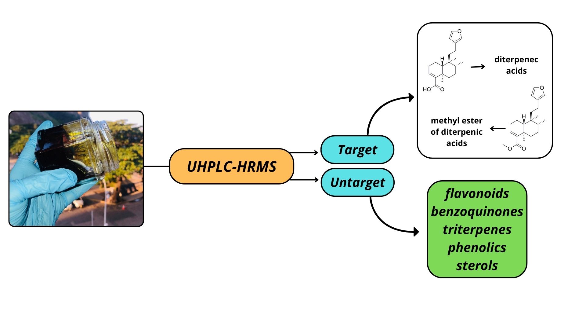
The third question of this study is to expand the chemical knowledge on E. oleifera. Since natural diterpenoid esters were never detected before in this oilresin, and terpenes are typically characterized as its main constituents, should other substances, from different natural biosynthetic classes, be present? The oleoresin of E. oleifera exhibits the physical characteristics of an oil, raising questions about the possible contribution of other compounds to its oily appearance. Analytical tools and software platforms such as Compound Discoverer are instrumental in the search for previously unidentified targets (Figure 1).
3.1. Characterization of Sesquiterpenes Using Gas Chromatography Mass Spectrometry (GC-MS)
To verify the presence of sesquiterpenes in the oleoresin of E. oleifera, the oleoresin of Copaifera multijuga was used as a reference due to the extensive amount of information available regarding the chemical characterization of this species and the huge amount of sesquiterpenes present, relating to the diterpenic acids [21,22]. Using GC-MS, it was possible to obtain a structured chromatogram based on the chemical profile of the oleoresins. The structured chromatogram is divided into three distinct regions: the sesquiterpene hydrocarbon region, the oxygenated sesquiterpene region, and the acidic diterpene region. Figure 2a illustrates the total ion chromatogram of the oleoresin from Copaifera multijuga.
The first region of the chromatogram corresponds to sesquiterpene hydrocarbons, which elute between 6 and 11 minutes. The main compounds identified in this region include α-copaene (6.62 min), β-caryophyllene (7.64 min), β-humulene (8.43 min), and β-bisabolene (9.96 min). The second region consists of oxygenated sesquiterpenes, eluting between 12 and 18 minutes. Notably, caryophyllene oxide elutes at 12.48 min within this region. In the final region, acidic diterpenes elute between 19 and 29 minutes. Among them, copalic acid (25.76 min), a labdane-type diterpenic acid, is considered a biomarker for species of the Copaifera genus.
The same analytical approach was applied to the Eperua oleifera oleoresin. No sesquiterpenes—neither hydrocarbon nor oxygenated forms—were detected; only acidic diterpenes and their corresponding esters were identified. Figure 2b shows the total ion chromatogram (TIC) of E. oleifera oleoresin. An expanded view of the sesquiterpene elution region is also provided to confirm that no sesquiterpenes were detected within the method’s detection limits. The absence of volatile compounds such as sesquiterpenes in E. oleifera supports the hypothesis that this material should be classified as a resin rather than an oleoresin, suggesting the need to investigate other compounds that may account for its semi-liquid or viscous appearance, rather than a purely solid form.
3.2. Chemical Characterization of Diterpenes (Targeted) by UHPLC-HRMS
The chemical composition of E. oleifera was investigated using UHPLC-HRMS in both negative and positive electrospray ionization (ESI) modes. Through a targeted analysis approach, eleven diterpenic acids and six of their corresponding methyl esters were identified (Table 1).
The first UHPLC-HRMS experiment was conducted using Full MS and DDA acquisition modes to identify the target compounds: diterpenic acids and their corresponding methyl esters (hardwickiate, patagonate, copalate, agathate, acetoxycopalate, and eperuate). The analytes were identified based on their exact masses (mass error < 0.5 ppm), and their elution order was established. To ensure effective ionization of the acids, formic acid and ammonium formate were used as mobile phase modifiers. This combination enhances the ionization of weak acids and bases, enabling ESI analysis while significantly improving peak resolution and separation. The mobile phase pH had a notable impact on retention times and chromatographic peak shapes, as it influenced the ionization state of the analytes. For diterpenic acids, most are better separated under slightly basic conditions where the acidic analytes are ionized. Solvent composition, acidity, and analyte polarity are key factors influencing ionization efficiency in negative ESI-MS mode [10,23].
The limited studies available on Eperua species indicate that labdane-type diterpenic acids are the most frequently identified [3,24]. As the resins of Eperua species are often described as being similar to those of the Copaifera genus, our findings further support this connection, given that most of the compounds identified in this study have been reported in both genera [25,26,27]. The main compounds found include hardwickiic acid, dihydroagathic acid, agathic acid, and copalic acid—all of which are also found in copaiba oils. Among the methyl esters, methyl hardwickiate was the most abundant. The ratio between hardwickiic acid and its methyl ester was approximately 1.3.

Table 1.
Diterpenes detected by UHPLC-HRMS and their parameters.
| Compound | Molecular Formula [M] | Retention time (min) | Precursor ion (m/z) [M-H]- | Precursor ion (m/z) [M-H]+ | (N)CE (%) |
Product ion (m/z) [M-H]- |
|---|---|---|---|---|---|---|
| Hardwickiic acid | C20H28O3 | 12.95 | 315.1966 | 40 | 301.18063 / 257.19086 | |
| Patagonic acid | C20H28O4 | 11.29 | 331.1915 | 40 | 287.20193 / 259.20685 / 243.17505 | |
| Copalic acid | C20H32O2 | 14.00 | 303.2330 | 40 | 285.18607 / 259.20685 / 243.17514 | |
| Agathic acid | C20H30O4 | 11.95 | 333.2071 | 40 | 301.18094 / 291.23303 / 273.22216 | |
| Dihydroagathic (pinifolic) acid | C20H32O4 | 12.22 | 335.2227 | 40 | 301.18109 / 291.23306 / 273.22263 | |
| Eperuic acid | C20H34O2 | 14.06 | 305.2486 | 40 | 287.23767 | |
| Kovalenic acid | C20H32O2 | 14.02 | 303.2329 | 40 | 285.18613 / 243.17538 / 84.02048 | |
| Clerod-3-en-15,18-dioic acid | C20H32O4 | 11.49 | 335.2227 | 40 | 285.18622 / 259.17053 / 245.19104 | |
| 14,15,16-trinor-hardwikiic acid** | C17H26O4 | 10.90/14.43 | 293.1758 | 40 | 96.95888 | |
| 2-oxokolavenic acid | C20H30O3 | 11.84 | 317.2122 | 40 | 301.18112 / 273.22256 / 257.19113 | |
| Methyl hardwickiate | C21H30O3 | 13.90 | 329.2122 | 40 | 301.18015 / 285.18555 / 257.19052 | |
| Methyl copalate | C21H34O2 | 14.90 | 317.2486 | 40 | 301.18112 / 285.18622 / 257.19128 | |
| Methyl 3β-hydroxy copalate | C21H34O3 | 12.81 | 319.2278 | 40 | 301.18073 / 273.22269 / 257.19122 | |
| Methyl 3β-acetoxy copalate | C23H36O4 | 13.88 | 375.2541 | 40 | 317.21210 / 301.18039 / 287.16553 | |
| Methyl patagonate | C21H32O4 | 13.89 | 345.2071 | 40 | 315.19662 / 301.21735 / 243.17531 | |
| Methyl agathate | C21H32O4 | 12.29 | 347.2227 | 40 | ||
| Methyl eperuate | C21H36O2 | 14.95 | 319.2642 | 40 | ||
| Cativic acid* | C20H34O2 | 14.10 | 305.2486 | 40 | ||
| 8,17-dihydroxy-13-labden-16,15-olid-19-oate* | C21H32O6 | 12.21 | 439.2340 [M-H-60]- |
|||
| Effusanin A* | C20H28O5 | 10.84 | 347.1865 | |||
| 18-hydroxy-clerod-3-en-15-oic acid* | C20H34O3 | 13.13 | 321.2437 | |||
| craterellin A* | C22H34O4 | 13.16 | 380.2792 [M+NH4]+ |
|||
| 14-Deoxy-11,12-didehydroandrographolide* | C20H28O4 | 11.94 | 315.1953 [M+H]-18 |
|||
| 12-hydroxy-7-carboxy- abiet-8(13)-en-18-oic acid* |
C20H30O4 | 12.53 | 335.2216 |
|||
| Aphidicolin* | C20H34O4 | 12.08 | 339.2529 |
|||
| 7-keto, 12-hydroxy, abiet-8-14-en-18-oic acid | C20H30O4 | 12.71 | 333.2071 | |||
| (-)-7β-hydroxycleroda-8(17),13E-diene-15-oic acid* | C20H32O3 | 13.47 | 319.2278 | |||
| 16-oxo-13,14H-hardwikiic acid* | C20H28O4 | 11.26 | 331.1914 | |||
| nor-hardwikiic acid* | C17H26O4 | 12.16 | 293.1758 | |||
| 7-oxo-labda-8-ene-15-oic acid* | C20H30O3 | 11.86 | 317.2122 | |||
| (-)-cleroda-7,13E-diene-15-oic acid* | C20H32O2 | 14.62 | 303.2329 | |||
| 6β,7β-Dihydroxykaurenoic acid* | C20H30O4 | 11.48 | 333.2071 | |||
| 8-Hydroxyoctadeca-9,12-dienoic acid* | C18H32O3 | 13.86 | 295.2278 | |||
| Ent-16β,17-dihydroxy-19-kaurenoic acid* | C20H32O4 | 13.05 | 335.2227 |
*untargeted; ** isomers.
Oleoresins are traditionally used for medicinal purposes in northern Brazil, with knowledge passed down through native populations. However, such uses still lack scientific validation. Chemical characterization helps bridge traditional knowledge with scientific understanding. Notably, some of the identified diterpenes—such as hardwickiic acid and eperuic acid—have demonstrated antitumor, anti-leishmania, and anti-inflammatory activities [28,29,30,31,32]. Both compounds are present in Eperua oleifera.
Although the evidence is compelling—given that the analysis involves acids in a mobile phase containing methanol (an alcohol) and acidic additives, which could promote ester formation through the well-known Fischer esterification reaction—additional experiments were conducted to assess the influence of the medium (mobile phase) on the analysis of diterpenic acids in oleoresins solubilized in methanol.
3.2.1. Evaluation of the Kinetics of Diterpenoate Methyl Ester Formation
The Fischer esterification is a method for forming esters from carboxylic acids and alcohols in the presence of an acid catalyst. The equilibrium is driven toward the ester product by using a substantial excess of alcohol. To evaluate the likelihood of this reaction occurring in our system, control experiments were conducted under the same chromatographic conditions but with variations in solvent composition. The oleoresin analysis repeated using a mobile phase devoid of the additives formic acid and ammonium formate made it impossible to detect the diterpene acids. In ESI, the first step to ensure detection is ionization; once the analytes are ionized, volatilization occurs, followed by detection. This experiment demonstrates the necessity of additives for the effective detection of diterpene acids.
3.2.1.1. Oleoresin Dissolved in Methanol Containing 0.1% Formic Acid
Based on the results described in Section 3.1 and considering the characteristics of electrospray ionization, a new experiment was conducted to evaluate the possible occurrence of Fischer esterification within the vial. To this end, following the sample preparation procedure outlined in Section 2.3.1, we assessed the potential formation of esters by analyzing the oleoresin sample, which was solubilized in methanol containing 0.1% formic acid, at pre-established time intervals (Table 2).
The data presented in Table 2 demonstrate that the medium does not catalyze any methyl ester formation. The area values obtained for both the diterpene acids and their corresponding esters show a coefficient of variation below 10%, indicating the repeatability of the measurement. If ester formation were occurring, we would expect to observe a progressive decrease in the acid peak areas, accompanied by an increase in the ester peak areas.
3.2.1.2. Oleoresin Dissolved in Acetonitrile
To evaluate whether the methyl group of the ester could be coming from methanol, we repeated the assessment of the kinetics of methyl ester formation from acidic diterpenes using acetonitrile as the solvent. Table 3 supports the findings from the experiment described in item 2.2.1, confirming that the medium does not promote the formation of methyl esters.
From Table 3, it is possible to observe a reduction of approximately one order of magnitude in all area values obtained for the samples dissolved in acetonitrile compared to those dissolved in methanol. The greater tendency for ionization easily explains this result, and thus detection by ESI, when methanol with 0.1% formic acid is used as the dilution solvent.
3.3. Other Substances Described in E. oleifera Resin, by UHPLC-HRMS Approach
High-resolution mass spectrometry provides greater mass accuracy, allowing for the identification of a broader range of compounds compared to other techniques. Both positive and negative ion modes were recorded using UHPLC-Q-Orbitrap HRMS. The untargeted approach, performed with Compound Discoverer 3.3 software, enabled the detection of compounds by comparing fragmented data with known fragmentation rules.
In addition to some non-targeted diterpenes (Table 1), other classes of natural products not previously reported in oleoresins were identified, including flavonoids, benzoquinones, triterpenes, and phenolics, among others (Table 4). Table 4 presents the compound assignments in both ESI positive and negative modes, including mass errors and the molecular formulas identified in the oleoresin.
Among the classes of natural products detected, flavonoids and phenolic acids have been reported as chemical constituents in the heartwood of Eperua falcata [33]. Additionally, triterpenes have been identified in the leaves of Eperua bijuga [34].
The untargeted study—employing tools that enable compound-focused searches—thus led to the detection of compounds not commonly reported in oleoresins or resins. This approach, using more sensitive analytical techniques, provided new insights into the chemical profile of resins that had previously gone unrecognized due to the limitations of earlier methodologies.
4. Conclusions
The evaluation of terpenoid classes in Eperua oleifera using GC-MS revealed the absence of peaks in the 120–200 °C temperature range, which is characteristic of volatile compounds such as sesquiterpenes. This confirms that E. oleifera should be classified as a resin, as it contains only the non-volatile diterpenic acid fraction. The development and application of a method using Full MS. Data-Dependent Acquisition (DDA) and Parallel Reaction Monitoring (PRM) acquisition modes on UHPLC-HRMS enabled the direct and simultaneous detection of acidic diterpenes and their methyl esters in the resin. For effective ionization, mobile phase modifiers—formic acid and ammonium formate—were used, enhancing both electrospray ionization (ESI) efficiency and chromatographic resolution. The UHPLC-HRMS method highlighted the critical role of additive concentration in optimizing ESI ionization and method accuracy. Key diterpenic acids identified included hardwickiic, dihydroagathic, agathic, and copalic acids, all of which are also found in Copaifera oleoresins. Among the methyl esters, methyl hardwickiate was the most abundant. Additional experiments using alternative solvents for both sample preparation and mobile phases confirmed that the observed methyl esters are naturally present and not artifacts from esterification during analysis. Finally, untargeted studies using Compound Discoverer software revealed the presence of flavonoids and phenolic acids not previously reported in resins or oleoresins, offering new insights into the chemical complexity of E. oleifera.
Funding
FAPERJ (Grant Numbers E-26/200.512/2023 and E-26/211.315/2021), and CNPq (Grant number 310782/2022-8)
Conflicts of Interest
The authors declare no conflict of interest.
References
- Do Brasil, F. Jardim Botânico Do Rio de Janeiro 2020.
- Gomes, F.T.A.; de Araújo Boleti, A.P.; Leandro, L.M.; Squinello, D.; Aranha, E.S.P.; Vasconcelos, M.C.; Cos, P.; Veiga-Junior, V.F.; Lima, E.S. Biological Activities and Cytotoxicity of Eperua Oleifera Ducke Oil-Resin. Pharmacogn Mag 2017, 13, 542. [Google Scholar]
- Leandro, L.M.; Veiga-Junior, V.F. O Gênero Eperua Aublet: Uma Revisão. Sci Amazonia 2012, 1, 14–22. [Google Scholar]
- Leandro, L.M.; da VEIGA-JUNIOR, V.F.; Sales, A.P.B.; do O Pessoa, C. Composição Química e Atividade Citotóxica Dos Óleos Essenciais Das Folhas e Talos de Eperua Duckeana Cowan. Bol Latinoam Caribe Plantas Med Aromat 2015, 14, 42–47. [Google Scholar]
- Ribeiro, R. ; Finotti, Padilha, M.C.; R.; Pereira, H.M.G.; Magalhães, A; Hallwass, F; Veiga-Junior, V.F. Eperua oleifera Ducke (Fabaceae) oilresin chemical composition and the isolation of a natural diterpenic acid methyl ester. Chem Biodivers, 2025; in press. [Google Scholar]
- de Sousa, J.P.B.; Brancalion, A.P.S.; Junior, M.G.; Bastos, J.K. A Validated Chromatographic Method for the Determination of Flavonoids in Copaifera Langsdorffii by HPLC. Nat Prod Commun 2012, 7, 1934578X1200700110. [Google Scholar] [CrossRef]
- Xavier Junior, F.H.; Gueutin, C.; do Vale Morais, A.R.; do Nascimento Alencar, E.; do Egito, E.S.T.; Vauthier, C. HPLC Method for the Dosage of Paclitaxel in Copaiba Oil: Development, Validation, Application to the Determination of the Solubility and Partition Coefficients. Chromatographia 2016, 79, 405–412. [Google Scholar] [CrossRef]
- Souza, A.B.; Moreira, M.R.; Borges, C.H.G.; Simão, M.R.; Bastos, J.K.; de Sousa, J.P.B.; Ambrosio, S.R.; Veneziani, R.C.S. Development and Validation of a Rapid RP-HPLC Method for Analysis of (−)-copalic Acid in Copaíba Oleoresin. Biomedical Chromatography 2013, 27, 280–283. [Google Scholar] [CrossRef] [PubMed]
- Al-Sulaiti, H.; Almaliti, J.; Naman, C.B.; Al Thani, A.A.; Yassine, H.M. Metabolomics Approaches for the Diagnosis, Treatment, and Better Disease Management of Viral Infections. Metabolites 2023, 13, 948. [Google Scholar] [CrossRef] [PubMed]
- Cui, L.; Lu, H.; Lee, Y.H. Challenges and Emergent Solutions for LC-MS/MS Based Untargeted Metabolomics in Diseases. Mass Spectrom Rev 2018, 37, 772–792. [Google Scholar] [CrossRef] [PubMed]
- Couttas, T.A.; Jieu, B.; Rohleder, C.; Leweke, F.M. Current State of Fluid Lipid Biomarkers for Personalized Diagnostics and Therapeutics in Schizophrenia Spectrum Disorders and Related Psychoses: A Narrative Review. Front Psychiatry 2022, 13, 885904. [Google Scholar] [CrossRef] [PubMed]
- Lazofsky, A.; Brinker, A.; Rivera-Núñez, Z.; Buckley, B. A Comparison of Four Liquid Chromatography–Mass Spectrometry Platforms for the Analysis of Zeranols in Urine. Anal Bioanal Chem 2023, 415, 4885–4899. [Google Scholar] [CrossRef] [PubMed]
- Li, C.; Chu, S.; Tan, S.; Yin, X.; Jiang, Y.; Dai, X.; Gong, X.; Fang, X.; Tian, D. Towards Higher Sensitivity of Mass Spectrometry: A Perspective from the Mass Analyzers. Front Chem 2021, 9, 813359. [Google Scholar] [CrossRef] [PubMed]
- Blaženović, I.; Kind, T.; Ji, J.; Fiehn, O. Software Tools and Approaches for Compound Identification of LC-MS/MS Data in Metabolomics. Metabolites 2018, 8, 31. [Google Scholar] [CrossRef] [PubMed]
- Ebbels, T.M.D.; van der Hooft, J.J.J.; Chatelaine, H.; Broeckling, C.; Zamboni, N.; Hassoun, S.; Mathé, E.A. Recent Advances in Mass Spectrometry-Based Computational Metabolomics. Curr Opin Chem Biol 2023, 74, 102288. [Google Scholar] [CrossRef] [PubMed]
- Kontou, E.E.; Walter, A.; Alka, O.; Pfeuffer, J.; Sachsenberg, T.; Mohite, O.S.; Nuhamunada, M.; Kohlbacher, O.; Weber, T. UmetaFlow: An Untargeted Metabolomics Workflow for High-Throughput Data Processing and Analysis. J Cheminform 2023, 15, 52. [Google Scholar] [CrossRef] [PubMed]
- Misra, B.B. New Software Tools, Databases, and Resources in Metabolomics: Updates from 2020. Metabolomics 2021, 17, 49. [Google Scholar] [CrossRef] [PubMed]
- Souza, A.L.; Patti, G.J. A Protocol for Untargeted Metabolomic Analysis: From Sample Preparation to Data Processing. In Mitochondrial Medicine: Volume 2: Assessing Mitochondria; Springer, 2021; pp. 357–382.
- Rivera-Pérez, A.; Garrido Frenich, A. Comparison of Data Processing Strategies Using Commercial vs. Open-Source Software in GC-Orbitrap-HRMS Untargeted Metabolomics Analysis for Food Authentication: Thyme Geographical Differentiation and Marker Identification as a Case Study. Anal Bioanal Chem 2024, 416, 4039–4055. [Google Scholar] [CrossRef] [PubMed]
- Shahzadi, I.; Nadeem, R.; Hanif, M.A.; Mumtaz, S.; Jilani, M.I.; Nisar, S. Chemistry and Biosynthesis Pathways of Plant Oleoresins: Important Drug Sources. Int J Chem Biochem Sci 2017, 12, 18–52. [Google Scholar]
- Patitucci, M.L.; Veiga Jr, V.F.; Pinto, A.C.; Zoghbi, M. das G.B.; Silva, J.R.A. Utilização de Cromatografia Gasosa de Alta Resolução Na Detecção de Classe de Terpenos Em Extratos Brutos Vegetais. Quim Nova 1995, 18, 262–265. [Google Scholar]
- da Silva Antonio, A.; Oliveira, D.S.; Dos Santos, G.R.C.; Pereira, H.M.G.; Wiedemann, L.S.M.; da Veiga-Junior, V.F. UHPLC-HRMS/MS on Untargeted Metabolomics: A Case Study with Copaifera (Fabaceae). RSC Adv 2021, 11, 25096–25103. [Google Scholar] [CrossRef] [PubMed]
- Cole, R.B.; Harrata, A.K. Solvent Effect on Analyte Charge State, Signal Intensity, and Stability in Negative Ion Electrospray Mass Spectrometry; Implications for the Mechanism of Negative Ion Formation. J Am Soc Mass Spectrom 1993, 4, 546–556. [Google Scholar] [CrossRef] [PubMed]
- Arruda, C.; Aldana Mejía, J.A.; Ribeiro, V.P.; Gambeta Borges, C.H.; Martins, C.H.G.; Sola Veneziani, R.C.; Ambrósio, S.R.; Bastos, J.K. Occurrence, Chemical Composition, Biological Activities and Analytical Methods on Copaifera Genus—A Review. Biomed. Pharmacother. 2019, 109, 1–20. [Google Scholar] [CrossRef] [PubMed]
- Barbosa, K. de S.; Yoshida, M.; Scudeller, V. V Detection of Adulterated Copaiba (Copaifera Multijuga Hayne) Oil-Resins by Refractive Index and Thin Layer Chromatography. Revista Brasileira de Farmacognosia 2009, 19, 57–60. [Google Scholar] [CrossRef]
- do Nascimento, M.E.; Zoghbi, M. das G.B.; Pinto, J.E.B.P.; Bertolucci, S.K.V. Chemical Variability of the Volatiles of Copaifera Langsdorffii Growing Wild in the Southeastern Part of Brazil. Biochem Syst Ecol 2012, 43, 1–6. [Google Scholar] [CrossRef]
- Silva, W.G. da; Cortesi, N.; Fusari, P. Copaiba Oleoresin: Evaluation of the Presence of Polycyclic Aromatic Hydrocarbons (PAHs). Brazilian Journal of Pharmaceutical Sciences 2010, 46, 597–602. [Google Scholar] [CrossRef]
- Cavalcanti, B.C.; Costa-Lotufo, L. V; Moraes, M.O.; Burbano, R.R.; Silveira, E.R.; Cunha, K.M.A.; Rao, V.S.N.; Moura, D.J.; Rosa, R.M.; Henriques, J.A.P. Genotoxicity Evaluation of Kaurenoic Acid, a Bioactive Diterpenoid Present in Copaiba Oil. Food and chemical toxicology 2006, 44, 388–392. [Google Scholar] [CrossRef] [PubMed]
- Ohsaki, A.; Yan, L.T.; Ito, S.; Edatsugi, H.; Iwata, D.; Komoda, Y. The Isolation and in Vivo Potent Antitumor Activity of Clerodane Diterpenoid from the Oleoresin of the Brazilian Medicinal Plant, Copaifera Langsdorfi Desfon. Bioorg Med Chem Lett 1994, 4, 2889–2892. [Google Scholar] [CrossRef]
- Yamamoto, T.; Yamamoto, K. Accelerator of Collagen Production 2005.
- Símaro, G.V.; Lemos, M.; da Silva, J.J.M.; Ribeiro, V.P.; Arruda, C.; Schneider, A.H.; de Souza Wanderley, C.W.; Carneiro, L.J.; Mariano, R.L.; Ambrósio, S.R. Antinociceptive and Anti-Inflammatory Activities of Copaifera Pubiflora Benth Oleoresin and Its Major Metabolite Ent-Hardwickiic Acid. J Ethnopharmacol 2021, 271, 113883. [Google Scholar] [CrossRef] [PubMed]
- Bandara, B.M.R.; Wimalasiri, W.R.; Bandara, K.A.N.P. Isolation and Insecticidal Activity of (-)-Hardwickiic Acid from Croton Aromaticus. Planta Med 1987, 53, 575. [Google Scholar] [CrossRef] [PubMed]
- Royer, M.; Stien, D.; Beauchêne, J.; Herbette, G.; McLean, J.P.; Thibaut, A.; Thibaut, B. Extractives of the Tropical Wood Wallaba (Eperua Falcata Aubl.) as Natural Anti-Swelling Agents. 2010.
- Braz Filho, R.; Gottlieb, O.R.; Pinho, S.L.V.; Monte, F.J.Q.; Da Rocha, A.I. Flavonoids from Amazonian Leguminosae. Phytochemistry.
Figure 1.
Scheme of the Eperua oleifera study.
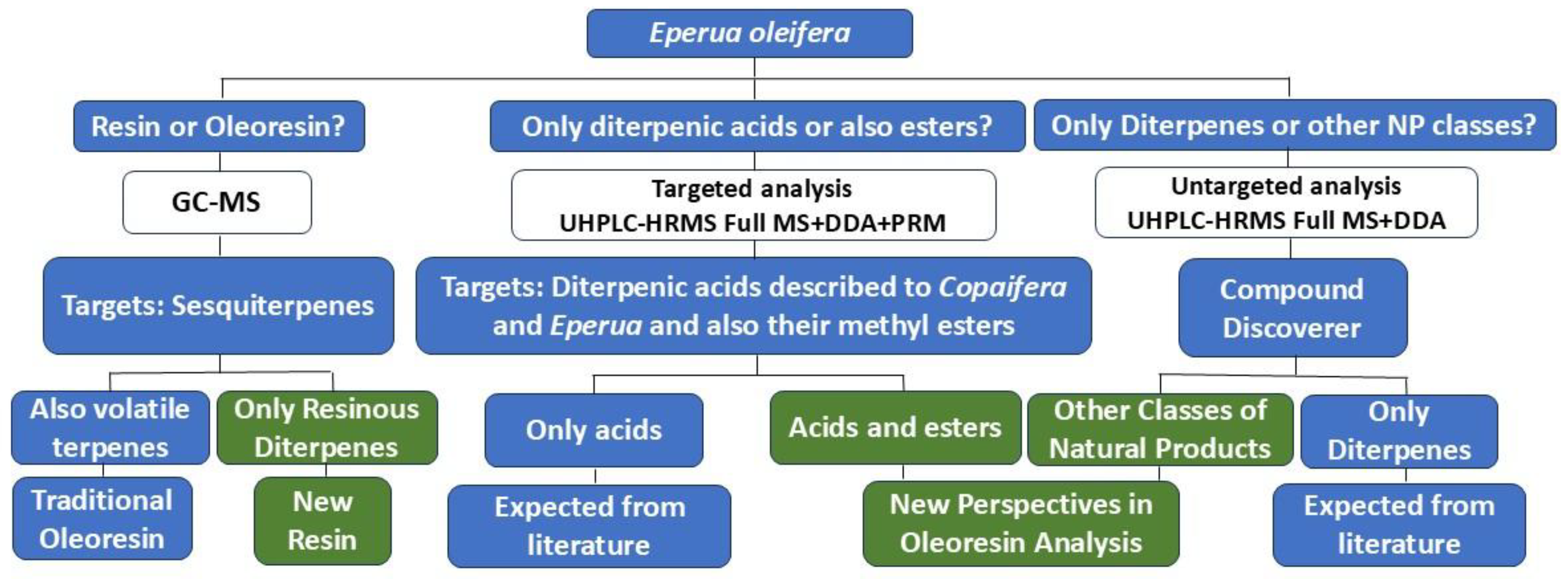
Figure 2.
Structured total ion chromatogram of Copaifera multijuga (a) and Eperua oleifera (b) oleoresins.
Figure 2.
Structured total ion chromatogram of Copaifera multijuga (a) and Eperua oleifera (b) oleoresins.
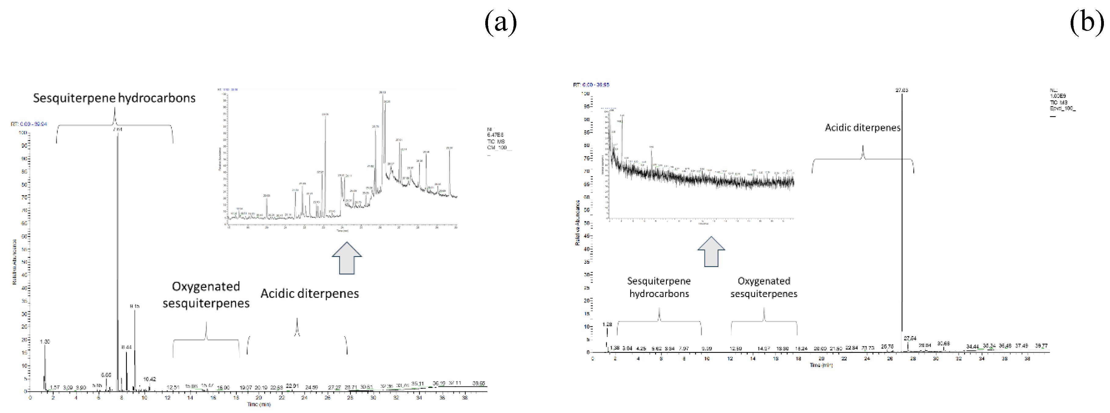
Table 2.
Area of the diterpene acids and their respective methyl esters. Oleoresin dissolved in methanol.
Table 2.
Area of the diterpene acids and their respective methyl esters. Oleoresin dissolved in methanol.
| Experiment dates |
Target analytes or Target substances | |||||||||||||||
|---|---|---|---|---|---|---|---|---|---|---|---|---|---|---|---|---|
| Hardwickiic acid | CV% | Methyl hardwickiate | CV% | Copalic acid | CV% | Methyl copalate | CV% | Patagonic acid | CV% | Methyl patagonate | CV% | Agathic acid | CV% | Methyl ester of agathic acid | CV% | |
| May 15, 2024 | 1975121726 | 8.0 | 1512316 | 5.1 | 1203063795 | 7.7 | 921009 | 4.4 | 74788718 | 7.7 | 891007 | 5.2 | 399396558 | 7.5 | 509843 | 6.2 |
| May 20, 2024 | 1926711350 | 1341619 | 1114681593 | 870510 | 64791889 | 862986 | 406791632 | 498032 | ||||||||
| May 25, 2024 | 1711628436 | 1470515 | 1223025777 | 941617 | 74937278 | 789675 | 453361272 | 569872 | ||||||||
| June 4, 2024 | 2075347716 | 1421008 | 1098075485 | 858979 | 77429128 | 871585 | 381595128 | 543929 | ||||||||
Table 3.
Area of the diterpene acids and their respective methyl esters. Oleoresin dissolved in acetonitrile.
Table 3.
Area of the diterpene acids and their respective methyl esters. Oleoresin dissolved in acetonitrile.
| Experiment dates |
Target analytes or Target substances | |||||||||||||||
|---|---|---|---|---|---|---|---|---|---|---|---|---|---|---|---|---|
| Hardwickiic acid | CV% | Methyl hardwickiate | CV% | Copalic acid | CV% | Methyl copalate | CV% | Patagonic acid | CV% | Methyl patagonate | CV% | Agathic acid | CV% | Methyl ester of agathic acid | CV% | |
| May 15, 2024 | 177534771 | 6.0 | 122541 | 6.1 | 109567508 | 5.2 | 76870 | 8.5 | 7278871 | 7.6 | 79100 | 8.5 | 37939253 | 4.9 | 48209 | 4.1 |
| May 20, 2024 | 169568903 | 130981 | 152698547 | 64191 | 6479188 | 75298 | 39891163 | 47981 | ||||||||
| May 25, 2024 | 162671135 | 131701 | 191469162 | 67078 | 7093297 | 75465 | 40459027 | 50629 | ||||||||
| June 4, 2024 | 186713428 | 142216 | 100330879 | 65132 | 7798235 | 89698 | 36289712 | 52298 | ||||||||
Table 4.
Analytes detected in the analysis of E. oleifera by UHPLC-HRMS, using an untargeted approach.
Table 4.
Analytes detected in the analysis of E. oleifera by UHPLC-HRMS, using an untargeted approach.
| Class of natural products | Substance detected | Molecular formula [M] | m/z [M-H]- | m/z [M+H]+ |
|---|---|---|---|---|
| Polyacetylene | (R)-(-)-Falcarinol | C17H24O | 243.17535 | |
| Benzoquinone | 5-O-ethyl embelin | C19H30O4 | 321.20731 | |
| Embelin | C17H26O4 | |||
| Fatty Acid | Methyl palmitate | C17H34O2 | 288.28931 | |
| (13Z)-8-hydroxyoctadecene-9,11-diynoic acid | C18H26O3 | 289.18121 | ||
| α-Linolenic acid | C18H30O2 | 277.21741 | ||
| Ricinoleic Acid | C18H34O3 | 297.24380 | ||
| Azelaic acid | C9H16O4 | 187.09711 | ||
| Amino Acid | L-Tyrosine methyl ester | C10H13NO3 | 194.08177 | |
| Polyene | (9cis)-Retinal | C20H28O | 285.22107 | |
| Diterpene | (E,E,E)-3,7,11,15-Tetramethylhexadeca-1,3,6,10,14-pentaene | C20H32 | 273.25748 | |
| Triterpene | Betulin | C30H50O2 | 443.38809 | |
| Ursolic acid | C30H48O3 | 455.35306 | ||
|
Phenolic |
1-(5-Hexyl-2,4-dihydroxyphenyl)ethenone | C14H20O3 | 254.17482 [M+NH4]+ | |
| 1-(2,6-Dihydroxyphenyl)-1,3-dodecanedione | C18H26O4 | 307.19009 | ||
| p-hydroxy benzoic acid | C7H6O3 | 137.02441 | ||
| Gallic acid | C27H20O5 | 425.13835 | ||
| Ellagic acid | C14H6O8 | 300.99899 | ||
| Flavonoids | 7-Hydroxy-2-methyl-4H-chromen-4-one | C10H8O3 | 177.05460 | |
| Catechin | C15H14O6 | 289.07176 | ||
| Epicatechin | C15H14O6 | 289.07176 | ||
| Quinic acid | C7H12O6 | 191.05611 | ||
| Quercitrin | C21H20O11 | 447.09328 | ||
| Quercetin | C15H10O7 | 301.03537 | ||
| Luteolin | C15H10O6 | 285.04046 | ||
| Apigenin | C15H10O5 | 269.04554 | ||
| Dihydromyricetin | C15H12O8 | 319.04594 |
*FA = Formic acid.
Disclaimer/Publisher’s Note: The statements, opinions and data contained in all publications are solely those of the individual author(s) and contributor(s) and not of MDPI and/or the editor(s). MDPI and/or the editor(s) disclaim responsibility for any injury to people or property resulting from any ideas, methods, instructions or products referred to in the content. |
© 2025 by the authors. Licensee MDPI, Basel, Switzerland. This article is an open access article distributed under the terms and conditions of the Creative Commons Attribution (CC BY) license (http://creativecommons.org/licenses/by/4.0/).
Copyright: This open access article is published under a Creative Commons CC BY 4.0 license, which permit the free download, distribution, and reuse, provided that the author and preprint are cited in any reuse.


