
Submitted:
26 June 2025
Posted:
30 June 2025
You are already at the latest version
Abstract
Micro- and nanoplastic particles (MNPs) are constantly formed through macroplastic fragmentation by sunlight, friction, or oxidation. MNPs potentialize the health risks en-tering the human body by ingestion, infusion, inhalation, and skin absorption. Still, the translocation among intracellular compartments must also be considered because MNPs can reach the circulatory system and be found in virtually all body fluids, tissues, and organs, potentially causing significant health impacts. The ability of MNPs to interact with macromolecules and cause damage to intracellular structures results in several physiopathological conditions, such as inflammation, oxidative imbalance, apoptosis and carcinogenesis. One major challenge in MNPs research is the development of reliable detection and quantification methods and effective sample separation processes. Alt-hough there is evidence directly linking MNPs to heart disease, the same cannot be said for diseases such as cancer, respiratory conditions and reproductive system disorders. Therefore, the impact of MNPs on human health and evaluate their effects on human health. We reviewed extensive scientific literature from the past years, focusing on ex-posure, aging, interactions and effects on entering MNPs into human metabolism and the physiological systems, which makes these particles particularly hazardous.
Keywords:
plastic additives
; inflammation
; apoptosis
; carcinogenesis
; neurotoxicity
1. Introduction
Plastics were once considered inert materials, but recent reports have shown they represent health risks to humans because they can be degraded into micro and nanoplastics (MNPs), becoming spreadable in air, water and soil. Because plastic materials are light and durable, produced at low cost, and widely used for various purposes, the disposal of plastic material is poorly managed and easily discarded. The manufacturing and utilization of plastics have increased annually due to irrational consumption driven by improved consumer purchasing power. Besides this, technological developments, allied with a lack of awareness about plastic, a pollutant material, lead to poor waste management since less than 35% of plastic waste is recycled. Worldwide, more than 330 million tons of plastic are produced annually, with a considerable increase in production during the COVID-19 pandemic period from 2019-2021 [1]. It is estimated that there are already 4.9 billion tons of plastic waste of varying sizes and chemical compositions, ubiquitous to all natural habitats, and those materials are spread in terrestrial and aquatic ecosystems [2]. In 2050, predictions indicate that this amount should increase by 12 billion metric tons [3].
MNPs particles have been distributed worldwide for a long time, but they are being considered emerging pollutants, and their potential health risks have been assessed [4]. These particles are an emerging global environmental contaminant that affects live beings and ecosystems; however, little is known about the effects of MNPs exposure and absorption by the human body [5]. The number of studies on the impact of these particles has been increasing, with emphasis on the polymers polyethylene (PE), polypropylene (PP), polyethylene vinyl chloride (PVC) and polystyrene (PS), which are the most produced plastics (Figure 1) [6,7]. Most discarded plastics are polystyrene and polypropylene, materials that could enter the circular economy [8]. However, the microparticles derived from biodegradable polylactic acid (PLA) also showed ecotoxicity against the mussel Mytillus coruscus, commonly used as a bioindicator organism [9].
More than 98% of plastics are produced from fossil sources. In addition, thousands of chemicals are added to polymers to impart specific properties such as color, flexibility, stability, water repellency, flame retardancy, and UV resistance. The critical environmental aspects of plastics led to the creation of the Minderoo-Monaco Commission that prospects these materials' impacts on human health [10]. Based on this, 175 nations have agreed to establish a legally binding international agreement to eliminate plastic pollution during the 2022 World Economic Forum [11].
The term "microplastic" (MPs) was first proposed by Thompson et al. in 2004 and refers to plastic particles with a size <5 mm, where nanoplastics (NPs), in turn, are defined by the European Food Safety Authority (EFSA) as 1 nm-10 nm particles [12]. The primary source of microplastics and nanoplastics in the environment is the improperly discarded macroplastics, transported to rivers and oceans and then fragmented mechanically or by solar irradiation in nature (Figure 1) [13].
There are large amounts of MPs below the surface of the Atlantic Ocean, highlighting the need for an in-depth assessment of the risks of plastic pollution since these tiny particles are dangerous to the health of all living organisms, including humans [14]. Those MPs, distributed in the human body, can cause several physiopathological conditions and the development of diseases, requiring studies that reveal how MPs interact broadly with components of the biological systems (Figure 2) [15].
2. MPs in the Environment
MPs have become a significant environmental concern as these contaminants accumulate and persist in all ecological compartments: air, water, soil, and even living organisms [16,17,18,19]. In addition, additives used to improve the properties of plastics and other toxic chemicals used for manufacturing are also carried by these particles, causing health problems [20]. Soil pollution by non-biodegradable MPs dates back to the widespread daily use of plastics. Over approximately 70 years (1950-2020), an enormous amount of plastic has accumulated on Earth, initially settling in the soil before being transported to water bodies and spreading in the air. There is now a concerted effort to trace better MPs in soils, including their sources, migration, distribution, biological effects, degradation, and the methodologies used to analyze them. The most frequently found MPs in soils are PE (78.8%), PP (78.8%), and PS (45.5%) [21]. In farming activities, MPs enter the agriculture chain through water, soil, silt, organic waste, fertilizers and airborne precipitates, negatively impacting agricultural production as they alter the soil microbial population and reduce nutrients, affecting plant growth [22,23,24,25,26].
There are two main routes for particles of MNPs to reach the environment: (i) particles formed during the production of macroplastics following direct human activity, and (ii) products of the decomposition of plastics or larger plastic fragments incorrectly discarded after usage [27]. These particles cause enormous economic losses in various sectors, including maritime transportation and fishing activities. Five trillion particles of MPs float in rivers and oceans or are deposited on beaches. Due to their size, they enter the food chain of the human beings that live in these habitats, but wisely than this, they affect all people consuming fish or seafood [28,29]. Surprisingly, the skin-exfoliating polymeric microspheres, an ingredient found in cosmetic products such as shampoos, toothpaste, soaps, and creams, are the new source of environmental contamination by MNPs.
Despite the advantages of recycling MPs, recycling facilities can generate a significant load of MPs, estimated at 59 and 1,184 tons annually [30]. Figure 3 shows some MPs of varying sizes collected from the sands of Praia das Dunas in Cabo Frio, RJ – Brazil, on 06/21/2024.
The toxicity of MPs to humans has not yet been fully clarified. However, they are increasingly recognized as a significant public health concern, as a potentially hazardous associated with several physiopathological conditions and diseases, including disorders in the lipid metabolism, inflammatory response and oxidative imbalance status on the organism, cancer and heart diseases [31,32,33,34,35]. Once in a human body or even other living organisms with a circulation system, NPs can be indistinctly distributed to every organ or tissue. Still, these plastic particles can penetrate the core of lipid bilayers. As discussed, several hazardous chemical compounds associated with those nanoparticles can be vectorized to plasmatic or intracellular membranes in living organisms [36].
The human body can be exposed to MPs through the inhalation of particles present in the air (especially in metropolitan cities), ingestion of contaminated water and or food, or through absorption by the skin, and also, less frequent, but not more critical by tubing of transfusion therapies (Figure 4) [37,38,39,40]. After entering the human body, MPs and NPs reach the bloodstream, are distributed and can accumulate in various tissues and organs [41]. MPs and NPs were detected in the oral, anal, and/or uterine/vaginal cavities, which are directly accessible because they are in contact with the external environment [42]. However, NPs can be translocated to any tissue or cell in living organisms since their size allows them to be diffused into the lipid environment of both plasmatic and intracellular membranes.
The ingestion of MNPs is a particular concern because of the presence of plastic particles in fish and seafood, which are an essential part of a balanced human diet, as already mentioned, but also for birds and mammals can be a source of MNPs for human beings. Fish tend to ingest plastic particles of specific colors, such as white, yellow, and blue [43]. However, the shape of MPs also plays an important role in ingestion by aquatic animals since some of them are very attractive; for example, fibre-type MPs resembling worms and eggs are often accidentally ingested by fish [44]. When consumed by other animals, these contaminated fish transfer their load of MPs and NPs to their predators, including humans. MPs were found in the stomach content of neotropical fishes in Brazil from the Paraná river basin. Microscopic analysis of 220 individuals belonging to 14 species were analyzed, and the results indicated the presence of small amounts of plastic particles of different shapes measuring from 1mm to 3 mm) [45]. MPs were also found in the gastrointestinal tract of fish specimens (18.1-34.5%) harvested in the middle Uruguay River in southern South America, and several species were collected in the rivers from the Amazon Basin, Brazil [46].
It is important to note that the qualitative and quantitative analyses of MPs in waterbodies still require optimization. No international standards are established for sampling, extraction, or detection of these contaminants, leading to inconsistent results in several reports [47,48]. After reviewing several studies, Koelmans and colleagues have proposed more appropriate methodologies for MPs quantitative evaluation in waterbodies [49]. In the later study, these researchers quantified the exposure limits and effects using probability density functions that describe the diversity of the MP particles [50].
Quantitative data, including information on MPs, were compiled from 26 studies, yielding 402 data points representing over 3,600 processed samples. Microplastic contamination via inhalation was evaluated using reported airborne MPs concentrations and respiration rates, as provided by the US Environmental Protection Agency (EPA) [51]. Regarding dietary exposure, MPs were found in food items consumed daily, such as seafood, sugar, salt, honey, alcohol, and tap and bottled waters. Based on the collected data, it was estimated that humans consume between 39,000 and 52,000 MPs annually, depending on their age and sex. If the inhalation route is considered, the estimative increases to between 74,000 and 121,000 MNPs annually [52]. However, the authors believed that these numbers are underestimated due to the methodology used and highlighted that individuals who consume bottled water may be ingesting nearly 90,000 additional MPs, compared to the 4,000 MPs ingested by those who consume piped water.
The zebrafish (Danio rerio), an animal model organism sharing 70% of its genome with humans, is widely used to study blood diseases and carcinomas, serving as a toxicological model. Zebrafish exposure to MPs had their metabolome and gut microbiome evaluated, revealing gut inflammation, unbalanced oxidative condition, and lipid metabolism disorders [53]. Within this context, Rahman et al. assessed the toxicity and cell viability of lung epithelial cells from mice under the effect of 11 types of MPs, prepared in the laboratory from commercial plastics [54]. The small-sized particles (1-5 μm) from PE and PET, prepared from disposable water bottles, induced maximum toxicity. These PET microparticles induced activation of the interferon signaling pathway because they were perceived by immune cells in a similar way that occurs to pathogens. Polyethylene terephthalate-MPs with the smallest size and heterogeneous shapes induced cell injury, triggering the inflammatory cascade, DNA damage, and cell death, characteristic of tissue injury in vivo.
A significant concern is the MPs potential to cause teratogenic effects. The teratogenic impact of exposition to plastic particles on the diatom Cocconeis placentula and the cnidarian Hydra vulgaris belongs to different trophic levels. A moderate teratogenic risk index was calculated for diatoms, even at a concentration of 0.1 ug. mL-1 of plastic particles, however, a low teratogenic risk was estimated for H. vulgaris even at the highest concentrations, but a slower regeneration was observed for these organisms. The study highlighted that the teratogenic effect depends on the concentration of plastic particles and can vary according to the habitat of the trophic level. However, the correlation between specific types of NPs or MPs and particular teratological forms was poorly evaluated [55].
The association of MPs with pesticides poses a significant threat to soil organisms such as earthworms (Eisenia fetida) that have been individually exposed to PE and PP, causing adverse effects on earthworms' digestive tract and side effects on soil ecosystems [56].
In addition to the concern about the effects of MPs and the human health risks, the additives used in plastic manufacture are also of concern. Additives are chemical compounds added to improve functionality and/or loading and are waste by-products of following plastics production. The chemical bonds between these additives and polymers are weak supramolecular interactions and are, therefore, released unintentionally during environmental exposure. There are approximately 2,712 known plastic additives, some of which are considered carcinogenic, causing DNA damage, apoptosis, immune system impairment, and certain types of cancers, including endocrine, biliary tract, pancreatic, and hepatocellular carcinomas. However, many of these compounds remain untested. Human exposure MNPs presents risks from the plastic particles and the toxic additives they carry, which can spread throughout the body [57].
3. Biological Effects of Polystyrene Particles (PS-MPand PS-NP)
Styrene, the monomer used to produce polystyrene (PS), is classified as a potentially carcinogenic substance (carcinogenicity class B2) by the International Agency for Research on Cancer (IARC) [58].
Recent studies involving micro and nanoparticles of PS and polyvinyl chloride (PVC) were tested on a triple culture model (Caco-2/HT29-MTX-E12/THP-1), mimicking the inflamed human intestine. The exposure to PVC particles during an active inflammatory process on gut cells was found to augment the release of IL-1β cytokine, causing the death of epithelial cells. No acute toxic effects were seen in the model of the healthy intestine following PS or PVC exposure. Busch et al. and Hesler et al. concluded in their respective works that polystyrene MPs did not present acute effects in the healthy gut model since they do not cross the intestinal and placental barriers and are weakly embryotoxic and non-genotoxic in vitro [59,60]. Although the polymer has different chemical characteristics than the styrene monomer, its use has raised several concerns. The polystyrene MPs and NPs accumulate along the food chain, promoting adverse effects on different organisms [61]. Wang et al. The photodegradation of PS-MPs increases its toxicity to marine organisms, disrupting hepatic lipid homeostasis and consequent liver injury, besides causing inhibition of growth [62]. Similarly, earthworms were more sensitive to freeze-thaw aging microplastics that caused oxidative imbalance in those organisms, changing their microbiota and possibly impacting the benefits of these invertebrates to the soil [63].
The tools used to predict the toxicity of organic compounds are important for the design of substances and are very efficient in predicting the toxicity of MPs. Three artificial intelligence algorithms that can predict the behavior of these particles on cells have identified particle size as the most critical characteristic for toxicity [64].
The most recent data indicate that PS is one of the most dangerous MPs, as it can associated with several organic macromolecules. Jones et al. conducted a critical literature review to address the question, 'Are micro- and nanoplastics toxic when ingested?' [65]. Their findings indicate that polystyrene microplastics are among the most toxic in vitro and in vivo, causing inflammation, altered cell proliferation, dysregulation of cell membrane permeability, and impairments in lipid and amino acid metabolism, ultimately leading to apoptosis. It is important to note that PE and PP also disrupt lipid metabolism and that PS is often ingested alongside other polymers.
Regarding size, particles < 10 μm pose the greatest danger, both in vitro and in vivo, suggesting that the chemical composition and size of MNPs are critical factors in their toxicity. This observation aligns with the known risks associated with other types of particles. In addition to the significant biological potential of micro- and nanoplastics (MNPs) and polystyrene nanoparticles (PS-NPs), Awet et al. identified the harmful effects of PS-NPs on the environment [66]. Their study, which evaluated the antimicrobial impact of PS-NPs on the native soil microbiome, suggests that PS-NPs may pose potential environmental risks. During 28 days, the activities of dehydrogenases, N-(leucine-aminopeptidase), P-(alkaline phosphatase), and C-(β-glucosidase and cellobiohydrolase) were significantly reduced, demonstrating the impact on the physiology and functionalities of soil microbiota.
Dong et al. used healthy human lung epithelial cells (BEAS-2B) in culture to investigate the relationship between lung toxicity and PS-MPs [67]. The results showed that these MPs cause cytotoxic and inflammatory effects on epithelial cells by increasing the production of reactive oxygen species and decreasing transepithelial electrical resistance, raising the risk of chronic obstructive pulmonary disease (COPD). In a further study by Danso et al. on the pulmonary toxicity of polystyrene, polypropylene, and polyvinyl chloride (PVC) using three strains of mice (C57BL/6, BALB/c, and ICR), it was found that polystyrene exposure led to an increase in inflammatory cells by stimulating the release of inflammatory cytokines in the bronchoalveolar lavage fluid of C57BL/6 and ICR mice [68]. These findings suggest that the inhalation of PS-MPs poses a significant risk for pulmonary toxicity Cao et al. also studied chronic exposure to polystyrene-MPs in mice and found that they induced lung lesions through activation of the TLR2/NF-κB pathway, triggering inflammation and oxidative stress in lung cells. In this way, MPs can aggravate apoptosis and induce pulmonary fibrosis in mice [69]. Wu et al. proved the protective antioxidant capacity of N-acetylcysteine against polystyrene NPs in mice by reversing the pulmonary toxicity of PS-MPs [70]. Li et al. tested several methods for the induction of pulmonary fibrosis in mice by oxidative stress and activation of the Wnt/β-catenin signaling pathway [71]. It has been found that the inhalation of these MPs induces pulmonary fibrosis in a dose-dependent manner, increasing oxidative stress in the lungs. This observation was proven when using melatonin (50 mg/kg), which relieved pulmonary fibrosis induced by polystyrene particles.
Kuroiwa et al. studied how phagocytes recognize MPs using Tim4, a surface protein receptor that plays an important role in the immune system [72]. Phagocytes absorb MPs, and it is possible that, in the case of polystyrene-derived-MPs, a supramolecular pi-stacking interaction occurs between the benzene aromatic rings of polystyrene and mucin 4 of Tim4 immunobiological cells. This may represent a new interface between MPs and biological systems through aromatic-aromatic interactions. As Tim4 mediates efferocytosis through its PtdSe-binding site, the macrophage engulfment of apoptotic cells can be blocked entirely by PS-MPs because the aromatic cluster of Tim4 is fully occupied by the PS-MPs binding, perturbing efferocytosis. Conversely, the authors have considered that continued exposure to PS microparticles can generate a chronic inflammatory status, predisposing individuals to autoimmune diseases.
Garcia et al. investigated the ingestion of various concentrations of MPs and how they affect the functioning of metabolic pathways within the colon, liver, kidney, and brain in mice [73] Polystyrene was administered in separate groups at concentrations of 0.2 or 4 mg/week, and light microscopy and spectroscopy were used to analyze the serum and tissues of the brain (prefrontal cortex), liver, kidney, and colon for the presence of MPs. The liver was the organ that most concentrated the particles, while the kidneys had the lowest concentration. These results suggest that the particles pass the intestinal epithelium, enter the circulatory system, and deposit in the organs.
Bingrui et al. studied the effect of exposure to polystyrene-MPs at various stages of nephrogenesis using human renal organoids, three-dimensional structures grown in the laboratory, i.e., in vitro [74]. MPs with a size of 1 μm adhered to the cell surface during the Nephron Progenitor Cell (NPC) stage and accumulated within glomerulus-like structures of renal organoids. This resulted in organoids with reduced nephron size, increased production of reactive oxygen species, apoptosis, decreased cell viability, and reduced NPC. However, the adverse effects of exposure to these polystyrene-MPs on the human renal system remain uncertain as there is no model for a proper comparison.
All these works undoubtedly point to the real danger that micro- and nanoplastics derived from polystyrene pose to human health and the environment. In this way, a discussion can be opened about the applicable alternatives for replacing this material and its gradual removal from contact with humans and the environment, especially as packaging or containers for food and plastic products that have a very short shelf life.
4. Occurrence of MNPs in the Bloodstream, Reproductive System and Gastrointestinal Tract
PS microplastics affect the male reproductive system by decreasing sperm count and damaging testicular structures. Wen et al. studied the effects of NPs (80 nm) and MPs (5 μm), specifically polystyrene, on the spermatogenesis of male C57BL/6 mice that orally ingested these particles for 60 days [75]. It was observed that different molecular mechanisms were affected. NPs affected the regulation of retinoic acid metabolism, while MPs mainly influenced pyruvate metabolism and thyroid hormone metabolism, which compromised the spermatogenesis of mice and, consequently, their reproduction (Figure 5 and Figure 6).
A common but unexpected source of MPs contamination is the disposable materials used for serum infusion or blood transfusion therapies, where the infusion bottles, infusion bags, and infusion tubes are made of plastic. Zhu et al. identified eight MPs, ranging from 4 μm to 148 μm at the concentration of 1-2 MNPs/unit [76]. The MNPs were found in PP-bottled, PE-bagged, and glass-bottled, but no particles in infusion tubes. The microplastic samples accounted for 11.66% of the total samples evaluated. This finding indicates that plastic materials used for infusion/transfusion therapies can be a source of MNPs in the bloodstream. Besides this, MPs absorbed from contaminated food can be secreted into saliva, gastric juice, and intestinal fluids, releasing toxic compounds into the gastrointestinal tract and distributing them to other organs through the bloodstream (Figure 5) [77].
The pioneering study of Halfar and coworkers showed the presence of MPs in the amniotic fluid and placenta by employing infrared spectroscopy with attenuated Fourier transform-total reflectance (FTIR-ATR) after alkaline digestion with KOH [78]. From 20 samples of amniotic fluid and placenta collected from 10 patients, 44 MPs or plastic additives were found in the amniotic fluid, placenta, or both in 90% of those patients. PVC and the calcium-zinc PVC stabilizer predominated with granulometry between 10 and 50 μm. It is important to note that if MPs were found in the placenta, both mother and fetus were or are being exposed to risks. Indeed, all women who participated in the study have experienced pregnancy complications, such as pregnancies with prelabour rupture of membranes before the onset of labor. However, no evidence correlates the breakage of the amniotic sac to the presence of MPs in the placenta.
In a qualitative study using the Fourier transform infrared microspectroscopy technique in a clinical setting to detect MPs in the human placenta and fetal meconium, ten types of MPs, derived mainly from PE, PP, and PS were detected. Braun et al. detected polyurethane particles in the operating room following atmospheric precipitation [79].
Zhu et al. recently conducted a quantitative and qualitative study on MPs in 17 placenta samples using direct laser infrared spectroscopy (LD-IR) [80]. 12 spherical or irregular-shaped MP different types were found in 4 placentas samples, and 3 were identified as PP. In all of them, there were MPs at concentrations ranging from 0.28 to 9.55 microparticles per gram. Eleven distinct polymers were found, where PVC was the most abundant (43.27%), PP at 14.55%, and polybutylene succinate (PBS) at 10.90%. While the study provided data on the size and types of polymers present, there was no correlation between the presence of plastic particles and potential diseases in mothers and fetuses. Using the Raman microspectroscopy technique, Ragusa et al. also studied the presence of MPs in human breast milk samples from 34 women [81]. MPs were found in 26 samples, and the most abundant polymers identified were PE, PVC and PP, with sizes ranging from 2 to 12 μm. Transmission to babies through breast milk is another form of vertical transmission of MPs. Considering another part of the female reproductive system, Qin et al. evaluated the presence and entry mode of MPs into the human endometrium [82]. After the investigation of the endometrial tissues of 22 patients, the contamination of MPs of the polyamide (PA), polyurethane (PU), PET, PP, PS and PE classes was found with sizes ranging from 2 μm to 200 μm. Tests with mice to determine whether contamination occurs via blood circulation, vaginal contamination, or vaginal-uterine contamination proved blood circulation as the route of contamination of the endometrium, with particles acquired during the diet. In addition, MP caused reduced fertility of the mice (P <0.05). After 3.5 months of intragastric exposure, a significant inflammatory response occurred in the endometrium (P <0.05). Studies have indicated that MP contamination in the human uterus can also have harmful effects on reproductive health.
An investigation into MPs in samples from 15 patients who underwent cardiac surgeries used a direct infrared laser chemical imaging system and scanning electron microscopy [83]. The analysis detected several of these particles in 5 tissue types, with the largest fragment being 469 μm in diameter. In addition, nine types of MPs, with a maximum diameter of 184 μm, were detected in blood samples collected before and after the surgeries.
A recent study investigated MPs in healthy liver tissues and cirrhotic liver tissues [84]. The analysis involved chemical digestion of the tissues application of the fluorescent dye red of the Nile (an azo dye), followed by fluorescence microscopy and Raman spectroscopy. All samples from healthy livers did not show MPs. However, the cirrhotic liver tissues tested positive for MPs. Six different polymers were identified, ranging from 4 to 30 μm in MPs. The authors claim that they do not have sufficient evidence to confirm the MPs in cirrhotic liver tissues as the cause of cirrhosis.
The physiology of the male reproductive system is sensitive to several environmental conditions, especially concerning sperm production [85,86]. NP and MPs can affect human reproduction, with a potential impact on the male reproductive system, based on a study that evaluated the effect of MPs on the sperm from dogs and humans using sensitive pyrolysis gas chromatography associated with mass spectrometry (Py-GC/MS), 12 types of MP were detected, with PE being the most prevalent, in the testicles of 47 dogs and 23 humans, reaching, in total, an average concentration of 122.63 mg/g in dogs and 328.44 mg/g in humans the testicle sizes was also increased [87].
Eleven human colectomy samples were analyzed from adult patients in Peninsular Malaysia (mean age 45.7 years) [88]. After chemical digestion, the samples were analyzed under stereo and FTIR microscopes in search of MPs (composition, abundance, length, shape, and color). The results showed the detection of MPs in all 11 samples, with an average of 331 particles/individual sample or 28.1 ± 15.4 particles/g of tissue. Filaments or fibers accounted for 96.1% of the particles, 73.1% of all filaments, 90% were polycarbonate, 50% were polyamide, and 40% were polypropylene. Cetin et al. conducted a study to investigate the effects of MPs in colorectal adenocarcinoma tissues compared to normal tissues [89]. They employed attenuated total reflection infrared spectroscopy with Fourier transform and Raman spectroscopy for detection and analysis. The study found that MPs were more prevalent in tumor tissues than in normal or control tissues, with particle sizes ranging from 0.001 to 1.2 mm and composed of polymers such as polyethylene, methyl methacrylate, and polyamide. However, the study did not establish a definitive connection between the presence of MPs and colorectal cancer.
Other in vivo and in vitro studies have investigated the potential for MPs to cross the blood-brain barrier and cause neurotoxicity by promoting oxidative stress, disrupting inflammatory balance, altering neurotransmitters (synaptic plasticity), inhibiting acetylcholinesterase activity, and impacting the gut-brain axis [90]. From the analyses, it was observed that lower MPs and NPs cause more severe damage to neuronal diseases. There are still no relevant reports of the effects in humans, but there is evidence that they can disrupt neurons and alter the memory and behavior of organisms. Urgency is needed to elucidate the neurotoxic danger of exposure to MPs and N.P.s [91,92].
5. Occurrence of MPs in the Respiratory Tract via Inhalation
MP particles (>5 mm up to >1nm) dispersed in the air also represent the most important source of exposure of the human body through respiration, aggravated by the fact that smaller particles ensure their prolonged permanence in the atmosphere [93,94]. MP particles lodged in the lungs can impair gas exchange. In addition, prolonged exposure to air MPs can worsen the condition of patients with lung diseases by airway barrier function, as mentioned, but causing dysfunctions in the biophysics of pulmonary surfactants, alveolar structure,95 and lung epithelial cell proliferation, with mechanistic studies in the initial phase [95,96,97].
Patients with a history of exposure to MPs had significantly higher levels of MPs in their lung tumors than those with lower exposure histories [98]. This suggests a potential link between MP inhalation and the development of Ground-Glass Nodules (GGNs) visible on CT scans. Individuals most affected reside in heavily polluted urban areas, particularly near major highways. For instance, a study of 12 nonsmoking women living close to major highways found elevated levels of MPs in their lung tissues. Analysis using infrared laser imaging spectroscopy and scanning electron microscopy identified 108 MPs, of which 12 were composed of various polymers, with an average concentration of 2.2 particles per gram. Particle sizes ranged from 20 to 100 μm, with polypropylene being the most prevalent polymer (34.2%), followed by PET (21.3%). Notably, MP contamination was positively correlated with elevated platelet and fibrinogen levels and negatively correlated with direct bilirubin levels, a degradation product of hemoglobin.
The SARS-CoV-2 coronavirus infection, which led to the COVID-19 pandemic, has as its main characteristic a pulmonary disease that can vary from mild to severe. It was predictable that lungs contaminated with particles of MPs could interfere with the course of this infection. Bishop et al. showed that MPs can dysregulate lung inflammation by SARS-CoV-2 by suppressing innate immune responses after two days of infection and increasing the release of proinflammatory cytokines six days after infection [99].
Human lung tissues obtained from autopsies were evaluated by Raman spectroscopy [100]. MPs were observed in 13 of the 20 tissue samples; all were smaller than 5.5 μm, and the fibers ranged from 8.12 to 16.8 μm. The most present polymers were PE and PP. Although they do not find a direct correlation between MP and some diseases, they can still harm the respiratory system.
One way to obtain samples is to investigate the deposition of MPs in bronchoalveolar lavage fluid (LPBA). This fluid is a source of samples of the material present in the pulmonary alveoli and bronchi. Qiu et al. conducted experiments to detect MPs in the respiratory tract. BALF of 18 nonsmoking individuals were analyzed by direct infrared laser spectroscopy combined with scanning electron microscopy [101]. 13 types of MPs were detected in the 18 samples of the liquid, with PE being the most frequent (86.1%), showing that MPs penetrate deep into the respiratory tract. MPs were also found in Chinese children's bronchoalveolar lavage fluid (BALF), with 89.6% of the samples containing MPs, averaging 4.31 ± 2.77 particles per 10 mL [102]. These data confirm inhalation as a significant route of exposure to MPs in the pediatric lungs. The MPs were made of PP (41.9%), PE (19.4%), and PS (13.6%), most of which were smaller than 20 μm, indicating a possible relationship between MPs and pediatric lung diseases.
MP particles lodged in the lungs can impair gas exchange. MPs were detected in human lung tissues by analyzing digested samples from 13 individuals using μFTIR spectroscopy, with a size detection limit of 3 μm [103]. They identified 39 MP particles in 11 of the 13 samples, with an average concentration of 1.42 ± 1.50 MPs per gram of tissue. The primary plastic components found were PET and polypropylene (PP), which were present in critical areas for gas exchange, including terminal bronchioles, alveolar ducts, and alveoli. Xu et al. demonstrated that MPs impair lung biophysical function through the hetero-aggregates formation of this interface, negatively affecting gas exchange [104]. Further evidence of the effect of MPs on the respiratory system was reported by Li et al. by demonstrating that the particles of MPs dispersed in the air, resulting from tire wear, induced pulmonary fibrotic injury through the rearrangement of proteins that provide structural support to the cytoskeleton of epithelial cells [105].
MPs and NPs can potentially disrupt the composition of nasal and lung microbiota, including bacteria, viruses, and fungi, though their effects are not yet fully understood [106]. Studies have shown that bacteria from Staphylococcus spp and Roseburia spp are more frequently associated with MPs, while Prevotella species are more commonly linked to NPs. These findings suggest that airborne MPs and NPs could contribute to microbial dysbiosis in both nasal and pulmonary environments.
Jiang et al. studied the contamination of the human upper respiratory tract by MPs, differentiating between internal and external workers [107]. It has been shown that indoor workers are more exposed to MP particles, while outdoor workers are more exposed to MP fibers. Outdoor workers can wear respirator masks as personal protective equipment, but they have the advantage of the wind dispersing MP particles.
6. MP Occurrence and the Cardiovascular System Treats
MP particles' size and chemical structure can significantly impact the cardiovascular system. The bloodstream can transport MPs to various organs, potentially leading to severe physiopathological conditions and diseases. For example, MPs can induce platelet aggregation and thrombus formation. However, there remains uncertainty regarding the full extent of pathologies associated with environmental exposure to MPs and the various routes [108]. Research into the relationship between MPs and heart disease is still emerging. Still, preliminary evidence suggests a potential cause-and-effect relationship where MPs may directly contribute to the development of heart disease. The primary challenge in this field has been accurately detecting and quantifying this relationship using analytical methods.
MPs pose a threat to the cardiovascular system. Data published over the past decade highlight the toxicity of MPs and their effects on cardiovascular mechanisms, showing that MPs impair cardiac function and cause microvascular toxicity. Direct cardiac effects of MPs include arrhythmias, myocardial dysfunction, pericardial edema, and myocardial fibrosis [109]. Microvascular lesions induce hemolysis, thrombosis, blood clotting, and vascular endothelial damage through oxidative stress, inflammation, apoptosis, and interaction of MPs with other cellular components. However, the authors recommend further studies for a more conclusive assessment of actual health risks.
MPs may play a significant role in blood clot formation. Their impact on fibrin clot formation was investigated using a simplified ex vivo human thrombin/fibrinogen clot model [110]. The clot formation was characterized by turbidity and thromboelastography. When the particles were pre-incubated with fibrinogen, a reduction in coagulation efficiency for amino polystyrene and polystyrene of up to 100 μg/mL was observed. The results demonstrated a significant impact on the rate and strength of clot formation by inhibitory effects on fibrin clot formation.
Further evidence of the interference of MPs in the dynamics of blood coagulation was reported by Christodoulides et al., who examined this question using the technique of thromboelastography, detecting particles of sizes of 50, 100 and 500 nm from three types of plastics [111]. Carboxypolystyrene was found to activate the coagulation cascade by enhancing fibrin polymerization rates. While polystyrene generally had minimal effects on coagulation dynamics, particles sized at 50 nm could trigger the coagulation cascade.
Environmental factors are considered one of the responsible factors for the cause of cardiovascular diseases, such as exposure to pollutants and chemicals (e.g. arsenic, lead, cadmium, polluting gases, airborne particles, solvents, pesticides and MP particles) through the air, water and food [112]. Daily exposure to these external agents has extensively been related to increased risk of cardiovascular diseases [113,114]. Recently, the pyrolysis gas chromatography coupled to mass spectrometry (Py-GC/MS) technique has emerged to detect MPs in organs and tissues and was used to investigate MPs in the atherosclerotic plaques of 17 samples from human vessels, coronary and carotid arteries and aorta, with a mean concentration of 118.66 μg/g of tissue. PET was found in 73.70% of the sample, polyamide-66 in 15.54%, PE-vinyl chloride in 9.69%) and PE in 1.07% [115].
For the first time, a prospective, multicenter, observational study has proven a direct link between MPs and human health. Marfella and coworkers conducted this historic study involving 304 patients, of whom 257 completed the mean follow-up of 33.7±6.9 months [116]. The patients underwent a surgical procedure to remove the accumulation of atheroma plaque from the carotid arteries (asymptomatic carotid endarterectomy) [117]. The basic principle was that the particles of MPs interact with fat molecules and can accumulate in atheromatous plaques in the epithelial lining of blood vessels. Analyses to identify the type and size of the MPs were performed using the pyrolysis technique, together with gas chromatography coupled to the mass spectrometer, stable isotope analysis, and electronic microscopy analysis. Electron microscopy showed the presence of irregular bubbles of MPs mixed with cells and other residues in samples from 150 participants. Chemical analyses revealed that most of the particles in the samples from 150 patients (58.4%) correspond to PE, with an average level of 21.7±24.5 μg per milligram of plaque. The detection of inflammatory biomarkers was evaluated by enzyme-linked immunosorbent and immunohistochemical tests. Patients with more MPs in atheroma plaque samples had higher inflammation biomarker levels. These data suggest that MPs may contribute to plaque rupture in patients with carotid atheroma plaque. Those in whom MPs and NPs were detected had a higher risk of myocardial infarction, stroke, or death from any cause at 34 months of follow-up compared to those in whom these particles were not detected.
7. MPs and the Development of Cancer
As previously mentioned, plastics may contain various additives, including phthalic esters, polycyclic aromatic hydrocarbons, polychlorinated biphenyls, plasticizers (particularly bisphenol A), and dyes. Phthalic esters, such as di-2-ethylhexyl-fatalate (DEHP), di-n-butyl phthalate (DBP) and diethyl phthalate (DEP) are among the most systemic toxics compounds displaying carcinogenic potential [118,119,120]. They can be adsorbed to MPs, and released when they reach the living organisms [121]. It has been demonstrated that MPs can transport and release phthalic esters in the intestines of mice. This suggests that MPs may carry plastic additives to the gastrointestinal tract, which could accumulate and cause toxic effects [121]. It can be considered that microplastics could adsorb and transport plastic additives to the gastrointestinal tract, where they accumulate and cause toxic effects. Deng et al. demonstrated for the first time that MPs can transport and release phthalic esters in the intestines of mice. As a result, MPs can adsorb and transport phthalic esters (PAEs) to the gastrointestinal tract, where they accumulate and cause toxic effects [122].
Another plasticizer widely used in plastics manufacture is bisphenol A, which has been the subject of major toxicological concerns for human health. Cheng et al. The study investigated the hepatotoxic effects of bisphenol when combined with polystyrene particles [123]. Initially, adding polystyrene to the culture medium led to a decrease in bisphenol levels, suggesting an interaction between the two substances. The most significant finding was the synergistic adverse effect of this combination, which resulted in hepatotoxicity and genomic interference related to lipid metabolism. This study highlighted a substantial metabolic health risk, such as hepatic steatosis, when polystyrene is exposed to bisphenol, even at low doses comparable to human internal exposure levels.
There is still little knowledge about the correlation between exposure to MPs and their additives and various types of human cancer. Kumar et al. discuss the possibilities of inhalation, ingestion and dermal exposure causing genotoxicity, cell division and viability, cytotoxicity, induction of oxidative stress, metabolism disruption, DNA damage, inflammation and immune responses in humans [124]. However, they conclude that with current knowledge about the potential risks to human health, there are still many gaps and recommend further investigation.
Rawle et al. investigated the potential of MP particles to induce colonic inflammation in rats that consumed approximately 80 μg/kg/day of 1 μm polystyrene through drinking water [125]. They observed that colonic inflammation was prolonged in rats exposed to MPs, suggesting that disturbances in the colon activate lymphoid cells and promote inflammatory responses. Regarding the effects of MPs on the colon, Li et al. reviewed the incidence of colorectal cancer with MPs [126]. The incidence of this type of cancer is increasing in people under 50 years of age, and this epidemiological change suggests that there may be a prevalent environmental cause, possibly contamination by MPs. This hypothesis is based on substantial evidence of the increasing presence of these MPs in the gastrointestinal tract from contaminated food. Particles of MPs transiting the gastrointestinal tract can interfere with the protective mucus layer of the colon and rectum, reducing its protective effect and increasing the likelihood of colorectal cancer.
A recent study revealed the presence of MPs in several human tumors [127]. The research identified the types and amounts of MPs in these tumors through qualitative and quantitative analysis using gas chromatography and mass spectrometry. Among the 26 samples analyzed, PS, PVC and PE were found in 6 types of tumors. The distribution of MPs varied among the different organs. In lung, stomach, colon, and cervical tumors, the detection rates of MPs were 80%, 40%, 50%, and 17%, respectively, with concentrations ranging from 7.1 to 545.9 ng/g. In pancreatic tumors, MPs were detected in 70% of the samples, with concentrations between 18.4 and 427.1 ng/g, while in esophageal tumors, no MP was detected. MPs may affect the tumor immune microenvironment (TIME), accelerating tumor progression and treatment resistance, which is still unclear.
The interactions between MPs and toxic heavy metals and their effects on human and animal health have also been studied. In aquatic environments, MPs can adsorb metals, glycolipids, pesticides, long-chain perfluoroalkyl substances (PFAS), and other contaminants, potentially leading to harmful biological effects [128]. MPs act as carriers of these toxic substances [129,130]. MPs undergo aging due to exposure to UV radiation and air, increasing the surface roughness and, consequently, enhancing the contact surface of the material. Godoy and colleagues investigated MPs made from various polymers and their ability to carry metals, pesticides, and pharmaceutical products [131]. Metals, originating from plastic manufacturing processes, can associate with MPs through environmental adsorption and be transferred along the food chain. PE, PS and PVC MPs quickly adsorb various metals, but chromium and lead are absorbed in more significant quantities. Co-exposure to PVC-derived MPs and cadmium damages the liver of female ducks, but its mechanism is still unknown. Cadmium is a toxic metal that interacts strongly with various polymers and can be carried into organisms [132]. Sun and colleagues studied the association of PVC with cadmium and its biological effects on female ducks over two months [133]. The ducks were treated with pure water, isolated components, or were co-exposed to both. The researchers observed liver shape changes, hepatocyte dysfunction, ultrastructural damage, and alterations in glycolipid metabolism, ultimately leading to apoptosis. The hepatotoxicity caused by polypropylene-MPs, artificially aged with UV radiation, was tested in hepatic organoids, generating oxidative stress at low doses and interfering with the mitochondrial respiratory chain [134].
8. Elimination and Clearance of MNPs
As mentioned, the main routes MPs and NPs can take to enter the human body are infusion tubing, inhalation, absorption by the skin, and ingesting food. MPs and NPs are then distributed via the bloodstream, accumulating in the respiratory and digestive system, reproductive system, liver, spleen and brain [135]. However, the accumulation of MPs and NPs does not necessarily imply direct damage; part of the MPs absorbed can be engulfed by macrophages and digested by gut microbiota. It may undergo an inflammatory reaction and a fibrous encapsulation. Most of them will be removed by the liver and spleen and excreted in the feces, the most common route for the MPs elimination absorbed from drinking water and food ingestion. Schwabl et al. were the first to examine the human feces of 8 healthy volunteers in the presence of MPs [136]. After the chemical digestion of the fecal samples, infrared microspectroscopy with a Fourier transform was used to analyze the presence and shape of 10 common MP types in the samples. The characteristics of the participants included: none of them were vegetarians; 6 consumed seafood, and food was commonly wrapped, packaged, or stored in plastic; 7 participants consumed beverages in plastic bottles daily; and three used cosmetics with synthetic polymers. All eight feces samples confirmed sizes particles between 50 and 500 μm were found in all stool samples. Nine plastics were identified, PP and PET being the most abundant. Subsequently, Yan and coworkers analyzed MPs in the feces of patients with inflammatory bowel disease and compared them with healthy people [137]. The fecal concentration of MPs in patients with inflammatory bowel disease (41.8 particles/g DM) was higher than in healthy individuals (28.0 particles/g DM). The study found a possible positive correlation between fecal MP concentration and inflammatory bowel disease. MPs were analyzed in the feces of both groups, with 15 types of non-MP sheets and fibers detected, the majority being polyethylene terephthalate (PET) at 22.3–34.0%.
Urine is another route for MP elimination, particularly after the biodegradation of polymers [138]. In samples from 6 volunteers from different cities in southern Italy (3 men and three women), Raman microspectroscopy. MPs and NPs ranging from 4-15 μm, with irregular colors and shapes. Polymers such as PP and PE were found in 4 samples, while polyvinyl acrylate (PVA) and PVC were observed in 1 female sample.
Inhalation is one of the involuntary routes of absorption of MPs; however, sputum secretion can eliminate part of the absorbed MPs, and analysis of human sputum can indicate the amount of these polymers absorbed by the lungs. Huang et al. investigated the sputum of 22 patients with different respiratory diseases employing laser infrared imaging spectrometry and Fourier transform infrared microscopy [139]. The analyses showed 21 types of MP < 500 μm, with PU being the most prevalent, followed by polyester, chlorinated polyethylene and alkyd varnish, which comprised 78.36% of the total MP. The types of MPs detected may be related to smoking, thus increasing the risks to human health.
9. Conclusion
Plastic is a relatively recent invention in human history, and it has become ubiquitous in our everyday lives due to its versatility, durability, and low cost. However, these same characteristics that have made plastic so popular are the ones that now pose a colossal challenge to the planet's health. Plastics are widespread in the environment at an alarming rate, and the proliferation of plastics is not only a problem of visual pollution but a profound threat to the health of ecosystems and living beings, with unpredictable consequences for the entire ecological system of our planet. Macroplastics, MPs and NPs can be found in every ecosystem, terrestrial, aquatic, and atmospheric, and through different routes, the MPs and NPs have already reached all human body organs. However, quantifying these micro- and nanoplastic particles by non-invasive methods in human tissues has not yet been available to determine the safe limits of exposure and accumulation in the body organs. Maybe for this reason, there are still few direct correlations between their presence and the development of diseases, with only suggestions and hypotheses of positive occurrence of certain physiopathological conditions such as inflammatory and oxidative status besides some diseases such as cancer or, intestinal, pulmonary and cardiovascular affections, that seems to be induced or aggravated by these materials.
Macroplastics, visible to the naked eye, are just a small chapter in this story. They slowly break down into MPs and NPs, enable to penetrate almost any environment, from the depths of the oceans to the most remote and pristine regions of the Earth, and living organisms, including human beings, since they reach the bloodstream, the different organs and tissues, even the human placenta, raising concerns about the long-term health effects. MPs and NPs can interact with other toxic chemicals such as pesticides, heavy metals, and persistent organic pollutants and then ingested or inhaled, and finally released into the body, potentially causing cellular damage, inflammation, oxidative imbalance and changing the pattern of gene expression.
It is possible to say that this part of the Anthropocene era could be called the era of plastics. Changing individual behaviors, corporations, and government institutions may be the key to solving the global crisis caused by plastics. Many actions can be implemented, but international politics will be mandatory, including expanding plastics collection and recycling, reducing production, reducing consumption, higher taxes and fees on single-use products, and encouraging the use of biodegradable plastic. Minimizing plastics should follow an approach that starts with prevention, reduction, reuse, recycling, and recovery and ends with elimination as the final option.
However, solving the plastics crisis requires a multi-pronged approach. Education and public awareness are crucial to changing consumer behavior, encouraging sustainable consumption practices and reducing single-use plastic use. Corporations also play a crucial role by innovating in sustainable packaging and promoting the circular economy, where products are designed to be reused and recycled rather than discarded.
Governments should implement strict policies encouraging reducing plastic use, promoting recycling, and penalizing waste. Measures such as banning single-use plastics, imposing taxes on plastic products, and subsidizing biodegradable alternatives can accelerate the transition to a society that is less dependent on plastics. International cooperation is essential, as plastic pollution is a global problem that transcends borders.
Author Contributions
A.S.S. – did the literature searches, delineated the main topics and wrote the original draft and created figures; P.G.F.; I.S.J.; R.P.R.F.O.; A.S.C.; L.C.D.R. – helped in the data investigation, and writing the original draft; P.R.P. helped in the data investigation, writing the original draft and created figures; V.M.F.P.; D.O.F. – worked in the writing - review & editing and supervision; V.F.F – responsible for project administration, Resources, Funding acquisition and writing - review & editing and supervision. All authors reviewed the manuscript.
Funding
This research was funded by FAPERJ grant numbers E-26/200.911/2021 (FAPERJ CNE), and CNPq 1A 301873/2019-4; SEI-260003/001178/2020 (thematic FAPERJ).
Institutional Review Board Statement
Not applicable
Informed Consent Statement
Not applicable
Data Availability Statement
Data are contained within the article.
Conflicts of Interest
The authors declare no conflicts of interest.
References
- Zeb A, Liu W, Ali N, Shi R, Wang J, Li J, Yin C, Liu J, Yu M, Liu J. Microplastic pollution in terrestrial ecosystems: Global implications and sustainable solutions. J Hazard. Mater. 2024, 461, 132636.
- Thushari GGN, Senevirathna JDM. Plastic pollution in the marine environment. Heliyon 2020, 6(8), e04709.
- Geyer R, Jambeck JR, Law KL. Production, Use, and Fate of All Plastics Ever Made. Sci. Adv. 2017, 3, e1700782.
- Li Y, Tao L, Wang Q, Wang F, Li G, Song M. Potential Health Impact of Microplastics: A Review of Environmental Distribution, Human Exposure, and Toxic Effects. Environ. Health 2023, 1, 249–257.
- Rahman A, Sarkar A, Yadav O, Achari G, Slobodnik J. Potential human health risks due to environmental exposure to nano- and microplastics and knowledge gaps: A scoping review. Sci. Total Environ. 2020, 757, 143872.
- Prinz N, Korez Š. Understanding How Microplastics Affect Marine Biota on the Cellular Level Is Important for Assessing Ecosystem Function: A Review. In: Jungblut S, Liebich V, Bode-Dalby M. (eds) YOUMARES 9 - The Oceans: Our Research, Our Future, Cham: Springer; 2020. 101-120 p.
- Proki MD, Radovanovi TB, Gavri JP, Faggio C. Ecotoxicological effects of microplastics: Examination of biomarkers, current state and future perspectives. TrAC Trends Anal. Chem. 2019, 111, 37–46.
- Ferreira PG, da Silva FC, Ferreira VF. The Importance of Chemistry for the Circular Economy. Rev. Virtual Quim. 2017, 9, 452–473.
- Zhong Z, Huang W, Yin Y, Wang S, Chen L, Chen Z, Wang J, Li L, Khalid M, Hu M, Wang Y. Tris(1-chloro-2-propyl)phosphate enhances the adverse effects of biodegradable polylatic acid microplastics on the mussel Mytilus coruscus. Environ. Pollut. 2024, 359, 124741.
- Landrigan, PJ, Raps H, Cropper M, Bald C, Brunner M, Canonizado EM, Charles D, Chiles TC, Donohue MJ, Enck J, Fenichel P, Fleming LE, Ferrier-Pages C, Fordham R, Gozt A, Griffin C, Hahn ME, Harayanto B, Hixson R, Ianelli H, James BD, Kumar P, Laborde A, Law KL, Martin K, Mu J, Mulders Y, Mustapha A, Niu J, Pahal S, Park Y, Pedrotti ML, Pitt JA, Ruchirawat M, Seewoo BJ, Spring M, Stegeman JJ, Suk W, Symeonides C, Takada H, Thompson RC, Vicini A, Wang Z, Whitiman E, Wirth D, Wolff M, Yousuf AK, Dunlop S. The Minderoo-Monaco Commission on Plastics and Human Health. Annals of Global Health 2023, 89 (1), 71.
- Davos AM24 – World Economic Forum. Landing an Ambitious Global Plastics Treaty. https://www.weforum.org/events/world-economic-forum-annual-meeting-2024/sessions/landing-an-ambitious-global-plastics-treaty/. Accessed December 3, 2024.
- Thompson RC, Olsen Y, Mitchell RP, Davis A, Rowland SJ, John AWG, McGonigle D, Russell AE. Lost at sea: where is all the plastic? Science 2004, 304, 838–838.
- Arthur C, Baker J, Bamford H. Proceedings of the International Research Workshop on the Occurrence, Effects, and Fate of Microplastic Marine Debris. https://repository.library.noaa.gov/view/noaa/2509. Accessed December 5, 2024.
- Pabortsava K, Lampitt R. High concentrations of plastic hidden beneath the surface of the Atlantic Ocean. Nat. Commun. 2020, 11, 4073.
- Damaj S, Trad F, Goevert D, Wilkesmann, J. Bridging the Gaps between Microplastics and Human Health. Microplastics 2024, 3, 46–66.
- Wu P, Huang J, Zheng Y, Yang Y, Zhang Y, He F, Chen H, Quan G, Yan J, Li T, Gao B. Environmental occurrences, fate, and impacts of microplastics. Ecotoxicol. Environ. Saf. 2019, 184, 109612.
- Akdogan Z, Guven B. Microplastics in the environment: A critical review of current understanding and identification of future research needs. Environ. Pollut. 2019, 254, Part A, 113011.
- Kumar M, Chen H, Sarsaiya S, Qin S, Liu H, Awasthi MK, Kumar S, Singh L, Zhang Z, Bolan NS, Pandey A, Varjani S, Taherzadeh MJ. Current research trends on micro-and nano-plastics as an emerging threat to global environment: A review J. Hazard. Mater. 2021, 409, 124967.
- Borah SJ, Gupta AK, Kumar V, Jhajharia P, Singh PP, Kumar R. DubeThe Peril of Plastics: Atmospheric Microplastics in Outdoor, Indoor, and Remote Environments. Sustain. Chem. 2024, 5, 149–162.
- Albazoni HJ, Al-Haidarey MJS, Nasir AS. A Review of Microplastic Pollution: Harmful Effect on Environment and Animals, Remediation Strategies. J. Ecol. Eng. 2024, 25, 140–157.
- Zhang S, Wang J, Yan P, Hao X, Xu B, Wang W, Aurangzeib M. Non-biodegradable microplastics in soils: a brief review and challenge. J. Hazard. Mater. 2021, 409, 124525.
- Nafea TH, Al-Maliki AJ, Al-Tameemi IM. Sources, fate, effects, and analysis of microplastic in wastewater treatment plants: A review. Environ. Eng. Res. 2024, 29, 230040.
- Bodor A, Feigl G, Kolossa B, Mészáros E, Laczi K, Kovács E, Perei K, Rákhely G. Soils in distress: The impacts and ecological risks of (micro)plastic pollution in the terrestrial environment. Ecotoxicol. Environ. Saf. 2024, 269, 115807.
- Corradini F, Meza P, Eguiluz R, Casado F, Huerta-Lwanga E, Geissen, V. Evidence of Microplastic Accumulation in Agricultural Soils from Sewage Sludge Disposal. Sci. Total Environ. 2019, 671, 411–420.
- Weithmann N, Möller JN, Loder MGJ, Piehl S, Laforsch C, Freitag R. Organic Fertilizer as a Vehicle for the Entry of Microplastic into the Environment. Sci. Adv. 2018, 4, eaap8060.
- Tariq M, Iqbal B, Khan I, Khan AR, Jho EH, Salam A, Zhou H, Zhao X, Li G, Du D. Microplastic contamination in the agricultural soil-mitigation strategies, heavy metals contamination, and impact on human health: a review. Plant Cell Rep. 2024, 43, 65.
- Rillig M, Lehmann A. Microplastic in terrestrial ecosystems. Science 2020, 368, 1430–1431.
- Sacco VA, Zuanazzi NR, Selinger A, Costa JHA, Lemunie ÉS, Comelli CL, Abilhoa V, Sousa FC, Fávaro LF, Mendoza LMR, Ghisi NC, Delariva RL. What are the global patterns of microplastic ingestion by fish? A scientometric review. Environ. Pollut. 2024, 350, 123972.
- Van Cauwenberghe L, Janssen CR. Microplastics in Bivalves Cultured for Human Consumption. Environ. Pollut. 2014, 193, 65–70.
- Brown E, MacDonald A, Allen S, Allen D. The potential for a plastic recycling facility to release microplastic pollution and possible filtration remediation effectiveness. J. Hazard. Mater. Adv. 2023, 10, 100309.
- Li P, Liu J. Micro(nano)plastics in the Human Body: Sources, Occurrences, Fates, and Health Risks. Environ. Sci. Technol. 2024, 58, 3065–3078.
- Eze CG, Nwankwo CE, Dey S, Sundaramurthy S, Okeke ES. Food chain microplastics contamination and impact on human health: a review. Environ. Chem. Lett. 2024, 22, 1889–1927.
- Al Mamun A, Prasetya TAE, Dewi IR, Ahmad M. Microplastics in human food chains: Food becoming a threat to health safety. Sci. Total Environ. 2023, 858, Part 1, 159834.
- Hu M, Palić D. Micro- and nano-plastics activation of oxidative and inflammatory adverse outcome pathways. Redox Biol. 2020, 37, 101620.
- Zhao X, You F. Microplastic Human Dietary Uptake from 1990 to 2018 Grew across 109 Major Developing and Industrialized Countries but Can Be Halved by Plastic Debris Removal. Environ. Sci. Technol. 2024, 58, 8709–8723.
- Fleury JB, Baulin VA. Synergistic Effects of Microplastics and Marine Pollutants on the Destabilization of Lipid Bilayers. J. Phys. Chem. B 2024, 128, 36, 8753–8761.
- Maurizi L, Simon-Sánchez L, Vianllo A, Nielsen AH, Vollertsen J. Every breath you take: high concentration of breathable microplastics in indoor environments. Chemosphere 2024, 20, 142553.
- Xu L, Bai X, Li K, Zhang G, Zhang M, Hu M, Huang Y. Human Exposure to Ambient Atmospheric Microplastics in a Megacity: Spatiotemporal Variation and Associated Microorganism-Related Health Risk. Environ. Sci. Technol. 2024, 58, 3702–3713.
- Toussaint B, Raffael B, Angers-Loustau A, Gilliland D, Kestens V, Petrillo M, Rio-Echevarria IM, Van den Eede G. Review of micro- and nanoplastic contamination in the food chain. Food Addit. Contam.: Part A 2019, 36, 639–673.
- Prata J, Costa J, Lopes I, Duarte A, Rocha-Santos T. Environmental exposure to microplastics: An overview on possible human health effects. Sci. Total Environ. 2019, 702, 134455.
- Hore M, Bhattacharyya S, Roy S, Sarkar D, Biswas JK. Human Exposure to Dietary Microplastics and Health Risk: A Comprehensive Review. Rev. Environ. Contam. Toxicol. 2024, 262, article number 14.
- Osman AI, Hosny M, Eltaweil AS, Omar S, Elgarahy AM, Farghali M, Yap PS, Wu YS, Nagandran S, Batumalaie K, Gopinath SCB, John, OD, Sekar M, Saikia T, Karunanithi P, Hatta MHM, Akinyede KA. Microplastic sources, formation, toxicity and remediation: a review. Environ. Chem. Lett. 2023, 21, 2129–2169.
- Bhutto SUA, You X. Spatial distribution of microplastics in Chinese freshwater ecosystem and impacts on food webs. Environ. Pollut. 2022, 293, 118494.
- Peters CA, Thomas PA, Rieper KB, Bratton SP. Foraging preferences influence microplastic ingestion by six marine fish species from the Texas Gulf Coast. Mar. Pollut. Bull. 2017, 124, 82–88. [Google Scholar] [CrossRef]
- Oliveira CWS, Corrêa CS, Smith WS. Food ecology and presence of microplastic in the stomach content of neotropical fish in an urban river of the upper Paraná River Basin. Rev. Ambient. Água 2020, 15, e255.
- Pegado TSS, Schmid K, Winemiller KO, Chelazzi D, Cincinelli A, Dei L, Giarrizzo T. First evidence of microplastic ingestion by fishes from the Amazon River estuary. Mar. Pollut. Bull. 2018, 133, 814–821.
- Kutralam-Muniasamy G, Shruti VC, Pérez-Guevara F, Roy PD. Microplastic diagnostics in humans: “The 3Ps” Progress, problems, and prospects. Sci. Total Environ. 2023, 856, 159164.
- Sharma P, Sharma P, Abhishek K. Sampling, separation, and characterization methodology for quantification of microplastic from the environment. J. Hazard. Mater. Adv. 2024, 14, 100416.
- Koelmans AA, Mohamed Nor NH, Hermsen E, Kooi M, Mintenig SM, De France J. Microplastics in freshwaters and drinking water: Critical review and assessment of data quality. Water Res. 2019, 15, 410–422.
- Koelmans AA, Redondo-Hasselerharm PE, Nor NHM, de Ruijter VN, Mintenig SM, Kooi M. Risk assessment of microplastic particles. Nat. Rev. Mater. 2022, 7, 138–152.
- U. S. Environmental Protection Agency. Microplastics Research. https://www.epa.gov/water-research/microplastics-research. Accessed , 2024. 20 December.
- Cox KD, Covernton GA, Davies HL, Dower JF, Juanes F, Dudas SE. Human consumption of microplastics. Environ. Sci. Technol. 2019, 53, 7068–7074.
- Qiao R, Sheng C, Lu Y, Zhang Y, Ren H, Lemos B. Microplastics induce intestinal inflammation, oxidative stress, and disorders of metabolome and microbiome in zebrafish. Sci. Total Environ. 2019, 662, 246–253.
- Rahman L, Williams A, Wu D, Halappanavar S. Polyethylene Terephthalate Microplastics Generated from Disposable Water Bottles Induce Interferon Signaling Pathways in Mouse Lung Epithelial Cells. Nanomaterials 2024, 14, 1287.
- Cesarini G, Secco S, Taurozzi D, Venditti I, Battocchio C, Marcheggiani S, Mancini L, Fratoddi I, Scalici M. Puccinelli, C. Teratogenic effects of environmental concentration of plastic particles on freshwater organisms. Sci. Total Environ. 2023, 898, 165564.
- Fu H, Zhu L, Chen L, Zhang L, Mao L, Wu C, Chang Y, Jiang J, Jiang H, Liu X. Metabolomics and microbiomics revealed the combined effects of different-sized polystyrene microplastics and imidacloprid on earthworm intestinal health and function. Environ. Pollut. 2024, 124799.
- Vincoff S, Schleupner B, Santos J, Morrison M, Zhang N, Dunphy-Daly MM, Eward WC, Armstrong AJ, Diana Z, Somarelli JA. The Known and Unknown: Investigating the Carcinogenic Potential of Plastic Additives. Environ. Sci. Technol. 2024, 58, 10445–10457.
- World Health Organization. IARC Monographs on the Identification of Carcinogenic Hazards to Humans. https://monographs.iarc.who.int/agents-classified-by-the-iarc/. Accessed December 28, 2024.
- Busch M, Bredeck G, Kämpfer A, Schins R. Investigations of acute effects of polystyrene and polyvinyl chloride micro- and nanoplastics in an advanced in vitro triple culture model of the healthy and inflamed intestine. Environ. Res. 2020, 110536.
- Hesler M, Aengenheister L, Ellinger B, Drexel R, Straskraba S, Jost C, Wagner S, Meier F, Briesen H, Büchel C, Wick P, Buerki-Thurnherr T, Kohl Y. Multi-endpoint toxicological assessment of polystyrene nano- and microparticles in different biological models in vitro. Toxicol. in Vitro 2019, 104610.
- Kik K, Bukowska B, Sicińska P. Polystyrene nanoparticles: Sources, occurrence in the environment, distribution in tissues, accumulation and toxicity to various organisms. Environ. Pollut. 2020, 262, 114297.
- Wang X, Zheng H, Zhao J, Luo X, Wang Z, Xing B. Photodegradation Elevated the Toxicity of Polystyrene Microplastics to Grouper (Epinephelus moara) through Disrupting Hepatic Lipid Homeostasis. Environ. Sci. Technol. 2020, 54 (10), 6202-6212.
- Li Y, Xu G, Wang J, Yu Y. Freeze-thaw aging increases the toxicity of microplastics to earthworms and enriches pollutant-degrading microbial genera. J. Hazard. Mater. 2024, 479, 135651.
- Liu C, Zong C, Chen S, Chu J, Yang Y, Pan Y, Yuan B, Zhang H. Machine learning-driven QSAR models for predicting the cytotoxicity of five common Microplastics. Toxicol. 2024, 508, 153918.
- Jones LR, Wright SJ, Gant TW. A critical review of microplastics toxicity and potential adverse outcome pathway in human gastrointestinal tract following oral exposure. Toxicol. Lett. 2023, 385, 51–60.
- Awet T, Kohl Y, Meier F, Straskraba S, Grün A, Ruf T, Jost C, Drexel R, Tunç E, Emmerling C. Effects of polystyrene nanoparticles on the microbiota and functional diversity of enzymes in soil. Environ. Sci. Eur. 2018, 30.
- Dong C, Chen C, Chen Y, Chen H, Lee J, Lin C. Polystyrene microplastic particles: In vitro pulmonary toxicity assessment. J. Hazard. Mater. 2019, 121575.
- Danso IK, Woo JH, Lee K. Pulmonary toxicity of polystyrene, polypropylene, and polyvinyl chloride microplastics in mice. Mol. 2022, 27, 7926.
- Cao J, Xu R, Geng Y, Xu S, Guo M. Exposure to polystyrene microplastics triggers lung injury via targeting toll-like receptor 2 and activation of the NF-kappaB signal in mice. Environ. Pollut. 2023, 320, 121068.
- Wu Y, Yao Y, Bai H, Shimizu K, Li R, Zhang C. Investigation of pulmonary toxicity evaluation on mice exposed to polystyrene nanoplastics: The potential protective role of the antioxidant N-acetylcysteine. Sci. Total Environ. 2023, 855, 158851.
- Li X, Zhang T, Lv W, Wang H, Chen H, Xu Q, Cai H, Dai J. Intratracheal administration of polystyrene microplastics induces pulmonary fibrosis by activating oxidative stress and Wnt/beta-catenin signaling pathway in mice. Ecotoxicol. Environ. Saf. 2022, 232, 113238.
- Kuroiwa M, Yamaguchi SI, Kato Y, Hori A, Toyoura S, Nakahara M, Morimoto N, Nakayama M. Tim4, a macrophage receptor for apoptotic cells, binds polystyrene microplastics via aromatic-aromatic interactions. Sci. Total Environ. 2023, 875, 162586.
- Garcia MM, Romero AS, Merkley SD, Meyer-Hagen JL, Forbes C, El Hayek E, Sciezka DP, Templeton R, Gonzalez-Estrella J, Jin Y, Gu H, Benavidez A, Hunter RP, Lucas S, Herbert G, Kim KJ, Cui JY, Gullapalli RR, In JG, Campen MJ, Castillo EF. In vivo tissue distribution of polystyrene or mixed polymer microspheres and metabolomic analysis after oral exposure in mice. Environ. Health Perspect. 2024, 132(4), 47005.
- Zhou B, Wei Y, Chen L, Zhang A, Liang T, Low JH, Liu Z, He S, Guo Z, Xie J. Microplastics exposure disrupts nephrogenesis and induces renal toxicity in human iPSC-derived kidney organoids. Environ. Pollut. 2024, 360, 124645.
- Wen Y, Cai J, Zhang H, Li Y, Yu M, Liu J, Han F. The Potential Mechanisms Involved in the Disruption of Spermatogenesis in Mice by Nanoplastics and Microplastics. Biomedicines 2024, 12, 1714.
- Zhu L, Ma M, Sun X, Wu Z, Yu Y, Kang Y, Liu Z, Xu Q, An L. Microplastics Entry into the Blood by Infusion Therapy: Few but a Direct Pathway. Environ. Sci. Technol. Lett. 2024, 11, 67–72. [Google Scholar] [CrossRef]
- Emecheta EE, Pfohl PM, Wohlleben W, Haase A, Roloff A. Desorption of Polycyclic Aromatic Hydrocarbons from Microplastics in Human Gastrointestinal Fluid Simulants-Implications for Exposure Assessment. ACS Omega 2024, 9, 24281–24290.
- Halfar, J. ,Čabanová K, Vávra K, Delongová P, Motyka O, Špaček R, Kukutschová J, Šimetka O, Heviánková S. Microplastics and additives in patients with preterm birth: The first evidence of their presence in both human amniotic fluid and placenta. Chemosphere 2023, 343, 140301. [Google Scholar]
- Braun T, Ehrlich L, Henrich W, Koeppel S, Lomako I, Schwabl P, LiebmannB. Detection of Microplastic in Human Placenta and Meconium in a Clinical Setting. Pharmaceutics 2021, 13, 921.
- Zhu, L, Zhu J, Zuo R, Xu Q, Qian Y, AN L. Identification of microplastics in human placenta using laser direct infrared spectroscopy. Sci. Total Environ. 2023, 856, Part 1,159060. [Google Scholar]
- Ragusa A, Notarstefano V, Svelato A, Belloni, A, Gioacchini G, Blondeel C, Zucchelli E, De Luca C, D’Avino S, Gulotta A, Carnevali O, Giorgini E. Raman Microspectroscopy Detection and Characterisation of Microplastics in Human Breastmilk. Polymers 2022, 14, 2700.
- Qin X, Cao M, Peng T, Shan H, Lian W, Yu Y, Shui G, Li R. Features, Potential Invasion Pathways, and Reproductive Health Risks of Microplastics Detected in Human Uterus. Environ. Sci. Technol. 2024, 58, 10482–10493.
- Yang Y, Xie E, Du Z, Peng Z, Han Z, Li L, Zhao R, Qin Y, Xue M, Li F, Hua K, Yang X. Detection of Various Microplastics in Patients Undergoing Cardiac Surgery. Environ. Sci. Technol. 2023, 57, 10911–10918.
- Horvatits T, Tamminga M, Liu B, Sebode M, Carambia A, Fischer L, Püschel K, Huber S, Fischer EK. Microplastics detected in cirrhotic liver tissue. EBioMedicine. 2022, 82, 104147.
- Huang C, Li B, Xu K, Liu D, Hu J, Yang Y, Nie H, Fan L, Zhu W. Decline in semen quality among 30,636 young Chinese men from 2001 to 2015. Fertil. Steril. 2017, 107, 83–88.
- Cannarella R, Condorelli RA, Mongioì LM, La Vignera S, Calogero AE. Molecular Biology of Spermatogenesis: Novel Targets of Apparently Idiopathic Male Infertility. Int. J. Mol. Sci. 2020, 21, 1728.
- Hu CJ, Garcia MA, Nihart A, Liu R, Yin L, Adolphi N, Gallego DF, Kang H, Campen MJ, Yu X. Microplastic presence in dog and human testis and its potential association with sperm count and weights of testis and epididymis. Toxicol. Sci. 2024, 1–6.
- Ibrahim YS, Anuar ST, Azmi AA, Khalik WMAWM, Lehata S, Hamzah SR, Ismail D, Ma ZF, Dzulkarnaen A, Zakaria Z, Mustaffa N, Sharif SET, Lee YY. Detection of microplastics in human colectomy specimens. JGH Open. 2021, 5, 116–121.
- Cetin M, Miloglu FD, Baygutalp NK, Ceylan O, Yildirim S, Eser G, Gul Hİ. Higher number of microplastics in tumoral colon tissues from patients with colorectal adenocarcinoma. Environ. Chem. Lett. 2023, 21, 639–646.
- Zheng Y, Xu S, Liu J, Liu Z. The effects of micro- and nanoplastic on the central nervous system: A New Threat to Humanity? Toxicol. 2024, 504, 153799.
- Prüst M, Meijer J, Westerink RHS. The plastic brain: neurotoxicity of micro- and nanoplastics. Part Fibre Toxicol. 2020, 17, 24.
- Savuca A, Curpan AS, Hritcu LD, Buzenchi Proca TM, Balmus IM, Lungu PF, Jijie R, Nicoara MN, Ciobica AS, Solcan G, Solcan C. Do Microplastics Have Neurological Implications in Relation to Schizophrenia Zebrafish Models? A Brain Immunohistochemistry, Neurotoxicity Assessment, and Oxidative Stress Analysis. Int. J. Mol. Sci. 2024, 25, 8331.
- Xu C, Zhang B, Gu C, Shen C, Yin S, Aamir M, Li F. Are we underestimating the sources of microplastic pollution in terrestrial environment? J. Hazard. Mater. 2020, 400, 123228.
- Dris R Gasperi J, Saad M, Mirande C, Tassin B. Synthetic fibers in atmospheric fallout: A source of microplastics in the environment? Mar. Pollut. Bull. 2016, 104 (1-2), 290-293.
- Chen C, Liu F, Quan S, Chen L, Shen A, Jiao A, Qi H, Yu G. Microplastics in the Bronchoalveolar Lavage Fluid of Chinese Children: Associations with Age, City Development, and Disease Features. Environ. Sci. Technol. 2023, 57 (34), 12594-12601.
- Lu K, Zhan D, Fang Y, Li L, Chen G, Chen S, Wang L. Microplastics, potential threat to patients with lung diseases. Front. Toxicol. 2022, 4, 958414. [Google Scholar]
- Goodman K, Hare J, Khamis Z, Hua T, Sang Q. Exposure of Human Lung Cells to Polystyrene Microplastics Significantly Retards Cell Proliferation and Triggers Morphological Changes. Chem. Res. Toxicol. 2021, 34(4), 1069–1081.
- Chen Q, Gao J, Yu H, Su H, Yang Y, Cao Y, Zhang Q, Ren Y, Hollert H, Shi H, Chen C, Liu H. An emerging role of microplastics in the etiology of lung ground glass nodules. Environ. Sci. Eur. 2021, 34, 1–15.
- Bishop C, Yan K, Nguyen W, Rawle D, Tang B, Larcher T, Suhrbier A. Microplastics dysregulate innate immunity in the SARS-CoV-2 infected lung. Front. Immunol. 2024, 15, 1382655.
- Amato-Lourenço L, Carvalho-Oliveira R, Júnior G, Galvão L, Ando R, Mauad T. Presence of airborne microplastics in human lung tissue. J. Hazard. Mater. 2021, 416, 126124.
- Qiu L, Lu W, Tu C, Li X, Zhang H, Wang S, Chen M, Zheng X, Wang Z, Lin M, Zhang Y, Zhong C, Li S, Liu Y, Liu J, Zhou Y. Evidence of microplastics in bronchoalveolar lavage fluid among never-smokers: A prospective case series. Environ. Sci. Technol. 2023, 57, 2435–44.
- Chen C, Liu F, Quan S, Chen L, Shen A, Jiao A, Qi H, Yu G. Microplastics in the bronchoalveolar lavage fluid of chinese children: associations with age, city development, and disease features. Environ. Sci. Technol. 2023, 57, 12594–12601.
- Jenner LC, Rotchell JM, Bennett RT, Cowen M, Tentzeris V, Sadofsky LR. Detection of microplastics in human lung tissue using μFTIR spectroscopy. Sci. Total Environ. 2022, 831, 154907.
- Xu X, Goros RA, Dong Z, Meng X, Li G, Chen W, Liu S, Ma J, Zuo YY. Microplastics and Nanoplastics Impair the Biophysical Function of Pulmonary Surfactant by Forming Heteroaggregates at the Alveolar-Capillary Interface. Environ. Sci. Technol. 2023, 57 (50), 21050-21060.
- Li Y, Shi T, Li X, Sun H, Xia X, Ji X, Zhang J, Liu M, Lin Y, Zhang R, Zheng Y, Tang J. Inhaled tire-wear microplastic particles induced pulmonary fibrotic injury via epithelial cytoskeleton rearrangement. Environ. Int. 2022, 164, 107257.
- Zha H, Xia J, Li S, Lv J, Zhuge A, Tang R, Wang S, Wang K, Chang K, Li L. Airborne polystyrene microplastics and nanoplastics induce nasal and lung microbial dysbiosis in mice. Chemosphere 2023, 310, 136764. [Google Scholar] [CrossRef]
- Jiang Y, Han J, Na J, Fang J, Qi C, Lu J, Liu X, Zhou C, Feng J, Zhu W, Liu L, Jiang H, Hua Z, Pan G, Yan L, Sun W, Yang Z. Exposure to microplastics in the upper respiratory tract of indoor and outdoor workers. Chemosphere 2022, 307, 136067.
- Lett Z, Hall A, Skidmore S, Alves NJ. Environmental microplastic and nanoplastic: exposure routes and effects on coagulation and the cardiovascular system. Environ. Pollut. 2021, 291, 118190.
- Zhu X, Wang C, Duan X, Liang B, Xu EG, Huang Z, Micro- and nanoplastics: A new cardiovascular risk factor? Environ. Int. 2023, 171, 107662. [CrossRef]
- Tran DQ, Stelflug N, Hall A, Nallan Chakravarthula T, Alves NJ. Microplastic effects on thrombin-fibrinogen clotting dynamics measured via turbidity and thromboelastography. Tran. Biomol. Ther. 2022, 12, 1864.
- Christodoulides A, Hall A, Alves NJ. Exploring microplastic impact on whole blood clotting dynamics utilizing thromboelastography. Front. Public Health. 2023, 11, 1215817.
- Zhu M, Li P, Xu T, Zhang G, Xu Z, Wang X, Zhao L, Yang H. Combined exposure to lead and microplastics increased risk of glucose metabolism in mice via the Nrf2 / NF-κB pathway. Environ. Toxicol. 2024, 39, 2502–2511.
- Bhatnagar, A. Cardiovascular pathophysiology of environmental pollutants. Am. J. Physiol. Heart Circ. Physiol. 2004, 286: H479-H485.
- Bhatnagar, A. Studying Mechanistic Links Between Pollution and Heart Disease. Environ.l Cardiol. 2006, 99, 692–705. [Google Scholar]
- Liu S, Wang C, Yang Y, Du Z, Li L, Zhang M, Ni S, Yue Z, Yang K, Wang Y, Li X, Yang Y, Qin Y, Li J, Yang Y, Zhang M. Microplastics in three types of human arteries detected by pyrolysis-gas chromatography/mass spectrometry (Py-GC/MS). J. Hazard. Mater. 2024, 469, 133855.
- Marfella R, Paolisso G, Prattichizzo F, Sardu C, Fulgenzi G, Graciotti L, Spadoni T, D’Onofrio N, Scisciola L, La Grotta R, Frigé C, Pellegrini V, Minicinò M, Siniscalchi M, Spinetti F, Vigliotti G, Vecchione C, Carrizzo A, Accarino G, Squillante A, Spaziano G, Mirra D, Esposito R, Altieri S, Falco G, Fenti A, Galoppo S, Canzano S, Sasso FC, Matacchione G, Olivieri F, Ferraraccio F, Panarese I, Paolisso P, Barbato E, Lubritto C, Balestrieri ML, Mauro C, Caballero AE, Rajagopalan S, Ceriello A, D’Agostino B, Iovino P, Paolisso G. Microplastics and Nanoplastics in Atheromas and Cardiovascular Events. N. Engl. J. Med. 2024, 390, 900–910.
- Kozlov, M. Landmark study links microplastics to serious health problems. Nature. 2024 Mar 06.
- Guo Q, Deng T, Du Y, Yao W, Tian W, Liao H, Wang Y, Li J, Yan W, Li Y. Impact of DEHP on mitochondria-associated endoplasmic reticulum membranes and reproductive toxicity in ovary. Ecotoxicol. Environ. Saf. 2024, 282, 116679.
- Xie Y, Feng NX, Huang L, Wu M, Li CX, Zhang F, Huang Y, Cai QY, Xiang L, Li YW, Zhao HM, Mo CH. Improving key gene expression and di-n-butyl phthalate (DBP) degrading ability in a novel Pseudochrobactrum sp. XF203 by ribosome engineering. Sci. Total Environ. 2024, 946, 174207.
- Almeida MC, Machado MR, Costa G,G, Oliveira GAR, Nunes HF, Veloso DFMC, Ishizawa TA, Pereira J, Oliveira TF. Influence of different concentrations of plasticizer diethyl phthalate (DEP) on toxicity of Lactuca sativa seeds, Artemia salina and Zebrafish. Heliyon 2023, 9 (9), e18855.
- Sharma M, Elanjickal A, Mankar J, Krupadam R. Assessment of cancer risk of microplastics enriched with polycyclic aromatic hydrocarbons. J. Hazard. Mater. 2020, 398, 122994.
- Deng Y, Yan Z, Shen R, Wang M, Huang Y, Ren H, Zhang Y, Lemos B. Microplastics release phthalate esters and cause aggravated adverse effects in the mouse gut. Environ. Int. 2020, 143, 105916.
- Cheng W, Zhou Y, Xie Y, Li Y, Zhou R, Wang H, Feng Y, Wang Y. Combined effect of polystyrene microplastics and bisphenol A on the human embryonic stem cells-derived liver organoids: The hepatotoxicity and lipid accumulation. Sci. Total Environ. 2023, 854, 158585.
- Kumar R, Manna C, Padha S, Verma A, Sharma P, Dhar A, Ghosh A, Bhattacharya P. Micro(nano)plastics pollution and human health: How plastics can induce carcinogenesis to humans? Chemosphere 2022, 298, 134267.
- Rawle DJ, Dumenil T, Tang B, Bishop CR, Yan K, Le TT, Suhrbier A. Microplastic consumption induces inflammatory signatures in the colon and prolongs a viral arthritis. Sci. Total Environ. 2022, 809, 152212.
- Li S, Keenan J, Shaw I, Frizelle F. Cancers 2023, 15(13), 3323.
- Zhao J, Zhang H, Shi L, Jia Y, Sheng H. Detection and quantification of microplastics in various types of human tumor tissues. Ecotoxicol. Environ. Saf. 2024, 283, 116818.
- Liu S, Huang J, Zhang W, Shi L, Yi K, Yu H, Zhang C, Li S, Li J. Microplastics as a vehicle of heavy metals in aquatic environments: A review of adsorption factors, mechanisms, and biological effects. J. Environ. Manage. 2021, 302 Part A, 113995.
- Guan J,Qi K, Wang J, Wang W, Wang Z, Lu N, Qu J. Microplastics as an emerging anthropogenic vector of trace metals in freshwater: Significance of biofilms and comparison with natural substrates. Water Res. 2020, 184, 116205.
- Liu S, Shi J, Wang J, Dai Y, Li H, Li J, Liu X, Chen X, Wang Z, Zhang P. Interactions Between Microplastics and Heavy Metals in Aquatic Environments: A Review. Front. Microbiol. 2021, 12.
- Godoy V, Blázquez G, Calero M, Quesada L, Martín-Lara M. The potential of microplastics as carriers of metals. Environ. Pollut. 2019, 255 Part 3, 113363.
- Sun J, Qu H, Ali W, Chen Y, Wang T, Ma Y, Yuan Y, Gu J, Bian J, Liu Z, Zou H. Co-exposure to cadmium and microplastics promotes liver fibrosis through the hemichannels-ATP-P2×7 pathway. Chemophere 2023, 344, 140372.
- Chen Y, Jin H, Ali W, Zhuang T, Sun J, Wang T, Song J, Ma Y, Yuan Y, Bian J, Liu Z, Zou H. Co-exposure of polyvinyl chloride microplastics with cadmium promotes non-alcoholic fatty liver disease in female ducks through oxidative stress and glycolipid accumulation. Poult. Sci. 2024, 104152.
- Cheng W, Chen H, Zhou Y, You Y, Lei D, Li Y, Feng Y, Wang Y. Aged fragmented-polypropylene microplastics induced ageing statues-dependent bioenergetic imbalance and reductive stress: In vivo and liver organoids-based in vitro study. Environ. Int. 2024, 108949.
- Prata, JC. Microplastics and human health: Integrating pharmacokinetics. Crit. Rev. Environ. Sci. Technol. 2023, 53, 1489–1511. [Google Scholar] [CrossRef]
- Schwabl P, Köppel S, Dipl-Ing Königshofer P, Bucsics T, Trauner M, Reiberger T, Liebmann B. Detection of Various Microplastics in Human Stool: A Prospective Case Series. Ann. Intern. Med. 2019, 171, 453–457.
- Yan Z, Liu Y, Zhang T, Zhang F, Ren H, Zhang Y. Analysis of Microplastics in Human Feces Reveals a Correlation between Fecal Microplastics and Inflammatory Bowel Disease Status. Environ. Sci. Technol. 2022, 56(1), 414–421.
- Pironti C, Notarstefano V, Ricciardi M, Motta O, Giorgini E, Montano L. First Evidence of Microplastics in Human Urine, a Preliminary Study of Intake in the Human Body. Toxics 2023, 11, 40. [Google Scholar]
- Huang S, Huang X, Bi R, Guo Q, Yu X, Zeng Q, Huang Z, Liu T, Wu H, Chen Y, Xu J, Wu Y, Guo P. Detection and Analysis of Microplastics in Human Sputum. Environ. Sci. Technol. 2022, 56(4), 2476–2486.
Figure 1.
Main polymers that make up plastic materials and classification by size.
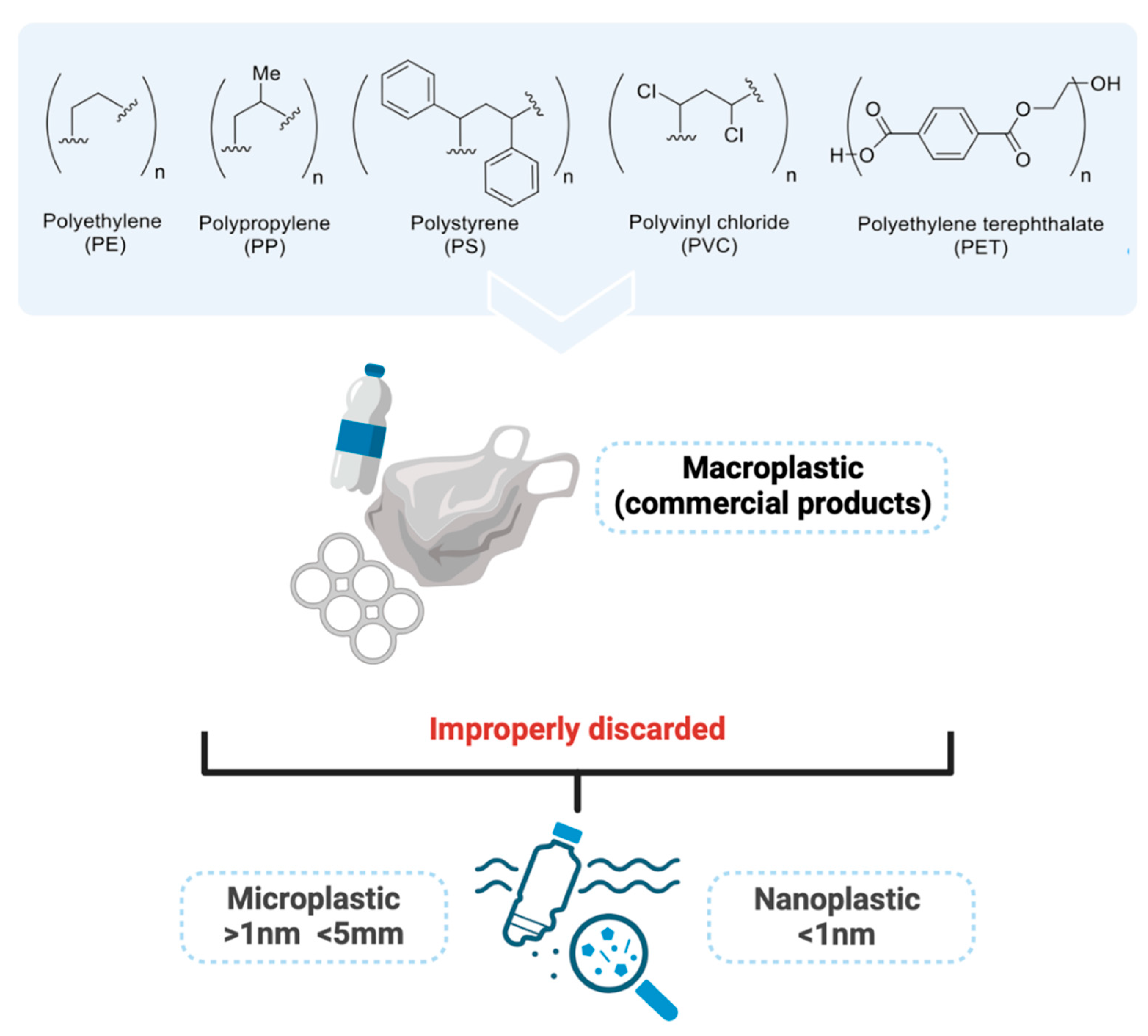
Figure 2.
MPs and NPs chain impact on human health.
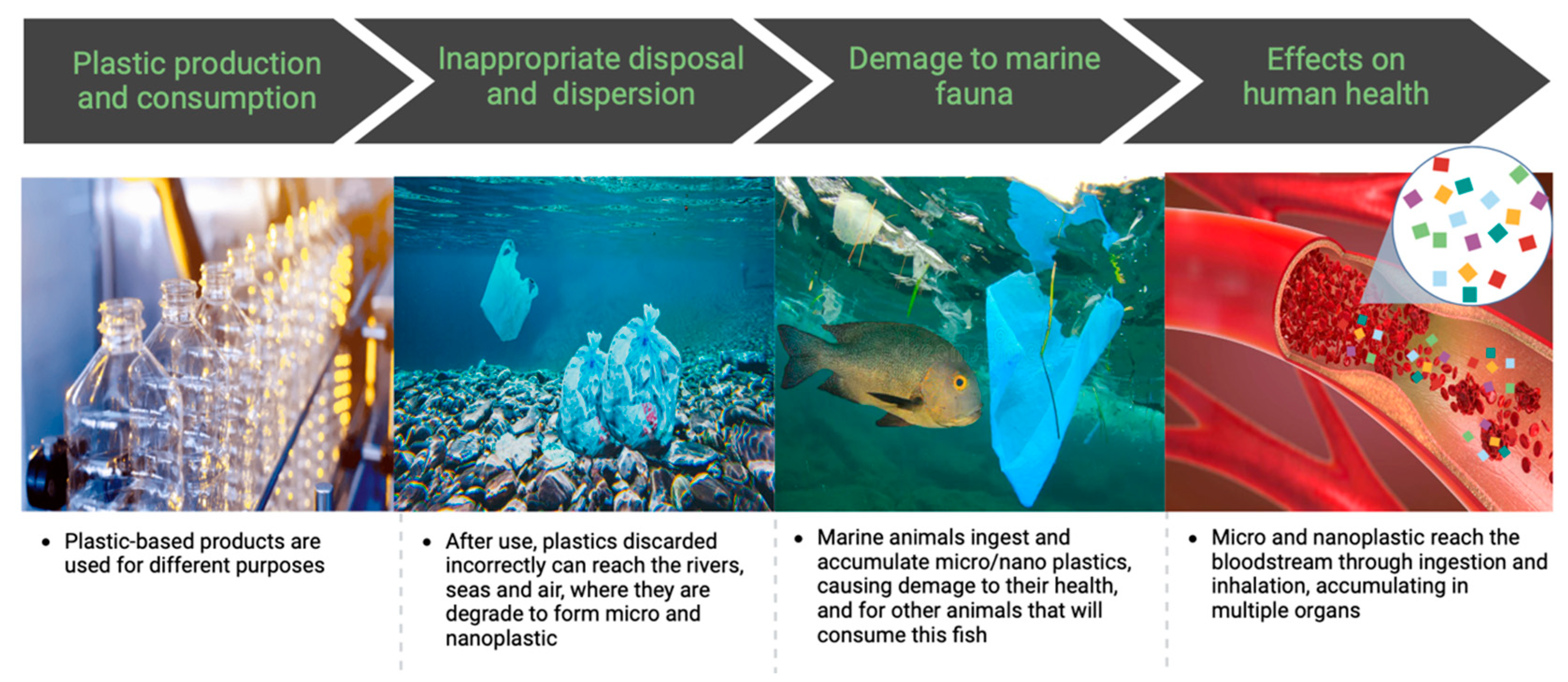
Figure 3.
MPs collected from the sands at Praia das Dunas in Cabo Frio, RJ, Brazil (22º53'37" S 42º01'31" W).
Figure 3.
MPs collected from the sands at Praia das Dunas in Cabo Frio, RJ, Brazil (22º53'37" S 42º01'31" W).
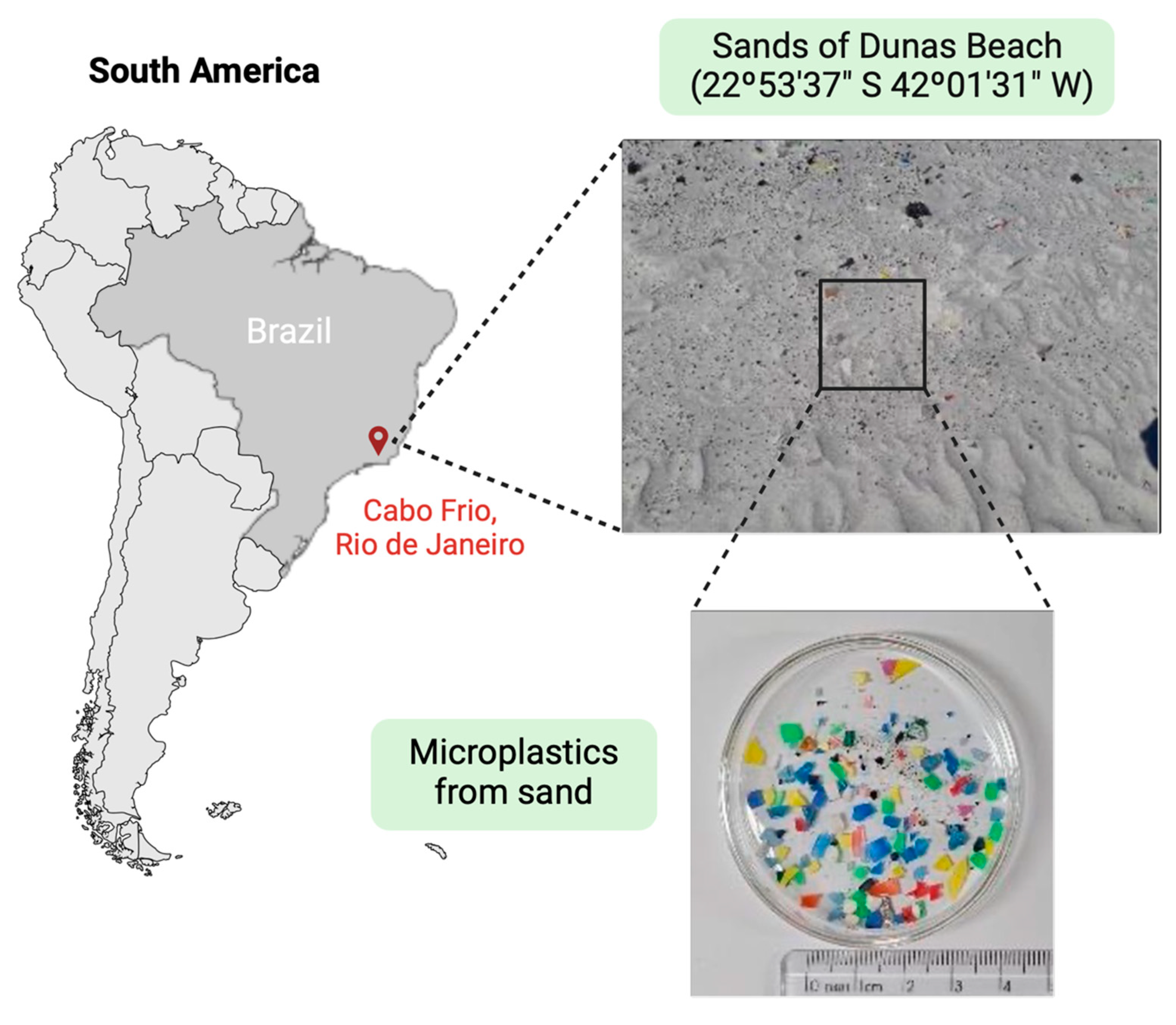
Figure 4.
Routes of human contamination by MPs and NPs.
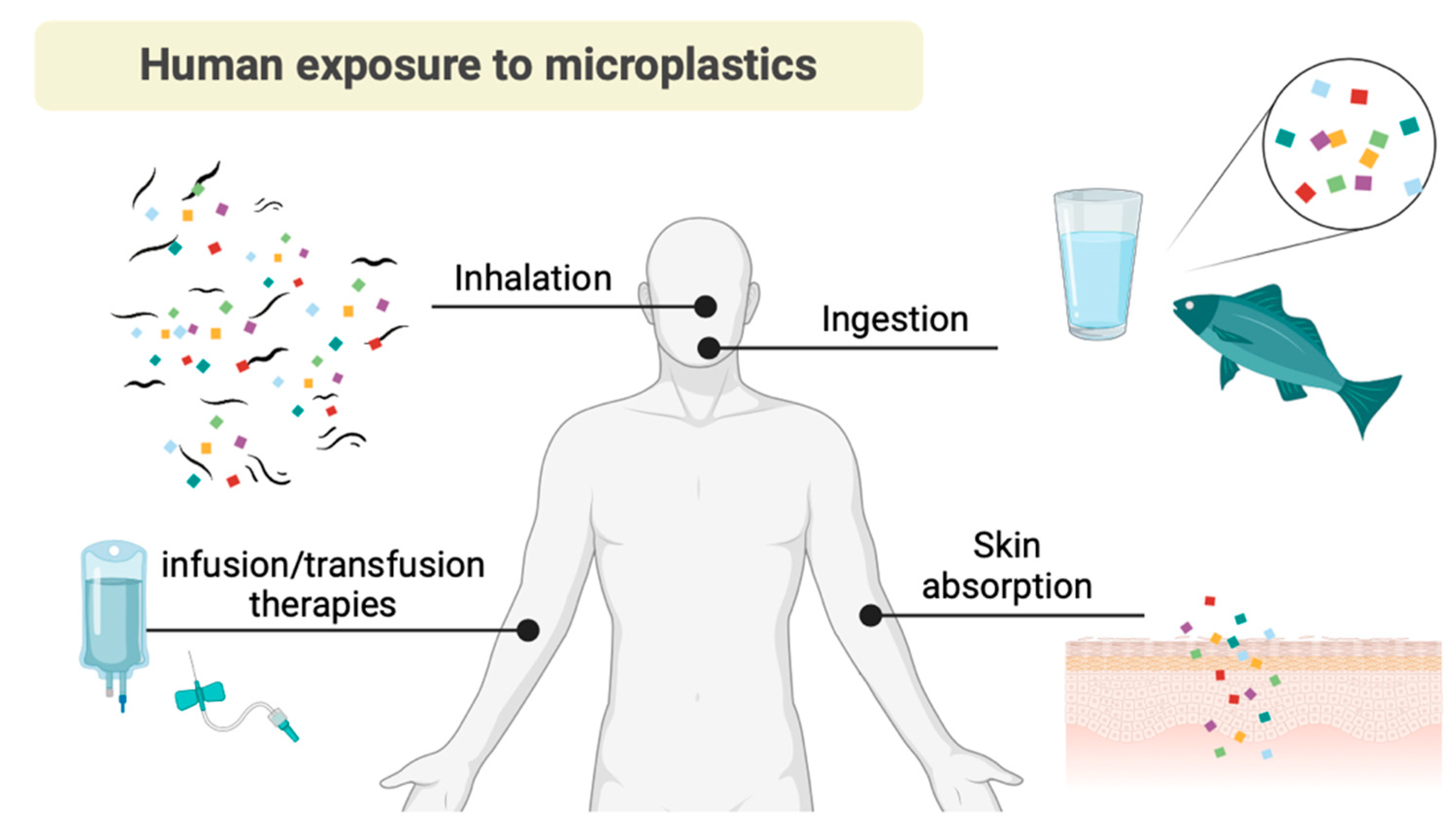
Figure 5.
Trajectory representation of the absorption of micro and nanoplastics through various entry points and distribution into human organs, systems and fluids before elimination.
Figure 5.
Trajectory representation of the absorption of micro and nanoplastics through various entry points and distribution into human organs, systems and fluids before elimination.
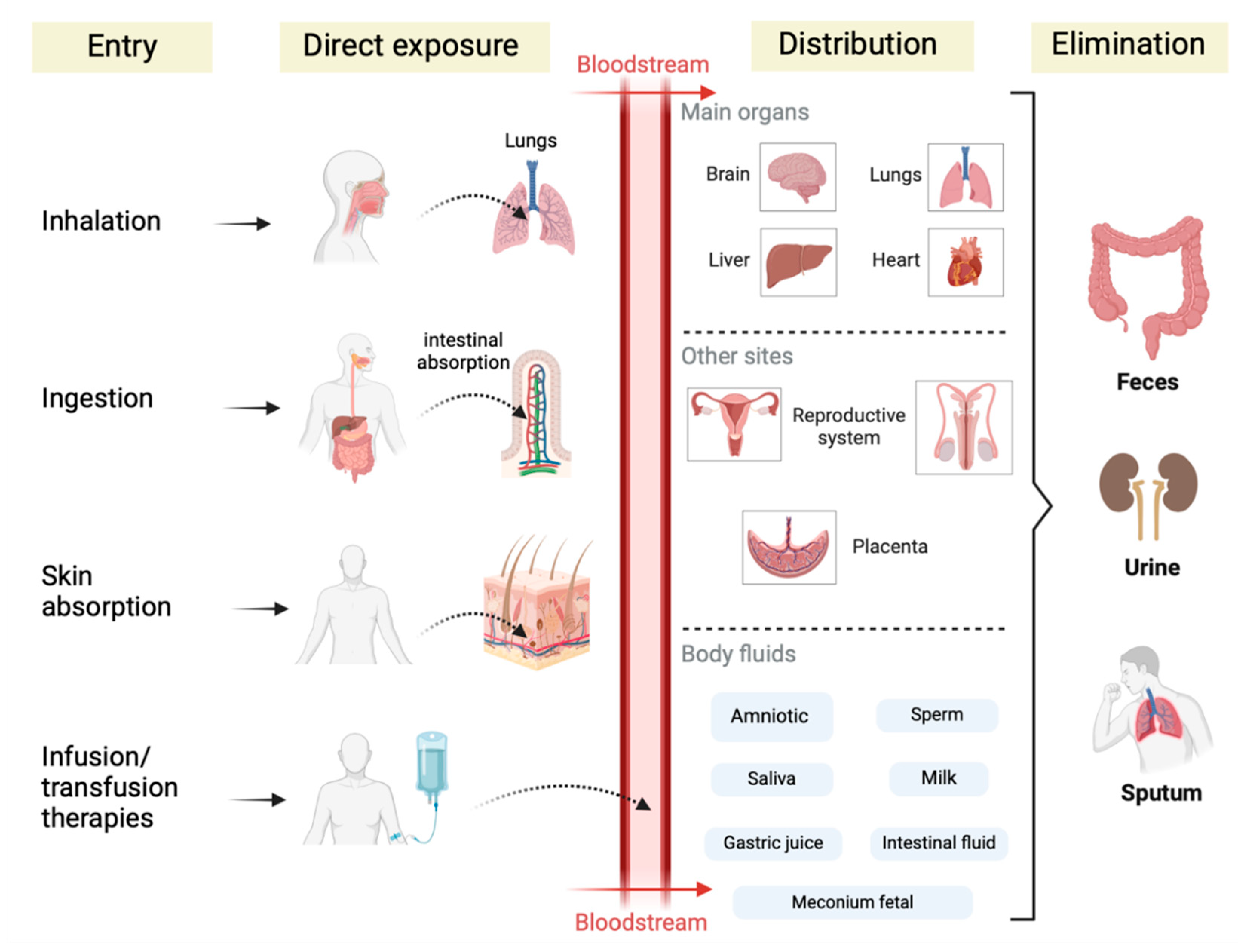
Figure 6.
Health damages that might be associated with micro and nanoplastic absorption and potential carcinogenic contributors.
Figure 6.
Health damages that might be associated with micro and nanoplastic absorption and potential carcinogenic contributors.

Disclaimer/Publisher’s Note: The statements, opinions and data contained in all publications are solely those of the individual author(s) and contributor(s) and not of MDPI and/or the editor(s). MDPI and/or the editor(s) disclaim responsibility for any injury to people or property resulting from any ideas, methods, instructions or products referred to in the content. |
© 2025 by the authors. Licensee MDPI, Basel, Switzerland. This article is an open access article distributed under the terms and conditions of the Creative Commons Attribution (CC BY) license (http://creativecommons.org/licenses/by/4.0/).
Copyright: This open access article is published under a Creative Commons CC BY 4.0 license, which permit the free download, distribution, and reuse, provided that the author and preprint are cited in any reuse.


