
Submitted:
06 June 2025
Posted:
06 June 2025
You are already at the latest version
Abstract
Abstract
The field of prosthetic dentistry has witnessed significant advancements in recent decades, largely driven by innovations in materials science. Among these innovations, nanoporous silica has emerged as a transformative material with the potential to revolutionize dental applications. This manuscript explores the multifaceted effects of nanoporous silica on prosthetic dental applications, focusing on its mechanical properties, biocompatibility, and its promising role in drug delivery systems. By examining these influences, we can gain insights into the future of dental prosthetics and the integration of innovative materials to enhance patient outcomes.
Keywords:
Nanoporous Silica
; Prosthetic Dentistry
; Biocompatibility
; Drug Delivery
1. Introduction
Prosthetic dentistry aims to restore both function and aesthetics for individuals with missing or damaged teeth [1]. Traditional materials, including metals, ceramics, and polymers, have been the mainstay in this field, yet they often present limitations in terms of mechanical strength, aesthetic appeal, and biocompatibility [2]. The introduction of nanoporous silica as a material for dental applications offers a promising solution to these challenges, paving the way for enhanced performance and patient satisfaction [3]. Nanoporous silica is characterized by its unique structure, featuring pores in the nanometer range (1 to 100 nm) [4]. This material exhibits exceptional physical and chemical properties, including a high surface area, tunable pore size, and enhanced mechanical strength [5]. The synthesis of nanoporous silica can be achieved through various methods, such as sol-gel processes [6], templating [7], and hydrothermal synthesis [8], each providing different advantages in controlling pore characteristics. The high surface area of nanoporous silica, often exceeding 1000 m²/g, facilitates substantial interactions with biological tissues and other materials, making it particularly advantageous in dental applications [9, 10]. This property enhances bonding and integration with surrounding tissues, which is crucial for the success of dental implants and other prosthetic devices. This manuscript aims to review the properties of nanoporous silica relevant to dental applications, discuss the impact of nanoporous silica on the mechanical properties of prosthetic materials, evaluate the biocompatibility of nanoporous silica in dental applications, explore the role of nanoporous silica in drug delivery systems for dental applications, and investigate future directions and potential applications of nanoporous silica in prosthetic dentistry.
2. Properties of Nanoporous Silica
2.1. High Surface Area and Porosity
Nanoporous silica typically possesses a high surface area, which enhances its interaction with biological tissues and other materials [11]. The porosity allows for the incorporation of bioactive agents, which can promote osseointegration and improve the overall success of dental implants [12]. The unique structure of nanoporous silica enables the formation of a three-dimensional network that facilitates nutrient and waste transport, critical in biological environments [13].
The tunable pore size and distribution can be tailored during synthesis, allowing for the design of materials that meet specific clinical needs [14]. Smaller pores may enhance the retention of bioactive molecules, while larger pores may facilitate cell migration and tissue integration [15]. This versatility makes nanoporous silica an attractive candidate for various dental applications, including implants, restorative materials, and drug delivery systems.
2.2. Mechanical Strength
Nanoporous silica exhibits excellent mechanical properties, including high tensile and compressive strength [16]. This strength is crucial for dental applications, where materials must withstand the forces of mastication and resist wear over time. The incorporation of nanoporous silica into dental composites can lead to a significant increase in their mechanical properties, making them more suitable for clinical use [17].
Research has demonstrated that the addition of nanoporous silica can enhance the modulus of elasticity and tensile strength of dental resins and ceramics [18]. For example, studies have shown that dental composites reinforced with nanoporous silica can achieve tensile strength increases of up to 30% compared to traditional materials [19, 20]. This enhancement is particularly beneficial for load-bearing applications, such as dental crowns and bridges, where mechanical performance is paramount.
2.3. Biocompatibility
The biocompatibility of nanoporous silica is a critical factor in its application in dentistry. Studies have shown that nanoporous silica can support cell adhesion and proliferation, making it an ideal candidate for use in dental implants and other prosthetic devices [21, 22]. The ability of nanoporous silica to interact positively with biological systems enhances its potential for successful integration into the oral environment [23]. Nanoporous silica's surface chemistry can be modified to further enhance its biocompatibility [24]. For instance, functionalizing the surface with bioactive molecules can promote specific cellular responses, such as osteogenic differentiation of stem cells [25]. This property is vital for the success of dental implants, as it promotes osseointegration and tissue integration [26]. Furthermore, the low cytotoxicity of nanoporous silica has been demonstrated in various in vitro and in vivo studies, indicating its safety for use in dental applications [27].
3. Impact on Mechanical Properties of Prosthetic Materials
3.1. Strength Enhancement
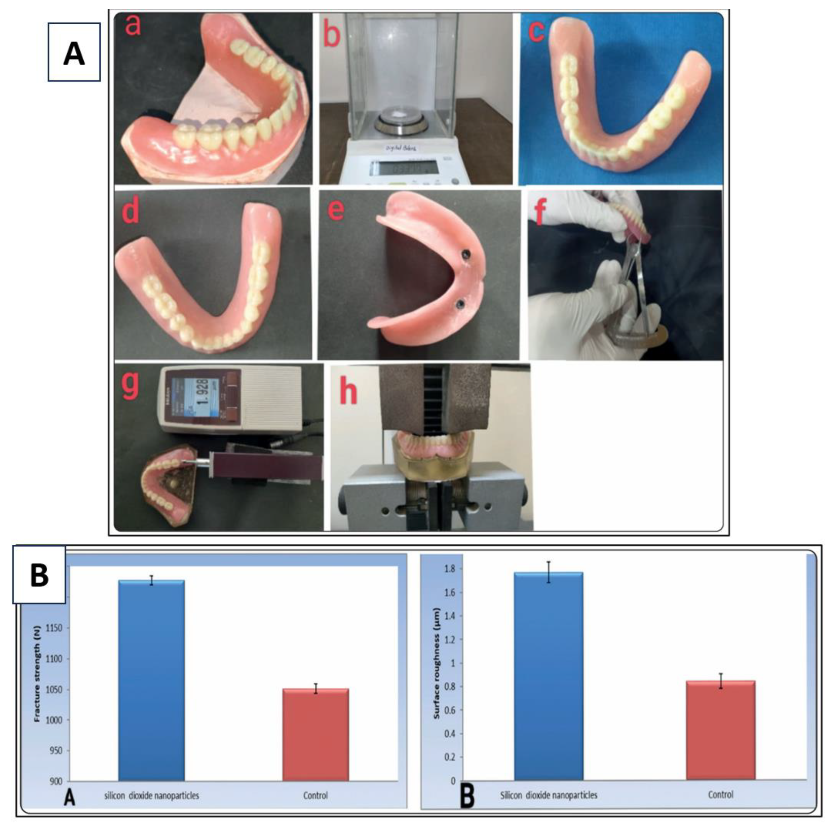
The incorporation of nanoporous silica into dental materials can significantly enhance their mechanical properties (Table 1). For instance, when added to polymer matrices, nanoporous silica can improve tensile strength and modulus, leading to more durable prosthetic restorations (Fig. 1) [28]. This enhancement is particularly beneficial for materials used in load-bearing applications, such as dental crowns and bridges. The mechanical enhancement is attributed to the reinforcement of the polymer matrix by the silica nanoparticles, which act as a barrier to crack propagation [29]. Recent studies have quantified the improvements in mechanical properties associated with nanoporous silica incorporation. For example, the tensile strength of a dental composite can increase by up to 30% with the addition of just 5% nanoporous silica by weight [30, 31]. This increase in strength translates to longer-lasting restorations that can withstand the rigors of everyday use.
3.2. Wear Resistance
Nanoporous silica has been shown to improve the wear resistance of dental materials [32]. This property is essential for prosthetic applications, as wear can lead to the degradation of the restoration and compromise its longevity [33]. By incorporating nanoporous silica, dental materials can achieve greater resistance to abrasion and wear, particularly in high-stress areas of the mouth [34]. The mechanisms behind the wear resistance of nanoporous silica include its ability to form a protective layer on the surface of dental materials and its role in enhancing the overall toughness of the composite. Studies have demonstrated that dental composites with nanoporous silica exhibit significantly lower wear rates compared to traditional materials, resulting in improved longevity and performance [35, 36].
3.3. Flexural Strength
Flexural strength is a critical property for dental materials, as it determines their ability to withstand bending forces. Nanoporous silica can enhance the flexural strength of ceramics and composites, making them more suitable for use in prosthetic applications where stress distribution is crucial [3]. The addition of nanoporous silica can lead to a more uniform stress distribution within the material, reducing the likelihood of failure [37]. Research has shown that the flexural strength of dental ceramics can increase by as much as 40% with the inclusion of nanoporous silica [38]. This improvement is particularly relevant for posterior restorations, where the forces exerted during chewing can be substantial. Enhanced flexural strength not only improves the durability of restorations but also contributes to patient satisfaction by reducing the risk of fracture [39].
3.4. Fatigue Resistance
In addition to static strength, the fatigue resistance of dental materials is vital for their long-term performance. Nanoporous silica can improve the fatigue life of dental composites by dissipating stress concentrations that can lead to crack initiation [40]. This property is particularly relevant for restorations subjected to cyclic loading, such as those found in posterior teeth. Studies have indicated that dental materials reinforced with nanoporous silica can withstand significantly more loading cycles before failure compared to traditional materials [17, 41]. This increased fatigue resistance is essential for ensuring the longevity of dental restorations, particularly in high-stress environments.
Figure 1.
A) Acrylic teeth on denture base wax, using silica nanoparticles overdenture, undergoing mechanical analysis to examine fracture strength and surface roughness, B) Fracture strength and surface roughness of dentures containing silica in comparison with control samples [42].
Figure 1.
A) Acrylic teeth on denture base wax, using silica nanoparticles overdenture, undergoing mechanical analysis to examine fracture strength and surface roughness, B) Fracture strength and surface roughness of dentures containing silica in comparison with control samples [42].
4. Biocompatibility of Nanoporous Silica
4.1. Cell Response
Biocompatibility is paramount in dental applications, as materials must interact favorably with surrounding tissues [47]. Research indicates that nanoporous silica supports the adhesion and proliferation of various cell types, including osteoblasts and fibroblasts [48-50]. This property is vital for the success of dental implants, as it promotes osseointegration and tissue integration. The high surface area of nanoporous silica facilitates the adsorption of proteins, which can enhance cell attachment and proliferation [51]. In vitro studies have demonstrated that osteoblasts exhibit increased proliferation and differentiation on nanoporous silica surfaces compared to traditional materials [52]. This enhanced cellular response is crucial for the integration of dental implants, as it directly impacts the stability and success of the restoration.
4.2. Inflammatory Response
The inflammatory response to dental materials can significantly impact the success of prosthetic restorations. Studies have shown that nanoporous silica exhibits minimal cytotoxicity and a favorable inflammatory response, making it a suitable choice for dental applications [53, 54]. The low inflammatory response is attributed to the biocompatible nature of silica, which does not elicit a significant immune response. Animal studies have further confirmed the favorable inflammatory response associated with nanoporous silica [55]. Histological evaluations have shown minimal signs of inflammation and good tissue integration around implants made from nanoporous silica, indicating its potential for clinical use in dental applications [22].
4.3. Long-Term Stability
The long-term stability of dental materials is crucial for ensuring the success of prosthetic restorations. Nanoporous silica has demonstrated resistance to degradation in biological environments, which can contribute to the longevity of dental prostheses [56]. This stability is essential for preventing the release of harmful degradation products into the surrounding tissues. Studies have shown that nanoporous silica maintains its structural integrity and mechanical properties over extended periods in biological environments [57]. This long-term stability is particularly important for dental implants and other prosthetic devices that are expected to function effectively for many years.
4.4. Interaction with Biological Fluids
The interaction of nanoporous silica with biological fluids is another important aspect of its biocompatibility. Studies have shown that nanoporous silica can absorb and release ions and biomolecules, which may enhance its integration with biological tissues [58]. This property can be harnessed to deliver therapeutic agents, such as growth factors or antimicrobial agents, directly to the site of implantation. The ability of nanoporous silica to serve as a drug delivery system opens new avenues for improving dental implant success. By incorporating bioactive molecules into the nanoporous structure, it is possible to create implants that not only integrate with bone but also promote healing and reduce the risk of infection [59].
5. Role of Nanoporous Silica in Drug Delivery Systems
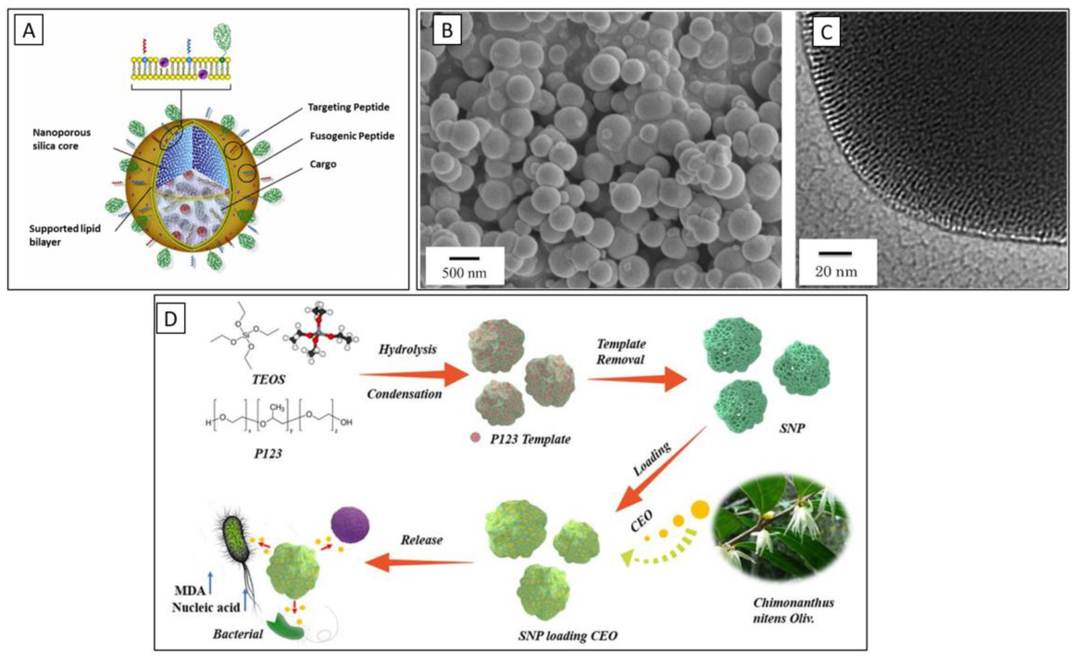
Nanoporous silica's unique structure and high surface area make it an ideal candidate for drug delivery applications in dentistry [59]. Its porous nature allows for the encapsulation of various therapeutic agents, including antibiotics, anti-inflammatory drugs, and growth factors, which can be released in a controlled manner [58]. This controlled release can enhance the therapeutic efficacy of the drugs while minimizing systemic side effects. The mechanisms of drug release from nanoporous silica can be influenced by several factors, including pore size, surface chemistry, and the nature of the encapsulated drug [60]. For instance, smaller pores may provide a sustained release profile, while larger pores can facilitate rapid drug release [61]. Additionally, surface modifications can be employed to tune the release kinetics, allowing for tailored therapeutic strategies (Fig. 2). The porous structure of silica nanoparticles provides a significant surface area for drug loading, enabling high encapsulation efficiency. This is crucial in dental applications where the dosage and localized delivery of drugs are essential for effective treatment. Smaller pores may provide a sustained release profile, allowing drugs to be released gradually over an extended period. This sustained release is particularly beneficial for maintaining therapeutic concentrations of antibiotics in the vicinity of dental implants or during periodontal treatments, thereby enhancing their efficacy and reducing the frequency of administration. Conversely, larger pores can facilitate rapid drug release, which may be advantageous in situations requiring immediate therapeutic action, such as in the case of analgesics for postoperative pain management. Surface chemistry also plays a vital role in the effectiveness of nanoporous silica as a drug delivery system. The chemical properties of the silica surface can be modified to enhance drug loading and control release kinetics. For instance, functionalizing the surface with different chemical groups can improve the interaction between the drug and the silica matrix, allowing for better encapsulation and controlled release profiles. Hydrophilic modifications can increase the wettability of the silica, promoting faster drug release, while hydrophobic modifications can slow down the release, providing a more sustained therapeutic effect. The specific characteristics of the drug itself, such as its molecular weight, solubility, and stability, also significantly impact how effectively it can be loaded into the nanoporous silica and how it will be released. Hydrophilic drugs may be better suited for smaller pores, while hydrophobic drugs may benefit from larger pore sizes and hydrophobic surface modifications. Surface modifications can be employed to tune the release kinetics, allowing for tailored therapeutic strategies. By adjusting the surface properties of nanoporous silica, researchers can create drug delivery systems that respond to specific physiological conditions, such as pH or temperature, thereby enhancing the precision of drug release in targeted areas. For example, pH-sensitive nanoporous silica can be designed to release drugs in response to the acidic environment of inflamed tissues, which is common in dental infections. This ensures that the drug is released precisely where it is needed most, maximizing therapeutic effects while minimizing side effects in other areas. Similarly, temperature-sensitive modifications can lead to the release of drugs in response to localized hyperthermia, which might occur during inflammation or infection. This responsiveness can be particularly useful in managing conditions like pulpitis or peri-implantitis, where localized treatment is crucial. The applications of nanoporous silica in drug delivery are vast and varied, making it a promising tool in modern dentistry. For instance, nanoporous silica can effectively deliver antibiotics directly to infected sites, such as periodontal pockets or around dental implants, significantly enhancing local drug concentrations and reducing systemic exposure. This targeted delivery can help combat antibiotic resistance by minimizing the need for systemic antibiotics. Additionally, controlled release of anti-inflammatory medications can provide prolonged relief from pain and swelling following dental procedures, improving patient comfort and recovery times. This is particularly useful in surgical contexts, such as tooth extractions or implant placements. Furthermore, the encapsulation and controlled release of growth factors can promote tissue regeneration and healing in dental applications. For example, delivering bone morphogenetic proteins (BMPs) in a controlled manner can enhance bone healing around dental implants or in grafting procedures, improving overall treatment outcomes. Incorporating local anesthetics into nanoporous silica matrices can also provide sustained pain relief during and after dental procedures, reducing the need for repeated injections and improving patient satisfaction. Despite the promising applications, there are challenges that need to be addressed for the successful implementation of nanoporous silica in drug delivery systems. Regulatory approval processes can be lengthy and complex, requiring thorough testing to ensure the safety and efficacy of nanoporous silica-based drug delivery systems in clinical settings. Additionally, developing scalable methods for producing nanoporous silica with consistent quality and performance is crucial for its widespread adoption in dental practice. Long-term stability of drug formulations within nanoporous silica also needs to be thoroughly investigated to ensure they maintain their efficacy until the point of use. In conclusion, nanoporous silica represents a versatile and innovative platform for drug delivery in dentistry, offering significant advantages in terms of controlled release, targeted therapy, and enhanced patient outcomes. Ongoing research and development in this field hold the potential to revolutionize dental treatments, making them more effective and patient-friendly.
Figure 2.
A) Schematic of nanoporous silica in drug delivery applications [62], B,C) SEM and TEM imaging of nanoporous silica used in nano medicine, showcasing particles with 200-300 nm in diameter [63], C) Chimonanthus nitens Oliv. essential oil loaded in sponge-like silica nanoporous particles [64].
6. Applications of Nanoporous Silica in Prosthetic Dentistry
6.1. Dental Implants
One of the most exciting uses of nanoporous silica in prosthetic dentistry is in the realm of dental implants. Thanks to its high surface area and excellent biocompatibility, nanoporous silica plays a crucial role in facilitating osseointegration—the process by which the implant fuses with the jawbone. This leads to improved stability and higher success rates for dental implants. Moreover, by integrating antimicrobial agents into the nanoporous silica matrix, we can effectively combat peri-implant infections, which are a common complication that can jeopardize the success of implants [22]. The localized release of these antimicrobial agents directly at the site of implantation ensures targeted therapy, reducing the need for systemic antibiotics and their associated side effects. Research has shown that dental implants coated with nanoporous silica infused with these agents experience significantly less bacterial colonization and demonstrate superior osseointegration compared to traditional implants [65]. The unique properties of nanoporous silica enhance the attachment of osteoblasts, the cells responsible for bone formation, which leads to faster and more robust integration with the surrounding bone [66]. Additionally, the ability to modify the surface characteristics of nanoporous silica allows for the inclusion of bioactive agents that further stimulate bone growth, making dental implants not only more effective but also more reliable [67]. (Fig. 3)
6.2. Crowns and Bridges
Nanoporous silica is making waves in the production of dental crowns and bridges, offering enhancements in both mechanical strength and aesthetic appeal. Its remarkable properties contribute to improved strength and wear resistance, making it an ideal choice for load-bearing applications in prosthetic dentistry. Furthermore, the incorporation of nanoporous silica enables the development of translucent materials that closely resemble the natural appearance of teeth [54]. Recent innovations in the fabrication of dental ceramics that include nanoporous silica have resulted in materials that not only boast enhanced mechanical properties but also exhibit superior aesthetic qualities [68]. These advanced materials can be tailored to match the shade and translucency of a patient’s natural teeth, ensuring that restorations blend seamlessly with their existing dentition. This ability to customize dental restorations enhances patient satisfaction and confidence in their smiles.
6.3. Dentures
Nanoporous silica also plays a significant role in the fabrication of dentures, providing benefits that enhance both comfort and functionality. Its lightweight nature, combined with favorable mechanical properties, contributes to the overall comfort of removable prostheses [69]. Additionally, the potential for bioactive coatings on dentures made from nanoporous silica can improve integration with oral tissues, promoting a more natural fit. Studies have demonstrated that dentures incorporating nanoporous silica exhibit enhanced mechanical properties, including increased strength and reduced wear over time [69]. Furthermore, the addition of bioactive agents within the denture material can promote tissue health, potentially reducing the risk of irritation or infection [70]. This means that patients can enjoy dentures that not only look good but also support their overall oral health.
6.4. Orthodontic Applications
In orthodontics, nanoporous silica can be used in the development of brackets and wires. The enhanced mechanical properties of nanoporous silica can lead to more durable orthodontic appliances, while its biocompatibility ensures a favorable interaction with oral tissues. Additionally, nanoporous silica can be utilized in the development of controlled-release systems for orthodontic adhesives, improving the bonding process [71]. Research indicates that orthodontic materials that incorporate nanoporous silica demonstrate superior mechanical characteristics, such as increased tensile strength and fatigue resistance [17]. These improvements can lead to more effective orthodontic treatments, providing better outcomes for patients who seek straighter teeth and improved bite alignment.
6.5. Bone Grafting Materials
Nanoporous silica is also emerging as a valuable scaffold material for bone grafting applications. Its porous structure facilitates the infiltration of bone cells, promoting the formation of new bone tissue. When combined with bioactive agents, nanoporous silica scaffolds can significantly enhance bone regeneration and healing, making it an excellent choice for dental and maxillofacial surgeries [72]. Studies have demonstrated that nanoporous silica scaffolds support the proliferation and differentiation of bone-forming cells, leading to improved bone regeneration in preclinical models [73]. This characteristic positions nanoporous silica as a promising option for applications in dental implantology and reconstructive surgery, where effective bone healing is crucial for successful outcomes.
6.6. Delivery of Growth Factors
Another innovative application of nanoporous silica lies in its ability to deliver growth factors, such as bone morphogenetic proteins (BMPs) or platelet-derived growth factor (PDGF), which are essential for promoting bone regeneration around dental implants [74]. The controlled release of these factors can significantly enhance osteogenesis, improving how well implants integrate with surrounding bone. Research has demonstrated that incorporating BMPs into nanoporous silica scaffolds can markedly boost bone formation in preclinical settings [75, 76]. This application holds great promise for enhancing the success of dental implants, particularly in patients who may have compromised bone quality, ultimately leading to better clinical outcomes.
6.7. Localized Pain Management
Beyond its regenerative and antimicrobial capabilities, nanoporous silica can also be utilized for localized pain management during dental procedures. By encapsulating analgesic agents within nanoporous silica, we can achieve sustained release at the surgical site, providing effective pain relief while minimizing systemic exposure to medications [77]. Ongoing research aims to explore the effectiveness of nanoporous silica in delivering analgesics and anti-inflammatory medications following dental treatments. This innovative approach could significantly enhance patient comfort and satisfaction throughout the recovery process, making dental visits more pleasant and less daunting for patients.
7. Future Directions
7.1. Research and Development
The journey into the world of nanoporous silica is just beginning, and ongoing research is vital to unlocking its full potential in prosthetic dentistry. Future investigations should aim to refine the processes used for synthesizing and functionalizing nanoporous silica, making it even more effective for dental applications. By exploring how different pore sizes and surface modifications impact its mechanical strength, biological compatibility, and drug delivery capabilities, researchers can gain crucial insights that will enhance its utility in dental care. Collaborative efforts among materials scientists, dental practitioners, and biomedical engineers are essential for bridging the gap between laboratory discoveries and real-world applications. This multidisciplinary approach can pave the way for groundbreaking dental materials that address the dynamic needs of both patients and dental professionals. (Fig. 4)
Figure 4.
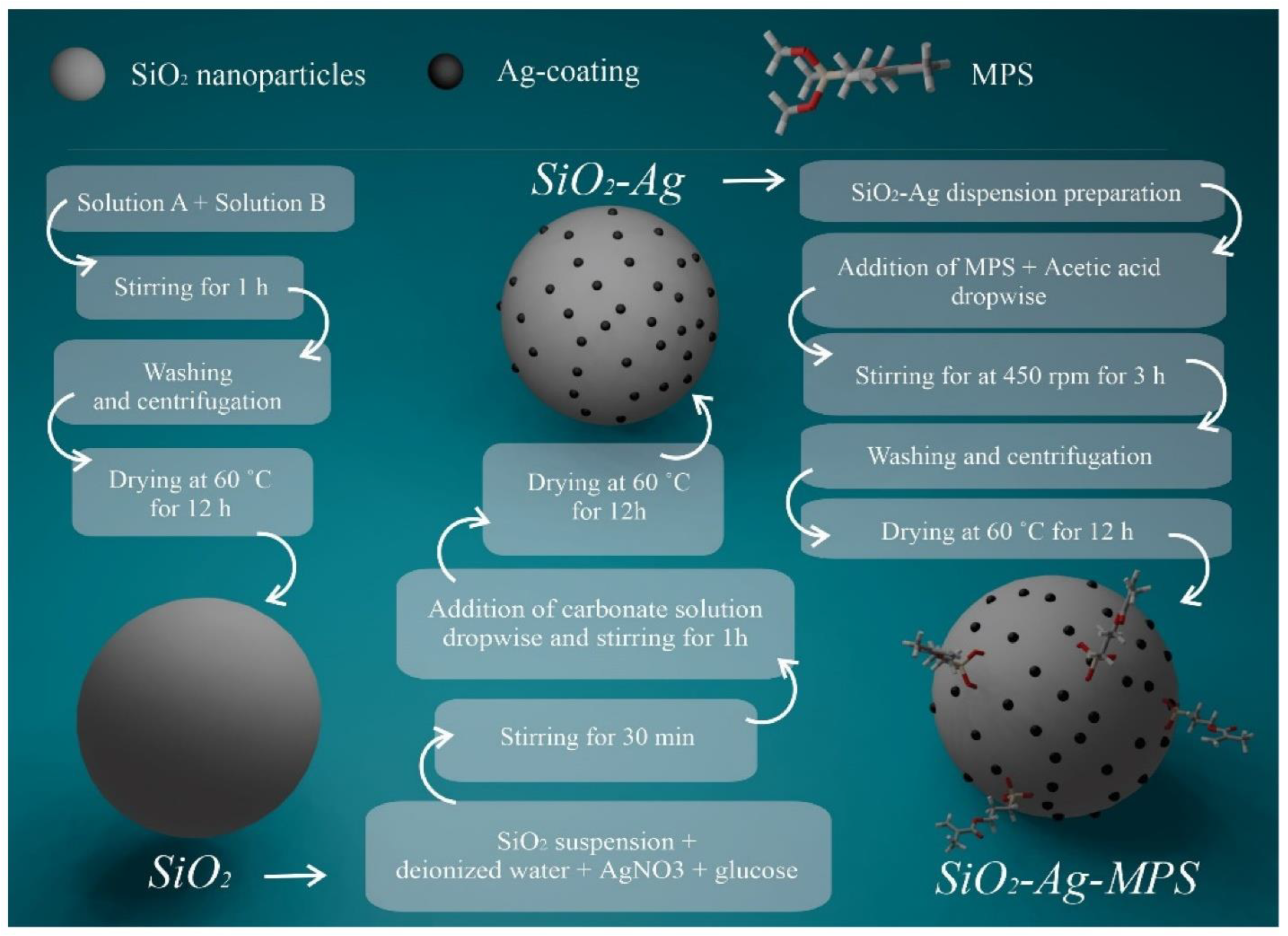
Silica nanoparticles ability to be elevated in dental prosthesis applications to enhance the required properties, using novel materials, such as metallic-based such as silver (Ag) and polymer-based such as methacryloxy propyl trimethoxy silane (MPS) [80].
Figure 4.
Silica nanoparticles ability to be elevated in dental prosthesis applications to enhance the required properties, using novel materials, such as metallic-based such as silver (Ag) and polymer-based such as methacryloxy propyl trimethoxy silane (MPS) [80].
7.2. Clinical Trials
To ensure that nanoporous silica-based dental materials are both effective and safe, rigorous clinical trials are imperative. These studies will shed light on the long-term performance and biocompatibility of these innovative materials in everyday dental practice. Establishing standardized testing protocols for assessing the biocompatibility and mechanical properties of nanoporous silica will be crucial for securing regulatory approval. Longitudinal studies that monitor the clinical outcomes of dental restorations utilizing nanoporous silica will play a key role in validating their efficacy and safety. Such research can help formulate best practices, guiding dental professionals in the optimal use of nanoporous silica across various applications.
7.3. Integration with Digital Dentistry
The fusion of nanoporous silica with cutting-edge digital dentistry technologies, such as CAD/CAM systems, holds the promise of revolutionizing prosthetic restorations. This synergy could lead to the creation of more precise and customized dental solutions, significantly enhancing patient satisfaction and treatment outcomes. The ability to design and fabricate tailored dental restorations using nanoporous silica not only improves the fit and function of prostheses but also streamlines the production process. Digital workflows can facilitate the creation of custom restorations with greater accuracy and efficiency, while the incorporation of nanoporous silica can further elevate both the mechanical durability and aesthetic appeal of the finished products.
7.4. Sustainability and Environmental Considerations
As the dental industry continues to evolve, it is increasingly important to prioritize sustainability in the materials we use. Research into eco-friendly methods for synthesizing nanoporous silica and the development of biodegradable composites can lead to more environmentally responsible dental practices. Additionally, investigating ways to recycle and repurpose dental materials can significantly mitigate the environmental footprint of dental care. Embracing sustainable practices not only benefits the planet but also enhances the reputation of dental practices among patients who value environmental stewardship. By implementing green initiatives, dental professionals can attract new patients and foster loyalty among their current clientele, creating a win-win situation for both the practice and the environment.
7.5. Educational Initiatives
For nanoporous silica to be successfully integrated into clinical practice, it is essential to educate dental professionals about its benefits and applications. Continuing education programs and hands-on workshops can empower practitioners with the knowledge they need to leverage nanoporous silica effectively in prosthetic dentistry. Collaboration among researchers, manufacturers, and dental professionals is key to translating cutting-edge research into practical applications. By cultivating a culture of continuous learning and innovation, dental professionals can stay abreast of the latest advancements in materials science and their implications for patient care. This ongoing education will ultimately lead to improved outcomes and enhanced experiences for patients seeking dental care.
8. Conclusions
Nanoporous silica represents a significant advancement in the field of prosthetic dentistry, offering unique properties that enhance the mechanical performance, biocompatibility, and drug delivery capabilities of dental materials. Its potential applications in dental implants, crowns, bridges, and drug delivery systems highlight its versatility and promise for the future of dental prosthetics. As research continues to explore the capabilities of nanoporous silica, it is likely to play an increasingly important role in improving patient outcomes and the overall success of dental restorations. The integration of nanoporous silica into dental materials not only enhances their mechanical properties and biocompatibility but also opens new avenues for innovation in dental practice. As the field of prosthetic dentistry continues to evolve, nanoporous silica will undoubtedly be at the forefront of material advancements, providing dental professionals with the tools necessary to deliver high-quality care to their patients.
References
- S. Safaee, H. Murata, The effect of incorporating nano-porous silica particles on the viscoelastic behavior of a commercial tissue conditioner and its long-lasting drug-release characteristics. Japan Prosthodontic Society 2024, 16, 385. [Google Scholar]
- Safaee, S. , et al., Multi-layered Coatings for Improving Implant Performance of Titanium. Ceramics International, 2025.
- Timpe, N. , et al., Nanoporous silica nanoparticles with spherical and anisotropic shape as fillers in dental composite materials. BioNanoMaterials 2014, 15, 89–99. [Google Scholar] [CrossRef]
- Jose, A. , et al. Porous inorganic nanomaterials: Their evolution towards hierarchical porous nanostructures. in Micro.
- Kankala, R.K. , et al., Nanoarchitectured structure and surface biofunctionality of mesoporous silica nanoparticles. Advanced materials 2020, 32, 1907035. [Google Scholar] [CrossRef] [PubMed]
- Singh, L.P. , et al., Sol-Gel processing of silica nanoparticles and their applications. Advances in colloid and interface science 2014, 214, 17–37. [Google Scholar] [CrossRef]
- Zhao, X. , et al. , Templating methods for preparation of porous structures. Journal of Materials Chemistry 2006, 16, 637–648. [Google Scholar]
- Yang, G. and S.-J. Park, Conventional and microwave hydrothermal synthesis and application of functional materials: A review. Materials 2019, 12, 1177. [Google Scholar] [CrossRef]
- SEHRAWAT¹, S. and M. AHUJA, ANISHA'ARYAN BOORA¹, BHAVNA ROHILLA¹. Nanomaterials: Synthesis and Applications 2024, 174.
- Hoskeri, J. , Mesoporous Materials for Tissue Engineering Applications. Materials Research Foundations. 173.
- Wang, S. , et al., Nanoporosity significantly enhances the biological performance of engineered glass tissue scaffolds. Tissue Engineering Part A 2013, 19, 1632–1640. [Google Scholar] [CrossRef]
- Zhang, C. , et al., Dental implants loaded with bioactive agents promote osseointegration in osteoporosis: a review. Frontiers in Bioengineering and Biotechnology 2021, 9, 591796. [Google Scholar]
- Chen, L. , et al., Silicon-containing nanomedicine and biomaterials: materials chemistry, multi-dimensional design, and biomedical application. Chemical Society Reviews 2024, 53, 1167–1315. [Google Scholar] [CrossRef]
- Yang, X.-Y. , et al., Hierarchically porous materials: synthesis strategies and structure design. Chemical Society Reviews 2017, 46, 481–558. [Google Scholar] [CrossRef]
- Bružauskaitė, I. , et al., Scaffolds and cells for tissue regeneration: different scaffold pore sizes—different cell effects. Cytotechnology 2016, 68, 355–369. [Google Scholar] [CrossRef] [PubMed]
- Rimsza, J. and J. Du, Structural and mechanical properties of nanoporous silica. Journal of the American Ceramic Society 2014, 97, 772–781. [Google Scholar] [CrossRef]
- Atai, M., A. Pahlavan, and N. Moin, Nano-porous thermally sintered nano silica as novel fillers for dental composites. Dental materials 2012, 28, 133–145. [Google Scholar] [CrossRef]
- Hata, K. , et al., Dental poly (methyl methacrylate)-based resin containing a nanoporous silica filler. Journal of functional biomaterials 2022, 13, 32. [Google Scholar] [CrossRef]
- Aminoroaya, A. , et al., Mesoporous silica aerogel reinforced dental composite: Effects of microstructure and surface modification. Journal of the Mechanical Behavior of Biomedical Materials 2022, 125, 104947. [Google Scholar] [CrossRef]
- Abid Althaqafi, K. , et al., Properties of a model self-healing microcapsule-based dental composite reinforced with silica nanoparticles. Journal of functional biomaterials 2022, 13, 19. [Google Scholar] [CrossRef]
- Inzunza, D. , et al., Synthesis of nanostructured porous silica coatings on titanium and their cell adhesive and osteogenic differentiation properties. Journal of Biomedical Materials Research Part A: An Official Journal of The Society for Biomaterials, The Japanese Society for Biomaterials, and The Australian Society for Biomaterials and the Korean Society for Biomaterials 2014, 102, 37–48. [Google Scholar] [CrossRef]
- Vandamme, K. , et al., Implant functionalization with mesoporous silica: A promising antibacterial strategy, but does such an implant osseointegrate? Clinical and Experimental Dental Research 2021, 7, 502–511. [Google Scholar] [CrossRef]
- Wang, X. , et al., Nano-porous silica aerogels as promising biomaterials for oral drug delivery of paclitaxel. Journal of Biomedical Nanotechnology 2019, 15, 1532–1545. [Google Scholar] [CrossRef]
- Shahbazi, M.-A. , et al., The mechanisms of surface chemistry effects of mesoporous silicon nanoparticles on immunotoxicity and biocompatibility. Biomaterials 2013, 34, 7776–7789. [Google Scholar] [CrossRef]
- Rana, D. , et al., Surface functionalization of nanobiomaterials for application in stem cell culture, tissue engineering, and regenerative medicine. Biotechnology progress 2016, 32, 554–567. [Google Scholar] [CrossRef] [PubMed]
- Anil, S. , et al. , Dental implant surface enhancement and osseointegration. Implant dentistry-a rapidly evolving practice 2011, 2011, 82–108. [Google Scholar]
- Attik, N. , et al., Mesoporous silica fillers and resin composition effect on dental composites cytocompatibility. Dental Materials 2017, 33, 166–174. [Google Scholar] [CrossRef]
- Topouzi, M. , et al. , Reinforcement of a PMMA resin for interim fixed prostheses with silica nanoparticles. Journal of the mechanical behavior of biomedical materials 2017, 69, 213–222. [Google Scholar]
- Su, C. , et al., Enhancement of mechanical behavior of FRP composites modified by silica nanoparticles. Construction and Building Materials 2020, 262, 120769. [Google Scholar] [CrossRef]
- Yang, Q. , et al., Characterization of mesoporous silica nanoparticle composites at low filler content. Journal of Composite Materials 2016, 50, 715–722. [Google Scholar] [CrossRef]
- Fonseca, R.B. , et al., Effect of short glass fiber/filler particle proportion on flexural and diametral tensile strength of a novel fiber-reinforced composite. Journal of prosthodontic research 2016, 60, 47–53. [Google Scholar] [CrossRef]
- Luo, J., J. J. Lannutti, and R.R. Seghi, Effect of filler porosity on the abrasion resistance of nanoporous silica gel/polymer composites. Dental Materials 1998, 14, 29–36. [Google Scholar] [CrossRef]
- Merola, M. and S. Affatato, Materials for hip prostheses: a review of wear and loading considerations. Materials 2019, 12, 495. [Google Scholar] [CrossRef]
- Alfaer, A.S. , et al., Wear Patterns in Modern Dental Materials Used in Extensive Tooth Replacement Treatments. 2025.
- Mirjalili, A., A. Zamanian, and S.M.M. Hadavi, The effect of TiO2 nanotubes reinforcement on the mechanical properties and wear resistance of silica micro-filled dental composites. Journal of Composite Materials 2019, 53, 3217–3228. [Google Scholar] [CrossRef]
- Chen, H. , et al., Micromechanical interlocking-inspired dendritic porous silica-based multimodal resin composites for the tooth restoration. Nano Research 2024, 17, 9065–9077. [Google Scholar] [CrossRef]
- Jin, H.-J., J. Weissmüller, and D. Farkas, Mechanical response of nanoporous metals: A story of size, surface stress, and severed struts. Mrs Bulletin 2018, 43, 35–42. [Google Scholar] [CrossRef]
- Zandinejad, A., M. Atai, and A. Pahlevan, The effect of ceramic and porous fillers on the mechanical properties of experimental dental composites. Dental Materials 2006, 22, 382–387. [Google Scholar] [CrossRef] [PubMed]
- Taha, N., J. Palamara, and H. Messer, Fracture strength and fracture patterns of root filled teeth restored with direct resin restorations. Journal of dentistry 2011, 39, 527–535. [Google Scholar] [CrossRef]
- Zamani, P., A. Jaamialahmadi, and L.F. Da Silva, The influence of GNP and nano-silica additives on fatigue life and crack initiation phase of Al-GFRP bonded lap joints subjected to four-point bending. Composites Part B: Engineering 2021, 207, 108589. [Google Scholar] [CrossRef]
- Zhang, J. , et al. , Antibacterial dental composites with chlorhexidine and mesoporous silica. Journal of dental research 2014, 93, 1283–1289. [Google Scholar]
- Abdelhamid, S.M. , et al., Influence of Reinforcement with Silicon Dioxide Nanoparticles on The Fracture Strength and Surface Roughness of Implant-Retained Mandibular Overdenture: An In Vitro Comparative Study. Egyptian Dental Journal 2024, 70, 1503–1513. [Google Scholar]
- Yang, D.-L. , et al., The properties of dental resin composites reinforced with silica colloidal nanoparticle clusters: Effects of heat treatment and filler composition. Composites Part B: Engineering 2020, 186, 107791. [Google Scholar] [CrossRef]
- Tah, R. , et al., Effect of zirconia silica nanofibers on flexural strength of feldspathic ceramic-An experimental study. Advanced Biomedical Research 2021, 10, 31. [Google Scholar] [CrossRef]
- Rezvani, M.B. , et al., The effect of silica nanoparticles on the mechanical properties of fiber-reinforced composite resins. Journal of Dental Research, Dental Clinics, Dental Prospects.
- Pourhajibagher, M. and A. Bahador, Effects of incorporation of nanoparticles into dental acrylic resins on antimicrobial and physico-mechanical properties: A meta-analysis of in vitro studies. Journal of Oral Biology and Craniofacial Research 2022, 12, 557–568. [Google Scholar] [CrossRef]
- Safaee, S. , et al., Fabrication of bioactive glass coating on pure titanium by sol-dip method: Dental applications. Dental Materials Journal 2021, 40, 949–956. [Google Scholar] [CrossRef] [PubMed]
- Bellino, M.G. , et al., Controlled adhesion and proliferation of a human osteoblastic cell line by tuning the nanoporosity of titania and silica coatings. Biomaterials Science 2013, 1, 186–189. [Google Scholar] [CrossRef] [PubMed]
- Malek, N.A.N.N., N. S. Sani, and A. Taufiq, Cell Adhesion and Proliferation of Silica Nanocomposite, in Biomedical Materials and Biofabrication for Regenerative Medicine. 2025, CRC Press. 166-180.
- Nesabi, M. , et al., A novel multi-structural reinforced treatment on Ti implant utilizing a combination of alkali solution and bioactive glass sol. Journal of the Mechanical Behavior of Biomedical Materials 2021, 124, 104837. [Google Scholar] [CrossRef] [PubMed]
- Saikia, J. , et al., Differential protein adsorption and cellular uptake of silica nanoparticles based on size and porosity. ACS applied materials & interfaces 2016, 8, 34820–34832. [Google Scholar]
- Ha, S.-W. , et al., Bioactive effects of silica nanoparticles on bone cells are size, surface, and composition dependent. Acta biomaterialia 2018, 82, 184–196. [Google Scholar] [CrossRef]
- Sivamaruthi, B.S. , et al., Mesoporous silica-based nanoplatforms are theranostic agents for the treatment of inflammatory disorders. Pharmaceutics 2023, 15, 439. [Google Scholar] [CrossRef]
- Chiang, Y.-C. , et al., A mesoporous silica biomaterial for dental biomimetic crystallization. ACS nano 2014, 8, 12502–12513. [Google Scholar] [CrossRef]
- Yu, Y. , et al., Short-term oral administration of mesoporous silica nanoparticles potentially induced colon inflammation in rats through alteration of gut microbiota. International Journal of Nanomedicine 2021, 881–893. [Google Scholar] [CrossRef]
- Pavanello, L. , et al., Physicochemical and biological properties of dental materials and formulations with silica nanoparticles: A narrative review. Dental Materials, 2024.
- Adiga, S.P. , et al. , Nanoporous membranes for medical and biological applications. Wiley Interdisciplinary Reviews: Nanomedicine and Nanobiotechnology 2009, 1, 568–581. [Google Scholar]
- Zhu, H., K. Zheng, and A.R. Boccaccini, Multi-functional silica-based mesoporous materials for simultaneous delivery of biologically active ions and therapeutic biomolecules. Acta Biomaterialia 2021, 129, 1–17. [Google Scholar] [CrossRef]
- Wang, G. , et al., Antibacterial peptides-loaded bioactive materials for the treatment of bone infection. Colloids and Surfaces B: Biointerfaces 2023, 225, 113255. [Google Scholar] [CrossRef] [PubMed]
- Jarvis, K.L., T. J. Barnes, and C.A. Prestidge, Surface chemistry of porous silicon and implications for drug encapsulation and delivery applications. Advances in Colloid and Interface Science 2012, 175, 25–38. [Google Scholar] [CrossRef] [PubMed]
- Maderuelo, C., A. Zarzuelo, and J.M. Lanao, Critical factors in the release of drugs from sustained release hydrophilic matrices. Journal of controlled release 2011, 154, 2–19. [Google Scholar] [CrossRef]
- Sun, J. , et al., Nanoporous silica-based protocells at multiple scales for designs of life and nanomedicine. Life 2015, 5, 214–229. [Google Scholar] [CrossRef]
- Nakamura, K. , et al., Calcium Charge and Release of Conventional Glass-Ionomer Cement Containing Nanoporous Silica. Materials 2018, 11, 1295. [Google Scholar] [CrossRef]
- Lai, H. , et al., Sponge-liked silica nanoporous particles for sustaining release and long-term antibacterial activity of natural essential oil. Molecules 2023, 28, 594. [Google Scholar] [CrossRef]
- Massa, M.A. , et al., Synthesis of new antibacterial composite coating for titanium based on highly ordered nanoporous silica and silver nanoparticles. Materials Science and Engineering: C 2014, 45, 146–153. [Google Scholar] [CrossRef]
- Wang, L. , et al., Osteoblast/bone-tissue responses to porous surface of polyetheretherketone–nanoporous lithium-doped magnesium silicate blends' integration with polyetheretherketone. International journal of nanomedicine 2019, 4975–4989. [Google Scholar] [CrossRef]
- Safaee, S. , et al., Osseointegration Dynamics: Insights into the Dental Bone-Implant Interface. The Journal of Applied Tissue Engineering.
- Pasha, M. , et al., Ceramic nanomaterials in dental applications. Nanoengineering of Biomaterials 2022, 123–144. [Google Scholar]
- Aati, S. , et al., Silver-loaded mesoporous silica nanoparticles enhanced the mechanical and antimicrobial properties of 3D printed denture base resin. Journal of the mechanical behavior of biomedical materials 2022, 134, 105421. [Google Scholar] [CrossRef]
- Al-Dulaijan, Y.A. and A.A. Balhaddad, Prospects on tuning bioactive and antimicrobial denture base resin materials: A narrative review. Polymers 2022, 15, 54. [Google Scholar] [CrossRef] [PubMed]
- Alkhazaleh, A.M. , Mechanical and chemical analysis of experimental adhesive containing drug-loaded mesoporous silica nanoparticles. 2022, The University of Iowa.
- Shadjou, N. and M. Hasanzadeh, Silica-based mesoporous nanobiomaterials as promoter of bone regeneration process. Journal of Biomedical Materials Research Part A 2015, 103, 3703–3716. [Google Scholar] [PubMed]
- Casarrubios, L. , et al., Silicon substituted hydroxyapatite/VEGF scaffolds stimulate bone regeneration in osteoporotic sheep. Acta biomaterialia 2020, 101, 544–553. [Google Scholar] [CrossRef] [PubMed]
- Satalov, A. , Nanoporous silica nanoparticles and bone morphogenetic protein 2 for bone regeneration. 2017.
- Neumann, A. , et al., BMP2-loaded nanoporous silica nanoparticles promote osteogenic differentiation of human mesenchymal stem cells. Rsc Advances 2013, 3, 24222–24230. [Google Scholar] [CrossRef]
- Zhang, Q. , et al., Porous nanofibrous scaffold incorporated with S1P loaded mesoporous silica nanoparticles and BMP-2 encapsulated PLGA microspheres for enhancing angiogenesis and osteogenesis. Journal of Materials Chemistry B 2018, 6, 6731–6743. [Google Scholar] [CrossRef]
- Wang, H. , et al., An injectable mesoporous silica-based analgesic delivery system prolongs the duration of sciatic nerve block in mice with minimal toxicity. Acta Biomaterialia 2021, 135, 638–649. [Google Scholar] [CrossRef]
- Covarrubias, C. , et al., Osseointegration properties of titanium dental implants modified with a nanostructured coating based on ordered porous silica and bioactive glass nanoparticles. Applied Surface Science 2016, 363, 286–295. [Google Scholar] [CrossRef]
- Fullriede, H. , et al., pH-responsive release of chlorhexidine from modified nanoporous silica nanoparticles for dental applications. BioNanoMaterials 2016, 17, 59–72. [Google Scholar] [CrossRef]
- Rossi, N.R. , et al., Silver-coated silica nanoparticles modified with MPS: potential antimicrobial biomaterials applied in glaze and soft reliner. Polymers 2022, 14, 4306. [Google Scholar] [CrossRef]

Table 1.
Effect of silica on the mechanical properties of biomaterials.
| Mechanical Properties | Material System Studied | Silica Details | Key Findings | References |
|---|---|---|---|---|
| Flexural Strength | Dental Resin Composite | Calcined Silica Colloidal Nanoparticle Clusters (CSCNCs) 60 wt% + SCNCs 10 wt% | Flexural strength: 143.5 ± 8.3 MPa (a 14% improvement compared to 70 wt% uncalcined SCNCs). Flexural Modulus: 8.91 ± 0.48 GPa (a 23% improvement). Calcination strengthened the nanoparticle clusters, leading to better reinforcement. | [43] |
| Flexural Strength | Feldspathic Ceramic | Zirconia-Silica Nanofibers | Incorporation of 5 wt% zirconia-silica nanofibers increased mean flexural strength from 141.08 ± 31.27 MPa (control) to 189.07 ± 5.52 MPa. With 7.5 wt%, it reached 196.71 ± 5.25 MPa. | [44] |
| Flexural Strength | Fiber-Reinforced Composite Resin (FRC) | Silica Nanoparticles (5 wt% in matrix resin) | For FRC with 2 fiber bundles, flexural strength increased from 64.2 ± 11.28 MPa (0 wt% silica) to 100.2 ± 18.41 MPa (5 wt% silica). Significant improvement was seen from 0.2 wt% onwards. | [45] |
| Flexural Strength & Hardness | Light Cured Dental Composite Resin | Silica Nanoparticles | 3 wt% silica significantly increased flexural strength (FS) and Vickers hardness (VH). FS (Control): approx. 85.78 MPa; FS (3% SiO₂): approx. 115.96 MPa. VH (Control): approx. 47.8 HV; VH (3% SiO₂): approx. 63.5 HV. | [46] |
| Compressive Strength | Dental Resin Composite | Calcined Silica Colloidal Nanoparticle Clusters (CSCNCs) 60 wt% + SCNCs 10 wt% | Compressive strength reached 260.3 ± 10.5 MPa. The study highlights how specifically engineered silica nanoparticle clusters can significantly bolster resin composites. | [43] |
| Wear Resistance | Light Cured Dental Composite Resin | Silica Nanoparticles (3 wt%) | Wear resistance improved; the increase in surface roughness after a brushing test was significantly less for the 3 wt% silica group (change of 0.14 µm) compared to the control group (change of 0.24 µm). This indicates a ~41.7% reduction in wear-induced surface roughness change. | [46] |
| Fatigue Resistance | Dental Resin Composites | Silica Colloidal Nanoparticle Clusters (SCNCs) | Enhanced static properties (like flexural strength) and improved structural integrity contribute to better fatigue life. | [43] |
Disclaimer/Publisher’s Note: The statements, opinions and data contained in all publications are solely those of the individual author(s) and contributor(s) and not of MDPI and/or the editor(s). MDPI and/or the editor(s) disclaim responsibility for any injury to people or property resulting from any ideas, methods, instructions or products referred to in the content. |
© 2025 by the authors. Licensee MDPI, Basel, Switzerland. This article is an open access article distributed under the terms and conditions of the Creative Commons Attribution (CC BY) license (http://creativecommons.org/licenses/by/4.0/).
Copyright: This open access article is published under a Creative Commons CC BY 4.0 license, which permit the free download, distribution, and reuse, provided that the author and preprint are cited in any reuse.


