Preprint
Article

This version is not peer-reviewed.

Effectiveness of Surgical Marginal Resection with Piezoelectric Device on Bisphosphonate-Related Osteonecrosis of the Jaws: A Retrospective Study

Submitted:

04 March 2025

Posted:

05 March 2025

You are already at the latest version

Abstract

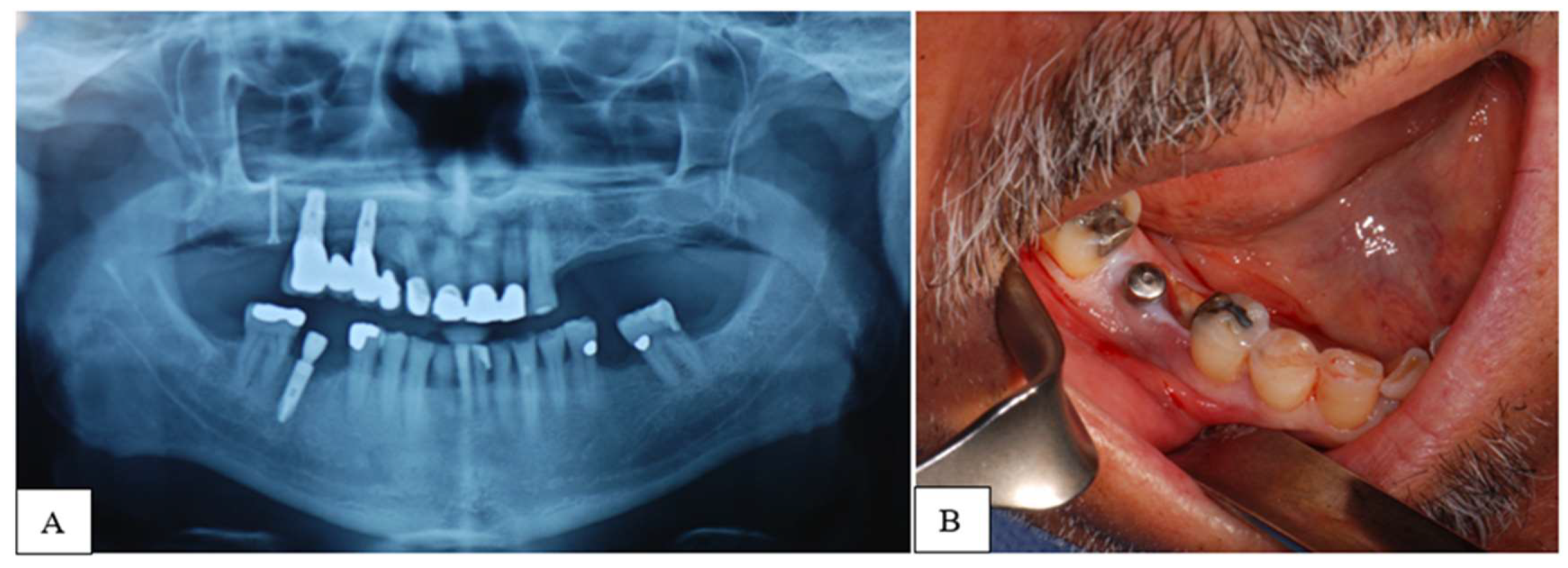
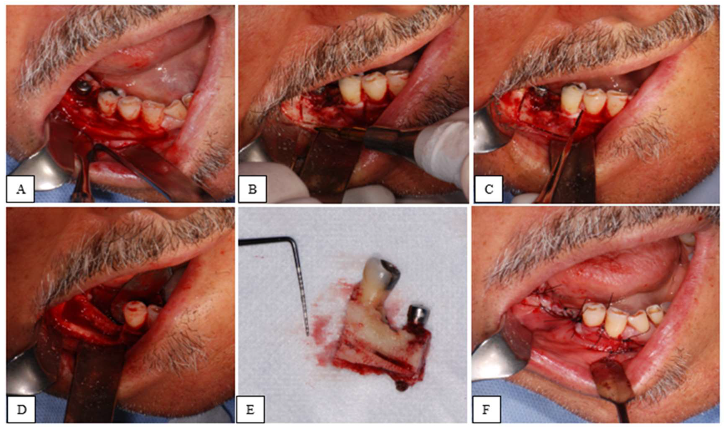
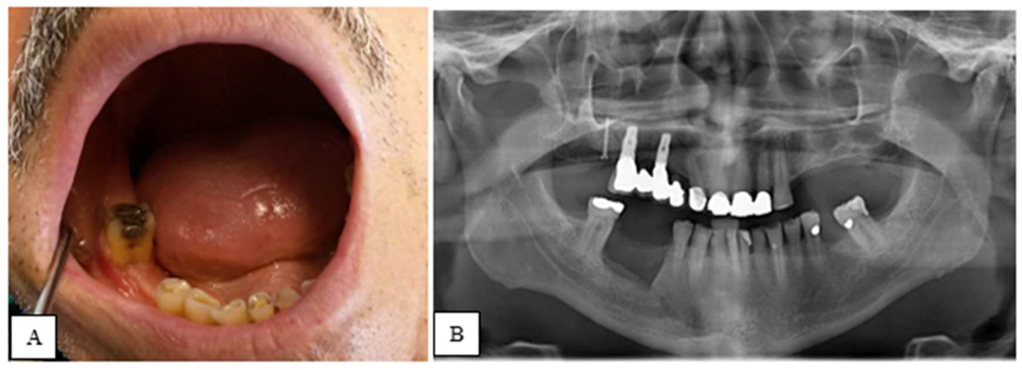
Objective: This study aims to investigate the efficacy of marginal resection using a piezoelectric device in patients with Bisphosphonate-Related Osteonecrosis of the Jaw (BRONJ). Methods: A retrospective study was conducted on subjects treated at the Dental Clinic University Hospital of Padua (Italy) from January 2017 to April 2024. Patients diagnosed with BRONJ (stages 1 and 2) who underwent marginal resection of the maxillae using a piezoelectric instrument were included. Exclusion criteria included patients who had received radiotherapy to the head and neck, those with Medication-Related Osteonecrosis of the Jaw (MRONJ), and those with primary tumors of the maxillary bones. Marginal resection was considered an effective treatment when complete epithelialization of the surgical site was achieved, with no signs or symptoms of disease, and the condition remained stable one-year post-operation. Results: 21 patients (17 females and 4 males) were selected. A single resection was performed for each patient, resulting in a total of 21 surgeries: 14 in the mandible and 7 in the maxilla. At one-year post-surgery, 20 patients showed no signs or symptoms of the disease. 1 patient experienced two recurrences, both of which were subsequently treated. Conclusions: The study demonstrates that marginal resection using a piezoelectric device is an effective procedure for the treatment of BRONJ, although it remains a relatively invasive and destructive therapeutic approach.

Keywords: 
;  ;  ;  ;  

1. Introduction

In 2020, the definition of Bisphosphonate-Related Osteonecrosis of the Jaws (BRONJ) was revised and redefined as an adverse drug-related reaction, characterized by the progressive destruction and necrosis of the mandibular and/or maxillary bone in individuals exposed to bisphosphate treatment, for which an increased risk of disease has been established, in the absence of prior head and neck radiotherapy. Along with this new definition, the terms “major” and “minor” diagnostic criteria for BRONJ diagnosis have been eliminated. The Italian Society of Oral Pathology and Medicine and the Italian Society of Maxillofacial Surgery (SIMPO-SICMF) have provided a list of clinical and radiographic signs and symptoms for the presentation of BRONJ, without distinguishing between major and minor criteria. There have also been changes in the recommended treatments: the surgical approach, previously reserved for advanced stages of the disease, is now indicated and extended to less severe stages of BRONJ. In this new context, the surgical marginal resection intervention performed using piezoelectric instruments in BRONJ patients is introduced [1]
Piezoelectric Devices in Medical-Dental Applications
Modern piezoelectric devices used in medical-dental fields are based on the so-called piezoelectric effect. The discovery of the piezoelectric effect dates to the 19th century, specifically in 1880, and is attributed to the insight of two physician brothers, Pierre and Jacques Curie. Following the Curie brothers’ discovery, physicist Gabriel Lippmann discovered the inverse piezoelectric effect. Since the early 2000s, the inverse piezoelectric effect has also found applications in the biomedical and healthcare fields with the patenting and widespread use of the first piezoelectric devices, commonly used today in dental surgery and other surgical specialties.
The piezoelectric effect occurs when mechanical deformation is applied to certain solid bodies with a crystalline structure. This force generates an electric charge, or more precisely, a potential difference called “piezoelectric current.” Conversely, the inverse piezoelectric effect is obtained by applying an electric field to a solid crystalline body, resulting in its mechanical deformation. The body, subjected to an electric current, expands and contracts, causing rapid dilation and contraction of the crystalline structure, which translates into vibration of the body itself.
Piezoelectric devices use materials with piezoelectric properties, specifically crystalline ceramics, which deform when exposed to an electric current. This deformation induces vibration, which is transferred via internal mechanisms to the working insert mounted on the piezoelectric device. Piezoelectric instruments and inserts operate at frequencies between 24 KHz and 40 KHz, hence they are also referred to as ultrasonic devices and inserts.
Ultrasonic piezoelectric devices are increasingly used in dental surgery due to their advantages over oral surgery performed with rotary tools and drills. The greatest advantage of ultrasound surgery is its selective cutting action on hard tissues such as bone. The use of ultrasonic inserts allows for osteotomy cuts on bone, reducing the risk of damage or iatrogenic injuries to sensitive structures such as the inferior alveolar nerve or soft tissues like the gingiva and Schneiderian membrane, in case of accidental contact. It can be used in several clinical situations, such as the accidental displacement of foreign bodies into the maxillary sinus, to gain access to teeth or alveolar bone lesions and to perform osteotomy in maxilla and mandible, such as in cases of bone lid surgery in posterior mandible [2]. Another characteristic of piezosurgery is its ability to create precise and thin osteotomy lines at the micron level, thanks to the microscopic vibration of the inserts. This is achieved without overheating the bone tissue. Additionally, piezoelectric devices have been shown to promote faster and better healing in terms of bone repair. These aspects have led to piezoelectric surgery being defined as atraumatic and minimally invasive. Regarding post-operative pain, it is generally lower than that associated with traditional surgery performed with rotary instruments [3,4].
Another advantage of piezoelectric surgery is related to cavitation effects: when the vibrating insert comes into contact with the physiological irrigation solution, micro-bubbles or gas cavities containing high-pressure steam are formed. These bubbles have a hemostatic effect because they implode, compressing blood capillaries and reducing bleeding, which improves intraoperative visibility for the surgeon. Additionally, the micro-bubbles have bactericidal effects and help remove bone debris formed on the bone surface [4]
Objective of the Study
This paper aims to evaluate the effectiveness of marginal surgical resection with piezoelectric devices, as part of minimally invasive ultrasonic surgery, in the treatment of bisphosphonate-related osteonecrosis of the jaws (BRONJ). Specifically, the surgical procedure will be assessed in terms of healing and recurrence, in line with the prognostic indices present in the clinical-therapeutic recommendations of the SIPMO-SICMF.

2. Materials and Methods

This retrospective study reviewed medical records and histological examinations of 274 potentially eligible patients treated at the Dental Clinic of Padova from January 2017 to April 2024. The study focused on a series of BRONJ cases that were unresponsive to medical therapy and underwent marginal surgical resection using piezoelectric devices.
Inclusion criteria – Inclusion criteria included adult patients with BRONJ stages 1 and 2, undergoing marginal resection with piezoelectric devices, patients followed by the same surgeon for both visits and surgery, availability of clinical documentation, and at least three follow-up visits at 1 month (T1), 6 months (T2), and 12 months (T3) post-surgery.
Exclusion criteria – Exclusion criteria included patients who had undergone head and neck radiotherapy, MRONJ patients, and those with primary or metastatic neoplasms of the maxillary bones.
For each patient, the following data were collected: gender, age, smoking status, comorbidities, BRONJ triggers, osteonecrosis resection site, bisphosphonate treatment indication, bisphosphonate type, and the presence of actinomyces in histological examinations.
In this study, we used the SIPMO SICMF staging, which differs in some details from the AAOMS staging system [5]. The corresponding stages are presented in Table 1 and Table 2. Presurgical (T0) clinical-radiographic signs and symptoms of BRONJ were recorded, as shown in Figure 1. From the Orthopantomography (OPG), Computed Tomography (CT) or Cone Beam Computed Tomography (CBCT), the dimensions in millimetres of the lesion to be excised were pre-defined.
The surgical procedures were performed in a sterile environment, under conscious sedation, and by the same lead surgeon. After local anesthesia (Articaine Hydrochloride and Bupivacaine Hydrochloride), a mucoperiosteal flap was raised to expose the necrotic tissue. The osteotomy lines were made using a piezoelectric device and occasionally completed with a manual chisel. Finally, the tissue was excised. Intra-operatively, the surgical margins were adjusted, if necessary, based on the consistency, color, and bleeding of the bone tissue. The closure was achieved by first intention using resorbable sutures. Histological examination was requested for all cases.
All surgical procedures are presented in Figure 2.
Specifically, the marginal bone resection was performed using a piezoelectric device (PIEZOSURGERY® touch, Mectron, Carasco, Genoa, Italy) set to bone mode with a high-frequency vibration of up to 36 kHz. The ultrasonic vibration tips required for the procedures were OT7, OT8L, OT8R, and OT12 for osteotomies, and OP1 for osteoplasty.
As shown in Figure 3, in the post-operative period, the clinical signs and symptoms of BRONJ were reassessed, while radiographic evaluation was performed only at T2 and T3 to minimize the patient’s exposure to X-rays. For 9 days (3 days before and 6 days after the procedure), dual antibiotic therapy was prescribed: Amoxicillin and Clavulanic Acid (3 g/day) and Metronidazole (750 mg/day). In the post-operative phase, analgesic therapy (Paracetamol 3 g/day and Ibuprofen 1800 mg/day) and antiseptic therapy (0.2% chlorhexidine spray) were recommended to reduce pain and the microbial load in the oral cavity.
Success Criteria
The surgical marginal resection with a piezoelectric device was considered effective upon achieving the following criteria 1 year after surgery: absence of exposed bone in the oral cavity, complete epithelialization of the surgical site, and no clinical or radiographic signs or symptoms of BRONJ.
The study was conducted in accordance with the ethical principles outlined in the Helsinki Declaration and adhered to the Strengthening the Reporting of Observational Studies in Epidemiology (STROBE) guidelines for reporting observational studies [6,7]. The project was registered with the local ethics committee. Informed consent was obtained from all patients.
Statistical Analysis
A descriptive statistical analysis was performed on the data. Qualitative variables were presented as absolute frequencies and percentages, while quantitative variables were analysed using the mean index. The data were analysed and recorded using Excel 18.0 - 2021.

3. Results

Characteristics of all clinical cases are presented in Table 3. Table 4 and Table 5 summarize preoperative clinical and radiographic signs and symptoms, based on SIPMO-SICMF staging criteria.
A total of 21 patients were selected: 17 women (80.95%) and 4 men (19.05%). The average age was 74.38 years (range 63-86). 9 patients (42.86%) were diagnosed with BRONJ stage 1, and 12 patients (57.14%) with BRONJ stage 2, based on the preoperative clinical and radiographic signs and symptoms and the SIPMO-SICMF staging criteria. Each stage was divided into asymptomatic (a) and symptomatic (b) cases. Regarding intra-oral triggers for osteonecrosis, the cause was not identified for 5 patients (23.81%). 7 patients (33.32%) were treated with bisphosphonates for osteometabolic reasons, mainly osteoporosis, and 14 patients (66.67%) for oncological reasons: 11 patients (52.38%) had bone metastases and 3 patients (14.29%) had Multiple Myeloma. Zoledronic acid (ZOL) had been prescribed for 15 patients (71.43%), Alendronate (ALE) for 4 patients (19.05%) and Ibandronate (IBA) for 2 patients (9.52%).
The most common comorbidities were hypertension, presented in 12 patients (57.14%), familial hypercholesterolemia presented in 5 patients (23.81%) and type II diabetes mellitus presented in 3 patients (14.29%). Other conditions, such as chronic kidney failure, were observed in less than 10% of the cases. 10 patients (47.62%) were smokers.
Each patient underwent a single marginal resection, with a total of 21 surgeries performed: 14 resections (66.67%) were carried out on the mandible, and 7 resections (33.33%) on the maxilla. All patients received histological confirmation of BRONJ, and in 9 samples (42.86%), actinomyces were found.
Table 6 presents clinical cases and corresponding clinical-radiographic signs and symptoms of BRONJ observed after piezoelectric marginal resection at follow-up (T1 - T2 - T3).
At the first follow-up (T1), 5 patients (23.81%) showed incomplete epithelialization of the surgical site, and other symptoms (trismus, lip dysesthesia) were present, although with reduced severity compared to the preoperative state. At 6 months post-resection (T2), in 1 patient (4.76%) (N°1), clinical and radiographic signs such as incomplete epithelialization, dysesthesia, and thickening of the trabecular bone were noted. At T3, the last follow-up, the same patient (N°1) showed worsening of the clinical picture with exposed bone in the oral cavity, lip dysesthesia, pain, and focal osteosclerosis. Pain was measured using the Visual Analogue Scale (VAS) and recorded a score of 4. This patient underwent two additional marginal resections using the piezoelectric device. For the following 3 years after the last resection, the patient was followed up biannually, with no further clinical or radiographic signs of BRONJ being observed.

4. Discussion

In this case series, the most frequent site of osteonecrosis was the mandible (66.67%). This result aligns with literature, where the mandible is reported to be more commonly affected by BRONJ compared to the maxilla. In one study, the mandible was affected in 70% of cases, and in another study, in 60% [8,9]. The most widely accepted hypothesis is that the mandible is more susceptible to osteonecrosis due to its terminal vascularization [10]. The average age of patients at the time of surgery was 74.38 years (range 63-86), indicating that BRONJ tends to manifest in the 60-80 age group. This finding is consistent with numerous other studies, where the mean age ranges from 60 to 77 years [11,12]. One systematic review reports a mean age of 66.5 ± 4.7 years [13]. While the majority of patients fall within this age range, the onset of the primary condition and the initiation of bisphosphonate therapy may play a role in the age of BRONJ manifestation, as some studies report a mean age of 55.4 years [14].
Women represent most patients (80.95%), which is consistent with the higher incidence of BRONJ in women. Some studies suggest a gender ratio of approximately 3:1. This could be attributed to women’s longer life expectancy, the rising incidence of breast cancer and the menopausal condition, which leads to a decrease in estrogen levels and consequently reduced bone mass. Regarding the primary condition for which bisphosphonates were prescribed, it can be said, in agreement with the international literature, that oncological patients develop BRONJ more frequently than osteometabolic patients [15]. The different potency of the drug, the route of administration, and the duration of bisphosphonate therapy all affect the outcomes. The role of periodontal disease as a risk factor for BRONJ, and more generally for inflammatory and infectious conditions in the oral cavity, has been extensively discussed [16]. Inflammation may induce bone necrosis both through the release of chemical mediators and via indirect action through edema, leading to reduced blood supply to the bone and subsequent necrosis [17].
Extraction surgery and implant therapy can connect the bone with the oral flora and induce trauma in tissues with altered metabolism and healing [18,19,20,21]. Poorly adjusted prosthetics also pose a local risk factor, as ill-fitting prostheses can exert excessive pressure, leading to thinning of soft tissues, ulceration, and bone exposure [22,23]. For 5 patients in this study (23.81%), no identifiable trigger for BRONJ was found. These cases may be due to anatomical predispositions or what is known as “spontaneous” drug-related ONJ, which may be associated with the patient’s pharmacogenetics, though there is insufficient evidence on this [24,25].
It is well-established that smoking causes vasoconstriction of blood vessels, leading to reduced blood flow to the bone and necrosis [26]. It also impairs wound healing, delaying the entire process [27]. In this study and in a similar paper, however, smoking did not seem to influence the surgical outcome, which might be due to patients either abstaining from smoking or reducing their daily cigarette consumption during the postoperative period [28].
In addition to the primary condition, comorbidities were observed, including hypertension, diabetes, and chronic kidney failure. The latter is considered a systemic risk factor for BRONJ, as excessive calcium excretion and inadequate renal reabsorption can disrupt calcium metabolism, also affecting the maxillary bones [29]. Regarding hypertension, some studies have identified a correlation between high blood pressure and increased BRONJ risk, though the exact causal relationship remains unclear [30]. For diabetes, the literature does not yet provide a clear pathophysiological mechanism linking it to BRONJ onset. However, some studies suggest that the microvascular damage caused by diabetes may also impact the bone tissue [31,32].
The histological confirmation of BRONJ was obtained for all cases, and the presence of actinomycetes was detected in the biopsy sample of 9 patients (42.86%), indicating superinfection of the necrotic bone.
The bacteria found in BRONJ are typically present in the oral cavity or are found in odontogenic and periodontal diseases. The most frequent pathogen in cases of bisphosphonate-induced osteonecrosis is, therefore, the actinomycete [33,34,35].
At T0, the clinical and radiographic manifestation of BRONJ in the selected cases aligns with what is reported in the literature: in no case were symptoms and/or signs found that differed from those already known. Pain, a frequent and debilitating component in BRONJ patients, was reported by approximately one-third of the patients (28.57%) [36] . The results indicate that, one year after surgery (T3), 95.24% of the treated cases meet the success criteria established by this study to evaluate the effectiveness of piezoelectric marginal resection in BRONJ patients: 20 patients, in fact, showed complete epithelialization of the surgical site and absence of clinical and radiographic signs and symptoms of the disease.
Similar case series show results comparable to those of this research. In these two studies, a total of 29 piezoelectric marginal resections were performed in BRONJ patients, both oncological and osteometabolic. These studies suggest that ultrasonic resection provides positive clinical outcomes in the treatment of bisphosphonate-induced osteonecrosis of the jaws [37,38]. Another case series monitored 6 BRONJ patients who underwent a single piezoelectric marginal resection for a period of 60 months, without observing signs or symptoms of the disease in the long term: 5 years after ultrasound surgery, no recurrences were reported [39].
At T3, only one patient (4.76%) presented, in addition to the persistence of the symptoms and signs recorded at T2, a worsening of the clinical condition. This clinical case was successfully re-treated with two additional piezoelectric marginal resections, and 4 years after the last surgery, no further signs or symptoms of BRONJ were observed.
While this paper analyses and describes an effective resective technique, the most effective approach to ONJ remains preventive (40).
A limitation of the research is represented by the sample size, which can be considered adequate in relation to study design but relatively small compared to larger cohorts of subjects. There are also limitations related to the evaluation of the study variables and the retrospective data collection. This case series was conducted in a single hospital facility, and the patients were selected only from those managed by a single medical team. Another limitation is the relatively short 12-month post-operative follow-up period.

5. Conclusions

This study demonstrates that piezoelectric marginal resection is effective in the treatment of BRONJ, although it remains an invasive procedure. Further studies are needed to include larger cohorts of patients. Additionally, there is hope for the future development of more efficient piezoelectric devices in order to establish this type of surgery as the “Gold Standard” for drug-related osteonecrosis.

Author Contributions

For research articles with several authors, a short paragraph specifying their individual contributions must be provided. The following statements should be used “Conceptualization, CB, MB; methodology, CB; validation, CB, CM; formal analysis, MT.; investigation, CM; resources, MT; data curation, CM.; writing—original draft preparation, MT.; writing—review and editing, CM; visualization, MB.; supervision, CB.; project administration, CBX.X.;. All authors have read and agreed to the published version of the manuscript.” Please turn to the CRediT taxonomy for the term explanation. Authorship must be limited to those who have contributed substantially to the work reported.

Funding

This research received no external funding

Institutional Review Board Statement

The study was conducted in accordance with the Declaration of Helsinki and approved by the Ethics Committee of Provincia di Padova, Code CESC 5647/A0/23 Code URC AOP2940 02/03/2023

Informed Consent Statement

Informed consent was obtained from all subjects involved in the study.

Conflicts of Interest

The authors declare no conflicts of interest.

Abbreviations

The following abbreviations are used in this manuscript:
OPG Orthopantomography
CBCT Cone Beam Computed Tomography
CT Computed Tomography
ZOL Zoledronic acid
ALE Alendronate
IBA Ibandronate

References

  1. Bedogni, A.; Mauceri, R.; Fusco, V.; Bertoldo, F.; Bettini, G.; Di Fede, O.; Lo Casto, A.; Marchetti, C.; Panzarella, V.; Saia, G.; Vescovi, P.; Campisi, G. (2024). Italian position paper (SIPMO-SICMF) on medication-related osteonecrosis of the jaw (MRONJ). In Oral Diseases (Vol. 30, pp. 3679–3709). John Wiley and Sons Inc. [CrossRef]
  2. Sivolella, S.; Brunello, G.; Berengo, M.; de Biagi, M.; Bacci, C. (2015). Rehabilitation With Implants After Bone Lid Surgery in the Posterior Mandible. Journal of Oral and Maxillofacial Surgery, 73, 1485–1492. [CrossRef]
  3. Rullo, R.; Piccirillo, A.; Femiano, F.; Nastri, L.; Festa, V. M. (2018). A Comparison between Piezoelectric Devices and Conventional Rotary Instruments in Bone Harvesting in Patients with Lip and Palate Cleft: A Retrospective Study with Clinical, Radiographical, and Histological Evaluation. BioMed Research International, 2018. [CrossRef]
  4. Labanca, M.; Azzola, F.; Vinci, R.; Rodella, L. F. (2008). Piezoelectric surgery: Twenty years of use. British Journal of Oral and Maxillofacial Surgery, 46, 265–269. [CrossRef]
  5. Ruggiero SL, Dodson TB, Aghaloo T, Carlson ER, Ward BB, Kademani D. (2020). American Association of Oral and Maxillofacial Surgeons’ Position Paper on Medication-Related Osteonecrosis of the Jaws-2022 Update. J Oral Maxillofac Surg. 2022 May;80(5):920-943. [CrossRef]
  6. World Medical Association declaration of Helsinki: Ethical principles for medical research involving human subjects. (2013). In JAMA (Vol. 310, pp. 2191–2194). American Medical Association. [CrossRef]
  7. Von Elm, E.; Altman, D. G.; Egger, M.; Pocock, S. J.; Gøtzsche, P. C.; Vandenbroucke, J. P. (2008). The Strengthening the Reporting of Observational Studies in Epidemiology (STROBE) statement: guidelines for reporting observational studies. Journal of Clinical Epidemiology, 61, 344–349. [CrossRef]
  8. Hoff, A. O.; Toth, B. B.; Altundag, K.; Johnson, M. M.; Warneke, C. L.; Hu, M.; Nooka, A.; Sayegh, G.; Guarneri, V.; Desrouleaux, K.; Cui, J.; Adamus, A.; Gagel, R. F.; Hortobagyi, G. N. (2008). Frequency and risk factors associated with osteonecrosis of the jaw in cancer patients treated with intravenous bisphosphonates. Journal of Bone and Mineral Research, 23, 826–836. [CrossRef]
  9. Estilo, C. L.; van Poznak, C. H.; Wiliams, T.; Bohle, G. C.; Lwin, P. T.; Zhou, Q.; Riedel, E. R.; Carlson, D. L.; Schoder, H.; Farooki, A.; Fornier, M.; Halpern, J. L.; Tunick, S. J.; Huryn, J. M. (2008). Osteonecrosis of the Maxilla and Mandible in Patients with Advanced Cancer Treated with Bisphosphonate Therapy. The Oncologist, 13, 911–920. [CrossRef]
  10. Eckert, A. W.; Maurer, P.; Meyer, L.; Kriwalsky, M. S.; Rohrberg, R.; Schneider, D.; Bilkenroth, U.; Schubert, J. (2007). Bisphosphonate-related jaw necrosis - Severe complication in Maxillofacial surgery. In Cancer Treatment Reviews (Vol. 33, pp. 58–63). [CrossRef]
  11. Thumbigere-Math, V.; Tu, L.; Huckabay, S.; Dudek, A. Z.; Lunos, S.; Basi, D. L.; Hughes, P. J.; Leach, J. W.; Swenson, K. K.; Gopalakrishnan, R. (2012). A retrospective study evaluating frequency and risk factors of osteonecrosis of the jaw in 576 cancer patients receiving intravenous bisphosphonates. American Journal of Clinical Oncology: Cancer Clinical Trials, 35, 386–392. [CrossRef]
  12. O’Ryan, F. S.; Lo, J. C. (2012). Bisphosphonate-related osteonecrosis of the jaw in patients with oral bisphosphonate exposure: Clinical course and outcomes. Journal of Oral and Maxillofacial Surgery, 70, 1844–1853. [CrossRef]
  13. Fliefel, R.; Tröltzsch, M.; Kühnisch, J.; Ehrenfeld, M.; Otto, S. (2015). Treatment strategies and outcomes of bisphosphonate-related osteonecrosis of the jaw (BRONJ) with characterization of patients: A systematic review. In International Journal of Oral and Maxillofacial Surgery (Vol. 44, pp. 568–585). Churchill Livingstone. [CrossRef]
  14. Atalay, B.; Yalcin, S.; Emes, Y.; Aktas, I.; Aybar, B.; Issever, H.; Mandel, N. M.; Cetin, O.; Oncu, B. (2011). Bisphosphonate-related osteonecrosis: Laser-assisted surgical treatment or conventional surgery? Lasers in Medical Science, 26, 815–823. [CrossRef]
  15. Vescovi, P.; Manfredi, M.; Merigo, E.; Meleti, M.; Guidotti, R.; Sarraj, A.; Mergoni, G.; Fornaini, C.; Bonanini, M.; Pizzi, S.; Rocca, J. P.; Nammour, S. (2012). Osteonecrosi dei mascellari e bisfosfonati: Terapia e follow-up a lungo termine in 160 pazienti. Dental Cadmos, 80, 9–21. [CrossRef]
  16. Thumbigere-Math, V.; Michalowicz, B. S.; Hodges, J. S.; Tsai, M. L.; Swenson, K. K.; Rockwell, L.; Gopalakrishnan, R. (2014). Periodontal Disease as a Risk Factor for Bisphosphonate-Related Osteonecrosis of the Jaw. Journal of Periodontology, 85, 226–233. [CrossRef]
  17. Lorenzo-Pouso, A. I.; Pérez-Sayáns, M.; Chamorro-Petronacci, C.; Gándara-Vila, P.; López-Jornet, P.; Carballo, J.; García-García, A. (2020). Association between periodontitis and medication-related osteonecrosis of the jaw: A systematic review and meta-analysis. In Journal of Oral Pathology and Medicine (Vol. 49, pp. 190–200). Blackwell Publishing Ltd. [CrossRef]
  18. Dioguardi, M.; di Cosola, M.; Copelli, C.; Cantore, S.; Quarta, C.; Nitsch, G.; Sovereto, D.; Spirito, F.; Caloro, G. A.; Cazzolla, A. P.; Aiuto, R.; Cascardi, E.; Greco Lucchina, A.; lo Muzio, L.; Ballini, A.; Mastrangelo, F. (2023). Oral bisphosphonate-induced osteonecrosis complications in patients undergoing tooth extraction: a systematic review and literature updates. European Review for Medical and Pharmacological Sciences, 27, 6359–6373. [CrossRef]
  19. Nisi, M.; la Ferla, F.; Karapetsa, D.; Gennai, S.; Miccoli, M.; Baggiani, A.; Graziani, F.; Gabriele, M. (2015). Risk factors influencing BRONJ staging in patients receiving intravenous bisphosphonates: A multivariate analysis. International Journal of Oral and Maxillofacial Surgery, 44, 586–591. [CrossRef]
  20. Bodem, J. P.; Kargus, S.; Eckstein, S.; Saure, D.; Engel, M.; Hoffmann, J.; Freudlsperger, C. (2015). Incidence of bisphosphonate-related osteonecrosis of the jaw in high-risk patients undergoing surgical tooth extraction. Journal of Cranio-Maxillofacial Surgery, 43, 510–514. [CrossRef]
  21. Holzinger, D.; Seemann, R.; Matoni, N.; Ewers, R.; Millesi, W.; Wutzl, A. (2014). Effect of dental implants on bisphosphonate-related osteonecrosis of the jaws. Journal of Oral and Maxillofacial Surgery, 72, 1937.e1-1937.e8. [CrossRef]
  22. Levin, L.; Laviv, A.; Schwartz-Arad, D. (2007). Denture-related osteonecrosis of the maxilla associated with oral bisphosphonate treatment. Journal of the American Dental Association, 138, 1218–1220. [CrossRef]
  23. Hasegawa, Y.; Kawabe, M.; Kimura, H.; Kurita, K.; Fukuta, J.; Urade, M. (2012). Influence of dentures in the initial occurrence site on the prognosis of bisphosphonate-related osteonecrosis of the jaws: A retrospective study. Oral Surgery, Oral Medicine, Oral Pathology and Oral Radiology, 114, 318–324. [CrossRef]
  24. Yang, Y.-L.; Xiang, Z.-J.; Yang, J.-H.; Wang, W.-J.; Xiang, R.-L. (2019). The incidence and relative risk of adverse events in patients treated with bisphosphonate therapy for breast cancer: a systematic review and meta-analysis. Therapeutic Advances in Medical Oncology, 11, 1758835919855235. [CrossRef]
  25. Fung, P.L.; Nicoletti, P.; Shen, Y.; Porter, S.; Fedele S (2015) Pharmacogenetics of Bisphosphonate-associated Osteonecrosis of the Jaw In Oral Maxillofacial Surgery Clinics of North America (Vol, 2.7.; pp 537–546), W.B.Fung, P. L.; Nicoletti, P.; Shen, Y.; Porter, S.; Fedele, S. (2015). Pharmacogenetics of Bisphosphonate-associated Osteonecrosis of the Jaw. In Oral and Maxillofacial Surgery Clinics of North America (Vol. 27, pp. 537–546). W.B. Saunders. [CrossRef]
  26. Apatzidou, D. A. (2022). The role of cigarette smoking in periodontal disease and treatment outcomes of dental implant therapy. In Periodontology 2000 (Vol. 90, pp. 45–61). John Wiley and Sons Inc. [CrossRef]
  27. Chambler, D.; Blincoe, T. (2018). Smoking and surgery. British Journal of Hospital Medicine (London, England: 2005), 79, 478. [CrossRef]
  28. Bacci, C.; Boccuto, M.; Cerrato, A.; Grigoletto, A.; Zanette, G.; Angelini, A.; Sbricoli, L. (2021). Safety and efficacy of sectorial resection with piezoelectric device in ONJ. [CrossRef]
  29. Shannon, J.; Shannon, J.; Modelevsky, S.; Grippo, A. A. (2011). Bisphosphonates and osteonecrosis of the jaw. Journal of the American Geriatrics Society, 59, 2350–2355. [CrossRef]
  30. Paek, S. J.; Park, W.-J.; Shin, H.-S.; Choi, M.-G.; Kwon, K.-H.; Choi, E. J. (2016). Diseases having an influence on inhibition of angiogenesis as risk factors of osteonecrosis of the jaw. Journal of the Korean Association of Oral and Maxillofacial Surgeons, 42, 271–277. [CrossRef]
  31. Molcho, S.; Peer, A.; Berg, T.; Futerman, B.; Khamaisi, M. (2013). Diabetes microvascular disease and the risk for bisphosphonate-related osteonecrosis of the jaw: A single center study. Journal of Clinical Endocrinology and Metabolism, 98. [CrossRef]
  32. Jarnbring, F.; Kashani, A.; Björk, A.; Hoffman, T.; Krawiec, K.; Ljungman, P.; Lund, B. (2015). Role of intravenous dosage regimens of bisphosphonates in relation to other aetiological factors in the development of osteonecrosis of the jaws in patients with myeloma. British Journal of Oral and Maxillofacial Surgery, 53, 1007–1011. [CrossRef]
  33. Anavi-Lev, K.; Anavi, Y.; Chaushu, G.; Alon, D. M.; Gal, G.; Kaplan, I. (2013). Bisphosphonate related osteonecrosis of the jaws: Clinico-pathological investigation and histomorphometric analysis. Oral Surgery, Oral Medicine, Oral Pathology and Oral Radiology, 115, 660–666. [CrossRef]
  34. Tiranathanagul, S.; Yongchaitrakul, T.; Pattamapun, K.; Pavasant, P. (2004). Actinobacillus actinomycetemcomitans Lipopolysaccharide Activates Matrix Metalloproteinase-2 and Increases Receptor Activator of Nuclear Factor-κB Ligand Expression in Human Periodontal Ligament Cells. Journal of Periodontology, 75, 1647–1654. [CrossRef]
  35. Jelin-Uhlig, S.; Weigel, M.; Ott, B.; Imirzalioglu, C.; Howaldt, H.-P.; Böttger, S.; Hain, T. (2024). Bisphosphonate-Related Osteonecrosis of the Jaw and Oral Microbiome: Clinical Risk Factors, Pathophysiology and Treatment Options. International Journal of Molecular Sciences, 25. [CrossRef]
  36. Miksad, R. A.; Lai, K.-C.; Dodson, T. B.; Woo, S.-B.; Treister, N. S.; Akinyemi, O.; Bihrle, M.; Maytal, G.; August, M.; Gazelle, G. S.; Swan, J. S. (2011). Quality of Life Implications of Bisphosphonate-Associated Osteonecrosis of the Jaw. The Oncologist, 16, 121–132. [CrossRef]
  37. Blus, C.; Szmukler-Moncler, S.; Giannelli, G.; Denotti, G.; Orrù, G. (2013). Use of Ultrasonic Bone Surgery (Piezosurgery) to Surgically Treat Bisphosphonate-Related Osteonecrosis of the Jaws (BRONJ). A Case Series Report with at Least 1 Year of Follow-Up. The Open Dentistry Journal, 7, 94–101. [CrossRef]
  38. Blus, C.; Giannelli, G.; Szmukler-Moncler, S.; Orru, G. (2017). Treatment of medication-related osteonecrosis of the jaws (MRONJ) with ultrasonic piezoelectric bone surgery. A case series of 20 treated sites. Oral and Maxillofacial Surgery, 21, 41–48. [CrossRef]
  39. Dipalma, G.; Inchingolo, A. M.; Malcangi, G.; Ferrara, I.; Viapiano, F.; Netti, A.; Patano, A.; Isacco, C. G.; Inchingolo, A. D.; Inchingolo, F. (2023). Sixty-Month Follow Up of Clinical MRONJ Cases Treated with CGF and Piezosurgery. Bioengineering (Basel, Switzerland), 10. [CrossRef]
  40. Bacci, C.; Cerrato, A.; Bardhi, E.; Frigo, A. C.; Djaballah, S. A.; Sivolella, S. (2022). A retrospective study on the incidence of medication-related osteonecrosis of the jaws (MRONJ) associated with different preventive dental care modalities. Supportive Care in Cancer, 30, 1723–1729. [CrossRef]
Figure 1. (A) Clinical case BRONJ stage 1b with osteonecrosis in the right mandible. Preoperative radiograph (T0). (B) Intra-oral clinical picture (T0) with distal bone exposure at second lower right premolar.
Preprints 151270 g001
Figure 2. (A) Full thickness surgical flap detachment; (B) Piezoelectric device osteotomy lines; (C) Completion of osteotomy; (D) Clinical picture after resection; (E) Tissue removed with second lower right premolar and implant seat in first lower right molar position; (F) Suture and closure by primary intention.
Preprints 151270 g002
Figure 3. (A) Clinical picture 12 months after surgery (T3); (B) Radiography 12 months after piezoelectric surgery (T3).
Preprints 151270 g003
Table 1. Clinical and Radiographic stages of MRONJ based on SIPMO-SICMF staging criteria [1].
STAGE CLINICAL SIGNS AND SYMPTOMS CT SIGNS
Stage 1 - FOCAL MRONJ
The presence of at least 1 clinical sign/symptom and increased bone density limited to the alveolar process at CT, with or without additional radiological signs.
Stage 1a: Asymptomatic (without pain)
Stage 1b: Symptomatic (the presence of pain and/or purulent discharge)

Abscess, bone exposure, halitosis, intraoral fistula, jaw pain of bone origin, mucosal inflammation, non-healing post-extraction socket, soft tissue swelling, spontaneous loss of bone fragments, sudden dental/implant mobility, purulent discharge, toothache and trismus.

Trabecular thickening and/or focal bone marrow sclerosis, with or without cortical erosion, osteolytic changes, thickening of the alveolar ridge, thickening of the lamina dura, persistent post-extraction socket, periodontal space widening, thickening of the inferior alveolar nerve canal, sequester formation.
Stage 2 - DIFFUSE MRONJ
The presence of at least 1 clinical sign/symptom and increased bone density extending to the basal bone at CT, with or without additional radiological signs.
Stage 2a: asymptomatic (without pain)
Stage 2b: symptomatic (presence of pain and/or purulent discharge)

Same as Stage 1, plus mandibular deformation and numbness of the lips.

Diffuse bone marrow sclerosis, with or without cortical erosion, osteolytic changes, thickening of the alveolar ridge, thickening of the lamina dura, persistent post-extraction socket, periodontal space widening, thickening of the inferior alveolar nerve canal, sequester formation, periosteal reaction and opacified maxillary sinus.
Stage 3 - COMPLICATED MRONJ
The presence of at least 1 clinical sign/symptom and increased bone density extended to the basal bone at CT, plus one or more of the following
Stage 3a: asymptomatic (without pain)
Stage 3b: symptomatic (presence of pain and/or purulent discharge)

Cutaneous fistula, mandible fracture, fluid discharge from the nose.

Osteosclerosis of adjacent bones (zygoma and hard palate), pathologic fracture, osteolysis extending to the maxillary sinus, sinus tract (oroantral, oronasal fistula, oro-cutaneous).
Table 2. Clinical and Radiographic stages of MRONJ based on AAOMS staging criteria [2].
STAGE SYMPOTMS CLINICAL FINDINGS RADIOGRAPHIC FINDINGS
Stage 0 Odontalgia not explained by an odontogenic cause.
Dull, aching bone pain in the jaw, which may radiate to the temporomandibular joint region.
Sinus pain, which may be associated with inflammation and thickening of the maxillary sinus wall.
Altered neurosensory function.
Loosening of teeth not explained by chronic periodontal disease.
Intraoral or extraoral swelling.
Alveolar bone loss or resorption not attributable to chronic periodontal disease.
Changes to trabecular pattern sclerotic bone and no new bone in extraction sockets.
Regions of osteosclerosis involving the alveolar bone and/or the surrounding basilar bone.
Thickening/obscuring of periodontal ligament (thickening of the lamina dura, sclerosis and decreased size of the periodontal ligament space)
Stage I Asymptomatic Exposed and necrotic bone or fistula that probes to the bone.
No evidence of infection/inflammation.
may present with radiographic findings mentioned for Stage 0 that are localized to the alveolar bone region
Stage II Symptomatic Exposed and necrotic bone, or fistula that probes to the bone.
Evidence of infection/inflammation.
may present with radiographic findings mentioned for Stage 0 localized to the alveolar bone region.


Stage III
Symptomatic Exposed and necrotic bone or fistulae that probes to the bone.
Evidence of infection
One or more of the following:
Exposed necrotic bone extending beyond the region of alveolar bone (i.e., inferior border and ramus in the mandible, maxillary sinus and zygoma in the maxilla).
Extraoral fistula.
Oral antral/oral-nasal communication.
May be present:
Pathologic fracture.
Osteolysis extending to the inferior border of the mandible or sinus floor
Table 3. Characteristics of clinical cases subjected to marginal surgical resection with piezoelectric device.
N° Patient Sex Age Primary Disease Comorbidities Smoking status BiP Treatment Triggering cause of BRONJ BRONJ Stage BRONJ Resection Site Presence of Actinomyces
1 M 66 Thyroid Cancer Hypertension yes ZOL Implant 1b Mandible (right side) yes
2 F 80 Osteoporosis Diabetes, Familial hypercholesterolemia no IBA Unknown 2a Mandible (right side) no
3 F 79 Osteoporosis Hypertension no IBA Unknown 2b Mandible (left side) no
4 F 63 Breast Cancer Hypertension yes ZOL Periodontal Disease 2b Mandible (right side) yes
5 F 81 Breast Cancer None no ZOL Extraction 2b Maxillary (left side) no
6 F 79 Osteoporosis Familial hypercholesterolemia yes ALE Extraction 1a Mandible (left side) no
7 F 77 Breast Cancer Hypertension yes ALE Prosthetic Decubitus 1a Mandible (left side) yes
8 M 72 Multiple Myeloma Hypertension yes ZOL Prosthetic Decubitus 2b Maxillary (left side) no
9 M 77 Thyroid Cancer Hypertension, Familial hypercholesterolemia yes ZOL Periodontal Disease 2a Maxillary (left side) no
10 F 68 Multiple Myeloma Hypertension,Familial hypercholesterolemia no ZOL Periodontal Disease 2a Mandible (left side) yes
11 F 68 Breast Cancer None no ZOL Unknown 1b Maxillary (right side) no
12 F 75 Multiple Myeloma Hypertension, Diabetes no ZOL Periodontal Disease 1b Mandible (right side) yes
13 F 67 Breast Cancer Hypertension no ZOL Extraction 2a Mandible (right side) yes
14 F 85 Breast Cancer Diabetes, Familial hypercholesterolemia no ZOL Periodontal Disease 2a Maxillary (left side) yes
15 F 73 Osteoporosis None no ALE Prosthetic Decubitus 1a Mandible (left side) no
16 M 69 Prostate Cancer Hypertension yes ZOL Periodontal Disease 1a Mandible (left side) no
17 F 65 Osteoporosis None yes ZOL Implants 2a Maxillary (left side) yes
18 F 83 Osteoporosis None yes ALE Unknown 1a Mandible (left side) no
19 F 86 Thyroid Cancer Chronic kidney failure no ZOL Extraction 2a Mandible (left side) no
20 F 63 Osteoporosis Hypertension no ZOL Unknown 1a Mandible (left side) no
21 F 86 Breast Cancer Hypertension yes ZOL Periodontal Disease 2b Maxillary (right side) yes
Table 4. Distribution of patients based on clinical signs and symptoms of BRONJ recorded preoperatively (T0).
Clinical Signs and Symptoms of BRONJ (T0) N° of Patients %
Exposed bone 21 100
Halitosis 11 52.38
Dental mobility 7 33.33
Pain 6 28.57
Trismus 5 23.81
Failure of post-extraction alveolar mucosa repair 4 19.05
Soft tissue swelling 3 14.29
Lip paresthesia/dysesthesia 3 14.29
Implant mobility 2 9.52
Suppuration 2 9.52
Table 5. Distribution of patients based on radiographic signs of BRONJ recorded preoperatively (T0).
Radiographic Signs of BRONJ (T0) N° of Patients %
Diffuse Osteosclerosis 6 28.57
Focal Medullary Osteosclerosis 5 23.81
Widening of the Periodontal Space 5 23.81
Persistence of Post-extraction Alveolus 4 19.05
Sinusitis 4 19.05
Thickening of the Alveolar Canal 2 9.52
Oro-antral Fistulas 2 9.52
Periosteal Reaction 2 9.52
Table 6. Clinical cases and corresponding clinical-radiographic signs and symptoms of BRONJ observed after piezoelectric marginal resection at follow-up (T1 - T2 - T3).
N° Patient T1 Clinical Signs and Symptoms of BRONJ T1 Radiographic Signs of BRONJ T2 Clinical Signs and Symptoms of BRONJ T2 Radiographic Signs of BRONJ T3 Clinical Signs and Symptoms of BRONJ T3 Radiographic Signs of BRONJ
1 Incomplete epithelialization, Dysesthesia X-rays not performed Incomplete epithelialization, Dysesthesia Trabecular thickening Exposed bone, Dysesthesia, Pain Focal medullary osteosclerosis
8 Incomplete epithelialization X-rays not performed Absent Absent Absent Absent
13 Incomplete epithelialization, Trismus X-rays not performed Absent Absent Absent Absent
14 Incomplete epithelialization X-rays not performed Absent Absent Absent Absent
19 Incomplete epithelialization, Lip Dysesthesia X-rays not performed Absent Absent Absent Absent
Disclaimer/Publisher’s Note: The statements, opinions and data contained in all publications are solely those of the individual author(s) and contributor(s) and not of MDPI and/or the editor(s). MDPI and/or the editor(s) disclaim responsibility for any injury to people or property resulting from any ideas, methods, instructions or products referred to in the content.
Copyright: This open access article is published under a Creative Commons CC BY 4.0 license, which permit the free download, distribution, and reuse, provided that the author and preprint are cited in any reuse.
Prerpints.org logo

Preprints.org is a free preprint server supported by MDPI in Basel, Switzerland.

Subscribe

Disclaimer

Terms of Use

Privacy Policy

Privacy Settings

© 2025 MDPI (Basel, Switzerland) unless otherwise stated