
Submitted:
06 June 2025
Posted:
09 June 2025
You are already at the latest version
Abstract
Temporomandibular joint (TMJ) dysfunction affects a significant portion of the population and can lead to pain and functional limitations, impacting quality of life. The etiology is multifactorial, requiring multifaceted therapeutic approaches. Nanotechnology, such as the Taopatch® device, offers promising support in pain relief and function improvement. This study evaluates the effect of Taopatch® on mouth opening and TMJ pain relief in both adults and children.
Keywords:
Pain
; pain relief
; TMJ
; maximum opening
; nanotechnology
; Taopatch
; placebo
1. Introduction
It can affect both children and adults and the therapeutic approach varies depending on the cause [3,4].
Temporomandibular joint pain is a multifactorial condition and the therapeutic approach must also be aimed at identifying the cause [11,12].
Treatment of temporomandibular joint pain depends on the symptoms, the treatment can be of different types: pharmacological therapy, physiotherapy, oral devices, surgery [13,14].
Pain and limitation when opening the mouth can be debilitating and negatively affect patients’ quality of life [10].
The limitation of mouth opening and the associated pain can be caused by various factors: temporomandibular dysfunction, inflammation, dental problems, muscle disorders [15,16].
Once the cause has been identified, treatment must be directed towards choosing the right therapy [17,18].
Although there is currently no sufficient scientific evidence to support the statement that nanotechnology is always effective in the treatment of pain and in limiting mouth opening, we can state that there are beginning to be studies aimed at verifying any therapeutic efficacy [19,20].
Among the most used therapeutic approaches in pain disorders, Taopach can help relieve pain, Taopatch® nanotechnology emits therapeutic light wavelengths that can have reduce pain [21,22].
The aim of study is to investigate the effect of the medical device Taopatch® in the daily orthodontic practice and their influence on the patient’s comfort as maximum opening of the mouth and pain relief in the area of the Temporo-mandibular joint (TMJ) and some of the masticatory muscles in both adults and children [23,24].
2. Treatment Procedure
The Taopatch® is a nanotechnological device in the form of a circular microchip measuring 16 mm in diameter and less than 1 mm in thickness.
It is applied directly to the skin using an adhesive patch and remains in place for the entire duration of the treatment.
The patch is coated with Mylar®, a hypoallergenic material, which encapsulates nanotechnological component specifically, quantum dots, a type of nanocrystal capable of emitting light at specific wavelengths when stimulated by light or electrical energy.
Mechanism of Action
The Taopatch® functions by converting photons produced by body heat into light at wavelengths similar to those used in Low-Level Laser Therapy (LLLT). This form of therapy uses red and near-infrared (NIR) light at low energy densities to trigger a photochemical reaction at the cellular level.
Exposure to this light increases the production of mitochondrial elements such as ATP, NADH, proteins, and RNA, and simultaneously enhances oxygen consumption. The primary chromophore responsible for this response is cytochrome C oxidase (CCO)—a key enzyme in the mitochondrial electron transport chain.
In normal conditions, CCO activity can be inhibited by nitric oxide (NO), produced by mitochondrial NO synthase. However, light at specific wavelengths causes photodissociation of NO from CCO, thereby restoring mitochondrial function and promoting ATP synthesis.
The quantum dots in the Taopatch® are activated by infrared photons generated from body heat. These are then converted into wavelengths typical of LLLT or Ultra-Low-Level Laser Therapy, such as 904 nm or 670 nm.
In clinical use, the device is typically positioned at two standard anatomical locations: posteriorly below the C7 vertebra and anteriorly on the xiphoid process. Additional devices may be applied to the side experiencing pain, either as a single patch or multiple, depending on the area involved. Each application lasts approximately two hours, corresponding to the average duration of a common painkiller’s effect, and follow-up continues over time.
3. Materials and Methods
In our research we decided to investigate the efficiency of a new nanotechnological device Taopatch®TMJ in the dental practice, evaluating the amount of maximum opening and the pain in the pterigoideus lateralis before using any applicator, using a placebo applicator and using the real Taopatch®.
We wanted to know also the level of stress in our 2 groups of patients – children and adults and found out that the adults suffer from almost twice more stress than the kids.
At the same time the pain during palpation without any patch in children was higher.
We find as an interesting result and might be explain with the fact that the kids may be more sensitive and are more tender for palpation or for them it is difficult to calculate the pain with the 0-to-10 scale.
On the other hand the children could determine their stress level with the same scale quite well, although some of them were wondering to answer to the question.
In our research we have examined a placebo group which was consistent of both kids and adults to find out if there is a statistically significant difference in the evaluated parameters using a tape with no applicator.
There were no changes found and makes the research quite valuable.
These results make us believe that Taopatch® is effective appliance with immediate effect on the body and patient’s well-being.
Were analayzed 149 patients (73 adults and 76 children) have been examined before (pre) and after (post) the application of Taopatch® type TMJ (Figure 1).
Mean age for the adult patients was 33.52±9.37-years-old and for the children was 10.62±2.49-years-old.
According to the gender the male adults were 27.4% from the group of adults and the female adults were 72.6%. In the group of children, the examined boys were 60,5%, while the girls were less – 39.5%.
A placebo group of 43 patients have been examined according to the same parameters.
Mean age 23.44±9.415 (from 7-years-old to 53-years-old).
From them male 34,9% and female 65,1%. The children were 18% of the patients in the placebo group. In this group we have attached in front of the tragus just a tape without the applicator.
The same tape that was used to attach Taopatch to the skin of the patient.
The pain was determined with a 0-to-10-scale where 0 is no pain and 10 is maximum pain.
The temporal tendon insertion on the medial aspect of the coronoid process was palpated with the index finger intraorally by one and the same operator both on the left and on the right side (Figure 2). The intraoral palpation was performed with the index finger behind the upper last molars, pressing upwards and inwards, laterally to the maxillary tuberosity and medial to coronoid process.
Although small, m. pterygoideus lateralis is believed to be the primary muscle affected in temporomandibular joint (TMJ) disturbances and that is why we decided to investigate the pain and discomfort in this muscle of mastication.
The maximum opening of the mouth was measured in mm by one and the same investigator using a metal measuring sliding caliper Dentaurum® (Figure 3).
Three measurements were done for each patient before and after the application of Taopatch®: maximum opening of the mouth, pain during palpation (on the right R and left L sides).
The applicator was placed in front of the tragus of the patient from both sides.
After applying the human upgrade device, the patient had a glass of water with approximately 150 ml and room temperature and walked 50 steps, moving energetically the head, the shoulders and the whole body.
The measurements of the investigated parameters were done immediately after walking which makes approximately 3 minutes after the application of the Taopatch® TMJ device.
One and the same investigator performed all palpations and measurements in order to reduce the risk of mistakes due to different strength for palpation.
The level of the life style stress in both groups was evaluated using the 0-to-10-scale where 0 is no stress and 10 is maximum stress.
3.1. Statistical Analysis
The statistical analysis included descriptive statistic, frequency analysis, the Independent Samples Test, the Paired samples Statistics and T-test. (Table 1, Table 2, Table 3 and Table 4) (Figure 5 and Figure 6).

Table 1.
Results before (pre) applying Taopatch TMJ and after (post) its application in adults.
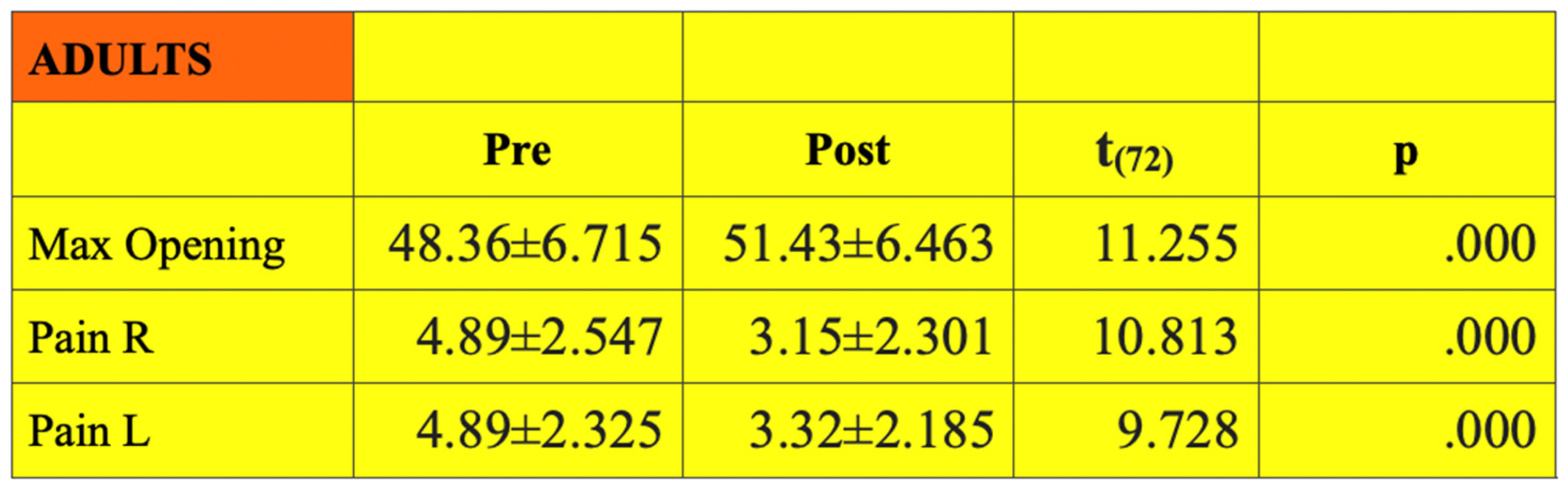 |
Table 2.
Results before (pre) applying Taopatch TMJ and after (post) its application in children.
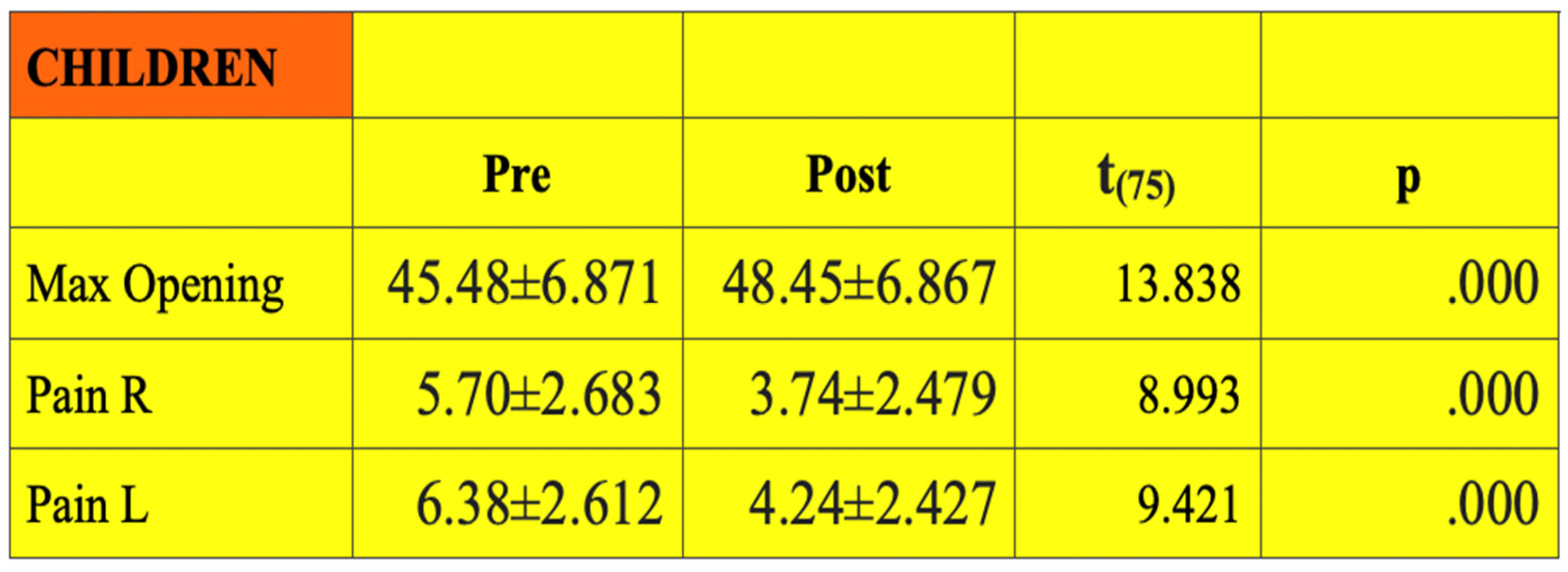 |
Table 3.
Paired Samples t-test for the placebo group – maximum mouth opening before and after the application of the placebo tape.
Table 3.
Paired Samples t-test for the placebo group – maximum mouth opening before and after the application of the placebo tape.
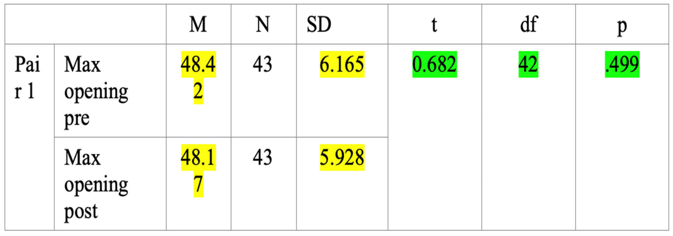 |
Table 4.
Paired Samples t-test for the placebo group - pain during palpation before and after the application of the placebo tape.
Table 4.
Paired Samples t-test for the placebo group - pain during palpation before and after the application of the placebo tape.
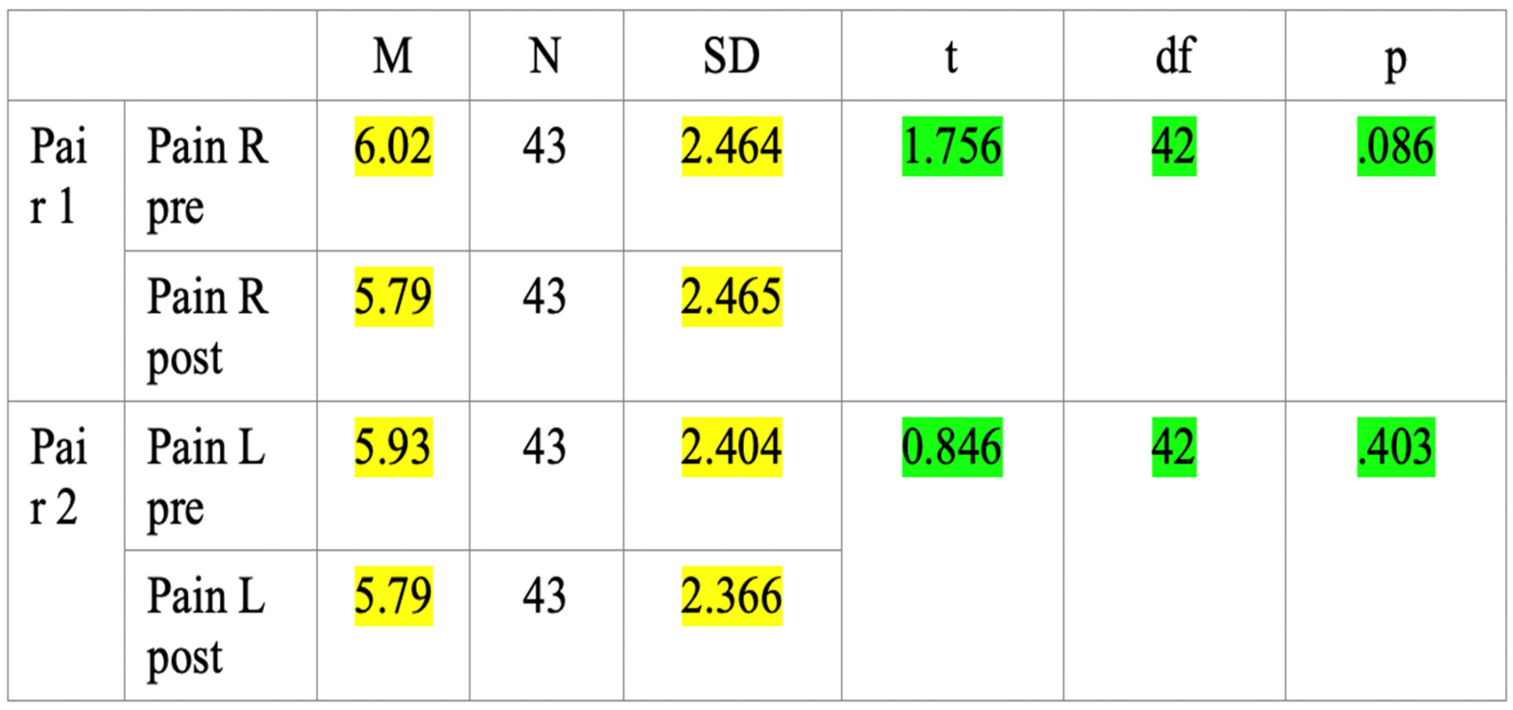 |
Table 5.
Improvement in the mouth opening and pain relief with Taopatch® TMJ in adults.
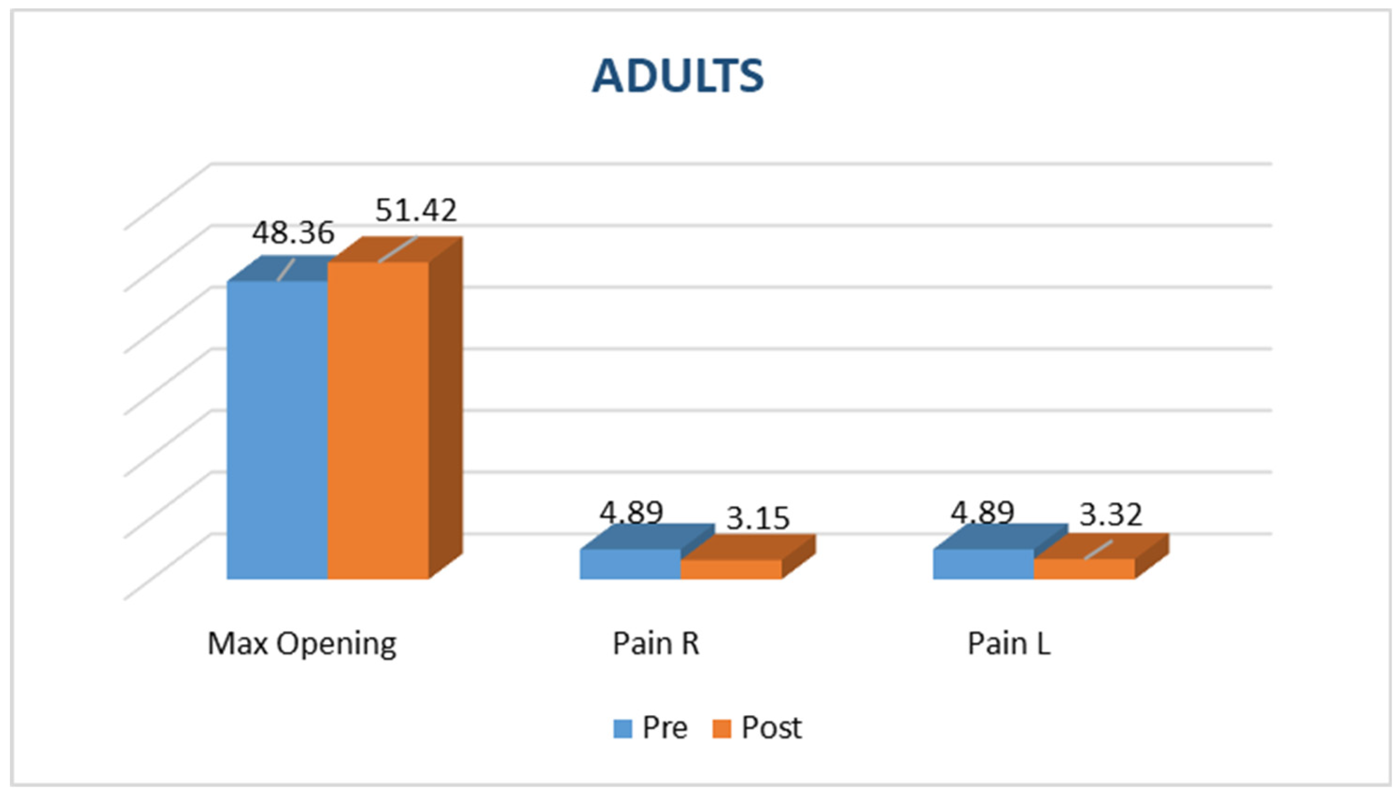 |
Table 6.
Improvement in the mouth opening and pain relief with Taopatch® TMJ in children.
 |
The Independent Samples Test, Levene’s Test for Equality of Variances, was used to find out statistically significant differences between the two groups of adults and children t(148)= 8.890, p= .000.
3.2. Results
1. All measured parameters have shown improvement after the application of the human upgrade device Taopatch® TMJ in all patients.
2. Both adults and children reacted positively to the therapy and the results were immediate.
3. In the group of adults the max opening has increased from 48.36±6.715 mm to 51.43±6.463 mm (t(72)=11.255, p=.000). The pain on the right side during palpation has reduced from 4.89±2.547 to 3.15±2.301. Statistically significant differences were found in the group of adults before and after the application of the nanotechnology t(72)=10.813, p=.000 for the right side and t(72)=9.728, p= .000 for the left side. (Figure 4)
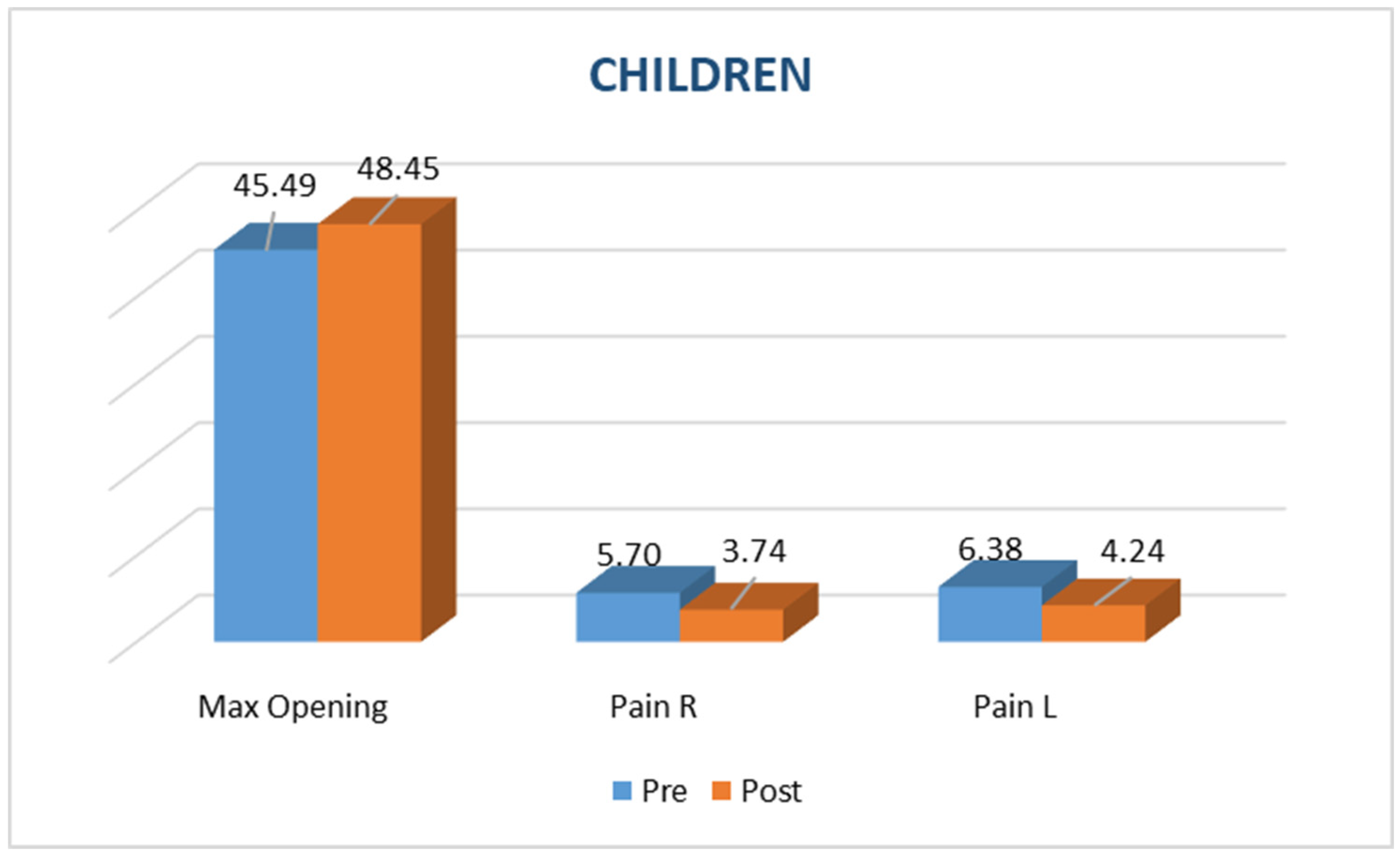
4. In the group of children the max opening has increased from 45.48±6.871 mm to 48.45±6.867 mm (t(75)=13.838, p= .000). The pain on the right side during palpation has reduced from 5.70±2.683 to 3.74±2.479. The pain on the left side has reduced from 6.38±2.612 to 4.24±2.427. Statistically significant differences were found in the group of children before and after the application of the nanotechnology t(75)=8.993, p=0.000 for the right side and t(75)=9.421, p= .000 for the left side.
5. The level of stress in adults was high 6.74±2.17, while in children the stress was half less 3.30±2.53. In the placebo group the level of stress was 6.33±2.201.
6. No statistically significant changes have been found in the placebo group in the maximum mouth opening before (pre) and after (post) the application of the placebo tape (t(42)= .682, p= .499).
7. No statistically significant changes have been found in the placebo group in the pain during palpation from the right or from the left side before (pre) and after (post) the application of the placebo tape. (right side t(42)= 1.756, p= .086; left side t(42)= .846, p= .403).
4. Discussion
Pain and limited mouth opening are among the most common symptoms in temporomandibular disorders (TMD), significantly impacting patients’ quality of life by interfering with essential functions such as speaking, chewing, and social interaction [25].
Among the various non-invasive therapeutic options, Taopatch® can be considered a valuable complementary tool alongside physical therapy and occlusal splints [26].
The combined use of traditional approaches and advanced technology, represents a promising direction in the multimodal management of TMD [27].
The results obtained in this study demonstrate immediate and statistically significant improvements in both maximum mouth opening and reduction of pain during muscle palpation.
These effects were observed in both adult and pediatric populations, with particularly strong responses in children.
Notably, the placebo group did not exhibit any meaningful change in the assessed parameters, confirming the intrinsic effectiveness of the device and reinforcing the credibility of the collected data. Furthermore, the absence of any adverse effects highlights the safety of the Taopatch® device, making it suitable for clinical use across a wide age range, including children.
However, it is important to acknowledge the current limitations in the scientific literature regarding the use of light-emitting nanotechnological devices for TMD management.
Larger, randomized controlled trials with long-term follow-up are necessary to confirm these findings and explore the full therapeutic potential of this technology.
Another use of Taopatch could be for the treatment of sleep disorders.
Sleep disorders represent a growing clinical issue, negatively impacting quality of life and daily cognitive functions [28].
Non-pharmacological solutions are receiving increasing attention due to their non-invasive nature and the absence of side effect [29].
In this context, a recent study focused on the ‘Repose Tao’ pillow, an innovative device that integrates Taopatch® technology within an ergonomic structure designed to provide proper cervical support during nighttime rest.
The study involved 30 participants who used the pillow over a four-week period. Sleep quality was monitored using the Pittsburgh Sleep Quality Index (PSQI), a validated tool for assessing subjective sleep quality.
The results showed significant improvements in various aspects: reduced sleep latency, increased perceived sleep duration, and overall enhancement in sleep quality. These findings support the device’s effectiveness as a supportive option for managing sleep disorders.
In conclusion, the use of advanced technologies such as that implemented in the Repose Tao pillow may represent a promising strategy for addressing sleep-related issues in a safe and non-invasive way [30].
5. Conclusions
Based on the findings of this study, the Taopatch® TMJ device emerges as an innovative and non-invasive tool for the management of pain and functional limitation associated with temporomandibular disorders in both adults and children.
Its ease of application, immediate effectiveness, lack of adverse effects, and suitability for daily dental practice make it a valuable addition to the clinician’s toolkit.
Improvements in mouth opening and pain reduction, achieved without the use of medication, underscore the potential of this technology to enhance both treatment outcomes and the overall patient experience.
Given these results, the Taopatch® may also hold promise in broader pain management and craniofacial rehabilitation contexts beyond TMD.
In conclusion, the Taopatch® TMJ represents a bridge between conventional therapies and next-generation technologies, offering tangible benefits in terms of clinical efficacy, patient comfort, and treatment innovation.
Its integration into routine dental and orthodontic procedures has the potential to improve therapeutic quality while opening new avenues in the field of nanotechnology assisted care.
References
- Zakrzewska, J. M. (2013). Differential diagnosis of facial pain and guidelines for management. In British Journal of Anaesthesia (Vol. 111, Issue 1, pp. 95–104). Elsevier BV. [CrossRef]
- Verrill, P. J. (1980). Pain control in dentistry. In British Dental Journal (Vol. 149, Issue 12, pp. 362–363). Springer Science and Business Media LLC. [CrossRef]
- Scoppa, F., Saccomanno, S., Bianco, G., & Pirino, A. (2020). Tongue Posture, Tongue Movements, Swallowing, and Cerebral Areas Activation: A Functional Magnetic Resonance Imaging Study. In Applied Sciences (Vol. 10, Issue 17, p. 6027). MDPI AG. [CrossRef]
- Valesan, L. F., Da-Cas, C. D., Réus, J. C., Denardin, A. C. S., Garanhani, R. R., Bonotto, D., Januzzi, E., & de Souza, B. D. M. (2021). Prevalence of temporomandibular joint disorders: a systematic review and meta-analysis. In Clinical Oral Investigations (Vol. 25, Issue 2, pp. 441–453). Springer Science and Business Media LLC. [CrossRef]
- SACCOMANNO, S., DELI, R., DI CINTIO, G., DE CORSO, E., PALUDETTI, G., & GRIPPAUDO, C. (2018). Retrospective epidemiological study of mandibular rotational types in patients with orthodontical malocclusion. In Acta Otorhinolaryngologica Italica (Vol. 38, Issue 2, pp. 160–165). Pacini Editore. [CrossRef]
- Calixtre, L. B., Oliveira, A. B., de Sena Rosa, L. R., Armijo-Olivo, S., Visscher, C. M., & Alburquerque-Sendín, F. (2018). Effectiveness of mobilisation of the upper cervical region and craniocervical flexor training on orofacial pain, mandibular function and headache in women withTMD. A randomised, controlled trial. In Journal of Oral Rehabilitation (Vol. 46, Issue 2, pp. 109–119). Wiley. [CrossRef]
- Messina, G. (2017). The tongue, mandible, hyoid system. In European Journal of Translational Myology (Vol. 27, Issue 1). PAGEPress Publications. [CrossRef]
- Saccomanno, S. (2021). Does a short lingual frenulum affect body posture? Assessment of posture in the sagittal plane before and after laser frenulotomy: a pilot study. In JOURNAL OF BIOLOGICAL REGULATORS AND HOMEOSTATIC AGENTS (Vol. 35, Issue 3_Suppl_1). Asia Pacific Academy of Science Pte. Ltd. [CrossRef]
- Gelb, M., Montrose, J., Paglia, L., Saccomanno, S., Quinzi, V., & Marzo, G. (2021). Myofunctional therapy Part 2: Prevention of dentofacial disorders [JB]. EUROPEAN JOURNAL OF PAEDIATRIC DENTISTRY, 22(2), 163–167. [CrossRef]
- Saccomanno, S., PhD, Saran, S., Dr, Pirino, A., Dr, Fontanella, R., Dr, Bruno, G., Dr, & Scoppa, F. Dr. (2025). A NEW APPROACH FOR PAIN IN TEMPOROMANDIBULAR DISORDERS ADRESSING THE INTERACTION OF DIET, EATING HABITS AND MEDICATIONS. In BULLETIN OF STOMATOLOGY AND MAXILLOFACIAL SURGERY (pp. 165–177).
- Saccomanno S, Antonini G, D’Alatri L, D’Angelantonio M, Fiorita A, Deli R. Patients treated with orthodontic-myofunctional therapeutic protocol. Eur J Paediatr Dent. 2012 Sep;13(3):241-3. [PubMed]
- Tran, C., Ghahreman, K., Huppa, C., & Gallagher, J. E. (2022). Management of temporomandibular disorders: a rapid review of systematic reviews and guidelines. In International Journal of Oral and Maxillofacial Surgery (Vol. 51, Issue 9, pp. 1211–1225). Elsevier BV. [CrossRef]
- Liu, F., & Steinkeler, A. (2013). Epidemiology, Diagnosis, and Treatment of Temporomandibular Disorders. In Dental Clinics of North America (Vol. 57, Issue 3, pp. 465–479). Elsevier BV. [CrossRef]
- Warzocha, J., Gadomska-Krasny, J., & Mrowiec, J. (2024). Etiologic Factors of Temporomandibular Disorders: A Systematic Review of Literature Containing Diagnostic Criteria for Temporomandibular Disorders (DC/TMD) and Research Diagnostic Criteria for Temporomandibular Disorders (RDC/TMD) from 2018 to 2022. In Healthcare (Vol. 12, Issue 5, p. 575). [CrossRef]
- Messina G, et al. A new road to improve vitamin D and balance through Taopatch® and proprioceptive protocol in Multiple Sclerosis patients. Eur J Transl Myol. 2022;32(4):10774. [CrossRef]
- Reddy, A. B., O’Neill, J. S. Healthy clocks, healthy body, healthy mind. Trends in cell biology 2010, 20, 36-44. [CrossRef]
- Morris, C. J., Aeschbach, D., Scheer, F. A. Circadian system, sleep and endocrinology. Molecular and cellular endocrinology 2012, 349, 91-104. [CrossRef]
- Taillard, J., Gronfier, C., Bioulac, S., Philip, P., Sagaspe, P. Sleep in normal aging, homeostatic and circadian regulation and vulnerability to sleep deprivation. Brain sciences 2021, 11, 1003-1022. [CrossRef]
- Lanzafame, R. J. Editorial: good vibrations: more evidence of reciprocity? Photomed Laser Surg 2007, 25, 465–466. [Google Scholar] [CrossRef] [PubMed]
- Bagnato, V. S., Rodrigues, T. Z., Garcia, V., Vidotti, H. G. M., Aquino Junior, A. E. D. Systemic effects of photobiomodulation and ultrasound as a potentiating tool in the treatment of sleep disorders: pilot study. J. Nov. Physiother 2024, 14, 1000703-1.
- Karadjova, M., Dimitrova, T., Dobreva, D., Petrova, J., Messina, G. Improvement of gait, balance and coordination after application of Taopatch® device. Eur. J. Transl. Myol. 2023, 33, 75-87. [CrossRef]
- Messina, G., Amato, A., Alioto, A., Stallone, R., Rizzo, F., Ragonese, P., Proia, P. A new road to improve vitamin D and balance through Taopatch® and proprioceptive protocol in Multiple Sclerosis patients. Eur. J. Transl. Myol. 2022, 32, 10774. [CrossRef]
- Costa, Y. M., Ariji, Y., Ferreira, D. M. A. O., Bonjardim, L. R., Conti, P. C. R., Ariji, E., & Svensson, P. (2018). Muscle hardness and masticatory myofascial pain: Assessment and clinical relevance. In Journal of Oral Rehabilitation (Vol. 45, Issue 8, pp. 640–646). Wiley. [CrossRef]
- Çetiner, R. B., Ergün-Kunt, G., Yüceer-Çetiner, E., & Schimmel, M. (2021). Masticatory function before and after masticatory muscle-related temporomandibular disorder treatment: An observational study. In Journal of Oral Rehabilitation (Vol. 49, Issue 3, pp. 295–300). [CrossRef]
- Ohrbach, R., & Dworkin, S. F. (2016). The Evolution of TMD Diagnosis. In Journal of Dental Research (Vol. 95, Issue 10, pp. 1093–1101). SAGE Publications. [CrossRef]
- Baig, N., Kammakakam, I., Falath, W. Nanomaterials: a review of synthesis methods, properties, recent progress, and challenges. Mater. Adv. 2021, 2, 1821–1871. [CrossRef]
- Gudin, J. A., Dietze, D. T., Hurwitz, P. L. Using Nanotechnology to Improve Pain and Function with a Novel, Drug-Free, Topical Pain-Relief Patch: An Interim Analysis. Anesth Pain Res 2020, 4, 1-10. [CrossRef]
- Almeneessier, A.S.; Gupta, R.; Pandi-Perumal, S.R.; BaHammam, A.S. Overview of sleep disorders. In The behavioral, molecular, pharmacological, and clinical basis of the sleep-wake cycle; Academic Press: New York, 2019; pp. 103–122. [Google Scholar]
- Valverde, A., Hamilton, C., Moro, C., Billeres, M., Magistretti, P., Mitrofanis, J. Lights at night: does photobiomodulation improve sleep? Neural regeneration research 2023, 18, 474-477. [CrossRef]
- Malik, S., Muhammad, K., Waheed, Y. Emerging Applications of Nanotechnology in Healthcare and Medicine. Molecules 2023, 28, 1-30. [CrossRef]
Figure 1.
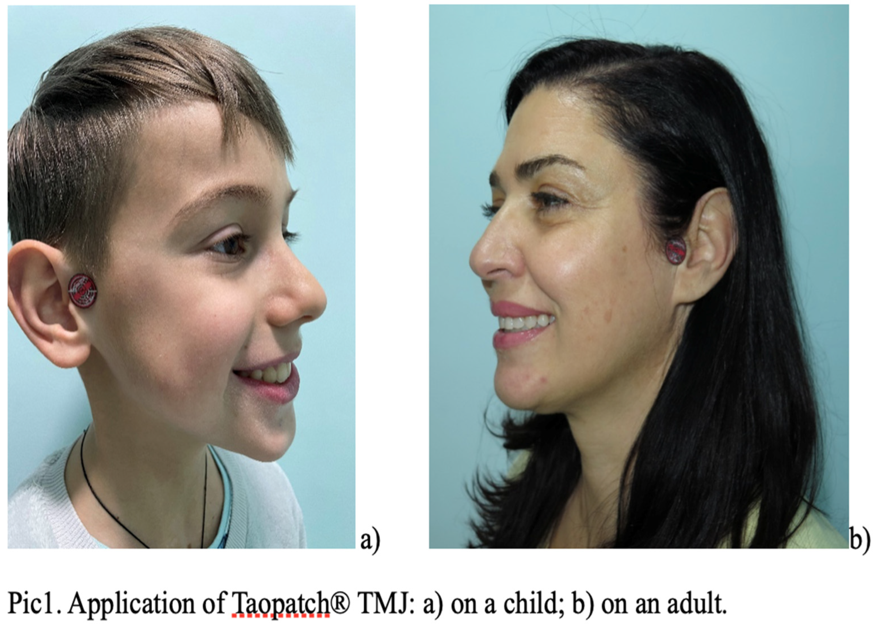
Figure 2.
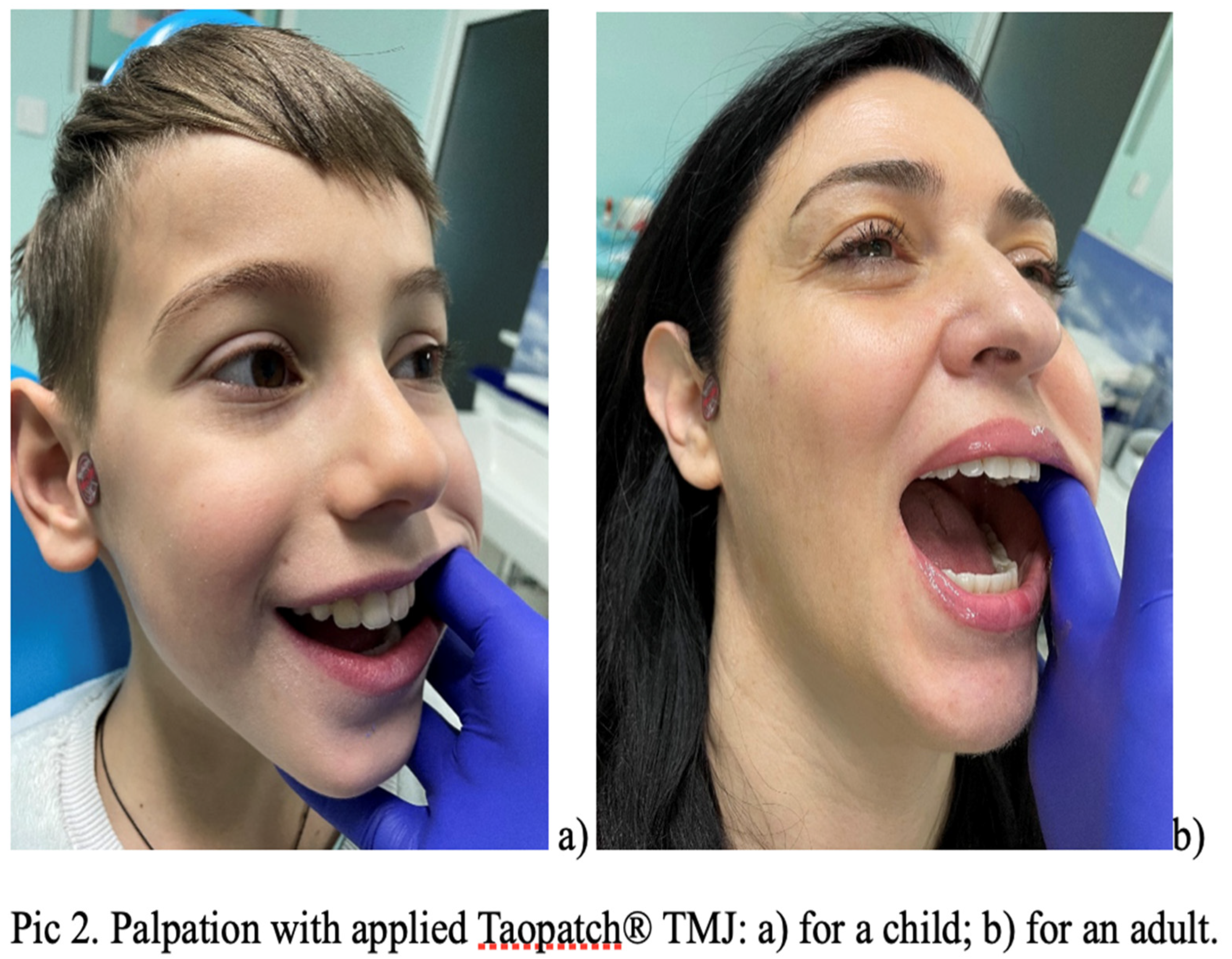
Figure 3.
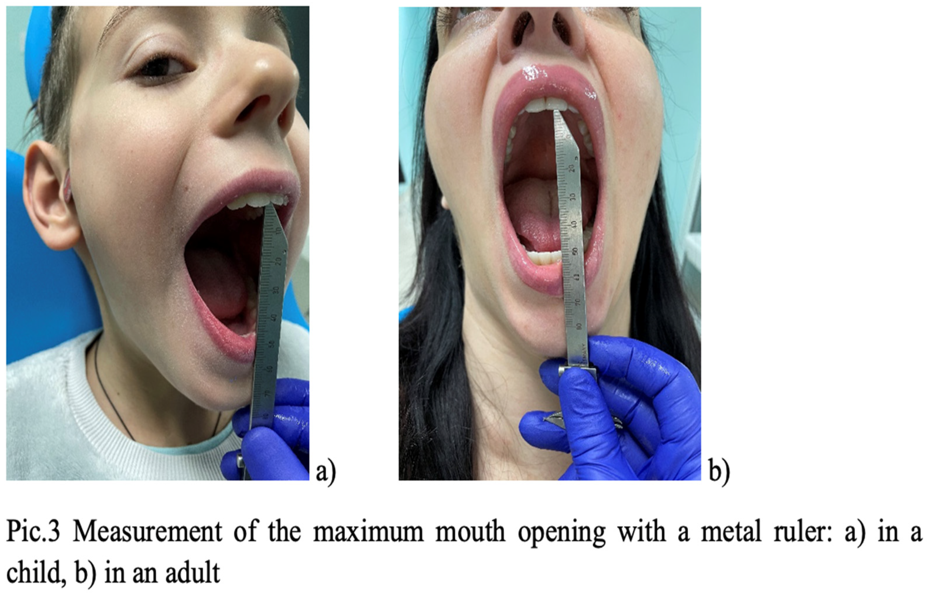
Figure 4.

Disclaimer/Publisher’s Note: The statements, opinions and data contained in all publications are solely those of the individual author(s) and contributor(s) and not of MDPI and/or the editor(s). MDPI and/or the editor(s) disclaim responsibility for any injury to people or property resulting from any ideas, methods, instructions or products referred to in the content. |
© 2025 by the authors. Licensee MDPI, Basel, Switzerland. This article is an open access article distributed under the terms and conditions of the Creative Commons Attribution (CC BY) license (http://creativecommons.org/licenses/by/4.0/).
Copyright: This open access article is published under a Creative Commons CC BY 4.0 license, which permit the free download, distribution, and reuse, provided that the author and preprint are cited in any reuse.


