
Submitted:
15 April 2025
Posted:
15 April 2025
You are already at the latest version
Abstract
Dictyocaulosis is a parasitic disease affecting ungulate species, including red deer (Cervus elaphus). Dictyocaulus genus consist of nine species but only four are reported to infect red deer. The disease is characterized by respiratory tract infection, particularly in lungs, bronchi and bronchioles, causing inflammatory and hemorrhagic microscopic lesions but also emphysema and edema. Biological cycle consists of a female that oviposits larvae eggs in the bronchi and trachea to be expelled to the exterior by coughing and releasing the L1 in the environment. In this study 106 adult red deer were collected from seven locations in Extremadura (Spain). Eight positive lungs were primarily assessed by morphological identification, leading to a mean intensity of 13.25 adult worms/infected lung, but globally descending to an average of 1.78 adults/sampled lung. The presence of adult worms in the upper and middle respiratory tract was confirmed by anatomopathological analysis. Molecular identification was made by sequencing the COI gene. As a result, we have detected the presence of three genetic groups supported by significant subdivision using ɸST measure but D. cervi and D. viviparus showing its respective matrilineal ancestry while D. eckerti and D. cervi showing matrilineal sharing. Consequently, introgression between these two species was suggested. D. viviparus has been identified in the same Spanish region on the basis of morphological characters, but D. cervi and D. eckerti were reported for the first time in the explored geographic area.
Keywords:
COI-barcoding
; Dictyocalulus
; Morfolofical and Molecular identification
; Red Deer
1. Introduction
Throughout the history of mankind, attention has been paid to the study of zoonoses and how wildlife has been involved in health alerts, which includes the transmissible diseases to livestock, as is the case of tuberculosis, protozoan or helminths as dictyocaulus [1,2]. Helminths parasitic diseases are among those of high importance for wild ruminants’ dynamics but four genera, Ascaris, Dictyocaulus, Strongyloides, and Trichuris are of special attention for health and husbandry in ruminant [3]. Among them Dictyocaulus species (Nematoda: Trichostrongyloidea) are distributed worldwide and affect both even-toed ungulates – D. viviparus, D. filaria, D. eckerti, D. murmanensis, D. africanus, D. capreolus, D. cervi, D. cameli - and odd-toed ungulates – D. arnfieldi, D. pandionis within Artiodactyla mammals.
The genus Dictyocaulus (Nematoda: Dictyocaulidae) was described by Karl Moritz (1851) for the first time based on morphological traits. Advances in optical microscopy and scanning electron microscopy from the end of the 19th to the 20th century, further the development of histological techniques, allowed more accurate morphological identification of these nematodes [4] improving species taxonomic classification of distinct species, as in the case of Dictyocaulus capreolus [5]. Despite this, most species of the genus Dictyocaulus are still identified by the parasitized host, even in the 21st century [3]. In Spain, Dictyocaulus viviparus and Dictyocaulus filaria have been reported in domestic ruminants [6,7]. However, some reports acknowledged major difficulties for species identification occurring in wild ruminants, due to the high level of morphological similarity exhibited among several species [8]. To this respect, prior to the advent of molecular identification, all infections of lung worms from red deer were identified as D. viviparus which parasitized to cattle. However, molecular characterization of Dictyocaulus spp. has revealed the existence of several distinct clades or species [2,3,7,9,10,11] and have also been of value in estimating phylogenetic relationships among trichostrongyloid and metastrongyloid nematodes [6,12]. Although the genus Dictyocaulus belongs in the monotypic family Dictyocaulidae with 18 nominal species, only five species have been confirmed valid based on molecular genetic data: D. viviparus, D. filaria, D. eckerti, D. capreolus, and D. cervi [3 and references therein].
This parasite is found in the small and large airways of the host, potentially causing parasitic bronchitis (dictyocaulosis), sometimes a fatal disease, especially in cattle, sheep and farmed red deer [13]. Under this scenario, a major breakthrough for the development of control strategies to prevent dictyocaulosis was a topic of concern because different species of cervids carrying these parasites behaves as true vectors to livestock [1,14,15]. In this regard, relevant researchs have provided control strategies based on vaccine preparations with effective immunological response against this parasite [16,17]. However, it was noticing as the nematodes became more resistant to the former vaccines reason why repeated improvement has been performed, until the “Bovilis Huskvac” vaccine was available offering a 95-98% level of protection [18]. Unfortunately, this vaccine against pulmonary verme is not of wide use despite its usefulness. Practical concerns, such as a short shelf life and the availability of anthelmintics with persistent efficacy against dictyocaulus, have apparently made vaccination a less attractive control option [18]. Currently, it has been described that resistance to antiparasitic drugs, such as fenbendazole and albendazole [19] and macrocyclic Lactones [20] in cattle reactivated the interest in these parasites. All lungworms were reported as Dictyocaulus viviparus in feral deer species [21,22] and a few studies on native Iberian red deer in Extremadura (Spain) suggested a low level of infection [23]. However, recent research advocates for investigating the full host range, epidemiology, the potential impacts and cross-transmission events with livestock of Dictyocaulus spp. with special attention on cervids world-wide [13,24] because levels of infection with lungworms in free-ranging deer remains largely unknown [7,13,24,25]. In addition, a global opinion suggests that objective methods of species-specific identification should be recommended [3], as cross-transmission has not yet been described [26]. For all these reasons, this report was aimed to contributed to knowledge of Dictyocaulus species by aspect such as the morphology, prevalence and associated lesions, as well as to carry out a genetic analysis using DNA molecular techniques in order to accurate specie-specific identification of parasite involved in dictyocaulosis in Extremadura at southwestern of Spain in free ranging red deer.
2. Materials and Methods
A total of 36 deer lungs from 7 fences in the province of Cáceres (Figure 1). Table 1 showed the fences/estates where sampling was done after carried out hunting activities. Carcass inspection was performed by dedicated official veterinarian (Junta de Extremadura, Spain) following the regulations (EC) No. 853/2004 of the European Parliament and the execution reglament (EU) 2015/1375. Accordingly, 5-6 lungs with their respective tracheas were collected in each site. Each lung was stored individually in an airtight bag and transported at 4ºC for preservation.
2.1. Necropsies and Microscopic Examination
The lungs were opened in a parasitology dedicated laboratory in the Clinical Veterinary Hospital (University of Extremadura, Spain) using appropriated preventive measures. After, careful examinations by compression were performed to check the presence of any vermecia. Macroscopic vermeus were introduced in a 150 mL propylene bottle containing 96% Ethanol and stored at 4ºC until processing. Finally, microscopic assessment confirmed Dictyocaulus spp. under a NIKON H550S (Japan) light microscope. Morphological traits were assayed following keys [4,27], focusing on the shape of the anterior end (mouth and esophagus) and on the spicules of males. In any case, the lungs were additionally subjected to complementary exams by scraping the trachea and bronchi to find eggs and/or larval stages. All data was recorded in an Excel sheet. Statistical agreement between macroscopic and microscopic was performed using Cohen's Kappa statistic in SPSS 15.0. (under UNEX License).
2.2. Anatomopathologival Study
The macroscopic and microscopic lesions caused by the worms were studied. Specially, affected areas of positive lungs were embedded in formalin. Subsequently, slices were processed by conventional hematoxylin eosin histopathological methods.
2.3. Molecular Procedures
2.3.1. DNA Extraction
A total of 15 worms were processed, 5 for each fence from the positive lung. Samples from Sierra Palomares (SP), Jabalina (JB) and Cerro Verde (CV) fences were processed for molecular analysis. Cuadrillas Bajas fence was rejected due to sampling conservation error. Excluding genitalia, half cm from the medial portion of adult or larvae parasites were stuffed for generic material (DNA) extraction under sterility condition in dedicated room, avoiding cross-contamination. DNA template was purified by a salting-out protocol [28] modified using Zymo-Spin II C columns and eluted in 400 μL molecular biology grade water.
2.3.2. End-Point PCR and Primers
End-point PCR was made with primers described [29] which targeting a portion of the COI mitochondrial gene as follows: COX I_F (5'-TTTTTTTTGGGCATCCTGAGGTTTAT-3') and COX I_R (5′-TAAAGAAAGAAAGAACATAATGAAAAAATG-3'). The PCR master-mix followed published protocols [30]. Briefly, 10 μL of each 10x NH4 buffer, 2mM dNTPs, 10 μM COX I_R and F primers and 3 μL MgCl2 (50mM) and completing up to 90 μL with molecular biology grade water. Individual assays were performed with 18 μL of 2 μL of template DNA. Thermocycler conditions were as follows: Pre-Denaturation at 95ºC for 5 minutes; 35 cycles at 94ºC 60 seconds, 50ºC 60 seconds, 72ºC 60 seconds and final extension of 72ºC 7 minutes. PCR products were visualized by 1.6% (w/v) agarose electrophoresis SYBR safe stains. Molecular weight 100 bp ladder profile 100 bp ladder was used to monitor amplicon size under 312 nm ultraviolet light transilluminator.
2.3.3. Sequencing, Alignment and Comparison Through Phylogenetic Networks
5 μL PCR products were intimately purified with Ex-Spure (NimaGen, The Netherlands) following manufacturer's recommendations and later diluted to 10 μL with ultrapure water. PCR product templates (10 µL) were sequenced using the Big Dye® ver 3.1 cycle sequencing Kit (Thermo Fisher Scientific) and residual eliminated with Performa®DTR cartridges (Edge Bio). Profile of sequencing products obtained by capillary electrophoresis (Applied Biosystems™ 3130 DNA Analyzer; USA) and analyzed with “ABI Sequencing Analysis” ver. 5.2 software (Applied Biosystems Company, USA).
Genbank database NCBI (USA) was filtered to find species belonging to the genus Dictyocaulus. The search was limited to 250 sequences but having the same or higher coverage with respect to the COI segment sequenced in this study. These sequences were selected based on a similarity cutoff level of ≥ 90% aimed to gather all sequences of the genus Dictyocaulus [31]. The last species downloaded was Arthrosoma spp. after filtering under these criteria. Thirty sequences among those downloaded belonged to the genus Dictyocaulus spp. with 100% coverage (320 bp) with respect to the 13 sequences from this study. As a result, a total of 115 sequences were selected but 102 from GenBank (NCBI, USA). These sequences were collapsed into haplotypes using DnaSP ver 6.1 [32]. These haplotypes were further used to create a standard Median-Joining (MJ) Network [33] of the genetic relationships with the program PopArt v. 1.7. PopArt v. 1.7 (Population Analysis with Reticulate Trees) [34] but their positions are visualized on a physical map using GPS data. Divergence among phylogenetic cluster was measured using the fixation index statistic (ɸST) by grouping sequenced data as follows: (1) D. cervi, (2) D. Eckerti/D.cervi and (3) D. vivparus, in PopArt v. 1.7.
3. Results
3.1. Prevalence of Dictyocaulosis in Deer at Sampling Location
Table 2 recorded the number of positive animals and the number of adults in each of the 36 lungs processed.
The frequency of infected lung was 13.89% after the macroscopic analysis but an averaged adult count of 12.80 (SD 7.12). However, higher frequency was found after microscopic analysis with 22.22% of positive lungs. The results highlight the relevance of the microscopic analysis over the macroscopic inspection because larvae are much more common in lungs than macroscopically observable adults. Although at first glance Cohen's Kappa test suggested substantial agreement (κ=0.722) between the macroscopic and microscopic analysis with significant p-value (p<0.001), there was not perfect agreement indicating some discrepancies. Furthermore, the mean value for adults suggested a large variability in the count of adult parasites per infected red deer. Globally, the mean abundance of worms in all the lungs was calculated, giving an average of 1.78 (SD = 5.09) adults/lung.
3.2. Morphological Identification and Anatomopathological Finding in Dictyocaulosis
After worm collection, preservation and preparation for microscopic observation, the adults were studied in detail for their correct identification. It was found that the buccal capsule was small with an oval shape and surrounded by simple lips (Figure 2A) as expected. Also, differences between males and females were examined. The male has an expansion of the cuticle (copulatory pouch) with a series of supporting ribs, as well as a series of rays for allowing its expansion during copulation. The posterior part in the female was conical in shape, showing a small and rounded protrusion called the vaginal diverticulum (Figure 2B,C). Finally, the presence of eggs in the uterus of the female is noteworthy with a uterus full of larvae eggs (L1 stage) (Figure 2C).
Anatomopathological study showed that dictyocaulus produces bronchopneumonia in red deer due to the presence of adult or larvae worms in the upper and middle respiratory tract. Figure 3A showed bronchioles inflammation and destruction of the bronchiolar epithelium (bronchiolitis) as well as intraluminal exudate of eosinophil, lymphocytes and plasmatic cells. Intraluminal parasitic structures (Figure 3B) compatible with the nematode surrounded by massive intraluminal hemorrhage and slight peri-bronchial lymphoid reaction were also identified. Figure 3C highlighted a clear parasitic membrane with three structural components: fibrous capsule (C), acellular laminated membrane (L), and germinative membrane (G). Figure 3D showed a non-specific interstitial pneumonia with a diffuse mononuclear cell infiltration that expands and widens the interalveolar septa, with little collagenization and a mild hyperplasia of the alveolar epithelium. No foci of fibroblastic proliferation and panalization phenomena.
3.3. Barcoding Finding Through Sequencing COI Gene
Sequences from this study resulted in thirteen successful COI profiles using Sanger sequencing. These sequences collapsed in seven different haplotypes (Hap_31 to Hap_37, Figure 4). These haplotypes were compared with 102 additional sequences which collect molecular information from twenty-four nematode worm genus or species (Table S1). Firstly, globally, the 115 sequences yielded up to 84 different haplotypes (Figure S1). The obtained overall sequence similarity varies between 96.97% and 99.66% respect to the sequences from Genebank (NCBI, USA) except one sequence from Cerro Verde (CV) that showed the highest similarity value (100%) and unclear species assignation that might better belong to D. eckerti ( see Hap_32 vs. Hap_73, Figure 4). All haplotypes drawn very conspicuous different clusters which mostly assigned haplotypes to taxonomic species level as reported by the GenBank entries (Figure S1). Particularly important was the fact that the haplotype network very clearly separated three species from the Dictyocaulus genus, D. viviparus, D. eckerti and D. cervi. For this reason, it was decided to analyze them separately for a better visualization of the results (Figure 4). Secondly, it was constructed another network containing exclusive haplotypes from Dictyocaulus genus. In this redraw picture the Dictyocalus genus showed three well distinguishable matrilineal groups. Of these, two groups contained haplotypes exclusively assigned to D. viviparus and D. cervi species. However, D. viviparus was never found in this study. Four haplotypes (Hap_33, 34, 36 and 37) belonged to a group containing solely D. cervi species. A third genetic groups collect very similar haplotypes assigned to D. cervi or D. eckerti (Hap_ 31 and 32, plus Hap_35) species. Because these lasts were assigned to different species despite sharing a most probable common ancestor respect to D. viviparus or D. cervi, assignment confusion or admixture throughout hybridization could be further investigated. Furthermore, Hap_32 was the only showing a 100% identity with other sequences from Genbank but assigned to D. cervi (Acc N. PP922991) within the admixed group (Figure 4).
According with phylogenetic analysis (color assignment in the network, Figure 4) should be noted that Hap_31 was found in SP (Sierra Palomares, n=1) and CV (n=1), Hap_32 in CV (n=1), Hap_34 in both CV (n=3) and JB (La Jabalina, n=2), Hap_36/Hap_37 in SP (n=1 each), Hap_33/hap _35 in JB (n=2 and n=1, respectively)(Figure 4). In addition to phylogenetic evidence of molecular divisions, also, AMOVA analysis demonstrated genetic subdivision among the three dictyocaulus groups with 89.82% genetic variance explained among groups, two with conspicuous species status, and 10.18% variance within groups, supporting by a significant ɸST index (p-value < 0.001). This result conforms with matrilineal ancestor isolation among haplotype groups with perfect match to species assignation. Conversely, D. cervi and D. eckerti shared matrilineal ancestor in one of the genetically differentiated groups, reason why this gene cluster (central cluster, Figure 4) does not match the species assignment (see table S1: haplotypes and species) with suggestive introgression. Furthermore, haplotypes of the admixed group and D. cervi groups were present in all sampling sites. Also, it can be stated that both D. cervi and D. eckerti coexist in the same host, since Hap_31, _32 (mixed group) and_34 (D. cervi group) were simultaneously identified in the only lung obtained at CV. As well the same scenario happened for haplotypes 31, 36 and 37, identified in one lung obtained from SP.
4. Discussion
4.1. Prevalence of Dictyocaulosis in Deer in Extremadura
If we focus on the adult count after macroscopic analysis, our results agreed with those obtained by other authors [13,35] who found an averaged intensity per positive lung of 11.7 adults and 6.33, respectively. However, extremely large discordances have been detected with others [4,36,37,38] because they reported disparate prevalence as less than one adult among 27 positive lungs or all positive deer presenting adults. All of these different results may be due to multifactorial reasons, ranging from the environmental and physiological status of the animals to the study design, but those particularly related to the age of the sampled animals and the state of their immune system have been suggested. Also, differences in sample size may be claimed. Several authors [13,23,35,39,40,41,42] suggested taking as large a possible number of samples, especially in the locations sampled. From an epidemiological point of view, a prevalence of 22.22% has been obtained, very similar to those obtained previously [40] in roe deer in the north of Spain and in deer in the Italian Alps [13]. However, prevalence rates of 50% and 54% in Norway have been found [40,43] respectively. Also, in Poland [4,38] and in Extremadura [23] reported prevalence three times higher in the order of 44%/68% and 62.5% in red deer, respectively. All these data suggest that a high variation in prevalence over time and geography would be expected.
Furthermore, it would have been interesting to perform coprological analysis to determine how many larvae, if any, they expel in their faeces [23]. Despite this application of feces, given that genetically different worms can be found from the same lung (even with small samples), caution is advised when using conventional sequencing analysis based on genetic material obtained from feces. Therefore, adults or larvae should be individualized when genetic analysis is among the objectives. This is especially relevant when hybridization is suspected. Furthermore, our results are significant because individualized specimens contribute 44.44% (4/9), 33.33% (2/6) of haplotypes to D. cervi and to admixed D. eckerti/D. cervi group, respectively, but 0% (0/14) to D. viviparus group; additionally, to those currently present in the NCBI databases.
4.2. Findings After Anatomopathological Study
In agreement to some authors [4,27] morphological traits observed in worms may only assess its belonging to the genus Dictyocaulus spp. However, these traits may distinguish Dictyocaulus from other genuses of pulmonary nematodes. Specifically, the adults collected in our study presented a simple buccal capsule, with a long esophagus, the presence of a copulatory pouch with ribs and rays that allow its expansion during copulation. In males, the dark-colored spicules with a boot or sock shape that are directed by the gubernaculum, and in females, the presence of the uterus filled with easily discernible larval eggs. However, identification at species level has been suggested to be extremely complicated by light microscopy since we have to rely on subjective measurements with no great difference among species. These reasons led to the need for more standardized diagnostic methods to identify dictyocaulus species level, such as molecular DNA methods. Furthermore, they open up new and unexpected scenarios out of the scope of traditional anatomopathological methods.
In the analysis of lung lesions caused by dictyocaulus worms, our findings show similarities and important differences compared to those reported in previous studies. Specifically, our observations present notable discrepancies with other authors [38] because no significant presence of macrophages was confirmed in the exudates of this study. In addition, they reported massive hyperplasia in follicular lymphoid follicles, bronchial vessels, and bronchiolar epithelium, leading to strong and widespread inflammatory and proliferative response in the affected lungs [38]. In contrast, our study showed only mild hyperplasia in the alveolar epithelium, which could indicate a less intense inflammatory response to different stages of the alveolar epithelium with reduced inflammatory response. Another study [44] largely corroborated the lesions described by us, reinforcing the consistency of many pathological patterns associated with different scenarios of dictyocaulosis. Also, compared with other study [45] findings were congruent, except for some specific details. Described the presence of slight fibroblastic proliferation, necrotic changes and proliferation of type II pneumocytes which were not a prominent observation in our study. These differences may be due to environmental and/or genetic variations between different red deer populations. Furthermore, we cannot discard differences in the histological and sampling techniques used. All these data may suggest multifactorial and differential infection stages, underscore possible differential responses among Dictyocaulus species and/or species mixtures.
4.3. Molecular of Dictyocaulus spp. Assessment
Mitochondrial COI gene outcomes were consistent with some authors [13,29] because relatively large molecular variability allowed for effective barcoding to discern all three Dictyocaulus species: D. viviparus, D. eckerti and D. cervi. Of the 18 nominal species within the genus only four are reported to infect European deer species: D. eckerti, D. capreolus, D. cervi and D. skrjabini [46]. Except D. viviparus from cattle, the remaining species in the genus were found in sheep and goats (D. filaria), donkeys and horses (D. arnfeldi), camels (D. cameli), African artiodactylids (D. africanus) and, also, D. skrjabini were not present in the data set due to either their lower similarity threshold, different sequence coverage or absence of data.
As a result, the downloaded molecular data only collected information from D. viviparus reinforcing a closer genetic relationship between these three species (D. viviparus, D. eckerti and D. cervi) within the genus Dictyocaulus. There are also subspecies, or possible new species, of D. viviparus within both European [47] and North American bisons [48] which were also in our data set (Hap_58 and Hap_57, Figure 4 and table S1) but it has not been possible to corroborate even different lineage level for this subspecies with the available data. All these facts gives support to a high genetic variability and longlife evolutionary history of this genus. The global phylogenetic analysis (115 sequences) allowed for the identification of three major groups in the genus Dictyocaulus supported by a great genetic variability between (89,9% variance) and less within (10,12% variance) groups, which agreed with last studies [4,13,38,48]. However, from the three genetic groups (D. eckerti, D. cervi and D. viviparus), only the two formers were found by us in red deer. Interestingly, this wide genetic variability has been associated with a possible adaptation to environmental changes of the nematode, even the development of resistance to anthelmintics. This resistance has been reported in a few studies from [19,20,49]. The first study carried out in the same region of Extremadura identified infection D. viviparus [23] but based on challenged morphological characters. However, a better barcoding-based model identified D. cervi and, probably, D. eckerti at this location (this study). A MJN analysis showed estrict genetic relationships within lineages of D. cervi and D. viviparus sequences but allocating D. eckerti with several D. cervi haplotypes (specially Hap_32) suggesting a mixed origin for this group (D. Eckerti/D. cervi groups, Figure 4). These findings were also reported [46]. ɸST statistic supported a significantly high divergence among the three groups from this study with strongly different mitochondrial COI alleles within the Dyctiocaulus genus was detected.
Accordingly, it was formerly confirmed the presence of D. eckerti and D. cervi in Extremadura and Spain. In addition, our results suggest a better and precise identification of Dictyocaulus species and discarding D. viviparus as causing dictyocaulosis in the examined red deer populations but discarding D. viviparus. According to some authors [4,13,38], there is firm evidence, even morphological, as D. cervi recovered from red deer was well distinguished from D. eckerti, assessing the importance of the use of COI sequences for its diagnosis. Despite, our results suggested more research must be performed to unravel the presence of two species in one of the three genetic groups, especially when hybridization might be a reasoned event since in this study both species have been found in the same red deer lung. The hybridization in nematode species has been demonstrated [50]. This state of hybridization in which they share genetic material could be facilitated by similarities in climatic conditions, between hosts and their life cycles. So, our findings further strengthen the idea that the genetic complexity and diversity among Dictyocaulus lungworms infecting wildlife ruminants is larger than previously believed and requires further investigation.
5. Conclusions
The prevalence of Dictyocaulus spp. was low, which leads to suggest as the immune system of red deer balanced these infestations and concordance between the macroscopic and microscopic diagnosis was also confirmed. The molecular analysis was valuable to assess taxonomic level among groups because of the high divergence of Dictyocalus species. So, the COI gene differentiated Dictyocaulus species in Spain for the first time, even when worms were sampled in the same infected lung. Furthermore, some sequences attributed to different species within the same genetic group were revealed, suggesting the need of new studies unraveling two competing questions aimed to differentiate between natural species mixtures or hybridization in these worms.
Supplementary Materials
The following supporting information can be downloaded at: Preprints.org, Figure S1: Graphical representation of the distribution of the different 81 haplotypes used for MJN analysis. Color were selected according to assigned species in genebank report, sampling sites when form those study and others from non Dictyocaulus genus.; Table S1: Species of worms from Genbank (NCBI, USA) with a higher 90% threshold similarity.
Author Contributions
Conceptualization, J.L.F-G., M.A.H. and M.A. H-P; methodology, A. E-S. and A. R.; software managemet, J.L.F-G., M. G-V. and M.A. H-P; validation, J.L.F-G., M.A.H. and M. G-V..; formal analysis, J.L.F-G., A. E-S, A.R., M. G-V. and C. B-P.; investigation, J.L.F-G., M.A.H., M. G-V., C. B-P, M.A. H-P., A. E-S. and A. R; resources, J.L.F-G. and M.A.H.; data curation, M. G-V., C. B-P..; writing—original draft preparation, M. G-V., C. B-P.; writing—review and editing, J.L.F-G., M.A.H. and A. E-S.; visualization, A. E-S. and A. R.; supervision, J.L.F-G., M.A.H.; funding acquisition, J.L.F-G. All authors have read and agreed to the published version of the manuscript.
Funding
No funding to declare.
Data Availability Statement
Data Availability on request to the authors.
Acknowledgments
not declared yet.
Conflicts of Interest
The authors declare no conflicts of interest.
References
- Huaman, J.L., Helbig, K.J., Carvalho, T.G., Doyle, M., Hampton, J., Forsyth, D.M., Pople, A.R., Pacioni, C. A Review Of Viral And Parasitic Infections In Wild Deer In Australia With Relevance To Livestock And Human Health. Wildl. Res., 50 (2023), Pp. 593-602. [CrossRef]
- S. Shamsi, K. Brown, N. Francis, D.P. Barton, D.J. Jenkins. First Findings Of Sarcocystis Species In Game Deer And Feral Pigs In Australia.Int. J. Food Microbiol. (2024). [CrossRef]
- Vainutis, K.S., Voronova, A.N., Andreev, M.E. et al. Morphological and molecular description of Dictyocaulus xanthopygus sp. nov. (Nematoda: Trichostrongyloidea) from the Manchurian wapiti Cervus elaphus xanthopygus. Syst Parasitol 100, 557–570 (2023). [CrossRef]
- Pyziel, A. M., Laskowski, Z., Demiaszkiewicz, A. W., Y Höglund, J. (2017). Interrelationships of Dictyocaulus Spp. In Wild Ruminants With Morphological Description Of Dictyocaulus Cervi N. Sp. (Nematoda: Trichostrongyloidea) From Red Deer, Cervus Elaphus. Journal Of Parasitology, 103(5), 506-518. [CrossRef]
- Gibbons, L. M., Y Höglund, J. (2002). Dictyocaulus Capreolus N. Sp. (Nematoda: Trichostrongyloidea) From Roe Deer, Capreolus Capreolus And Moose, Alces Alces In Sweden. Journal Of Helminthology, 76(2), 119-124. [CrossRef]
- R. A. Carreno And S. A. Nadler . 2003. Phylogenetic Analysis Of The Metastrongyloidea (Nematoda: Strongylida) Inferred From Ribosomal Rna Gene Sequences. Journal Of Parasitology 89:965–973.
- Carreno, R. A., Diez-Baños, N., Hidalgo-Argüello, M. del R., & Nadler, S. A. (2009). Characterization of Dictyocaulus Species (Nematoda: Trichostrongyloidea) from Three Species of Wild Ruminants in Northwestern Spain. Journal of Parasitology, 95(4), 966-970. [CrossRef]
- L. M. Gibbons And L. F. Khalil . 1988. A Revision Of The Genus Dictyocaulus Railliet And Henry, 1907 (Nematoda: Trichostrongyloidea) With The Description Of D. Africanus N. Sp. From African Artiodactylids. Revue De Zoologie Africaine 102:151–175.
- J. Höglund, D. A. Morrison, B. P. Divina, E. Wilhelmsson, And J. G. Mattsson . 2003. Phylogeny Of Dictyocaulus (Lungworms) From Eight Species Of Ruminants Based On Analyses Of Ribosomal Rna Data. Parasitology 127:179–187. [CrossRef]
- R.B. Gasser, A. Jabbar, N. Mohandas, J. Höglund, R.S. Hall, D.T.J. Littlewood, A.R. Jex. Assessment Of The Genetic Relationship Between Dictyocaulus Species From Bos Taurus And Cervus Elaphus Using Complete Mitochondrial Genomic Datasets. Parasites Vectors, 5 (2012), P. 241. [CrossRef]
- Pyziel, A. M., Laskowski, Z., Klich, D., Demiaszkiewicz, A. W., Kaczor, S., Merta, D., Kobielski, J., Nowakowska, J., Anusz, K., & Höglund, J. (2023). Distribution of large lungworms (Nematoda: Dictyocaulidae) in free-roaming populations of red deer Cervus elaphus (L.) with the description of Dictyocaulus skrjabini n. sp. Parasitology, 150(10), 956-966. [CrossRef]
- Chilton, N. B., Huby-Chilton, F., Gasser, R. B., & Beveridge, I. (2006). The evolutionary origins of nematodes within the order Strongylida are related to predilection sites within hosts. Molecular Phylogenetics and Evolution, 40(1), 118-128. [CrossRef]
- Cafiso, M. Castelli, P. Tedesco, G. Poglayen, C. Buccheri-Pederzoli, S. Robetto, R. Orusa, L. Corlatti, C. Bazzocchi, C. Luzzago. Molecular Characterization Of Dictyocaulus Nematodes In Wild Red Deer Cervus Elaphus In Two Areas Of The Italian Alps. Parasitol. Res., 122 (2023), Pp. 881-887. [CrossRef]
- D. Jenkins, A. Baker, M. Porter, S. Shamsi, D.P. Barton. Wild Fallow Deer (Dama Dama) As Definitive Hosts Of Fasciola Hepatica (Liver Fluke) In Alpine New South Wales. Aust. Vet. J., 98 (2020), Pp. 546-549. [CrossRef]
- J. Lamb, E. Doyle, J. Barwick, M. Chambers, L. Kakhn. Prevalence And Pathology Of Liver Fluke (Fasciola Hepatica) In Fallow Deer (Dama Dama). Vet. Parasitol., 293 (2021), Article 109427. [CrossRef]
- Jarrett, W. F. H., Jennings, F. W., Mcintyre, W. I. M., Mulligan, W., Thomas, B. A. C., Y Urquhart, G. M. (1959). Immunological Studies On Dictyocaulus Viviparus Infection. Immunology, 2(3), 252-261.
- Jarrett, W. F. H., Y Sharp, N. C. C. (1963). Vaccination Against Parasitic Disease: Reactions In Vaccinated And Immune Hosts In Dictyocaulus Viviparus Infection. The Journal Of Parasitology, 49(2), 177-189. [CrossRef]
- Claerebout, E., Y Geldhof, P. (2020). Helminth Vaccines In Ruminants: From Development To Application. Veterinary Clinics: Food Animal Practice, 36(1), 159-171. [CrossRef]
- Molina, V. M., Arbeláez, J. M., Prada, J. A., Blanco, R. D., Y Oviedo, C. A. (2016). Posible Resistencia De Dictyocaulus Viviparus Al Fenbendazol En Un Bovino. Revista De La Facultad De Medicina Veterinaria Y De Zootecnia, 63(1), 54-63. [CrossRef]
- Campbell, P., Forbes, A., Mcintyre, J., Bartoschek, T., Devine, K., O’neill, K., Laing, R., Y Ellis, K. (2024). Inefficacy Of Ivermectin And Moxidectin Treatments Against Dictyocaulus Viviparus In Dairy Calves. Veterinary Record, E4265. [CrossRef]
- R.A. Mckenzie, P.E. Green, A.M. Thornton, Y.S. Chung, A.R. Mackenzie, D.H. Cybinski, T.D. St George. Diseases Of Deer In South Eastern Queensland Aust. Vet. J., 62 (1985), P. 424. G.E. Mylrea, R.C. Mulley, A.W. English Gastrointestinal Helminths In Fallow Deer (Dama Dama) And Their Response To Treatment With Anthelminthics. Aust. Vet. J., 68 (1991), Pp. 74-75.
- G.E. Mylrea, R.C. Mulley, A.W. English. Gastrointestinal Helminths In Fallow Deer (Dama Dama) And Their Response To Treatment With Anthelminthics. Aust. Vet. J., 68 (1991), Pp. 74-75.
- Habela Martínez-Estéllez, M. Á., Moreno Casero, A. M., Peña, J., Montes, G., Gómez Carmona, J. M., Y Hermoso De Mendoza Salcedo, J. (2006). Parásitos Asociados A Tuberculosis En Ciervos (Cervus Elaphus) De Extremadura. Xxxi Jornadas Científicas Y X Internacionales De La Sociedad Española De Ovinotecnia Y Caprinotecnia (Seoc): Zamora, 20-22 De Septiembre De 2006, 2006, Isbn 84-934535-8-7, Págs. 337-339, 337 339. https://dialnet.unirioja.es/servlet/articulo?codigo=8691504.
- A.M. Pyziel, Z. Laskowski, J. Höglund. Development Of Amultiplex Pcr For Identification Of Dictyocaulus Lungworms In Domestic And Wild Ruminants. Parasitol. Res., 114 (2015), Pp. 3923-3926. [CrossRef]
- P. Halvarsson, P. Baltrušis, P. Kjellander, J. Höglund.Parasitic Strongyle Nemabiome Communities In Wild Ruminants In Sweden. Parasites Vectors, 15 (2022), P. 341. [CrossRef]
- Bangoura, B., Brinegar, B., & Creekmore, T. E. (2020). DICTYOCAULUS CERVI-LIKE LUNGWORM INFECTION IN A ROCKY MOUNTAIN ELK (CERVUS CANADENSIS NELSONI) FROM WYOMING, USA. Journal of Wildlife Diseases, 57(1), 71-81. [CrossRef]
- Pato Rivero, F. J. (2012). Estudio Epidemiológico De Las Infecciones Que Afectan Al Aparato Respiratorio Y Gastrointestinal De Los Corzos En Galicia [Tesis Doctoral]. https://minerva.usc.es/xmlui/handle/10347/3700.
- Miller, S. A., Dykes, D. D., Y Polesky, H. F. (1988). A Simple Salting Out Procedure For Extracting Dna From Human Nucleated Cells. Nucleic Acids Research, 16(3), 1215.
- Bowles, J., Blair, D., Y Mcmanus, D. P. (1992). Genetic Variants Within The Genus Echinococcus Identified By Mitochondrial Dna Sequencing. Molecular And Biochemical Parasitology, 54(2), 165-173. [CrossRef]
- Zhang, D. X., Y Hewitt, G. M. (1997). Assessment Of The Universality And Utility Of A Set Of Conserved Mitochondrial Coi Primers In Insects. Insect Molecular Biology, 6(2), 143 150. [CrossRef]
- Wollan, G. T., & Quevedo, E. M. (2024). Molecular Methods Used to Identify a New Species of Dictyocaulus (Family Dictyocaulidae) in White-Tailed Deer.
- Rozas Liras, J. A., Librado Sanz, P., Sánchez Del Barrio, J. C., Messeguer Peypoch, X., Y Rozas, R. (2010). Dnasp Version 5. Dna Sequence Polymorphism. Programari (Genètica, Microbiologia I Estadística). https://diposit.ub.edu/dspace/handle/2445/53451.
- Bandelt, H. J., Forster, P., Y Röhl, A. (1999). Median-Joining Networks For Inferring Intraspecific Phylogenies. Molecular Biology And Evolution, 16(1), 37-48. [CrossRef]
- Leigh, J. W., Y Bryant, D. (2015). Popart: Full-Feature Software For Haplotype Network Construction. Methods In Ecology And Evolution, 6(9), 1110-1116. [CrossRef]
- Panadero, R., Carrillo, E. B., López, C., Dı́ez-Baños, N., Dı́ez-Baños, P., Y Morrondo, M. P. (2001). Bronchopulmonary Helminths Of Roe Deer (Capreolus Capreolus) In The 45 Northwest Of Spain. Veterinary Parasitology, 99(3), 221-229. [CrossRef]
- Borgsteede, F. H. M., Jansen, J., Van Nispen Tot Pannerden, H. P. M., Van Der Burg, W. P. J., Noorman, N., Poutsma, J., Y Kotter, J. F. (1990). Untersuchungen Über Die Helminthen-Fauna Beim Reh (Capreolus Capreolus L.) In Den Niederlanden. Zeitschrift Für Jagdwissenschaft, 36(2), 104-109. [CrossRef]
- Shimalov, V., Y Shimalov, V. (2002). Helminth Fauna Of Cervids In Belorussian Polesie. Parasitology Research, 89(1), 75-76. [CrossRef]
- Pyziel, A. M., Dolka, I., Werszko, J., Laskowski, Z., Steiner-Bogdaszewska, Ż., Wiśniewski, J., Demiaszkiewicz, A. W., Y Anusz, K. (2018). Pathological Lesions In The Lungs Of Red Deer Cervus Elaphus (L.) Induced By A Newly-Described Dictyocaulus Cervi (Nematoda: Trichostrongyloidea). Veterinary Parasitology, 261, 22-26. [CrossRef]
- Hugonnet, L., Y Cabaret, J. (1987). Infection Of Roe-Deer In France By The Lung Nematode, Dictyocaulus Eckerti Skrjabin, 1931 (Trichostrongyloidea): Influence Of Environmental Factors And Host Density. Journal Of Wildlife Diseases, 23(1), 109-112. [CrossRef]
- Dacal, V., Vázquez, L., Pato, F. J., Cienfuegos, S., Panadero-Fontán, R., López Sández, C., Y Morrondo, P. (2010). Cambios En La Capacidad Pulmonar En Corzos (Capreolus Capreolus) Del Noroeste De España Infectados Por Nematodos Broncopulmonares. Galemys 22, 22, 233-242. [CrossRef]
- Kuzmina, T., Kharchenko, V., Y Malega, A. (2010). Helminth Fauna Of Roe Deer (Capreolus Capreolus) In Ukraine: Biodiversity And Parasite Community. Vestnik Zoologii, 44(1), E-12-E-19. [CrossRef]
- Handeland, K., Davidson, R. K., Viljugrein, H., Mossing, A., Meisingset, E. L., Heum, M., Strand, O., Y Isaksen, K. (2019). Elaphostrongylus And Dictyocaulus Infections In Norwegian Wild Reindeer And Red Deer Populations In Relation To Summer Pasture Altitude And Climate. International Journal For Parasitology: Parasites And Wildlife, 10, 188-195. [CrossRef]
- Stoican, E.; Olteanu, G. (1959). Beitriige Zum Studium Der Helminthofauna Des Rehes (C. Capreolus) In Rumiinien. Probleme Der Parazitologie, 7: 38-46.
- Llada, I. M., Gianechini, L. S., Lloberas, M. M., Morrell, E. L., Odriozola, E. R., Y Cantón, G. J. (2020). Dictiocaulosis En Vacas De Cría En La Provincia De Buenos Aires, Argentina: Descripción De Dos Brotes. Analecta Veterinaria, 40(1). https://revistas.unlp.edu.ar/analecta/article/download/9495/9286?inline=1#redalyc_2 51357005_ref2.
- Mahmood, F., Khan, A., Hussain, R., Y Anjum, M. S. (2011). Prevalence And Pathology Of Dictyocaulus Viviparus Infection In Cattle And Buffaloes. Veterinary Record, 169(19), 494-494. [CrossRef]
- Keira Brown, David J. Jenkins, Alexander W. Gofton, Ina Smith, Nidhish Francis, Shokoofeh Shamsi, Diane P. Barton, The First Finding Of Dictyocaulus Cervi And Dictyocaulus Skrjabini (Nematoda) In Feral Fallow Deer (Dama Dama) In Australia, International Journal For Parasitology: Parasites And Wildlife, Volume 24, 2024, 100953. Issn 2213-2244. [CrossRef]
- Pyziel, A. M., Laskowski, Z., Dolka, I., Kołodziej-Sobocińska, M., Nowakowska, J., Klich, D., Bielecki, W., Żygowska, M., Moazzami, M., Anusz, K., & Höglund, J. (2020). Large lungworms (Nematoda: Dictyocaulidae) recovered from the European bison may represent a new nematode subspecies. International Journal for Parasitology: Parasites and Wildlife, 13, 213-220. [CrossRef]
- H.A. Danks, C. Sobotyk, M.N. Saleh, M. Kulpa, J.L. Luksovsky, L.C. Kones, G.G. Verocai. Opening A Can Of Lungworms: Molecular Characterization Of Dictyocaulus (Nematoda: Dictyocaulidae) Infecting North American Bison (Bison Bison). Int. J. Parasitol.: Parasites And Wildlife, 18 (2022), Pp. 128-134. [CrossRef]
- Molento, M. B., Depner, R. A., & Mello, M. H. A. (2006). Suppressive treatment of abamectin against Dictyocaulus viviparus and the occurrence of resistance in first-grazing-season calves. Veterinary Parasitology, 141(3), 373-376. [CrossRef]
- Blanc-Mathieu, R., Perfus-Barbeoch, L., Aury, J.-M., Rocha, M. D., Gouzy, J., Sallet, E., Martin-Jimenez, C., Bailly-Bechet, M., Castagnone-Sereno, P., Flot, J.-F., Kozlowski, D. K., Cazareth, J., Couloux, A., Silva, C. D., Guy, J., Kim-Jo, Y.-J., Rancurel, C., Schiex, T., Abad, P., … Danchin, E. G. J. (2017). Hybridization And Polyploidy Enable Genomic Plasticity Without Sex In The Most Devastating Plant-Parasitic Nematodes. Plos Genetics, 13(6), E1006777. [CrossRef]
Figure 1.
Maps of collected áreas in north Extremadura.
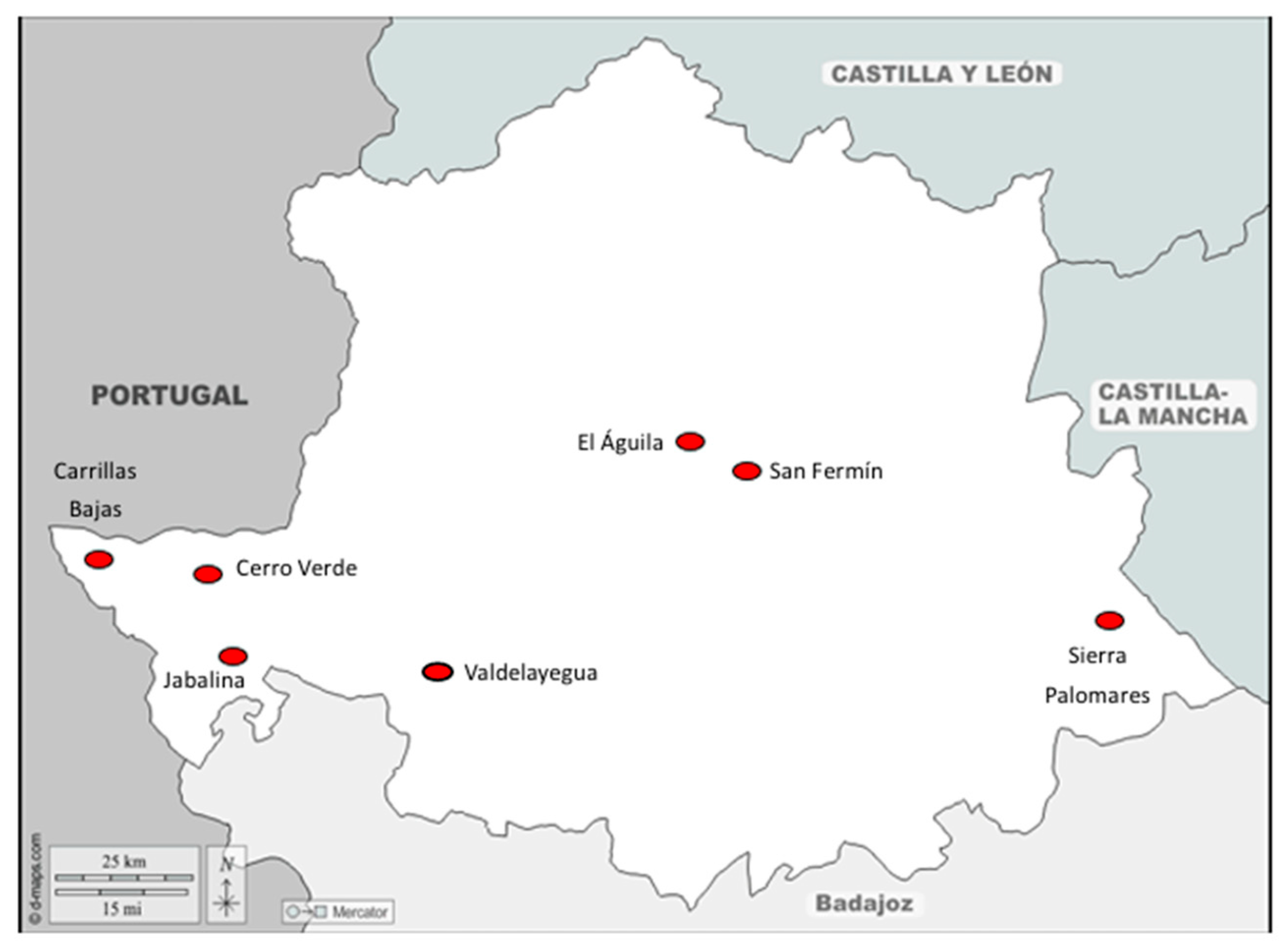
Figure 2.
(A) Anterior part of Dictyocaulus spp.: Buccal capsule; (B) and (C ) posterior part of male and female, respectively.
Figure 2.
(A) Anterior part of Dictyocaulus spp.: Buccal capsule; (B) and (C ) posterior part of male and female, respectively.
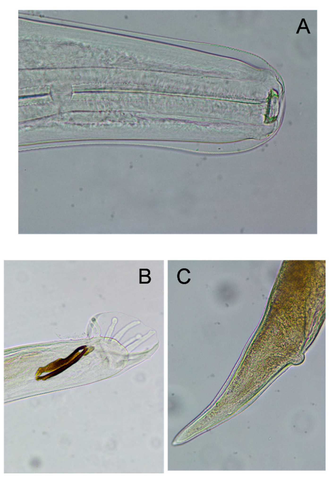
Figure 3.
(A) Bronchioitis with a destructuring of the bronchiolar epithelium; (B). Cross section of adult Dictyocaulus spp. in the bronchial lumen, surrounded by hemorrhages. (C ). Cross section of adult Dictyocaulus sp.. int he bronchial lumen and structural components: fibrous capsule (C9, acellular laminated membrane (L) and germinative membrane 8G). (D) Non-specific interstitial pneumonia and mild hyperplasia of the alveolar epithelium.
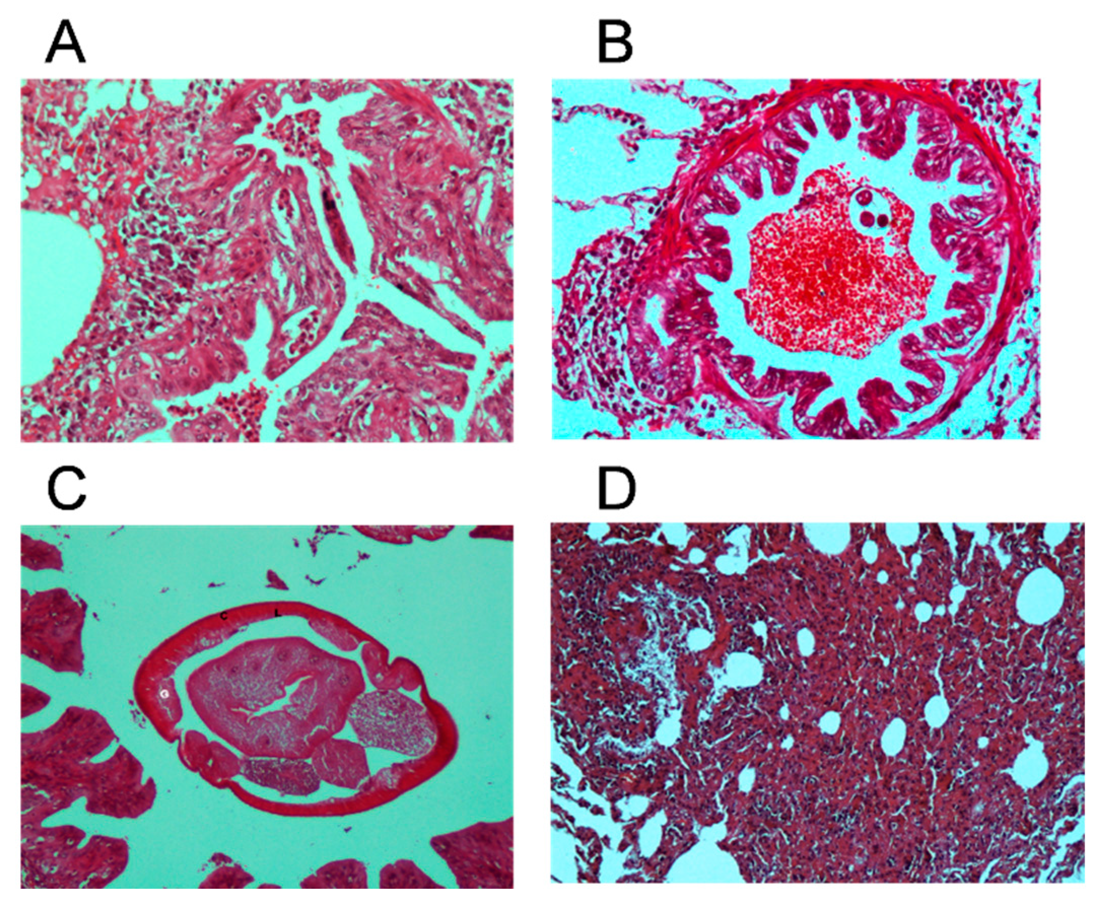
Figure 4.
Graphical philogenetic relationship among the three Dictyocaulus spp. genetic groups under a similarity threshold greater then 90% respect to Genebank sequences (NCBI, USA). Central group consist of conflicting species assignations.
Figure 4.
Graphical philogenetic relationship among the three Dictyocaulus spp. genetic groups under a similarity threshold greater then 90% respect to Genebank sequences (NCBI, USA). Central group consist of conflicting species assignations.
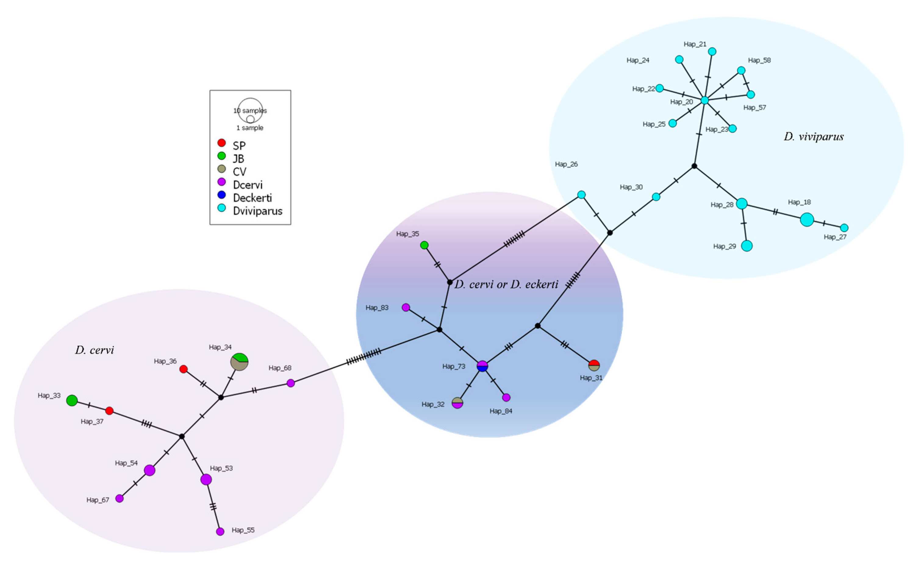

Table 1.
Location and date of material collection.
| Name | Location | Date |
|---|---|---|
| Cuadrillas Bajas | Cedillo | 21/09/23 |
| San Fermín | Torrejón el Rubio | 03/12/23 |
| Sierra Palomares | Alía | 27/01/24 |
| Jabalina | Salorino | 02/02/24 |
| Valdelayegua | Aliseda | 09/02/24 |
| Cerro Verde | Carbajo | 11/02/24 |
| El Águila | Serradilla | 17/02/24 |
Table 2.
Number of deers, location, macroscopic, microscopic results and adults found.
| Lungs | Location | Macroscopic | Microscopic | Adult number |
| Deer 1 | Carrillas | + | + | 4 |
| Deer 2 | Carrillas | - | + | |
| Deer 3 | Carrillas | - | - | |
| Deer 4 | Carrillas | - | - | |
| Deer 5 | Carrillas | - | - | |
| Deer 6 | San Fermín | - | - | |
| Deer 7 | San Fermín | - | - | |
| Deer 8 | San Fermín | - | - | |
| Deer 9 | San Fermín | - | + | |
| Deer 10 | San Fermín | - | - | |
| Deer 11 | San Fermín | - | - | |
| Deer 12 | Sierrra Paloma | - | - | |
| Deer 13 | Sierrra Paloma | - | - | |
| Deer 14 | Sierrra Paloma | - | - | |
| Deer 15 | Sierrra Paloma | - | + | |
| Deer 16 | Sierrra Paloma | + | + | 8 |
| Deer 17 | Jabalina | + | + | 17 |
| Deer 18 | Jabalina | + | + | 22 |
| Deer 19 | Jabalina | - | - | |
| Deer 20 | Jabalina | - | - | |
| Deer 21 | Jabalina | - | - | |
| Deer 22 | Valdelayegua | - | - | |
| Deer 23 | Valdelayegua | - | - | |
| Deer 24 | Valdelayegua | - | - | |
| Deer 25 | Valdelayegua | - | - | |
| Deer 26 | Valdelayegua | - | - | |
| Deer 27 | Cerro Verde | - | - | |
| Deer 28 | Cerro Verde | - | - | |
| Deer 29 | Cerro Verde | - | - | |
| Deer 30 | Cerro Verde | - | - | |
| Deer 31 | Cerro Verde | + | + | 13 |
| Deer 32 | El Águila | - | - | |
| Deer 33 | El Águila | - | - | |
| Deer 34 | El Águila | - | - | |
| Deer 35 | El Águila | - | - | |
| Deer 36 | El Águila | - | - | |
| TOTAL | 5 | 8 | 64 |
Disclaimer/Publisher’s Note: The statements, opinions and data contained in all publications are solely those of the individual author(s) and contributor(s) and not of MDPI and/or the editor(s). MDPI and/or the editor(s) disclaim responsibility for any injury to people or property resulting from any ideas, methods, instructions or products referred to in the content. |
© 2025 by the authors. Licensee MDPI, Basel, Switzerland. This article is an open access article distributed under the terms and conditions of the Creative Commons Attribution (CC BY) license (http://creativecommons.org/licenses/by/4.0/).
Copyright: This open access article is published under a Creative Commons CC BY 4.0 license, which permit the free download, distribution, and reuse, provided that the author and preprint are cited in any reuse.


