
Submitted:
08 April 2025
Posted:
11 April 2025
You are already at the latest version
Abstract
Introduction: The treatment of MBC patients is guided by receptor status, with re-biopsy at relapse recommended to reassess hormone receptor (HR) status. Various treatment options are available for HER2- MBC, including CDK4/6 inhibitors, PARP inhibitors, and checkpoint inhibitors. The study highlights the importance of determining receptor subtype for guiding treatment choices. Patients and Methods: The GIM 13 AMBRA study is a longitudinal cohort study involving 42 centers in Italy. It includes data from 939 HER2- MBC patients enrolled between May 2015 and September 2020. The study analyzes the impact of HR expression changes on clinical outcomes using Kaplan-Meier survival curves and other statistical methods. Results: Among the 939 patients, 588 were re-biopsied at first relapse. The study found no significant differences in disease-free survival (DFS), progression-free survival (PFS), or overall survival (OS) between patients whose tumors changed molecular subtype and those who did not. However, post-progression survival from first-line treatment (PPS1) was different between the two groups. Discussion: The study confirms the phenomenon of receptor discordance between primary tumors and metastases. It emphasizes the need for re-biopsy in recurrent MBC to guide treatment strategies. The findings align with previous studies and highlight the importance of understanding receptor changes for improving patient outcomes. Conclusion: The GIM 13 AMBRA study provides valuable insights into the impact of molecular subtype changes on survival outcomes in Luminal MBC patients. It underscores the importance of re-biopsy and personalized treatment strategies in managing metastatic breast cancer.
Keywords:
Luminal metastatic breast cancer
; overall survival
; progression free survival
1. Introduction
Breast cancer (BC) continues to be the leading cause of cancer death among women worldwide [1]. In Italy, incidence accounts for approximately 55,000 new diagnosed case per year and mortality for 13,000 deaths [2]. Despite the great progress in the treatment of the primary tumor, about 30% of breast patients are destined to develop distant metastases [3]. Among the most important factors associated with disease recurrence and overall survival, nodal involvement, hormone receptor status, Human Epidermal growth factor receptor-2 (HER2) expression and proliferation index play a key role [4]. The clinical course of metastatic breast cancer (MBC) is very heterogeneous in terms of growth rate and response to systemic therapies, however medical treatments remain a palliative cure. Median survival is about 2 years for some subtypes. Treatment of metastatic breast cancer (MBC) patients is mostly based on the receptor status [5], independently of the setting (adjuvant vs metastatic) and the line of therapy. In Human Epidermal growth factor Receptor 2-negative (HER2-) patients, a first biopsy at the time of relapse is strongly recommended to re-assess Hormone Receptor status, especially in HR+, considering the wide choice of multiple therapeutic agents [6]. In recent years, various treatment options became available for HER2- MBC: in the first-line setting, these options have included Cycline-Dependent Kinases 4/6 inhibitors (CDK4/6i) in combination with aromatase inhibitors or tamoxifen for HR+ patients, [7,8,9,10], PARP inhibitors for patients with BRCA1/2 mutations [11,12], and checkpoint inhibitors for PD-L1-positive triple-negative disease (TNBC) [13]. Following these developments, it has become increasingly important to determine the receptor subtype of the tumor in guiding systemic treatment choices, even in second- and further-line settings due to the potential changes in HR and HER2 expression between primary tumor and metastatic sites, or before and after a first- or second-line treatment. A recent meta-analysis, including 39 prospective and retrospective studies, found discordance rates in terms of estrogen receptor (ER), progesterone receptor (PR), and HER2 status between primary breast tumor and loco-regional or distant recurrences of 19% for ER, 31% for PR, and 10% for HER2, respectively [14]. In terms of clinical implication, a change in receptor subtype often leads to an adjustment of the treatment strategy, which can also translate into different outcomes, as different authors reported [15]. The GIM 13 - AMBRA study is a longitudinal, cohort study aiming to describe therapeutic choices in HER2- MBC in the Italian real-life setting. The main objectives of the GIM 13-AMBRA study have been previously described [16] and briefly included the description of the strategies used as first, second or subsequent lines of treatment in patients receiving at least one chemotherapy line (CHT), evaluation of possible correlations between the choice of treatment, both in the adjuvant phase and for metastatic disease and patient characteristics (age, menopausal status, comorbidities) and the evaluation of adherence to literature recommendations for therapeutic sequences in the clinical practice. Here, we report data regarding the impact of molecular subtype change (lack of Hormone Receptors at the first biopsy for metastatic disease) on clinical outcomes, on real-word Progression Free Survival (rwPFS), Time to Treatment Change (TTC) and Overall Survival (OS).
2. Patients and Methods
2.1. Study Design
The GIM 13 AMBRA is a longitudinal cohort study, which has collected data of the first 50 consecutive HER2- MBC patients, who started a first-, second- or subsequent line CHT between January 2012 and December 2016. Forty-two centers have been selected from the 192 national Centers listed in the “Libro Bianco 2012 of the Italian Association of Medical Oncology – AIOM), according to hospital type and geographical distribution. Eligible patients were females aged 18 years or more, with newly diagnosed MBC and who provided written informed consent. All Centers have been authorized by their Ethical Committees (ECs), after the approval of Coordinating Center EC (CE Brianza).
2.2. Objectives
The primary objective of the main study was to describe the strategies in terms of first, second and subsequent lines of treatment in patients receiving at least one chemotherapy line (CHT) and the relative outcome parameters, whose results have already been reported [13]. In the present analysis, we report data regarding the impact of HR expression change or not measured at the first biopsy done at relapse on the main outcome parameters in HR+/HER2- tumors.
2.3. Statistical Analysis
The clinical outcomes were real-world Progression-Free Survival at first- (rwPFS1) or second-line treatment (rwPFS2), defined as the time between first/second-line therapy start and time to progression, according to investigator, or censored to date of latest news; Time to Treatment Change of first- (TTC1) or second-line (TTC2) therapy, defined as the time between the start date, declared by Investigator, of first- or second-line treatment and the date, not defined a priori due to the observational design of the study, of subsequent therapy start. OS was defined as the time between the date of diagnosis of metastatic disease and date of death (any cause) or censored to the date of latest news. The variables were analyzed in the different groups of patients according to whether the hormone receptor status changed at relapse. Normal probability plots have been used to identify substantive deviations from original evolution. This includes identifying outliers, skewness, kurtosis, a need for transformations, and mixtures. Analyses were carried out using NCSS® 12 statistical Software 2018 (Kaysville, Utah, USA). Continuous variables have been evaluated with descriptive statistics (including number of patients, mean, standard deviation, median, minimum, 25th and 75th percentiles, maximum). Categorical variables have been evaluated with frequency and percentage. Tumor subtypes were defined according to the definition provided by Prat et al. [17]
3. Results
3.1. Patients’ and Tumor Characteristics at First Relapse
Between May 2015 and September 2020, 1071 patients have been enrolled in the main study, of whom 132 (12.3%) were not considered eligible due to 1) incomplete information about 1st-line treatment, 2) other reasons, leading to a total of 939 (Luminal N=794; TNBC, N=145) evaluable patients for the main analysis. Demographic characteristics have been described elsewhere in our previous paper. Briefly, median age at primary tumor diagnosis was 51.9 years (range 50.6-52.9) and most of the patients received adjuvant CHT (71.8%), mainly a combination of anthracycline + taxanes (305, 31.5%), or anthracycline + other drugs (266, 28.3%). Re-biopsy at relapse was done in 588 out of 939 patients (62.6%) (Supplementary Figure 1).
Scheme 1.
Consort diagram describes the enrolled population, and the final Luminal population considered for the present analysis.
Scheme 1.
Consort diagram describes the enrolled population, and the final Luminal population considered for the present analysis.
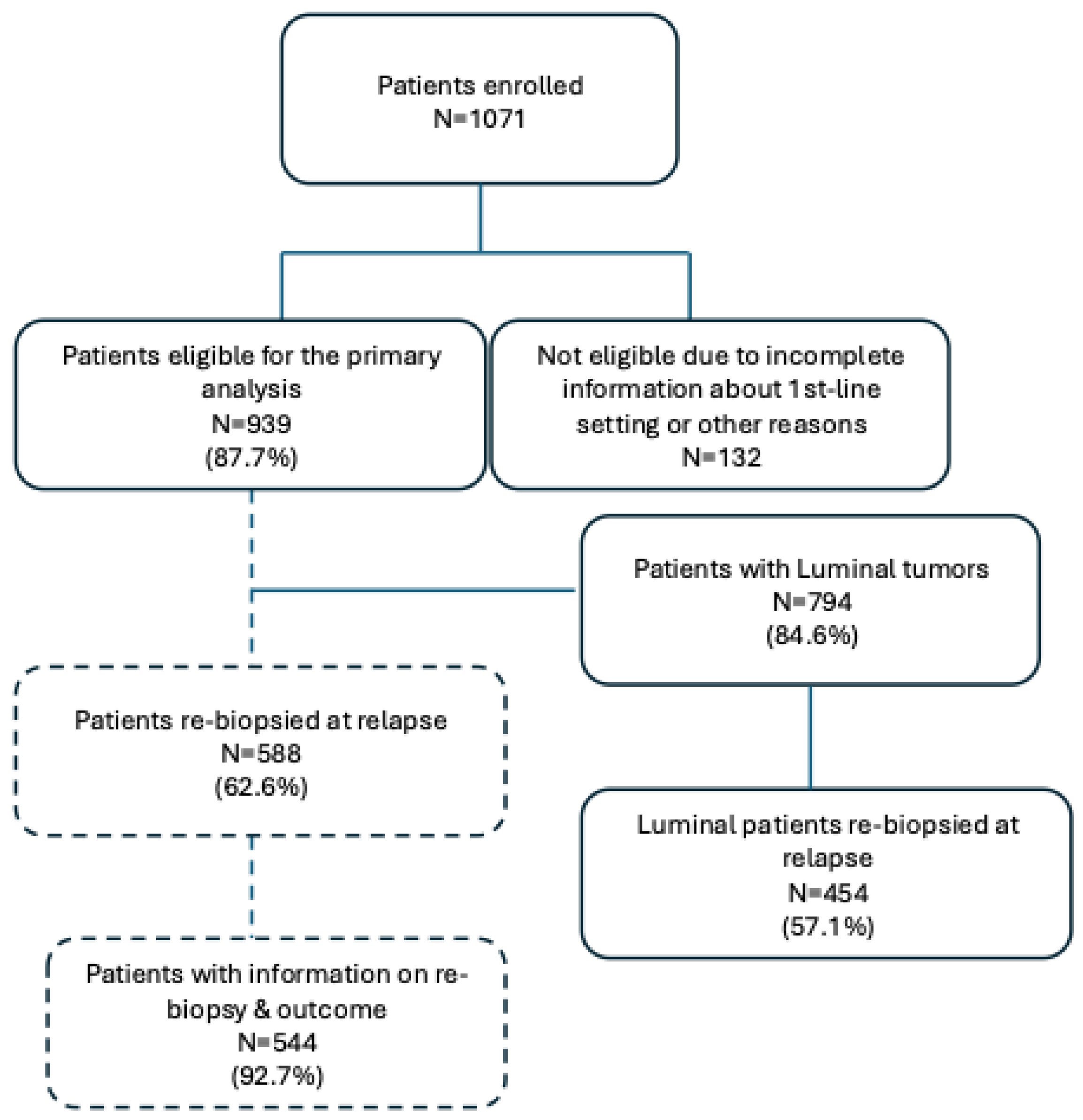
Percentages of tumors rebiopsed at relapse did not significantly differ according the main ORIGINAL molecular subtype. Details are reported in Supplementary Table 1.
A total of 49 tumors (9.1%) among those re-biopsied changed their molecular category at relapse: 36 Luminal A or Luminal B tumors became TNBC (7.9%), and 13 TNBC became Luminal B (13%). As previously declared and considering the occasional change into Luminal subtype occurring in TNBC tumors as well as the low clinical relevance of this event, we report here outcome data only for Luminal tumors.
Among the 454 Luminal patients re-biopsied at relapse, 288 (63%) have received a sequence of chemo- and endocrine therapy (ET) and 109 (24%) endocrine therapy alone as adjuvant treatment. The type of treatment at first relapse was available for 441 (97.1%): the majority received endocrine therapy (151, 34.2%), 144 (32.7%) were treated with chemotherapy and 41 (31.9%) with chemotherapy followed by endocrine treatment.
No difference in terms of median Disease-Free Survival (DFS) was observed in Luminal tumors according to the change of molecular subtype in comparison to those which did not change (Luminal: 74.6 months, 95% CI: 66.8-82.1 vs Luminal becoming TNBC: 89.7 months, 95% CI: 44.7-103.5).
Subtype change did not significantly affect the main progression outcomes: no difference in median rwPFS1 was observed between the Luminal tumors which remain as such (14.4 months, 95% CI 12.4-16) and those changed into TNBC subtype (11.1 months, 95%CI 7.2-16.8; p=0.36) (Figure 1).
Only median post-progression survival from 1st line treatment (PPS1) was different in the 2 groups (33.1 months, 95% CI: 30.1-36.6 vs 24 months (95% CI: 15.8-30.9; p=0.031) (Figure 3a), while no difference in median OS was observed between the two groups according to the change of molecular subtype (9.4 years, 95% CI: 8.7-9.8 vs 9.7 years, 95% CI: 5.6-11.2) (Figure 3b).
4. Discussion
Discordance in ER, PgR and HER2 status between primary breast tumors and metastases is a well-known phenomenon, previously described by many authors and analyzed in different, even small, series [15,16]. However, the magnitude of this change in affecting patients’ management and survival outcomes remains important to define therapeutic strategies, particularly in Luminal patients. The GIM 13 – AMBRA study is a cohort, real-world study which collected data of more than 900 HER2- metastatic breast cancer patients in Italy from diagnosis until death, or data censoring, so far representing a priceless opportunity to collect insights useful for future trials. ESMO guidelines [2] strongly support the practice to have a biopsy in patients with recurrent MBC, to confirm histology and to re-assess ER, PgR and HER2 status. Among the 939 patients registered into the study, 588 (62.6%) have been re-biopsied at first relapse, without any difference among Luminal A, Luminal B and TNBC patients. In a large retrospective study, Meegdes et Al [18] conducted a large real-world study which included all patients diagnosed with MBC between 2007–2018 in seven hospitals in Southeast Netherlands and registered in the SONABRE registry, with the aim of assessing the biopsy rate and the factors associated with taking a biopsy of a metastatic site at MBC diagnosis. Interestingly, they found that 60% of patients had a biopsy of a metastatic site at presentation : decision to have a biopsy was higher in academic hospitals (73%), in comparison to teaching hospitals (60%) and non-teaching hospitals (59%). The Author also found that a more recent period of MBC diagnosis was associated with a higher biopsy rate: 67% in 2016–2018 compared with 51% in 2007–2009 (OR=2.14; 95% CI: 1.70–2.70) and that biopsy was performed more frequently in younger patients in comparison to older ones (56–75 years versus > 75 years: OR=1.80; 95% CI: 1.48–2.19; ≤ 55 years versus > 75 years: OR=2.20; 95% CI: 1.74–2.78)
The percentage of re-biopsies done in our study is among the highest, if compared to also those reported in other retrospective studies, the largest one being the ESME database [16]. Grinda et al. reported that re-biopsy was done in 17.6% of the 16703 patients included in the ESME database.
In our analysis, a total of 49 tumors (9.1%) among those re-biopsied changed their molecular category at relapse: 36 Luminal A or Luminal B tumors became TNBC (7.9%), and 13 TNBC became Luminal B (13%). Discordance rates between primary and metastases greatly varies according to the different series, strongly depending on the size of population analyzed. Meegdes et Al., reported an overall receptor discordance rate of 18%, in particular discordance rates were 13% for HR+/HER2- and 12% for TNBC. Mellouli et Al. [19] collected data on 68 BC patients at a single-center in a retrospective study, reporting a discordance in ER status in 20 patients (29.4 %, p = 0.041), with ER-negative conversion in 15 patients (22 %) and ER-positive conversion in 5 patients (7.3 %). They observed a difference in PR status in 27 of the cases (39.7 %, p =0.001): in 24 of the patients (35.2%), the PR status had changed from positive in PBC to negative in metastatic lesions, while in 3 of the patients (4.4 %), the PR status had changed from negative to positive. HR status conversion was detected in 20 of cases (29.4 %, p =0.04): 15 of patients (22 %) had changed from HR-positive status in primary breast cancer to HR-negative status in the metastatic tissues, and 5 of patients (7.35 %) from HR-negative to HR-positive. In the ESME cohort [20], one of the most important analysis ever conducted in a large population, regarding discordance rate in HR expression between the primary site and the metastatic ones, Grind et Al reported a change rate for HR status of 14.2% [95% CI 12.5–16.0] with expression loss in 72.5% and expression gain in 27.5%. For ER status, 15.1% [95% CI 13.3–17.0] of cases showed a change with loss in 67.7% and gain in 32.3%. For PR status, a modification was observed in 31.1% [95% CI 28.7–33.5] with loss in 75.3% and gain in 24.7%. So far, our results in terms of percentages of discordance between primary and metastases are in the middle of those reported by other authors [17], and this can explained in part by the absence of a central review, as often happens in real-world studies, as well the unavailability of information regarding the site where biopsy taken. Other Authors have thus showed that among the factors associated with HR discordance there were metastases to bone only and primary tumor treatments with endocrine therapy [20].
The clinical meaning of discordance and moreover the impact in terms of treatment choice remains a matter of debate. In our analysis, we evaluated outcomes (rwPFS1, rwPFS2, PPS1 and OS) of 454 out of 490 re-biopsied Luminal patients, finding no differences in rwPFS1, rwPFS2 and OS. Differences among the various series remain wide, indicating there is probably the need for larger, prospective trials. Schwieger et al. [18] recently reported that change from an ER or PgR+/HER2− at primary tumor to TNBC paired metastatic one was not associated with decreased survival (p > 0.05) in 258 analyzed patients. Similarly, Peng et al. ( [21] found no statistical difference in PFS according to the subtype of the recurrent or metastatic breast cancer (p > 0.05). In another retrospective study, Yang et al. [19] evaluated the frequency and the prognostic impact of changes in HR and HER2 between primary and recurrent/metastatic lesions in 133 patients: in their population, the ER-discordant cases and ER-loss cases experienced a worse overall survival (OS) (p=0.001 and p=0.016, respectively) and post-recurrence survival (PRS) (p=0.001 and p=0.018, respectively), compared with the respective concordant cases.
In the contemporary era, the treatment choice for HR+ BC which do not change hormone receptor expression from primary to metastases and those which change into TNBC can significantly differ, both in terms of drugs, as well as in terms of additional molecular tests: like an example, first-line treatment of TNBC patients is largely guided by the expression of programmed-death ligand 1 (PDL-1), while HR+ tumors may derive a benefit from more personalized approaches based on the detection of ESR1, AKT or PIK3CA mutations. So far, when and how should we biopsy our metastatic patients? The availability of liquid biopsy, a technique which allows to detect breast cancer cell mutations in different organic fluids, mainly blood and urine, has revolutionized the approach to MBC patients treatment:, as it is easier to perform than tissue biopsies and is virtually non-invasive to the patient. However, it is our opinion that tissue biopsy should remain mandatory to assess ER and PgR status as first approach for a potential subsequent use of liquid biopsy to detect mutations specific of the molecular subtype. Several questions remain open: like an example, considering the high heterogeneity of breast cancers, should we test for PDL-1 expression those Luminal tumors which lost the HR expression becoming TNBC at the time of relapse? How much a biopsy obtain from a single metastatic site is representative of the whole disease? To settle these and other questions, an answer is becoming more and more important, considering the high costs of the new drugs and, above all, the different safety profiles of the various drugs.
Declaration of Interest
None declared
Supplementary Materials
The following supporting information can be downloaded at the website of this paper posted on Preprints.org.
Acknowledgments
The study was supported by Celgene.
References
- H. Sung et al., “Global Cancer Statistics 2020: GLOBOCAN Estimates of Incidence and Mortality Worldwide for 36 Cancers in 185 Countries,” CA Cancer J Clin, vol. 71, no. 3, pp. 209–249, May 2021. [CrossRef]
- “Cos’è il Cancro | AIRC.” Accessed: Apr. 04, 2025. [Online]. Available: https://www.airc.it/cancro/informazioni-tumori/cose-il-cancro/numeri-del-cancro.
- T. Sørlie et al., “Gene expression patterns of breast carcinomas distinguish tumor subclasses with clinical implications,” Proc Natl Acad Sci U S A, vol. 98, no. 19, pp. 10869–10874, Sep. 2001. [CrossRef]
- K. H. Allison, “Prognostic and predictive parameters in breast pathology: a pathologist’s primer,” Modern Pathology, vol. 34, pp. 94–106, Jan. 2021. [CrossRef]
- G. Curigliano et al., “De-escalating and escalating treatments for early-stage breast cancer: the St. Gallen International Expert Consensus Conference on the Primary Therapy of Early Breast Cancer 2017,” Ann Oncol, vol. 28, no. 8, pp. 1700–1712, Aug. 2017. [CrossRef]
- Gennari et al., “ESMO Clinical Practice Guideline for the diagnosis, staging and treatment of patients with metastatic breast cancer 5 behalf of the ESMO Guidelines Committee,” Annals of Oncology, vol. 32, pp. 1475–1495, 2021. [CrossRef]
- D. J. Slamon et al., “Overall Survival With Palbociclib Plus Letrozole in Advanced Breast Cancer,” J Clin Oncol, vol. 42, no. 9, pp. 994–1000, Mar. 2024. [CrossRef]
- G. N. Hortobagyi et al., “Overall Survival with Ribociclib plus Letrozole in Advanced Breast Cancer,” N Engl J Med, vol. 386, no. 10, pp. 942–950, Mar. 2022. [CrossRef]
- M. P. Goetz et al., “Abemaciclib plus a nonsteroidal aromatase inhibitor as initial therapy for HR+, HER2- advanced breast cancer: final overall survival results of MONARCH 3,” Ann Oncol, vol. 35, no. 8, pp. 718–727, Aug. 2024. [CrossRef]
- Y. S. Lu et al., “Updated Overall Survival of Ribociclib plus Endocrine Therapy versus Endocrine Therapy Alone in Pre- and Perimenopausal Patients with HR+/HER2- Advanced Breast Cancer in MONALEESA-7: A Phase III Randomized Clinical Trial,” Clin Cancer Res, vol. 28, no. 5, pp. 851–859, Mar. 2022. [CrossRef]
- M. Robson et al., “Olaparib for Metastatic Breast Cancer in Patients with a Germline BRCA Mutation,” N Engl J Med, vol. 377, no. 6, pp. 523–533, Aug. 2017. [CrossRef]
- M. E. Robson et al., “OlympiAD final overall survival and tolerability results: Olaparib versus chemotherapy treatment of physician’s choice in patients with a germline BRCA mutation and HER2-negative metastatic breast cancer,” Annals of Oncology, vol. 30, no. 4, pp. 558–566, Apr. 2019. [CrossRef]
- P. Schmid et al., “Pembrolizumab for Early Triple-Negative Breast Cancer,” New England Journal of Medicine, vol. 382, no. 9, pp. 810–821, Feb. 2020. [CrossRef]
- W. A. M. E. Schrijver, K. P. M. Suijkerbuijk, C. H. Van Gils, E. Van Der Wall, C. B. Moelans, and P. J. Van Diest, “Receptor Conversion in Distant Breast Cancer Metastases: A Systematic Review and Meta-analysis,” J Natl Cancer Inst, vol. 110, no. 6, pp. 568–580, Jun. 2018. [CrossRef]
- Kolberg-Liedtke et al., “Phenotype Discordance between Primary Tumor and Metastasis Impacts Metastasis Site and Outcome: Results of WSG-DETECT-PriMet,” Breast Care (Basel), vol. 16, no. 5, pp. 475–483, Oct. 2021. [CrossRef]
- M. E. Cazzaniga et al., “Clinical Outcomes of HER2-Negative Metastatic Breast Cancer Patients in Italy in the Last Decade: Results of the GIM 13-AMBRA Study,” Cancers (Basel), vol. 16, no. 1, 2024. [CrossRef]
- Prat, B. Adamo, M. C. U. Cheang, C. K. Anders, L. A. Carey, and C. M. Perou, “Molecular characterization of basal-like and non-basal-like triple-negative breast cancer,” Oncologist, vol. 18, no. 2, pp. 123–133, Feb. 2013. [CrossRef]
- M. Meegdes et al., “The initial hormone receptor/HER2 subtype is the main determinator of subtype discordance in advanced breast cancer: a study of the SONABRE registry,” Breast Cancer Res Treat, vol. 192, no. 2, pp. 331–342, Apr. 2022. [CrossRef]
- M. Mellouli et al., “Discordance in receptor status between primary and metastatic breast cancer and overall survival: A single-center analysis,” Ann Diagn Pathol, vol. 61, Dec. 2022. [CrossRef]
- T. Grinda et al., “Phenotypic discordance between primary and metastatic breast cancer in the large-scale real-life multicenter French ESME cohort,” NPJ Breast Cancer, vol. 7, no. 1, Dec. 2021. [CrossRef]
- L. Peng, Z. Zhang, D. Zhao, J. Zhao, F. Mao, and Q. Sun, “Discordance in ER, PR, HER2, and Ki-67 Expression Between Primary and Recurrent/Metastatic Lesions in Patients with Primary Early Stage Breast Cancer and the Clinical Significance: Retrospective Analysis of 75 Cases,” Pathology and Oncology Research, vol. 27, Apr. 2021. [CrossRef]
Figure 1.
Normal probability plots of rwPFS1 according to the change of molecular. Median rwPFS2 was 8.1 months (95% CI: 7.3-9.2) and 7.1 months (95% CI: 5.3-9.1) respectively (p=0.59) (Figure 2).
Figure 1.
Normal probability plots of rwPFS1 according to the change of molecular. Median rwPFS2 was 8.1 months (95% CI: 7.3-9.2) and 7.1 months (95% CI: 5.3-9.1) respectively (p=0.59) (Figure 2).
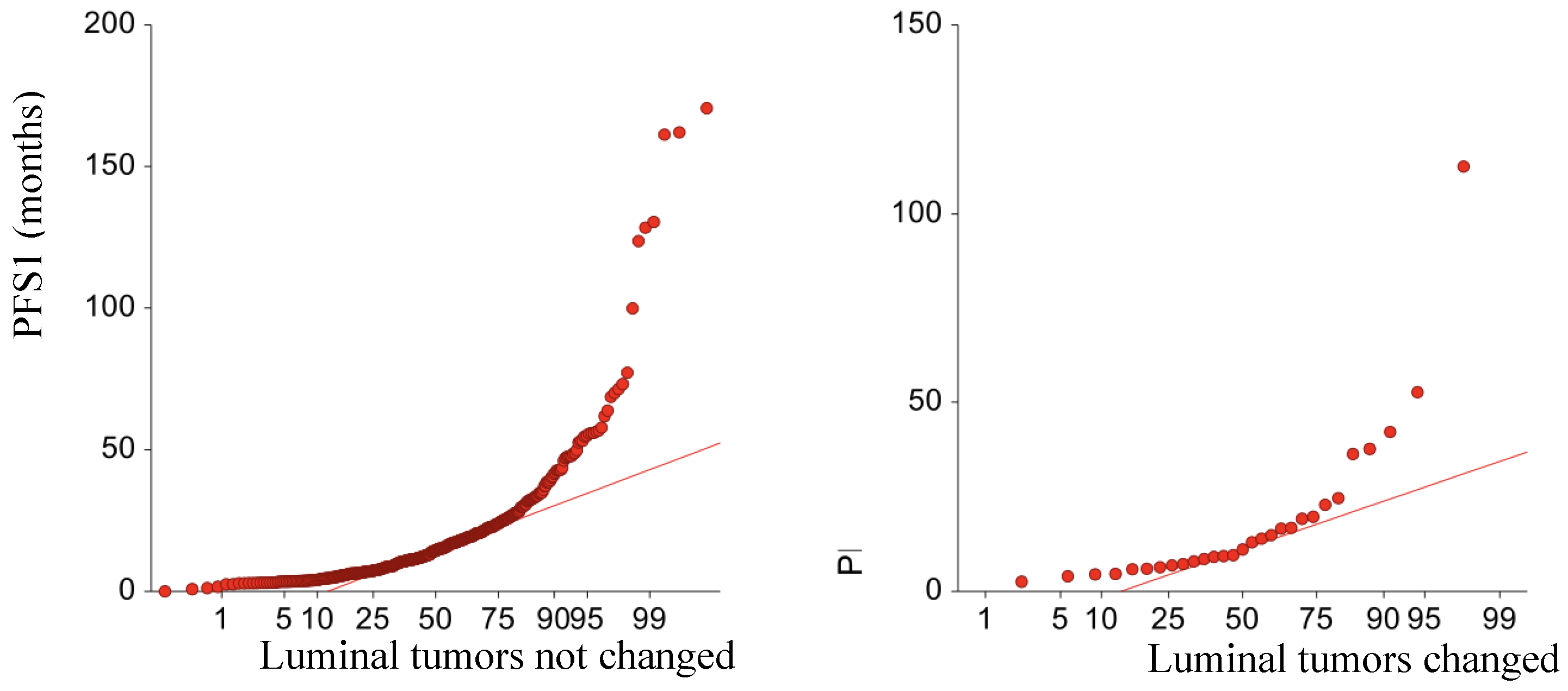
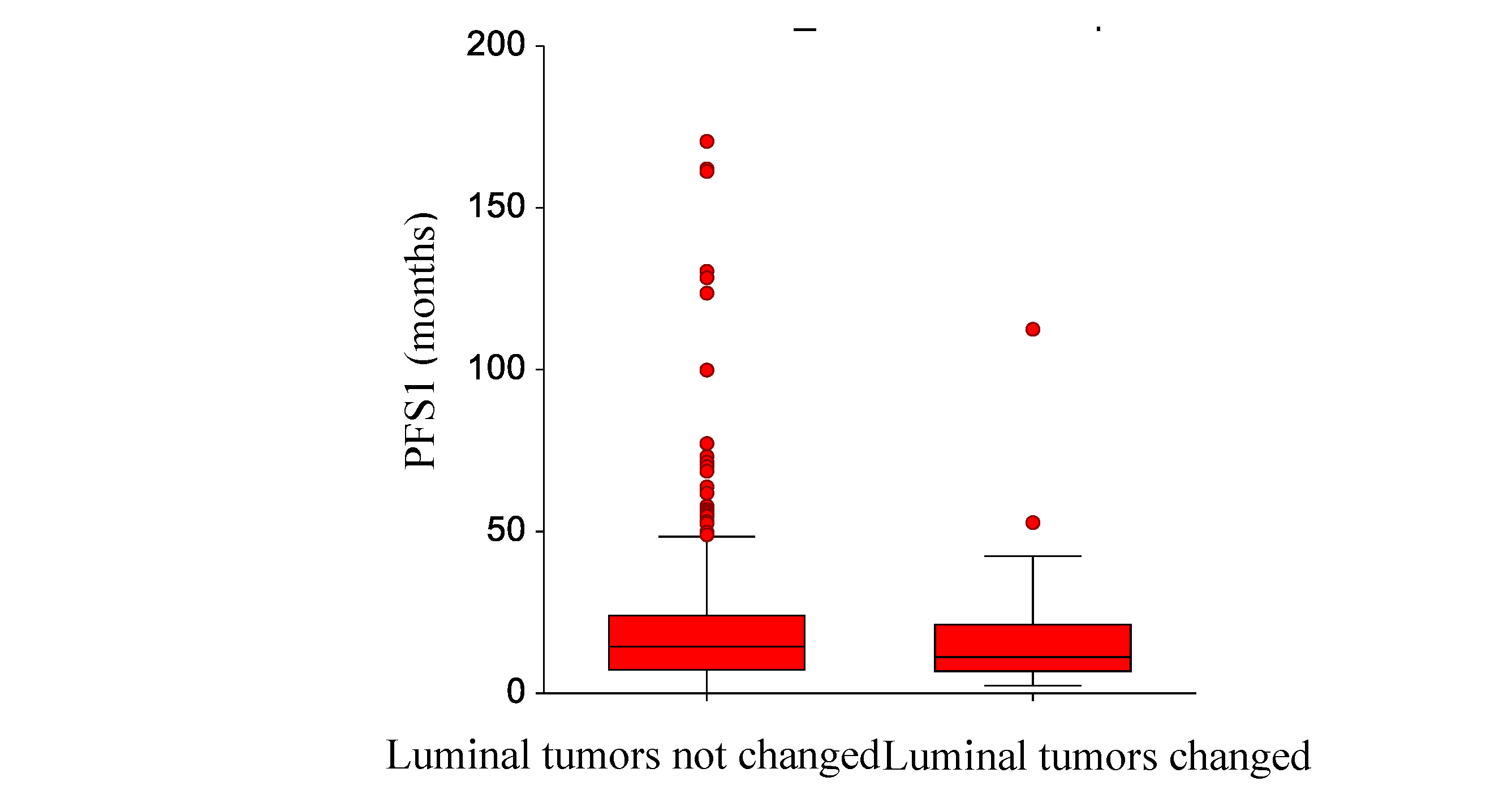
Figure 2.
Normal probability plots of rwPFS2 (months) according to the change or not of molecular subtype.
Figure 2.
Normal probability plots of rwPFS2 (months) according to the change or not of molecular subtype.
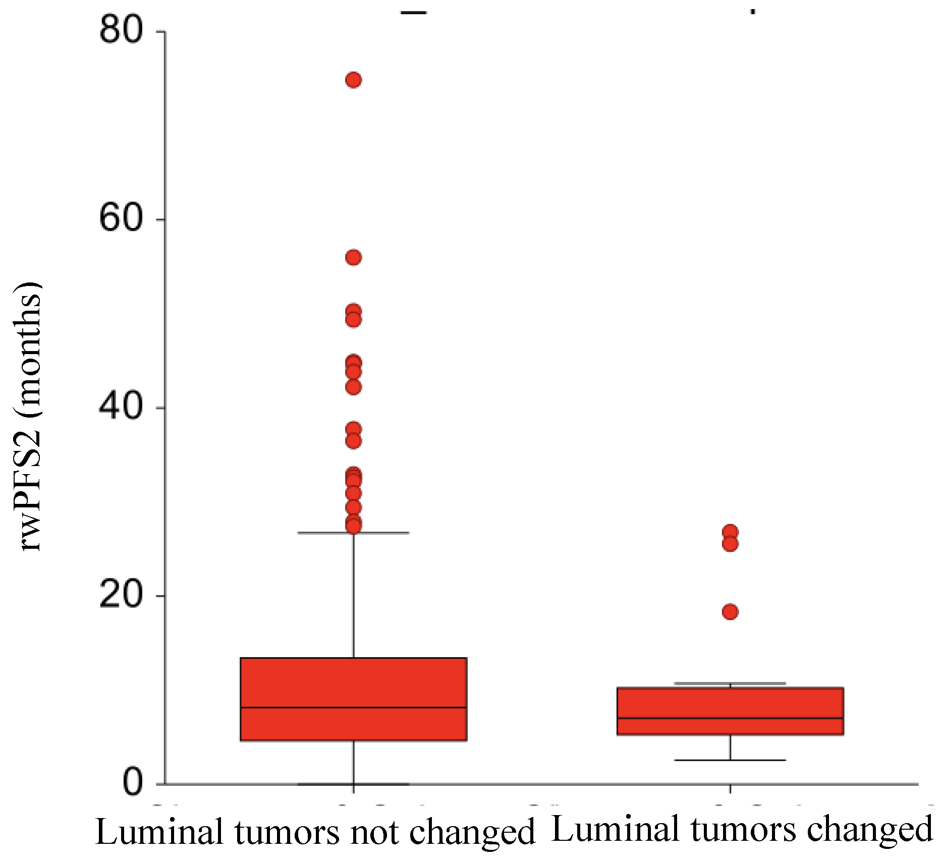
Figure 3.
Normal probability plots of (a) PPS1 (months) and (b) OS (years) according to the change of molecular subtype.
Figure 3.
Normal probability plots of (a) PPS1 (months) and (b) OS (years) according to the change of molecular subtype.
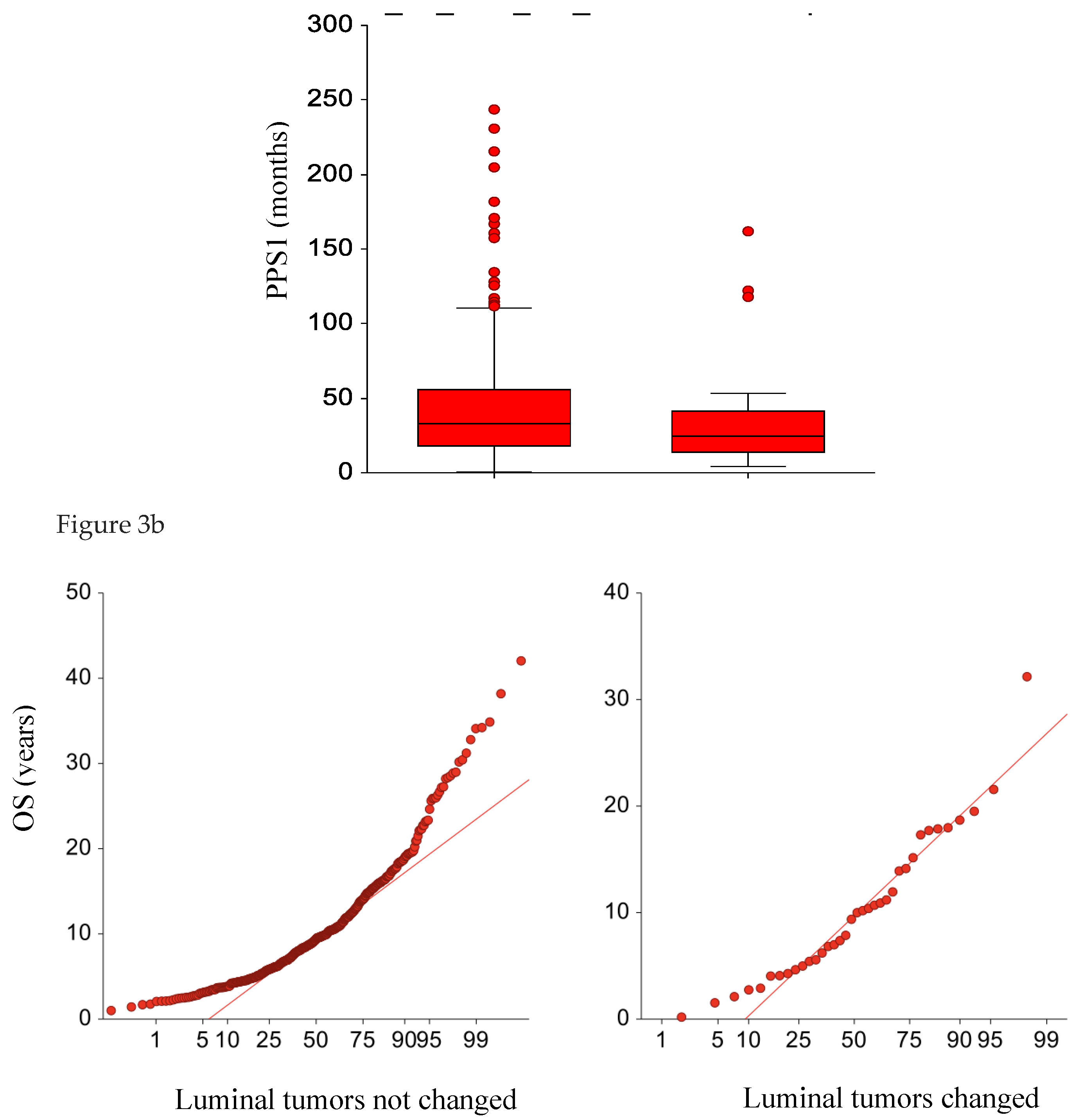

Table 1.
Proportion of tumors re-biopsied according to molecular subtype.
| Luminal A | Luminal B | TNBC |
| 233/38660.4% | 257/40862.9% | 100/14568.9% |
Disclaimer/Publisher’s Note: The statements, opinions and data contained in all publications are solely those of the individual author(s) and contributor(s) and not of MDPI and/or the editor(s). MDPI and/or the editor(s) disclaim responsibility for any injury to people or property resulting from any ideas, methods, instructions or products referred to in the content. |
© 2025 by the authors. Licensee MDPI, Basel, Switzerland. This article is an open access article distributed under the terms and conditions of the Creative Commons Attribution (CC BY) license (http://creativecommons.org/licenses/by/4.0/).
Copyright: This open access article is published under a Creative Commons CC BY 4.0 license, which permit the free download, distribution, and reuse, provided that the author and preprint are cited in any reuse.


