
Submitted:
01 April 2025
Posted:
01 April 2025
You are already at the latest version
Abstract
Staphylococcus aureus, particularly its methicillin-resistant strains (MRSA), poses significant challenges in healthcare settings due to its propensity to form biofilms on various surfaces. These biofilms enhance bacterial survival and increase resistance to conventional treatments, complicating infection control efforts. This study investigates the efficacy of a novel approach combining povidone-iodine (PVP-I) and hydrogen peroxide (H₂O₂) to combat Staphylococcus aureus biofilm formation in hospital environments. A range of methodologies were employed, including biofilm formation assays, gene expression analysis, colony-forming unit (CFU) enumeration, and confocal microscopy, to assess the effects of PVP-I and H₂O₂, both individually and in combination. The results demonstrated a synergistic effect when these agents were combined, significantly reducing biofilm formation compared to the use of either compound alone. PVP-I, an io-dine-based antiseptic, and H₂O₂, a potent oxidizing agent, each exhibit potential in disrupting bacterial biofilms when used independently. However, their combined application appears to enhance penetration into the biofilm matrix and increase overall antimicrobial activity. These findings have important implications for infection control strategies in healthcare settings. By harnessing the complementary mechanisms of PVP-I and H₂O₂, this approach offers a promising solution for preventing and eradicating biofilm-associated infections on medical devices and hospital surfaces. Additionally, the use of two agents with different modes of action may help mitigate the risk of developing antimicrobial resistance. This study lays the groundwork for future research aimed at optimizing concentrations, application methods, and assessing the long-term efficacy and safety of this combined approach. Ultimately, this research contributes to improved patient outcomes and a reduction in the economic burden associated with biofilm-related complications.
Keywords:
Staphylococcus aureus
; povidone-iodine (PVP-I)
; hydrogen peroxide (H₂O₂)
; biofilms
; Anti-Biofilm activity
1. Introduction
Periprosthetic joint infection (PJI) represents one of the most severe complications following arthroplasty joint replacement (AJR) [1]. Bacterial pathogens are responsible for over 98% of PJIs, with Staphylococcus aureus and coagulase-negative staphylococci accounting for 50% to 60% of all cases [2,3]. Staphylococcus aureus (S. aureus) is the most prevalent cause of infections related to implanted biomaterials [4]. Particularly, methicillin-resistant Staphylococcus aureus (MRSA) continues to be a significant global health challenge in hospital settings. This Gram-positive bacterium is a leading cause of hospital-associated infections (HAIs), contributing substantially to patient morbidity, mortality, and escalating healthcare costs worldwide. The prevalence of MRSA varies considerably across different countries and regions. According to the World Health Organization’s (WHO) 2014 report on antimicrobial resistance, MRSA prevalence in hospitals exceeded 20% in all regions, with some areas reporting infection rates as high as 80%. In South Korea, the burden of hospital-acquired Staphylococcus aureus bloodstream infections (SA-BSI) is notably high. A national study conducted in 2011 estimated the incidence of hospital-acquired MRSA-BSI at 0.12 cases per 1,000 patient-days, compared to 0.04 cases per 1,000 patient-days for methicillin-sensitive Staphylococcus aureus bloodstream infections (MSSA-BSI). This translates to approximately 2,946 cases of MRSA-BSI and 986 cases of MSSA-BSI annually in hospitals with more than 500 beds. The mortality rate associated with these infections is alarmingly high, with MRSA-BSI at 31.9% and MSSA-BSI at 14.1%. The economic impact of these infections is also significant, with the additional cost per case estimated at $20,494 for MRSA-BSI and $6,914 for MSSA-BSI, leading to an annual total cost exceeding $67.2 million. In recent years, MRSA prevalence in South Korean hospitals has increased, with MRSA accounting for 60-70% of all Staphylococcus aureus infections. This high prevalence underscores the urgent need for effective infection control measures within healthcare facilities to mitigate the spread and impact of MRSA [5]. Given the substantial clinical and economic burden of Staphylococcus aureus infections in South Korean hospitals, there is an urgent need to develop innovative strategies for the prevention and control of these infections. The use of povidone-iodine (PVP-I) and hydrogen peroxide (H2O2) has shown considerable potential in addressing these infections and offers several advantages over traditional methods. Iodine-based compounds, such as PVP-I, have long been recognized for their broad-spectrum antimicrobial activity. They are effective against a wide range of pathogens, including antibiotic-resistant strains like MRSA. These agents act by releasing free iodine, which penetrates microbial cell walls and disrupts critical cellular processes, leading to bacterial death. Their efficacy in reducing biofilm formation makes them particularly valuable in hospital settings [6,7]. Biofilms are complex systems comprising sessile bacteria encased within an extracellular polymeric substance (EPS), which serves as the structural scaffold of the biofilm. EPS also acts as a physical and chemical barrier, making it difficult for antibiotics to penetrate the biofilm, thus providing a protective shield against antimicrobial agents [8,9]. Hydrogen peroxide is a potent oxidizing agent that generates free radicals, causing oxidative stress and damage to bacterial cells. Research indicates that H2O2 is effective against various hospital pathogens, including Staphylococcus aureus. The ability of H2O2 to penetrate biofilms, along with its relatively low toxicity to human cells at appropriate concentrations, makes it an attractive option for infection control [10]. The combination of iodine-based compounds with H2O2 provides a synergistic approach to infection prevention and control [11,12,13]. This dual strategy has the potential to overcome the limitations of monotherapy, offering a more comprehensive solution to combat Staphylococcus aureus infections [14]. By targeting different aspects of bacterial survival and proliferation, this combination may enhance overall efficacy and reduce the risk of resistance development. Moreover, both agents are cost-effective and readily available, making them practical for implementation in hospital infection control programs. Their use could significantly reduce infection rates, thereby lowering patient morbidity and mortality, shortening hospital stays, and alleviating the economic burden on healthcare systems. This study investigates the effects of PVP-I and H2O2, both individually and in combination, on the eradication of Staphylococcus aureus biofilm formation. By leveraging the complementary mechanisms of these two agents, we aim to develop a more effective strategy to combat biofilm-associated infections in hospital environments, ultimately improving patient outcomes and reducing the economic burden associated with these infections.
2. Results
2.1. Morphological Analysis of Staphylococcus aureus
Staphylococcus aureus was analyzed using Mannitol Salt Agar (MSA) and Gram staining. As a Gram-positive bacterium, S. aureus appeared purple under the microscope and exhibited a grape-like cluster arrangement, which is crucial for distinguishing it from other bacterial species. MSA is both a selective and differential medium used to isolate and identify S. aureus. It contains mannitol, which S. aureus can ferment. The fermentation process produces acid, lowering the pH and changing the color of the phenol red indicator, resulting in yellow colonies. This color change differentiates S. aureus from other non-mannitol fermenting staphylococci, such as coagulase-negative staphylococci, which form pink colonies on MSA. These morphological and biochemical tests are critical for diagnosing and managing infections in clinical microbiology (Figure 1).
2.2. Evaluation of PVP-I and H2O2 Effectiveness on Biofilm Removal
The effectiveness of PVP-I and H2O2, both alone and in combination, in removing biofilms was assessed using a biofilm formation assay. After biofilm formation, the metal discs were treated with these agents. Quantitative analysis showed a slight reduction in biofilm biomass with individual treatments of PVP-I and H2O2 compared to the untreated control. However, the combination treatment with PVP-I and H2O2 led to a significant reduction in biofilm quality, indicating a synergistic effect. This combined treatment was notably more effective, highlighting its potential as a strategy for managing biofilm-related infections (Figure 2).
2.3. Expression of Biofilm-Associated Genes Under PVP-I and H2O2 Treatment
The study also investigated the impact of PVP-I and H2O2 treatment on the expression of biofilm-associated genes. The levels of these genes were analyzed to understand their role in biofilm formation and maintenance under oxidative stress conditions. Results indicated that PVP-I and H2O2 treatment significantly modulated the expression of key biofilm-related genes, affecting biofilm structural integrity and resistance characteristics. These findings provide insights into the molecular mechanisms by which these agents influence biofilm dynamics and suggest potential therapeutic strategies for biofilm-associated infections (Figure 3).
2.4. Live/Dead Assay of Biofilms Treated with PVP-I and H2O2
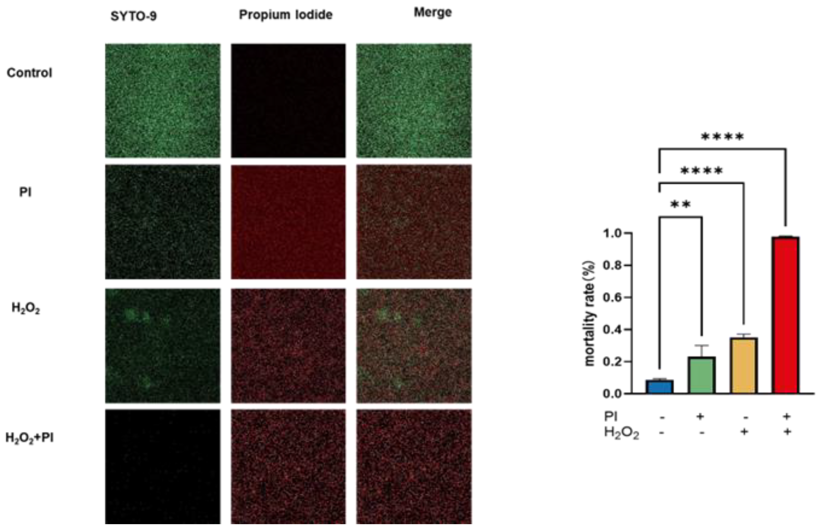
A Live/Dead assay was conducted to evaluate the effectiveness of PVP-I and H2O2, both individually and in combination, in removing biofilms. After treatment, the biofilms were assessed for viability. Quantitative analysis revealed a slight reduction in biofilm biomass with individual treatments of PVP-I and H2O2 compared to the untreated control. The Live/Dead assay demonstrated that both treatments increased the proportion of dead cells, as evidenced by an increase in red fluorescence. However, the combination treatment with PVP-I and H2O2 showed a more pronounced effect, significantly increasing the number of dead cells and thereby more effectively reducing biofilm viability. These findings underscore the potential of combining PVP-I and H2O2 in managing biofilm-related infections (Figure 4).
3. Discussion
Biofilms pose a significant risk in hospital settings due to their ability to form on various surfaces and medical devices, leading to increased infection rates. Periprosthetic joint infections (PJI) are among the most severe complications following joint replacement surgeries [15,16,17]. The unique structure of biofilms makes it challenging for disinfectants like PVP-I to penetrate and eradicate the biofilm, often resulting in diminished antimicrobial effectiveness. Studies indicate that biofilm resistance to antimicrobial agents can be 100 to 1,000 times greater than that of planktonic bacteria [8,18]. Biofilms provide a protective environment for bacteria, rendering conventional antimicrobial treatments ineffective and leading to chronic infections. The prevalence of biofilm-forming microorganisms in hospital environments is high, with Staphylococcus aureus, coagulase-negative staphylococci, and Pseudomonas aeruginosa exhibiting biofilm formation rates of 100% [19]. This high prevalence underscores the clinical challenge posed by biofilms, which complicates patient management and infection control efforts. Continuous research and development of effective biofilm prevention and removal techniques are essential. In hospital settings, PVP-I and hydrogen peroxide have been explored as alternative strategies for removing infections caused by Staphylococcus aureus, including methicillin-resistant strains (MRSA). However, the efficacy of these agents in completely removing biofilms remains a challenge.
Recent studies provide compelling evidence of PVP-I and polyhexamethylene biguanide (PHMB) showing strong antibiofilm activity in vitro [20]. Both agents demonstrated significant activity against biofilms formed by various microorganisms, including MRSA and Pseudomonas aeruginosa. Notably, PVP-I exhibited a faster action against MRSA biofilms compared to PHMB, as evidenced by colony-forming unit (CFU) counts and microscopy analysis. Additionally, another study demonstrated that the combination of PVP-I and vancomycin effectively reduced early biofilm formation by MRSA and methicillin-sensitive Staphylococcus aureus (MSSA) on titanium surfaces [21]. Research indicates that a 5-minute exposure to PVP-I can reduce CFU/cm² of S. aureus biofilm on prosthetic materials by approximately three orders of magnitude. This reduction was confirmed by confocal imaging and crystal violet staining. The study also noted reduced expression of biofilm-related genes such as icaACDR and clfA, suggesting that PVP-I effectively diminishes biofilm formation by affecting bacterial adhesion and virulence [6]. Some studies have shown that hydrogen peroxide can downregulate the expression of biofilm-related genes at certain stages of biofilm development, such as the sod gene in Pseudomonas aeruginosa. However, the effectiveness of hydrogen peroxide in completely removing biofilms formed by S. aureus on infected implants is inconsistent. A recent study demonstrated that sustained application of hydrogen peroxide could effectively control biofilm growth and remove biofilms from dental water systems [22].
This study addresses the persistent challenge posed by biofilms in medical environments, particularly MRSA. These biofilms not only enhance bacterial survival but also confer resistance to conventional antimicrobial treatments, complicating infection control efforts. Our research explored the efficacy of combining PVP-I and H2O2 as a novel strategy for combating S. aureus biofilm formation. The results indicate a synergistic effect between PVP-I and H2O2 that surpasses the effectiveness of either agent alone. This synergy may stem from their complementary mechanisms: PVP-I, an iodine-based antiseptic, disrupts bacterial cell walls [23], while H2O2, a potent oxidizer, generates reactive oxygen species that damage the biofilm’s organic matrix, leading to its breakdown. The release of reactive oxygen species: H2O2 decomposes upon contact with biofilms to produce oxygen and water, while releasing highly reactive oxygen species. These species further damage microbial cells and organic matter within the biofilm. Disruption of microbial cells: The oxidative action of H2O2 and released reactive oxygen species can damage microbial cell membranes and internal structures, resulting in cell death. Physical detachment: As the organic matter and microorganisms within the biofilm are damaged, the biofilm structure becomes loosened, facilitating physical removal (e.g., washing) [24]. Therefore, theoretically, using PVP-I after H2O2 disrupts the biofilm structure can kill more bacteria [25,26]. The combined treatment likely enhances penetration into the biofilm matrix, increasing overall antimicrobial activity and effectively reducing biofilm biomass. These findings have significant implications for infection control practices in hospital environments. Utilizing the complementary mechanisms of PVP-I and H2O2 provides a promising approach to addressing biofilm-related infections on medical devices and hospital surfaces. Employing agents with different mechanisms of action may also help reduce the risk of antimicrobial resistance development, a key concern in managing biofilm-related infections. Although the results are promising, further research is needed to optimize concentrations, application methods, and evaluate the long-term efficacy and safety of this combination approach. Additionally, exploring the impact of this treatment on different S. aureus strains and other biofilm-forming pathogens could expand its applicability.
4. Materials and Methods
4.1. Bacterial Strains
The study utilized Staphylococcus aureus (S. aureus) strains. Representative colonies were obtained from a specified source and suspended in Tryptic Soy Broth (TSB; Fushenbio, Shanghai, China). The suspension was incubated overnight at 37°C with shaking at 200 rpm. After incubation, the bacterial suspension was collected and adjusted to a turbidity equivalent to the 1 McFarland standard. The suspension was then diluted 1:300 to achieve an approximate final cell concentration of 1 × 10⁶ CFU/mL.
4.2. Biofilm Formation Assay
To prepare the overnight culture of S. aureus, the bacteria were cultured in TSB supplemented with 0.2% glucose. After adjusting the bacterial suspension, metal discs were placed in a 24-well tissue culture plate, with each well receiving 2 mL of the bacterial suspension to ensure complete coverage of the metal discs. The plates were incubated at 37°C with 5% CO2 for 48 hours [27].
4.3. Treatment Procedures
After incubation, the metal discs were removed from the bacterial suspension and divided into four groups. Following multiple preliminary experiments, the final study employed 1% povidone-iodine (PVP-I) and 3.5% hydrogen peroxide (H2O2). The metal discs were exposed to either PBS, 1% PVP-I, or 3.5% H2O2 for 5 minutes. For the combination treatment, the discs were first exposed to 3.5% H2O2 for 5 minutes, then transferred to 1% PVP-I for an additional 5 minutes. The experiment was repeated at least five times.
4.4. Colony-Forming Unit (CFU) Enumeration
Following treatment, the metal discs were placed in 1 mL of sterile PBS and subjected to ultrasonic treatment at 35 kHz for 15 minutes, with 10-second shaking intervals every 5 minutes. Serial tenfold dilutions were prepared, and 50 μL of the appropriately diluted samples were plated on Mannitol Salt Agar (MSA). The plates were incubated at 37°C for 24 hours, and CFU counts were determined by colony enumeration.
4.5. Biofilm Detection
After treatment with PVP-I, H2O2, and PBS, the wells were washed twice with PBS to remove any loosely attached bacteria. For biofilm staining, each metal disc was stained with 1 mL of 0.1% crystal violet solution. The plates were incubated at room temperature for 25 minutes. After staining, the dye was removed, and the discs were washed three times with PBS and dried at 37°C. The biofilm was then dissolved in 1 mL of 95% ethanol. Absorbance was measured at λ = 590 nm using a microplate reader, with the experiment repeated eight times [28].
4.6. Gene Expression Analysis (PCR)
Previous studies have linked PVP-I’s biofilm inhibition to decreased transcription of the icaADBC operon and activation of icaA transcription repression in S. aureus [29,30,31,32]. Total RNA was extracted, and 0.5 μg of total RNA was used to synthesize first-strand cDNA in a 20 μL reaction mix containing 200 U M-MLV reverse transcriptase, 0.25 mM DTT, and 250 μM each of dATP, dCTP, dGTP, and dTTP. The reverse transcription conditions were as follows: initial incubation at room temperature for 10 minutes, followed by 12 cycles at 25°C for 30 seconds, 45°C for 4 minutes, and 55°C for 30 seconds, with a final 5-minute heat step at 95°C and storage at 4°C. The cDNA was then amplified using AccuPower® GreenStar PCR PreMix (Bioneer, Daejeon, South Korea) in a MyGenie™ 96/384 thermal cycler. Gene expression analysis targeted clfA, icaA, icaB, icaD, icaR, and 16S rRNA, with 16S rRNA used as the housekeeping gene for normalization and assessment [33].
4.7. Live/Dead Assay
Biofilms from control and treated groups were imaged using confocal microscopy to verify CFU data. The SYTO-9/PI Live/Dead BacLight Bacterial Viability Kit (Fushenbio, Shanghai, China) was used to visualize bacterial biofilms according to the manufacturer’s instructions. SYTO-9 stains live bacteria green, while dead cells are stained red due to the uptake of propidium iodide (PI) through compromised cell membranes. After treatment, the biofilm samples were gently washed with sterile water to remove loosely attached bacteria and debris. The samples were then stained in the dark at room temperature for 15 minutes, washed with PBS to remove extracellular dye, and observed using the Zeiss confocal microscope.
5. Conclusions
In summary, this study provides significant contributions to improving patient outcomes and reducing the economic burden associated with biofilm-related complications. The synergistic use of povidone-iodine (PVP-I) and hydrogen peroxide (H2O2) represents a novel and effective strategy for managing biofilm-related infections, warranting further investigation and potential clinical integration.
Author Contributions
Le Wan, Jaishree Sankaranarayanan: conceptualization, methodology, data curation and analysis, and original draft preparation: writing—review and editing; Taek Rim Yoon: Assistance, Zhou Hongyan: Assistance, Jong Keun Seon: supervision and assistance, Kyung Soon Park: conceptualization, supervision, project administration, final approval, and funding acquisition. All authors have read and agreed to this version of the manuscript. All authors have read and agreed to the published version of the manuscript.
Funding
This study was supported by a grant (HCRI 21012) of the Chonnam National University Hwasun hospital Research Institute of Clinical Medicine.
Institutional Review Board Statement
Not applicable.
Informed Consent Statement
Not applicable.
Data Availability Statement
All data generated or analyzed in this study are included in the article.
Conflicts of Interest
The authors declare no conflicts of interest.
References
- Patel R. (2023). Periprosthetic Joint Infection. The New England journal of medicine, 388(3), 251–262. [CrossRef]
- Tande, A. J., & Patel, R. (2014). Prosthetic joint infection. Clinical microbiology reviews, 27(2), 302–345. [CrossRef]
- Gross, C. E., Della Valle, C. J., Rex, J. C., Traven, S. A., & Durante, E. C. (2021). Fungal Periprosthetic Joint Infection: A Review of Demographics and Management. The Journal of arthroplasty, 36(5), 1758–1764. [CrossRef]
- Pietrocola, G., Campoccia, D., Motta, C., Montanaro, L., Arciola, C. R., & Speziale, P. (2022). Colonization and Infection of Indwelling Medical Devices by Staphylococcus aureus with an Emphasis on Orthopedic Implants. International journal of molecular sciences, 23(11), 5958. [CrossRef]
- Kim, C. J., Kim, H. B., Oh, M. D., Kim, Y., Kim, A., Oh, S. H., Song, K. H., Kim, E., Cho, Y., Choi, Y., Park, J., Kim, B. N., Kim, N. J., Kim, K. H., Lee, E., Jun, J. B., Kim, Y., Kiem, S., Choi, H., Choo, E., … KIND Study group (Korea Infectious Diseases Study group) (2014). The burden of nosocomial staphylococcus aureus bloodstream infection in South Korea: a prospective hospital-based nationwide study. BMC infectious diseases, 14, 590. [CrossRef]
- Chen, S., Jiang, Y., Wang, W., Chen, J., & Zhu, J. (2023). The effect and mechanism of iodophors on the adhesion and virulence of Staphylococcus aureus biofilms attached to artificial joint materials. Journal of orthopaedic surgery and research, 18(1), 756. [CrossRef]
- Ruder, J. A., & Springer, B. D. (2017). Treatment of Periprosthetic Joint Infection Using Antimicrobials: Dilute Povidone-Iodine Lavage. Journal of bone and joint infection, 2(1), 10–14. [CrossRef]
- Singh, S., Singh, S. K., Chowdhury, I., & Singh, R. (2017). Understanding the Mechanism of Bacterial Biofilms Resistance to Antimicrobial Agents. The open microbiology journal, 11, 53–62. [CrossRef]
- Nadar, S., Khan, T., Patching, S. G., & Omri, A. (2022). Development of Antibiofilm Therapeutics Strategies to Overcome Antimicrobial Drug Resistance. Microorganisms, 10(2), 303. [CrossRef]
- Raval, Y. S., Mohamed, A., Song, J., Greenwood-Quaintance, K. E., Beyenal, H., & Patel, R. (2020). Hydrogen Peroxide-Generating Electrochemical Scaffold Activity against Trispecies Biofilms. Antimicrobial agents and chemotherapy, 64(4), e02332-19. [CrossRef]
- Wang, M., Gu, K., Wan, M., Gan, L., Chen, J., Zhao, W., Shi, H., & Li, J. (2023). Hydrogen peroxide enhanced photoinactivation of Candida albicans by a novel boron-dipyrromethene (BODIPY) derivative. Photochemical & photobiological sciences : Official journal of the European Photochemistry Association and the European Society for Photobiology, 22(7), 1695–1706. [CrossRef]
- Wang, M., Gu, K., Wan, M., Gan, L., Chen, J., Zhao, W., Shi, H., & Li, J. (2023). Hydrogen peroxide enhanced photoinactivation of Candida albicans by a novel boron-dipyrromethene (BODIPY) derivative. Photochemical & photobiological sciences : Official journal of the European Photochemistry Association and the European Society for Photobiology, 22(7), 1695–1706. [CrossRef]
- Yang, S. M., Lee, D. W., Park, H. J., Kwak, M. H., Park, J. M., & Choi, M. G. (2019). Hydrogen Peroxide Enhances the Antibacterial Effect of Methylene Blue-based Photodynamic Therapy on Biofilm-forming Bacteria. Photochemistry and photobiology, 95(3), 833–838. [CrossRef]
- Mishra, S., Gupta, A., Upadhye, V., Singh, S. C., Sinha, R. P., & Häder, D. P. (2023). Therapeutic Strategies against Biofilm Infections. Life (Basel, Switzerland), 13(1), 172. [CrossRef]
- Shoji, M. M., & Chen, A. F. (2020). Biofilms in Periprosthetic Joint Infections: A Review of Diagnostic Modalities, Current Treatments, and Future Directions. The journal of knee surgery, 33(2), 119–131. [CrossRef]
- Wildemann, B., & Jandt, K. D. (2021). Infections @ Trauma/Orthopedic Implants: Recent Advances on Materials, Methods, and Microbes-A Mini-Review. Materials (Basel, Switzerland), 14(19), 5834. [CrossRef]
- Migliorini, F., Weber, C. D., Bell, A., Betsch, M., Maffulli, N., Poth, V., Hofmann, U. K., Hildebrand, F., & Driessen, A. (2023). Bacterial pathogens and in-hospital mortality in revision surgery for periprosthetic joint infection of the hip and knee: analysis of 346 patients. European journal of medical research, 28(1), 177. [CrossRef]
- Hoogenkamp, M. A., Mazurel, D., Deutekom-Mulder, E., & de Soet, J. J. (2023). The consistent application of hydrogen peroxide controls biofilm growth and removes Vermamoeba vermiformis from multi-kingdom in-vitro dental unit water biofilms. Biofilm, 5, 100132. [CrossRef]
- Ciofu, O., Moser, C., Jensen, P. Ø., & Høiby, N. (2022). Tolerance and resistance of microbial biofilms. Nature reviews. Microbiology, 20(10), 621–635. [CrossRef]
- Gryson, L., Meaume, S., Feldkaemper, I., & Favalli, F. (2023). Anti-biofilm Activity of Povidone-Iodine and Polyhexamethylene Biguanide: Evidence from In Vitro Tests. Current microbiology, 80(5), 161. [CrossRef]
- Taha, M., Arulanandam, R., Chen, A., Diallo, J. S., & Abdelbary, H. (2023). Combining povidone-iodine with vancomycin can be beneficial in reducing early biofilm formation of methicillin-resistant Staphylococcus aureus and methicillin-sensitive S. aureus on titanium surface. Journal of biomedical materials research. Part B, Applied biomaterials, 111(5), 1133–1141. [CrossRef]
- Lepelletier, D., Maillard, J. Y., Pozzetto, B., & Simon, A. (2020). Povidone Iodine: Properties, Mechanisms of Action, and Role in Infection Control and Staphylococcus aureus Decolonization. Antimicrobial agents and chemotherapy, 64(9), e00682-20. [CrossRef]
- Lee, S. W., Carnicelli, J., Getya, D., Gitsov, I., Phillips, K. S., & Ren, D. (2021). Biofilm Removal by Reversible Shape Recovery of the Substrate. ACS applied materials & interfaces, 13(15), 17174–17182. [CrossRef]
- Sindi, A., Chawn, M. V. B., Hernandez, M. E., Green, K., Islam, M. K., Locher, C., & Hammer, K. (2019). Anti-biofilm effects and characterisation of the hydrogen peroxide activity of a range of Western Australian honeys compared to Manuka and multifloral honeys. Scientific reports, 9(1), 17666. [CrossRef]
- Lineback, C. B., Nkemngong, C. A., Wu, S. T., Li, X., Teska, P. J., & Oliver, H. F. (2018). Hydrogen peroxide and sodium hypochlorite disinfectants are more effective against Staphylococcus aureus and Pseudomonas aeruginosa biofilms than quaternary ammonium compounds. Antimicrobial resistance and infection control, 7, 154. [CrossRef]
- Paulitsch-Fuchs, A. H., Bödendorfer, B., Wolrab, L., Eck, N., Dyer, N. P., & Lohberger, B. (2022). Effect of Cobalt-Chromium-Molybdenum Implant Surface Modifications on Biofilm Development of S. aureus and S. epidermidis. Frontiers in cellular and infection microbiology, 12, 837124. [CrossRef]
- Grossman, A. B., Burgin, D. J., & Rice, K. C. (2021). Quantification of Staphylococcus aureus Biofilm Formation by Crystal Violet and Confocal Microscopy. Methods in molecular biology (Clifton, N.J.), 2341, 69–78. [CrossRef]
- Vijayakumar, K., Muhilvannan, S., & Arun Vignesh, M. (2022). Hesperidin inhibits biofilm formation, virulence and staphyloxanthin synthesis in methicillin resistant Staphylococcus aureus by targeting SarA and CrtM: an in vitro and in silico approach. World journal of microbiology & biotechnology, 38(3), 44. [CrossRef]
- Gajewska, J., & Chajęcka-Wierzchowska, W. (2020). Biofilm Formation Ability and Presence of Adhesion Genes among Coagulase-Negative and Coagulase-Positive Staphylococci Isolates from Raw Cow’s Milk. Pathogens (Basel, Switzerland), 9(8), 654. [CrossRef]
- Oduwole, K. O., Glynn, A. A., Molony, D. C., Murray, D., Rowe, S., Holland, L. M., McCormack, D. J., & O’Gara, J. P. (2010). Anti-biofilm activity of sub-inhibitory povidone-iodine concentrations against Staphylococcus epidermidis and Staphylococcus aureus. Journal of orthopaedic research : official publication of the Orthopaedic Research Society, 28(9), 1252–1256. [CrossRef]
- Nourbakhsh, F., & Namvar, A. E. (2016). Detection of genes involved in biofilm formation in Staphylococcus aureus isolates. GMS hygiene and infection control, 11, Doc07. [CrossRef]
- Atshan, S. S., Shamsudin, M. N., Karunanidhi, A., van Belkum, A., Lung, L. T., Sekawi, Z., Nathan, J. J., Ling, K. H., Seng, J. S., Ali, A. M., Abduljaleel, S. A., & Hamat, R. A. (2013). Quantitative PCR analysis of genes expressed during biofilm development of methicillin resistant Staphylococcus aureus (MRSA). Infection, genetics and evolution : journal of molecular epidemiology and evolutionary genetics in infectious diseases, 18, 106–112. [CrossRef]
- Deivanayagam, C. C., Wann, E. R., Chen, W., Carson, M., Rajashankar, K. R., Höök, M., & Narayana, S. V. (2002). A novel variant of the immunoglobulin fold in surface adhesins of Staphylococcus aureus: crystal structure of the fibrinogen-binding MSCRAMM, clumping factor A. The EMBO journal, 21(24), 6660–6672. [CrossRef]
Figure 1.
Morphology of Staphylococcus aureus in MSA medium.
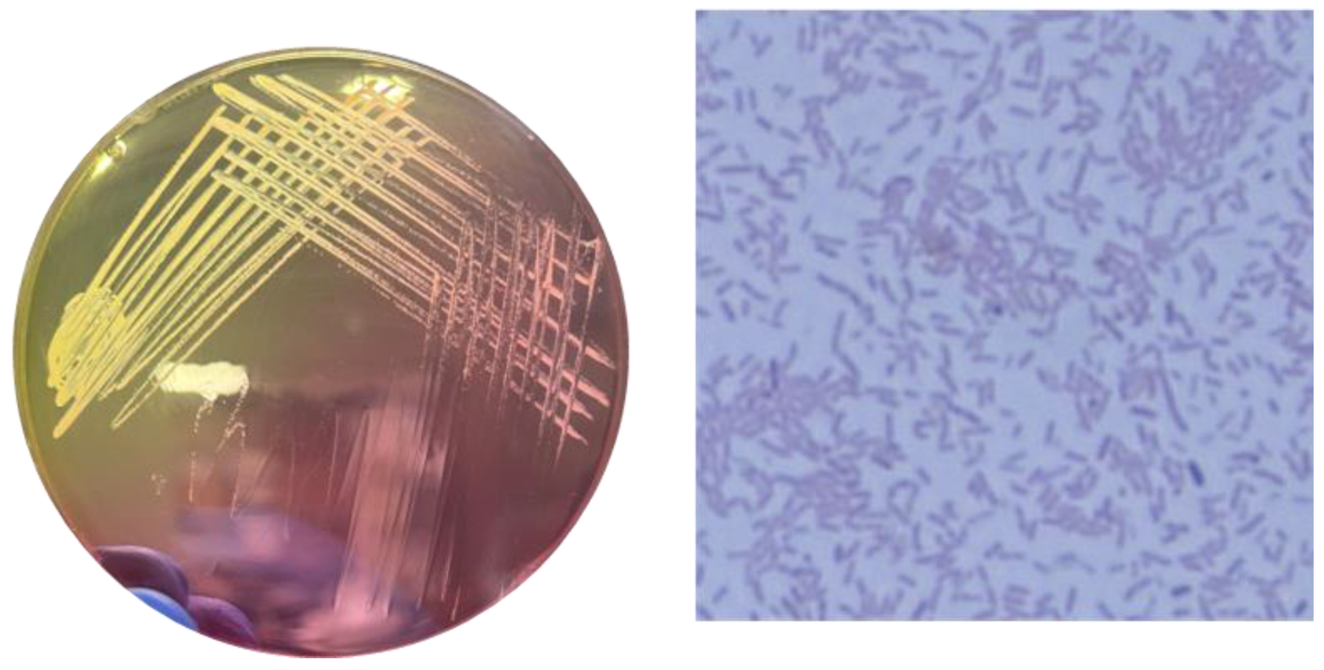
Figure 2.
Biofilm formation Assay showing the reduction of biofilm in PI and H2O2 treated groups. Data are presented as the mean ± standard deviation (n = 3). The treatment group showed an approximate three-log reduction in CFU/cm2 compared to the control group. Statistical significance is indicated as follows: ns = non-significant, *p < 0.05 , ** p < 0.01, *** p < 0.001, and **** p < 0.0001 compared to the control group.
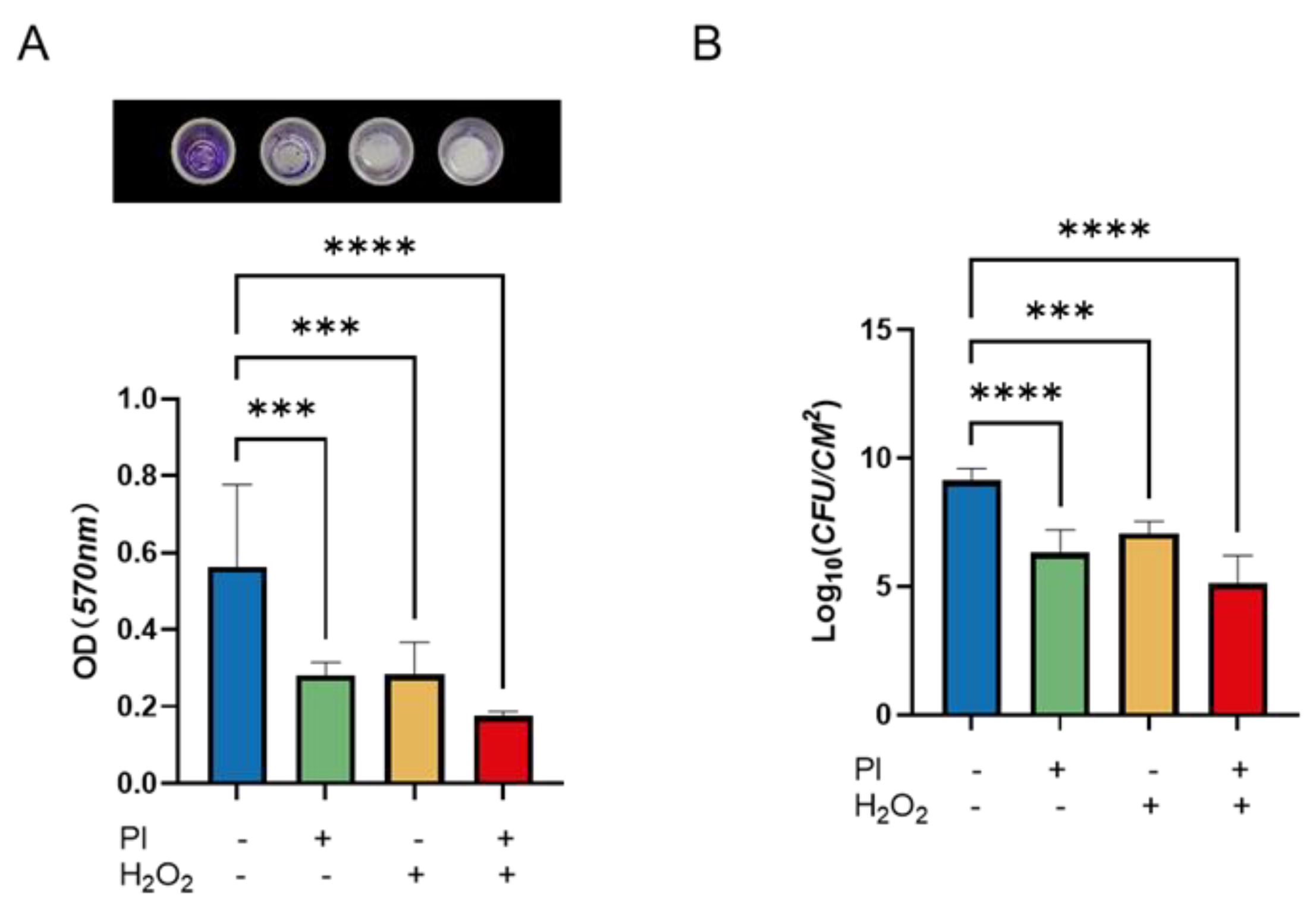
Figure 3.
(A)Expression levels of biofilm-associated genes (icaABDR, ClfA) demonstrate a reduction in the PI and H2O2 treated groups (B) followed by quantitative analysis. Data are expressed as mean ± standard deviation (n = 3). Statistical significance is indicated as follows: ns = non-significant, *p < 0.05 , ** p < 0.01, *** p < 0.001, and **** p < 0.0001 compared to the control group.
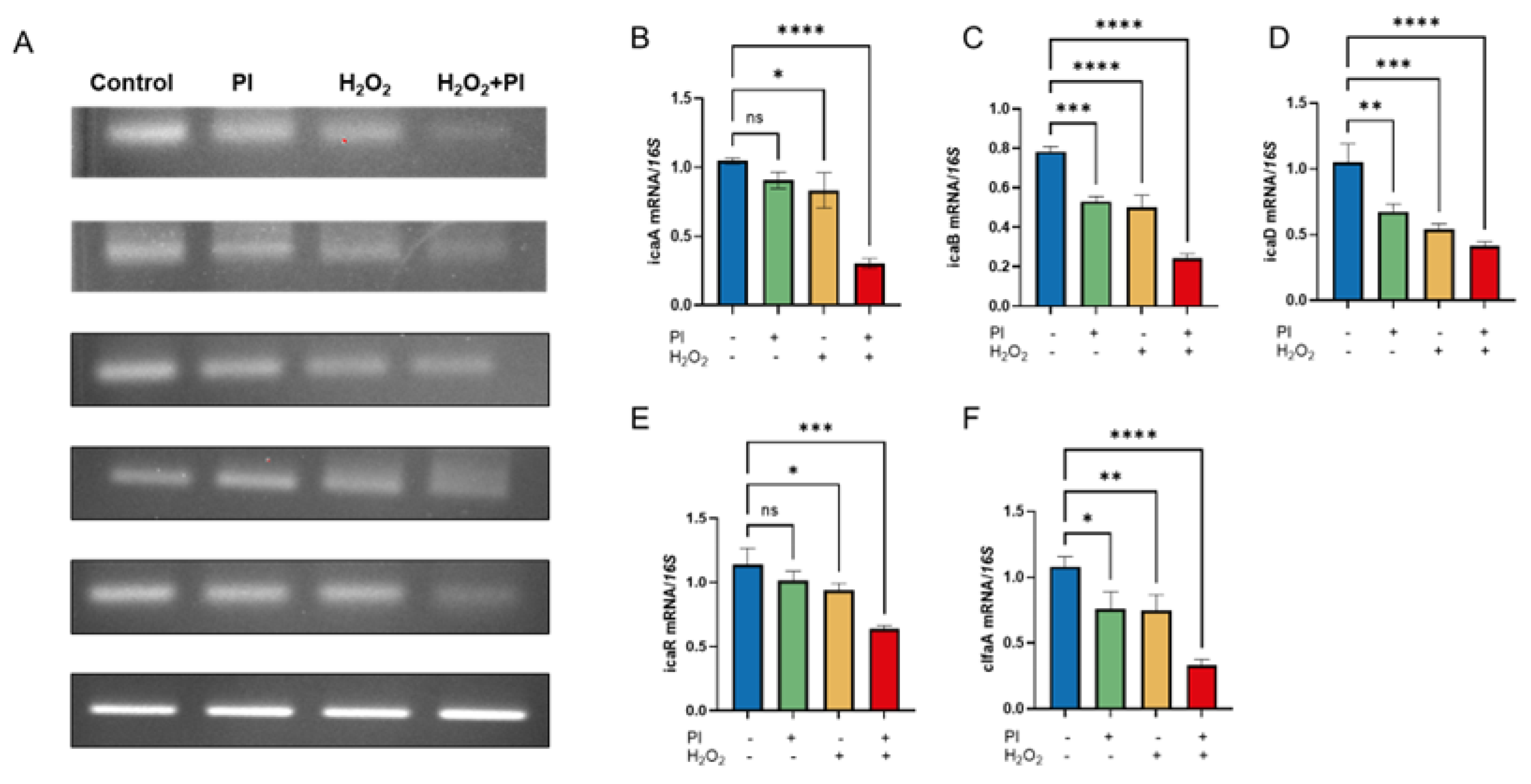
Figure 4.
Confocal microscopy images from a Live/Dead assay demonstrate a reduction in biofilm viability in the PI and H2O2 treated groups, indicating increased cell death within the biofilms. Images were captured at 40x magnification with a scale bar of 100 µm. Statistical significance is indicated as follows: ns = non-significant, ** p < 0.01, *** p < 0.001, and **** p < 0.0001 compared to the control group.
Disclaimer/Publisher’s Note: The statements, opinions and data contained in all publications are solely those of the individual author(s) and contributor(s) and not of MDPI and/or the editor(s). MDPI and/or the editor(s) disclaim responsibility for any injury to people or property resulting from any ideas, methods, instructions or products referred to in the content. |
© 2025 by the authors. Licensee MDPI, Basel, Switzerland. This article is an open access article distributed under the terms and conditions of the Creative Commons Attribution (CC BY) license (http://creativecommons.org/licenses/by/4.0/).
Copyright: This open access article is published under a Creative Commons CC BY 4.0 license, which permit the free download, distribution, and reuse, provided that the author and preprint are cited in any reuse.


