
Submitted:
14 March 2025
Posted:
17 March 2025
You are already at the latest version
Abstract
The antifouling performance of a zwitterionic Sulfobetaine-Hydroxyethyl containing-Polymethylmethacrylate ter-co-polymer (PSBM) is evaluated against three photosynthetic strains, namely Chlorella sp., Nannochloropsis sp., and Arthrospira maxima. PSBM coated polymethylmethacrylate (PMMA) surfaces displayed significantly reduced propensity for biofilm formation, compared to rough and untreated controls, leaving clean surfaces after 7 days of exposure. A tribological approach was adopted to estimate the long-term durability of the PSBM coating. Repeated cycles of exposure to Chlorella sp., Nannochloropsis sp., and A. maxima biomass subject the coating to stress and continuous biofilm challenge. After several cycles, the PSBM coating maintains a higher antifouling efficacy than untreated PMMA surfaces, suggesting stability and high potential in Photobioreactors application.
Keywords:
biofouling
; biofilm
; photobioreactor
; polymethylmethacrylate
; Chlorella sp.
; Nannochloropsis sp
; Arthrospira maxima
; biomass
; zwitterionic coating
1. Introduction
The microalgae and cyanobacteria biomass cultivation are promising bio-factories because they can be used in food, feed, bioactive bulk substances, wastewater treatment, and fuel production [1]. They provide valuable natural antibiotics, pigments, vitamins, and bioactive compounds [2,3,4]. Microalgae use CO2 as a carbon sourse to generate biomass through a photosynthetic process. This makes them active materials in CO2 sequestration to mitigate global warming and for the sustainable production of bioenergy and other value-added products [5,6,7,8].
The most prevalent approaches for large-scale phototrophic biomass production are open systems like ponds and artificial basins or closed photobioreactors (PBRs) [9]. Open systems are cheaper to design, build, and maintain, while the closed ones, despite a higher initial investment, produce biomass more efficiently for fine chemical, nutraceutical, and pharmaceutical applications.
However, large-scale microalgae production presents several challenges, including selecting the correct algal strain and biomass-producing methods, long-term efficiency and harvesting [1,10]. Furthermore, bioproducts and microorganisms, which adhere and multiply on the inner surfaces, cause a counterproductive process called biofouling [11,12]. This reduces solar/UV radiation penetration through the PBRs walls and biomass production, also causing cell pigmentation alterations, culture degradation, and invasive microbe contamination [13].
Biofilm growth on PBR surfaces, depends on several factors and its understanding requires a holistic approach as it involves bio, engineering, and chemico-physical experties. PBR material and geometry, algae typology, culture medium ionic strength, fluid dynamics [12,13,14,15] are among the most demanding.
Biofouling on PBR surfaces begins with organic molecule adsorption, creating a conditioning film to which microalgae adhere and secrete exopolymeric substances (EPS), increasing the surface biofouling potential [14,15]. Environmental variables including temperature, light intensity, and nutritional ratios impact it. High temperatures and light, especially in outdoor culture, boost EPS synthesis and cell adhesion and biofouling [14].
PBR walls are often made of acrylate, polymethylmethacrylates, and polyethylene plastics due to their transparency and mechanical properties. These polymers are hydrophobic and, over the time, their wettability reduces while cell adherence increases [9,12]. Even surface roughness, often caused by ageing, can promote cell adhesion and biofilm growth [11,12].
Recent study highlights innovative surface modifications to reduce biofouling [14,15,16,17]. A very promising method is surface grafting of hydrophilic "fouling resistant coatings" (FRCs). Examples include PEG, PA, hydrogel, and ZW coatings [16,18,19,20,21,22,23]. These coatings are biomimetic [24] since, like peptides and glycoproteins, in water generate a surface hydration layer that prevents proteins, algae, and other generic foulants from settling [25].
Among these, zwitterionic polymers, a family of polyampholytes with high polarisable unit pairs of cations and anions, were extensively investigated in the context of marine, bio, and medical fields, due to their excellent biocompatibility, stability, and minimal fouling [26,27]. Due to well established synthetic procedures, a large variety of- sulfo- and carboxy-betaine methylmethacrylates based ZW polymers have been developed. These display a plethora of properties such as conformational electrolyte-responsiveness [28], AF activity retention in high salt concentrations [26,29], and composition-controlled surface activity, which make them suitable candidates for the engineering of AF coatings for PBRs surface application. Nevertheless, only a few examples are reported. These concern omo-polysulfobetaine- and sulfobetaine copolymers grafted on chemically activated PE (polyethylene) and EVA (ethylene-vinylacetate copolymer) substrates [30,31], which displayed satisfactory AF behavior against Chlorella sp [30].
Biofouling and surface topography highly affect material surface performance and lifespan. Prevention methods included surface engineering and coatings design to inhibit biological material adherence and wear effects [32]. Tribology is the multidisciplinary science which studies friction effects, wear, and lubrication of surfaces under extreme environments. It is essential for assessing surface material life, mechanical contact efficiency, degradation properties of polymer coatings [32,33] and, at the same time, to improve their biodegradability and of end-of-cycle recycling [34]. Literature reports [36] emphasize the importance of using this technique to create tribosystems with coatings that function in the intended wear regime, enhancing surface life cycle.
To overcome the issue of biofouling inside PBR walls the zwitterionic Sulfobetaine-Hydroxyethyl containing-Polymethylmethacrylate ter-co-polymer coating (PSBM) was developed, whose antifouling activity was excellent against Chlorella sp. [37]. In the present study, we report on PSBM antifouling characteristics compared to different photosynthetic strains, namely Chlorella sp., Nannochloropsis sp. and Arthrospira maxima. Antifouling tests were also performed on untreated PMMA and mechanically aged/rough coupons for 7 days for comparison. To test the long-term working of PSBM coated PMMA coupons, a tribological approach is used here. To this aim, the PSBM coupons were repeatedly exposed to Nannochloropsis sp., A. maxima, and Chlorella sp. and their behavior compared to uncoated PMMA controls.
2. Materials and Methods
2.1. PMMA Coupons
PSBM coated PMMA coupons were prepared according to an established procedure [37]. Untreated PMMA (UT) and rough PMMA (R), obtained by using sandpaper of Grit size P800, were used for comparative tests. The dimension of all three types of coupons were 16 mm x 12 mm x 2 mm and used in duplicate in each set of experiments. Each side of the coupons were sterilized under ultraviolet light (UV) for 10 minutes each. The coupons were glued on glass slides and sterilized under UV and then inserted in double into Erlenmeyer flasks containing the growth medium for the AF tests (Figure 1a) For the tribological experiments were carried out using glass Petri dishes Ø 110 mm (Figure 1b). Coupons were hydrated before experimentation by covering them with the appropriate medium for 24 hrs.
2.2. Photosynthetic Strains
The three photosynthetic strains used to test the AF coating performance were the Chlorella sp. strain coming from the culture collection of the Laboratory of Microbiology Applied to the Environment and Cultural Heritage (Micro ABC), Nannochloropsis sp. and the filamentous cyanobacterium A. maxima from the culture collection of Laboratory of Microbial Ecology (Figure 2).
Chlorella sp. and Nannochloropsis sp. microalgal strains were grown in BG 11 medium [38], while the cyanobacterium A. maxima was grown in Zarrouk's medium [39]. For all experiments, fresh biomass was obtained by growing each strain in the respective liquid medium for 7 days, under a constant light intensity of 3000 lux and an agitation at 100 rpm, at room temperature of about 24°C. Biomass was then collected by centrifugation at 10,000 rpm per 30 minutes at 10°C, rinsed and centrifuged three times with sterile physiological solution and the pellet wet weight was then determined. The biomass amount inoculated into the flasks or into the Petri dishes was adjusted to be about ≥ 1 g/L for all the strains.
2.3. Antifouling Assays
The AF experiments for each strain were carried out in duplicate on Erlenmeyer flasks, each containing two sets of glass slides with attached coupons (PSBM, UT and R), 400 mL of liquid medium, and biomass adjusted to a final value > 1g/L (Figure 1a). Then, the flasks were placed on the orbital shaker for 7 days under a continuous light exposure of 3000 lux and agitation (100 rpm) at room temperature (~24°C). At the end of the 7 days cycle, the biomass was collected by centrifugation as previously described and the wet weight obtained was calculated and compared to the starter amount to determine the biomass production. The coupons were detached from the glass slide, rinsed with sterile distilled water and observed under a light microscope (Leica DM750) and fluorescent microscope (Leica DMRE). Each image, representative of the general behavior of algal cells, were analysed by Leica LAS EZ Application (Version 3.4.0 Switzerland), and further processed by ImageJ software (Version 1.46r). Cell counts, area coverage/mm2, and percentage of coverage of the coupons were determined.
2.4. Tribological Tests
For running tribological cycles, untreated (UT) and PSBM PMMA coupons coming from the antifouling tests described in section 2.3 were employed (this cycle was considered as cycle nr. 1). The experiments were carried out using glass Petri plates Ø 110 mm instead of Erlenmeyer flasks (Figure 1). For each cycle the same coupons were placed at the bottom of sterile glass Petri plates and sterilized by UV radiation for 10 minutes. The starting biomass was obtained as described in par. 2.2 and adjusted at ≥1 g/L when inoculated onto Petri plates with hydrated PMMA coupons. The inoculated Petri dishes were placed on an orbital agitator at 80 rpm to evenly distribute biomass on all surfaces. Light intensity was 3000 lux, and temperature was 25°C. After each 7-day experimental cycle, biomass was collected and weighted as indicated in section 2.3 and the coupons were rinsed with sterile deionized water to eliminate loose biomass. Then, they were observed under the light microscope and ImageJ software (ver. 1.46) was used to assess biofouling and performance over time. Finally, the coupons were gently washed and rinsed and used for the next cycle.
3. Results and Discussion
3.1. Biomass Production
The biomass production was not affected by the presence of the coupons, and it reached very satisfactory values for all the 3 strains. Biomass production doubled after 7 days of incubation (from 1.3 g/L, to 2.65 g/L for Chlorella sp, from 0.85 g/L, to 1.7 g/L for Nannochloropsis sp, and from 1.0 g/L, to 2.0 g/L, for A. maxima). This indicates a robust and proportional biomass increase for all strains under the given incubation conditions.
3.2. Antifouling Performance
The antifouling performance of R, UT and PSBM PMMA surfaces was evaluated by the image analysis obtained under light and fluorescent microscopy. The results are reported in Figure 3 and Figure 4 and Table 1. These clearly show that after a 7-day cycle, rough PMMA allowed the highest adhesion of cells for all the three strains. For Chlorella sp. the average number of cells/mm2 was 4500 cells/mm², corresponding to an area coverage of 9%, while, for Nannochloropsis sp., an average of 6063 cells/mm² was observed with 3% of occupied area. For A. maxima only the area coverage was , due to the poor persistence of the filaments on R and UT surfaces as shown in Figure 4 g and h. The value of 37.72 % considers also the presence of biofilm and cell debris. The UT coupons showed for Chlorella sp. a cell adhesion of 3705 cells/mm² and a surface coverage of 7.5%, while for Nannochloropsis sp. the average of cell adhesion was 4044 cells/mm² with a 2% of area coverage. For A. maxima, no significant presence of filaments could be detected on the surfaces (Figure 6i), but the biofouling observed was approximately 5.33% per mm2 of surface. In contrast, microscopic observations showed that PSBM coated PMMA surface were characterized by a very low cell adhesion of Chlorella sp. and Nannochloropsis sp. cells being respectively 2 cells/mm² and 222 cells/mm2. PSBM surfaces showed a negligible area coverage being 0.004% for Chlorella, 0.1% for Nannochloropsis and 0.037% for A. maxima, respectively. Therefore, it was evident that PSBM coated surfaces display remarkable antifouling properties, against all the model strains here tested.
Fluorescent microscopy (FM) images (Figure 4) showed, for both the microalgae, a scattered adhesion pattern on R surfaces (Figure 4a and 4d) while, on UT surfaces, typical aggregates due to liquid agitation were observed (Figure 4b and 4e). In the case of A. maxima, the images showed mainly debris traces of filaments attachment on the surfaces (Figure 4g and 4h). Extremely low cell adhesion and almost no biofilm coverage for all tested strains was observed on the PSBM surface (Figure 4c, 4f, 4i).
Rough surfaces, as expected, were those that showed the most intense fouling, especially in the case of A. maxima, with biofilm observed on most of the surface. These findings agreed with previously reported studies, which suggest that roughness or other irregularities promote microbial attachment [40]. Besides, the microscopic images of A. maxima show generally debris and biofilm coverage and, sometimes, fingerprint of the attached filaments, but never real filaments, as already reported [41,42,43,44]. A comparison between R and UT coupons shows that the latter, in early stage of use, is less prone to fouling and that cell aggregations depend on liquid movement. Very low cell adhesion and almost no biofilm coverage for all tested strains was observed on the PSBM surfaces. This data supports, once more, that zwitterionic based coatings can provide a non-fouling environment due to their ability to maintain a hydration layer, which prevents protein adsorption reduction and microbial adhesion [45,46].
3.3. Long Term Effectiveness of the PSBM
3.3.1. Biomass Production over Cycles
For all the tested strains, Chlorella sp., Nannochloropsis sp. and A. maxima, biomass yield was doubled or even more respect the initial weight of ≥1 g/L. This increase was constant in each cycle. The average and Standard deviations were calculated over 9 successive cycles. For Chlorella sp., the gain in biomass across tribological cycles was found to be 2.45 ± 0.40 g/L, and for Nannochloropsis sp., the gain was 2.25 ± 0.38 g/L, while for A. maxima was 2.09 ± 0.35 g/L.
3.3.2. Antifouling Performance of Strains Through Tribological Approach
The antifouling performance of PSBM coated surface was analyzed over multiple (9) cycles and compared to that of UT PMMA. The results obtained clearly shows that the PSBM led to a remarkable reduction of cell adhesion with respect to UT PMMA . Furthemore, along the cycles, only a light increment of attached cells was observed which, on the contrary, was consistent on UT. Surface resistance to biofouling was slightly variable when compared against the three different strains Chlorella sp., Nannochloropsis sp. and A. maxima. Results are reported in the Table 2, Table 3, Figure 5a and 5b and Figure 8. As shown in Table 2, on UT PMMA, the number of cells increased along the cycles. In fact, cell counts were at beginning 763 cells/mm² and 525.93, respectively for Chlorella and Nannochloropsis, while at the end were 3,861 and 3,768.52 cells/mm², respectively. On the other hand, PSBM-coated PMMA demonstrated good antifouling behaviour with low and consistent cell counts from Cycle 2 (7 and 10 cells/mm2) to Cycle 10 (23 and 100 cells/mm2).
The logarithmic trends of biofouling on UT PMMA and PSBM surfaces for the two strains, along nine cycles, are reported in Figure 5a and b. These clearly show that the PMMA is more prone to fouling than the PSBM treated PMMA.
Regarding the extent of biofouling observed on UT PMMA vs PSBM, as summarized in Table 3 and Figure 5, it is evident that the PSBM coating significantly reduced cell adhesion across all cycles. This finding aligns with the antifouling performance observed in the previous sections, highlighting the superior resistance of PSBM against biofouling, particularly with Chlorella sp. and Nannochloropsis sp. The consistency of these results across multiple cycles further supports the long-term effectiveness of PSBM coatings in preventing biofouling.
The comparation of surface covered by debris and biofilm due to A. maxima growth on untreated PMMA surfaces vs those coated with PSBM is reported in Figure 6. The results show that the PSBM coating effectively resisted biofouling for nine cycles, maintaining minimal coverage by A. maxima. In contrast, the untreated PMMA surface experienced significant biofouling, with a high area covered by A. maxima.
Figure 6.
Graphical representation of area covered by A. maxima on UT PMMA surface vs PSBM surface.

The data obtained show that advanced surface coating technologies such as the PSBM antifouling coating on PMMA can significantly enhance wear resistance and durability of a given material. The tribological approach shows the importance of testing antifouling coatings, such as PSBM, under environmental and prolonged exposure circumstances to determine the coating durability and performance over multiple utilization cycles [47,48]. In our opinion, this study represents an important step in this direction showing the need to test the durability and performance of PSBM surfaces in repeated biofouling cycles to assess coating integrity and antifouling efficacy during multiple mechanical and environmental challenges [49].
4. Conclusions
In conclusion, our study demonstrates that the sulfobetaine-zwitterionic PMMA (PSBM) was effective in:
- (a)
- mitigate the biofouling due to the three photosynthetic strains considered as model: Chlorella sp., Nannochloropsis sp. and A. maxima,
- (b)
- maintenance of optical transparency
- (c)
- surface protection against wear.
Thus, the use of PSBM coating can contribute to lowering maintenance costs, increasing reactor lifespan, and to increasing overall productivity by reducing biofouling and maintaining reactor efficiency.
This study provides significant findings to advance the development of more sustainable and biocompatible alternatives to conventional antifouling treatments in industrial and biotechnological applications against biofouling, addressing an important lack of data in literature within the long-term assessment of antifouling coatings.
5. Patents
No patents resulting from the work reported in this manuscript.
Author Contributions
Conceptualization, SLS and CU; methodology, SLS, CU and RHA; software, RHA; validation, SLS and CU; formal analysis, SLS, CU and RHA; investigation, RHA, MN and VZ.; resources SLS, FDL and CU; data curation, SLS and CU.; writing—original draft preparation, RHA; writing—review and editing, SLS, CU and RHA; visualization, RHA; supervision, SLS and CU; project administration, SLS and CU; funding acquisition, SLS, FDL and CU All authors have read and agreed to the published version of the manuscript.
Funding
This research was partially carried out with the financial contribution of FFABR 2022 (Italian fund for basic research activities). RHA carried out his doctorate work with a scholarship in the frame of (RISORSE PON “RICERCA E INNOVAZIONE” 2014–2020).
Institutional Review Board Statement
“Not applicable” our research did not involve humans or animals.
Data Availability Statement
Data are contained within the article.
Acknowledgments
We thank Dr. Luciano Falqui from Di Bella Costruzioni srl, Catania, for his valuable suggestions throughout the project.
Conflicts of Interest
The authors declare no conflicts of interest and the funders had no role in the design of the study; in the collection, analyses, or interpretation of data; in the writing of the manuscript; or in the decision to publish the results.
Abbreviations
The following abbreviations are used in this manuscript:
| PMMA | Polymethylmethacrylate |
| PSBM | Poly sulfobetaine methacrylate / Zwitterionic Sulfobetaine-Hydroxyethyl containing-Polymethylmethacrylate ter-co-polymer |
| AF | Antifouling |
| UT | Untreated |
| R | Rough |
References
- Joy, S.R. , Anju, T.R. (2023). Microalgal Biomass: Introduction and Production Methods. In: Thomas, S., Hosur, M., Pasquini, D., Jose Chirayil, C. (eds) Handbook of Biomass. Springer, Singapore. [CrossRef]
- López-Hernández, J.-F.; Kean-Meng, T.; Asencio-Alcudia, G.-G.; Asyraf-Kassim, M.; Alvarez-González, C.-A.; Márquez-Rocha, F.-J. Sustainable Microalgae and Cyanobacteria Biotechnology. Applied Sciences 2022, 12, 6887. [Google Scholar] [CrossRef]
- Mobin SM, A.; Chowdhury, H.; Alam, F. Commercially important bioproducts from microalgae and their current applications–A review. Energy Procedia 2019, 160, 752–760. [Google Scholar] [CrossRef]
- Rizwan, M.; Mujtaba, G.; Memon, S.A.; Lee, K.; Rashid, N. Exploring the potential of microalgae for new biotechnology applications and beyond: A review. Renewable and Sustainable Energy Reviews 2018, 92, 394–404. [Google Scholar] [CrossRef]
- Khan, M.I.; Shin, J.H.; Kim, J.D. The promising future of microalgae: current status, challenges, and optimization of a sustainable and renewable industry for biofuels, feed, and other products. Microb. Cell Fact. 2018, 17, 36. [Google Scholar] [CrossRef]
- Eze, C.N.; Onyejiaka, C.K.; Ihim, S.A.; Ayoka, T.O.; Aduba, C.C.; Ndukwe, J.K.; Nwaiwu, O.; Onyeaka, H. Bioactive compounds by microalgae and potentials for the management of some human disease conditions. AIMS Microbiol. 2023, 7, 55–74. [Google Scholar] [CrossRef]
- Sun, Y.; Huang, Y.; Liao, Q.; Fu, Q.; Zhu, X. Enhancement of microalgae production by embedding hollow light guides to a flat-plate photobioreactor. Bioresour. Technol. 2016, 207, 31–38. [Google Scholar] [CrossRef] [PubMed]
- Kaur, M.; Bhatia, S.; Gupta, U.; Decker, E.; Tak, Y.; Bali, M.; Gupta, V.K.; Dar, R.A.; Bala, S. Microalgal bioactive metabolites as promising implements in nutraceuticals and pharmaceuticals: inspiring therapy for health benefits. Phytochem. Rev. 2023, 22, 903–933. [Google Scholar] [CrossRef]
- Chanquia, S.N.; Vernet, G.; Kara, S. Photobioreactors for cultivation and synthesis: Specifications, challenges, and perspectives. Eng. Life Sci. 2022, 12, 712–724. [Google Scholar] [CrossRef]
- Yu Chen, Zhiwu Yu * 2024 Low-melting mixture solvents: extension of deep eutectic solvents and ionic liquids for broadening green solvents and green chemistry. Green Chemical Engineering Perspective. [CrossRef]
- Soriano-Jerez, Y.; Macías-de la Rosa, A.; García-Abad, L.; López-Rosales, L.; Maza-Márquez, P.; García-Camacho, F.; Bressy, C.; Cerón-García, M.C.; Molina-Grima, E. Transparent antibiofouling coating to improve the efficiency of Nannochloropsis gaditana and Chlorella sorokiniana culture photobioreactors at the pilot-plant scale. Chemosphere 2024, 347, 140669. [Google Scholar] [CrossRef]
- Zeriouh, O.; Reinoso-Moreno, J.V.; López-Rosales, L.; Cerón-García, M.C.; Sánchez-Mirón, A.; García-Camacho, F.; Molina-Grima, E. Biofouling in photobioreactors for marine microalgae. Critical Reviews in Biotechnology 2017, 37, 1006–1023. [Google Scholar] [CrossRef]
- Talluri, S.N.L.; Winter, R.M.; Salem, D.R. Conditioning film formation and its influence on the initial adhesion and biofilm formation by a cyanobacterium on photobioreactor materials. Biofouling 2020, 36, 183–199. [Google Scholar] [CrossRef] [PubMed]
- Damodaran, V.B.; Murthy, N.S. Bio-inspired strategies for designing antifouling biomaterials. Biomater. Res. 2016, 20, 1–11. [Google Scholar] [CrossRef]
- García-Abad, L.; López-Rosales, L.; Cerón-García, M.D.C.; Fernández-García, M.; García-Camacho, F.; Molina-Grima, E. Influence of abiotic conditions on the biofouling formation of flagellated microalgae culture. Biofouling 2022, 38, 507–520. [Google Scholar] [CrossRef] [PubMed]
- Magin, C.M.; Cooper, S.P.; Brennan, A.B. Non-toxic antifouling strategies. Materials Today 2010, 13, 36–44. [Google Scholar] [CrossRef]
- Maan, A.M.C.; Hofman, A.H.; de Vos, W.M.; Kamperman, M. Recent developments and practical feasibility of polymer-based antifouling coatings. Adv. Funct. Mater. 2020, 30, 2000936. [Google Scholar] [CrossRef]
- Banerjee, I.; Pangule, R.C.; Kane, R.S. Antifouling coatings: recent developments in the design of surfaces that prevent fouling by proteins, bacteria, and marine organisms. Adv. Mater. 2011, 23, 690–718. [Google Scholar] [CrossRef]
- Soriano-Jerez, Y.; López-Rosales, L.; Cerón-García, M.C.; Sánchez-Mirón, A.; Gallardo-Rodríguez, J.J.; García-Camacho, F.; Molina-Grima, E. Long-term biofouling formation mediated by extracellular proteins in Nannochloropsis gaditana microalga cultures at different medium N/P ratios. Biotechnol. Bioeng. 2021, 118, 1152–1165. [Google Scholar] [CrossRef]
- Borucinska, E.; Zamojski, P.; Grodzki, W.; Blaszczak, U.; Zglobicka, I.; Zielinski, M.; Kurzydlowski, K.J. Degradation of polymethylmethacrylate (PMMA) bioreactors used for algal cultivation. Materials 2023, 16, 4873. [Google Scholar] [CrossRef]
- Tang, J.; Liu, B.; Gao, L.; Wang, W.; Liu, T.; Su, G. Impacts of surface wettability and roughness of styrene-acrylic resin films on adhesion behavior of microalgae Chlorella sp. Colloids and Surfaces B: Biointerfaces 2021, 199, 111522. [Google Scholar] [CrossRef]
- Tong, C.Y.; Derek CJ, C. Membrane surface roughness promotes rapid initial cell adhesion and long-term microalgal biofilm stability. Environmental Research 2022, 206, 112602. [Google Scholar] [CrossRef]
- Zeriouh; Ouassim; Reinoso-Moreno, J. V.; López-Rosales, L.; del Carmen Cerón-García, M.; Mirón, A.S.; García-Camacho, F.; Molina-Grima, E. Assessment of a photobioreactor-coupled modified Robbins device to compare the adhesion of Nannochloropsis gaditana on different materials. Algal Research 2019, 37, 277–287. [Google Scholar]
- Zhang, H.; Wang, F.; Guo, Z. The antifouling mechanism and application of bio-inspired superwetting surfaces with effective antifouling performance. Adv. Colloid Interface Sci. 2024, 325, 103097. [Google Scholar] [CrossRef]
- Zheng, L.; Sundaram, H.S.; Wei, Z.; Li, C.; Yuan, Z. Applications of zwitterionic polymers, reactive and functional polymers 2017, 118, 51–61. [CrossRef]
- Li, M.; Zhuang, B.; Yu, J. Functional zwitterionic polymers on surface: structures and applications. Chem. Asian J. 2020, 15, 2060–2075. [Google Scholar] [CrossRef] [PubMed]
- Qu, K.; Yuan, Z.; Wang, Y.; Song, Z.; Gong, X.; Zhao, Y.; Mu, Q.; Zhan, Q.; Xu, W.; Wang, L. Structures, properties, and applications of zwitterionic polymers. ChemPhysMater. 2022, 1, 294–309. [Google Scholar] [CrossRef]
- Xiao, S.; Ren, B.; Huang, L.; Shen, M.; Zhang, Y.; Zhong, M.; Yang, J.; Zheng, J. Salt-responsive zwitterionic polymer brushes with anti-polyelectrolyte property. Curr. Opin. Chem. Eng. 2018, 19, 86–93. [Google Scholar] [CrossRef]
- Higaki, Y.; Nishida, J.; Takenaka, A.; Yoshimatsu, R.; Kobayash, M.; Takahara, A.A. Versatile inhibition of marine organism settlement by zwitterionic polymer brushes. Polym. 2015, 47, 811–818. [Google Scholar] [CrossRef]
- Wang, D.; Wu, X.; Long, L.; Yuan, X.; Zhang, Q.; Xue, S.; Wen, S.; Yan, C.; Wange, J.; Cong, W. Improved antifouling properties of photobioreactors by surface grafted sulfobetaine polymers. Biofouling 2017, 33, 970–979. [Google Scholar] [CrossRef]
- Wang, Y.; Chen, C.; Wu, X.; Wang, Z.; Wen, S.; Yu, J.; Yan, C.; Cong, W. Improved antibiofouling properties of photobioreactor with amphiphilic sulfobetaine copolymer coatings. Progress in Organic Coatings 2020, 144, 105666. [Google Scholar] [CrossRef]
- Akhaia, S.; Wadhwa, A.S. Recent advances in bio-tribology from joint lubrication to medical implants: A review. Journal of Materials and Engineering 2024, 2, 125–135. [Google Scholar] [CrossRef]
- Simson, D. , & Subbu, S. K. (2024). Investigating the tribological performance of bioimplants. In Bioimplants Manufacturing (pp. 258–283). CRC Press. eBook ISBN 9781003509943.
- Prabhu, A.; Raghavan, S.; Kumar, S. Recent review of tribology, rheology of biodegradable and FDM compatible polymers. Journal of Materials Research and Technology 2020, 9, 12345–12367. [Google Scholar] [CrossRef]
- Chen, M.; Song, Z.; Yang, X.; Song, Z.; Luo, X. Antifouling strategies for protecting bioelectronic devices. APL Materials 2021, 9, 020701. [Google Scholar] [CrossRef]
- Jakovljević, J. , Rakić, D., & Vuković, M. Special issue: Tribological coatings—Properties, mechanisms, and applications in surface engineering. Materials 2023, 16, 1234–1256. [Google Scholar] [CrossRef]
- Lo Schiavo, S. , Gulino, A., Fragalà, M. E., Mineo, P., Nicosia, A., Ali, R. H.,... & Urzì, C. A sulfobetaine containing-polymethylmethacrylate surface coating as an excellent antifouling agent against Chlorella sp. Progress in Organic Coatings 2025, 199, 108940. [Google Scholar]
- Stanier, R.Y.; Kunisawa, R.; Mandel MC, B.G.; Cohen-Bazire, G. Purification and properties of unicellular blue-green algae (order Chroococcales). Bacteriological Reviews 1971, 35, 171–205. [Google Scholar] [CrossRef]
- Madkour, F.F.; Kamil, A.E.-W.; Nasr, H.S. Production and nutritive value of Spirulina platensis in reduced-cost media. The Egyptian Journal of Aquatic Research 2012, 38, 51–57. [Google Scholar] [CrossRef]
- Meireles, A. , Gonçalves, A. L., Gomes, I. B., Simões, L. C., & Simões, M. (2015). Methods to study microbial adhesion on abiotic surfaces.
- Liao, Y.; Fatehi, P.; Liao, B. Microalgae cell adhesions on hydrophobic membrane substrates using quartz crystal microbalance with dissipation. Colloids and Surfaces B: Biointerfaces 2023, 230, 113514. [Google Scholar] [CrossRef]
- El-Sapagh, S.; El-Shenody, R.; Pereira, L.; Elshobary, M. Unveiling the potential of algal extracts as promising antibacterial and antibiofilm agents against multidrug-resistant Pseudomonas aeruginosa: in vitro and in silico studies including molecular docking. Plants 2023, 12, 3324. [Google Scholar] [CrossRef]
- Zhang, H.; Zhu, S.; Yang, J.; Ma, A. Advancing strategies of biofouling control in water-treated polymeric membranes. Polymers 2022, 14, 1167. [Google Scholar] [CrossRef]
- Passos, L.F.; Berneira, L.M.; Poletti, T.; Mariotti KD, C.; Carreño, N.L.; Hartwig, C.A.; Pereira, C.M. Evaluation and characterization of algal biomass applied to the development of fingermarks on glass surfaces. Australian Journal of Forensic Sciences 2021, 53, 337–346. [Google Scholar]
- He, M.; Gao, K.; Zhou, L.; Jiao, Z.; Wu, M.; Cao, J.; You, X.; Cai, Z.; Su, Y.; Jiang, Z. Zwitterionic materials for antifouling membrane surface construction. Acta Biomaterialia 2016, 40, 142–152. [Google Scholar] [CrossRef]
- Murali, S.; Agirre, A.; Arrizabalaga, J.; Rafaniello, I.; Schäfer, T.; Tomovska, R. Zwitterionic stabilized water-borne polymer colloids for antifouling coatings. Reactive and Functional Polymers 2024, 196, 105843. [Google Scholar] [CrossRef]
- Xue, Y.J.; Zhang, Y.Z. Surface coatings in tribological and wear-resistant applications. International Heat Treatment and Surface Engineering 2009, 3, 17–25. [Google Scholar] [CrossRef]
- Tay, S.P.; Hu, X.; Fleming, P.; Forrester, S. Tribological investigation into achieving skin-friendly artificial turf surfaces. Materials & Design 2016, 89, 177–182. [Google Scholar] [CrossRef]
- Gee, M.; Kamps, T.; Woolliams, P.; Nunn, J.; Mingard, K. In situ real-time observation of tribological behaviour of coatings. Surface and Coatings Technology 2022, 442, 128233. [Google Scholar] [CrossRef]
Figure 1.
Erlenmeyer flask (a) and glass Petri dish (b) with coupons before the addition of algal suspension.
Figure 1.
Erlenmeyer flask (a) and glass Petri dish (b) with coupons before the addition of algal suspension.
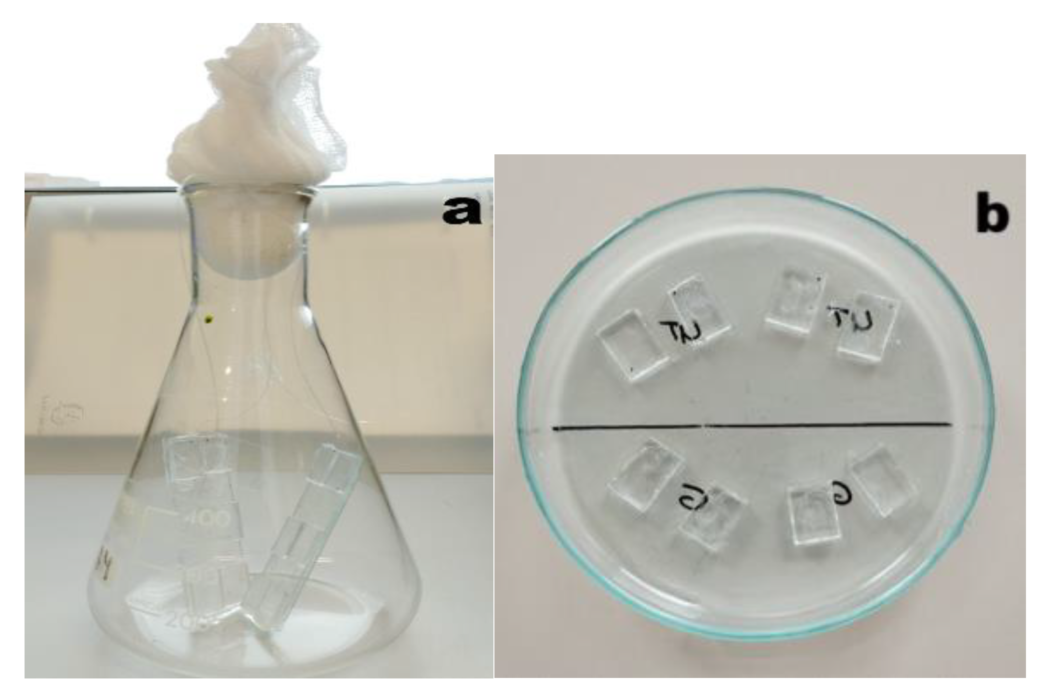
Figure 2.
Light microscopy images of the 3 strains used. a) Chlorella sp.; b) Nannochloropsis sp. and c) A. maxima. Bars are 50 µm.
Figure 2.
Light microscopy images of the 3 strains used. a) Chlorella sp.; b) Nannochloropsis sp. and c) A. maxima. Bars are 50 µm.
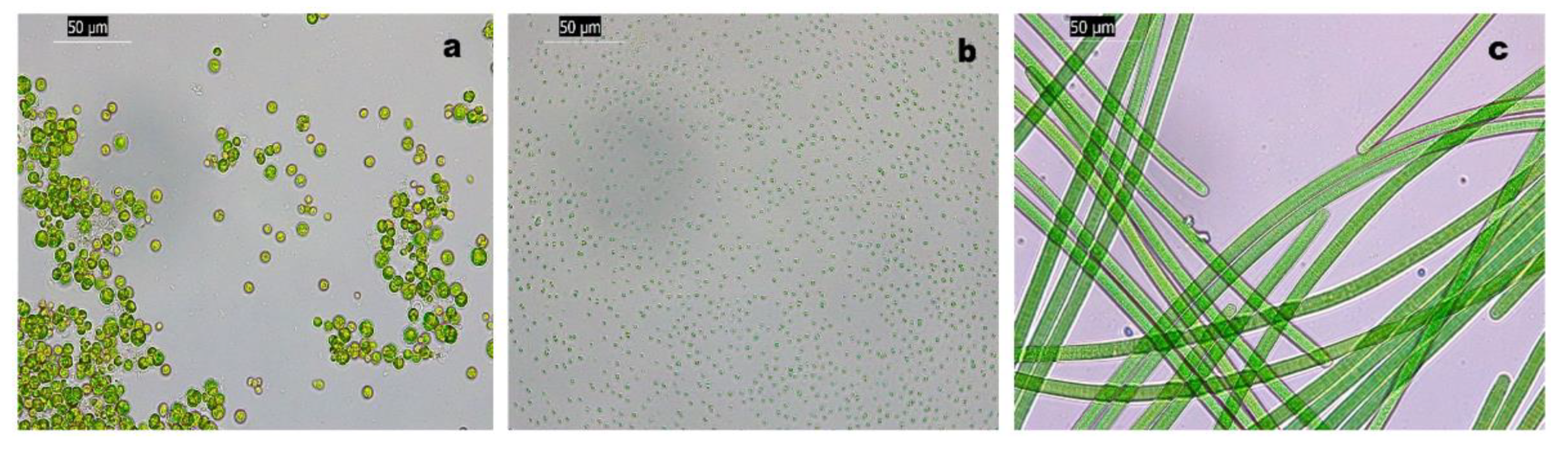
Figure 3.
Graphical representation of area coverage/mm2 for each strain on three different types of PMMA surfaces.
Figure 3.
Graphical representation of area coverage/mm2 for each strain on three different types of PMMA surfaces.
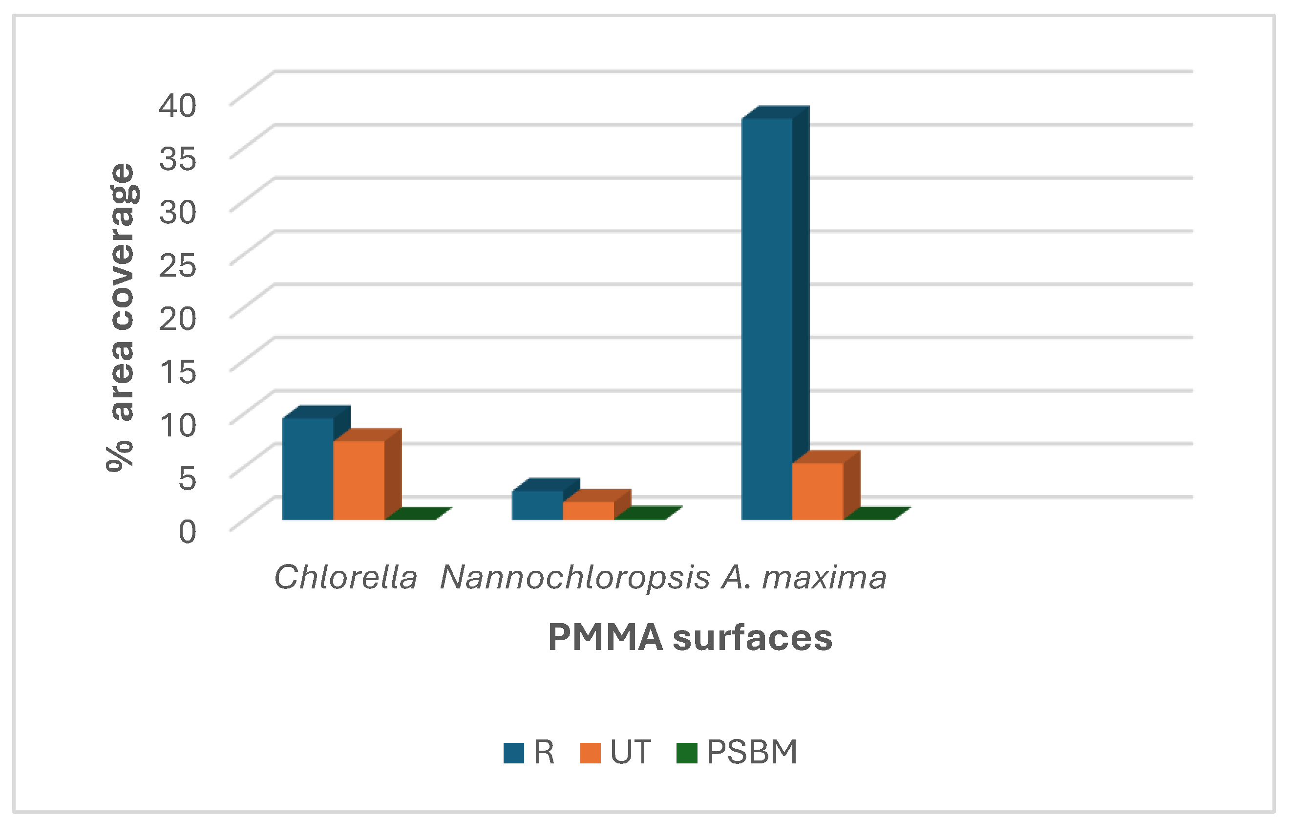
Figure 4.
Antifouling performance of different PMMA surfaces (R on the left, UT in the center and PSBM on the right) against the three strains as seen under fluorescent microscopy (FM). a-c) Chlorella strain adhesion; d-f) Nannochloropsis strain adhesion; g-i) A. maxima strain biofilm. The R surfaces are mostly prone to fouling showing a uniform distribution of adhering cells pattern, while UT PMMA presented typical aggregates for both microalgae; in the case of A. maxima, the FM images show mostly debris and fingerprints of filaments, but no attached filaments. Bar is 20 µm. The few seen cells on PSBM coated PMMA (c, f and i) are shown by magnifying indication.
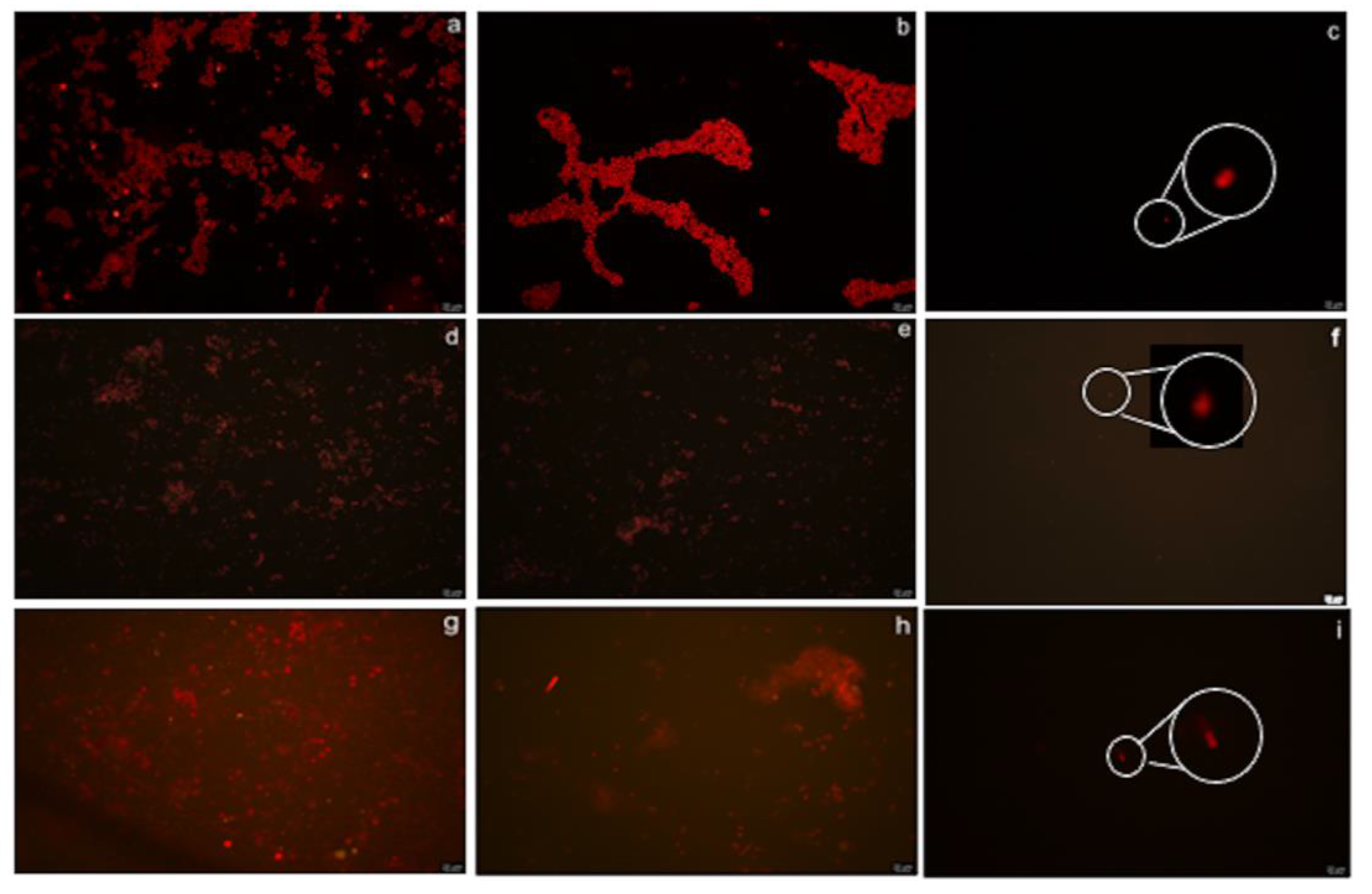
Figure 5.
Log of adhering cells/mm2 of Chlorella sp. (a) and Nannochloropsis (b) on the UT surface (control) vs PSBM surface along 9 tribological cycles.
Figure 5.
Log of adhering cells/mm2 of Chlorella sp. (a) and Nannochloropsis (b) on the UT surface (control) vs PSBM surface along 9 tribological cycles.
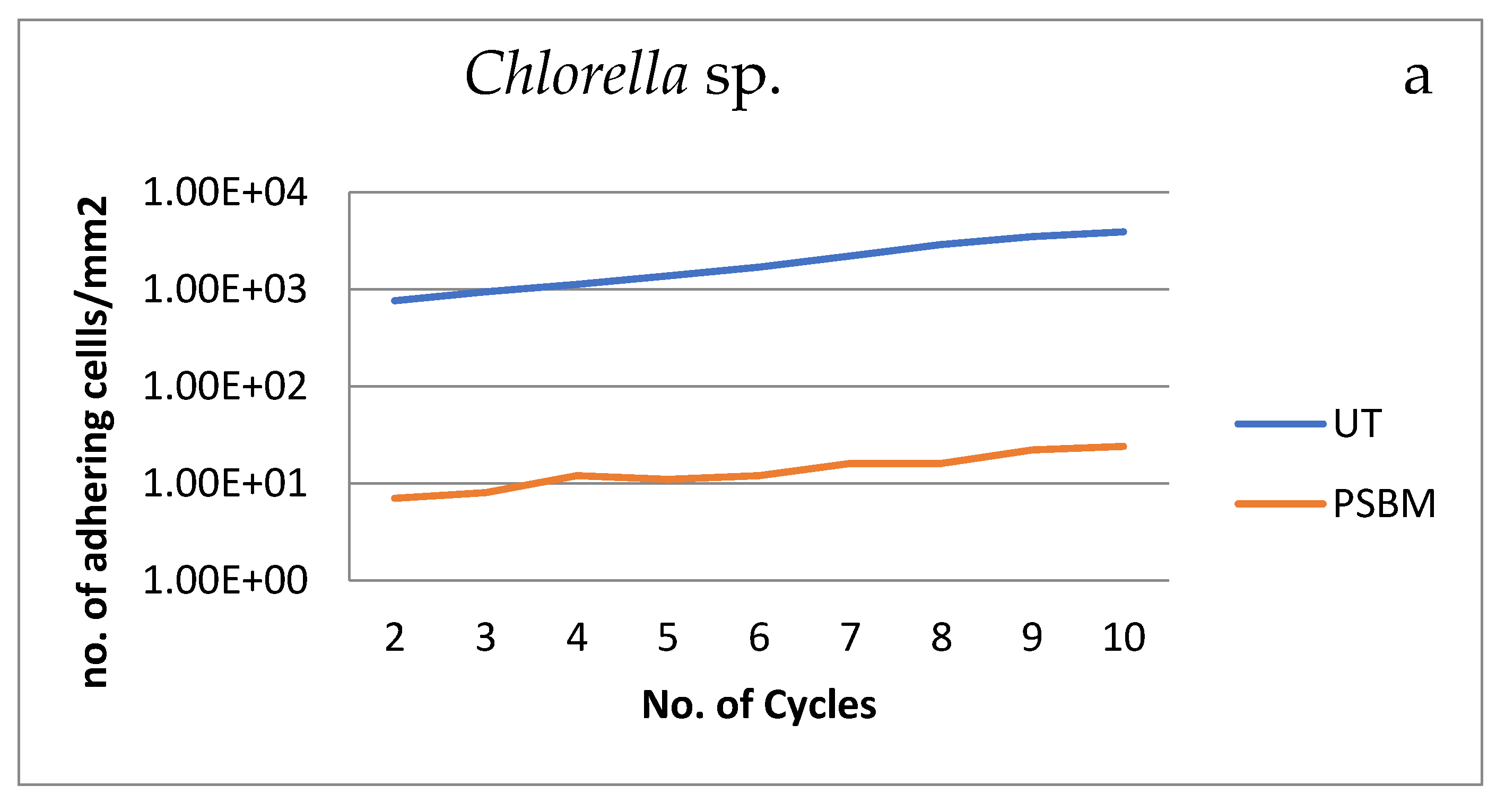
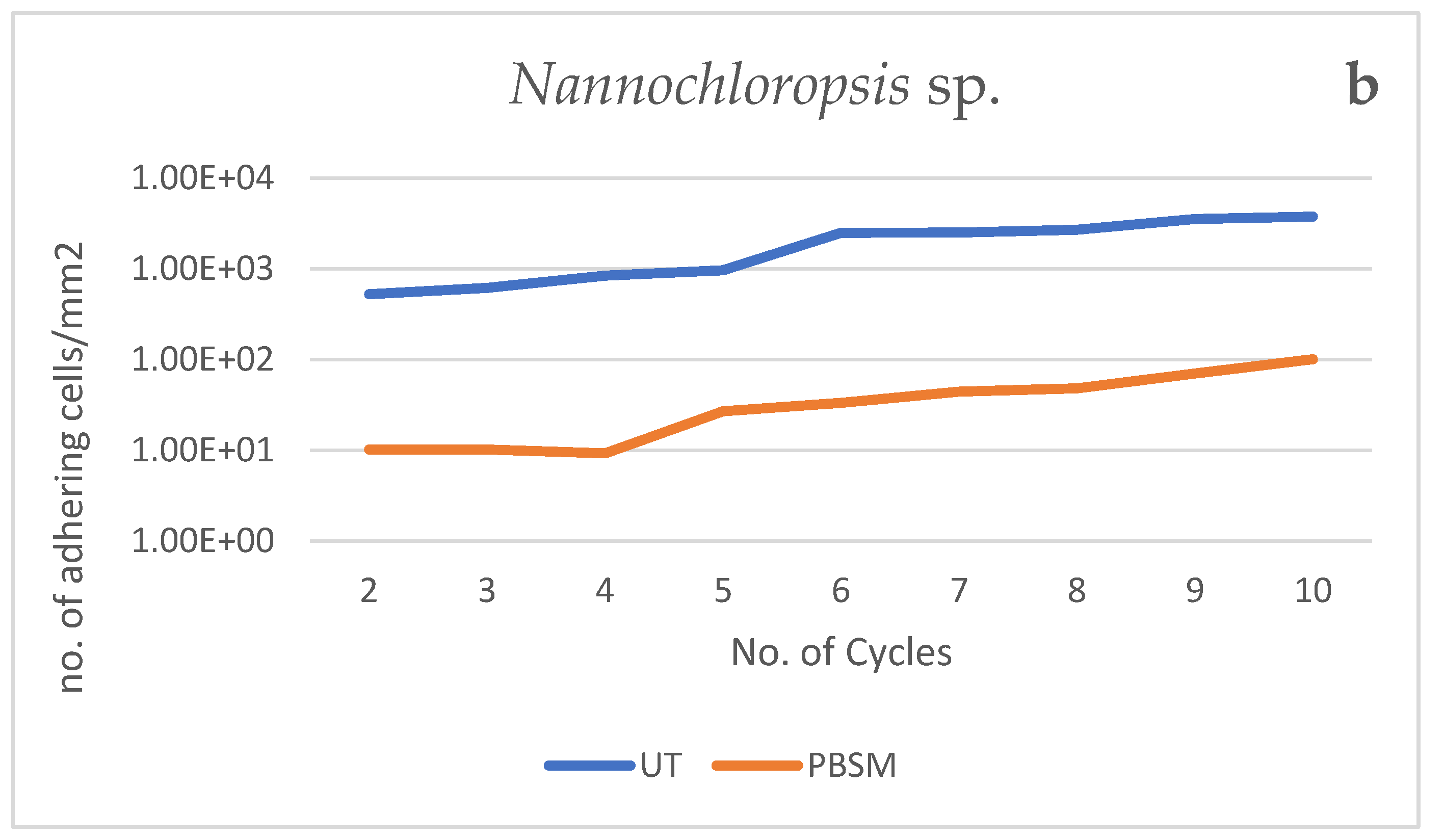

Table 1.
Average of adhering cell/mm2 and area coverage (%/mm2) of the three algal strains (Chlorella sp., Nannochloropsis and A. maxima (only area coverage) as determined under Light and Fluorescent microscopy and Image J analysis.
Table 1.
Average of adhering cell/mm2 and area coverage (%/mm2) of the three algal strains (Chlorella sp., Nannochloropsis and A. maxima (only area coverage) as determined under Light and Fluorescent microscopy and Image J analysis.
| Strains | Average cells/mm2 | % Area coverage/mm2 | ||||
| R | UT | PSBM | R | UT | PSBM | |
| Chlorella | 4500 | 3705 | 2 | 9 | 7,5 | 0.004 |
| Nannochloropsis | 6063 | 4044 | 222 | 3 | 2 | 0.1 |
| A. maxima | N.A. | N.A. | N.A. | 37,72 | 5,33 | 0.037 |
Table 2.
Antifouling performance of UT vs PSBM surface for Chlorella sp. and Nannochloropsis sp. expressed as no. of cells/mm2.
Table 2.
Antifouling performance of UT vs PSBM surface for Chlorella sp. and Nannochloropsis sp. expressed as no. of cells/mm2.
| UT | PSBM | |||
| Cell/mm2 | ||||
| No.Cycles | Chlorella | Nannochloropsis | Chlorella | Nannochloropsis |
| 2 | 763 | 525.93 | 7 | 10.19 |
| 3 | 970 | 618.52 | 8 | 10.19 |
| 4 | 1118 | 840.74 | 12 | 9.26 |
| 5 | 1370 | 963.89 | 11 | 26.85 |
| 6 | 1677 | 2495.37 | 12 | 33.33 |
| 7 | 2196 | 2515.74 | 16 | 44.42 |
| 8 | 2885 | 2700.93 | 16 | 48.15 |
| 9 | 3481 | 3554.57 | 22 | 70.37 |
| 10 | 3861 | 3768.52 | 23 | 100.93 |
Table 3.
Comparison of percentage of area coverage/mm2 for Chlorella, Nannochloropsis and A. maxima on UT PMMA and PSBM surface.
Table 3.
Comparison of percentage of area coverage/mm2 for Chlorella, Nannochloropsis and A. maxima on UT PMMA and PSBM surface.
| Chlorella | Nannochloropsis | A. maxima | ||||
| No. cycles | UT | PSBM | UT | PSBM | UT | PSBM |
| 2 | 1.52 | 0.014 | 0.17 | 0.0032 | 8.78 | 0.218 |
| 3 | 1.94 | 0.016 | 0.19 | 0.0032 | 10.48 | 0.489 |
| 4 | 2.23 | 0.024 | 0.26 | 0.0029 | 10.68 | 0.885 |
| 5 | 2.74 | 0.026 | 0.30 | 0.0084 | 9.37 | 1.685 |
| 6 | 3.35 | 0.024 | 0.78 | 0.0105 | 9.74 | 1.244 |
| 7 | 4.39 | 0.032 | 0.79 | 0.0140 | 25.22 | 2.026 |
| 8 | 5.77 | 0.032 | 0.85 | 0.0151 | 19.26 | 1.752 |
| 9 | 6.69 | 0.044 | 1.11 | 0.0221 | 34.04 | 2.204 |
| 10 | 7.72 | 0.046 | 1.18 | 0.0317 | 44.70 | 3.481 |
Disclaimer/Publisher’s Note: The statements, opinions and data contained in all publications are solely those of the individual author(s) and contributor(s) and not of MDPI and/or the editor(s). MDPI and/or the editor(s) disclaim responsibility for any injury to people or property resulting from any ideas, methods, instructions or products referred to in the content. |
© 2025 by the authors. Licensee MDPI, Basel, Switzerland. This article is an open access article distributed under the terms and conditions of the Creative Commons Attribution (CC BY) license (http://creativecommons.org/licenses/by/4.0/).
Copyright: This open access article is published under a Creative Commons CC BY 4.0 license, which permit the free download, distribution, and reuse, provided that the author and preprint are cited in any reuse.


