Preprint
Article

This version is not peer-reviewed.

Lactobacillus plantarum 17‐1 Ameliorate DSSInduced Colitis by Modulating Colonic Microbiota Composition and Metabolome in Mice

A peer-reviewed article of this preprint also exists.

Submitted:

13 March 2025

Posted:

13 March 2025

You are already at the latest version

Abstract

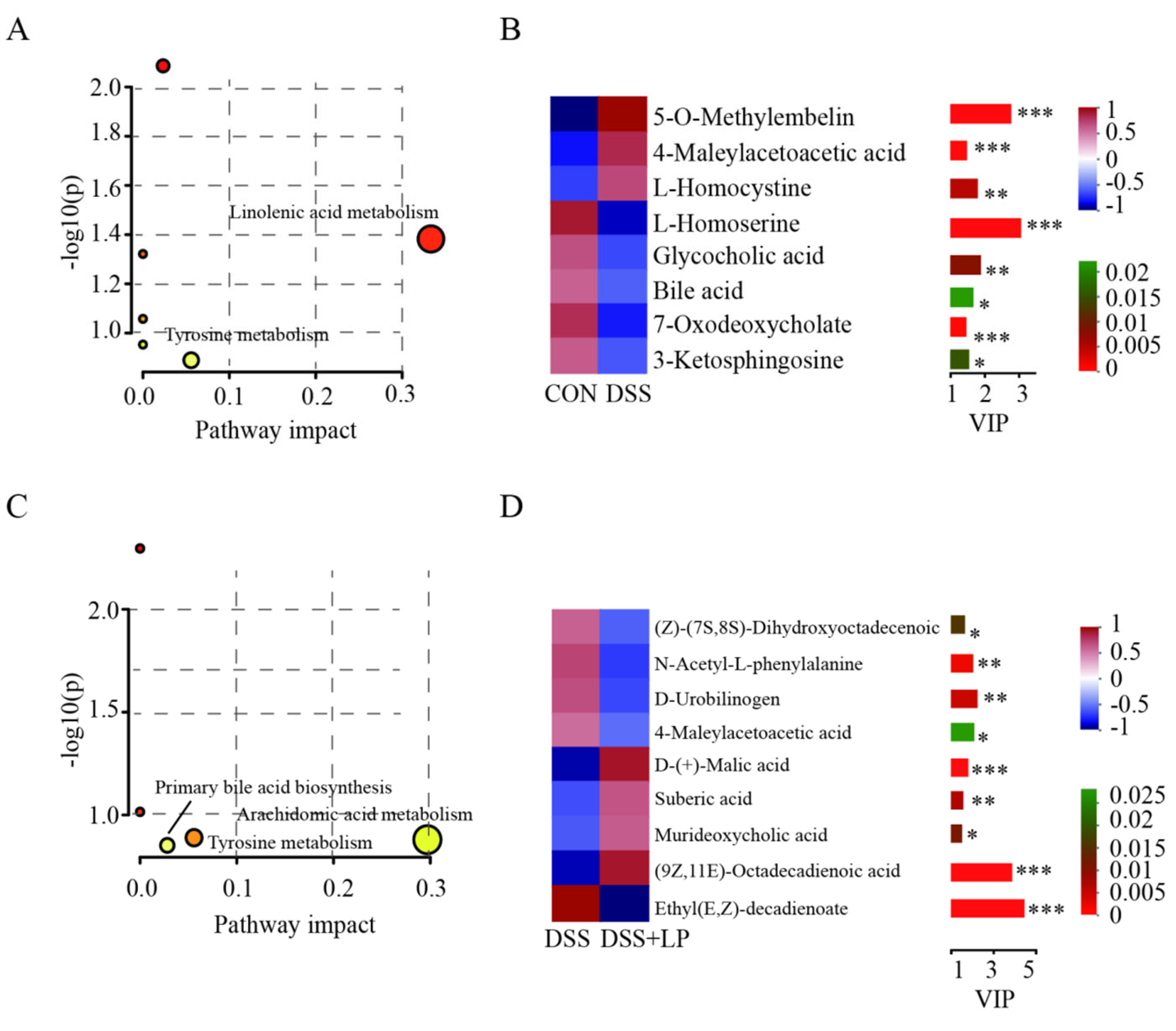
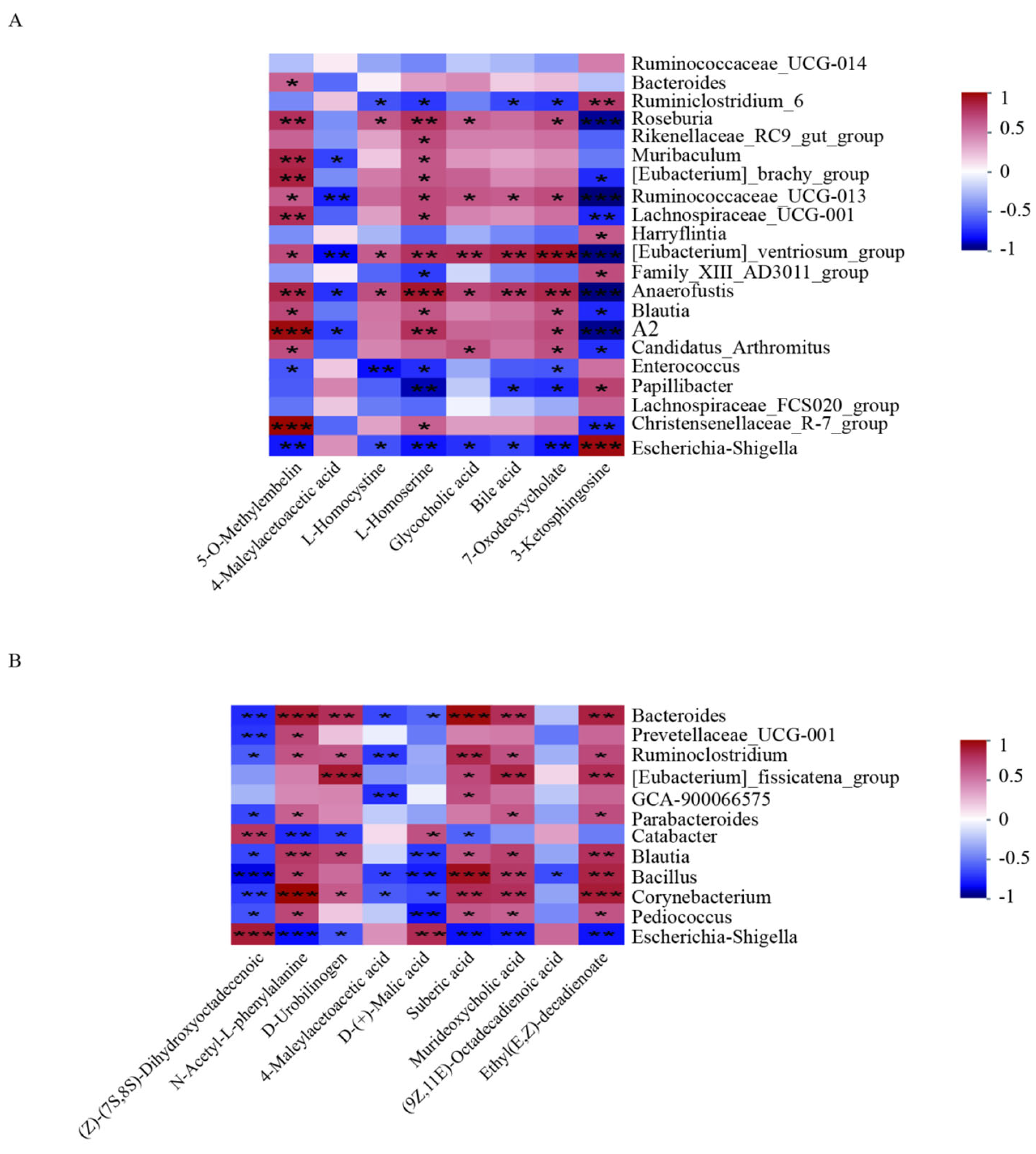
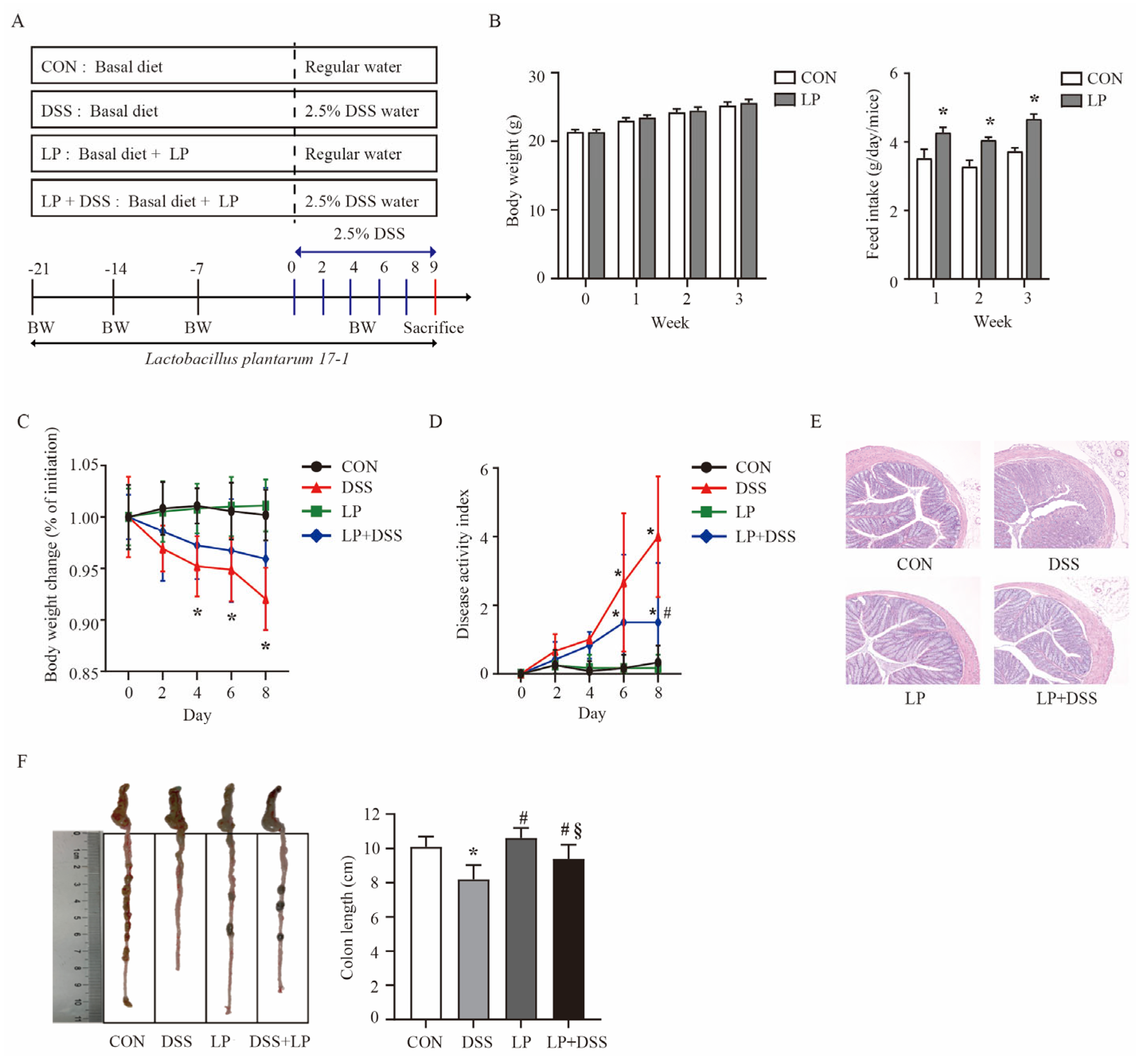
Background/Objectives: Lactobacillus strains are widely used as probiotics in the functional food industry and show potential for treating inflammatory bowel disease (IBD). The aim of this study was to investigate the effects of dietary supplementation with microencapsulated Lactobacillus plantarum 17-1 on the intestinal immune response and gut microbiota in mice with colitis. Methods: Mice were pre-fed a diet containing microencapsulated Lactobacillus plantarum 17-1 for three weeks, followed by colitis induction using 2.5% dextran sulfate sodium (DSS) in drinking water for 8 days. Results: Lactobacillus plantarum 17-1 significantly improved clinical symptoms and histopathological features in colitis-affected mice. Additionally, it effectively suppressed the up-regulation of pro-inflammatory cytokines IL-6 and IL-17 in the colon tissue of the mice. The probiotic administration increased the linear discriminant analysis score for several beneficial bacterial taxa, including Ruminococcaceae_UCG_014, Bacteroides, Prevotellaceae_UCG_001, Lactococcus, Weissella, Pediococcus, and so on. Also, Lactobacillus plantarum 17-1 regulated the abundance of inflammation-related metabolites which involved in linolenic acid metabolism, arachidonic acid metabolism, primary bile acid biosynthesis and tyrosine metabolism. Conclusions: These findings indicate that microencapsulated Lactobacillus plantarum 17-1 has a significant anti-inflammatory effect in the DSS-induced colitis model and may serve as a potential therapeutic strategy for IBD.

Keywords: 
;  ;  ;  ;  

1. Introduction

Ulcerative colitis (UC) is a subtype of inflammatory bowel disease (IBD) characterized by chronic, ulcerative inflammation of the mucosa and submucosa, typically confined to the colon [1]. It is a chronic, incurable idiopathic enteritis requiring lifelong management [2]. The pathogenesis of UC is multifactorial, involving genetic predispositions, disruption of the intestinal barrier, and activation of the mucosal immune system [3,4]. Furthermore, recent evidence highlights a significant association between UC and intestinal microbial dysbiosis. Specifically, the disruption of homeostasis between the intestinal microbiota and the mucosal immune system is emerging as a major contributing factor to the development and progression of UC [5,6]. The metabolic products of the resident gut microbiota, encompassing both beneficial and detrimental substances, are strongly correlated with the development of inflammation [7,8], which may constitute a primary mechanism of host-microbiota interaction. Traditionally used drugs for treating IBD including 5-aminosalicylic acid, steroids, and immunosuppressive agents, have severe side effects (such as leucopenia, pancreatitis and increased risk of malignancy) and are not intended for long-term treatment [9,10]. In recent years, some probiotics, while administered at effective doses, have been proved to protect against UC in both animal model and human studies [11,12]. The beneficial effects of probiotics in UC patients have been elucidated to exhibit immunomodulatory activity, protect intestinal barrier function and regulate intestinal homeostasis [13,14,15].
Lactobacillus plantarum (L. plantarum) is a lactic acid bacterium, widely used in the food and dairy industry. It is known to have effective capacity in UC therapy through suppressing pro-inflammatory cytokine expressions, regulating NF-κB signaling pathway, enhancing the intestinal epithelial barrier as well as modulating gut microbiota [16]. However, the beneficial effects of probiotics in UC patients rely on the heterogeneity among strains [17]. Moreover, the specific effects of L. plantarum on the gut microbiota and their metabolites, as well as the mechanisms underlying its ability to alleviate UC, are still not fully understood. Dextran sulfate sodium (DSS) induced colitis in mouse model can simulate the key pathological features of human ulcerative colitis and is a common model for studying the anti-inflammatory effects of probiotics. The aim of this study was to investigate the alleviating effect and potential mechanism of microencapsulated L. plantarum 17-1 on DSS induced colitis in mice, which include: (1) the effect of L. plantarum 17-1 on clinical symptoms and histopathological features of colitis mice; (2) the effects of L. plantarum 17-1 on the levels of inflammation-related cytokines in colon tissues; (3) the regulatory effects of L. plantarum 17-1 on intestinal microbial composition and metabolome. We hypothesized that microencapsulated L. plantarum 17-1 could significantly improve the symptoms of DSS induced colitis in mice and inhibit the inflammatory response by modulating the gut microbiome and metabolome.

2. Materials and Methods

2.1. Materials

Lactobacillus plantarum 17-1 was isolated from naturally fermented dairy products in China and identified through 16S rDNA similarity analysis. The strain was preserved and cultured anaerobically in de Man, Rogosa, and Sharpe (MRS) broth at 37 °C for 24 hours. Microencapsulation of L. plantarum 17-1 was conducted using a modified version of the emulsion method described by Qi et al [18]. Specifically, after two generations of activation, 700 mL of the bacterial broth (109 CFU/mL) was combined with 5 L of sterile 2.0% (w/v) sodium alginate. This mixture was then combined with 75 g of calcium carbonate and introduced into a paraffin oil phase containing 0.2% (w/v) Span 80. Emulsification was achieved by stirring at 400 rpm for 5 minutes under sterile conditions in a 100 mL fermenter. Subsequently, 90 mL of glacial acetic acid was added with continuous stirring for 10 minutes. Next, 200 mL of a saturated calcium chloride solution was added to precipitate the micro-beads into the aqueous phase at the base of the fermenter. The oil phase, located in the upper layer, was removed by aspiration. The microencapsulated bacteria were then transferred into 70 L of MRS liquid medium for a continuous growth period of 20 hours. Finally, the proliferated bacteria were harvested using a tubular bowl centrifuge, combined with corn starch as a diluent carrier, and dried using a fluidized bed dryer.

2.2. Animal Treatments

A total of 48 male BALB/c mice, aged 5 weeks, were procured from Beijing HFK Bioscience Co., Ltd. (Beijing, China). The mice were housed in individually ventilated cages, with four mice per cage, under standardized environmental conditions (temperature 23 ± 2°C, relative humidity 50%, and a 12-hour light/dark cycle). The experimental protocol was approved by the Animal Ethics Committee of the Academy of National Food and Strategic Reserves Administration (registration number: 2019M06). Based on the feasibility of the experimental design and the experience of the preliminary study, after an acclimation period for 1 week, the mice were assigned to 4 groups (n = 12): the control group (CON) and the DSS-induced colitis group (DSS) were fed the basal diet and given distilled water, the L. plantarum fed group (LP) and the LP + DSS (LP + DSS) group were fed the basal diet supplemented with 107 CFU/g L. plantarum 17-1 for a period of 29 days. To minimize potential confounders, we randomly assigned mice to the experimental group and randomly determined the order of treatment and measurement, while randomizing the location of the animals and cages using Excel’s randomization function. To induce colitis, the mice in DSS group and LP + DSS group were received 2.5% (w/v) DSS in drinking water from d 22 to d 29, as DSS treated mice developed visually blood or occult blood fecal in d 29 (Figure 1A).
At the time of sacrifice using cervical dislocation method, all of the colon was isolated for evaluation of the disease activity index and histopathology. Colon tissue and cecal contents from eight mice close to the average body weight in each group were chosen to evaluate colon cytokines, microbial composition and metabolite profiles. In order to reduce bias and improve the reliability of the results, we use a partially blind design in animal experiments, where only the experimental operator is aware of the grouping, and the results evaluators and data analysts remain in the dark until the data analysis is complete.

2.3. Evaluation of the Disease Activity Index and Histopathology

The disease activity index (DAI) scores were calculated based on body weight (BW) loss, stool consistency, and the presence of blood in stool. Colon length was measured from the ileum-cecal junction to the proximal rectum. Histopathological analysis was performed according to the method by Xia et al [19]. Briefly, 1 mm sections of colon tissue were fixed in 4% paraformaldehyde for 24 hours, dehydrated through a graded ethanol series, embedded in paraffin, sectioned, and stained with hematoxylin and eosin (HE). The stained sections were examined under a light microscope and photographed using a Canon camera.

2.4. Measurement of Colon Cytokines

Colon tissues were weighed and homogenized in ice-cold phosphate-buffered saline (PBS) at a ratio of 1:9 (w/v). The homogenates were then centrifuged at 5, 000 × g for 10 minutes at 4°C. The resulting supernatants were analyzed to quantify the concentrations of tumor necrosis factor alpha (TNF-α), tumor necrosis factor beta (TNF-β), interleukin-6 (IL-6), interleukin-10 (IL-10), and interleukin-17 (IL-17) using commercial enzyme-linked immunosorbent assay (ELISA) kits, following the manufacturer's instructions (Elabscience Biotechnology Co., Ltd, Wuhan, China).

2.5. DNA Extraction, 16S rRNA Gene Sequencing and Microbial Analysis

Genomic DNA from the microbial community present in the cecal contents was isolated utilizing the E.Z.N.A.® Stool DNA Kit (Omega Bio-tek, Norcross, GA, USA) following the manufacturer's protocol. The integrity of the extracted genomic DNA was assessed via agarose gel electrophoresis, while its concentration and purity were measured using a NanoDrop 2000 UV-Vis spectrophotometer (Thermo Scientific, Wilmington, USA). The V3-V4 region of the bacterial 16S rRNA genes was amplified using primers 338F (5'-ACTCCTACGGGAGGCAGCAG-3') and 806R (5'-GGACTACHVGGGTWTCTAAT-3') in a PCR thermocycler (GeneAmp 9700, ABI, CA, USA). The resulting PCR products were purified with the AxyPrep DNA Gel Extraction Kit (Axygen Biosciences, CA, USA) and quantified using a Quantus™ Fluorometer (Promega, USA). Subsequently, the purified amplicons were pooled in equimolar concentrations and subjected to paired-end sequencing on an Illumina MiSeq platform (Illumina, San Diego, USA) in accordance with the standard protocols at Majorbio Bio-Pharm Technology Co. Ltd. (Shanghai, China).
A total of 6,987,098 raw reads were acquired and subsequently merged using FLASH software version 1.2.7 [20]. Quality filtering was performed with fastp version 0.20.0 [21]. Following the removal of chimeric sequences, operational taxonomic units (OTUs) were clustered with 97% similarity using UPARSE software version 7.1 [22]. Taxonomic classification of OTUs was executed using the RDP Classifier software version 2.2 against the SILVA database. QIIME software version 1.7.0 was employed to assess α- and β-diversity. The linear discriminant analysis effect size (LEfSe) method was utilized to identify biomarkers that characterize differences in abundant bacterial taxa between groups [23]. The associations between bacterial taxa and colitis-related indices were evaluated using Spearman’s rank correlation coefficients, with significance determined at P < 0.05, as calculated with R software version 3.3.1.

2.6. Untargeted Metabolome Profiling Analysis

Metabolic profiling of cecal contents was performed utilizing an ultra-high-performance liquid chromatography-mass spectrometry (UHPLC-MS) methodology, as described by Yao et al [24], with subsequent analysis conducted via the Majorbio Cloud Platform (www.majorbio.com). Specifically, a precisely weighed sample of cecal content (50 mg) was combined with 400 μL of a methanol/water solution (4:1, v/v) and 20 μL of an internal standard solution (2-chloro-l-phenylalanine in acetonitrile, 0.3 mg/mL). The mixture underwent freezing, grinding using a high-throughput tissue crusher, and homogenization via ultrasound. Following centrifugation at 13, 000 × g at 4°C for 15 minutes, the supernatant was collected and transferred into sample vials for UHPLC-MS analysis. A quality control (QC) sample was generated by pooling aliquots from each individual sample. The LC-MS/MS analysis of sample was conducted on a Thermo UHPLC-Q Exactive system equipped with an ACQUITY HSS T3 column (100 mm × 2.1 mm i.d., 1.8 μm; Waters, USA). The UPLC system was coupled to a Thermo UHPLC-Q Exactive Mass Spectrometer
equipped with an electrospray ionization (ESI) source operating in positive mode and negative mode. The initial data underwent pre-processing using Progenesis QI version 2.3 (Waters, Milford, MA, USA). Metabolite annotation was performed utilizing publicly available biochemical databases, including the Human Metabolome Database (HMDB, http://www.hmdb.ca/), METLIN (https://metlin.scripps.edu/), and a proprietary untargeted database from Majorbio (Shanghai, China). To assess the variations in metabolic compounds across different groups, principal component analysis (PCA) and partial least-squares discrimination analysis (PLS-DA) were conducted using the ropls R package (Version 1.6.2, http://bioconductor.org/packages/release/bioc/html/ropls.html). The variable importance in the projection (VIP) scores were computed within the PLS-DA model. P-values were calculated using paired Student's t-tests to compare metabolite concentrations across the groups. Metabolites with a VIP score greater than 1 and a P-value less than 0.05 were identified as significantly differential metabolites within each group. These differential metabolites were verified through the Kyoto Encyclopedia of Genes and Genomes (KEGG) database and cross-referenced with KEGG pathways. Significantly enriched pathways were validated using Fisher’s exact test, implemented via the scipy.stats package in Python. Pearson's correlations between the differentially abundant cecal microbiota metabolites across groups were analyzed using R software version 3.6.0.

2.7. Statistical Analysis

Values are expressed as the mean ± standard deviation (SD). The statistically significant differences among group means were determined using one-way ANOVA, followed by the Student-Newman-Keuls comparison test of SAS (SAS Institute Inc., NC, USA). Values of P < 0.05 were considered statistically significant..

3. Results

3.1. L. Plantarum 17-1 Ameliorated Clinical Features and Intestinal Injury in DSS-Induced Colitis Mice

To evaluate the progression of colitis symptoms during DSS treatment across different groups, we assessed changes in BW, DAI, colon length, and colonic histopathology. Prior to DSS exposure, no significant differences in body weight were observed between the control (CON) and L. plantarum (LP) treatment groups over a three-week period (P > 0.05). However, feed intake was significantly higher in the LP treatment group compared to the CON group (P < 0.05, figure 1B). From the fourth day of DSS exposure, mice treated with DSS exhibited a significant reduction in body weight compared to those in the CON and LP groups (P < 0.05, Figure 1C). Additionally, a significant increase in the DAI score was observed on the sixth day in the DSS group compared to the CON and LP groups (P < 0.05), with a subsequent decrease noted on the eighth day in the LP + DSS group relative to the DSS group (P < 0.05, Figure 1D). Furthermore, colon length was significantly reduced in the DSS group compared to the CON and LP groups (P < 0.05). However, supplementation with L. plantarum 17-1 restored colon length to a level comparable to that of the CON group (P > 0.05, Figure 1F). To observe the effects of L. plantarum 17-1 on pathological changes in colonic tissue of mice, HE staining was performed. As shown in Figure 1E, DSS-induced colon injury was characterized by partial destruction of the epithelial structure, damage to the crypt structure, fibrous hyperplasia, and submucosal edema, accompanied by inflammatory cell infiltration. In contrast, supplementation with L. plantarum 17-1 significantly alleviated these pathological changes. Specifically, it repaired the epithelial architecture and reduced inflammatory cell infiltration in the colon tissue.

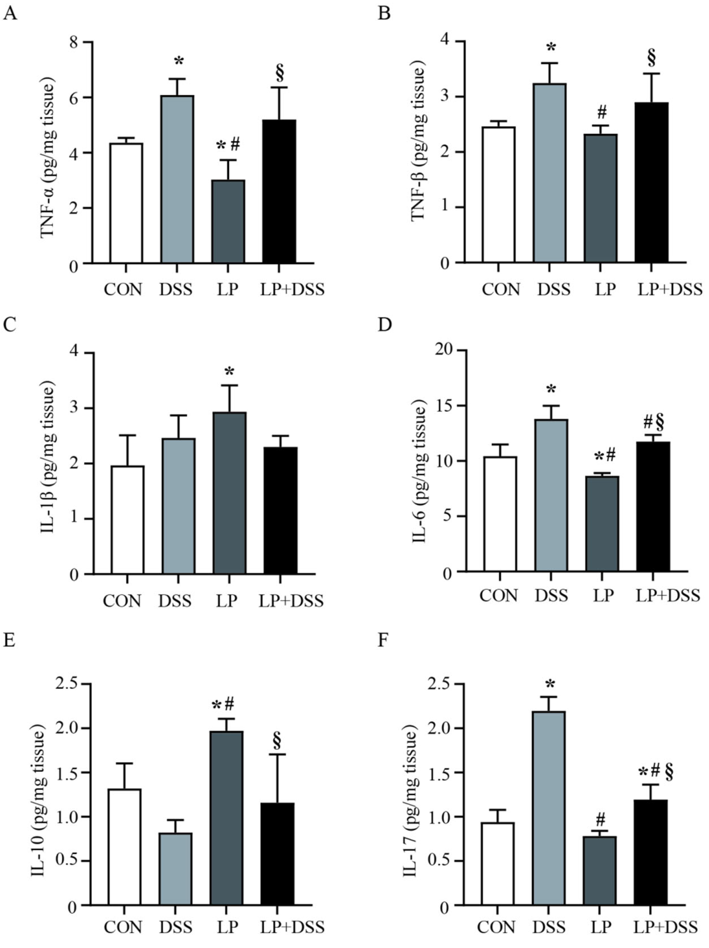
3.2. L. Plantarum 17-1 Regulated the Production of Cytokines in DSS-Induced Colitis Mice

The study investigated the prophylactic effects of L. plantarum 17-1 on intestinal inflammation by assessing cytokine levels in colon tissue (figure 2). In mice with DSS-induced colitis, significant increases in the levels of pro-inflammatory cytokines TNF-α, TNF-β, IL-6, and IL-17 were observed, alongside a decrease in the anti-inflammatory cytokine IL-10, compared to the control group (P < 0.05). Notably, administration of L. plantarum 17-1 to DSS-exposed mice resulted in a significant reduction in IL-6 and IL-17 levels (P < 0.05) compared to the DSS group. Furthermore, in the LP group, the production of TNF-α and IL-6 was significantly lower (P < 0.05), while the levels of IL-1β and IL-10 were significantly higher (P < 0.05) than those observed in the control group.
Figure 2. Cytokine levels in colon tissue of mice. Data are expressed as the mean ± SD (n = 8). Values with different superscripts letters are very significantly different within the level. *P < 0.05, compared with the control group; #P < 0.05, compared with the DSS group.
Preprints 152269 g002

3.3. L. Plantarum 17-1 Changed Caecal Microbiota Diversity in DSS-Induced Colitis Mice

To elucidate the gut microbiota's response to a diet containing L. plantarum 17-1 in mice with DSS-induced colitis, microbiota composition was assessed using high-throughput 16S rRNA gene sequencing.
In the analysis of α-diversity, the Sobs and Shannon indices were used to represent taxonomic richness and evenness, respectively. As illustrated in figure 3A, the Sobs index showed a significant reduction (P < 0.05) in the bacterial communities within the colonic digesta of mice following DSS treatment. Conversely, administration of L. plantarum 17-1 mitigated this reduction, resulting in a significant increase (P < 0.05) in microbial community richness in DSS-induced mice. The Shannon index indicated a higher (P < 0.05) diversity of microbiota in the LP group compared to the CON group; however, no significant differences were observed among the CON, DSS, and DSS + LP groups (P > 0.05). Based on unweighted UniFrac distances, the Principal Coordinate Analysis (PCoA) demonstrated a clear separation of microbiota profiles in the colonic digesta of the four groups (R = 0.2405, P = 0.001, ANOSIM, figure 3B). These findings suggest that L. plantarum 17-1 may alleviate alterations in gut microbial richness and structure induced by DSS.
Figure 3. Impact of L. Plantarum 17-1 on gut microbiota diversity in DSS-induced colitis mice. Data are expressed as the mean ± SD (n = 8). Values with different superscripts letters are very significantly different within the level. *P < 0.05, compared with the control group; #P < 0.05, compared with the DSS group. (A) Shannon and Sobs index; (B) PCoA plot based on the unweighted UniFrac distances; (C) microbiota composition at phylum and genus level.
Preprints 152269 g003

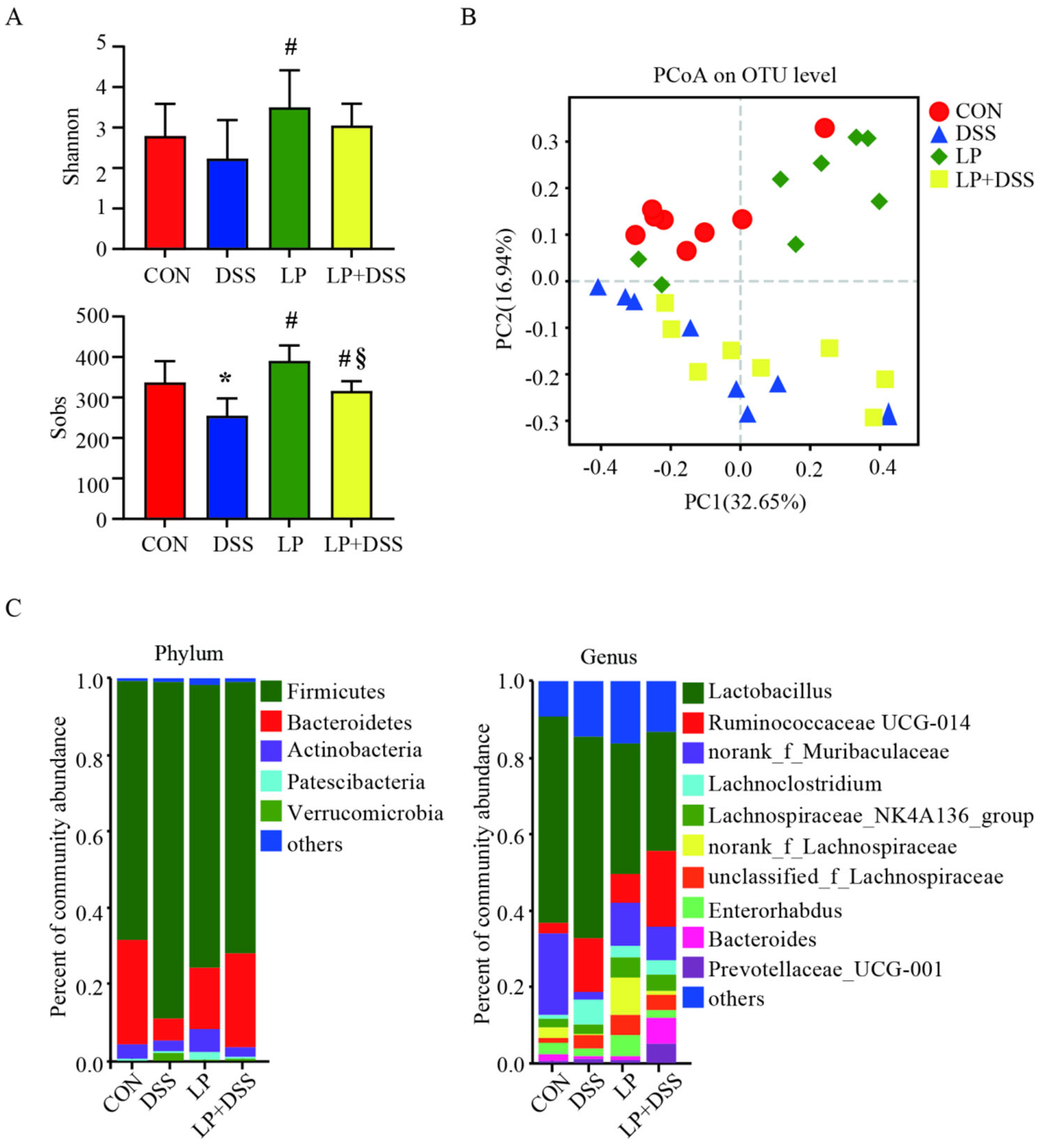
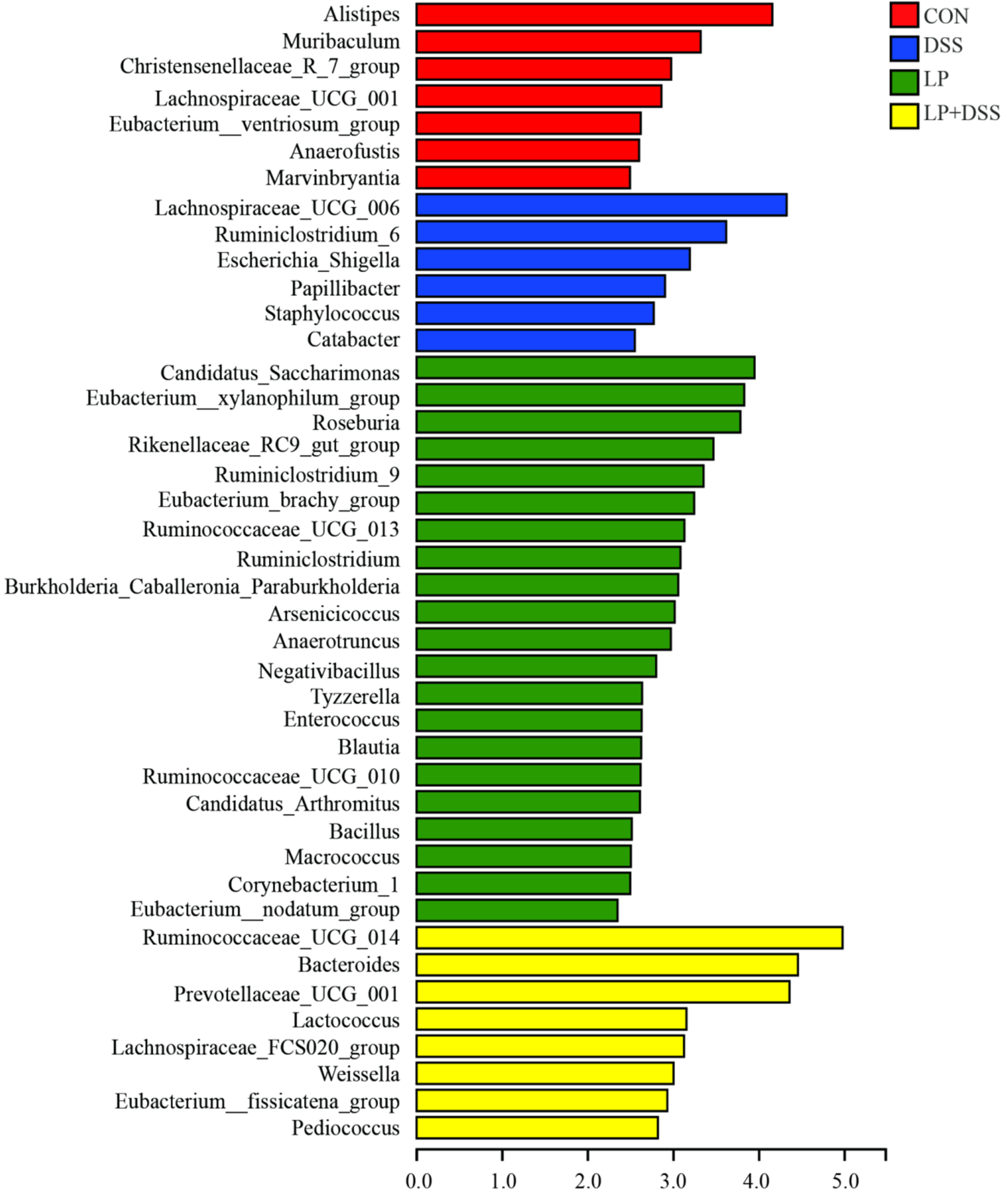
3.4. L. Plantarum 17-1 Induced Shift in Gut Microbiota Composition in DSS-Induced Colitis Mice

The composition of the microbiota was analyzed at both the phylum and genus levels, as illustrated in figure 3C. At the phylum level, the microbiota in the colonic digesta of mice was predominantly composed of Firmicutes, Bacteroidetes, and Actinobacteria. At the genus level, OTUs associated with Lactobacillus, Ruminococcaceae_UCG_014, norank_f_Muribaculaceae, Lachnoclostridium, Lachnospiraceae_NK4A136_group, norank_f_Lachnospiraceae, unclassified_f_Lachnospiraceae, Enterorhabdus, Bacteroides and Prevotellaceae_UCG_001 were predominant in the colonic digesta of mice. Subsequently, LEfSe analysis was conducted to identify changes in the relative abundance of genera between the groups, as depicted in figure 4. The relative abundance of 6 genera, including Lachnospiraceae_UCG_006, Ruminiclostridium_6, Escherichia_Shigella, Papillibacter, Staphylococcus, and Catabacter, was significantly higher (P < 0.05) in the colonic digesta of mice in DSS group. Conversely, the relative abundance of 8 genera, including Ruminococcaceae_UCG_014, Bacteroides, Prevotellaceae_UCG_001, Lactococcus, Lachnospiraceae_FCS020_group, Weissella, Eubacterium_fissicatena_group and Pediococcus, were significantly higher (P < 0.05) in mice from the LP + DSS group. Additionally, 21 genera, such as Candidatus_Saccharimonas, Eubacterium_xylanophilum_group, and Roseburia, exhibited a significantly higher abundance (P < 0.05) in mice from the LP group.
Figure 4. LEfSe analysis at genus level.
Preprints 152269 g004

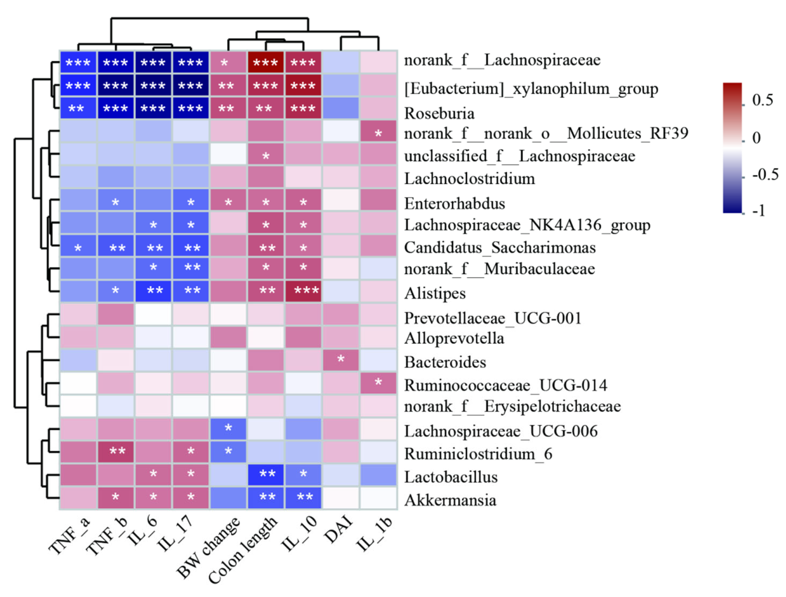
3.5. Spearman Correlation Analysis of Different Abundant Genera and Colitis Injury Indices

To further elucidate the relationship between colitis injury indices and shifts in gut microbiota at the genus level, correlation analysis was conducted involving the top 20 microbial genera with significantly altered relative abundance across all four groups. These genera were analyzed in relation to BW changes, colon length, DAI, and inflammatory cytokines (figure 5). The genera norank_f_Lachnospiraceae, Eubacterium_xylanophilum_group, Roseburia, Enterorhabdus, Candidatus_Saccharimonas, and Alistipes exhibited significant negative correlations with inflammatory cytokines such as TNF-α, TNF-β, IL-6, and IL-17, while showing positive correlations with the anti-inflammatory cytokine IL-10, as well as with BW change and colon length in mice. Conversely, the genera Ruminiclostridium_6, Lactobacillus, and Akkermansia demonstrated opposite correlations with these parameters, thereby identifying specific gut bacterial populations that may be associated with colitis.
Figure 5. Spearman correlation analysis of different abundant genera and environmental factors. Values with different superscripts letters are very significantly different within the level. *P < 0.05, **P < 0.01, ***P < 0.001.
Preprints 152269 g005

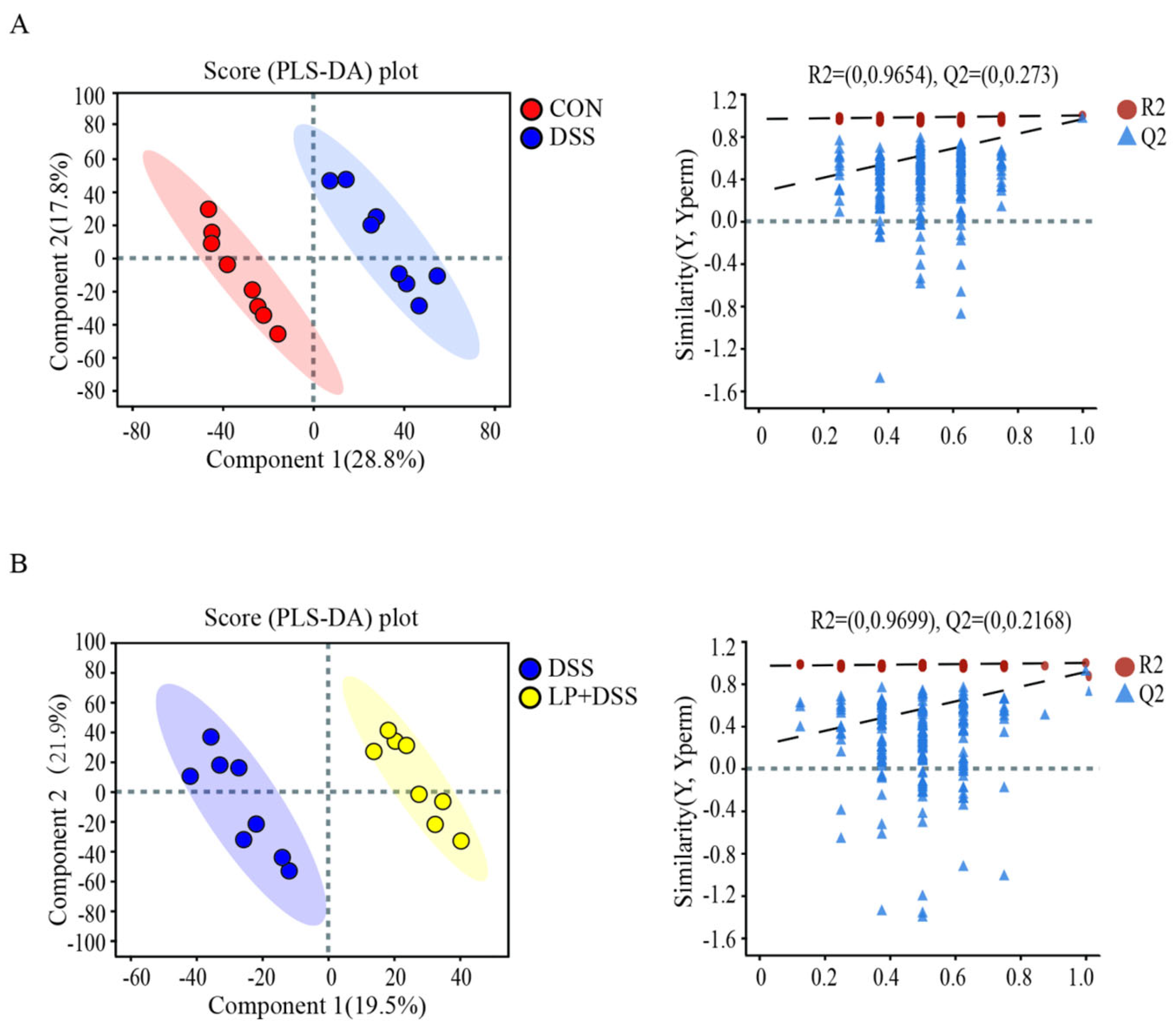
3.6. L. Plantarum 17-1 Altered Gut Metabolic Profiles in DSS-Induced Colitis Mice

To assess the impact of L. plantarum 17-1 on gut metabolic profiles in DSS-induced colitis mice, colonic digesta were analyzed using UHPLC-MS across CON, DSS, and LP + DSS groups. PLS-DA demonstrated significant differentiation in metabolic profiles between the CON and DSS groups (R² = 0.9654, Q² = 0.273; figure 6A), as well as between the DSS and LP + DSS groups (R² = 0.9699, Q² = 0.2168; figure 6B).
Figure 6. PLS-DA score plots of caecal digesta metabolites between (A) CON and DSS groups; (B) DSS and DSS + LP groups.
Preprints 152269 g006
The selection of altered metabolites between groups was based on a threshold of VIP values greater than 1 from the PLS-DA model and a P-value less than 0.05 from Student's t-test. A total of 20 differential metabolites between DSS and CON groups were annotated in the KEGG database (Supplementary Table 1), which primarily associated with linolenic acid metabolism and tyrosine metabolism (figure 7A). Among these, 8 metabolites with VIP values exceeding 1.5 are shown in figure 7B. In the comparison between the DSS and LP + DSS groups, 24 metabolites that can be annotated in the KEGG database showed significant differences (Supplementary Table 2), with 9 metabolites having VIP values greater than 1.5 (figure 7D), predominantly related to arachidonic acid metabolism, primary bile acid biosynthesis, and tyrosine metabolism (figure 7C).
Figure 7. Significantly changed metabolics and pathways between (A) CON and DSS groups; (B) DSS and DSS + LP groups. Values with different superscripts letters are very significantly different within the level. *P < 0.05, **P < 0.01, ***P < 0.001.
Preprints 152269 g007

3.7. Spearman Correlation Analysis of Different Abundant Genera and Metabolites

Furthermore, we conducted an analysis of the correlation between bacterial taxa and metabolite levels across the CON, DSS, and LP + DSS groups (figure 8). In the comparison between the CON and DSS groups, 3-Ketosphingosine exhibited positive correlations with the genera Ruminiclostridium_6, Escherichia_Shigella, and Papillibacter. Conversely, L-Homoserine, bile acid, and 7-Oxodeoxycholate demonstrated negative correlations with these same genera (refer to figure 8A). In the analysis between the DSS and LP + DSS groups, N-Acetyl-L-phenylalanine, suberic acid, ethyl(E,Z)-Decadienoate were positively correlated with the genera Bacteroides and Pediococcus, whereas (Z)-(7S,8S)-Dihydroxyoctadecenoic acid and D-(+)-Malic acid were negatively correlated with these genera (refer to figure 8B).
Figure 8. Correlation analysis of different abundant genus and metabolics between (A) CON and DSS groups; (B) DSS and DSS + LP groups. Values with different superscripts letters are very significantly different within the level. *P < 0.05, **P < 0.01, ***P < 0.001.
Preprints 152269 g008

4. Discussion

An increasing number of research indicates that L. plantarum has the potential to restore disrupted intestinal flora and serve as a probiotic to ameliorate colitis [25,26]. However, due to its limited stress resistance ability, L. plantarum has predominantly been administered by gavage in most studies, which have shown promising results but are often constrained by factors such as gastric acid degradation and inconsistent delivery to the colon. In the present study, we employed microencapsulation techniques for L. plantarum 17-1 and subsequently incorporated it into the diet of mice, which not only enhances the survivability of L. plantarum through the harsh conditions of the gastrointestinal tract, but also ensures sustained release within the colon, where its therapeutic effects are most needed. The results demonstrated that dietary inclusion of microencapsulated L. plantarum 17-1 effectively alleviated disease symptoms and pathological damage, modulated inflammatory cytokines, and maintained the dynamic equilibrium of intestinal flora in mice with DSS-induced colitis.
The characteristic manifestations of DSS-induced colitis include weight loss, colon shortening, fecal bleeding and so on, which were observed in DSS-treated mice after one week in the present study. However, administration of L. plantarum 17-1 demonstrated a mitigating effect on these episodic symptoms and exhibited synergistic benefits (Figure 1). Prior to DSS treatment, mice were pre-fed with a probiotic-supplemented diet for three week, which resulted in increased food intake. Although there was no observed change in body weight at this stage, and intestinal morphology and microbial composition were not assessed, subsequent results indicated that a probiotic-supplemented diet can enhance intestinal morphology and microbial composition, thereby preventing colitis in mice. This early intervention phase underscores the importance of establishing a favorable gut environment before the onset of disease. By promoting the growth of beneficial bacteria and inhibiting pathogenic species, L. plantarum may help maintain a balanced gut microbiota, which is crucial for preventing recurrent episodes of colitis [27,28].
Cytokines are deeply implicated in the pathogenesis of IBD, with clinical studies consistently indicating that the severity of the disease is closely associated with the equilibrium between pro-inflammatory and anti-inflammatory cytokines [29]. L. plantarum, a well-studied probiotic strain, exerts a protective effect by modulating gut-associated lymphoid and epithelial cells [30]. These interactions stimulate the production of various cytokines, including TNF-α, IL-1β, IL-6, IL-10, and IL-12, which collectively influence the immune response and intestinal homeostasis. For instance, studies have shown that L. plantarum can significantly reduce pro-inflammatory cytokine levels while enhancing anti-inflammatory cytokine secretion [31]. This dual action helps restore the disrupted balance in IBD patients, potentially alleviating symptoms and promoting recovery. Furthermore, the ability of L. plantarum to interact with gut epithelial cells reinforces its role as a key player in gut barrier function and immune modulation [32,33]. Several researchers have demonstrated that IL-10 deficiency is a primary initiating event in chronic intestinal inflammation using an IL-10-deficient mouse model. The findings revealed that mice lacking IL-10 exhibited increased levels of TNF-α, TNF-β, IL-6, and IL-17, along with decreased levels of IL-10 when subjected to DSS-induced colitis [34,35,36]. The result in our study further validating the critical role of IL-10 in maintaining intestinal homeostasis. Also, treatment with L. plantarum 17-1 significantly reduced IL-6 and IL-17 levels in DSS-induced colitis mice, as evidenced by experimental data presented in figure 2, which reminder that the regulation of cytokine networks is crucial for maintaining gut health and preventing inflammatory conditions.
Research utilizing animal models has consistently highlighted the critical role of bacterial colonization in intestinal inflammation, emphasizing the significance of gut microbiota in IBD [37]. Despite ongoing uncertainties regarding the precise mechanisms driving IBD development, it is widely accepted that dysregulation in the balance between gut microbiota and the mucosal immune system plays a pivotal role. In our study, we observed elevated levels of specific bacterial taxa, including Lachnospiraceae_UCG_006, Ruminiclostridium_6, Escherichia_Shigella, Papillibacter, Staphylococcus, and Catabacter, within the colonic digesta of mice treated with DSS (figure 5). Notably, the abundance of Ruminiclostridium_6 demonstrated a positive correlation with cytokine levels, the DAI, and pathological scores, suggesting its potential as an indicator of colitis progression. These findings align with previous studies highlighting the pathogenic nature of Escherichia_Shigella, which was found to be significantly more abundant in the intestines of colitics mice [38]. Our results further corroborate this by identifying a strong positive correlation between Escherichia_Shigella and pro-inflammatory cytokines IL-17 and IFN-γ. This indicates that these bacteria may contribute to UC exacerbation by disrupting cytokine homeostasis. The reduction in Escherichia_Shigella abundance and down-regulation of pro-inflammatory cytokines following treatment with L. plantarum underscores its therapeutic potential, consistent with other studies demonstrating the beneficial effects of probiotics in modulating gut flora and mitigating inflammatory responses [39,40]. Additionally, our data revealed a significant increase in Staphylococcus following DSS treatment, potentially linking it to inflammatory diseases. This observation resonates with recent research suggesting that certain Staphylococcus species can promote intestinal inflammation through toxin production or immune system modulation [41]. Additionally, we noted a significant increase in the Verrucomicrobia phylum in DSS mice (figure 3C), primarily due to the proliferation of Akkermansia. Akkermansia also showed positive correlations with inflammatory cytokines and negative correlations with the anti-inflammatory cytokine as well as colon length (figure 5), which may resides within the mucus layer and degrades mucin [42,43]. While our findings reinforce the importance of microbial balance in maintaining gut health, they also highlight the complex interplay between various bacterial taxa and their influence on disease outcomes.
Probiotics, as live microorganisms, play a pivotal role in modulating gut microbiota composition and restoring the mucosal immune responses disrupted by chronic intestinal inflammation [44]. In this study, we demonstrated that L. plantarum 17-1 administration significantly alleviated inflammatory symptoms in colitis mice, promoting microbial diversity and restructuring the gut microbiota at various taxonomic levels (figure 3). This aligns with previous studies indicating the therapeutic potential of probiotics in mitigating IBD. Furthermore, our results indicate that L. plantarum 17-1 enhances beneficial bacteria such as Ruminococcaceae_UCG_014, Bacteroides, Prevotellaceae_UCG_001, Lactococcus, Lachnospiraceae_FCS020_group, Weissella, Eubacterium_fissicatena_group and Pediococcus (figure 4). SCFAs have been shown to reduce intestinal inflammation through the induction of regulatory T Treg cells, consistent with observations from other studies [45,46]. The effect of Ruminococcaceae_UCG_014 in producing SCFA and mitigate colonic barrier dysfunction and inflammation were observed in several disease modes [47,48,49]. Notably, Bacteroides are known to enhance intestinal barrier function by promoting the expression of intestinal tight junction proteins and reducing apoptosis of epithelial cells [50,51], the increase of Bacteroides in our study suggests that L. plantarum 17-1 may restore the intestinal microbiota to maintain the stability and function of the internal environment. The abundance of Bacteroidetes and Prevotellaceae_UCG-001, belong to the Bacteroidetes phylum, were increased in L. plantarum 17-1 treated DSS mice, which may degrade polysaccharides and supplying nutrients to other probiotics to producing anti-inflammatory metabolites [52]. The immunomodulatory and mucosal repair properties of Lactococcus, Weissella and Pediococcus were also widely concluded in several colitis models [53,54,55,56].
Metabolites are the main substances that microorganisms regulate the local microenvironment and affect the function of the organisms. In this study, UHPLC-MS was used to analyze the metabolic characteristics of colonic contents in three groups of mice. The significantly changed metabolites between CON, DSS, LP + DSS group were mainly involved in linolenic acid metabolism, tyrosine metabolism, arachidonic acid metabolism and primary bile acid biosynthesis pathways (figure 7). Linolenic acid is an important polyunsaturated fatty acid, and its metabolites may be involved in regulating the production of inflammatory mediators and cell signaling [57]. Studies have shown that changes in metabolites in the linolenic acid metabolic pathway are closely related to the severity of inflammation in mouse models of DSS induced colitis. By regulating the metabolism of linolenic acid, the symptoms of DSS induced colitis can be significantly reduced [58]. In a mouse model of DSS induced UC, arachidonic acid metabolism is one of the main metabolic pathways affected, which is closely related to the regulation of inflammatory response [59]. Tyrosine metabolism is significantly altered in DSS induced colitis, and these changes may influence the development of the disease by modulating immune cell signaling and inflammatory responses [60]. Probiotics may indirectly affect tyrosine metabolism in DSS induced colitis by regulating the composition and function of the gut microbiota. This regulatory effect may be achieved by improving inflammatory response and enhancing intestinal barrier function [61]. 3-Ketosphingosine was up-regulated in DSS group and exhibited positive correlations with Ruminiclostridium_6, Escherichia_Shigella, and Papillibacter (figure 8A), which act as an intermediate of sphingolipid metabolism and may affect intestinal health by regulating cell signaling pathways and inflammatory response [62]. Studies have shown that gut microbiota is able to convert primary bile acids into secondary bile acids, which are strongly associated with colon inflammation [63]. In addition, some microorganisms such as Escherichia_Shigella may participate in the metabolism of bile acids, thus affecting the inflammatory response, which is consistent with our results that bile acid was down-regulated in DSS group [64]. L-Homoserine and 7-Oxodeoxycholate are metabolites of amino acids and bile acids, and although there is currently a lack of direct research into the role of these metabolites in DSS models, their potential role in intestinal health has been widely demonstrated [62,65,66].
Collectively, these data underscore the pivotal role of microencapsulated L.plantarum 17-1 in modulating gut microbiota composition and maintaining intestinal homeostasis. However, there are still some limitations. Although we have characterized the composition and diversity of the gut microbiota using 16S rRNA gene sequencing technology, our investigation into the functional aspects of the microbiota remains relatively superficial. Therefore, future research should consider exploring the combined application of L. plantarum 17-1 with other probiotics or prebiotics to potentially enhance therapeutic efficacy and further elucidate the mechanisms underlying its beneficial effects.

5. Conclusions

In summary, microencapsulated Lactobacillus plantarum 17-1 significantly alleviated colitis symptoms and inflammation in a DSS-induced mouse model by modulating the gut microbiota and metabolites. These results highlight its potential as a therapeutic strategy for IBD and warrant further clinical exploration.

Supplementary Materials

The following supporting information can be downloaded at: www.mdpi.com/xxx/s1, Table S1: Differential metabolites between DSS and CON groups annotated in KEGG database; Table S2: Differential metabolites between DSS and LP + DSS groups annotated in KEGG database.

Author Contributions

Beibei He, methodology, validation, investigation, data curation, writing – original draft. Tao Duan and Dandan Hu: methodology, validation, investigation, data curation. Lixian Chen and Lin Qiao: investigation, data curation. Dan Song and Li Wang: resources, investigation. Shijie Fan and Kunru Teng: resources, investigation. Weiwei Wang and Aike Li: conceptualization, resources, supervision, project administration, funding acquisition, writing– original draft, writing – review & editing. All the authors have read and approved the final version of the manuscript.

Funding

This study was supported by Youth Talent Support Program by the National Food and Strategic Re serves Administration (QN2024402).

Institutional Review Board Statement

The experimental protocol was approved by the Animal Ethics Committee of the Academy of National Food and Strategic Reserves Administration (registration number: 2019M06).

Data Availability Statement

The raw reads of 16S rRNA gene sequencing were deposited into the NCBI Sequence Read Archive database (accession number: PRJNA1219830; http://www.ncbi.nlm.nih.gov/bioproject/1219830 ). Raw metabolomics data were uploaded to the MetaboLights database (accession number: MTBLS12244; https://www.ebi.ac.uk/metabolights/reviewer48d14ea1-23ef-4c78-bfd8-8b82d3403eed).

Conflicts of Interest

The authors declare no conflicts of interest.

Abbreviations

The following abbreviations are used in this manuscript:
BW body weight
DSS dextran sulfate sodium
DAI disease activity index
ELISA enzyme-linked immunosorbent assay
ESI electrospray ionization
LEfSe linear discriminant analysis effect size
IBD inflammatory bowel disease
IL interleukin
OTU operational taxonomic unit
SD standard deviation
TNF tumor necrosis factor
UC ulcerative colitis
UHPLC-MS ultra-high-performance liquid chromatography-mass spectrometry
PCA principal component analysis
PLS-DA partial least-squares discrimination analysis
QC quality control
VIP variable importance in the projection

References

  1. Ungaro, R.; Mehandru, S.; Allen, P. B.; Peyrin-Biroulet, L. and Colombel, J. F. Ulcerative colitis. Lancet 2017, 389, 1756-1770.
  2. Baumgart, D. C. and Sandborn, W. J. Crohn's disease. Lancet 2012, 380, 1590-1605.
  3. Kostic, A.D.; Xavier, R.J.; Gevers, D. The Microbiome in Inflammatory Bowel Disease: Current Status and the Future Ahead. Gastroenterology 2014, 146, 1489–1499. [CrossRef]
  4. Ramos, G. P. and Papadakis, K. A. Mechanisms of disease: Inflammatory bowel diseases. Mayo Clin Proc 2019, 94, 155-165.
  5. Nishida, A.; Inoue, R.; Inatomi, O.; Bamba, S.; Naito, Y.; Andoh, A. Gut microbiota in the pathogenesis of inflammatory bowel disease. Clin. J. Gastroenterol. 2017, 11, 1–10. [CrossRef]
  6. Robertson, S.J.; Goethel, A.; Girardin, S.E.; Philpott, D.J. Innate Immune Influences on the Gut Microbiome: Lessons from Mouse Models. Trends Immunol. 2018, 39, 992–1004. [CrossRef]
  7. Louis, P.; Hold, G.L.; Flint, H.J. The gut microbiota, bacterial metabolites and colorectal cancer. Nat. Rev. Microbiol. 2014, 12, 661–672. [CrossRef]
  8. Nusbaum, D.J.; Sun, F.; Ren, J.; Zhu, Z.; Ramsy, N.; Pervolarakis, N.; Kunde, S.; England, W.; Gao, B.; Fiehn, O.; et al. Gut microbial and metabolomic profiles after fecal microbiota transplantation in pediatric ulcerative colitis patients. FEMS Microbiol. Ecol. 2018, 94. [CrossRef]
  9. Wilson, D.; Thomas, A.; Croft, N.; Newby, E.; Akobeng, A.; Sawczenko, A.; Fell, J.; Murphy, M.S.; Beattie, R.; Sandhu, B.; et al. Systematic Review of the Evidence Base for the Medical Treatment of Paediatric Inflammatory Bowel Disease. J. Pediatr. Gastroenterol. Nutr. 2010, 50, S14–34. [CrossRef]
  10. Troncone, E.; Monteleone, G. The safety of non-biological treatments in Ulcerative Colitis. Expert Opin. Drug Saf. 2017, 16, 779–789. [CrossRef]
  11. Bian, X.; Yang, L.; Wu, W.; Lv, L.; Jiang, X.; Wang, Q.; Wu, J. J.; Li, Y. T.; Ye, J. Z. et al. Pediococcus pentosaceus LI05 alleviates DSS-induced colitis by modulating immunological profiles, the gut microbiota and short-chain fatty acid levels in a mouse model. Microb Biotechnol 2020, 13, 1228-1244.
  12. Chen, P.; Xu, H.; Tang, H.; Zhao, F.; Yang, C.; Kwok, L.; Cong, C.; Wu, Y.; Zhang, W.; Zhou, X.; et al. Modulation of gut mucosal microbiota as a mechanism of probiotics-based adjunctive therapy for ulcerative colitis. Microb. Biotechnol. 2020, 13, 2032–2043. [CrossRef]
  13. Choi, J. W.; Kang, H. O.; Seo, M. J.; Park, S. H. and Lee, D. H. Microencapsulation of Lactobacillus plantarum K159 using alginate and gellan gum mixture: Optimization of formulation variables using response surface methodology. J Microbiol Biotechnol 2015, 25, 10-18.
  14. Kim, D.H.; Kim, S.; Ahn, J.B.; Kim, J.H.; Ma, H.W.; Seo, D.H.; Che, X.; Park, K.C.; Jeon, J.Y.; Kim, S.Y.; et al. Lactobacillus plantarum CBT LP3 ameliorates colitis via modulating T cells in mice. Int. J. Med Microbiol. 2020, 310, 151391. [CrossRef]
  15. Kim, S.-Y.; Shin, J.-S.; Chung, K.-S.; Han, H.-S.; Lee, H.-H.; Lee, J.-H.; Kim, S.-Y.; Ji, Y.W.; Ha, Y.; Kang, J.; et al. Immunostimulatory Effects of Live Lactobacillus sakei K040706 on the CYP-Induced Immunosuppression Mouse Model. Nutrients 2020, 12, 3573. [CrossRef]
  16. Sun, M.; Liu, Y.; Song, Y.; Gao, Y.; Zhao, F.; Luo, Y.; Qian, F.; Mu, G.; Tuo, Y. The ameliorative effect ofLactobacillus plantarum-12 on DSS-induced murine colitis. Food Funct. 2020, 11, 5205–5222. [CrossRef]
  17. Campana, R.; van Hemert, S.; Baffone, W. Strain-specific probiotic properties of lactic acid bacteria and their interference with human intestinal pathogens invasion. Gut Pathog. 2017, 9, 1–12. [CrossRef]
  18. Qi, W.; Liang, X.; Yun, T.; Guo, W. Growth and survival of microencapsulated probiotics prepared by emulsion and internal gelation. J. Food Sci. Technol. 2019, 56, 1398–1404. [CrossRef]
  19. Xia, Y.; Chen, Y.; Wang, G.; Yang, Y.; Song, X.; Xiong, Z.; Zhang, H.; Lai, P.; Wang, S.; Ai, L. Lactobacillus plantarum AR113 alleviates DSS-induced colitis by regulating the TLR4/MyD88/NF-κB pathway and gut microbiota composition. J. Funct. Foods 2020, 67. [CrossRef]
  20. Magoč, T.; Salzberg, S.L. FLASH: fast length adjustment of short reads to improve genome assemblies. Bioinformatics 2011, 27, 2957–2963. [CrossRef]
  21. Chen, S.; Zhou, Y.; Chen, Y.; Gu, J. fastp: an ultra-fast all-in-one FASTQ preprocessor. Bioinformatics 2018, 34, i884–i890, doi:10.1093/bioinformatics/bty560.
  22. Edgar, R.C. UPARSE: highly accurate OTU sequences from microbial amplicon reads. Nat. Methods 2013, 10, 996–998. [CrossRef]
  23. Segata, N.; Izard, J.; Waldron, L.; Gevers, D.; Miropolsky, L.; Garrett, W.S.; Huttenhower, C. Metagenomic biomarker discovery and explanation. Genome Biol. 2011, 12, R60. [CrossRef]
  24. Yao, J.; Lu, H.; Wang, Z.; Wang, T.; Fang, F.; Wang, J.; Yu, J.; Gao, R. A sensitive method for the determination of the gender difference of neuroactive metabolites in tryptophan and dopamine pathways in mouse serum and brain by UHPLC-MS/MS. J. Chromatogr. B 2018, 1093-1094, 91–99. [CrossRef]
  25. Liu, Y.; Liu, G.; Fang, J. Progress on the mechanisms of Lactobacillus plantarum to improve intestinal barrier function in ulcerative colitis. J. Nutr. Biochem. 2023, 124, 109505. [CrossRef]
  26. Huang, C.; Hao, W.; Wang, X.; Zhou, R.; Lin, Q. Probiotics for the treatment of ulcerative colitis: a review of experimental research from 2018 to 2022. Front. Microbiol. 2023, 14, 1211271. [CrossRef]
  27. Ouwehand, A.C.; Forssten, S.; Hibberd, A.A.; Lyra, A.; Stahl, B. Probiotic approach to prevent antibiotic resistance. Ann. Med. 2016, 48, 246–255. [CrossRef]
  28. Zhang, X.; Zhang, D.; Jia, H.; Feng, Q.; Wang, D.; Liang, D.; Wu, X.; Li, J.; Tang, L.; Li, Y.; et al. The oral and gut microbiomes are perturbed in rheumatoid arthritis and partly normalized after treatment. Nat. Med. 2015, 21, 895–905. [CrossRef]
  29. Haneishi, Y.; Furuya, Y.; Hasegawa, M.; Picarelli, A.; Rossi, M.; Miyamoto, J. Inflammatory Bowel Diseases and Gut Microbiota. Int. J. Mol. Sci. 2023, 24, 3817. [CrossRef]
  30. Yang, B.; Yue, Y.; Chen, Y.; Ding, M.; Li, B.; Wang, L.; Wang, Q.; Stanton, C.; Ross, R.P.; Zhao, J.; et al. Lactobacillus plantarum CCFM1143 Alleviates Chronic Diarrhea via Inflammation Regulation and Gut Microbiota Modulation: A Double-Blind, Randomized, Placebo-Controlled Study. Front. Immunol. 2021, 12. [CrossRef]
  31. Vidal, A.; Guarner, F.; Pérez-Arribas, L. V.; Sánchez, B.; Borruel, N. and Manichanh, C. Probiotics as anti-inflammatory agents in IBD: Mechanisms of action and clinical evidence. Front Microbiol 2019, 10, 2832.
  32. Liu, Y.; Liu, Q.; Zhao, J.; Zhang, H.; Zhai, Q.; Chen, W. Strain-specific regulative effects of Lactobacillus plantarum on intestinal barrier dysfunction are associated with their capsular polysaccharides. Int. J. Biol. Macromol. 2022, 222, 1343–1352. [CrossRef]
  33. Wang, L.; Li, L.; Lv, Y.; Chen, Q.; Feng, J.; Zhao, X. Lactobacillus plantarum Restores Intestinal Permeability Disrupted by Salmonella Infection in Newly-hatched Chicks. Sci. Rep. 2018, 8, 2229. [CrossRef]
  34. Keubler, L. M.; Buettner, M.; Häger, C. and Bleich, A. A. A multihit model: Colitis lessons from the interleukin-10-deficient mouse. Inflamm Bowel Dis 2015, 21, 1967-1975.
  35. Yuan, Y.; Li, N.; Fu, M.; Ye, M. Identification of Critical Modules and Biomarkers of Ulcerative Colitis by Using WGCNA. J. Inflamm. Res. 2023, ume 16, 1611–1628. [CrossRef]
  36. Papoutsopoulou, S.; Pollock, L.; Walker, C.; Tench, W.; Samad, S.S.; Bergey, F.; Lenzi, L.; Sheibani-Tezerji, R.; Rosenstiel, P.; Alam, M.T.; et al. Impact of Interleukin 10 Deficiency on Intestinal Epithelium Responses to Inflammatory Signals. Front. Immunol. 2021, 12. [CrossRef]
  37. Frank, D.N.; St Amand, A.L.; Feldman, R.A.; Boedeker, E.C.; Harpaz, N.; Pace, N.R. Molecular-phylogenetic characterization of microbial community imbalances in human inflammatory bowel diseases. Proc. Natl. Acad. Sci. USA 2007, 104, 13780–13785. [CrossRef]
  38. Shao, J.; Xia, Y.; Wang, G.; Xiong, Z.; Yang, Y.; Song, X.; Wang, Y.; Ai, L. The bsh1 gene of Lactobacillus plantarum AR113 ameliorates liver injury in colitis mice. npj Sci. Food 2025, 9, 1–12. [CrossRef]
  39. Palmela, C.; Chevarin, C.; Xu, Z.; Torres, J.; Sevrin, G.; Hirten, R.; Barnich, N.; Ng, S.C.; Colombel, J.-F. Adherent-invasive Escherichia coli in inflammatory bowel disease. Gut 2017, 67, 574–587. [CrossRef]
  40. Chingwaru, W.; Vidmar, J. Potential of Zimbabwean commercial probiotic products and strains of Lactobacillus plantarum as prophylaxis and therapy against diarrhoea caused by Escherichia coli in children. Asian Pac. J. Trop. Med. 2017, 10, 57–63. [CrossRef]
  41. Liu, C.; Wang, X.; Zhang, Y. and Li, J. Staphylococcus aureus enterotoxins: A trigger for gut inflammation and barrier dysfunction. Front Cell Infect Microbiol 2023, 13, 1234567.
  42. Han, Y.; Teng, T.M.; Han, J.; Kim, H.S. Antibiotic-associated changes in Akkermansia muciniphila alter its effects on host metabolic health. Microbiome 2025, 13, 1–15. [CrossRef]
  43. Cani, P.D.; Depommier, C.; Derrien, M.; Everard, A.; de Vos, W.M. Akkermansia muciniphila: paradigm for next-generation beneficial microorganisms. Nat. Rev. Gastroenterol. Hepatol. 2022, 19, 625–637. [CrossRef]
  44. Hill, C.; Guarner, F.; Reid, G.; Gibson, G.R.; Merenstein, D.J.; Pot, B.; Morelli, L.; Canani, R.B.; Flint, H.J.; Salminen, S.; et al. Expert consensus document: The International Scientific Association for Probiotics and Prebiotics consensus statement on the scope and appropriate use of the term probiotic. Nat. Rev. Gastroenterol. Hepatol. 2014, 11, 506–514. [CrossRef]
  45. Arpaia, N.; Campbell, C.; Fan, X.; Dikiy, S.; Van Der Veeken, J.; DeRoos, P.; Liu, H.; Cross, J.R.; Pfeffer, K.; Coffer, P.J.; et al. Metabolites produced by commensal bacteria promote peripheral regulatory T-cell generation. Nature 2013, 504, 451–455. [CrossRef]
  46. Smith, P. M.; Howitt, M. R.; Panikov, N.; Michaud, M.; Gallini, C. A.; Bohlooly-Y, M.; Glickman, J. N. and Garrett, W. S. The microbial metabolites, short-chain fatty acids, regulate colonic Treg cell homeostasis. Science 2013, 341, 569-573.
  47. Tian, B.; Zhao, J.; Zhang, M.; Chen, Z.; Ma, Q.; Liu, H.; Nie, C.; Zhang, Z.; An, W.; Li, J. Lycium ruthenicum Anthocyanins Attenuate High-Fat Diet-Induced Colonic Barrier Dysfunction and Inflammation in Mice by Modulating the Gut Microbiota. Mol. Nutr. Food Res. 2021, 65, 2000745. [CrossRef]
  48. Xu, Y.; Yang, Y.; Li, B.; Xie, Y.; Shi, Y.; Le, G. Dietary methionine restriction improves gut microbiota composition and prevents cognitive impairment ind-galactose-induced aging mice. Food Funct. 2022, 13, 12896–12914. [CrossRef]
  49. Tian, B.; Liu, R.; Xu, T.; Cai, M.; Mao, R.; Huang, L.; Yang, K.; Zeng, X.; Peilong, S. Modulating effects of Hericium erinaceus polysaccharides on the immune response by regulating gut microbiota in cyclophosphamide-treated mice. J. Sci. Food Agric. 2023, 103, 3050–3064. [CrossRef]
  50. Dong, S.; Zhu, M.; Wang, K.; Zhao, X.; Hu, L.; Jing, W.; Lu, H.; Wang, S. Dihydromyricetin improves DSS-induced colitis in mice via modulation of fecal-bacteria-related bile acid metabolism. Pharmacol. Res. 2021, 171, 105767. [CrossRef]
  51. Cai, Y.; Li, S.; Zhang, X.; Cao, X.; Liu, D.; Zhu, Y.; Ye, S.; Xu, Z.; Liao, Q.; Hong, Y.; et al. Integrated microbiome-metabolomics analysis reveals the potential therapeutic mechanism of Zuo-Jin-Wan in ulcerative colitis. Phytomedicine 2022, 98, 153914. [CrossRef]
  52. Zafar, H.; Saier, M.H. Gut Bacteroides species in health and disease. Gut Microbes 2021, 13, 1–20. [CrossRef]
  53. Alves, J.L.; Lemos, L.; Rodrigues, N.M.; Pereira, V.B.; Barros, P.A.V.; Canesso, M.C.C.; Guimarães, M.A.F.; Cara, D.C.; Miyoshi, A.; Azevedo, V.A.; et al. Immunomodulatory effects of different strains of Lactococcus lactis in DSS-induced colitis. Braz. J. Microbiol. 2023, 54, 1203–1215. [CrossRef]
  54. Wong, C.C.M.; Zhang, L.; Wu, W.K.K.; Shen, J.; Chan, R.L.Y.; Lu, L.; Hu, W.; Li, M.X.; Li, L.F.; Ren, S.X.; et al. Cathelicidin-encoding Lactococcus lactis promotes mucosal repair in murine experimental colitis. J. Gastroenterol. Hepatol. 2017, 32, 609–619. [CrossRef]
  55. Li, S.; Chen, J.; Zheng, Y.; Zhang, Y. Weissella paramesenteroides NRIC1542 inhibits dextran sodium sulfate-induced colitis in mice through regulating gut microbiota and SIRT1/NF-κB signaling pathway. FASEB Journal 2024, 38, e23791.
  56. Lee, J.-H.; Chung, K.-S.; Shin, J.-S.; Jung, S.-H.; Lee, S.; Lee, M.-K.; Hong, H.-D.; Rhee, Y.K.; Lee, K.-T. Anti-Colitic Effect of an Exopolysaccharide Fraction from Pediococcus pentosaceus KFT-18 on Dextran Sulfate Sodium-Induced Colitis through Suppression of Inflammatory Mediators. Polymers 2022, 14, 3594. [CrossRef]
  57. Awad, K.; Newhart, S.L.; Brotto, L.; Brotto, M. Lipidomics profiling of the linoleic acid metabolites after whole-body vibration in humans. Methods Mol Biol 2024, 2816, 241-252.
  58. Li, X.; Duan, W.; Zhu, Y.; Ji, R.; Feng, K.; Kathuria, Y.; Xiao, H.; Yu, Y.; Cao, Y. Transcriptomics and metabolomics reveal the alleviation effect of pectic polysaccharide on dextran sodium sulfate-induced colitis mice. Int. J. Biol. Macromol. 2024, 288, 138755. [CrossRef]
  59. Kim, J.; Ahn, M.; Choi, Y.; Kang, T.; Kim, J.; Lee, N.H.; Kim, G.O.; Shin, T. Alpha-Linolenic Acid Alleviates Dextran Sulfate Sodium-Induced Ulcerative Colitis in Mice. Inflammation 2020, 43, 1876–1883. [CrossRef]
  60. Zhu, J.; Hou, B.-L.; Cheng, W.; Wang, T.; Wang, Z.; Liang, Y.-N. [Mechanism of tryptanthrin in treatment of ulcerative colitis in mice based on serum metabolomics].. 2023, 48, 2193–2202. [CrossRef]
  61. Sun, Q.; Li, B.-R.; Li, D.-H.; Wang, X.-Y.; Wang, Q.-Y.; Jiang, Z.-M.; Ning, S.-B.; Sun, T. WKB ameliorates DSS-induced colitis through inhibiting enteric glial cells activation and altering the intestinal microbiota. J. Transl. Med. 2025, 23, 1–19. [CrossRef]
  62. Yang, S.; Shang, J.; Liu, L.; Tang, Z.; Meng, X. Strains producing different short-chain fatty acids alleviate DSS-induced ulcerative colitis by regulating intestinal microecology. Food Funct. 2022, 13, 12156–12169. [CrossRef]
  63. Liu, T.; Fan, S.; Meng, P.; Ma, M.; Wang, Y.; Han, J.; Wu, Y.; Li, X.; Su, X.; Lu, C. Dietary Dihydroquercetin Alleviated Colitis via the Short-Chain Fatty Acids/miR-10a-5p/PI3K-Akt Signaling Pathway. J. Agric. Food Chem. 2024, 72, 23211–23223. [CrossRef]
  64. Chen, L.; Ye, Z.; Li, J.; Wang, L.; Chen, Y.; Yu, M.; Han, J.; Huang, J.; Li, D.; Lv, Y.; et al. Gut bacteria Prevotellaceae related lithocholic acid metabolism promotes colonic inflammation. J. Transl. Med. 2025, 23, 1–17. [CrossRef]
  65. Yang, X.; Xu, Y.; Li, J.l Ran, X.; Gu, Z.; Song, L.; Zhang, L.; Wen, L.; Ji, G.; Wang, R. Bile acid-gut microbiota imbalance in cholestasis and its long-term effect in mice. mSystems 2024, 9, e0012724.
  66. Tang, J.; Xu, W.; Yu, Y.; Yin, S.; Ye, B.-C.; Zhou, Y. The role of the gut microbial metabolism of sterols and bile acids in human health. Biochimie 2024, 230, 43–54. [CrossRef]
  67. Wang, J.; Zhong, M.-Y.; Liu, Y.-X.; Yu, J.-Y.; Wang, Y.-B.; Zhang, X.-J.; Sun, H.-P. Branched-chain amino acids promote hepatic Cyp7a1 expression and bile acid synthesis via suppressing FGF21-ERK pathway. Acta Pharmacol. Sin. 2024, 46, 662–671. [CrossRef]
Figure 1. Improvement of the apparent characteristics of DSS-induced colitis by Lactobacillus plantarum 17-1. Data are expressed as the mean ± SD (n = 12). Values with different superscripts letters are very significantly different within the level. *P < 0.05, compared with the control group; #P < 0.05, compared with the DSS group. (A) experimental design; (B) body weight and food intake before DSS treatment; (C) body weight change after DSS treatment; (D) disease activity index after DSS treatment; (E) histopathological analysis; (F) colon length.
Preprints 152269 g001
Disclaimer/Publisher’s Note: The statements, opinions and data contained in all publications are solely those of the individual author(s) and contributor(s) and not of MDPI and/or the editor(s). MDPI and/or the editor(s) disclaim responsibility for any injury to people or property resulting from any ideas, methods, instructions or products referred to in the content.
Copyright: This open access article is published under a Creative Commons CC BY 4.0 license, which permit the free download, distribution, and reuse, provided that the author and preprint are cited in any reuse.
Prerpints.org logo

Preprints.org is a free preprint server supported by MDPI in Basel, Switzerland.

Subscribe

Disclaimer

Terms of Use

Privacy Policy

Privacy Settings

© 2025 MDPI (Basel, Switzerland) unless otherwise stated