Preprint
Article

This version is not peer-reviewed.

Resistance Pattern of Bacterial Isolates from Clinical Specimens of Chittagong Medical College Hospital, Bangladesh: A Three-Year Retrospective Study

Submitted:

11 March 2025

Posted:

11 March 2025

You are already at the latest version

Abstract

Background: Antimicrobial resistance (AMR) is a pressing global health concern, leading to increased treatment costs, prolonged hospital stays, and higher mortality rates. This study analyzes the prevalence and trends of AMR in pathogenic bacteria isolated from various clinical specimens from Chattogram Medical College Hospital (CMCH) in Chittagong, Bangladesh. The objective is to track AMR over an extended period and provide comparative analytics for local and global surveillance efforts. Methods: Retrospective data from June 2017 to November 2019 were collected from a tertiary care hospital, en-compassing both inpatients and outpatients. Bacterial identification and antibiotic susceptibility testing followed standard methods. WHONET and Quick Analysis of AMR Patterns and Trends (QAAPT) software were utilized for data management and analysis. Results: The analysis included 6,896 records, with an average bacterial growth positivity rate of 39%. The most common specimen type was urine, accounting for 48.9% of all specimens. Among the bacterial isolates, variations in AMR prevalence were observed, particularly with E. coli displaying high resistance to commonly used antibiotics. Soft tissue and blood fluid samples exhibited a high positivity rate for bacterial growth. The study underscores the urgent need for AMR surveillance and evidence-based treatment guidelines tailored to local antibiotic susceptibility patterns. Conclusion: This study highlights the significance of monitoring AMR trends in Chittagong, Bangladesh. By understanding and addressing AMR patterns, policymakers, and stakeholders can develop informed national policies and strategies to combat AMR effectively. Sharing these findings with relevant parties is crucial for creating awareness and promoting evidence-based practices. The study emphasizes the importance of ongoing surveillance efforts and the development of targeted interventions to mitigate the impact of AMR and improve patient outcomes in the region.

Keywords: 
;  ;  ;  ;  ;  

1. Introduction

Antimicrobial resistance (AMR) has become one of the top public health threats worldwide [1]. Infections caused by drug-resistant bacteria are associated with increased treatment costs, hospital stays, increased mortality rate, and overall severe economic loss to the patient and nation [1,2,3]. It is estimated that about 4.95 million deaths in 2019 were due to bacterial AMR [2]. By 2050, it will be one of the leading causes of death, surpassing the mortality rate of malignant illnesses [3]. Excessive and uncontrolled use of antibiotics in humans, animals, and the environment, along with weak regulations and inefficient health systems, poor hygiene and sanitation leading to excessive infection, and gaps in surveillance of AMR are all factors contributing to the rise in resistant strains globally, particularly in developing countries [4].
Bangladesh is a densely populated, lower-middle-income country in Southeast Asia. Infection is common in communities and health care settings due to the tropical climate, lack of safe water supply and sanitation system, congested living patterns, and lack of infection control activities in health care settings [5]. Moreover, excessive antibiotics are being used to treat infections due to self-medication with over-the-counter (OTC) antibiotics, irrational prescribing of antibiotics, and indiscriminate use of antibiotics in poultry, dairy, and other agro farmers [6,7,8,9,10,11,12,13]. Consequently, antibiotic resistance was highly prevalent in pathogenic bacteria isolated from different clinical specimens in Dhaka and other regions of Bangladesh. Among the uropathogens, E. coli was found to be highly resistant to commonly used antibiotics such as cephradine, amoxicillin, cefixime, azithromycin, and ciprofloxacin [14,15].
Similarly, high levels of resistance to commonly used antibiotics have been detected in Chittagong, south-eastern area of Bangladesh, in limited studies based on selected patients or specific bacterial pathogens [16]. A study by Nusrat T et al. in 2021 found Acinetobacter sp., Klebsiella sp., Pseudomonas sp. were the most common bacterial isolates in ventilator-associated pneumonia of the Intensive Care Unit (ICU) at Chattogram Medical College Hospital (CMCH). These isolates had multidrug resistance to amoxiclav, amikacin, azithromycin, ceftazidime, ceftriaxone, ciprofloxacin, and gentamicin [17]. Another study in community-acquired pneumonia in CMCH found Klebsiella pneumonia, Pseudomonas aeruginosa, Staphylococcus aureus, and E. coli were the most common causes [18]. Similarly high levels of resistance have been found in specimens isolated from urinary tract infections, community acquired pneumonia, and septicemia [16,19].
Even though many studies have been performed on the prevalence of antibiotic resistance in selected pathogenic bacteria or isolated from selected infections in Chittagong, no attempts have been made. Yet, no study examined the trends in antimicrobial resistance amongst common pathogens over an extended period. However, this study will help track trends of antibiotic resistance among clinical isolates and provide comparative analytics that can be used to compare with antimicrobial resistance locally, helping to find AMR hotspots around the country. Additionally, this research will aid in the prioritization of pathogenic bacteria for AMR surveillance. It will inform clinical decision-making and challenge local guidelines and protocols for using antibiotics for empiric therapy based on local antibiotic susceptibility patterns. Moreover, a detailed picture of AMR must be shared with the policymakers and stakeholders of AMR so that national policies and strategies can be aligned with the AMR situation.

2. Materials and Methods

Study Design and Setting

A retrospective study was conducted in the microbiology department of CMCH between June 2017 and November 2019. The CMCH is a tertiary care hospital and referral health facility serving over thirty million people in Bangladesh’s Southeast region. Since 1998, the microbiology laboratory has operated at CMCH, where the results of bacteriological cultures from various clinical materials are recorded.

Data Collection

Data was collected with the support of the CAPTURA (The Capturing Data on Antimicrobial Resistance Patterns and Trends in Use in Regions of Asia) consortium [20]. Manual data was digitized using the WHONET software, a free desktop application for managing and analyzing microbiology laboratory data, particularly for antimicrobial resistance surveillance [21]. All data in our study period were stored on paper registers. Thus, the first step was retrieving the registers and entering them into WHONET. The data included the patient registration number, age, sex, specimen type, specimen collection date, patient location (in-patient or out-patient), department, organisms, and susceptibility testing results. The data was collected from all inpatients and outpatients attending CMCH with suspected infections and advised by the doctors on duty to do bacteriological culture and sensitivity testing of different specimens. All the isolated pathogens were included in this study.

Microbiological Testing

The standard method was followed for bacterial identification and testing for antibiotic susceptibility. All the antimicrobial susceptibility testing (AST) data were done by disk diffusion method and expressed in resistant, intermediate, and susceptible (RIS) format according to the Clinical and Laboratory Standards Institute’s (CLSI) definitions for the breakpoints and susceptibility interpretive factors.

Statistical Analysis

WHONET and the Quick Analysis of AMR Patterns and Trends (QAAPT) software were used to prepare the results [22]. WHONET was used for the data collection, management, quality control, and analysis of multi-drug resistance. QAAPT utilized the WHONET exports and visualized the descriptive statistics, including gender, sample and location distributions, findings of the most common organisms, AMR resistance patterns, and antibiograms.

Ethical and Project Approval

The CAPTURA consortium project received official approval from the Communicable Disease Control, DGHS, MoHFW, dated May 17, 2020. The reference number is DGHS/DC/ARC/2020/1708. Before the data collection, a tri-party collaborative agreement was made among the DGHS, Chattogram Medical College and Hospital, and the International Vaccine Institute- CAPTURA on 22 September 2020.
The project was exempted from ethical review at the Institutional Review Board (IRB) of IVI since the project does not involve intervention or interaction with individuals, and the information collected is not individually identifiable. This exemption is according to the definition of the IVI IRB SOP, D-RB-4-003.

3. Results

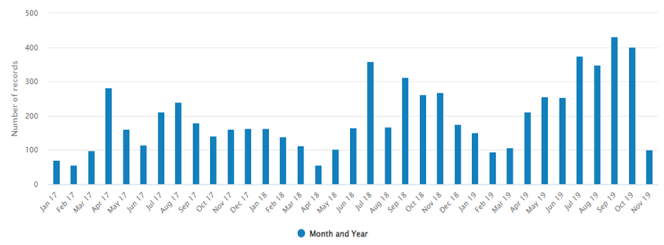
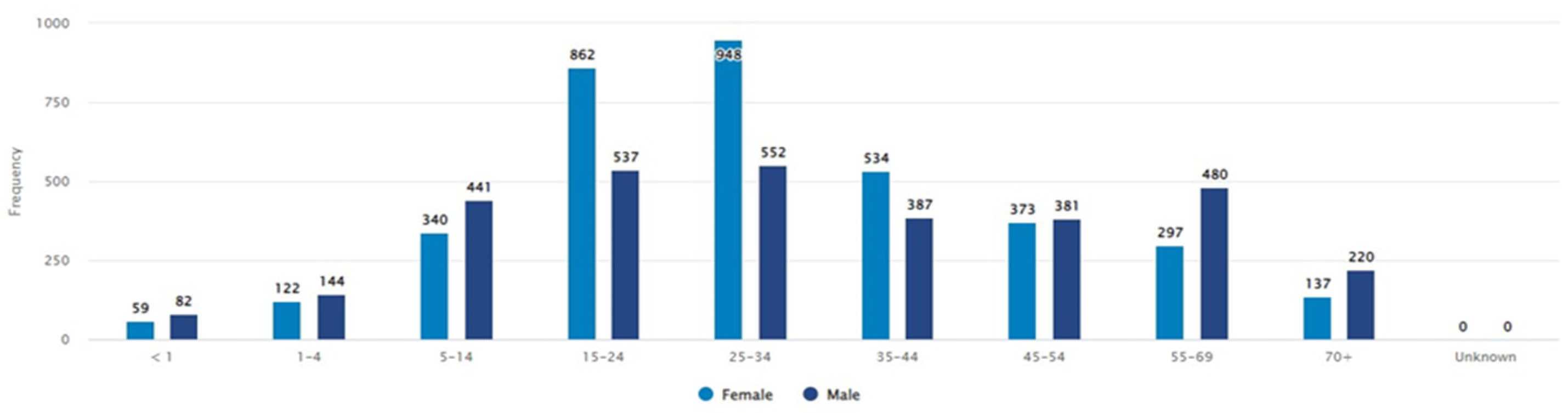
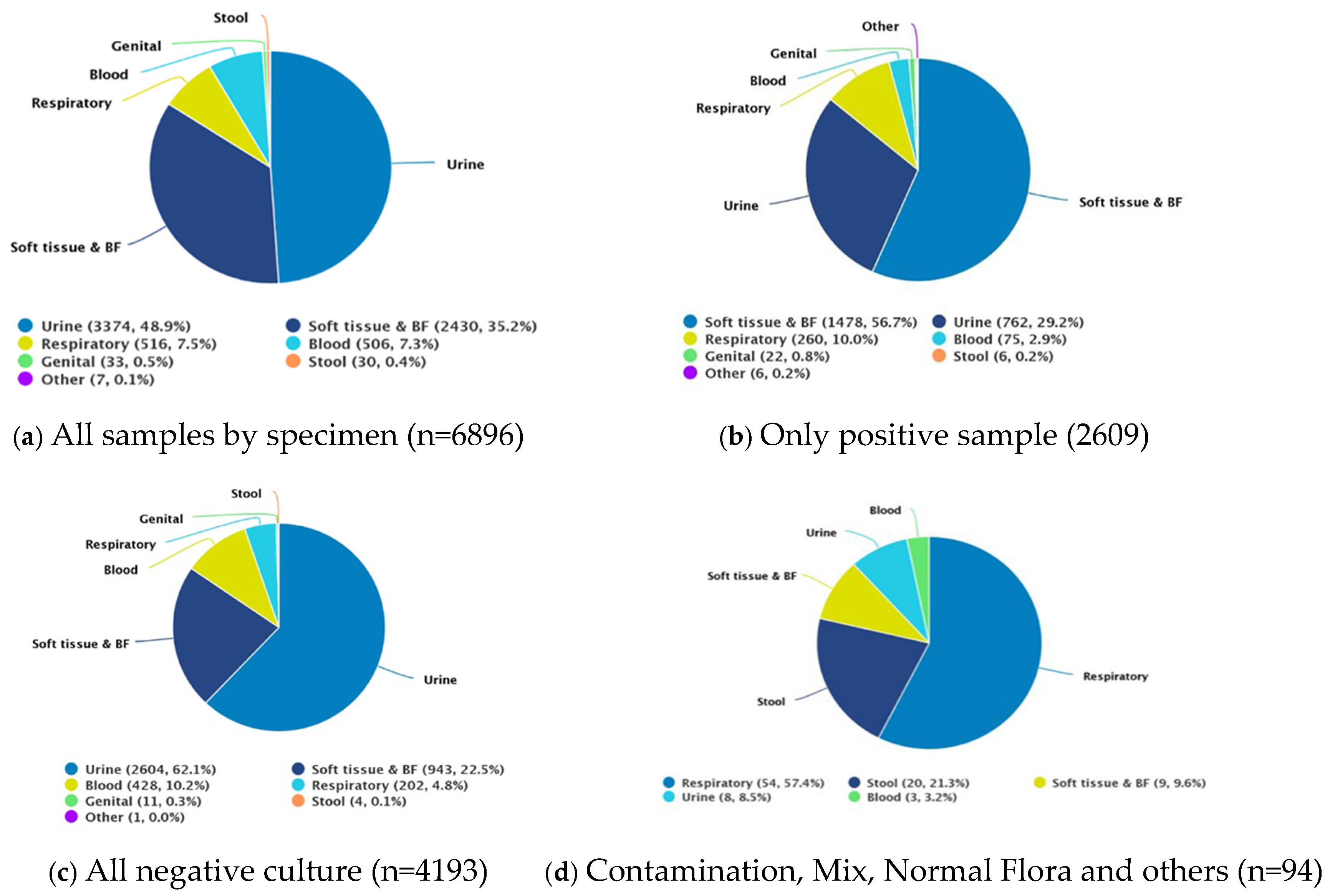
3.1. Demography and Bacterial Culture

Overall, 6,896 records were included in the analysis from 2017-2019. Around 40% were from 2019, 33% from 2018, and 27% from 2017, with an average positivity for bacterial growth of 39% in all isolates. The full breakdown table of the data volume can be found in (Supplement Table 1). Figure 1a presents the monthly distribution of the samples over the study period. Furthermore, A total of 3111 patients (45.1%) were admitted into different departments at CMCH, and 3785 (54.9%) were outpatients (supplementary Table 2). Figure 2 denotes the sex and age distribution, where females accounted for 53.2% (n=3672) and males were 46.8% (n= 3224).
Figure 3 shows the disaggregation of the isolates by specimen type. Urine was the highest specimen type of all isolates (48.9%), yet representing only 29.2% of all positive cultures, thus showing a 22.6% positivity in urine cultures. Furthermore, soft tissue and body fluid (BF) represented more than 50% of all positive cultures and 35.2% of all samples, showing a 61% positivity in all soft tissue and BF samples. Overall, only 94 (1.4%) isolates were contaminated, mostly from respiratory samples, followed by stool samples (Figure 3d).

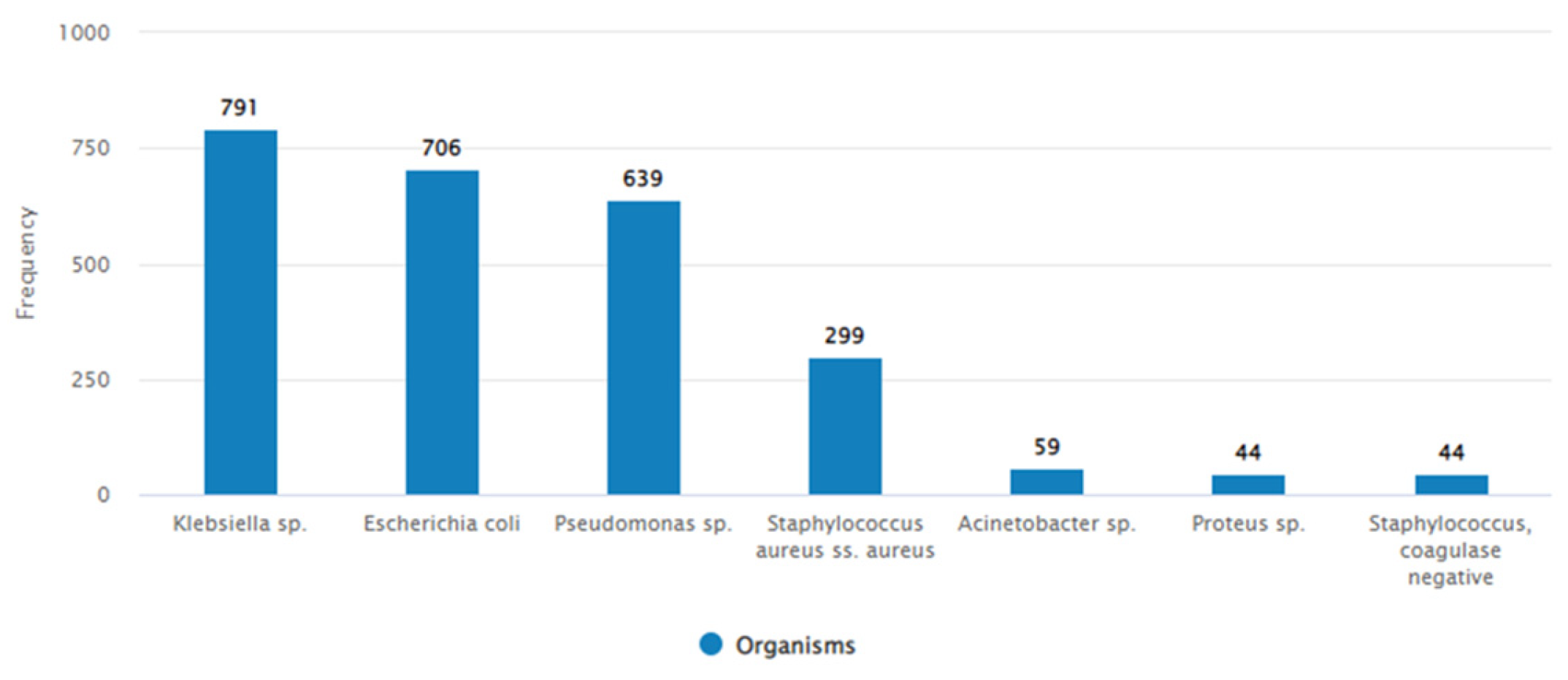
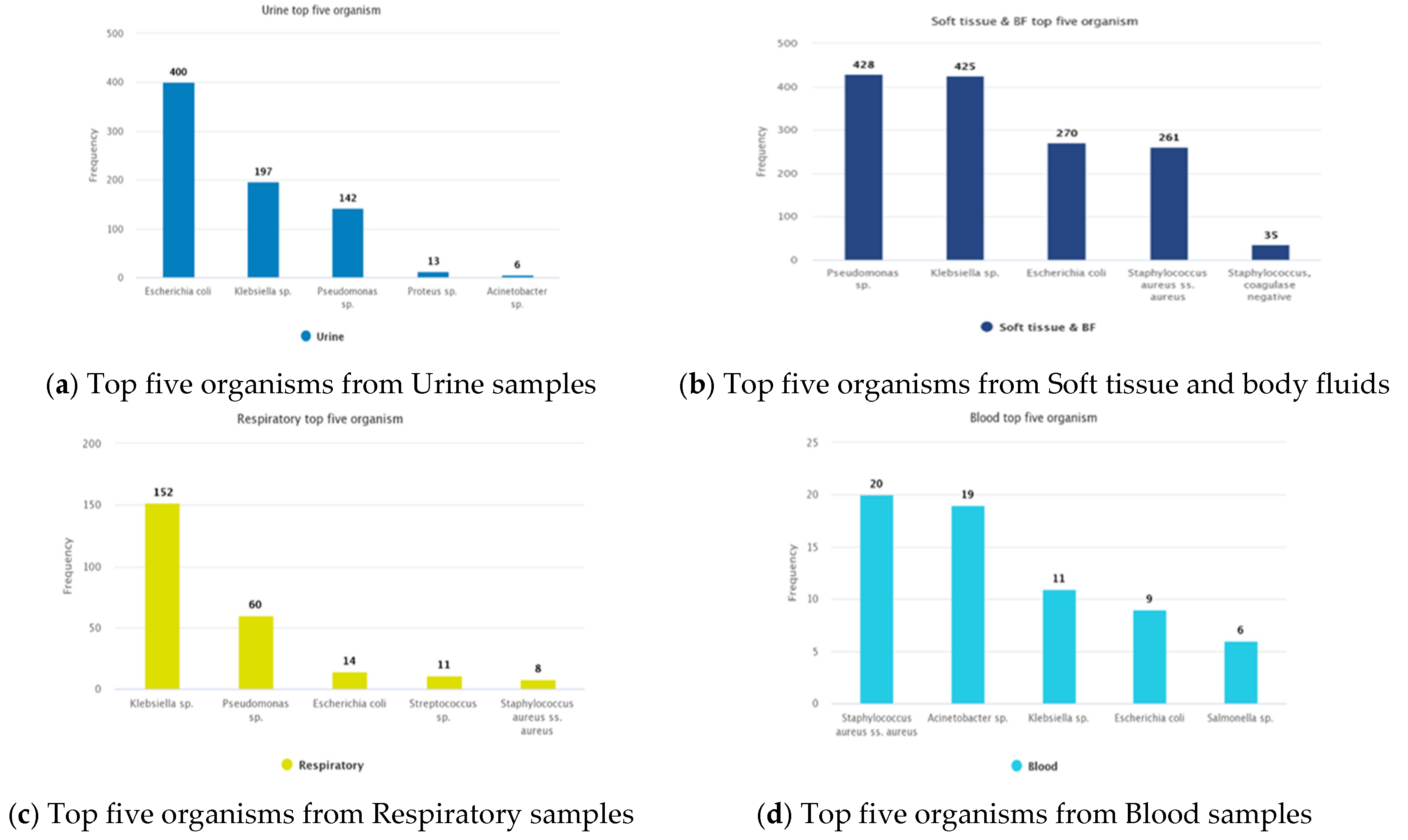
3.1.1. Isolated Bacteria and Their Resistance Pattern

The most frequently isolated bacteria in all samples were Klebsiella sp. (30.32%, n=791), Escherichia coli (26.06%, n=706), and Pseudomonas sp. (24.49%, n=639), as shown in Figure 4. Other less frequent isolates were Enterococcus sp., Salmonella typhi and other Salmonella sp., Streptococcus pyogenes, Neisseria gonorrhoae, which were excluded from the graph below.
The top five bacteria isolated from each sample type can be found in Figure 5. Overall, Gram-negative bacteria were more dominant bacteria isolated than Gram-positive bacteria across all sample types. Collectively, the most frequent Gram-negative bacteria was Klebsiella sp., and the Gram-positive was Staphylococcus aureus. Escherichia coli was the most common in urine isolates, representing more than 50% of all urine isolates. Whereas in soft tissue and body fluid, Pseudomonas sp., Klebsiella sp., were the most common, each representing 30% of the isolates. Klebsiella sp., was the most isolated from the respiratory samples, and Staphylococcus aureus, Acinetobacter sp., in blood samples.

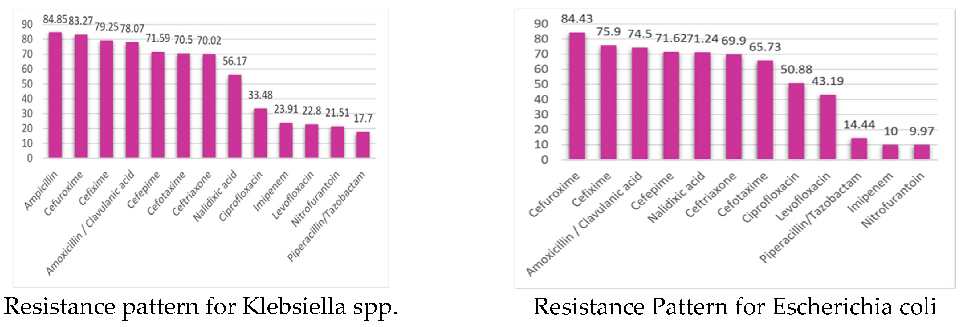
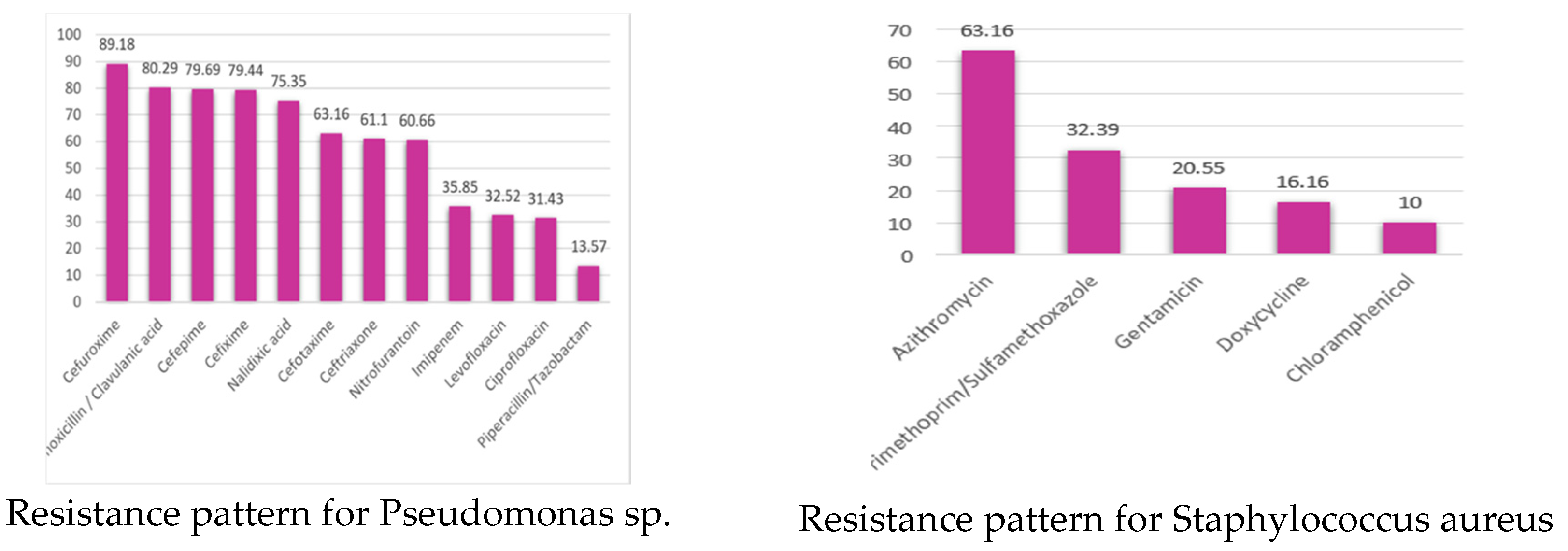
3.1.2. Resistant Pattern of Isolated Bacteria

Among the gram-negative bacteria, Klebsiella sp. isolated in this study showed higher resistance against ampicillin (85%), cefuroxime (83%), amoxicillin / clavulanic acid (79%), cefixime (78%), and lowest resistance was observed against imipenem (24%). Furthermore, the most potent antibiotic was piperacillin and tazobactam combination (82% sensitive). Similarly, a high resistance pattern of Escherichia coli was also observed against cefuroxime (84 %), amoxicillin / clavulanic acid (75%), cefixime (76%), and the lowest resistance was seen against nitrofurantoin (10%) and imipenem (10%). E coli was most sensitive to nitrofurantoin (90%). Pseudomonas sp. showed resistance to cefuroxime (89 %), amoxicillin / clavulanic acid (80%), cefepime (80%), cefixime (80%) and most sensitive to combination preparation of piperacillin and tazobactam (87%).
Among the gram-positive bacteria, Staphylococcus aureus showed the highest resistance to azithromycin (63%) and cotrimoxazole (68%). Staphylococcus aureus was mostly sensitive to chloramphenicol (90%), doxycycline (76%), and gentamicin (80%). For the full breakdown of resistance patterns, check Figure 6.

3.1.3. Drug Resistance Pattern (MDR, XDR, and PDR Categorization of Clinical Isolates)

Around two thirds of the isolates of Staphylococcus aureus (63%), Escherichia coli (61%) and Klebsiella sp. (63%), as well as nearly half (44%) of the samples of Acinetobacter sp., were identified in the CMCH dataset as being MDR. A quarter of Staphylococcus aureus (23%) and Pseudomonas sp. (25%) and more than half of Escherichia coli (56%) and Klebsiella sp. (57%) were possible XDR bacteria. Approximately 10% of isolates of Escherichia coli, 10% of isolates of Pseudomonas sp., 7% of isolates of Klebsiella sp., and one isolate of Acinetobacter sp. were found to be antibiotic-resistant or possibly XDR.

4. Discussion

This study presented the bacterial AMR trends and patterns of CMCH Hospital, Bangladesh, between January 2017 and November 2019. This retrospective study of 6,896 bacterial culture records of the Microbiology laboratory of CMCH showed that the most frequently isolated bacteria in all samples were Klebsiella sp., Escherichia coli, and Pseudomonas sp. followed by Staphylococcus aureus, Acinetobacter sp. These bacteria, except E. coli, are members of “ESKAPE”. ESKAPE represents six bacterial pathogens: Enterococcus faecium, Staphylococcus aureus, Klebsiella pneumoniae, Acinetobacter baumannii, Pseudomonas aeruginosa, and Enterobacter sp. ESKAPE organisms are the leading cause of healthcare-acquired infections worldwide [26], particularly in immunocompromised individuals. Pseudomonas aeruginosa and Staphylococcus aureus are some of the most widespread pathogens frequently found in the gut flora, soil, and water. These pathogens can potentially be transmitted to patients in healthcare settings, both directly and indirectly, and be present in biofilms within healthcare environments. [27,28]. Klebsiella pneumonia and Enterococcus faecium exhibit thicker biofilms, showcasing a robust ability to adhere firmly to neighboring cells, enabling their growth in diverse settings, including medical devices like urinary catheters and prosthetic heart valves within the human body. Acinetobacter baumannii thrives in numerous challenging conditions due to its ability to endure various temperatures, pH levels, nutrient concentrations, and even dry surroundings. Isolation of these bacteria in hospital-admitted patients indicates a high rate of hospital-acquired infection in CMCH. This finding is consistent with a similar study in another tertiary hospital, BIRDEM in Bangladesh, that found Escherichia coli, Klebsiella sp., Pseudomonas sp., and Acinetobacter sp. were the most common Gram-negative bacteria and Staphylococcus aureus, and Enterococcus sp. were the most common Gram-positive isolated bacteria [25]. These findings are consistent with other studies from the same hospital and other tertiary hospitals in Bangladesh [14,15,16,17,18,19,20,21,22,23,24,25,26,27,28,29,30,31]. In contrast, Escherichia coli was the most commonly isolated pathogen, in the Indian national surveillance during the year 2021, followed by the Klebsiella pneumoniae, Acinetobacter baumannii, Pseudomonas aeruginosa, and Staphylococcus aureus [29].
The most significant finding of this study was that the majority of pathogenic bacteria isolated from different clinical specimens were highly resistant to antibiotics such as amoxiclav (combination of amoxicillin and clavulanic acid), cotrimoxazole, cefuroxime, cefixime, azithromycin, and doxycycline. These antibiotics are the most commonly used in Bangladesh [30,31,32,33,34,35]. The resistance rate of Escherichia coli to the above-mentioned group was 61%–87%, and among Klebsiella sp. was 59%-85%. This finding is similar to other studies conducted in other tertiary care hospitals in Bangladesh, including BSMMU Hospital and Dhaka Medical College Hospital (DMCH) [14,15,16,23].
Another finding of this study was significantly high resistance of Escherichia coli, Klebsiella sp., Pseudomonas sp., and acinetobacter sp. to cefotaxime, ceftazidime, ceftriaxone which may be an indication of extended-spectrum beta-lactamase (ESBL) production. When a bacterium displays resistance or reduced sensitivity to any of these five agents, namely cefpodoxime, ceftazidime, aztreonam, cefotaxime, or ceftriaxone, it raises concerns about ESBL production. In such cases, it is advisable to conduct phenotypic confirmatory tests to confirm the diagnosis. [36,37]. ESBL’s enzymes break down and destroy some commonly used antibiotics, including penicillins and cephalosporins. These are a group of diverse, complex, and plasmid-mediated enzymes that undergo rapid evolution. In addition, ESBL-producing organisms are more challenging to treat, with fewer antibiotic options, as they usually exhibit co-resistance to many other antibiotics. Addressing ESBL-producing bacteria presents a significant therapeutic dilemma when treating hospitalized and community-based cases. [38]. Resistance of Escherichia coli to ceftriaxone and cefotaxime were 69.9%, 65.73% respectively. Similar susceptibility was also found in Klebsiella sp., which were highly resistant to cefotaxime (70.5%) and ceftriaxone (70.02%). This indicates that more than two-thirds of Escherichia coli and Klebsiella sp. were ESBL producers. Similarly, high-level resistance to cefotaxime and ceftazidime was found in Escherichia coli and Klebsiella sp. in Indian AMR surveillance [29]. Not only among members of Enterobacteriaceae but high levels of resistance to third-generation cephalosporins were also found in Pseudomonas sp. and Acinetobacter sp. About two-thirds of Pseudomonas sp. showed resistance to cefotaxime, ceftriaxone, and ceftazadime, and around three-fourths of Acinetobacter sp. were found resistant to cefixime, ceftriaxone, and ceftazadime. ESBL-producing Enterobacteriaceae was also highly prevalent in other referral hospitals in Bangladesh [25,39].
The high prevalence of ESBLs has limited the treatment options and led to increased use of carbapenems [37]. ESBL producers are best treated with carbapenems. Recently, this antibiotic has been highly used in Bangladesh. This would lead to increased risk and association with increased cost and carbapenem-resistant Enterobacterales emergence [40,41]. In this study, imipenem resistance in Escherichia coli was 10%, in Klebsiella sp. at 23.91%, and in Acinetobacter baumannii at 35.85%. This finding is coherent with another study in DMCH by Begum N et al. (2016) found 14% imipenem resistant among Gram-negative uropathogens [15]. In contrast, carbapenem (meropenem) resistance was 40% in non-fermenting entrobacterales and 35% in Pseudomonas aeruginosa isolates in Indian country surveillance [29].
The majority of pathogenic bacteria isolated from different clinical specimens were MDR. Multidrug-resistant bacterial infections have become a real threat in Bangladesh [23,24]. Around two-thirds of isolates of Staphylococcus aureus (63%), Escherichia coli (61%), and Klebsiella sp. (63%), and nearly half Acinetobacter sp. (44%) were MDR. The high prevalence of MDR bacteria may be because the samples were collected from referred cases of outpatient departments and admitted patients. Many of them may be suffering from healthcare-associated infections, usually caused by MDR bacteria. This scenario is also supported by other studies on hospitalized patients in Bangladesh [25]. Similar high-level bacteria have been observed in other countries of Southeast Asia and other developing countries of the world. A study of bacterial isolates in India from a tertiary-care healthcare facility observed lower rates of drug-resistant GNB. The study found that 66.12% were MDR strains, 34.32% were XDR strains, and 0.98% were PDR strains [42]. Another study in Saudi Arabia on Escherichia coli reported a prevalence rate of 67% for MDR infections [43]. A large study on bacterial resistance patterns in isolates from an emergency department in Hungary unveiled that 23.8% exhibited MDR. Antibiotic resistance was found to be comparatively low in western developed countries. In 2019, in the United States, a national report showed that the proportion of Enterobacteriaceae isolates with MDR in inpatient cases was 6.6% and 42.4% in Acinetobacter sp [44].
Moreover, more than half of Escherichia coli (56%) and Klebsiella sp. (57%), and a quarter of Staphylococcus aureus (23%) and Pseudomonas sp. (25%) were possibly XDR in the sense of the antibiotics used in this study. As a limited number of antibiotic discs were used in this study to test the antibiotic sensitivity testing, these isolates may not be truly XDR and PDR. Around one-tenth of Escherichia coli (11%) and Pseudomonas sp. (10%), Klebsiella sp. (7%) isolates, and one isolate of Acinetobacter sp. were found resistant to all antibiotics used for antibiotic sensitivity testing (possibly XDR).
This study has several limitations that should be considered when interpreting the findings. Because of the study’s retrospective nature, important epidemiological information about patients could not be obtained. AST was dependent on the availability of antibiotic discs at that particular time. The implementation of the AST did not strictly follow CLSI guidelines due to limited resources and disc availability. Since CMCH is a government-owned medical college, the Ministry controls the purchasing of antibiotic discs and other reagents, not the laboratory administration. Moreover, specific tests were not done due to limited resources and/or capacities, including inducible clindamycin resistance (D test), ESBL testing, and isolation of Anaerobic bacteria. Additionally, the test results were collected in RIS format, which would restrict reinterpretation based on updated guidelines.
Moreover, the number of isolates may be considered small for a tertiary hospital of 1500 beds thus, understanding the local context is crucial. Traditionally, most patients (inpatient and outpatient) are treated with empirical therapy, which may contribute to the low number of samples received in this microbiology laboratory [45]. However, this is a governmental laboratory; some patients may prefer to send their samples to other microbiology laboratories due to the complexity of government diagnostic services.
Several key policy recommendations and implications can be highlighted to address AMR in Bangladesh effectively. First, it is crucial to strengthen AMR testing infrastructure by allocating sufficient resources, enhancing laboratory infrastructure, and training personnel. Quality assurance measures and standardized testing methods should be enforced to ensure accurate and consistent results [46]. This should also include a robust stock-out motoring system in the facilities. Secondly, a comprehensive national surveillance system for monitoring AMR patterns should be established, promoting collaboration between facilities to establish national baselines for acceptable resistance. This system should use such a wealth of data to incorporate real-time data sharing and utilize electronic health records and digital platforms for efficient data analysis.
Furthermore, data sharing and reporting should be facilitated through mechanisms that encourage the timely sharing of surveillance data and the standardization of data collection formats. Accessible dashboards or portals can be created to disseminate surveillance data transparently. Implementing antimicrobial stewardship programs requires responsible and judicious use of antimicrobials through awareness campaigns and regulatory frameworks. International collaborations should be fostered to facilitate knowledge exchange, capacity building, and research on AMR surveillance and testing methods, particularly supporting low- and middle-income countries. Research and development efforts should focus on innovative diagnostic tools, alternative therapies, and public-private partnerships to develop new antibiotics. Lastly, education and training should be incorporated into curricula for healthcare professionals and the public to raise awareness about AMR prevention, responsible antibiotic use, and the role of individuals in combating AMR.

5. Conclusions

This study revealed that Klebsiella sp., Escherichia coli, Pseudomonas sp., Staphylococcus aureus, and Acinetobacter sp. were the most common bacteria isolated from clinical samples. Most of the bacteria were MDR (25% to 63%). Most common isolated bacteria showed resistance to some of the most commonly used antibiotics—amoxyclav, cotrimoxazole, cefuroxime, cefixime, azithromycin, and doxycycline Therefore, treatment and clinical guidelines, need to be updated and guided by the resistances patterns of common bacterial infections.

Author Contributions

MJS reports leading the data collection, management, analysis, interpretation, and coordination with co-authors. AH reports interpretation. HTB, SMSR, AR, ZHH, ATA, JS, and NP reports to review the manuscript.

Funding

The capacity development and data digitization on WHONET software was done by the Capturing Data on Antimicrobial Resistance Patterns and Trends in Use in Regions of Asia (CAPTURA) project funded by the Fleming Fund regional grant and implemented by the International Vaccine Institute, Seoul, South Korea.

Institutional Review Board Statement

The CAPTURA project was exempt from ethical review by the Institutional Review Board (IRB) of the IVI because the project did not involve intervention or interaction with individuals and the information collected was not individually identifiable. This exemption is per the IVI IRB SOP D-RB-4-003. The CAPTURA project undertook the retrospective data collection and curation, and the authors used the digitized data to prepare this manuscript.

Informed Consent Statement

The CAPTURA consortium project received official approval from the Communicable Disease Control, DGHS, MoHFW, dated May 17, 2020. The reference number is DGHS/DC/ARC/2020/1708. Before the data collection, a tri-party collaborative agreement was made among the DGHS, Chattogram Medical College and Hospital, and the International Vaccine Institute- CAPTURA on 22 September 2020.

Data Availability Statement

The dataset will be shared upon request.

Acknowledgments

We would like to express our gratitude to the microbiologist, data entry operators and laboratory technologists at the microbiology lab.

Conflicts of Interest

Declare conflicts of interest or state “The authors declare no conflicts of interest.”

Abbreviations

The following abbreviations are used in this manuscript:
CMCH Chittagong Medical College Hospital, Chattogram
CAPTURA Capturing Data on Antimicrobial Resistance Patterns and Trends in Use in Regions of Asia
IEDCR Institute of Epidemiology Disease Control & Research
CDC Communicable Disease Control
MoHFW Ministry of Health and Family Welfare
QAAPT Quick Analysis of Antimicrobial Patterns and Trends
CLSI Clinical & Laboratory Standards Institute
EUCAST European Committee for Antimicrobial Susceptibility Testing
IQR Interquartile range
MDR Multidrug-resistant
PDR Multidrug-resistant
WHO World Health Organization
XDR Extensively drug-resistant

References

  1. WHO Antimicrobial Resistance. https://www.who.int/news-room/fact-sheets/detail/antimicrobial-resistance Date: 2021 Date accessed: April 19, 2023.
  2. Antimicrobial Resistance Collaborators. Global burden of bacterial antimicrobial resistance in 2019: a systematic analysis. The Lancet 2022; 399(10325):P629-655. Epub 2022 Jan 19. Erratum in: Lancet. 2022 Oct 1;400(10358):1102. PMID: 35065702; PMCID: PMC8841637. [CrossRef]
  3. O’Neill J. Tackling drug-resistant infections globally: final report and recommendations. Review on Antimicrobial Resistance, London 2016 Available at:https://amr-review.org/sites/default/files/160518_Final%20paper_with%20cover.pdf. Accessed on 20 January 2021.
  4. Laxminarayan, R; Heymann, DL (2012) Challenges of drug resistance in the developing world. BMJ (Clinical research ed), 344. e1567. ISSN 0959-8138. [CrossRef]
  5. Muhammad et al. (2016), ′ Public Health problems in Bangladesh’ South East Asia Journal of Public Health 2016;6(2):11-16. [CrossRef]
  6. Hossain MM, Barman AA, Rahim MM, Hassan MT, Begum M, Bhattacharjee D.(2018) Oxytetracycline residues in Thai pangas Pangasianodon hypophthalmus sampled from Sylhet sadar upazila, Bangladesh. Bangladesh Journal of Zoology. 2018. July 26;46(1):81–90. [CrossRef]
  7. Islam A, Saifuddin A, Al Faruq A, Islam S, Shano S, Alam M, et al. Antimicrobial residues in tissues and eggs of laying hens at Chittagong, Bangladesh. Int J One Health. 2016;2(11):75–80. [CrossRef]
  8. Rana MM, Karim MR, Islam MR, Mondal MNI, Wadood MA, Bakar SMA et al. Knowledge and Practice about the Rational Use of Antibiotic among the Rural Adults in Rajshahi District, Bangladesh: A Community Clinic Based Study. Human Biology Review 2018;7 (3):259–271.
  9. Saha T, Saha T. Awareness Level of Patients Regarding usage of Antibiotics in a Slum Area of Dhaka City, Bangladesh. SSRG Int J Med Science 2018;5(9):10–16.
  10. Haque MU, Kumar A, Barik SMA and Islam MAU. Prevalence, practice and irrationality of self-medicated antibiotics among people in northern and southern region of Bangladesh. Int J Res Pharm Biosciences 2017;4(10): 17–24.
  11. Shamsuddin AK, Rahman MS, Sultana N, Islam MS, Shaha AK, Karim MR, et al. Current Trend of Antibiotic Practice in Paediatric Surgery in Bangladesh. IOSR Journal of Dental and Medical Sciences (IOSR-JDMS) 2019;18(4):28–32.
  12. Al Rasel Bin Mahabub Zaman M, Hasan M, Rashid HA. Indication-based use of antibiotic in the treatment of patients attending at the primary health care facility. Cough. 2018;44:22–0.
  13. Biswas M, Roy DN, Rahman MM, Islam M, Parvez GM, Haque MU et al. Doctor’s prescribing trends of antibiotics for out-patients in Bangladesh: A cross-sectional health survey conducted in three districts. Int J Pharma Sciences Res. 2015;6(2):669–75. [CrossRef]
  14. Nahar A, Hasnat S, Akhter H, Begum N. (2017) Evaluation of antimicrobial resistance pattern of uropathogens in a tertiary care hospital in Dhaka city, Bangladesh. South East Asia J Public Health. 2017;7(2):12–8. [CrossRef]
  15. Begum N, Shamsuzzaman S. Emergence of carbapenemase-producing urinary isolates at a tertiary care hospital in Dhaka, Bangladesh. Tzu Chi Medical Journal. 2016; 28(3):94–8. 10.1016/j.tcmj.2016.04.005. [CrossRef]
  16. Chowdhury S., Parial R. Antibiotic susceptibility patterns of bacteria among urinary tract infection patients in Chittagong, Bangladesh. SMU Med J, 2 (1) (2015), pp. 114-127.
  17. Nusrat, Tanzina and Akter, Nasima and Rahman, Nor Azlina A and Godman, Brian and Rozario, Diana Thecla D. and Haque, Mainul (2020) Antibiotic resistance and sensitivity pattern of Metallo-β-Lactamase producing gram-negative Bacilli in ventilator-associated pneumonia in the intensive care unit of a public medical school hospital in Bangladesh. Hospital Practice, 48 (3). pp. 128-136. ISSN 2377-1003. [CrossRef]
  18. Mohammad , A. K., Nasir, M., Paul, S., Rahman, H., Abul, K., & Ahmed, F. U. (2020). Common Pathogens and Their Resistance to Antimicrobials in Community Acquired Pneumonia (CAP): A Single Center Study in Bangladesh. International Journal of Medical Science and Clinical Invention, 7(12), 5144–5153. [CrossRef]
  19. Tanni AA, Hasan MM, Sultana N, Ahmed W, Mannan A (2021) Prevalence and molecular characterization of antibiotic resistance and associated genes in Klebsiella pneumoniae isolates: A clinical observational study in different hospitals in Chattogram, Bangladesh. PLoS ONE 16(9): e0257419. [CrossRef]
  20. Capturing Data on Antimicrobial Resistance Patterns and Trends in Use in Regions of Asia (CAPTURA). https://captura.ivi.int.
  21. WHONET. Boston, MA, USA. https://whonet.org.
  22. Sujan MJ, Gautam S, Aboushady AT, Clark A, Kwon S, Joh HS, Holm M, Stelling J, Marks F and Poudyal N (2025) QAAPT: an interoperable web-based open-source tool for antimicrobial resistance data analysis and visualisation. Front. Microbiol. 16:1513454. [CrossRef]
  23. Rahman MS, Huda S. Antimicrobial resistance and related issues: An overview of Bangladesh situation. Bangladesh J Pharmacol. 2014; 9 (2):218–24. [CrossRef]
  24. Ahmed I, Rabbi MB, Sultana S. Antibiotic resistance in Bangladesh: A systematic review. Int J Infect Dis. 2019;80:54-61. [CrossRef]
  25. Barai, L., Saha, M. R., Rahman, T., Khandaker, T., Dutta, S., Hasan, R., & Haq, J. A. (2018). Antibiotic Resistance: Situation Analysis In a Tertiary Care Hospital of Bangladesh. Bangladesh Journal of Microbiology, 34(1), 15–19. Retrieved from https://www.banglajol.info/index.php/BJM/article/view/39226.
  26. Santajit S, Indrawattana N. (2016) Mechanisms of Antimicrobial Resistance in ESKAPE Pathogens. BioMed research international.; 2016:2475067 (2016). [CrossRef]
  27. Center for Disease Control and Prevention: Pseudomonas aeruginosa in Healthcare Settings available at https://www.cdc.gov/hai/organisms/pseudomonas.html; Last Reviewed: November 13, 2019 ; Last date of accession: 24th august, 2023.
  28. Centers for Disease Control and Prevention: Antibiotic Resistance Threats in the United States, 2019. Atlanta, GA: Centers for Disease Control and Prevention; Available at: https://www.cdc.gov/drugresistance/pdf/threatsreport/2019-ar-threats-report-508.pdf.
  29. Indian Council of Medical Research. Annual report of Antimicrobial Resistance Surveillance Network, Division of Epidemiology communicable Disease, Indian Council of Medical Research, January 2021-December 2021 [Internet] [cited 2023 May 105]. Available from: https://main.icmr.nic.in/sites/default/files/upload_documents/AMR_Annual_Report_2021.pdf.
  30. Haque MU, Kumar A, Barik SMA and Islam MAU. Prevalence, practice and irrationality of self-medicated antibiotics among people in northern and southern region of Bangladesh. Int J Res Pharm Biosciences 2017;4(10): 17–24.
  31. Nishat C, Rashedul M. Prevalence of self-medication of antibiotics among people in Bangladesh. Ind J Pharma Prac. 2012;5(4):65–72.
  32. Paul TR, Hamid MR, Alam MS, Nishuty NL, Hossain MM, Sarker T et al. Prescription Pattern and Use of Antibiotics Among Pediatric Out Patients in Rajshahi City of Bangladesh. Int J Pharm Sci Res. 2018;9(9):3964–70.
  33. Chouduri AU, Biswas M, Haque MU, Arman MS, Uddin N, Kona N, et al. Cephalosporin-3G, Highly Prescribed Antibiotic to Outpatients in Rajshahi, Bangladesh: Prescription Errors, Carelessness, Irrational Uses are the Triggering Causes of Antibiotic Resistance. J Appl Pharm Sci. 2018;8(06):105–12. [CrossRef]
  34. Sayeed MA, Iqbal N, Ali MS, Rahman MM, Islam MR, Jakaria M.2015 Survey on Antibiotic Practices in Chittagong City of Bangladesh. Bangladesh Pharmaceutical Journal. 2015;18(2):174–8. [CrossRef]
  35. Begum MM, Uddin MS, Rahman MS, Nure MA, Saha RR, Begum T, et al. (2017) Analysis of prescription pattern of antibiotic drugs on patients suffering from ENT infection within Dhaka Metropolis, Bangladesh. Int J Basic Clinic Pharmacology. 2017;6(2):257–64. [CrossRef]
  36. Deepti Rawat,Deepthi Nair, 2010.Extended-spectrum-lactamases in Gram negative bacteria. J. Global Infect. Dis., 2010 Sep-Dec; 2(3): 263274. [CrossRef]
  37. Shaikh, S., Fatima, J., Shakil, S., Rizvi, S.M.D. and Kamal, M.A. (2015) Antibiotic Resistance and Extended Spectrum Beta-Lactamases: Types, Epidemiology and Treatment. Saudi Journal of Biological Sciences, 22, 90-101. [CrossRef]
  38. Rawat D, Nair D. Extended-spectrum β-lactamases in gram negative bacteria. J Glob Infect Dis.2010; 2(3): 263–74. [CrossRef]
  39. Rahman MM, Haq JA, Hossain MA, Sultana R, Islam F and Islam AHMS. 2004. Prevalence of extended-spectrum- b lactamase-producing Escherechia coli and Klebsiella pneumoniae in an urban hospital in Dhaka, Bangladesh. Int J Antimic Agents. 24:508-510. [CrossRef]
  40. Lutgring J. D. Carbapenem-resistant Enterobacteriaceae: an emerging bacterial threat. Seminars in Diagnostic Pathology . 2019;36(3):182–186. [CrossRef]
  41. Begum N, Shamsuzzaman S. Emergence of carbapenemase-producing urinary isolates at a tertiary care hospital in Dhaka, Bangladesh. Tzu Chi MedJ. 2016;28(3):94–98. [CrossRef]
  42. Pattnaik, D., Panda, S. S., Singh, N., Sahoo, S., Mohapatra, I., & Jena, J. (2019). Multidrug resistant, extensively drug resistant and pan drug resistant gram negative bacteria at a tertiary care centre in Bhubaneswar. International Journal Of Community Medicine And Public Health, 6(2), 567–572. [CrossRef]
  43. Hadeel Alkofide, Abdullah M Alhammad, Alya Alruwaili, Ahmed Aldemerdash, Thamer A Almangour, Aseel Alsuwayegh, Daad Almoqbel, Aljohara Albati, Aljohara Alsaud & Mushira Enani (2020) Multidrug-Resistant and Extensively Drug-Resistant Enterobacteriaceae: Prevalence, Treatments, and Outcomes—A Retrospective Cohort Study, Infection and Drug Resistance, , 4653-4662. [CrossRef]
  44. Gupta V., Gang Yea , Melanie Oleskyb , Kenneth Lawrenceb , John Murraya , Kalvin Yua (2019) National prevalence estimates for resistant Enterobacteriaceae and Acinetobacter species in hospitalized patients in the United States, International Journal of Infectious Diseases 85 (2019) 203–211. [CrossRef]
  45. Sutradhar, K., Saha, A., Huda, N. H., & Uddin, R. (2014). Irrational Use of Antibiotics and Antibiotic Resistance in Southern Rural Bangladesh: Perspectives from Both the Physicians and Patients. Annual Research & Review in Biology, 4(9), 1421-1430. [CrossRef]
  46. Musa K, Okoliegbe I, Abdalaziz T, Aboushady AT, Stelling J, Gould IM. Laboratory Surveillance, Quality Management, and Its Role in Addressing Antimicrobial Resistance in Africa: A Narrative Review. Antibiotics (Basel). 2023 Aug 14;12(8):1313. [CrossRef]
Figure 1. The distribution of culture records over time, including negative results.
Preprints 151965 g001
Figure 2. Distribution of the number of culture records by sex and age group for all records.
Preprints 151965 g002
Figure 3. The number of culture records stratified by specimen category and cumulative records culture positive showing bacterial growth.
Preprints 151965 g003
Figure 4. Most common bacteria isolated from all samples over the reported period.
Preprints 151965 g004
Figure 5. Top five isolated bacteria from different samples. 5a: Top five organisms from Urine samples; 5b: Top five organisms from soft tissue and body fluids samples; 5c: Top five organisms from Respiratory samples; 5d: Top five organisms from Blood samples.
Preprints 151965 g005
Figure 6. Resistance pattern of major bacteria to common antibiotics.
Preprints 151965 g006aPreprints 151965 g006b
Table 1. Summary of MDR, possible XDR, and possible PDR results.
Organism Number of isolates MDR Possible XDR Possible PDR
Staphylococcus aureus 299 187 (63%) 70 (23%) -
Escherichia coli 706 432 (61%) 398 (56%) 80 (11%)
Acinetobacter sp. 59 26 (44%) 26 (44%) 1 (2%)
Klebsiella sp. 791 496 (63%) 450 (57%) 54 (7%)
Pseudomonas sp. 369 93 (25%) 93 (25%) 38 (!0%)
Table 2. Gram positive antibiogram. The numbers indicate % susceptible.
Disclaimer/Publisher’s Note: The statements, opinions and data contained in all publications are solely those of the individual author(s) and contributor(s) and not of MDPI and/or the editor(s). MDPI and/or the editor(s) disclaim responsibility for any injury to people or property resulting from any ideas, methods, instructions or products referred to in the content.
Copyright: This open access article is published under a Creative Commons CC BY 4.0 license, which permit the free download, distribution, and reuse, provided that the author and preprint are cited in any reuse.
Prerpints.org logo

Preprints.org is a free preprint server supported by MDPI in Basel, Switzerland.

Subscribe

Disclaimer

Terms of Use

Privacy Policy

Privacy Settings

© 2025 MDPI (Basel, Switzerland) unless otherwise stated