Submitted:
13 February 2025
Posted:
13 February 2025
You are already at the latest version
Abstract
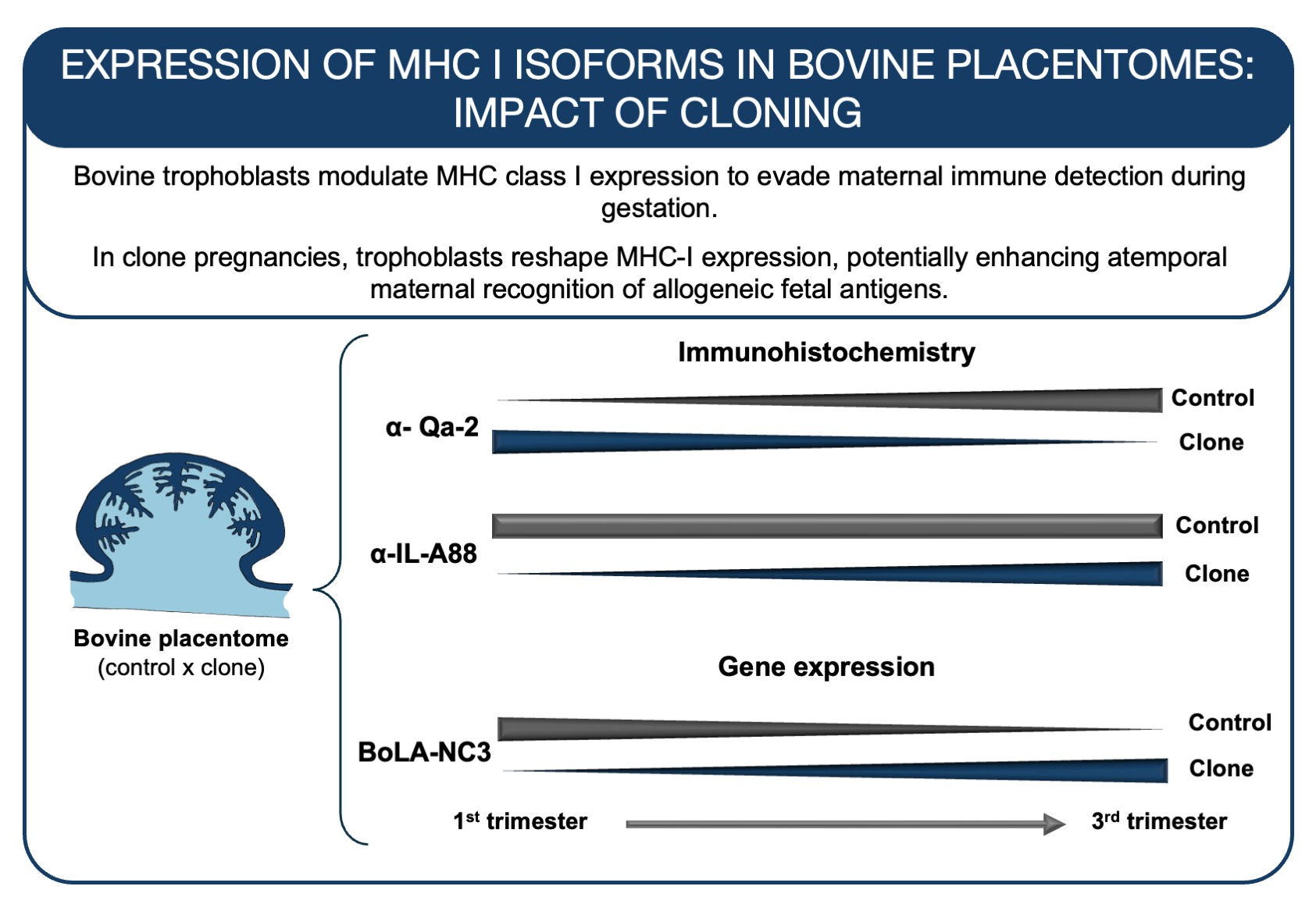
As observed in other species during pregnancy, bovine placentomes are assumed to suppress classical MHC I antigens, whereas overexpress non-classical MHC I to be recognized but not rejected by maternal immune system. However, in cloned bovine classical antigens were overexpress early in the pregnancy instead of non-classical ones. Then, MHC I antigens were investigated by immunohistochemistry (N=23) and RT-qPCR (N=16) in control and cloned bovine placentomes in early gestation and near term. In controls, IL-A88 staining was present in all stages, majorly in maternal tissues, whereas Qa-2 reacted only near term. Clones had an opposite pattern. Trophoblast giant cells near the uterine epithelium were weakly stained. By PCR, especially the non-classical isoform BoLA-NC3 resulted in significant higher expression in early gestation and down-regulation near term in controls compared to clones, as stain produced by Qa-2 antibody. The presence of MHC I in the placentomes indicated that bovine epitheliochorial placentas followed similar principles than that known for other species. Controls and clones reacted differently to the applied antibodies and differ partly in gene expression, which may explain pregnancy problems.
Keywords:
BoLA
; Bovine placenta
; chorionic villi
; cloned bovine
; IL-A88
; Qa-2
1. Introduction
During pregnancy, the communication between mother and fetus occurs through by expression and transfer of placental-specific factors [1], such as the fetal expression of Major Histocompatibility Complex (MHC) proteins [2]. In hemochorial placental models such as in mice and humans, semi-allogeneic trophoblastic cells (fetal cells) decrease the expression of MHC-I molecules early in gestation to avoid the recognition of paternal antigens by the maternal immune system [3]. With the low to no expression of MHC-I, the fetal cells may become a target to uterine natural killer (uNK) cells [4]. To prevent uNK-cells activation, the trophoblast cells express a unique form of MHC molecules: the nonclassical (NC) MHC-I isoforms [5], described as human leukocyte antigen G (HLA-G) or murine preimplantation embryo development (PED) genes [2]. NC MHC-I genes are expressed in isoforms: membrane-bound or soluble proteins [6]. Soluble HLA-G is detected in the maternal blood in successful pregnancies, suggesting a systemic modulation of the maternal immune system. [7]. High expression of HLA-G is associated with an invasive phenotype of the trophoblast cells in vitro, the extravillous trophoblast (EVT) [8]. However, trophoblast invasion and adhesion to the decidua are regulated not only by HLA-G expression but also through interactions with inhibitory receptors on NK cells and macrophages, such as KIR2DL4/CD158d, LILRB1/2, and ILT2/4.[8,9]. HLA-G expression by EVT modulates the local maternal immune system not only through the uNK cell system and its interactions with macrophages and other immune cells. Regulatory T (Treg) cells play an essential role in supporting the semi-allogeneic fetus in successful pregnancies [10], inhibiting T cell-mediated lysis of trophoblastic cells, and promoting a supporting immune environment [11]. Abnormal expression of HLAG appears deleterious: its down-regulation correlates with reduced numbers of CD56brightCD16- uNK cells in the maternofetal interface, which can contribute to miscarriages [12]. An abnormal invasion of EVT and uNK cells can impair the development and remodeling of maternal spiral arteries, predisposing intrauterine to restrict growth and preeclampsia [13]. For example, when extravillous trophoblasts (EVT) from mid-gestation samples are cultured in conditioned uNK-cell media, their ability to invade is significantly increased in in vitro assays [14]. Moreover, the co-culture of HLAG+ EVT and CD4+ cells increases the expression of forkhead box P3 (FOXP3) by CD4+CD25high T reg cells, supporting the HLA-G effects on the Th immune response at the maternofetal interface [10].
In contrast to humans and mice, the bovine displays a less invasive or epitheliochorial type of placentation which all maternal tissue layers are maintained throughout pregnancy [14,15]. Unlike other species with epitheliochorial placenta, the ruminant placenta is unique and appears more permeable to fetal products [19,20]. In cows, the trophoblastic giant cells (TGC) migrate toward the maternal epithelium and form a hybrid syncytium with the maternal epithelium [16] to deliver fetal products to the maternal system [17]. Despite the maintenance of all tissue layers in both dam and fetus sides, the dam immune system displays similar local and systemically modulation as observed in human and mice. Such as the increase of circulating CD4+CD25high T reg cells as early pregnancy[18] and alternative activation of uterine macrophage near-term [19]. The more intimate maternofetal communication in cows primes the maternal system to semi-allogeneic fetal antigens [20]. In contrast to humans and mice, MHC-I or Bovine Leukocyte Antigen class I (BoLA-I) expression in early gestation is unknown. In cows, trophoblast in the arcade zones and interplacentomal chorionic regions express BoLA-I around 6 months of pregnancy and BoLA-I expression is absent deep in the chorionic villi [21,22,23,24]. It suggested that the suppression of BoLA-I expression in bovine placentomes occurs in areas of close contact of fetal cells with maternal epithelium [21,22,23,25]. In clones, BoLA-I is expressed as early as day 36 in the chorioallantois membrane, which later differentiates into chorionic villi as placentome development progresses [25]. It suggests that BoLA-I expression in early pregnancy may represent a nonclassical isoform of BoLA class I (BoLA-NC), like murine PED and human HLA-G [26]. Furthermore, BoLA-I atemporal and increased expression observed in cloned pregnancies may have contributed to abnormal pregnancy/placentation and losses in the early stages of pregnancy [8,27].
The expression patterns of classical and nonclassical isoforms of BoLA class I molecules remains uncharacterized, and their role in local maternal-fetal immune modulation remains unclear. In this study, we describe the expression of classical and nonclassical MHC class I molecules in bovine placentomes from both cloned and naturally conceived pregnancies, examined during early and term stages of gestation.
2. Materials and Methods
For immunohistochemistry (IHC), mouse Qa-2 (clone 69H1 9 9, #11-5996; eBioscience, San Diego, CA, USA) and mouse bovine monomorphic MHC (clone IL-A88, #MCA2444GA, AbDSerotec, Kidlington, OX, UK) were used as primary antibodies. For control, we used anti-mouse IgG (clone MOPC-21, #M5284; Sigma-Aldrich, Saint Luis, MS, USA). Reactions were tagged with Dako Advance tm HRP (#K4069) and revealed with 3,3 – diaminobenzidine (DAB, Liquid DAB+ Substrate Chromogen System, #K346811), both purchased from DAKO (Carpinteria, CA, USA).
For real-time quantitative PCR (RT-qPCR), RNeasy Plus Mini kit (#74136, Qiagen - Valencia, CA, USA) for rna extraction, DNAse I amplification grade (#18068-015), RNAseOut Recombinant Ribonuclease Inhibitor (#10777-019), High Capacity cDNA Reverse Transcription Kit with RNase Inhibitor (#4374967) and SyBr Green PCR master mix (#4344463), all purchased from Invitrogen (Foster City, CA, EUA). Primers were purchased from Exxtend (Paulínia, SP, Brazil).
Monohydrate citric acid, sodium citrate tribasic dehydrate, sodium chloride (NaCL), hydrogen peroxide (H2O2), potassium chloride (KCl), trizma base (C4H11NO3), and 3-(Aminopropyl)triethoxysilane for buffer solutions were purchased from Sigma-Aldrich.
Clustering of BoLA isoforms and sequences recognized by Qa2 and IL-A88 antibodies
Published protein sequences of classical and non-classical BoLA isoforms were retrieved from the IPD-MHC database (ftp://ftp.ebi.ac.uk/pub/databases/ipd/mhc/bola/bola.n.prot.fasta) and peptide sequence recognized by the anti-Qa2 (GenBank: X80933.1) and anti-IL-A88 (GenBank: NM_001201460.1) antibodies were retrieved in FASTA format for further analysis. Phylogenetic analyses were conducted using Clustal Omega (https://www.ebi.ac.uk/jdispatcher/msa/clustalo) [28] within a Singularity container to ensure reproducibility and control over computational resources. Sequences were aligned with the –dealign option to eliminate pre-existing gaps, ensuring alignments based solely on sequence data. The software utilized MBED-like clustering for both guide-tree construction and iterative refinements to enhance alignment precision. The output sequences were maintained in their aligned order, and default settings were used for combined iterations, maximum guide tree, and HMM iterations, balancing efficiency and detailed analysis.
In silico analysis of bovine classical and non-classical MHC proteins solubility.
SignalP 4.1 (http://www.cbs.dtu.dk/services/SignalP/) was used to predict secretion via the classical secretory pathway by the presence of signal peptide, and SecretomeP 2.0 Server (http://www.cbs.dtu.dk/services/SecretomeP/) was used to predict non-classical secretory pathways. Only the entries that were predicted to be secreted by SignalP 4.1 or SecretomeP 2.0 Server were used for further analysis.
The secreted predicted sequences were further analyzed by TMHMM Server v. 2.0 (http://www.cbs.dtu.dk/services/TMHMM/) for the presence of transmembrane helices. Furthermore, to confirm the secretory nature of the analyzed sequences, two web tools were used to predict subcellular location of predict secreted proteins. TargetP 1.1 Server (http://www.cbs.dtu.dk/services/TargetP/), which utilizes neural networks to predict subcellular location of a given protein sequence, was used. And similar analysis was performed using WoLF PSORT (http://www.genscript.com/wolf-psort.html), which predicts the protein subcellular location using a dataset of 12771 animal proteins obtained from UniProt (http://www.uniprot.org) version 45 annotated by Gene Ontology (http://geneontology.org).
Placentome samples
Placentomes from pregnancies of bovine embryos produced by somatic cell nuclear transfer (SCNT) [29] were collected after surgical uteri recovery, and control placentomes were obtained from a local abattoir. For both groups, to placentome assessment, bovine uteri were incised in pregnant horn antimesometrial line. Then membranes, endometrium and fetuses were morphologically analyzed. For controls, we only used placentomes derived from pregnancies without gross abnormalities in vasculature, membranes, and placentome number and size. However, for clones, is know that some placental abnormalities could be present [31,32,33], then only ones without exacerbated abnormalities were used. Placentomes of clones from early gestation (60 and 90 days; n = 5) and near-term (285 ± 0 days; n = 6) were investigated, as well as controls of early gestation (72.8 ± 10.6 days, respectively; n = 6) and near term (251 ± 21 days; n = 6) that were determinate according to their crown-rump length. [30]. For RT-qPCR, 16 samples were included, grouped into the following: early gestation (75 ± 17.3 days; n = 4) and near term (285 days; n = 4) for clones and early gestation (73 ± 16.2 days; n = 4) and near term (240 ± 7.4 days; n = 4). The research was approved by the Ethical Committee of the College of Veterinary Medicine and Animal Science of the University of Sao Paulo (protocol number: 9408110315).
Immunohistochemistry with Qa-2 and IL-A88 antibodies
Samples were fixed in 4% buffered paraformaldehyde, dehydrated and paraffin embedded. Sections of 5 µm were mounted on pre-coated slides, deparaffinized and rehydrated. The antigen retrieval was performed by microwaving the slides in high potency 4 times for 5 minutes in citrate buffer (1.83 mM of monohydrate citric acid and 8.9 mM of sodium citrate tribasic dehydrate, pH 6.0). Endogenous peroxidase was blocked with 3% hydrogen peroxide in distillated water during 1 hour in the dark. Nonspecific protein interaction was blocked with 10% of normal goat serum in Tris Buffer Solution (TBS – 2.0 mM of Trizma base and 1.36 mM of sodium chloride, pH 7.5) for 30 minutes, followed by incubation with specific primary antibodies Qa2 (1:50), IL-A88 (1:50) or irrelevant anti-mouse IgG overnight in a humid chamber at 4° C. The reaction was detected by Dako Advance tm HRP, and the color developed with DAB. The slides were counter stained with hematoxylin. Between each step, slides were rinsed 3 times for 5 minutes in TBS; and after antibody incubation they were rinsed in TBS containing 1% of normal goat serum. The slides were analyzed using an upright Zeiss Axioplan 2 microscope and Axiocan HRc camera (Zeiss, Jenna, DE).
Detection of MHC genes expression by RT-qPCR
The RNA from all samples was extracted using the RNeasy Plus Mini kit; DNA digestion was performed with DNAse I amplification grade and cDNA conversion with High Capacity cDNA Reverse Transcription Kit; all following the manufacturer instructions. For each real time qPCR reactions, 7.5 μL of SyBr Green PCR master mix, 1.0 mM of each primer (Table 1) [34,35,36,37,38], 1.0 μL of diluted cDNA (63.0 μL of ultrapure water and 1.0 μL of cDNA) and ultrapure water for a total of 15.0 μL were used. Was used the default cycle program from 7500 fast real-time PCR system (Invitrogen). Data were analyzed according to the dCt method from Livak et al. [39] and statistical analysis was performed by least square means analysis of variance with GLM procedure of SAS (version 5.1). The model included main effects (group and animal) and standard errors. Graphs were plotted on SigmaPlot software (version 12.0).
3. Results
3.1. Qa-2 and IL-A88 Antibodies Recognizes Putative Soluble MHC-I Proteins
The sequence alignment of BoLA isoforms and specific peptides recognized by Qa-2 and IL-A88 antibodies revealed that the Qa-2 antibody predominantly clustered with non-classical bovine MHC I isoforms, while the IL-A88 antibody clustered with classical isoforms, as illustrated in the generated guide tree (Figure 1).
3.2. Immunohistochemistry with Distinct Pattern Between Qa2 and IL-A88 Antibodies
In early gestation, controls showed no labeling for Qa-2 antibody in all tissues of the placentomes (Figure 2A and B). Near term, a strong staining against Qa2 antibody stain was observed in uninucleate trophoblasts and the maternal uterine epithelium; whereas bi-nucleated trophoblast was weakly stained, and the mesenchyme and endometrial stroma were largely negative (Figure 2C). In clones, trophoblasts and the maternal epithelial cells were strongly labeled against Qa2 antibody in early gestation (Figure 2 D and E), but only weakly stained near term (Figure 2F). In all positive cases, the Qa-2 antibody labelling was stronger in the arcade zones than in the villous areas. In addition, trophoblastic giant cells (TGC) that were closely apposed to or fused with maternal epithelial cells showed weak to no labeling for Qa2 (Figure 3).
In controls, at 60 days of gestation, both trophoblast and maternal epithelial cells exhibited moderate labeling for IL-A88 antigen (Figure 4A). By day 90 of gestation, while the labeling intensity of IL-A88 in maternal cells remained consistent, it was markedly decreased in the trophoblast cells (Figure 4B). Near term, all samples displayed faint to weak staining for IL-A88 (Figure 4C). In clone samples, the IL-A88 antibody showed mild to moderate labeling was observed only in maternal cells during early gestation (Figure 4D and E). Moreover, in the near term, the IL-A88 antibody labeling intensity was evident in both the trophoblast and maternal epithelium (Figure 4F). Additionally, similarly observed with Qa2 antibody, the TGCs that were closely apposed or fused with the maternal epithelium exhibited markedly weak to no labeling for IL-A88.
3.3. Expression and Regulation of MHC I Gene Isoforms
Among the four non-classical MHC I isoforms investigated (Figure 5A-F), BoLA-NC3 exhibited significant differences in expression throughout pregnancy between the control and cloned groups (P < 0.01). In the controls compared to the clones, BoLA-NC3 expression was upregulated in early gestation (P < 0.01) and downregulated near term (P = 0.03) (Figure 5C). Expression of BoLA-NC1 differed only near term, showing downregulation in controls relative to clones (P < 0.02) (Figure 5A). Similarly, BoLA-NC2 expression differed only near term, being downregulated in controls compared to clones (P < 0.02). Additionally, controls exhibited a decrease in BoLA-NC2 expression near term compared to early gestation (P < 0.01), whereas its expression remained constant throughout pregnancy in clones (Figure 5B). In contrast, the expression of BoLA-NC4 and the classical isoform JSP-1 did not show significant differences at any analyzed stages of pregnancy or between groups (Figure 5D and F).
4. Discussion
In contrast to what was known before, MHC-I isoforms were detected in both control and clone bovine placentomes throughout pregnancy at mRNA and protein levels. In our study, two different antibodies for immunohistochemistry were tested, and the antibody-antigen reaction was revealed with a biotin-free polymer-based secondary antibody system. Such second-generation antibody systems are more sensitive and specific. [45] then the formerly used biotinylated ones [22,23,26,28,43,44].We concluded that newer, more sensitive reagents are more suitable for detecting MHC I expression, particularly during early bovine gestation when the MHC class I protein expression is evident in our current work. It is possible that the antigen retrieval and blockage of nonspecific site methods previously required by older reagents may have disrupted the integrity of the antigen-antibody binding process, potentially altering epitope availability, reducing binding affinity, or increasing background signal, thereby affecting assay specificity and sensitivity.
We observed intense labeling in uninucleate trophoblasts compared to trophoblast giant cells (TGCs). All the TGCs composing the syncytium with the maternal epithelium exhibited a complete loss of labeling. A similar pattern of heterogeneous MHC-I expression in trophoblasts has been described in humans[46,47]. Only, migratory EVT cells express HLAG to suppress activation of uterine NK cells and resident macrophages. The EVT interacts with maternal immune cells hence HLAG expression, whereas syncytiotrophoblast near to the maternal blood system have only light expression of soluble HLAG and do not express membrane bound HLAG [48]. Thus, comparable to the human we suppose that also in the bovine the fused cells at the feto-maternal interface were characterized by suppressed MHC I expression. This may cause their vulnerability regarding the maternal immune system, which may be involved in the process of apoptosis of the syncytia [49] and the release of their products into the maternal system [16]. The strong staining on the maternal side, and putative soluble pattern of BoLA isoforms suggested the presence of: (a) membrane-bound, classical MHC I as specific for an epithelium and (b) soluble isoforms that must have been released by the trophoblast and perhaps “accumulated” in the uterine epithelium [50,51]. In humans, the maternal epithelium displays similar “accumulation” of non-classical MHC I as observed in bovine placenta. Even though the bovine placenta is generally less invasive, studies proofed a permeability of fetal products to the maternal system [17,52,53,54]. Recently, cloned bovine endometrial immune response seems to be regulated by trophoblast BoLA expression [55], and hypoxic conditions [56] that is common feature in cloned pregnancies [57,58].
Our results highlighted differences between the two antibodies used. In our in-silico analysis, the Qa-2 sequence appeared closer to non-classical BoLA isoforms. Moreover, Qa2 antigens were not detected early gestation with markedly increased labeling near term, in the control samples. In contrast, the human placenta expresses HLA-G from the first trimester onwards. [59], probably due to more invasive placenta. In contrast, clones showed intense labeling in early gestation that decreased close to term, indicating that an altered overexpression of non-classical MHC I during early gestation may lead to pregnancy abnormalities as evidenced its expression by chorioallantoic membranes prior placentome formation[25], being even higher in MHC-I heterozygous incompatible clones [60]. In our in-silico analysis, IL-A88 protein sequences appeared closer to classical MHC I proteins. n control samples, IHC for IL-A88 showed consistent labeling in the maternal epithelium at both early and later stages of gestation. In contrast, clones exhibited weak to no IL-A88 labeling in the maternal epithelium during early gestation, but intense labeling in both maternal and fetal epithelium near term. Data suggests that IL-A88 may recognize the characteristic, i.e. classical antigens of epithelia that become down-regulated on the fetal side as previously observed for humans and mice [61,62]. However, in clones near term the trophoblast labeled for IL-A88 antigens. The anti-Qa2 and anti-IL-A88 monoclonal antibodies differed pattern of labeling in control and cloned sample, it could be a consequence of an abnormal chromatin remodeling due to abnormal patterns of methylation of the trophoblast during preimplantation period that perpetuated during the placental development [60].
MHC I mRNA expression was detected in all bovine placentome samples throughout pregnancy, consistent with the isoform evaluations by Santos et al. l [64]. Our findings reveal significant variations in the non-classical isoforms, with BoLA-NC3 being the most regulated gene during pregnancy. Its expression patterns closely correlate with the immunohistochemistry results for Qa-2 antigens observed in this study. Especially, the abnormal Qa-2 labeling aligned with non-classical isoforms expression in cloned samples. In addition, BoLA NC 1 and 2 were significantly upregulated in near term stages of clones, but the functional meaning remained unclear.
5. Conclusion
Our data suggest that MHC-I protein and gene expression in bovine placenta is similar observed in more invasive types of placenta such as in humans and mouse, suggesting bovine epitheliochorial placentas possess similar mechanisms of trophoblast evasion from maternal immune system during gestation. Controls and clones had distinct protein expression profile for Qa2 and IL-A88 antigens, which is somehow observed on the differences of expression of nonclassical MHC-I molecules. The disparity of MHC-I regulation in clones may lead to the failure to proper pregnancy signaling potentially leading to gestational failures.
Supplementary Materials
The following supporting information can be downloaded at the website of this paper posted on Preprints.org. Table S1: In silico prediction of soluble motifs of bovine MHC I isotypes.
Author Contributions
Experiments and data generation (RSNB and ACFM), data analysis and writing (RSNB, ACFM, LJO), conceptualization and review of the paper (all authors).
Funding
This research was funded by CAPES (grant number 2009/06702-1) and FAPESP (grant number CEPID-CTC 2013/08135-2 and 2021/05445-7).
Institutional Review Board Statement
The research was approved by the Ethical Committee of the College of Veterinary Medicine and Animal Science of the University of Sao Paulo (protocol number: 9408110315).
Conflicts of Interest
The authors declare no conflicts of interest.
References
- Zumkeller, W. Current Topic: The Role of Growth Hormone and Insulin-like Growth Factors for Placental Growth and Development. Placenta 2000, 21, 451–467. [CrossRef]
- Rizzo, R.; Melchiorri, L.; Stignani, M.; Baricordi, O.R. HLA-G Expression Is a Fundamental Prerequisite to Pregnancy. Hum Immunol 2007, 68, 244–250. [CrossRef]
- Moffett-King, A. Natural Killer Cells and Pregnancy. Nat Rev Immunol 2002, 2, 656–663. [CrossRef]
- Heemskerk, B.; Lankester, A.C.; van Vreeswijk, T.; Beersma, M.F.C.; Claas, E.C.J.; Veltrop-Duits, L.A.; Kroes, A.C.M.; Vossen, J.M.J.J.; Schilham, M.W.; van Tol, M.J.D. Immune Reconstitution and Clearance of Human Adenovirus Viremia in Pediatric Stem-Cell Recipients. J Infect Dis 2005, 191, 520–530. [CrossRef]
- Bulmer, J.N.; Johnson, P.M. Antigen Expression by Trophoblast Populations in the Human Placenta and Their Possible Immunobiological Relevance. Placenta 1985, 6, 127–140. [CrossRef]
- Hunt, J.S.; Petroff, M.G.; McIntire, R.H.; Ober, C. HLA-G and Immune Tolerance in Pregnancy. FASEB Journal 2005, 19, 681–693. [CrossRef]
- Steinborn, A.; Varkonyi, T.; Scharf, A.; Bahlmann, F.; Klee, A.; Sohn, C. Early Detection of Decreased Soluble HLA-G Levels in the Maternal Circulation Predicts the Occurrence of Preeclampsia and Intrauterine Growth Retardation during Further Course of Pregnancy. American Journal of Reproductive Immunology 2007, 57, 277–286. [CrossRef]
- Li, C.; Houser, B.L.; Nicotra, M.L.; Strominger, J.L. HLA-G Homodimer-Induced Cytokine Secretion through HLA-G Receptors on Human Decidual Macrophages and Natural Killer Cells. Proc Natl Acad Sci U S A 2009, 106, 5767–5772. [CrossRef]
- Wang, Q.; Song, H.; Cheng, H.; Qi, J.; Nam, G.; Tan, S.; Wang, J.; Fang, M.; Shi, Y.; Tian, Z.; et al. Structures of the Four Ig-like Domain LILRB2 and the Four-Domain LILRB1 and HLA-G1 Complex. Cell Mol Immunol 2020, 17. [CrossRef]
- Tilburgs, T.; Crespo, Â.C.; van der Zwan, A.; Rybalov, B.; Raj, T.; Stranger, B.; Gardner, L.; Moffett, A.; Strominger, J.L. Human HLA-G+ Extravillous Trophoblasts: Immune-Activating Cells That Interact with Decidual Leukocytes. Proceedings of the National Academy of Sciences 2015, 112, 7219–7224. [CrossRef]
- Djurisic, S.; Teiblum, S.; Tolstrup, C.K.; Christiansen, O.B.; Hviid, T.V.F. Allelic Imbalance Modulates Surface Expression of the Tolerance-Inducing HLA-G Molecule on Primary Trophoblast Cells. Mol Hum Reprod 2014, 21, 281–295. [CrossRef]
- Rizzo, R.; Lo Monte, G.; Bortolotti, D.; Graziano, A.; Gentili, V.; Di Luca, D.; Marci, R. Impact of Soluble HLA-G Levels and Endometrial NK Cells in Uterine Flushing Samples from Primary and Secondary Unexplained Infertile Women. Int J Mol Sci 2015, 16, 5510–5516. [CrossRef]
- Leonard, S.; Murrant, C.; Tayade, C.; Vandenheuvel, M.; Watering, R.; Croy, B. Mechanisms Regulating Immune Cell Contributions to Spiral Artery Modification – Facts and Hypotheses – A Review. Placenta 2006, 27, 40–46. [CrossRef]
- Carter, A.M.; Enders, A.C. Comparative Aspects of Trophoblast Development and Placentation. Reproductive biology and endocrinology 2004, 2, 46. [CrossRef]
- Carter, A.M. Evolution of Placental Function in Mammals: The Molecular Basis of Gas and Nutrient Transfer, Hormone Secretion, and Immune Responses. Physiol Rev 2012, 92, 1543–1576. [CrossRef]
- Wooding, F.B. Current Topic: The Synepitheliochorial Placenta of Ruminants: Binucleate Cell Fusions and Hormone Production. Placenta 1992, 13, 101–113.
- Pereira, F.T.V.; Oliveira, L.J.; Barreto, R.S.N.; Mess, A.; Perecin, F.; Bressan, F.F.; Mesquita, L.G.; Miglino, M.A.; Pimentel, J.R.; Neto, P.F.; et al. Fetal-Maternal Interactions in the Synepitheliochorial Placenta Using the EGFP Cloned Cattle Model. PLoS One 2013, 8, e64399. [CrossRef]
- Oliveira, L.J.; Hansen, P.J. Deviations in Populations of Peripheral Blood Mononuclear Cells and Endometrial Macrophages in the Cow during Pregnancy. Reproduction 2008, 136. [CrossRef]
- Oliveira, L.J.; McClellan, S.; Hansen, P.J. Differentiation of the Endometrial Macrophage during Pregnancy in the Cow. PLoS One 2010, 5. [CrossRef]
- Moffett, A.; Loke, C. Immunology of Placentation in Eutherian Mammals. Nat Rev Immunol 2006, 6, 584–594. [CrossRef]
- Chavatte-Palmer, P.; Guillomot, M.; Roïz, J.; Heyman, Y.; Laigre, P.; Servely, J.L.; Constant, F.; Hue, I.; Ellis, S. a Placental Expression of Major Histocompatibility Complex Class I in Bovine Somatic Clones. Cloning Stem Cells 2007, 9, 346–356. [CrossRef]
- Low, B.G.; Hansen, P.J.; Drost, M.; Gogolin-Ewens, K.J. Expression of Major Histocompatibility Complex Antigens on the Bovine Placenta. J Reprod Fertil 1990, 90, 235–243. [CrossRef]
- Davies, C.J.; Fisher, P.J.; Schlafer, D.H. Temporal and Regional Regulation of Major Histocompatibility Complex Class I Expression at the Bovine Uterine / Placental Interface. Placenta 2000, 21, 194–202. [CrossRef]
- Davies, C.J.; Hill, J.R.; Edwards, J.L.; Schrick, F.N.; Fisher, P.J.; Eldridge, J.A.; Schlafer, D.H. Major Histocompatibility Antigen Expression on the Bovine Placenta: Its Relationship to Abnormal Pregnancies and Retained Placenta. Anim Reprod Sci 2004, 82–83, 267–280. [CrossRef]
- Hill, J.R.; Schlafer, D.H.; Fisher, P.J.; Davies, C.J.; Al, H.E.T. Abnormal Expression of Trophoblast Major Histocompatibility Complex Class I Antigens in Cloned Bovine Pregnancies Is Associated with a Pronounced Endometrial Lymphocytic Response 1. Biol Reprod 2002, 67, 55–63. [CrossRef]
- Fair, T.; Gutierrez-Adan, A.; Murphy, M.; Rizos, D.; Martin, F.; Boland, M.P.; Lonergan, P. Search for the Bovine Homolog of the Murine Ped Gene and Characterization of Its Messenger RNA Expression during Bovine Preimplantation Development. Biol Reprod 2004, 70, 488–494. [CrossRef]
- Rutigliano, H.M.; Thomas, A.J.; Wilhelm, A.; Sessions, B.R.; Hicks, B.A.; Schlafer, D.H.; White, K.L.; Davies, C.J. Trophoblast Major Histocompatibility Complex Class I Expression Is Associated with Immune-Mediated Rejection of Bovine Fetuses Produced by Cloning. Biol Reprod 2016, 95, 39–39. [CrossRef]
- Li, W.; Cowley, A.; Uludag, M.; Gur, T.; McWilliam, H.; Squizzato, S.; Park, Y.M.; Buso, N.; Lopez, R. The EMBL-EBI Bioinformatics Web and Programmatic Tools Framework. Nucleic Acids Res 2015, 43. [CrossRef]
- Sangalli, J.R.; De Bem, T.H.C.; Perecin, F.; Chiaratti, M.R.; Oliveira, L. de J.; de Araújo, R.R.; Valim Pimentel, J.R.; Smith, L.C.; Meirelles, F.V. Treatment of Nuclear-Donor Cells or Cloned Zygotes with Chromatin-Modifying Agents Increases Histone Acetylation but Does Not Improve Full-Term Development of Cloned Cattle. Cell Reprogram 2012, 14, 235–247. [CrossRef]
- Evans, H.E.; Sack, W.O. Prenatal Development of Domestic and Laboratory Mammals: Growth Curves, External Features and Selected References. Anatomia, Histologia, Embryologia: Journal of Veterinary Medicine Series C 1973, 2, 11–45. [CrossRef]
- Miglino, M. a; Pereira, F.T. V; Visintin, J. a; Garcia, J.M.; Meirelles, F. V; Rumpf, R.; Ambrósio, C.E.; Papa, P.C.; Santos, T.C.; Carvalho, a F.; et al. Placentation in Cloned Cattle: Structure and Microvascular Architecture. Theriogenology 2007, 68, 604–617. [CrossRef]
- Mess, A.M.; Oliveira Carreira, A.C.; Oliveira, C.M. de; Fratini, P.; Favaron, P.O.; Barreto, R. da S.N.; Pfarrer, C.; Meirelles, F.V.; Miglino, M.A. Vascularization and VEGF Expression Altered in Bovine Yolk Sacs from IVF and NT Technologies. Theriogenology 2017, 87, 290–297. [CrossRef]
- Hill, J.R.; Burghardt, R.C.; Jones, K.; Long, C.R.; Looney, C.R.; Shin, T.; Spencer, T.E.; Thompson, J.A.; Winger, Q.A.; Westhusin, M.E. Evidence for Placental Abnormality as the Major Cause of Mortality in First-Trimester Somatic Cell Cloned Bovine Fetuses. Biol Reprod 2000, 63, 1787–1794. [CrossRef]
- O’Gorman, G.M.; Al Naib, A.; Naib, A. Al; Ellis, S. a; Mamo, S.; O’Doherty, A.M.; Lonergan, P.; Fair, T. Regulation of a Bovine Nonclassical Major Histocompatibility Complex Class I Gene Promoter. Biol Reprod 2010, 83, 296–306. [CrossRef]
- Al Naib, a; Mamo, S.; O’Gorman, G.M.; Lonergan, P.; Swales, A.; Fair, T. Regulation of Non-Classical Major Histocompatability Complex Class I MRNA Expression in Bovine Embryos. J Reprod Immunol 2011, 91, 31–40. [CrossRef]
- Mansouri-Attia, N.; Oliveira, L.J.; Forde, N.; Fahey, A.G.; Browne, J.A.; Roche, J.F.; Sandra, O.; Reinaud, P.; Lonergan, P.; Fair, T. Pivotal Role for Monocytes/Macrophages and Dendritic Cells in Maternal Immune Response to the Developing Embryo in Cattle. Biol Reprod 2012, 87, 123. [CrossRef]
- Goossens, K.; Van Poucke, M.; Van Soom, A.; Vandesompele, J.; Van Zeveren, A.; Peelman, L.J. Selection of Reference Genes for Quantitative Real-Time PCR in Bovine Preimplantation Embryos. BMC Dev Biol 2005, 5, 1–9. [CrossRef]
- Mansouri-Attia, N.; Sandra, O.; Aubert, J.; Degrelle, S.; Everts, R.E.; Giraud-Delville, C.; Heyman, Y.; Galio, L.; Hue, I.; Yang, X.; et al. Endometrium as an Early Sensor of in Vitro Embryo Manipulation Technologies. Proc Natl Acad Sci U S A 2009, 106, 5687–5692. [CrossRef]
- Livak, K.J.; Schmittgen, T.D. Analysis of Relative Gene Expression Data Using Real-Time Quantitative PCR and the 2(-Delta Delta C(T)) Method. Methods 2001, 25, 402–408. [CrossRef]
- O’Gorman, G.M.; Al Naib, A.; Naib, A. Al; Ellis, S. a; Mamo, S.; O’Doherty, A.M.; Lonergan, P.; Fair, T. Regulation of a Bovine Nonclassical Major Histocompatibility Complex Class I Gene Promoter. Biol Reprod 2010, 83, 296–306. [CrossRef]
- Al Naib, a; Mamo, S.; O’Gorman, G.M.; Lonergan, P.; Swales, A.; Fair, T. Regulation of Non-Classical Major Histocompatability Complex Class I MRNA Expression in Bovine Embryos. J Reprod Immunol 2011, 91, 31–40. [CrossRef]
- Mansouri-Attia, N.; Sandra, O.; Aubert, J.; Degrelle, S.; Everts, R.E.; Giraud-Delville, C.; Heyman, Y.; Galio, L.; Hue, I.; Yang, X.; et al. Endometrium as an Early Sensor of in Vitro Embryo Manipulation Technologies. Proc Natl Acad Sci U S A 2009, 106, 5687–5692. [CrossRef]
- Goossens, K.; Van Poucke, M.; Van Soom, A.; Vandesompele, J.; Van Zeveren, A.; Peelman, L.J. Selection of Reference Genes for Quantitative Real-Time PCR in Bovine Preimplantation Embryos. BMC Dev Biol 2005, 5, 1–9. [CrossRef]
- Mansouri-Attia, N.; Oliveira, L.J.; Forde, N.; Fahey, A.G.; Browne, J.A.; Roche, J.F.; Sandra, O.; Reinaud, P.; Lonergan, P.; Fair, T. Pivotal Role for Monocytes/Macrophages and Dendritic Cells in Maternal Immune Response to the Developing Embryo in Cattle. Biol Reprod 2012, 87, 123. [CrossRef]
- Rocha, R.M.; Miller, K.; Soares, F.; Vassallo, J.; Shenka, N.; Gobbi, H. The Use of the Immunohistochemical Biotin-Free Visualization Systems for Estrogen Receptor Evaluation of Breast Cancer. Applied Cancer Research 2009, 29, 112–117.
- Ellis, S.A.; Sargent, I.L.; Charleston, B.; Bainbridge, D.R.J. Regulation of MHC Class I Gene Expression Is at Transcriptional and Post-Transcriptional Level in Bovine Placenta. J Reprod Immunol 1998, 37, 103–115. [CrossRef]
- Bainbridge, D.R.J.; Sargent, I.L.; Ellis, S.A. Increased Expression of Major Histocompatibility Complex (MHC) Class I Transplantation Antigens in Bovine Trophoblast Cells before Fusion with Maternal Cells. Reproduction 2001, 122, 907–913. [CrossRef]
- Ishitani, A.; Sageshima, N.; Lee, N.; Dorofeeva, N.; Hatake, K.; Marquardt, H.; Geraghty, D.E. Protein Expression and Peptide Binding Suggest Unique and Interacting Functional Roles for HLA-E, F, and G in Maternal-Placental Immune Recognition. Journal of Immunology 2003, 171, 1376–1384. [CrossRef]
- Pinto, L. de M.; Ambrósio, C.E.; Teixeira, D.G.; Araújo, K.P.C.; Júnior, J.R.K.; Junior, J.C.M.; Morini, A.C.; Rici, R.E.G.; Ferreira, G.J.B. de C.; Martins, D. dos S.; et al. Behavior of Binucleate Giant Cells in Nelore Cow Placenta (Bos Indicus - Linnaeus, 1758) [Comportamento Das Células Trofoblásticas Gigantes Na Placenta de Vacas Nelore (Bos Indicus -Linnaeus, 1758)]. Rev Bras Reprod Anim 2008, 32, 110–121.
- Birch, J.; Codner, G.; Guzman, E.; Ellis, S. a Genomic Location and Characterisation of Nonclassical MHC Class I Genes in Cattle. Immunogenetics 2008, 60, 267–273. [CrossRef]
- Davies, C.J. Why Is the Fetal Allograft Not Rejected? J Anim Sci 2007, 85, E32-5. [CrossRef]
- Hiendleder, S.; Zakhartchenko, V.; Wenigerkind, H.; Reichenbach, H.-D.; Brüggerhoff, K.; Prelle, K.; Brem, G.; Stojkovic, M.; Wolf, E. Heteroplasmy in Bovine Fetuses Produced by Intra- and Inter-Subspecific Somatic Cell Nuclear Transfer: Neutral Segregation of Nuclear Donor Mitochondrial DNA in Various Tissues and Evidence for Recipient Cow Mitochondria in Fetal Blood. Biol Reprod 2003, 68, 159–166. [CrossRef]
- Turin, L.; Invernizzi, P.; Woodcock, M.; Grati, F.R.; Riva, F.; Tribbioli, G.; Laible, G. Bovine Fetal Microchimerism in Normal and Embryo Transfer Pregnancies and Its Implications for Biotechnology Applications in Cattle. Biotechnol J 2007, 2, 486–491. [CrossRef]
- Lemos, D.C.; Takeuchi, P.L.; Rios, A.F.L.; Araújo, A.; Lemos, H.C.; Ramos, E.S. Bovine Fetal DNA in the Maternal Circulation: Applications and Implications. Placenta 2011, 32, 912–913. [CrossRef]
- Rutigliano, H.M.; Thomas, A.J.; Umbaugh, J.J.; Wilhelm, A.; Sessions, B.R.; Kaundal, R.; Duhan, N.; Hicks, B.A.; Schlafer, D.H.; White, K.L.; et al. Increased Expression of Pro-Inflammatory Cytokines at the Fetal–Maternal Interface in Bovine Pregnancies Produced by Cloning. American Journal of Reproductive Immunology 2022, 87. [CrossRef]
- Wu, H.; Jiang, K.; Zhang, T.; Zhao, G.; Shaukat, A.; Deng, G. The Expression of Major Histocompatibility Complex Class I in Endometrial Epithelial Cells from Dairy Cow under a Simulating Hypoxic Environment. Res Vet Sci 2018, 118, 61–65. [CrossRef]
- Meirelles, F. V; Birgel, E.H.; Perecin, F.; Bertolini, M.; Traldi, A.S.; Pimentel, J.R. V; Komninou, E.R.; Sangalli, J.R.; Neto, P.F.; Nunes, M.T.; et al. Delivery of Cloned Offspring: Experience in Zebu Cattle (Bos Indicus). Reprod Fertil Dev 2010, 22, 88–97. [CrossRef]
- Hoffert-Goeres, K.A.; Batchelder, C.A.; Bertolini, M.; Moyer, A.L.; Famula, T.R.; Anderson, G.B. Angiogenesis in Day-30 Bovine Pregnancies Derived from Nuclear Transfer. Cloning Stem Cells 2007, 9, 595–607. [CrossRef]
- Bouteiller, P. Le; Blaschitz, A. The Functionality of HLA-G Is Emerging. Immunol Rev 1999, 167, 233–244. [CrossRef]
- Rutigliano, H.M.; Thomas, A.J.; Wilhelm, A.; Sessions, B.R.; Hicks, B.A.; Schlafer, D.H.; White, K.L.; Davies, C.J. Trophoblast Major Histocompatibility Complex Class I Expression Is Associated with Immune-Mediated Rejection of Bovine Fetuses Produced by Cloning. Biol Reprod 2016, 95, 39–39. [CrossRef]
- Hunt, J.; Manning, L.; Mitchell, D.; Selanders, J.; Wood, G. Localization and Characterization of Macrophages in Murine Uterus. J Leukoc Biol 1985, 38, 255–265. [CrossRef]
- Billington, W.D. Species Diversity in the Immunogenetic Relationship between Mother and Fetus: Is Trophoblast Insusceptibility to Immunological Destruction the Only Essential Common Feature for the Maintenance of Allogeneic Pregnancy? Exp Clin Immunogenet 1993, 10, 73–84.
- Shi, B.; Thomas, A.J.; Benninghoff, A.D.; Sessions, B.R.; Meng, Q.; Parasar, P.; Rutigliano, H.M.; White, K.L.; Davies, C.J. Genetic and Epigenetic Regulation of Major Histocompatibility Complex Class I Gene Expression in Bovine Trophoblast Cells. American Journal of Reproductive Immunology 2018, 79. [CrossRef]
- dos Santos, L.S.; da Silva Mol, J.P.; de Macedo, A.A.; Silva, A.P.C.; dos Santos Ribeiro, D.L.; Santos, R.L.; da Paixão, T.A.; de Carvalho Neta, A.V. Transcription of Non-Classic Major Histocompatibility Complex (MHC) Class I in the Bovine Placenta throughout Gestation and after Brucella Abortus Infection. Vet Immunol Immunopathol 2015, 167, 166–170. [CrossRef]
Figure 1.
Cluster analysis of sequences of MHC I isotypes and peptides recognized by Qa-2 (X80933.1) and IL-A88 (NM_001201460.1) antibodies.
Figure 1.
Cluster analysis of sequences of MHC I isotypes and peptides recognized by Qa-2 (X80933.1) and IL-A88 (NM_001201460.1) antibodies.
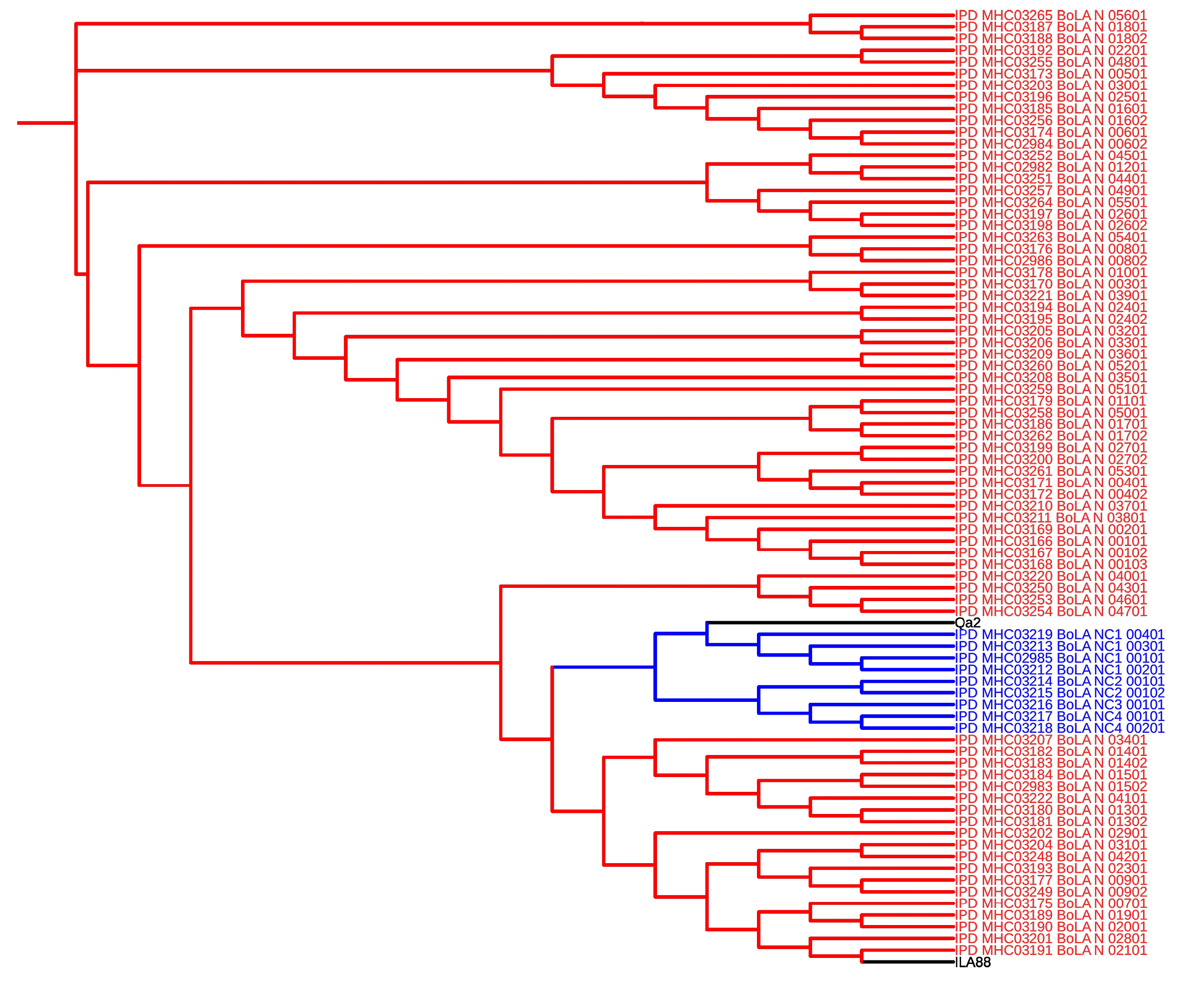
Figure 2.
Detection of MHC by Qa-2 antibody in bovine placentomes from control and cloned pregnancies. In A, control at 60 days of pregnancy. Absence of Qa-2 in all tissues. In B, control at 90 days of pregnancy. Absence of Qa-2 expression. In C, control at term. Increase of Qa-2 expression restricted to trophoblast and maternal epithelium. In D, cloned at 60 days of pregnancy. Qa-2 restricted to trophoblast and maternal epithelium. In E, cloned at 90 days of pregnancy. Increase of Qa-2 labeling in trophoblast and maternal epithelium, and low labelling in trophoblast giant cells near to maternal epithelium. In F, cloned at term. Same pattern as E but with low intensity. In F`, IgG control. Brown, immune reaction product; blue, hematoxylin counterstain. Endometrial stroma (S), maternal epithelium (E), trophoblast (T) and mesenchyme (M). Bar = 20 µm.
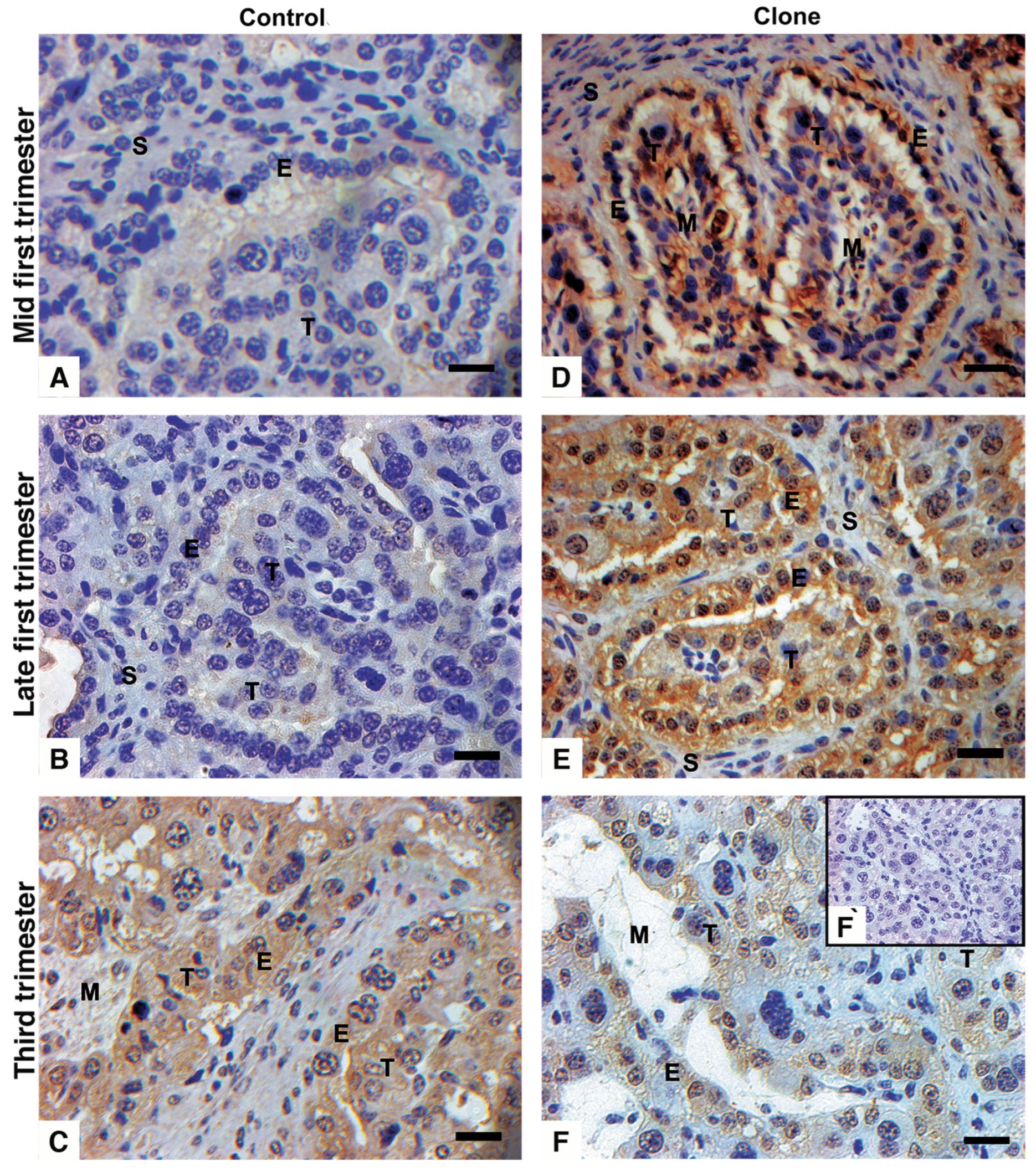
Figure 3.
Detection of MHC by Qa-2 antibody in bovine cloned placentome at 90 days of pregnancy. Migration process of trophoblastic giant cells (TGC - arrow) into disorganized maternal epithelium (E). See that the more deeply TGC are in maternal epithelium lower is the Qa-2 expression. Brown: immune reaction product; blue: hematoxylin counterstain. Endometrial stroma (S), maternal epithelium (E), trophoblast (T) and TGC (arrow). Bar = 20 µm.
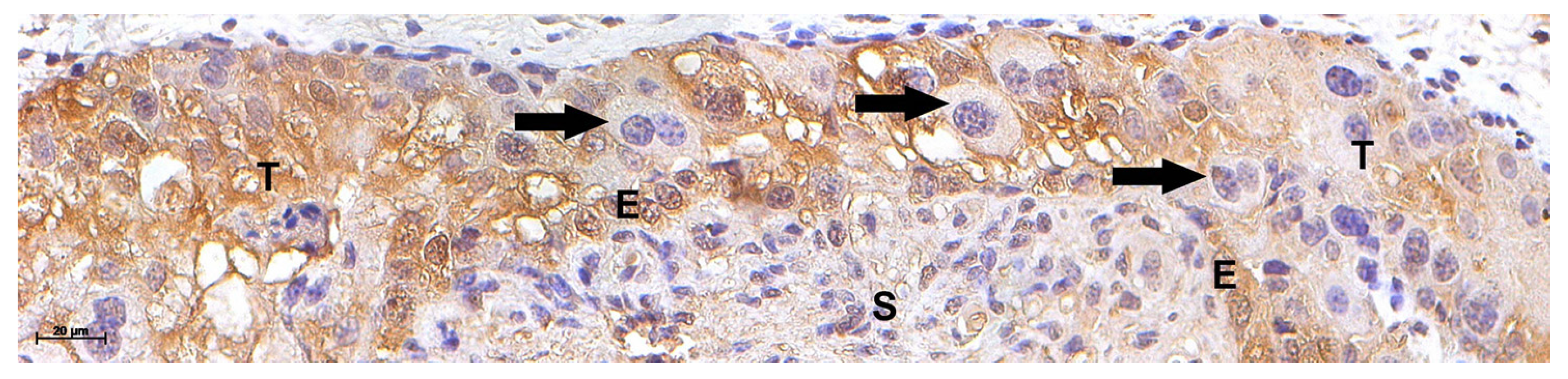
Figure 4.
Immunohistochemistry for IL-A88 in bovine placentomes from control and cloned pregnancies. In A, control at 60 days of pregnancy, observe anti-IL-A88 signal in maternal epithelium and light expression in trophoblast. In B, control at 90 days of pregnancy, anti-IL-A88 is absent in trophoblast. In C, control at term, low presence of anti-IL-A88 in maternal epithelium and trophoblast. In D, cloned at 60 days of pregnancy. In E, cloned at 90 days of pregnancy. Observe that both in D and E the maternal epithelium has a light IL-A88 signal, whereas is absent in trophoblast. In F, cloned at term, presence of anti-IL-A88 in maternal epithelium and trophoblast with more intensity than in control (C). Brown, immune reaction product; blue, hematoxylin counterstain. In F`, IgG control. Endometrial stroma (S), maternal epithelium (E), trophoblast (T) and mesenchyme (M). Bar = 20 µm.
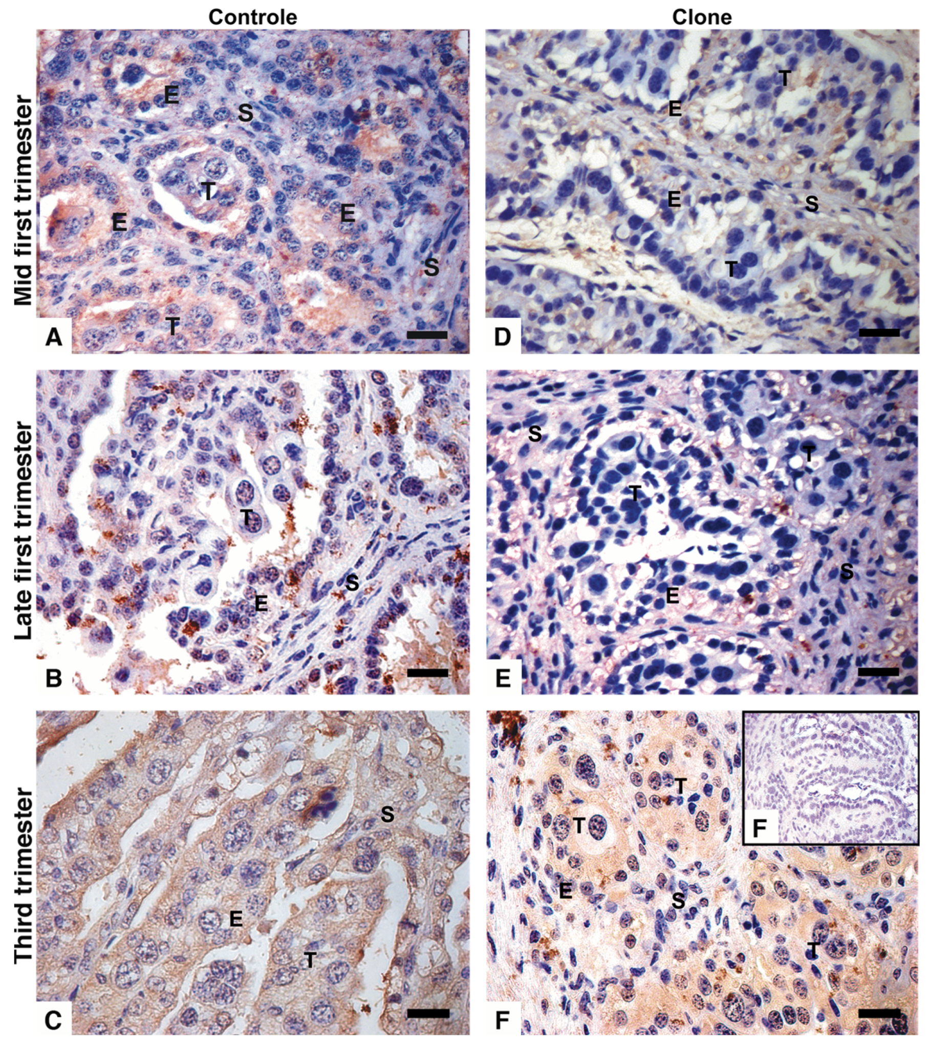
Figure 5.
Expression of BoLA-NC1 (A), BoLA-NC2 (B), BoLA-NC3 (C), BoLA-NC4 (D) and JSP-1 (E) in bovine placentomes from control (dark bar) and cloned (grey bar) pregnancies. In X axis, pregnancy period (first or third trimester); and Y axis, gene expression shown in dCt. Data are least-squares means ±S.E.M. Lowercase letters represent significance (p ≤ 0.05) between control placentomes. Uppercase letters represent significance (p ≤ 0.05) between cloned placentomes. Continuous bars represent significance (p ≤ 0.05) inside pregnancy age.
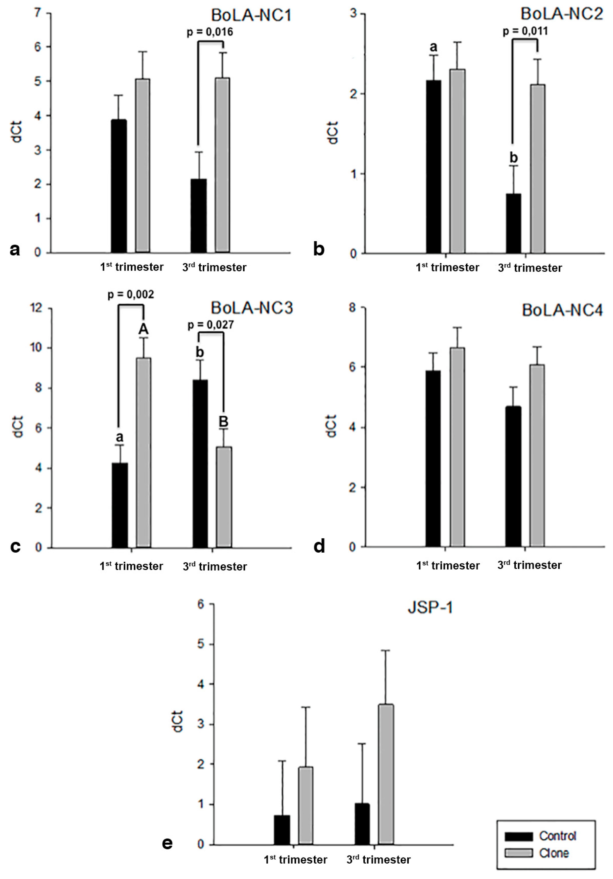

Table 1.
Details of primers used for classical and non-classical transcripts expression.
| Gene Symbol | Gene Name | Gene ID | Sequence (5’ – 3’) | Amplicon | Ref. |
|---|---|---|---|---|---|
| BOLA-NC1 | MHC 1 non-classical 1 | F: GGATACAAAAGGAAAACACACAGACTT | 77 | [40] | |
| R: GGCCTCGCTCTGGTTGTAGT | |||||
| BOLA-NC2 | MHC 1 non-classical 2 | F: TGCGCGGCTACTACAACCA | 67 | Mansouri-Attia in press | |
| R: ACGTCGCAGCCGTACATCTC | |||||
| BOLA-NC3 | MHC 1 non-classical 3 | F: CCTGCGCGGCTACTACAAC | 70 | Mansouri-Attia in press | |
| R: CACTTCGCAGCCAAACATCA | |||||
| BOLA-NC4 | MHC 1 non-classical 4 | F: TTGCGAACGGGCTTGAAC | 74 | [41] | |
| R: ACCCACTGGAGGGTGTGAGA | |||||
| JSP-1 | MHC-class I JSP-1 | 407173 | F: TAGGAGTTAGGGAGACTGGCCC | 121 | [42] |
| R: AGCCTCGTCTCTGCTGGAAC | |||||
| YWHAZ | tyrosine 3-monooxygenase/tryptophan 5-monooxygenase activation protein, zeta | 287022 | F: GCATCCCACAGACTATTTCC | 150 | [43] |
| R: GCAAAGACAATGACAGACCA | |||||
| ACTB | Beta-actin | 280979 | F: CAGCAGATGTGGATCAGCAAGC | 108 | [44] |
| R: AACGCAGCTAACAGTCCGCC | |||||
| PPIA | Peptidylprolyl isomerase A | 281418 | F: CATACAGGTCCTGGCATC | 91 | [44] |
| R: CACGTGCTTGCCATCCAA |
Disclaimer/Publisher’s Note: The statements, opinions and data contained in all publications are solely those of the individual author(s) and contributor(s) and not of MDPI and/or the editor(s). MDPI and/or the editor(s) disclaim responsibility for any injury to people or property resulting from any ideas, methods, instructions or products referred to in the content. |
© 2025 by the authors. Licensee MDPI, Basel, Switzerland. This article is an open access article distributed under the terms and conditions of the Creative Commons Attribution (CC BY) license (http://creativecommons.org/licenses/by/4.0/).
Copyright: This open access article is published under a Creative Commons CC BY 4.0 license, which permit the free download, distribution, and reuse, provided that the author and preprint are cited in any reuse.


