Preprint
Review

This version is not peer-reviewed.

Breast Cancer-Derived Exosomes: Formation, Biological Function and Applications

Submitted:

06 February 2025

Posted:

08 February 2025

You are already at the latest version

Abstract

The exosomes are a class of multi-vesicular bodies structures that originated from the cytoplasmic nucleus endosome, which can be synthesized by different cells and released into the extracellular environment. Breast cancer-derived exosomes can promote breast cancer proliferation, metastasis, escape and angiogenesis, which plays a significant role in the occurrence and development of breast cancer. In this article, we mainly review the roles of breast cancer-derived exosomes in tumor progression and immune suppression from the following aspects. Firstly, the exosomes are mainly introduced, including structure, formation mechanism and analytics. Then it describes the effect of the breast cancer-derived exosomes in the tumor microenvironment, and the applications of exosomes in biomedicine. For example, the applications of the breast cancer-derived exosomes as a biomarker and the value of exosomes in breast cancer treatment and prognostic. Finally, the application potential of the breast cancer-derived exosomes in breast cancer diagnosis and treatment is summarized and prospected, which provides a new idea for the accurate treatment of breast cancer.

Keywords: 
;  ;  

1. Introduction

Global Cancer Statistics 2024 recently reported that in 2022, Female breast cancer is the fourth leading cause of cancer mortality worldwide, with 666,000 deaths (6.9% of all cancer deaths). Among women, breast cancer is the most commonly diagnosed cancer, and it is the leading cause of cancer deaths globally [1]. However, the treatments of breast cancer have shown high success rates, but many cases of recurrence and drug resistance are still reported [2]. Currently, the main treatments for breast cancer include surgery, radiation therapy, chemotherapy, targeted therapy, gene therapy, and immunotherapy. Regardless of the treatment method, the tumor microenvironment plays a key role in the treatment of breast cancer. It is a source of nutrition for tumor cells and is closely related to cancer growth, metastasis, immune escape and drug resistance [3].
To realize the early detection and treatment of tumors and improve the cure rate of patients, researchers have done a lot of research in the early diagnosis and prognosis evaluation of tumors and found a variety of diagnostic biomarkers, such as exosomes [4,5]. Exosomes are produced and released by almost all types of cells. They are widely distributed in body fluids such as extracellular fluid, blood and cerebrospinal fluid [6]. Researchers found that exosomes can participate in the transmission of information between cells through autocrine, paracrine and endocrine mechanisms to transmit the genetic information to the recipient cells, and then affect the protein expression level of the receptor cells [7,8]. Their function depends on the kind of cells from which it originates, and they participate in the physiological and pathological processes of the body by mediating cell signal transduction, regulating cell homeostasis, participating in immune response and inflammatory reaction, etc. [9,10]. They are of great significance to study the occurrence, development, diagnosis and treatment of tumors, neurodegenerative diseases and cardiovascular diseases [11,12].
Tumor-derived exosomes of nanometer size have the characteristics of easy passage through the blood-brain barrier, natural homing property, stability of bilayer lipid structure and high biocompatibility [13]. Therefore, these exosomes are becoming an ideal carrier for tumor-targeted therapy, especially the engineered exosomes show excellent effects of tumor-targeted intervention and enhanced drug therapy [14,15]. Last several years, breast cancer-derived exosomes have made great progress in breast cancer diagnosis, treatment, targeted drug design and vaccine application.
In this review, we review the current understanding of breast cancer-derived exosomes, and also summarize the progress of breast cancer-derived exosomes in tumor progression and immune suppression in recent years.

2. Structure and biological function of exosomes

In 1983, researchers at the University of Washington discovered a vesicle-like structure secreted and released by sheep reticulocytes. A few years later, the researchers successfully isolated and extracted it by ultracentrifugation, and officially named it "exosome" [16,17]. Extracellular vesicles (EVs) can be divided into three main types: exosomes, microvesicles and apoptotic bodies, according to the formation pathway, size and membrane surface markers [18,19]. Exosomes are formed by the fusion of multi-vesicular bodies(MVBs) containing intraluminal vesicles (ILVs) and the plasma membrane [20]. The diameter is between 30 and 100nm. Big EVs are called microvesicles (MVs), also known as microparticles (MPs) or extracellular bodies. The diameter is 100-1000nm and exfoliates directly from the cell surface. Apoptotic bodies are formed during apoptosis, with diameters ranging from 1000 to 5000nm. The exosome showed a round bilayer membrane structure under a frozen electron microscope, and the concentration was between 1.13~1.19g/ml [21].
Almost all cells could secrete exosomes, including saliva, urine, blood, semen, amniotic fluid, ascites, bronchioalveolar lavage fluid, milk, joint synovial fluid and cerebrospinal fluid [22]. It can be synthesized and released into the extracellular environment by different types of cells, which can promote tumor proliferation, angiogenesis, invasion of adjacent normal tissue structure, distant metastasis, formation of chemotherapy resistance and so on [23,24].

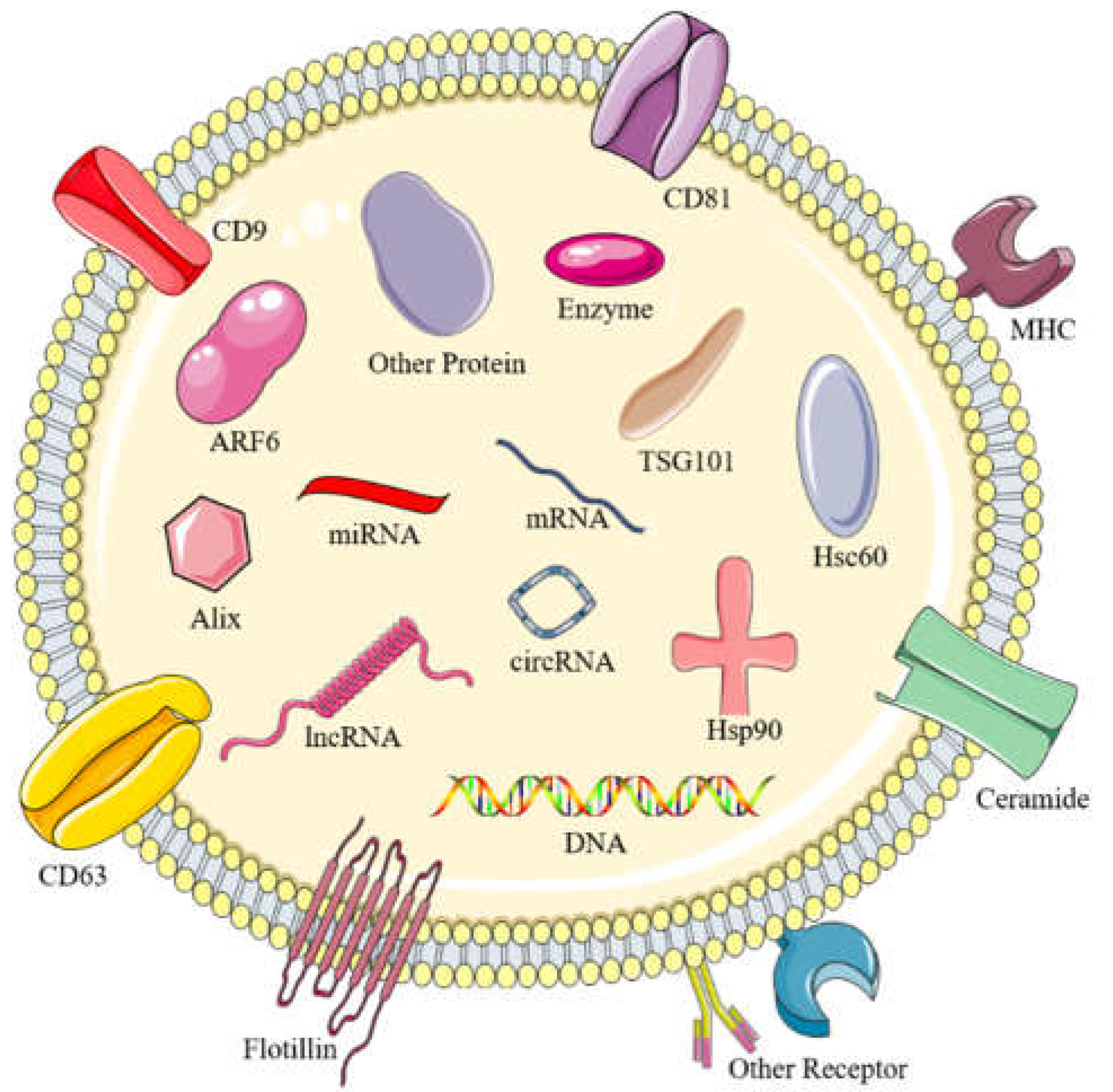
2.1. The structure of exosomes

According to the latest updated list of exosomes database (http://www.exocarta.org), exosomes are made up of a variety of substances. They contain 9769 kinds of proteins, 3408 kinds of mRNAs, 2838 kinds of miRNAs and 1116 kinds of lipids. They also contain DNA, for example, single-stranded DNA, double-stranded DNA, genomic DNA and mitochondrial DNA and so on [25,26]. A variety of bioactive substances in exosomes, such as proteins, mRNA and non-coding RNA (such as circ-RNA, miRNA, lncRNA) (Figure 1), play a variety of biological roles by transmitting internal active substances to mediate cellular communication [27,28].
Proteins are a vital constitution. It has been found that more than 4000 proteins can be isolated from exosomes. Some of these proteins are evolutionarily conserved and have nothing to do with the source cells, and the other part varies with different source cells. Exosomes contain proteins including membrane transport and fusion proteins such as GTP enzyme, annexins, rab, flotillin [29]. Proteins involved in MVEs syntheses, such as Tsg101 and Alix. Alix is a marker protein of exosomes, which plays a momentous effect in the biogenesis of exosomes [30]. In addition, there are four transmembrane proteins (such as CD9, CD81, CD82, CD83, CD63) and heat shock proteins(such as Hsp60, Hsp90) [31]. Besides, cytoskeleton proteins, lipid-related proteins, antigen presentation-related proteins (MHCⅠ, Ⅱ, etc.) and cellular signaling pathway proteins are also widely present in exosomes [32]. Four transmembrane proteins (Alix, Flotillin, Tsg101 and Rab56) were used as molecular markers of exosome specificity [33,34]. It has been shown that the proteolipid protein is transferred to ILV in an ESCRT-independent manner from the lipid-rich parts of the intima, such as cholesterol, ceramide and sphingomyelin [35].
Exosomes can transfer extracellular RNAs (exRNAs) to other cells and tissues. RNAs in exosomes are promising components in tumor therapy and diagnosis. One study described exosomes as capable of carrying and transferring mRNA and microRNA between cells, and established their role as carriers of intercellular communication [36]. The exosomes of tumor cells are rich in a variety of carcinogenic miRNAs, including miR-21, miR-23a, miR-23a, miR-221, miR-451 and so on [37]. The exosomes carry carcinogenic miRNAs between malignant tumor cells, which may be the mechanism of regulating tumor growth and invasion [36]. Exosomal non-coding RNAs (exo-ncRNAs) and exosomal microRNAs (exo-miRNAs) as natural carriers of long noncoding RNAs (lncRNAs) and microRNAs have attracted more and more attention through the programming process [38]. A recent study has shown that exosomal miR-21-5p inhibits the expression of cyclin-dependent kinase 6 (CDK6) by targeting the 3'- untranslated region (3'- UTR) of CDK6 at the mRNA and protein levels [39]. MiR-138-5p was delivered from breast cancer cells to tumor-associated macrophages through exosomes to downregulate KDM6B expression, restrain M1 polarization, and accelerate M2 polarization [40]. Exosomal miR-138-5p may be a potential target for the treatment of breast cancer.
Lipids can be used as carriers and can also participate in the synthesis of exosomes. The difference in protein content in exosomes mainly depends on the source of cells, which is rich in a variety of lipids and can be used as lipid carriers to transport a variety of bioactive lipids to recipient cells [41]. Exosomes are rich in sphingomyelin, cholesterol, phosphatidylserine, phosphatidylinositol, phosphatidic acid, ceramide and other components, mainly similar components of the plasma membrane and special lipids that change with different source cells [42]. Among them, bone morphogenetic protein, cholesterol, oxysterols, ceramide, phosphatidic acid and adenosine triphosphate-binding cassette (ABC) transporter are involved in the formation mechanism of exosomes [43]. The recognition of exosomes by target cells is related to the participation of lipid receptors. In addition, exosomes carry bioactive lipid molecules between cells, which play a certain role in regulating cellular lipid metabolism [44].

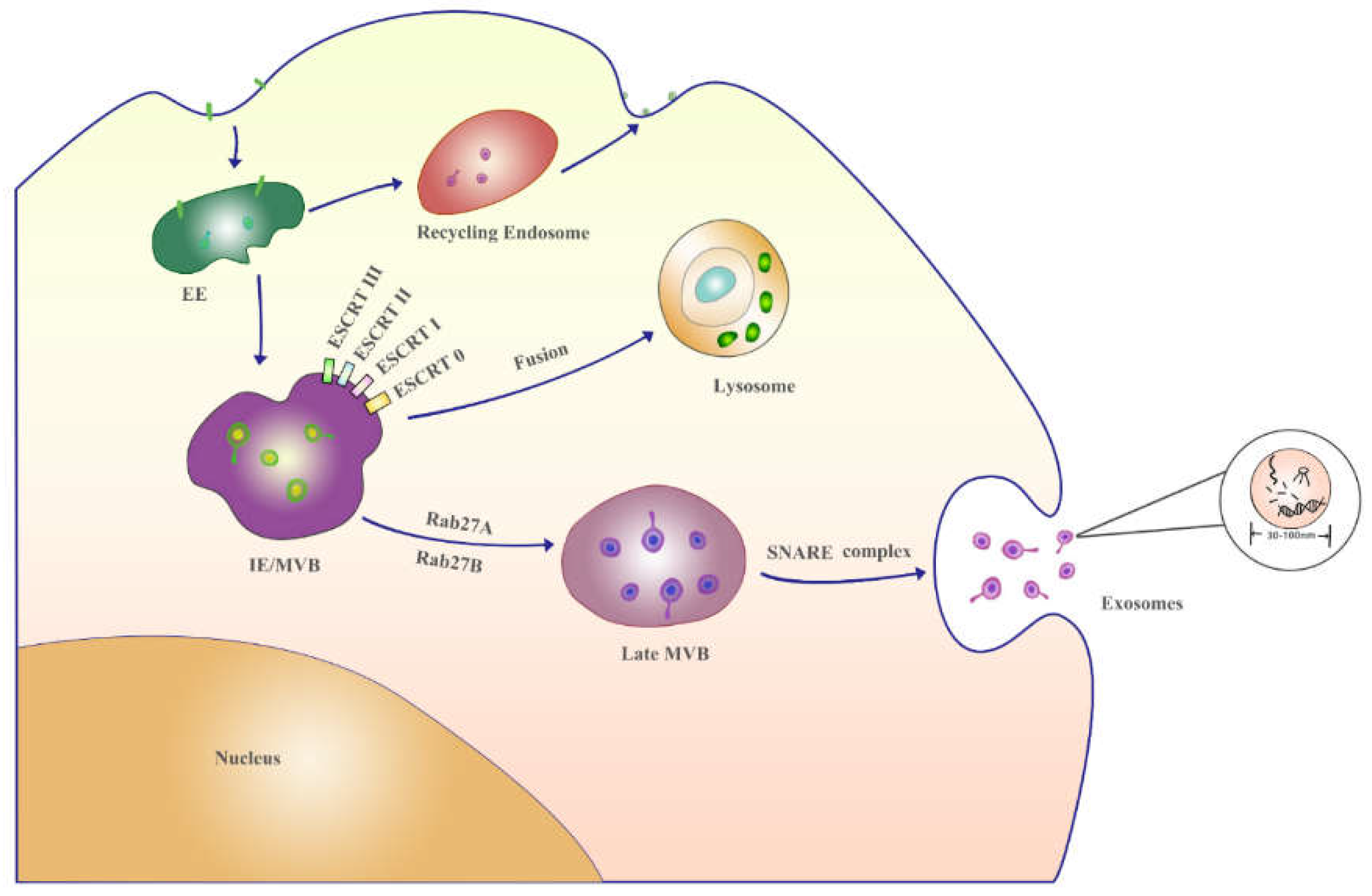
2.2. The formation mechanism and analytics of exosomes

Compared with other extracellular vesicles in vivo, exosomes have a special process of formation. First of all, cytoskeletal proteins (such as actin and tubulin) interact with grid protein to make the cell membrane invagination, forming vesicles covered with grid protein. These vesicles are called endosomes after removing the grid protein coating. Subsequently, the early endosomes sprouted in reverse and formed small molecular substances to form intraluminal vesicle exosomes. The late endosomes gradually matured and evolved into the multi-vesicular body (MVB) containing exosomes, and then MVB released the exosomes into the microenvironment by plasma membrane fusion to form exosomes [20,38]. The protein sorting of intraluminal vesicles (ILV) is a highly regulated mechanism, which in most cases depends on the endosomal sorting complexes required for transport (ESCRT). ESCRT-0、ESCRT-I、ESCRT-II and ESCRT-III are the four complexes that make up the ESCRT device [45]. At the beginning of the ESCRT-dependent pathway, it is determined by the protein ubiquitin (UB) checkpoint. In the UB dependent pathway, all ESCRT subunits are involved [31,46]. It is essential to understand the biogenesis and release of exosomes (Figure 2), which will help researchers to develop new treatment strategies.
According to the structural characteristics of the exosomes, their isolation and detection methods are as follows. The separation techniques of exosomes include ultracentrifugation, ultrafiltration, immunoaffinity capture, microfluidic control, size exclusion chromatography and polymer-based precipitation separation [47,48]. Although each method gives priority to different characteristics in the separation of exosomes, a common requirement is that their structural and biological activities should remain unchanged after separation [49]. The detection methods of exosomes include scanning electron microscope (SEM), transmission electron microscope (TEM), atomic force microscope (AFM), enzyme-linked immunosorbent assay (ELISA), dynamic light scattering (DLS), nanoparticle tracking analysis (NTA), atomic force microscopy measurements (AFMM), flow cytometry–like analytics and single-particle interferometric reflectance (SPIR) and so on [31,50]. These methods can detect the shape, size, quantity and surface protein content of exosomes. With the rapid development of microfluidic technology, it has been widely used in exosome detection. It is helpful to realize the detection of exosomes with high throughput and high precision [50]. A recent study developed a microfluidic device for one-step detection of breast cancer-derived exosomal mRNA in blood using signal-amplifiable 3D nanostructure [51]. Based on accelerated, rapid and localized in situ deposition of dopamine by HRP, a highly sensitive and selective colorimetric aptasensor was developed for the detection of cancer-derived exosomes [52]. At present, many exosome detection techniques have been widely used in the prediction and treatment of various diseases.

3. Exosomes in cancer development

3.1. Growth and proliferation

The bioactive components contained in the exosome of breast cancer cells enter the tumor microenvironment with the exosome, which will lead to changes in the downstream signal pathway, and then affect the growth, proliferation and invasion of tumor cells, making it more conducive to the development of tumor [16,53]. Survivin and heat shock proteins contained in tumor exosomes can inhibit apoptosis and promote tumor proliferation [54]. For example, Survivin in breast cancer-derived exosomes upregulates SOD1 expression and then converts them into myofibroblasts, whose feedback promotes cancer proliferation and metastasis [55]. To sum up, the proteins and RNAs contained in tumor exosomes play an essential function in regulating tumor growth and proliferation. When tumor cells reach a new metastatic site, exosomes derived from tumor cells can enhance the formation of new blood vessels and promote the growth of tumor cells at the metastatic site [56]. Mesenchymal stem cell-derived exosomes shuttled exosomal miR-100 inhibits angiogenesis in vitro by modulating the mTOR/HIF-1α/VEGF signaling axis in breast cancer cells [57]. Therefore, exosomes secreted by tumor cells push forward an immense influence on promoting angiogenesis in the tumor microenvironment (TME).

3.2. Migration and invasion

Exosomes produced by tumors can promote tumor invasion and metastasis by regulating a variety of signal pathways. Distant invasion and metastasis are important features of the tumor [58]. Exosomes produced by tumors can promote tumor invasion and metastasis by regulating a variety of signal pathways. Wang et. al found that the different macrophage balance fraction (MBF) environments can affect the migration and invasion ability of breast cancer cells. And the mechanism of MBF changes in breast cancer may be affected via breast cancer-derived exosomes [59]. A study showed that cancer-derived exosomal miR-7641 can promote breast cancer progression and metastasis by intercellular communication [60]. Breast cancer-derived exosomes promote the activation of CAFs via miR-146a that inhibit thioredoxin interacting protein and then activate the Wnt pathway, thereby enhancing migration and invasion of breast cancer cells [61]. Studies have shown that breast cancer-derived miR-122 can reprogram glucose metabolism to promote cancer cell metastasis [62]. Transfection of exogenous miR-223 into breast cancer cells can reduce the expression of MEF2C protein, facilitate the translocation of β-catenin, and eventually lead to cell invasion [63]. A recent study has shown that exosomes derived from human umbilical cord mesenchymal stem cells inhibit the migration and invasion of breast cancer cells through miR-21-5p/ZNF367 pathway. ZNF367 participates in the regulation of hucMSC-exos-mediated miR-21-5p on the metastasis of breast cancer cells, which may become a new intervention method for the clinical treatment of breast cancer [64]. In addition, the exosomes can carry functional proteins and nucleic acid molecules to release towards serum, urine, cerebrospinal fluid, saliva and other body fluids, and even the exosomes of the central nervous system can release to the blood circulation through the blood-brain barrier [65]. It is reasonable to speculate that exosomes play a major function in the process of tumor invasion and metastasis.

3.3. Immune evasion

Immune escape of breast cancer cells contributes to breast cancer pathogenesis. Exosomes can affect the development, maturation and antitumor activity of immune cells by transmitting inhibitory proteins. Tumor-derived exosomes can induce tumor cells to produce immunosuppressive function and transmit genomic DNA, mRNA and miRNA through immune cells, so that response cells have the role of reprogramming and promoting tumor progression [66]. Tumor-derived exosomes can not only cause T and NK cells dysfunction by blocking the activation, proliferation or apoptosis, but also inhibit antigen-presenting cells and anti-tumor immune response [67]. The main mechanism of tumor cell immune escape is the change in the antigen presentation mechanism. In addition, tumor-derived exosomes adjust host immunity by changing the behavior of macrophages [56]. Heat shock protein 70-80 and MHC-1 molecules derived from tumor antigens and tumor exosomes can interact with dendritic cells to induce mouse tumor T cells to produce an effective CD8+-dependent antitumor effect [68]. Tumor exosome-derived miR-9 and miR-181a activated the Janus Kinase (JAK)/Signal Transducer and Activator of Transcription (STAT) signaling pathway by targeting suppressor of cytokine signaling 3 (SOCS3) and protein inhibitors of activated STAT3 (PIAS3), respectively promoted the expansion of early-stage myeloid-derived suppressor cells (eMDSCs) which may provide a potential therapeutic target for high IL-6 breast cancer treatment [69]. Tumors can evade immune attack by upregulating the expression of programmed death-ligand 1 (PD-L1), which interacts with receptors on T cells to activate immunosuppressive signals. A result demonstrates that the TME can induce bone marrow-derived cell-exosomes to carry PD-L1, which effectively inhibits CD8+ T cell responses in vitro and in vivo. PD-L1 on bone marrow-derived cell-exosomes contributes to tumor immunosuppression and tumor growth [70]. Exosomal miR-27a-3p promotes immune evasion through membrane-associated guanylate kinase inverted 2 (MAGI2) / phosphatase and tensin homolog (PTEN) / phosphoinositide 3-kinase axis upregulating PD-L1 in breast cancer [71]. This mechanism of action may provide ideas and directions for the development of new breast cancer therapeutic targets in the future.

3.4. Drug resistance

Resistance is not only caused by genetic and phenotypic changes in the tumor itself, but may also be caused by the surrounding microenvironment that prevents drug internalization [72]. Exosomes promote drug resistance in donor cells by reducing intracellular drug concentrations and treating pro-apoptotic proteins. Exosomes from the drug-resistant cells are released into the extracellular medium that ultimately confers drug resistance in the recipient cells [73]. Recent studies indicate that exosomal miR-567 reverses trastuzumab resistance via inhibiting autophagy-related 5 (ATG5) in breast cancer. It may help develop more effective strategies to reverse chemotherapy resistance [74]. Exosomal miR-4443 can promote resistance to cisplatin and facilitate tumor growth through iron deficiency mediated by FSP1 m6A [75].

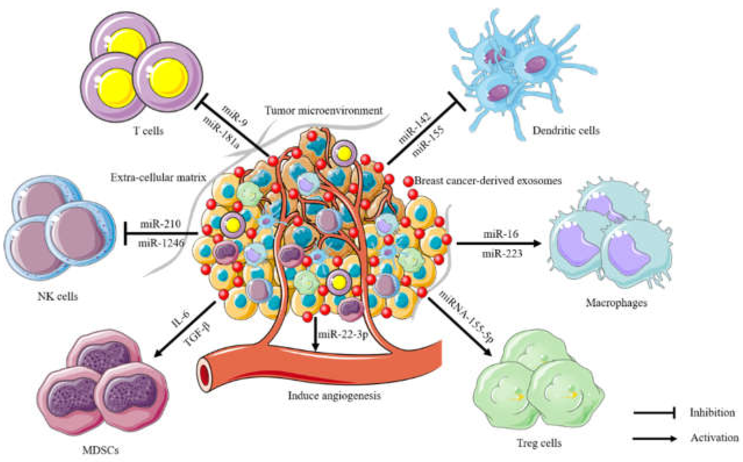
4. The immune suppression of breast cancer-derived exosomes in the tumor microenvironment

TME is a special biological environment formed by tumor cells, infiltrating immune cells, stromal cells, blood vessels, extracellular matrix, secretory factors and so on [76]. On the one hand, TME provides a suitable environment for tumor growth and malignant progression, on the other hand, it interferes with the function of immune cells in the environment to help tumors escape host immune surveillance and promote tumor metastasis and drug resistance [77].
Breast cancer-derived exosomes are considered to be specific signal carriers in TME, which can induce malignant phenotypic transformation and invasion and metastasis of tumors [78]. They can mediate the phenotypic transformation of stromal cells in TME and reshape the tumor microenvironment through epithelial-mesenchymal transition (EMT), extracellular matrix (ECM) degradation and neovascularization [79]. Studies have shown that breast cancer-derived exosomes secreted by normal cells can transfer tumor suppressor genes to cancer cells and inhibit the growth of tumor cells by inhibiting the expression of oncogenes. But tumor cell-derived exosomes secreted by tumor cells can affect the local microenvironment of proximal tumor cells and stromal cells, and regulate tumor neovascularization, pre-metastasis microenvironment, therapeutic drug resistance and metastasis diffusion [80].
Breast cancer-derived exosomes can transport proteins, mRNAs, and miRNAs, and act as hubs between the tumor microenvironment and stromal cells. They can be absorbed by cancer-associated fibroblast (CAF). CAF promotes the progression and metastasis of breast cancer through exosomal miR-500a-5p [81]. CAF is the main cell group on TME in most cancers, changing its biological activity and reprogramming energy metabolism [82]. In most cases, breast cancer-derived can induce the polarization of M2 macrophages. Some studies have confirmed that TDE can induce the polarization of M1 macrophages [83]. Overexpression of miR-130 and miR-33 in exosomes can reduce tumor progression by polarizing macrophages from an M2 to an M1 phenotype [84].
As communication between tumor and microenvironment, breast cancer-derived exosomes regulate the signal pathway and functional state of stromal cells in TME by delivering tumor-derived bioactive substances, thus remodeling TME. They can induce stromal cells to differentiate into tumor-associated cells. The exosomes of tumor-associated stromal cells trigger each other's EMT of tumor cells. It is worth noting that TDE has a different affinity with various types of stromal cells in TME [78]. In the normal microenvironment, phenotypic and functional changes occur after stromal cells absorb TDE. It provides support for the malignant development of tumors in the aspects of stimulating angiogenesis and lymphatic vessel formation, reprogramming energy metabolism, mediating inflammatory cell infiltration, inducing immunosuppression, etc. [8,19]. Therefore, blocking drugs and antibodies against specific bioactive substances of TDE is a new strategy for tumor therapy (Figure 3).

5. Applications of breast cancer-derived exosomes in biomedicine

5.1. Breast cancer-derived exosomes as a biomarker

Exosomes are more and more widely used in tumor diagnosis. MiRNAs released from exosomes were found to be resistant to freeze-thaw cycles. The high stability of exo-miRNAs makes it a promising biomarker for disease monitoring. The expression of CD82 in breast cancer tissues is significantly lower than that in healthy and benign breast diseases, b ut it is highly expressed in exosomes extracted from peripheral blood [85]. The level of serum exosomal miR-1910-3p from breast cancer patients was expressively higher than that of healthy controls. ROC curve analysis showed that serum exosomal miR-1910-3p had higher sensitivity, specificity and diagnostic efficiency than traditional tumor biomarker CA153 [86]. The level of serum exosomal miR-148a decreased significantly in breast cancer patients, which was closely related to the poor clinical results such as tumor lymph node metastasis, TNM staging and differentiation of breast cancer [87]. Therefore, CD82, miR-1910-3p, miR-148a, miR-21 and miR-200c can be used as biomarkers for breast cancer diagnosis. A recent study showed that breast cancer-derived exosomes may contribute to impaired neurodevelopment in brain organoids [88]. The identification and analysis of biomolecules in specific pathogenic states in exosomes make them an attractive diagnostic marker.

5.2. The value of breast cancer-derived exosomes in cancer treatment and prognostic

The fluidity of exosomes provides an opportunity for them to develop as therapeutic agents and nanoparticles drug carriers [89]. The exosomes containing the anti-inflammatory drug curcumin have been shown to increase bioavailability, and the preparation protects mice from lipopolysaccharide-induced shock [90]. The use of exosomes to deliver doxorubicin to the mouse tumor tissue model showed that doxorubicin containing exosomes accumulated in the target organs and led to the inhibition of tumor growth [91]. At present, the application of exosomes in the tumor is mainly used in immunomodulatory therapy, as a carrier or therapeutic target for the delivery of chemotherapy drugs. As a drug delivery carrier, exosomes have their natural advantages: exosome has the characteristics of stability, permeability, low immunogenicity, low toxicity and good biocompatibility [92]. Exosomes have a small molecular weight, inherent cell targeting, stability in the circulatory system, can freely pass the blood-brain barrier and come from the human body, so they can successfully avoid immune rejection. Similar to the nanoparticles currently used in drug delivery systems, they have been used as drug carriers [93]. The combination of exosomes and nanomaterials has become a research hotspot in the field of nanoparticle therapy. Targeting exosomes may disrupt cell-to-cell communication, so exosomes can be considered therapeutic targets.

6. Summary and prospect

The exosome is a kind of vesicle with potential in diagnosis, treatment and drug delivery. Exosomes exert an enormous function in communication between tumor cells and the tumor microenvironment. Exosomal RNAs and proteins are the main groups involved in cancer diagnosis. These RNAs and proteins molecules are involved in the formation of the tumor microenvironment, tumor metastasis and angiogenesis. This review also introduces the application of exosomes as biomarkers in biomedicine. These exosomes can be used as cancer treatment tools because they are good candidates for cancer diagnosis and therapy, such as biomarker and drug carriers. It is necessary to deeply understand the mechanism of bidirectional crosstalk between tumor and microenvironment for discovering new treatment strategies. Although breast cancer-derived exosomes have been studied in tumor-related fields, more detailed molecular mechanisms are needed to clarify their internal relationship.
Despite numerous studies and literature reports on exosomes as a potential tool in the diagnosis and treatment of tumors, the mechanism of their intercellular communication needs to be further studied. First, how to identify exosomes accurately? We need a wider understanding of how exosomes develop at every stage of cancer. In addition, how to apply the isolated exosomes for diagnosis or treatment of cancer patients? Besides, how to reduce exosome resistance also faces more and more challenges. Although exosomes have been identified as drug carriers or target molecules in clinical studies, we are still far from fully understanding the therapeutic effects of these vesicles. If the formation and release of exosomes are regulated, it will not only provide a new target for improving the chemotherapy effect of the tumor, but also provide a new idea for the development of precise treatment of the tumor. Importantly, it is still a great challenge to transform these basic scientific researches into clinical applications. We firmly believe the exosome-based cancer treatment strategy will make great progress to benefit the majority of cancer patients in the future.

Author Contributions

Conceptualization, N.Q. and T.S.; Writing, N.Q., G.M., Y.W., X.Q., Y.Z., M.X., W.J. and T.S; Funding Acquisition, N.Q. and T.S.; Supervision N.Q. and T.S. All authors have read and agreed to the published version of the manuscript.

Funding

This research was funded by the Natural Science Foundation of Hunan Province of China (No.2024JJ7324); Scientific Research Fund of Hunan Provincial Education Department (No.23B1042; No.24B1080); the Scientific Research Project of Hunan Provincial Health Commission (No.202113022028) and Projects of Innovation and Entrepreneurship for College Students in Hunan (No.S202312214027; No.S202212214006).

Institutional Review Board Statement

Not applicable.

Informed Consent Statement

Not applicable.

Data Availability Statement

The data generated during the current study are available from the corresponding author on reasonable request.

Acknowledgments

None.

Conflicts of Interest

The authors declare no conflicts of interest.

Abbreviations

EVs Extracellular vesicles
MVBs multi-vesicular bodies
ILVs intraluminal vesicles
MPs microparticles
MVs microvesicles
exRNAs extracellular RNAs
exo-ncRNAs Exosomal non-coding RNAs
exo-miRNAs exosomal microRNAs
lncRNAs long noncoding RNAs
CDK6 cyclin-dependent kinase 6
3'- UTR 3'- untranslated region
MVB multi-vesicular body
ILV intraluminal vesicles
ESCRT endosomal sorting complexes required for transport
UB ubiquitin
SEM scanning electron microscope
TEM transmission electron microscope
AFM atomic force microscope
ELISA enzyme-linked immunosorbent assay
DLS dynamic light scattering
NTA nanoparticle tracking analysis
AFMM atomic force microscopy measurements
SPIR single-particle interferometric reflectance
EE early endosomes
MVB multivesicular body
TME tumor microenvironment
MBF macrophage balance fraction
JAK Janus Kinase
STAT Signal Transducer and Activator of Transcription
SOCS3 suppressor of cytokine signaling 3
PIAS3 protein inhibitors of activated STAT3
eMDSCs early-stage myeloid-derived suppressor cells
PD-L1 programmed death-ligand 1
MAGI2 membrane-associated guanylate kinase inverted 2
PTEN phosphatase and tensin homolog
ATG5 autophagy-related 5
EMT epithelial-mesenchymal transition
ECM extracellular matrix
CAF cancer-associated fibroblast

References

  1. Bray, F.; Laversanne, M.; Sung, H.; Ferlay, J.; Siegel, R.L.; Soerjomataram, I.; Jemal, A. Global cancer statistics 2022: GLOBOCAN estimates of incidence and mortality worldwide for 36 cancers in 185 countries. CA Cancer J. Clin. 2024, 74, 229–263. [Google Scholar] [CrossRef] [PubMed]
  2. Wang, W.; Zhong, F.; Wang, D.; Zhao, Y.; Peng, D.; Li, S.; Ning, Q.; Tang, S.; Yu, C.; Wei, H. Dual gatekeepers-modified mesoporous organic silica nanoparticles for synergistic photothermal-chemotherapy of breast cancer. J. Colloid. Interface Sci. 2023, 646, 118–128. [Google Scholar] [CrossRef] [PubMed]
  3. Harris, M. A.; Savas, P.; Virassamy, B.; O'Malley, M.; Kay, J.; Mueller, S. N.; Mackay, L. K.; Salgado, R.; Loi, S. Towards targeting the breast cancer immune microenvironment. Nat. Rev. Cancer 2024, 24, 554–577. [Google Scholar] [CrossRef] [PubMed]
  4. O'Neill, R.S.; Stoita, A. Biomarkers in the diagnosis of pancreatic cancer: Are we closer to finding the golden ticket? World J. Gastroenterol. 2021, 27, 4045–4087. [Google Scholar] [CrossRef]
  5. Sanchez, J.I.; Jiao, J.; Kwan, S.Y.; Veillon, L.; Warmoes, M.O.; Tan, L.; Odewole, M.; Rich, N.E.; Wei, P.; Lorenzi, P.L.; Singal, A.G.; Beretta, L. Lipidomic Profiles of Plasma Exosomes Identify Candidate Biomarkers for Early Detection of Hepatocellular Carcinoma in Patients with Cirrhosis. Cancer Prev. Res. 2021, 14, 955–962. [Google Scholar] [CrossRef]
  6. Skotland, T.; Sandvig, K.; Llorente, A. Lipids in exosomes: Current knowledge and the way forward. Prog. Lipid Res. 2017, 66, 30–41. [Google Scholar] [CrossRef]
  7. Bahrami, A.; Moradi Binabaj, M. Exosomes: Emerging modulators of signal transduction in colorectal cancer from molecular understanding to clinical application. Biomed. Pharmacother. 2021, 141, 111882. [Google Scholar] [CrossRef]
  8. Ahmadi, M.; Jafari, R.; Mahmoodi, M.; Rezaie, J. The tumorigenic and therapeutic functions of exosomes in colorectal cancer: Opportunity and challenges. Cell Biochem. Funct. 2021, 39, 468–477. [Google Scholar] [CrossRef]
  9. Bhat, E.A.; Sajjad, N.; Thokar, F.M. Current advancement of exosomes as biomarkers for cancer diagnosis and forecasting. Cancer Treat. Res. Commun. 2021, 28, 100417. [Google Scholar] [CrossRef]
  10. Boriachek, K.; Islam, M.N.; Möller, A.; Salomon, C.; Nguyen, N.T.; Hossain, M.S.A.; Yamauchi, Y.; Shiddiky, M.J.A. Biological Functions and Current Advances in Isolation and Detection Strategies for Exosome Nanovesicles. Small 2018, 14, 02153. [Google Scholar] [CrossRef]
  11. Macedo-da-Silva, J.; Santiago, V.F.; Rosa-Fernandes, L.; Marinho, C.R.F.; Palmisano, G. Protein glycosylation in extracellular vesicles: Structural characterization and biological functions. Mol. Immunol. 2021, 135, 226–246. [Google Scholar] [CrossRef] [PubMed]
  12. Ruiz, G.P.; Camara, H.; Fazolini, N.P.B.; Mori, M.A. Extracellular miRNAs in redox signaling: Health, disease and potential therapies. Free Radic. Biol. Med. 2021, 173, 170–187. [Google Scholar] [CrossRef]
  13. Gebeyehu, A.; Kommineni, N.; Meckes, D.G.; Sachdeva, M.S. Role of Exosomes for Delivery of Chemotherapeutic Drugs. Crit. Rev. Ther. Drug Carrier Syst. 2021, 38, 53–97. [Google Scholar] [CrossRef] [PubMed]
  14. Jain, V.; Kumar, H.; Anod, H.V.; Chand, P.; Gupta, N.V.; Dey, S.; Kesharwani, S.S. A review of nanotechnology-based approaches for breast cancer and triple-negative breast cancer. J. Control Release 2020, 326, 628–647. [Google Scholar] [CrossRef] [PubMed]
  15. Huyan, T.; Li, H.; Peng, H.; Chen, J.; Yang, R.; Zhang, W.; Li, Q. Extracellular Vesicles - Advanced Nanocarriers in Cancer Therapy: Progress and Achievements. Int. J. Nanomed. 2020, 15, 6485–6502. [Google Scholar] [CrossRef]
  16. Azmi, A.S.; Bao, B.; Sarkar, F.H. Exosomes in cancer development, metastasis, and drug resistance: A comprehensive review. Cancer Metastasis Rev. 2013, 32, 623–642. [Google Scholar] [CrossRef]
  17. Harding, C.V.; Heuser, J.E.; Stahl, P.D. Exosomes: Looking back three decades and into the future. J Cell Biol 2013, 200, 367–371. [Google Scholar] [CrossRef]
  18. yorgy, B.; Szabo, T.G.; Pasztoi, M.; Pal, Z.; Misjak, P.; Aradi, B.; Laszlo, V.; Pallinger, E.; Pap, E.; Kittel, A.; Nagy, G.; Falus, A.; Buzas, E.I. Membrane vesicles, current state-of-the-art: Emerging role of extracellular vesicles. Cell Mol. Life Sci. 2011, 68, 2667–2688. [Google Scholar] [CrossRef]
  19. Lakshmi, S.; Hughes, T.A.; Priya, S. Exosomes and exosomal RNAs in breast cancer: A status update. Eur. J. Cancer 2021, 144, 252–268. [Google Scholar] [CrossRef]
  20. Kowal, J.; Tkach, M.; Thery, C. Biogenesis and secretion of exosomes. Curr. Opin. Cell Biol. 2014, 29, 116–125. [Google Scholar] [CrossRef]
  21. Li, K.; Chen, Y.; Li, A.; Tan, C.; Liu, X. Exosomes play roles in sequential processes of tumor metastasis. Int. J. Cancer 2019, 144, 1486–1495. [Google Scholar] [CrossRef] [PubMed]
  22. Lazaro-Ibanez, E.; Faruqu, F.N.; Saleh, A.F.; Silva, A.M.; Tzu-WenWang, J.; Rak, J.; Al-Jamal, K.T.; Dekker, N. Selection of Fluorescent, Bioluminescent, and Radioactive Tracers to Accurately Reflect Extracellular Vesicle Biodistribution in Vivo. ACS Nano 2021, 15, 3212–3227. [Google Scholar] [CrossRef] [PubMed]
  23. Li, X.; Wang, X. The emerging roles and therapeutic potential of exosomes in epithelial ovarian cancer. Mol. Cancer 2017, 16, 92. [Google Scholar] [CrossRef] [PubMed]
  24. Kang, Y.T.; Kim, Y.J.; Bu, J.; Cho, Y.H.; Han, S.W.; Moon, B.I. High-purity capture and release of circulating exosomes using an exosome-specific dual-patterned immunofiltration (ExoDIF) device. Nanoscale 2017, 9, 13495–13505. [Google Scholar] [CrossRef]
  25. Thakur, B.K.; Zhang, H.; Becker, A.; Matei, I.; Huang, Y.; Costa-Silva, B.; Zheng, Y.; Hoshino, A.; Brazier, H.; Xiang, J.; Williams, C.; Rodriguez-Barrueco, R.; Silva, J.M.; Zhang, W.; Hearn, S.; Elemento, O.; Paknejad, N.; Manova-Todorova, K.; Welte, K.; Bromberg, J.; Peinado, H.; Lyden, D. Double-stranded DNA in exosomes: A novel biomarker in cancer detection. Cell Res. 2014, 24, 766–769. [Google Scholar]
  26. Sansone, P.; Savini, C.; Kurelac, I.; Chang, Q.; Amato, L.B.; Strillacci, A.; Stepanova, A.; Iommarini, L.; Mastroleo, C.; Daly, L.; Galkin, A.; Thakur, B.K.; Soplop, N.; Uryu, K.; Hoshino, A.; Norton, L.; Bonafe, M.; Cricca, M.; Gasparre, G.; Lyden, D.; Bromberg, J. Packaging and transfer of mitochondrial DNA via exosomes regulate escape from dormancy in hormonal therapy-resistant breast cancer. Proc. Natl. Acad. Sci. USA 2017, 114, E9066–E9075. [Google Scholar] [CrossRef]
  27. Kok, V.C.; Yu, C.C. Cancer-Derived Exosomes: Their Role in Cancer Biology and Biomarker Development. Int. J. Nanomed. 2020, 15, 8019–8036. [Google Scholar] [CrossRef]
  28. Pegtel, D.M.; Gould, S.J. Exosomes. Annu. Rev. Biochem. 2019, 88, 487–514. [Google Scholar] [CrossRef]
  29. Rincon-Riveros, A.; Lopez, L.; Villegas, E.V.; Antonia Rodriguez, J. Regulation of Antitumor Immune Responses by Exosomes Derived from Tumor and Immune Cells. Cancers 2021, 13, 847. [Google Scholar] [CrossRef]
  30. Wei, D.; Zhan, W.; Gao, Y.; Huang, L.; Gong, R.; Wang, W.; Zhang, R.; Wu, Y.; Gao, S.; Kang, T. RAB31 marks and controls an ESCRT-independent exosome pathway. Cell Res. 2021, 31, 157–177. [Google Scholar] [CrossRef]
  31. van Niel, G.; Porto-Carreiro, I.; Simoes, S.; Raposo, G. Exosomes: A common pathway for a specialized function. J. Biochem. 2006, 140, 13–21. [Google Scholar] [CrossRef] [PubMed]
  32. Gross, J.C.; Zelarayan, L.C. The Mingle-Mangle of Wnt Signaling and Extracellular Vesicles: Functional Implications for Heart Research. Front. Cardiovasc. Med. 2018, 5, 10. [Google Scholar] [CrossRef] [PubMed]
  33. Salehi, M.; Sharifi, M. Exosomal miRNAs as novel cancer biomarkers: Challenges and opportunities. J. Cell Physiol. 2018, 233, 6370–6380. [Google Scholar] [CrossRef]
  34. Poliakov, A.; Spilman, M.; Dokland, T.; Amling, C.L.; Mobley, J.A. Structural heterogeneity and protein composition of exosome-like vesicles (prostasomes) in human semen. Prostate 2009, 69, 159–167. [Google Scholar] [CrossRef] [PubMed]
  35. McGough, I.J.; Vincent, J.P. Exosomes in developmental signalling. Development 2016, 143, 2482–2493. [Google Scholar] [CrossRef]
  36. Bushati, N.; Cohen, S.M. microRNA functions. Annu. Rev. Cell Dev. Biol. 2007, 23, 175–205. [Google Scholar] [CrossRef]
  37. Dobre, E.G.; Dinescu, S.; Costache, M. Connecting the Missing Dots: ncRNAs as Critical Regulators of Therapeutic Susceptibility in Breast Cancer. Cancers 2020, 12, 2698. [Google Scholar] [CrossRef]
  38. Hashemipour, M.; Boroumand, H.; Mollazadeh, S.; Tajiknia, V.; Nourollahzadeh, Z.; RohaniBorj, M.; Pourghadamyari, H.; Rahimian, N.; Hamblin, M.R.; Mirzaei, H. Exosomal microRNAs and exosomal long non-coding RNAs in gynecologic cancers. Gynecol. Oncol. 2021, 161, 314–327. [Google Scholar] [CrossRef]
  39. Cao, J.; Zhang, Y.; Mu, J.; Yang, D.; Gu, X.; Zhang, J. Exosomal miR-21-5p contributes to ovarian cancer progression by regulating CDK6. Hum. Cell 2021, 34, 1185–1196. [Google Scholar] [CrossRef]
  40. Xun, J.; Du, L.; Gao, R.; Shen, L.; Wang, D.; Kang, L.; Chen, C.; Zhang, Z.; Zhang, Y.; Yue, S.; Feng, S.; Xiang, R.; Mi, X.; Tan, X. Cancer-derived exosomal miR-138-5p modulates polarization of tumor-associated macrophages through inhibition of KDM6B. Theranostics 2021, 11, 6847–6859. [Google Scholar] [CrossRef]
  41. Donoso-Quezada, J.; Ayala-Mar, S.; Gonzalez-Valdez, J. The role of lipids in exosome biology and intercellular communication: Function, analytics and applications. Traffic 2021, 22, 204–220. [Google Scholar] [CrossRef] [PubMed]
  42. Dai, J.; Su, Y.; Zhong, S.; Cong, L.; Liu, B.; Yang, J.; Tao, Y.; He, Z.; Chen, C.; Jiang, Y. Exosomes: Key players in cancer and potential therapeutic strategy. Signal Transduct. Target. Ther. 2020, 5, 145. [Google Scholar] [CrossRef]
  43. Deb, A.; Gupta, S.; Mazumder, P.B. Exosomes: A new horizon in modern medicine. Life Sci. 2021, 264, 118623. [Google Scholar] [CrossRef] [PubMed]
  44. Mashouri, L.; Yousefi, H.; Aref, A.R.; Ahadi, A.M.; Molaei, F. Alahari, S.K. Exosomes: Composition, biogenesis, and mechanisms in cancer metastasis and drug resistance. Mol. Cancer 2019, 18, 75. [Google Scholar] [CrossRef] [PubMed]
  45. Gowda, R.; Robertson, B.M.; Iyer, S.; Barry, J.; Dinavahi, S.S.; Robertson, G.P. The role of exosomes in metastasis and progression of melanoma. Cancer Treat. Rev. 2020, 85, 101975. [Google Scholar] [CrossRef]
  46. Liu, Y.; Shi, K.; Chen, Y.; Wu, X.; Chen, Z.; Cao, K.; Tao, Y.; Chen, X.; Liao, J.; Zhou, J. Exosomes and Their Role in Cancer Progression. Front. Oncol. 2021, 11, 639159. [Google Scholar] [CrossRef]
  47. Yu, L.L.; Zhu, J.; Liu, J.X.; Jiang, F.; Ni, W.K.; Qu, L.S.; Ni, R.Z.; Lu, C.H.; Xiao, M.B. A Comparison of Traditional and Novel Methods for the Separation of Exosomes from Human Samples. Biomed. Res. Int. 2018, 2018, 3634563. [Google Scholar] [CrossRef]
  48. Gandham, S.; Su, X.; Wood, J.; Nocera, A.L.; Alli, S.C.; Milane, L.; Zimmerman, A.; Amiji, M.; Ivanov, A.R. Technologies and Standardization in Research on Extracellular Vesicles. Trends Biotechnol. 2020, 38, 1066–1098. [Google Scholar] [CrossRef]
  49. Buschmann, D.; Mussack, V.; Byrd, J.B. Separation, characterization, and standardization of extracellular vesicles for drug delivery applications. Adv. Drug Deliv. Rev. 2021, 174, 348–368. [Google Scholar] [CrossRef]
  50. Lin, S.; Yu, Z.; Chen, D.; Wang, Z.; Miao, J.; Li, Q.; Zhang, D.; Song, J.; Cui, D. Progress in Microfluidics-Based Exosome Separation and Detection Technologies for Diagnostic Applications. Small 2020, 16, e1903916. [Google Scholar] [CrossRef]
  51. Lim, J.; Kang, B.; Son, H.Y.; Mun, B.; Huh, Y.M.; Rho, H.W.; Kang, T.; Moon, J.; Lee, J.J.; Seo, S.B.; Jang, S.; Son, S.U.; Jung, J.; Haam, S.; Lim, E.K. Microfluidic device for one-step detection of breast cancer-derived exosomal mRNA in blood using signal-amplifiable 3D nanostructure. Biosens. Bioelectron. 2022, 197, 113753. [Google Scholar] [CrossRef] [PubMed]
  52. Xu, L.; Chopdat, R.; Li, D.; Al-Jamal, K.T. Development of a simple, sensitive and selective colorimetric aptasensor for the detection of cancer-derived exosomes. Biosens. Bioelectron. 2020, 169, 112576. [Google Scholar] [CrossRef] [PubMed]
  53. Zhang, H.G.; Grizzle, W.E. Exosomes: A novel pathway of local and distant intercellular communication that facilitates the growth and metastasis of neoplastic lesions. Am. J. Pathol. 2014, 184, 28–41. [Google Scholar] [CrossRef] [PubMed]
  54. Stefanius, K.; Servage, K.; Orth, K. Exosomes in cancer development. Curr. Opin. Genet. Dev. 2021, 66, 83–92. [Google Scholar] [CrossRef]
  55. Li, K.; Liu, T.; Chen, J.; Ni, H.; Li, W. Survivin in breast cancer-derived exosomes activates fibroblasts by up-regulating SOD1, whose feedback promotes cancer proliferation and metastasis. J. Biol. Chem. 2020, 295, 13737–13752. [Google Scholar] [CrossRef]
  56. Gao, Z.; Pang, B.; Li, J.; Gao, N.; Fan, T.; Li, Y. Emerging Role of Exosomes in Liquid Biopsy for Monitoring Prostate Cancer Invasion and Metastasis. Front. Cell Dev. Biol. 2021, 9, 679527. [Google Scholar] [CrossRef]
  57. Pakravan, K.; Babashah, S.; Sadeghizadeh, M.; Mowla, S.J.; Mossahebi-Mohammadi, M.; Ataei, F.; Dana, N.; Javan, M. MicroRNA-100 shuttled by mesenchymal stem cell-derived exosomes suppresses in vitro angiogenesis through modulating the mTOR/HIF-1alpha/VEGF signaling axis in breast cancer cells. Cell Oncol. 2017, 40, 457–470. [Google Scholar] [CrossRef]
  58. Hanahan, D.; Weinberg, R.A. Hallmarks of cancer: The next generation. Cell 2011, 144, 646–674. [Google Scholar] [CrossRef]
  59. Wang, C.; Cao, M.; Jiang, X.; Yao, Y.; Liu, Z.; Luo, D. Macrophage balance fraction determines the degree of immunosuppression and metastatic ability of breast cancer. Int. Immunopharmacol. 2021, 97, 107682. [Google Scholar] [CrossRef]
  60. Shen, S.; Song, Y.; Zhao, B.; Xu, Y.; Ren, X.; Zhou, Y.; Sun, Q. Cancer-derived exosomal miR-7641 promotes breast cancer progression and metastasis. Cell Commun. Signal 2021, 19, 20. [Google Scholar] [CrossRef]
  61. Yang, S.S.; Ma, S.; Dou, H.; Liu, F.; Zhang, S.Y.; Jiang, C.; Xiao, M.; Huang, Y.X. Breast cancer-derived exosomes regulate cell invasion and metastasis in breast cancer via miR-146a to activate cancer associated fibroblasts in tumor microenvironment. Exp. Cell Res. 2020, 391, 111983. [Google Scholar] [CrossRef] [PubMed]
  62. Fong, M.Y.; Zhou, W.; Liu, L.; Alontaga, A.Y.; Chandra, M.; Ashby, J.; Chow, A.; O'Connor, S.T.; Li, S.; Chin, A.R.; Somlo, G.; Palomares, M.; Li, Z.; Tremblay, J.R.; Tsuyada, A.; Sun, G.; Reid, M.A.; Wu, X.; Swiderski, P.; Ren, X.; Shi, Y.; Kong, M.; Zhong, W.; Chen, Y.; Wang, S.E. Breast-cancer-secreted miR-122 reprograms glucose metabolism in premetastatic niche to promote metastasis. Nat. Cell Biol. 2015, 17, 183–194. [Google Scholar] [CrossRef] [PubMed]
  63. Jiao, P.; Wang, X.P.; Luoreng, Z.M.; Yang, J.; Jia, L.; Ma, Y.; Wei, D.W. miR-223: An Effective Regulator of Immune Cell Differentiation and Inflammation. Int. J. Biol. Sci. 2021, 17, 2308–2322. [Google Scholar] [CrossRef] [PubMed]
  64. Du, L.; Tao, X.; Shen, X. Human umbilical cord mesenchymal stem cell-derived exosomes inhibit migration and invasion of breast cancer cells via miR-21-5p/ZNF367 pathway. Breast Cancer 2021, 28, 829–837. [Google Scholar] [CrossRef]
  65. Shi, M.; Sheng, L.; Stewart, T.; Zabetian, C.P.; Zhang, J. New windows into the brain: Central nervous system-derived extracellular vesicles in blood. Prog. Neurobiol. 2019, 175, 96–106. [Google Scholar] [CrossRef]
  66. Schwarzenbach, H.; Gahan, P.B. Exosomes in Immune Regulation. Noncoding RNA 2021, 1871, 455–468. [Google Scholar] [CrossRef]
  67. Kugeratski, F.G.; Kalluri, R. Exosomes as mediators of immune regulation and immunotherapy in cancer. FEBS J. 2021, 288, 10–35. [Google Scholar] [CrossRef]
  68. Wang, X.; Zhou, Y.; Ding, K. Roles of exosomes in cancer chemotherapy resistance, progression, metastasis and immunity, and their clinical applications (Review). Int. J. Oncol. 2021, 59, 44. [Google Scholar] [CrossRef]
  69. Jiang, M.; Zhang, W.; Zhang, R.; Liu, P.; Ye, Y.; Yu, W.; Guo, X.; Yu, J. Cancer exosome-derived miR-9 and miR-181a promote the development of early-stage MDSCs via interfering with SOCS3 and PIAS3 respectively in breast cancer. Oncogene 2020, 39, 4681–4694. [Google Scholar] [CrossRef]
  70. Sun, Y.; Guo, J.; Yu, L.; Guo, T.; Wang, J.; Wang, X.; Chen, Y. PD-L1(+) exosomes from bone marrow-derived cells of tumor-bearing mice inhibit antitumor immunity. Cell Mol. Immunol. 2021, 18, 2402–2409. [Google Scholar] [CrossRef]
  71. Yao, X.; Tu, Y.; Xu, Y.; Guo, Y.; Yao, F.; Zhang, X. Endoplasmic reticulum stress-induced exosomal miR-27a-3p promotes immune escape in breast cancer via regulating PD-L1 expression in macrophages. J. Cell Mol. Med. 2020, 24, 9560–9573. [Google Scholar] [CrossRef] [PubMed]
  72. Mansoori, B.; Mohammadi, A.; Davudian, S.; Shirjang, S.; Baradaran, B. The Different Mechanisms of Cancer Drug Resistance: A Brief Review. Adv. Pharm. Bull. 2017, 7, 339–348. [Google Scholar] [CrossRef] [PubMed]
  73. Jena, B.C.; Mandal, M. The emerging roles of exosomes in anti-cancer drug resistance and tumor progression: An insight towards tumor-microenvironment interaction. Biochim. Biophys. Acta Rev. Cancer 2021, 1875, 188488. [Google Scholar] [CrossRef] [PubMed]
  74. Han, M.; Hu, J.; Lu, P.; Cao, H.; Yu, C.; Li, X.; Qian, X.; Yang, X.; Yang, Y.; Han, N.; Dou, D.; Zhang, F.; Ye, M.; Yang, C.; Gu, Y.; Dong, H. Exosome-transmitted miR-567 reverses trastuzumab resistance by inhibiting ATG5 in breast cancer. Cell Death Dis. 2020, 11, 43. [Google Scholar] [CrossRef]
  75. Song, Z.; Jia, G.; Ma, P.; Cang, S. Exosomal miR-4443 promotes cisplatin resistance in non-small cell lung carcinoma by regulating FSP1 m6A modification-mediated ferroptosis. Life Sci. 2021, 276, 119399. [Google Scholar] [CrossRef]
  76. Plaks, V.; Kong, N.; Werb, Z. The cancer stem cell niche: How essential is the niche in regulating stemness of tumor cells? Cell Stem Cell 2015, 16, 225–238. [Google Scholar] [CrossRef]
  77. Gajewski, T.F.; Schreiber, H.; Fu, Y.X. Innate and adaptive immune cells in the tumor microenvironment. Nat. Immunol. 2013, 14, 1014–1022. [Google Scholar] [CrossRef]
  78. Balaji, S.; Kim, U.; Muthukkaruppan, V.; Vanniarajan, A. Emerging role of tumor microenvironment derived exosomes in therapeutic resistance and metastasis through epithelial-to-mesenchymal transition. Life Sci. 2021, 280, 119750. [Google Scholar] [CrossRef]
  79. Yang, E.; Wang, X.; Gong, Z.; Yu, M.; Wu, H.; Zhang, D. Exosome-mediated metabolic reprogramming: The emerging role in tumor microenvironment remodeling and its influence on cancer progression. Signal Transduct. Target. Ther. 2020, 5, 242. [Google Scholar] [CrossRef]
  80. Wu, Q.; Zhou, L.; Lv, D.; Zhu, X.; Tang, H. Exosome-mediated communication in the tumor microenvironment contributes to hepatocellular carcinoma development and progression. J. Hematol. Oncol. 2019, 12, 53. [Google Scholar] [CrossRef]
  81. Chen, B.; Sang, Y.; Song, X.; Zhang, D.; Wang, L.; Zhao, W.; Liang, Y.; Zhang, N.; Yang, Q. Exosomal miR-500a-5p derived from cancer-associated fibroblasts promotes breast cancer cell proliferation and metastasis through targeting USP28. Theranostics 2021, 11, 3932–3947. [Google Scholar] [CrossRef] [PubMed]
  82. Zhang, Q.; Peng, C. Cancer-associated fibroblasts regulate the biological behavior of cancer cells and stroma in gastric cancer. Oncol. Lett. 2018, 15, 691–698. [Google Scholar] [CrossRef] [PubMed]
  83. Hou, P.P.; Luo, L.J.; Chen, H.Z.; Chen, Q.T.; Bian, X.L.; Wu, S.F.; Zhou, J.X.; Zhao, W.X.; Liu, J.M.; Wang, X.M.; Zhang, Z.Y.; Yao, L.M.; Chen, Q.; Zhou, D.; Wu, Q. Ectosomal PKM2 Promotes HCC by Inducing Macrophage Differentiation and Remodeling the Tumor Microenvironment. Mol. Cell 2020, 78, 1192–1206. [Google Scholar] [CrossRef] [PubMed]
  84. Moradi-Chaleshtori, M.; Bandehpour, M.; Soudi, S.; Mohammadi-Yeganeh, S.; Hashemi, S.M. In vitro and in vivo evaluation of anti-tumoral effect of M1 phenotype induction in macrophages by miR-130 and miR-33 containing exosomes. Cancer Immunol. Immunother. 2021, 70, 1323–1339. [Google Scholar] [CrossRef]
  85. Wang, X.; Zhong, W.; Bu, J.; Li, Y.; Li, R.; Nie, R.; Xiao, C.; Ma, K.; Huang, X.; Li, Y. Exosomal protein CD82 as a diagnostic biomarker for precision medicine for breast cancer. Mol. Carcinog. 2019, 58, 674–685. [Google Scholar] [CrossRef]
  86. Wang, B.; Mao, J.H.; Wang, B.Y.; Wang, L.X.; Wen, H.Y.; Xu, L.J.; Fu, J.X.; Yang, H. Exosomal miR-1910-3p promotes proliferation, metastasis, and autophagy of breast cancer cells by targeting MTMR3 and activating the NF-kappaB signaling pathway. Cancer Lett. 2020, 489, 87–99. [Google Scholar] [CrossRef]
  87. Li, D.; Wang, J.; Ma, L.J.; Yang, H.B.; Jing, J.F.; Jia, M.M.; Zhang, X.J.; Guo, F.; Gao, J.N. Identification of serum exosomal miR-148a as a novel prognostic biomarker for breast cancer. Eur. Rev. Med. Pharmacol. Sci. 2020, 24, 7303–7309. [Google Scholar]
  88. Cui, K.; Chen, W.; Cao, R.; Xie, Y.; Wang, P.; Wu, Y.; Wang, Y.; Qin, J. Brain organoid-on-chip system to study the effects of breast cancer derived exosomes on the neurodevelopment of brain. Cell Regen. 2022, 11, 7. [Google Scholar] [CrossRef]
  89. Li, X.; Corbett, A.L.; Taatizadeh, E.; Tasnim, N.; Little, J.P.; Garnis, C.; Daugaard, M.; Guns, E.; Hoorfar, M.; Li, I.T.S. Challenges and opportunities in exosome research-Perspectives from biology, engineering, and cancer therapy. APL Bioeng. 2019, 3, 011503. [Google Scholar] [CrossRef]
  90. Sun, D.; Zhuang, X.; Xiang, X.; Liu, Y.; Zhang, S.; Liu, C.; Barnes, S.; Grizzle, W.; Miller, D.; Zhang, H.G. A novel nanoparticle drug delivery system: The anti-inflammatory activity of curcumin is enhanced when encapsulated in exosomes. Mol. Ther. 2010, 18, 1606–1614. [Google Scholar] [CrossRef]
  91. Tian, Y.; Li, S.; Song, J.; Ji, T.; Zhu, M.; Anderson, G.J.; Wei, J.; Nie, G. A doxorubicin delivery platform using engineered natural membrane vesicle exosomes for targeted tumor therapy. Biomaterials 2014, 35, 2383–2390. [Google Scholar] [CrossRef] [PubMed]
  92. Norouzi-Barough, L.; Shirian, S.; Gorji, A.; Sadeghi, M. Therapeutic potential of mesenchymal stem cell-derived exosomes as a cell-free therapy approach for the treatment of skin, bone, and cartilage defects. Connect. Tissue Res. 2021, 1–14. [Google Scholar] [CrossRef] [PubMed]
  93. Xie, X.; Wu, H.; Li, M.; Chen, X.; Xu, X.; Ni, W.; Lu, C.; Ni, R.; Bao, B.; Xiao, M. Progress in the application of exosomes as therapeutic vectors in tumor-targeted therapy. Cytotherapy 2019, 21, 509–524. [Google Scholar] [CrossRef] [PubMed]
Figure 1. The structure of exosomes. The lipid bilayer membrane with many transmembrane proteins of different classes. The components include flotillins, enzymes, nucleic acids, mRNA, miRNA, LncRNA, cytoskeletal proteins, proteins for vesicular biogenesis and signaling molecules and so on.
Preprints 148474 g001
Figure 2. The formation mechanism of exosomes. Exosomes are produced as intraluminal vesicles by inward budding of early endosomes (EE), forming a multivesicular body (MVB). MVBs can either fuse with lysosomes or with the cell membrane, thereby releasing exosomes into the extracellular milieu.
Preprints 148474 g002
Figure 3. Breast cancer-derived exosomes and their implications in cancer immunity. Exosomes-mediated signaling secreted by breast cancer interferes with the function of immune cells at multiple levels, and this communication network is entirely tumor-driven and aims to participate in the development, growth, metastasis, angiogenesis, and drug tolerance of tumors by suppressing antitumor immune responses. miRNA, microRNA; NK cells, natural killer cell; MDSCs, Myeloid-derived suppressor cells; IL-6, Interleukin-6; TGF-β, transforming growth factor-β; Treg cells, regulatory T cells.
Preprints 148474 g003
Disclaimer/Publisher’s Note: The statements, opinions and data contained in all publications are solely those of the individual author(s) and contributor(s) and not of MDPI and/or the editor(s). MDPI and/or the editor(s) disclaim responsibility for any injury to people or property resulting from any ideas, methods, instructions or products referred to in the content.
Copyright: This open access article is published under a Creative Commons CC BY 4.0 license, which permit the free download, distribution, and reuse, provided that the author and preprint are cited in any reuse.
Prerpints.org logo

Preprints.org is a free preprint server supported by MDPI in Basel, Switzerland.

Subscribe

Disclaimer

Terms of Use

Privacy Policy

Privacy Settings

© 2025 MDPI (Basel, Switzerland) unless otherwise stated