Preprint
Article

This version is not peer-reviewed.

Mechanism of Astragalus Polysaccharide in Alleviating Bovine Mammary Fibrosis through ROS/NLRP3 Inhibition and EMT Regulation

Submitted:

06 February 2025

Posted:

07 February 2025

You are already at the latest version

Abstract

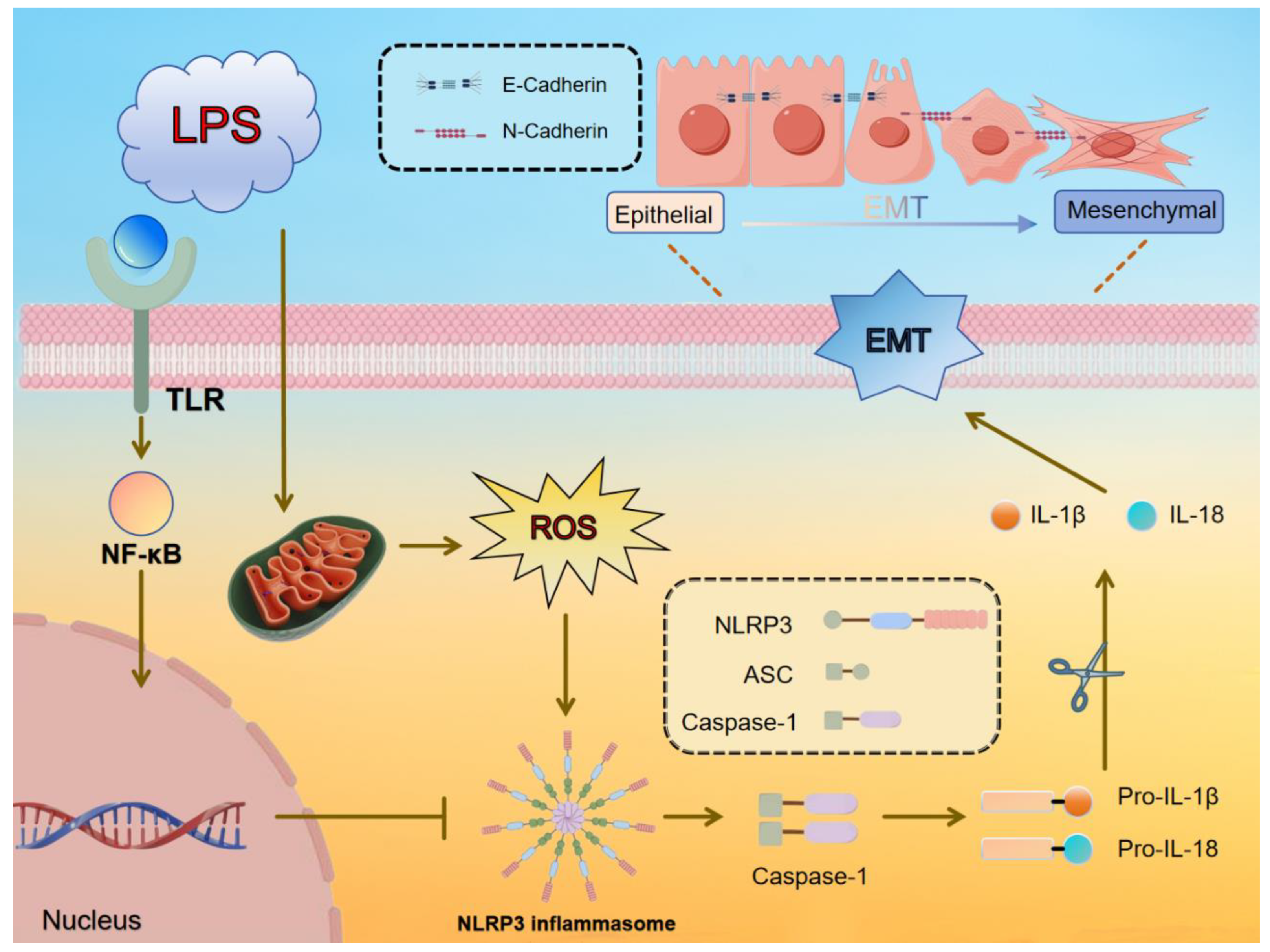
Mastitis in dairy cows, typically caused by bacterial infection, is a common inflammatory condition of the mammary tissue that leads to fibrosis, adversely affecting cow health, milk production, and dairy product quality. Astragalus polysaccharide (APS) has shown effectiveness in alleviating inflammation and fibrosis in various organs. In this study, a lipopolysaccharide (LPS)-induced fibrosis model was established using a bovine mammary epithelial cell line (MAC-T) and Kunming rats. Key parameters, including relative gene mRNA expression, protein levels, and reactive oxygen species (ROS) content, were assessed using RT-qPCR, Western blotting (WB), and DCFH-DA techniques, while histological analysis of breast tissue was performed using H&E and Masson trichrome staining. Oxidative stress markers, such as malondialdehyde (MDA) concentration and enzymatic activities of superoxide dismutase (SOD) and catalase (CAT), were also evaluated in mouse mammary glands. APS treatment modulated fibrosis markers (E-cadherin, N-cadherin, and α-SMA) and inflammation markers (NLRP3, ASC, Caspase-1, and IL-1β) at both mRNA and protein levels, significantly reduced ROS concentrations (P<0.01), restored oxidative stress balance in mice (P<0.05), and mitigated fibrosis and inflammation, as revealed by histological observations. These findings demonstrate that APS can mitigate ROS accumulation, reduce inflammation, and inhibit epithelial-mesenchymal transition (EMT) in vitro and mammary fibrosis in vivo, supporting its potential as an alternative therapeutic agent for mammary fibrosis treatment.

Keywords: 
;  ;  ;  ;  

1. Introduction

Mastitis in dairy cows is an inflammatory condition of the mammary tissue, typically caused by bacterial infection. This disease not only affects the health of the cows but also significantly reduces milk quality and yield, making it one of the most common and critical diseases in dairy farming [1]. Chronic mastitis has been closely linked to fibrosis in mammary tissues [2,3]. Mammary fibrosis occurs when epithelial cells undergo a series of molecular and cellular transformations into mesenchymal-like cells, a process known as epithelial-mesenchymal transition (EMT) [4]. This leads to the production of large amounts of extracellular matrix, changes in cell morphology, and the acquisition of fibroblast-like characteristics, such as the loss of epithelial cell polarity, the transformation from a cobblestone shape to a long spindle shape, and an increase in mesenchymal markers like N-cadherin and α-smooth muscle actin (α-SMA), alongside a decrease in the epithelial marker E-cadherin, which is crucial for cell adhesion and lateral junctions. EMT plays an essential role in both physiological and pathological processes, with the transformation of epithelial cells into fibroblast-like cells being a hallmark mechanism of fibrosis.
Chronic inflammation leads to the production of large amounts of reactive oxygen species (ROS) by immune cells [5], which contribute to the fibrotic process via several pathways [6]. For instance, diseases such as pulmonary fibrosis [7], hepatic fibrosis [8], renal fibrosis [9], and cardiac fibrosis [10] are closely associated with excessive ROS production. ROS act as signaling molecules, and their accumulation activates the NOD-like receptor protein 3 (NLRP3) inflammasome, which binds to ASC, activating caspase-1. This process leads to the maturation of pro-inflammatory cytokines IL-1β and IL-18 [11,12], which play a key role in triggering local and systemic inflammatory responses [13]. Inhibiting ROS production and NLRP3 activation has been shown to effectively attenuate inflammation-induced fibrosis [14].
Astragalus polysaccharide (APS), a key active compound extracted from Astragalus membranaceus, a plant from the legume family, has been used in traditional Chinese medicine (TCM) for thousands of years and is recognized as an important tonic. APS possesses a broad spectrum of bioactivities, particularly in immune regulation, anti-inflammation, antioxidant, and antifibrotic effects. It is widely used in treating cardiovascular diseases, diabetes, cancer, respiratory diseases, and neurological conditions [15,16,17,18]. APS has also demonstrated therapeutic potential in various LPS-induced chronic inflammation-related disorders [19], cellular inflammation models [20], and fibrotic diseases such as hepatic fibrosis [21], myocardial fibrosis [22], and renal fibrosis [23]. In therapeutic applications, APS promotes thymocyte proliferation, reduces drug-induced apoptosis, and improves immune function in immunosuppressed mice by increasing thymic index and immunoglobulin levels while inhibiting IL-2 overproduction [24]. In cancer models, APS has been shown to inhibit mitochondrial apoptosis pathways, upregulate anti-apoptotic proteins (BCL-2, BCL-X2), and downregulate pro-apoptotic proteins (BAX) [25].
This study aims to investigate whether APS can alleviate LPS-induced mammary fibrosis by inhibiting ROS accumulation and modulating NLRP3 activity in both in vitro cellular experiments and in vivo mouse models. Additionally, the study will explore the molecular mechanisms underlying its protective effects on the mammary gland of dairy cows, providing insights and references for the development of new therapeutic strategies and preventive measures for mammary fibrosis.

2. Results

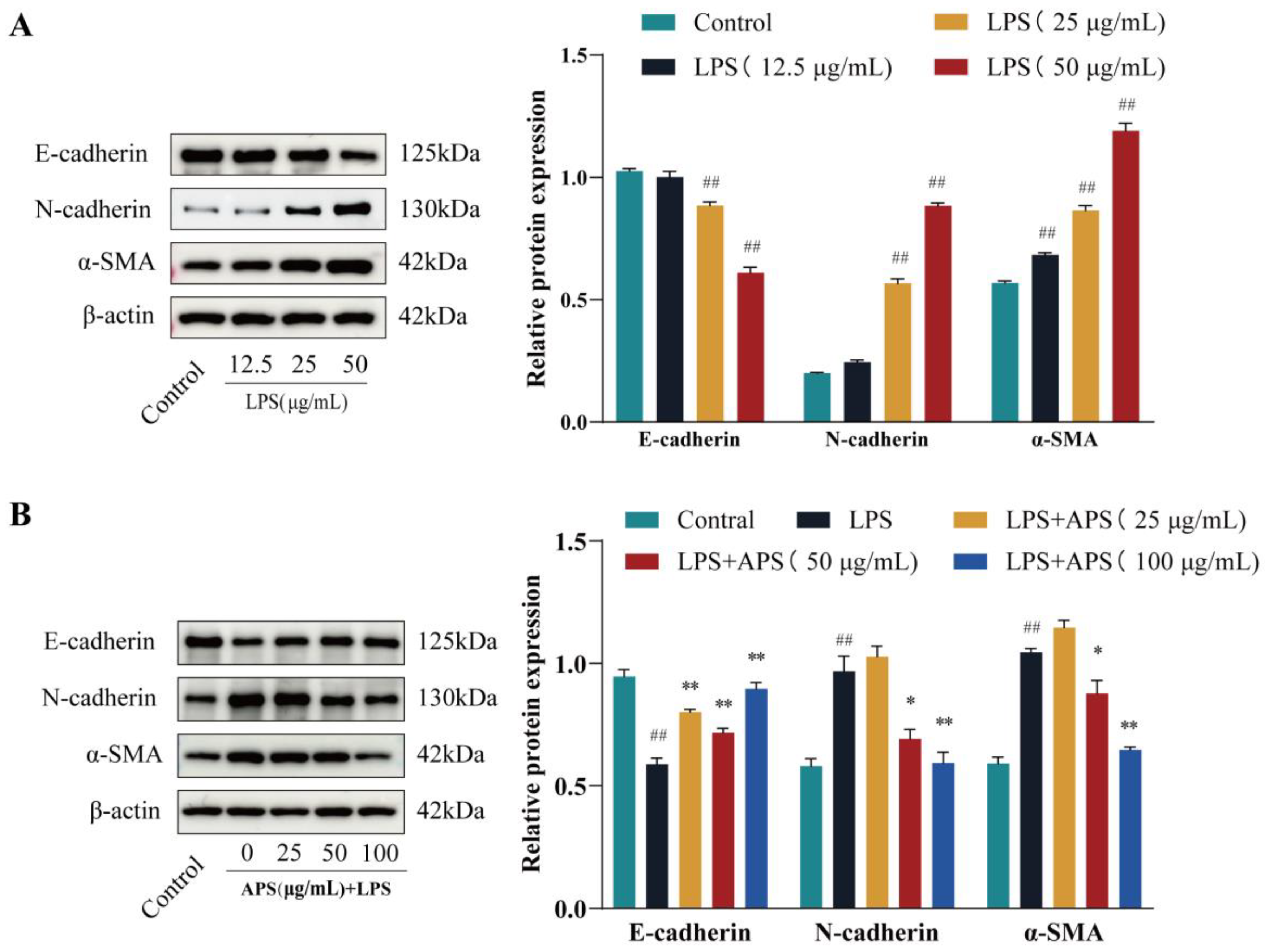
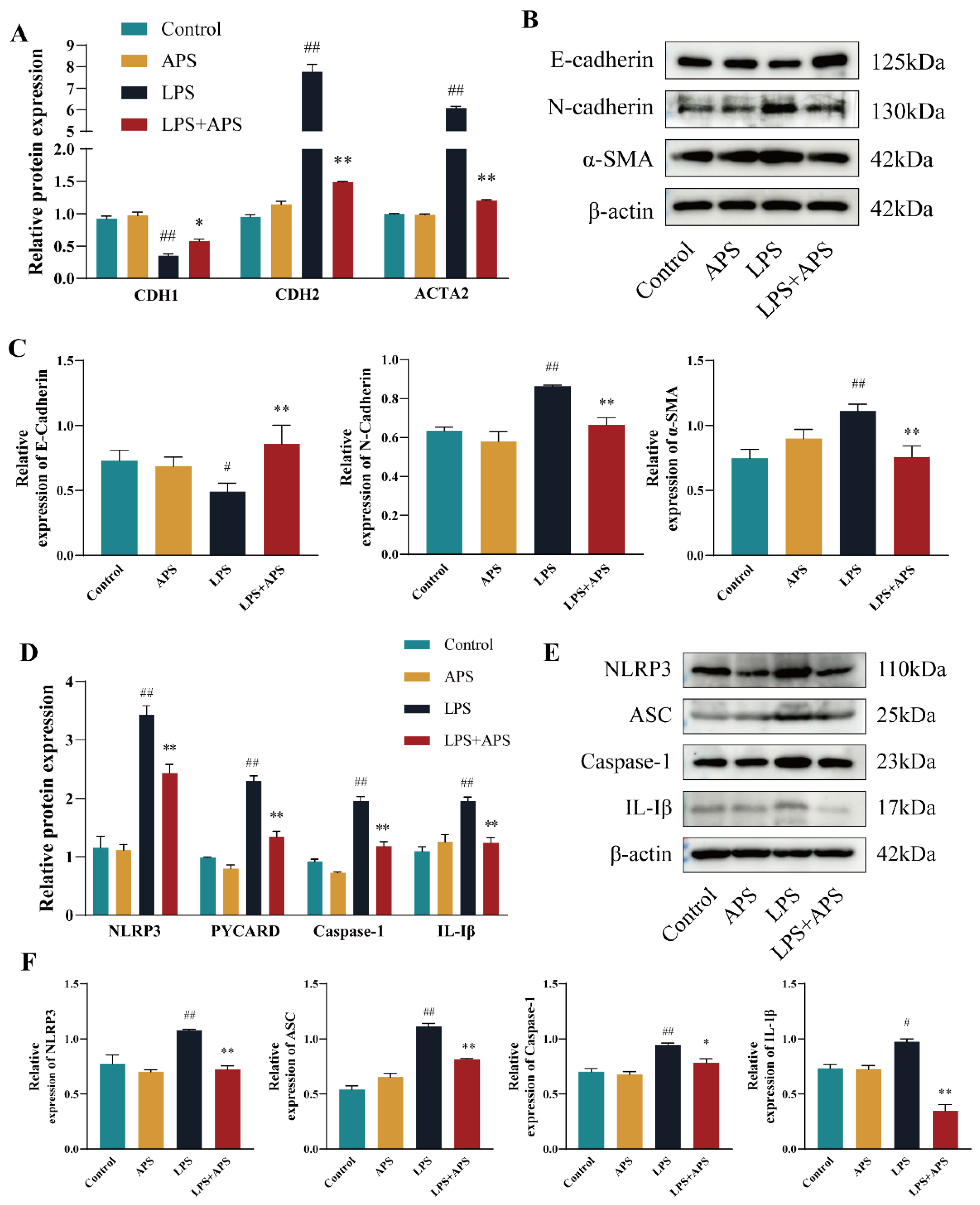
2.1. APS Inhibits LPS-Induced EMT Studies in MAC-T Cells

In this experiment, cells were induced with three LPS concentrations 12.5 µg/mL, 25 µg/mL, and 50 µg/mL and fibrosis marker expression was assessed to determine the optimal concentration for the LPS induced fibrosis model.The results, shown in (Figure 1A), revealed that at a concentration of 50 µg/mL LPS, E-cadherin expression significantly decreased (P<0.01), while α-SMA and N-cadherin expressions significantly increased (P<0.01) compared to the control group. To investigate whether APS could inhibit LPS-induced fibrosis, MAC-T cells were treated with 50 µg/mL LPS in combination with 0, 25, 50, and 100 µg/mL APS, and compared to the control group. The results in (Figure 1B) indicated that APS treatment showed a concentration-dependent effect, with E-cadherin expression increasing and N-cadherin and α-SMA expression decreasing. Notably, the 100 µg/mL APS-treated group exhibited a significant increase in E-cadherin expression (P<0.01) and a significant decrease in N-cadherin and α-SMA expression levels (P<0.01) compared to the control group.

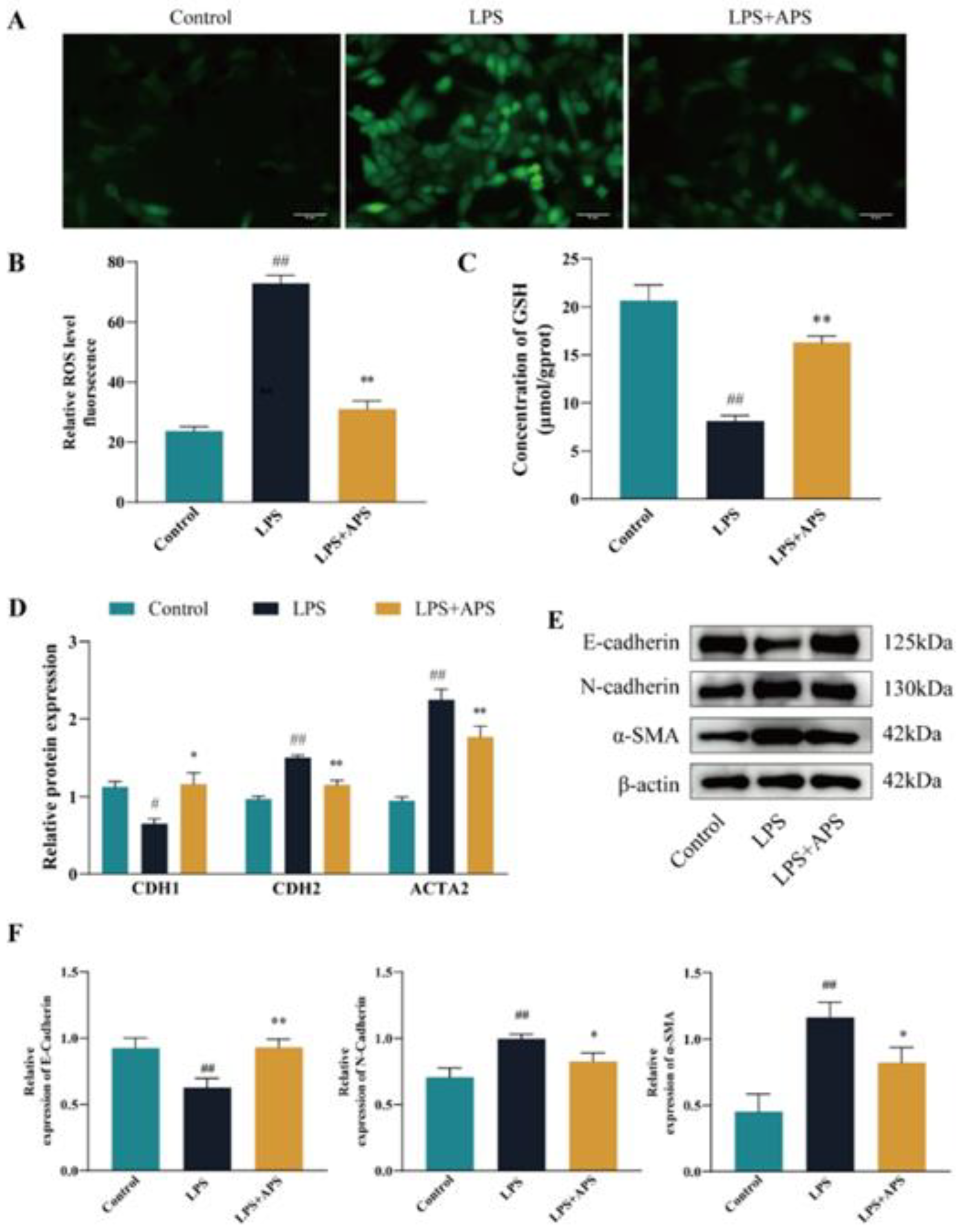
2.2. Inhibition of ROS Production by APS Alleviates LPS-Induced EMT in MAC-T Cells

To evaluate the effect of APS on ROS levels in LPS-stimulated MAC-T cells, ROS fluorescence signals and glutathione (GSH) enzyme activities were assessed. As shown in (Figure 2A-C), the LPS-treated group exhibited strong ROS positivity, while the APS-treated group displayed significantly weaker ROS positivity (P<0.01). Furthermore, the LPS-treated group showed a significant decrease in GSH content (P<0.01), whereas APS treatment significantly increased GSH levels (P<0.01). To investigate whether APS alleviates LPS-induced fibrosis by inhibiting ROS production, the expression of fibrogenic markers at both mRNA and protein levels was analyzed via qPCR and Western blot. As depicted in Figure 2D, mRNA levels of CDH2 and ACTA2 were significantly reduced in the APS-treated group compared to the LPS group (P<0.01), while CDH1 gene expression increased significantly (P<0.05). Protein analysis (Figure 2E-F) revealed that APS treatment significantly increased E-cadherin expression (P<0.01) and significantly decreased N-cadherin and α-SMA expression (P<0.05), consistent with the mRNA expression patterns.

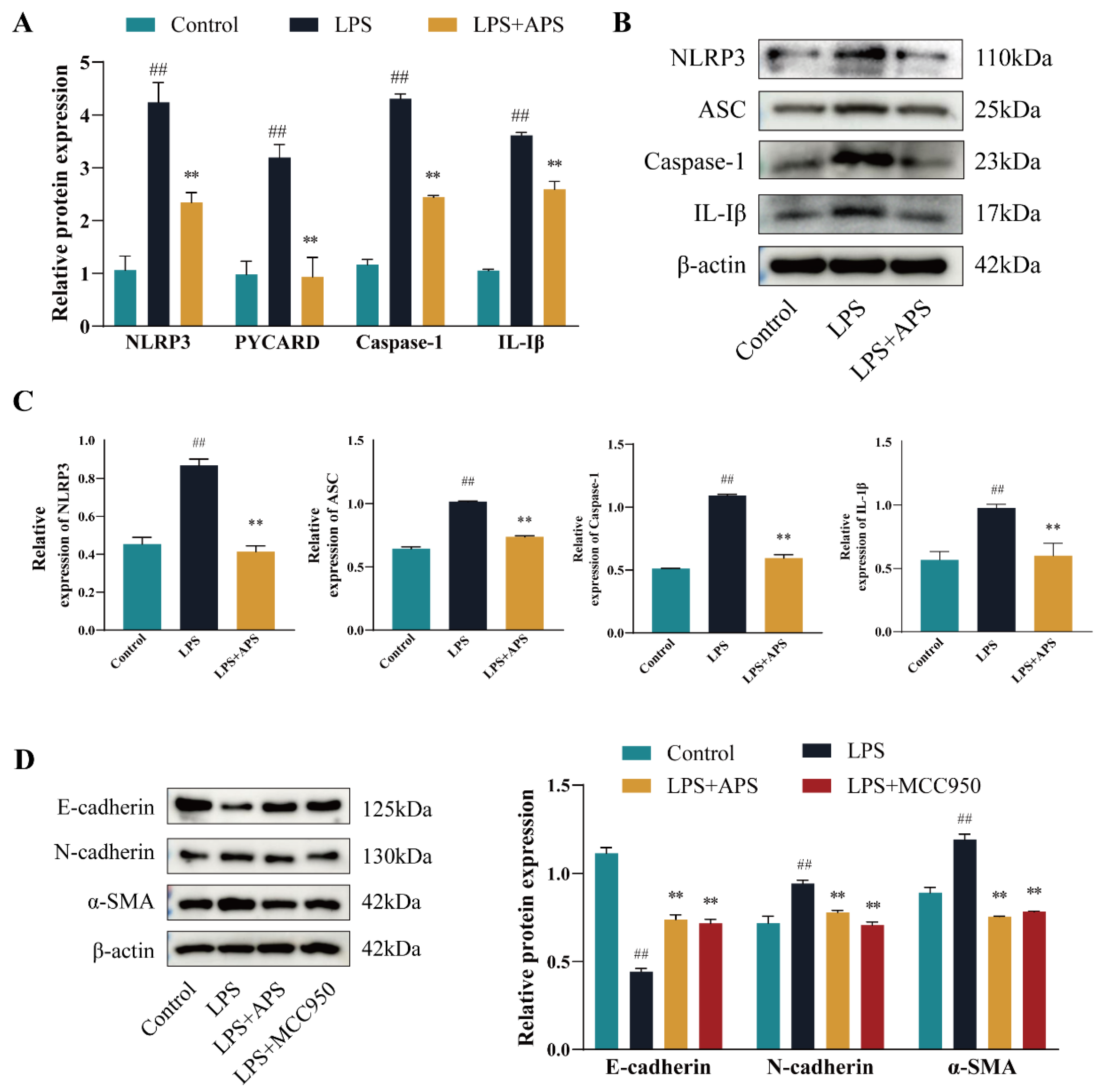
2.3. APS Alleviates EMT Progression in MAC-T Cells by Inhibiting NLRP3 Signaling

Pathway To investigate whether APS reduces ROS to regulate NLRP3 pathway expression and ultimately alleviate cell fibrosis, the researchers evaluated the expression levels of NLRP3, ASC, Caspase-1, and IL-1β across different groups. The results showed that, compared to the control group, the mRNA (Figure 3A) and protein (Figure 3B,C) levels of these factors were significantly elevated in the LPS group (P<0.01). In contrast, the expression levels of these inflammatory markers were significantly lower in the APS group compared to the LPS group (P<0.01). To further assess the critical role of NLRP3 in cell fibrosis, the researchers conducted an analysis using the MCC950 inhibitor. As shown in (Figure 3D), both the APS and MCC950 groups exhibited a highly significant increase in E-cadherin expression (P<0.01), while N-cadherin and α-SMA expression levels were significantly reduced (P<0.01) compared to the LPS group.

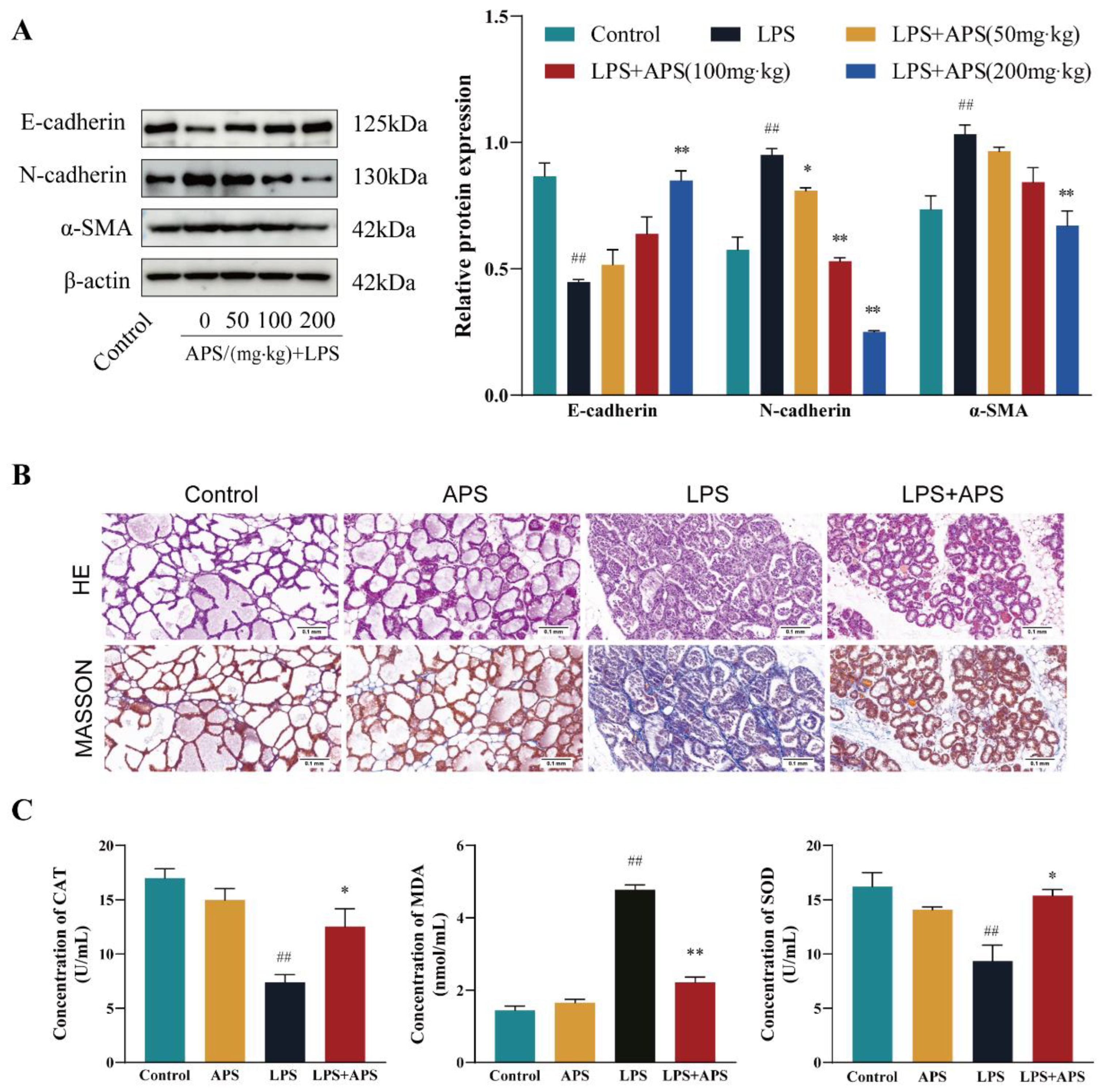
2.4. APS Inhibition of Mammary ROS and EMT Study in Mice

In this study, fibrosis-related factors were assessed across different APS treatment concentrations. As shown in (Figure 4A) , the E-cadherin levels in the LPS group and the APS 200 mg/kg group were significantly different (P<0.01). The expression of N-cadherin and α-SMA also showed a highly significant change (P<0.01) in the APS treatment group. H&E and Masson staining were performed, and the results (Figure 4B) revealed that the mammary gland structure was normal in both the control and APS groups. In contrast, the LPS group exhibited an incomplete basal layer structure of the mammary gland with increased neutrophilic and fibrous components. APS treatment provided a better protective effect against LPS-induced mastitis. Masson staining also demonstrated mild collagen deposition and an EMT trend in the mastitis group, with a significant reduction in the fibrosis rate after APS treatment. As for oxidative stress indicators (Figure 4C), no significant differences were observed in mammary gland CAT, MDA, and SOD levels between the control and APS groups. However, the LPS group showed a significant decrease (P<0.01) in CAT and SOD, along with a significant increase (P<0.01) in MDA compared to the control group. APS treatment led to a significant increase (P<0.05) in CAT and SOD, while MDA levels were significantly decreased (P<0.01).

2.5. APS Attenuated Mouse Mammary Fibrosis Triggered by NLRP3 Activation

APS attenuated LPS-induced mammary fibrosis in mice triggered by NLRP3 activation. In this study, the expression of inflammatory and fibrotic factors at both mRNA and protein levels was assessed using RT-qPCR and WB. The results revealed no significant differences between the control and APS groups. As shown in Figure 5A-D, mRNA expression was highly significantly different between the LPS and control groups (P<0.01). After APS treatment, CDH1 expression was significantly higher (P<0.05), while the mRNA levels of CDH2, ACTA2, and inflammatory factors such as NLRP3, PYCARD, Caspase-1, and IL-1β were significantly reduced (P<0.01). WB analysis (Figure 5B, C, E, F) further confirmed that E-cadherin protein expression was significantly lower (P<0.05) and IL-1β levels were significantly higher (P<0.05) in the LPS group compared to the control group. The remaining factors showed highly significant increases (P<0.01). Following APS treatment, inflammatory and fibrotic factors were largely restored (P<0.01) compared to the LPS group, except for caspase-1 expression, which was significantly reduced (P<0.05).

3. Discussion

Mastitis in dairy cows presents a significant threat not only to the health of the cows but also to the quality of dairy products, economic efficiency, and reproductive capacity [26]. Inflammation and fibrosis are closely linked physiological processes. Research has shown that chronic inflammation, particularly due to prolonged immune system activation, promotes the proliferation of fibroblasts and excessive deposition of matrix components such as collagen, ultimately contributing to fibrosis development [2,27]. E-cadherin, a calcium-dependent transmembrane protein, plays a pivotal role in cell-to-cell adhesion, maintaining epithelial cell polarity and the integrity of the mammary barrier [28]. During epithelial-to-mesenchymal transition (EMT), a reduction in E-cadherin leads to the loss of cell polarity and intercellular adhesion, signifying a loss of epithelial characteristics. The upregulation of N-cadherin promotes cell migration and infiltration, while increased α-SMA expression typically signals the acquisition of mesenchymal traits, making the cells contractile and involved in tissue remodeling and fibrosis [4]. The co-expression of these proteins in epithelial cells often reflects enhanced migratory ability and an increased capacity to secrete extracellular matrix. Lipopolysaccharide (LPS), a key pro-inflammatory factor in Gram-negative bacteria, is a potent activator of innate immunity and can stimulate tissue fibrosis [29,30] In particular, LPS was utilised in the construction of a cow endometrial epithelial cell line (BEND)[31] and a mouse adipose tissue fibrosis model [32]. Thus, LPS was utilized as a virulence factor to establish a fibrosis model in this study. The results showed that the expression of E-cadherin decreased with increasing LPS concentration, while the levels of α-SMA and N-cadherin increased, indicating that LPS induces fibrosis in MAC-T cells.The anti-inflammatory and antifibrotic effects of APS have been confirmed in numerous experimental and clinical studies [13], especially its capacity to inhibit the EMT process in hepatic stellate cells (LX-2) [33] and rat renal tubular epithelial cells (NRK-52E) [34]. In this study, an in vitro LPS-induced fibrosis model was established using MAC-T cells. The results demonstrated that APS treatment significantly reduced the expression of mesenchymal markers N-cadherin and α-SMA, while restoring the expression of the epithelial marker E-cadherin. As both an antioxidant and anti-inflammatory agent, APS not only inhibited LPS-induced ROS production in osteoblasts [35], but also protected chick embryo fibroblasts from autophagic damage by reducing ROS levels [36]. ROS, a powerful oxidizing agent, plays a central role in the initiation and progression of diseases. One of the underlying mechanisms of chronic inflammation is oxidative stress caused by the accumulation of excessive ROS [5]. The buildup of ROS is not only a hallmark of inflammation but also a key factor in tissue and organ fibrosis [6]. Elevated ROS levels are strongly linked to the onset of disease phenotypes [37]. For example, higher ROS levels are closely associated with the fibrosis process, where they promote the activation of hepatic stellate cells, induce their excessive proliferation, increase their ability to secrete extracellular matrix, and ultimately lead to the development of liver fibrosis [38]. Consequently, the timely removal of accumulated ROS can help mitigate fibrosis [39]. In this study, APS inhibited cellular EMT by reducing ROS generation induced by LPS (Figure 6).
In addition it has been shown that ROS levels are significantly elevated when cells are subjected to oxidative stress.The increase in ROS activates and interacts with the NLRP3 receptor either directly or through oxidative modification of other molecules to promote NLRP3 polymerisation to co-assemble with ASC proteins and Caspase-1 to form inflammatory vesicles.The activation of caspase-1 drives the maturation of cytokines such as IL-1β and IL-18 and triggers an inflammatory response and modifies the activation or increases the activity of transcription factors such as Snail[40], Slug[41], Twist[25] etc. These factors promote EMT in a variety of diseases, for example they promote EMT in cancer stem cells (CSC) [42]and inhibit tumour growth.This study utilized MCC950 [43] as a potent and selective NLRP3 inhibitor. The results revealed that the effects of APS and MCC950 on cells were largely consistent; both were capable of reducing the protein expression levels of α-SMA and N-cadherin induced by LPS, while promoting the expression of E-cadherin. These findings align with results observed with MCC950 inhibitors in liver fibrosis [44], myocardial fibrosis [45], and renal fibrosis [46]. This further supports the notion that APS mitigates the activation of the NLRP3 inflammasome through its antioxidant properties, thereby alleviating tissue and organ fibrosis.
To determine whether APS operates via a similar mechanism in an LPS-induced mammary fibrosis animal model, we conducted validation experiments in mice. The results showed that LPS promoted mammary fibrosis, while APS treatment restored the mammary gland structure, which is consistent with findings from diabetic mice with renal fibrosis [34]. This suggests that APS also alleviates mammary fibrosis in mice. Additionally, APS treatment increased E-cadherin expression and decreased the levels of α-SMA and N-cadherin in the mammary glands of LPS-induced mice, indicating that APS reversed mammary EMT. Recent studies have highlighted that inflammation and EMT are key contributors to organ fibrosis. In peritonitis muscularis atrophica mice, APS significantly mitigated EMT in HMrSV5 cells and peritoneal fibrosis [47]. In this study, the expression of ASC, Caspase-1, IL-1β, and NLRP3 was significantly elevated in the LPS-treated group compared to controls, but all were reduced after APS treatment. These results align with findings in rats with allergic rhinitis, suggesting that APS inhibits the activation of NLRP3 inflammasomes and reduces inflammatory markers such as Caspase-1, IL-1β, and ASC in rats. Studies indicate that APS can effectively improve nasal mucosal inflammation in rats by downregulating NLRP3 inflammatory factors [48]. Therefore, our results suggest that APS exerts a similar beneficial effect in animal models.

4. Materials and Methods

4.1. Cell Samples

MAC-T cells from the Lanzhou Veterinary Research Institute, Chinese Academy of Ag ricultural Sciences, Lanzhou, China, were provided by Jian Xi Li and cultivated in DMEM/F12 medium (Gibco, NY, USA) with 10% foetal bovine blood (Invigentech, Irvine, CA, USA). The cells were maintained at 37°C in a 5% CO2 incubator. Following the attainment of the logarithmic growth phase, MAC-T cells were inoculated into six-well plates using 0.25% trypsin-EDTA (Gibco, NY, USA). The cells were then exposed to various concentrations of APS (25, 50 and 100 µg) and LPS (2.5, 25 and 50 µg/ml) for a duration of 24 hours. The source of the APS was Solarbio (Beijing, China), while the LPS was obtained from Sigma (Shanghai, China). In the inhibition studies, MCC950 (a NLRP3 inhibitor from Macklin, Shanghai, China) was utilised at a concentration of 10 µM, and it was incubated for two hours prior to treatment, concurrently with the addition of 50 µg/mL LPS.

4.2. Animal Samples

Ninety 8-week-old female Kunming mice and 45 male Kunming mice were obtained from the Chinese Academy of Agricultural Sciences. The animals were housed under standard conditions (temperature: 20-25°C; humidity: 40–70%) with a 12-hour light/dark cycle. Mice had unrestricted access to water and food throughout the experiment. After a 1-week acclimatization period, the animals were mated for 3 days at a 2:1 male-to-female ratio. Pregnant mice with comparable body conditions were selected, and after 7 days of lactation, five groups of 10 mice each were formed: the control group, LPS group (Solarbio, Beijing, China) (200 µg/mL), and APS (50, 100, and 200 mg/kg) treatment groups. The remaining mice were allocated into four additional groups: control, APS, LPS, and APS+LPS. All mice were injected into the milk ducts of the fourth pair of mammary glands: the control group received 50 µL of saline, the APS group received saline injections and daily APS gavage, the LPS group was injected with 50 µL of 200 µg/mL LPS, and the APS+LPS group received both LPS injections and APS gavage at the corresponding doses. Gavage treatment commenced on the first day of lactation. On day 6, all animals were administered intraperitoneal pentobarbital sodium (150 mg/kg) to induce whole-body anesthesia, followed by euthanasia. One part of the excised mammary glands was immediately frozen at -80°C for analysis, while the other part was fixed in 4% paraformaldehyde for histological examination. All procedures were conducted in accordance with ethical guidelines approved by the Animal Protection Committee of Gansu Agricultural University (GSAU-Eth-LST-2021-003).

4.3. RT-qPCR

Taking MAC-T cells and mouse mammary tissue, and total RNA was extracted using TRIzol reagent (Transgen, Beijing, China). cDNA synthesis was performed following the manufacturer's protocol using the RevertAid First Strand cDNA Synthesis Kit (Thermo Fisher Scientific, Shanghai, China). Quantitative PCR (qPCR) was then conducted to assess the expression of fibrosis-related genes CDH1, CDH2, and ACTA2, as well as the inflammatory markers NLRP3, PYCARD, Caspase-1, and IL-1β. Primers for qPCR were designed and synthesized with Primer Premier 3.0 (Tsingke, Beijing, China) (Table 1). Relative mRNA expression levels were determined using the 2^−ΔΔCt method.

4.4. Western Blot

Western blot (WB) analysis was performed to assess the expression of breast tissue fibrosis markers, including E-cadherin (1:20,000 dilution; Proteintech, Wuhan, China), N-cadherin (1:2,000 dilution; Proteintech, Wuhan, China), and α-smooth muscle actin (α-SMA; 1:1,000 dilution; Proteintech, Wuhan, China), as well as inflammatory mediators such as NLRP3 (1:1,000 dilution; Proteintech, Wuhan, China), ASC (1:1,000 dilution; ABclonal, Wuhan, China), Caspase-1 (1:1,000 dilution; Abmart, Shanghai, China), and IL-1β (1:1,000 dilution; Abmart, Shanghai, China). Total protein was extracted from breast tissues and cells using cold RIPA buffer (Solarbio, Beijing, China).Primary antibodies were incubated overnight at 4°C, followed by incubation with the appropriate secondary antibodies (goat anti-rabbit IgG, 1:10,000 dilution; SAB, Greenfield, MD, USA; goat anti-mouse IgG, 1:10,000 dilution; SAB, Greenfield, MD, USA) for 1 hour at 37°C. Protein bands were visualized using an enhanced chemiluminescence (ECL) detection system (Solarbio, Beijing, China). Band intensities were quantified and analyzed using ImageJ software (version 1.52a) for signal quantification.

4.5. Determination of Mitochondrial ROS Levels

Reactive Oxygen Assay Kit (Solarbio, Beijing, China) was used to detect the total cellular ROS level. Firstly, MAC-T cells were inoculated into 6-well plates and followed up according to the relevant instructions. Subsequently, the cells were incubated with 10 µmol/L DCFH-DA reagent away from light (37°C, 30 min) and gently rinsed with PBS for 3 times. Subsequently, and images were captured using an inverted fluorescence microscope (Revolve Omega, apexbio, Suzhou, China).

4.6. H&E and Masson Staining

Mouse mammary tissue samples were fixed in 4% paraformaldehyde (0.04 g/mL) for 14 days. Following fixation, the tissues were rinsed in running water, subjected to dehydration through a graded alcohol series, cleared in xylene, and embedded in paraffin. Paraffin blocks were sectioned into 5 µm thick slices, which were then stained with Hematoxylin and Eosin (H&E) and Masson’s trichrome stain. Dynamic histological changes in the mouse mammary gland were observed using a Zeiss microscope (Axiocam 208 color, Zeiss, Germany), And image acquisition using a slice scanner.(Dynamax, Shanghai, China).

4.7. Detection of Mammary Gland Oxidative Stress Levels

The level of lipid oxidation was detected using Malondialdehyde (MDA) content assay kit (Solarbio, Beijing, China). Superoxide dismutase (SOD) activity assay kit (Solarbio, Beijing, China) was used to detect H2O2 generating enzyme activity. Catalase (CAT) activity assay kit (Solarbio, Beijing, China) detects H2O2 scavenging enzyme activity.

4.8. Data Processing and Statistical Analysis

Statistical analysis was performed using SPSS version 22.0 (SPSS Inc., Chicago, IL, USA). Data are presented as mean ± standard error of the mean (SEM). One-way analysis of variance (ANOVA) was used to compare differences among multiple groups, with post hoc pairwise comparisons conducted using the least significant difference (LSD) test. Prior to performing ANOVA, the data were assessed for normality to ensure that they conformed to a normal distribution. P<0.05 was considered statistically significant, while P<0.01 was considered highly significant. Graphical analysis was conducted using GraphPad Prism version 9.0 (GraphPad Software Inc., CA, USA).

5. Conclusions

This study demonstrated that APS can alleviate LPS-induced mammary fibrosis in both cells and mice, primarily by inhibiting ROS accumulation. This, in turn, modulates the expression of NLRP3 signaling pathway-related factors, ultimately regulating EMT. Therefore, APS shows promise as a potential alternative therapeutic agent for the treatment of mammary fibrosis.

Author Contributions

Conceptualization, Z.J.; methodology, L.K.; software, Y.T.; validation, D.H.;formal analysis, Z.J.; investigation, Z.J.; resources, Z.Z.; data curation, Z.J.; writing—original draftpreparation, Z.J.; writing—review and editing, Z.Q.; visualization, X.L.; project administration, W.D.;funding acquisition, Z.Y. All authors have read and agreed to the published version of the manuscript.

Funding

This study was supported by the “National Natural Science Foundation of China (NSFC),grant Number U21A20262”, Gansu Provincial Department of Education: Young PhD Support Program(2024QB-064)” , “ Gansu Youth Science and Technology Foundation(24JRRA657)”.

Institutional Review Board Statement

This study involving animals was approved by the Animal Ethics Committee of Gansu Agricultural University (Approval number 2006-398).

Data Availability Statement

The data presented in this study are available upon request from the corresponding author.

Conflicts of Interest

The funders had no participation in the study design, data collection and. analysis, decision to publish or preparation of the manuscript.

References

  1. Magro, G.; Biffani, S.; Minozzi, G.; Ehricht, R.; Monecke, S.; Luini, M.; Piccinini, R. Virulence Genes of S. aureus from Dairy Cow Mastitis and Contagiousness Risk. Toxins 2017, 9. [Google Scholar] [CrossRef]
  2. Miao, Z.; Ding, Y.; Bi, Y.; Chen, M.; Cao, X.; Wang, F. Staphylococcus aureus on the effect of expression of MMPs/TIMPs and uPA system in bovine mammary fibroblasts. Journal of microbiology, immunology, and infection = Wei mian yu gan ran za zhi 2021, 54, 411–419. [Google Scholar] [CrossRef] [PubMed]
  3. Kan, X.; Hu, G.; Liu, Y.; Xu, P.; Huang, Y.; Cai, X.; Guo, W.; Fu, S.; Liu, J. Mammary Fibrosis Tendency and Mitochondrial Adaptability in Dairy Cows with Mastitis. Metabolites 2022, 12. [Google Scholar] [CrossRef]
  4. Sisto, M.; Ribatti, D.; Lisi, S. Organ Fibrosis and Autoimmunity: The Role of Inflammation in TGFβ-Dependent EMT. Biomolecules 2021, 11. [Google Scholar] [CrossRef] [PubMed]
  5. El-Kenawi, A.; Ruffell, B. Inflammation, ROS, and Mutagenesis. Cancer cell 2017, 32, 727–729. [Google Scholar] [CrossRef]
  6. Zhao, Y.; Wang, Z.; Feng, D.; Zhao, H.; Lin, M.; Hu, Y.; Zhang, N.; Lv, L.; Gao, Z.; Zhai, X.; et al. p66Shc Contributes to Liver Fibrosis through the Regulation of Mitochondrial Reactive Oxygen Species. Theranostics 2019, 9, 1510–1522. [Google Scholar] [CrossRef] [PubMed]
  7. Gonzalez-Gonzalez, F.J.; Chandel, N.S.; Jain, M.; Budinger, G.R.S. Reactive oxygen species as signaling molecules in the development of lung fibrosis. Translational research : the journal of laboratory and clinical medicine 2017, 190, 61–68. [Google Scholar] [CrossRef]
  8. Lv, F.; Li, N.; Kong, M.; Wu, J.; Fan, Z.; Miao, D.; Xu, Y.; Ye, Q.; Wang, Y. CDKN2a/p16 Antagonizes Hepatic Stellate Cell Activation and Liver Fibrosis by Modulating ROS Levels. Frontiers in cell and developmental biology 2020, 8, 176. [Google Scholar] [CrossRef]
  9. Xiong, W.; Xiong, Z.; Song, A.; Lei, C.; Ye, C.; Su, H.; Zhang, C. UCP1 alleviates renal interstitial fibrosis progression through oxidative stress pathway mediated by SIRT3 protein stability. Journal of translational medicine 2023, 21, 521. [Google Scholar] [CrossRef]
  10. Li, J.; Ding, H.; Li, Y.; Zhou, H.; Wang, W.; Mei, Y.; Zhang, R. Alarin alleviated cardiac fibrosis via attenuating oxidative stress in heart failure rats. Amino acids 2021, 53, 1079–1089. [Google Scholar] [CrossRef]
  11. Zahid, A.; Li, B.; Kombe, A.J.K.; Jin, T.; Tao, J. Pharmacological Inhibitors of the NLRP3 Inflammasome. Frontiers in immunology 2019, 10, 2538. [Google Scholar] [CrossRef] [PubMed]
  12. Elliott, E.I.; Sutterwala, F.S. Initiation and perpetuation of NLRP3 inflammasome activation and assembly. Immunological reviews 2015, 265, 35–52. [Google Scholar] [CrossRef]
  13. Li, C.X.; Liu, Y.; Zhang, Y.Z.; Li, J.C.; Lai, J. Astragalus polysaccharide: a review of its immunomodulatory effect. Archives of pharmacal research 2022, 45, 367–389. [Google Scholar] [CrossRef]
  14. Liu, W.; Han, X.; Li, Q.; Sun, L.; Wang, J. Iguratimod ameliorates bleomycin-induced pulmonary fibrosis by inhibiting the EMT process and NLRP3 inflammasome activation. Biomedicine & pharmacotherapy = Biomedecine & pharmacotherapie 2022, 153, 113460. [Google Scholar] [CrossRef]
  15. Zhang, L.; Shergis, J.L.; Yang, L.; Zhang, A.L.; Guo, X.; Zhang, L.; Zhou, S.; Zeng, L.; Mao, W.; Xue, C.C. Astragalus membranaceus (Huang Qi) as adjunctive therapy for diabetic kidney disease: An updated systematic review and meta-analysis. Journal of ethnopharmacology 2019, 239, 111921. [Google Scholar] [CrossRef] [PubMed]
  16. Jung, Y.; Jerng, U.; Lee, S. A systematic review of anticancer effects of radix astragali. Chinese journal of integrative medicine 2016, 22, 225–236. [Google Scholar] [CrossRef]
  17. Qin, Q.; Niu, J.; Wang, Z.; Xu, W.; Qiao, Z.; Gu, Y. Astragalus embranaceus extract activates immune response in macrophages via heparanase. Molecules (Basel, Switzerland) 2012, 17, 7232–7240. [Google Scholar] [CrossRef]
  18. Song, Y.; Yang, J.; Bai, W.L.; Ji, W.Y. Antitumor and immunoregulatory effects of astragalus on nasopharyngeal carcinoma in vivo and in vitro. Phytotherapy research : PTR 2011, 25, 909–915. [Google Scholar] [CrossRef]
  19. Wang, X.; Shen, J.; Li, S.; Zhi, L.; Yang, X.; Yao, J. Sulfated Astragalus polysaccharide regulates the inflammatory reaction in LPS-infected broiler chicks. International journal of biological macromolecules 2014, 69, 146–150. [Google Scholar] [CrossRef]
  20. Wang, X.; Li, Y.; Yang, X.; Yao, J. Astragalus polysaccharide reduces inflammatory response by decreasing permeability of LPS-infected Caco2 cells. International journal of biological macromolecules 2013, 61, 347–352. [Google Scholar] [CrossRef] [PubMed]
  21. Sun, X.; Zheng, Y.; Tian, Y.; Xu, Q.; Liu, S.; Li, H.; Cheng, K.; Yuan, J.; Liu, H.; Zhu, P. Astragalus polysaccharide alleviates alcoholic-induced hepatic fibrosis by inhibiting polymerase I and transcript release factor and the TLR4/JNK/NF-κB/MyD88 pathway. Journal of ethnopharmacology 2023, 314, 116662. [Google Scholar] [CrossRef] [PubMed]
  22. Ren, C.; Zhao, X.; Liu, K.; Wang, L.; Chen, Q.; Jiang, H.; Gao, X.; Lv, X.; Zhi, X.; Wu, X.; et al. Research progress of natural medicine Astragalus mongholicus Bunge in treatment of myocardial fibrosis. Journal of ethnopharmacology 2023, 305, 116128. [Google Scholar] [CrossRef] [PubMed]
  23. Xu, C.; Chen, Y.; Liu, Z.; Fu, X. Hedysarum polybotrys polysaccharide attenuates renal inflammatory infiltration and fibrosis in diabetic mice by inhibiting the HMGB1/RAGE/TLR4 pathway. Experimental and therapeutic medicine 2023, 26, 493. [Google Scholar] [CrossRef] [PubMed]
  24. Meng, F.; Xu, P.; Wang, X.; Huang, Y.; Wu, L.; Chen, Y.; Teng, L.; Wang, D. Investigation on the immunomodulatory activities of Sarcodon imbricatus extracts in a cyclophosphamide (CTX)-induced immunosuppressanted mouse model. Saudi pharmaceutical journal : SPJ : the official publication of the Saudi Pharmaceutical Society 2017, 25, 460–463. [Google Scholar] [CrossRef]
  25. Zhou, X.; Liu, Z.; Long, T.; Zhou, L.; Bao, Y. Immunomodulatory effects of herbal formula of astragalus polysaccharide (APS) and polysaccharopeptide (PSP) in mice with lung cancer. International journal of biological macromolecules 2018, 106, 596–601. [Google Scholar] [CrossRef]
  26. Pakrashi, A.; Ryan, C.; Guéret, C.; Berry, D.P.; Corcoran, M.; Keane, M.T.; Mac Namee, B. Early detection of subclinical mastitis in lactating dairy cows using cow-level features. Journal of dairy science 2023, 106, 4978–4990. [Google Scholar] [CrossRef]
  27. Ng, B.; Dong, J.; Viswanathan, S.; Widjaja, A.A.; Paleja, B.S.; Adami, E.; Ko, N.S.J.; Wang, M.; Lim, S.; Tan, J.; et al. Fibroblast-specific IL11 signaling drives chronic inflammation in murine fibrotic lung disease. FASEB journal : official publication of the Federation of American Societies for Experimental Biology 2020, 34, 11802–11815. [Google Scholar] [CrossRef]
  28. Corso, G.; Figueiredo, J.; De Angelis, S.P.; Corso, F.; Girardi, A.; Pereira, J.; Seruca, R.; Bonanni, B.; Carneiro, P.; Pravettoni, G.; et al. E-cadherin deregulation in breast cancer. Journal of cellular and molecular medicine 2020, 24, 5930–5936. [Google Scholar] [CrossRef]
  29. Gao, F.; Shen, J.; Zhao, L.; Hao, Q.; Yang, Y. Curcumin Alleviates Lipopolysaccharide (LPS)-Activated Neuroinflammation via Modulation of miR-199b-5p/IκB Kinase β (IKKβ)/Nuclear Factor Kappa B (NF-κB) Pathway in Microglia. Medical science monitor : international medical journal of experimental and clinical research 2019, 25, 9801–9810. [Google Scholar] [CrossRef]
  30. Matsuwaki, T.; Shionoya, K.; Ihnatko, R.; Eskilsson, A.; Kakuta, S.; Dufour, S.; Schwaninger, M.; Waisman, A.; Müller, W.; Pinteaux, E.; et al. Involvement of interleukin-1 type 1 receptors in lipopolysaccharide-induced sickness responses. Brain, behavior, and immunity 2017, 66, 165–176. [Google Scholar] [CrossRef]
  31. Song, L.; Wang, L.; Li, X.; Xiao, L. Ginsenoside Rg1 Alleviates Lipopolysaccharide-Induced Fibrosis of Endometrial Epithelial Cells in Dairy Cows by Inhibiting Reactive Oxygen Species-Activated NLRP3. Animals : an open access journal from MDPI 2023, 13. [Google Scholar] [CrossRef]
  32. Yang, Y.; Zhang, Y.; Zhou, X.; Chen, D.; Ouyang, G.; Liu, Y.; Cui, D. Periostin deficiency attenuates lipopolysaccharide- and obesity-induced adipose tissue fibrosis. FEBS letters 2021, 595, 2099–2112. [Google Scholar] [CrossRef] [PubMed]
  33. Zheng, J.; Ma, L.T.; Ren, Q.Y.; Li, L.; Zhang, Y.; Shi, H.J.; Liu, Y.; Li, C.H.; Dou, Y.Q.; Li, S.D.; et al. The influence of astragalus polysaccharide and β-elemene on LX-2 cell growth, apoptosis and activation. BMC gastroenterology 2014, 14, 224. [Google Scholar] [CrossRef] [PubMed]
  34. Chen, Z.; Liang, H.; Yan, X.; Liang, Q.; Bai, Z.; Xie, T.; Dai, J.; Zhao, X.; Xiao, Y. Astragalus polysaccharide promotes autophagy and alleviates diabetic nephropathy by targeting the lncRNA Gm41268/PRLR pathway. Renal failure 2023, 45, 2284211. [Google Scholar] [CrossRef]
  35. Yang, J.; Qin, L.; Huang, J.; Li, Y.; Xu, S.; Wang, H.; Zhu, S.; Wang, J.; Zhu, B.; Li, F.; et al. Astragalus polysaccharide attenuates LPS-related inflammatory osteolysis by suppressing osteoclastogenesis by reducing the MAPK signalling pathway. Journal of cellular and molecular medicine 2021, 25, 6800–6814. [Google Scholar] [CrossRef]
  36. Shen, X.; Tang, Z.; Bai, Y.; Wan, M.; Yu, M.; Chen, J.; Li, G.; Zhang, R.; Ge, M. Astragalus Polysaccharide Protects Against Cadmium-Induced Autophagy Injury Through Reactive Oxygen Species (ROS) Pathway in Chicken Embryo Fibroblast. Biological trace element research 2022, 200, 318–329. [Google Scholar] [CrossRef]
  37. Zorov, D.B.; Juhaszova, M.; Sollott, S.J. Mitochondrial reactive oxygen species (ROS) and ROS-induced ROS release. Physiological reviews 2014, 94, 909–950. [Google Scholar] [CrossRef]
  38. Gandhi, C.R. Oxidative Stress and Hepatic Stellate Cells: A PARADOXICAL RELATIONSHIP. Trends in cell & molecular biology 2012, 7, 1–10. [Google Scholar]
  39. Qiu, Y.N.; Wang, G.H.; Zhou, F.; Hao, J.J.; Tian, L.; Guan, L.F.; Geng, X.K.; Ding, Y.C.; Wu, H.W.; Zhang, K.Z. PM2.5 induces liver fibrosis via triggering ROS-mediated mitophagy. Ecotoxicology and environmental safety 2019, 167, 178–187. [Google Scholar] [CrossRef]
  40. Iqbal, S.; Jabeen, F.; Aslam, N.; Manan, M. Anti-EMT properties of ergothioneine attenuate lipopolysaccharide-induced oxidative stress-mediated acute lung injury via modulating TGF-β/smad/snail signaling pathway. Human & experimental toxicology 2023, 42, 9603271231178015. [Google Scholar] [CrossRef]
  41. Steinbichler, T.B.; Dudas, J.; Ingruber, J.; Glueckert, R.; Sprung, S.; Fleischer, F.; Cidlinsky, N.; Dejaco, D.; Kofler, B.; Giotakis, A.I.; et al. Slug Is A Surrogate Marker of Epithelial to Mesenchymal Transition (EMT) in Head and Neck Cancer. Journal of clinical medicine 2020, 9. [Google Scholar] [CrossRef] [PubMed]
  42. Kim, M.S.; Lee, H.S.; Kim, Y.J.; Lee, D.Y.; Kang, S.G.; Jin, W. MEST induces Twist-1-mediated EMT through STAT3 activation in breast cancers. Cell death and differentiation 2019, 26, 2594–2606. [Google Scholar] [CrossRef] [PubMed]
  43. Corcoran, S.E.; Halai, R.; Cooper, M.A. Pharmacological Inhibition of the Nod-Like Receptor Family Pyrin Domain Containing 3 Inflammasome with MCC950. Pharmacological reviews 2021, 73, 968–1000. [Google Scholar] [CrossRef] [PubMed]
  44. Qu, J.; Yuan, Z.; Wang, G.; Wang, X.; Li, K. The selective NLRP3 inflammasome inhibitor MCC950 alleviates cholestatic liver injury and fibrosis in mice. International immunopharmacology 2019, 70, 147–155. [Google Scholar] [CrossRef]
  45. Gao, R.; Shi, H.; Chang, S.; Gao, Y.; Li, X.; Lv, C.; Yang, H.; Xiang, H.; Yang, J.; Xu, L.; et al. The selective NLRP3-inflammasome inhibitor MCC950 reduces myocardial fibrosis and improves cardiac remodeling in a mouse model of myocardial infarction. International immunopharmacology 2019, 74, 105575. [Google Scholar] [CrossRef]
  46. Ludwig-Portugall, I.; Bartok, E.; Dhana, E.; Evers, B.D.; Primiano, M.J.; Hall, J.P.; Franklin, B.S.; Knolle, P.A.; Hornung, V.; Hartmann, G.; et al. An NLRP3-specific inflammasome inhibitor attenuates crystal-induced kidney fibrosis in mice. Kidney international 2016, 90, 525–539. [Google Scholar] [CrossRef]
  47. Sheng, L.; Sun, J.; Huang, L.; Yu, M.; Meng, X.; Shan, Y.; Dai, H.; Wang, F.; Shi, J.; Sheng, M. Astragalus membranaceus and its monomers treat peritoneal fibrosis and related muscle atrophy through the AR/TGF-β1 pathway. Frontiers in pharmacology 2024, 15, 1418485. [Google Scholar] [CrossRef]
  48. Xu, J.; Zhang, Q.; Li, Z.; Gao, Y.; Pang, Z.; Wu, Y.; Li, G.; Lu, D.; Zhang, L.; Li, D. Astragalus Polysaccharides Attenuate Ovalbumin-Induced Allergic Rhinitis in Rats by Inhibiting NLRP3 Inflammasome Activation and NOD2-Mediated NF-κB Activation. Journal of medicinal food 2021, 24, 1–9. [Google Scholar] [CrossRef]
Figure 1. APS inhibits LPS-induced EMT progression in MAC-T cells. WB Detection of EMT-associated proteins N-cadherin, E-cadherin, and α-SMA, respectively (n=3) (A) MAC-T cells were treated with LPS (12.5, 25, and 50µg/mL) for 24 hours. (B) MAC-T cells were treated with LPS at a concentration of 50µg/mL and different concentrations of APS (25, 50 and 100µg/mL) for 24 hours. β-actin was used as loading control. Values are expressed using mean ± SEM; ##P < 0.01 vs Control group, **P < 0.01, *P < 0.05 vs LPS group.
Preprints 148488 g001
Figure 2. APS alleviates LPS-induced EMT in MAC-T cells by inhibiting ROS production. mac-t cells were treated with 50µg/mL LPS and 100µg/mL APS. cells were treated for 24 hours. (A, B) DCFH-DA assay for total reactive oxygen species content (scale bar = 50µM). (C) GSH levels (n=6). E-cadherin, α-SMA and N-cadherin mRNAs and proteins associated with EMT were detected by qPCR (D) with WB (EF) (n=3), and β-actin in the control group. mean ± SEM was chosen to represent the values, ##P<0.01, #P<0.05 compared with Control group, **P<0.01, *P< 0.05 compared with LPS group.
Preprints 148488 g002
Figure 3. APS alleviates EMT progression in MAC-T cells by inhibiting NLRP3 signaling. Expression of NLRP3, ASC, Caspase-1 and IL-1β was detected by qPCR (A) with WB (B, C) (n=3). mac-t cells were treated with 100µg/mL APS, 10µM MCC950 and 50µg/mL LPS for 24 h(D), WB assay was performed to detect EMT-associated proteins E-cadherin, N-cadherin, and α-SMA (n=3) .Control group was β-actin. Mean ±SEM was chosen to express the values, ##P<0.01 versus Control group, **P<0.01 versus LPS group.
Preprints 148488 g003
Figure 4. APS inhibits mammary ROS and EMT progression in mice. (A) Gavage using different concentrations of APS (50, 100, and 200 mg/kg) after treatment with LPS (200 µg/mL). WB assay for EMT-associated proteins E-cadherin, N-cadherin, and α-SMA (n = 3), and β-actin for the control group. Enrolled mice were divided into a control group (no drug administered), an APS group ( 200 mg/kg), model group (200 µg/mL LPS) and APS-treated group (200 mg/kg APS+LPS). (B) Breast tissue stained by H&E and Masson. (C) CAT, MDA, and SOD contents in breast tissues. The mean ± SEM was chosen to represent the values, ##P < 0.01 to compare with Control group, **P < 0.01, *P < 0.05 to compare with LPS group.
Preprints 148488 g004
Figure 5. APS attenuated mammary fibrosis in mice triggered by NLRP3 activation. Expression of E-cadherin, α-SMA and N-cadherin associated with EMT and with the inflammatory factors NLRP3, ASC, Caspase-1 and IL-1β was detected by qPCR (AD) with WB (BCEF) (n=3), and β-actin in the Control group. Mean± SEM was chosen to express the values, ##P<0.01, ## P<0.05 vs Control group, **P<0.01, *P<0.05 vs LPS group.
Preprints 148488 g005
Figure 6. Pathway map of LPS-induced ROS/NLRP3 regulation of EMT in mammary epithelial cells.
Preprints 148488 g006
Table 1. Primer information.
Gene Name Species Primer Sequence Product Length Tm/℃
GAPDH Cow FGGAGCGAGACCCCACTAACAT 247 61
RTAAGGGGGCTAAGCAGTTGGT
Mouse FGGCTGTATTCCCCTCCATCG 154 60
RCCAGTTGGTAACAATGCCATGT
CDH1 Cow FCAACAAGGAAACAGGCGTCA 175 59
RTGGGTTGAATCTGGGAGCAT
Mouse FGGCACTCTTCTCCTGGTCCTG 110 61
R: AAGATGGTGATGATATGAGGCTGTG
CDH2 Cow FCAGTGTGATTCCAACGGGGA 146 60
RTCCCGGCGTTTCATCCATAC
Mouse FACAGCCCCTTCTCAATGTGA 231 59
RTCAGGTAGGGCTGGTTTGAG
ACTA2 Cow FACCATCGGGAATGAGCGTTT 97 60
RTGTTGTACGTGGTCTCGTGG
Mouse FGCATGCAGAAGGAGATCACG 157 59
RTCGTCGTACTCCTGTTTGCT
NLRP3 Cow FCAACGGGGAAGAGAAGGCAT 297 60
RTTGAGGTTCACGCTCTCACC
Mouse FGGCCAAAGAGGAATCGGACA 483 60
RCTACGGCCGTCTACGTCTTC
PYCARD Cow FTGAGCAAGGGCCCTAGAAAC 137 60
RATCCAGAACCCCATCCACGA
Mouse FGTGAGCTCCAAGCCATACGA 124 60
RTGACAGTGCAACTGCGAGAA
Caspase-1 Cow FACAGCTATGGATAGAGCCCGA 135 60
RACTTTCTGAAGTGAGCCCCAG
Mouse FTCCTTGTTTCTCTCCACGGC 124 60
RCGAGGGTTGGAGCTCAAGTT
IL-1β Cow FTCCGACGAGTTTCTGTGTGA 206 59
RATACCCAAGGCCACAGGAAT
Mouse FGGAGCCTGTAGTGCAGTTGT 208 60
RAGCTTCAGGCAGGCAGTATC
Disclaimer/Publisher’s Note: The statements, opinions and data contained in all publications are solely those of the individual author(s) and contributor(s) and not of MDPI and/or the editor(s). MDPI and/or the editor(s) disclaim responsibility for any injury to people or property resulting from any ideas, methods, instructions or products referred to in the content.
Copyright: This open access article is published under a Creative Commons CC BY 4.0 license, which permit the free download, distribution, and reuse, provided that the author and preprint are cited in any reuse.
Prerpints.org logo

Preprints.org is a free preprint server supported by MDPI in Basel, Switzerland.

Subscribe

Disclaimer

Terms of Use

Privacy Policy

Privacy Settings

© 2025 MDPI (Basel, Switzerland) unless otherwise stated