
Submitted:
23 August 2024
Posted:
27 August 2024
You are already at the latest version
Abstract
Inflammation and immune cell dysfunction are critical facilitators of endometriosis pathophysiology. The diverse cellular fractions in the endometriotic microenvironment are represented mainly by glands and stroma, which are infiltrated by immune cells, including macrophages. Macrophages are renowned for stimulating lesion growth, vascularization, and innervation and contributing to pain in the disorder. By combining macrophages and endometriotic cells, we determined if resveratrol and its natural analogs can target the immune dysregulation and oxidative imbalance in endometriosis. We studied the effect of analyzed compounds on the expression of key inflammatory and oxidative stress markers, cytokines release, and ROS production by applying q-PCR, ELISA, Cytometric Beads Array, and multiplexed fluorogenic staining and flow cytometry analysis with bioimaging. The results showed that endometriosis-related macrophages treated with resveratrol or its derivatives have impaired expression of pro-inflammatory markers such as IL6, IL8, IL1B, TNF, CCL2, CXCL10, and PTGS2. This study also reported a pronounced antioxidant effect of the compounds by reducing the accumulation of free radicals in macrophages, which correlated with the transcript activation of detoxification enzymes (SOD, GPx). Considering the harmful macrophage action in endometriosis, we described the prospects for potential therapy based on regulating the immune microenvironment and reducing the accumulation of free radicals.
Keywords:
endometriosis
; inflammation
; macrophages
; co-culture
; transwell system
; oxidative stress
; resveratrol
1. Introduction
Endometrial macrophages play key roles in mediating the menstrual cycle through the initiation of the breakdown of endometrium during menstruation [1] or contribution to tissue remodeling during the establishment of pregnancy [2]. Macrophages exhibit extensive plasticity, and their phenotype and function are highly specific to the local tissue milieu. Dysregulation of their activity occurs in abnormalities and pathologies of the uterus including endometriosis. It is evident that macrophages are abundant in endometrial lesions and are critical for their establishment and growth. Their enhanced ability to produce pro-inflammatory cytokines contributes to a microenvironment that favors pelvic inflammation and promotes other immune cells’ recruitment, endometriotic cells’ proliferation, angiogenesis, and innervation as well as leads to the generation of endometriosis-associated pain [3,4]. Therefore, the reciprocal communication between macrophages and endometriotic cells appears, and their intercellular dialogue sustains the formation of endometriosis lesions. As macrophages are the predominant cellular component in endometriotic tissue, the targeting of macrophage alternation could be critical in developing potential therapeutic candidates.
The pathological processes associated with endometriosis like altered inflammatory microenvironment and oxidative stress, can be targets of natural bioactive compounds [5]. Numerous studies suggest that resveratrol (Res) demonstrated an immunomodulatory role in immunologic disorders, like cancers, autoimmune, neurodegenerative, metabolic, cardiovascular, and infectious diseases [6,7]. Regarding the immune cells, it has been found that resveratrol affects the anti-inflammatory profile in macrophages, exhibit inhibitory function on T cells, reduces the suppressive function of CD4+CD25+ regulatory T cells, and stimulates the killing activity of NK. Studies on macrophages revealed that the resveratrol regulates TLR-mediated inflammatory responses, NF-κB activation and COX-2 expression and attenuates TLR4-TRAF6, mitogen-activated protein kinase (MAPK), and AKT pathways in LPS-induced macrophages. Resveratrol reduced the production of granulocyte-macrophage colony-stimulating factor (GM-CSF), decreased LPS-induced pro-oxidant effect and modulated the immune response linked to prostaglandin E2 (PGE2) and Sirtuin 1 (Sirt1) level [6,8]. In searching for potential health-beneficial agents with increased bioavailability, it is necessary to investigate the biological mechanisms for which resveratrol analogs and metabolites can reach the target tissues and exert biological activity. Experiments utilizing analogs demonstrated that hydroxylation of resveratrol to piceatannol (3,5,3′,4′-tetrahydroxy-trans-stilbene) attributed the antioxidant and growth-inhibitory effects of the compound [9].Whereas the structure of pterostilbene (3,5-dimethoxy-4′-hydroxystilbene) with two extra methoxy groups showed more lipophilicity than resveratrol and thus possesses higher intestinal permeability and cellular uptake and enhanced stability [10]. Polydatin (3,4′,5-trihydroxystilbene-3-β-D-glucoside) as a glycoside form of resveratrol resulted in different biological characteristics like greater bioavailability and anti-inflammatory and antioxidative properties [11,12]. Since endometriosis is an inflammatory pathology, the study aims to investigate the comparative anti-inflammatory effects of resveratrol, pterostilbene, piceatannol and polydatin on endometriosis. It is suggested that macrophage influence on endometriotic cells may be fundamentally important for endometriosis development; however, the cell interactions have not been fully understood. Therefore, in this study, we explored the effect of stilbenes in the co-culture model of macrophages and endometriotic cells. We investigated the effects of compounds on the expression of key inflammatory and oxidative stress markers, production of cytokines, and radical scavenging activity. Whether the co-culture system could mediate the inflammatory niche in endometriosis was also addressed and discussed.
2. Materials and Methods
2.1. Chemicals
Unless stated otherwise, all chemicals were purchased from Merck KGaA (Darmstadt, Germany).
2.2. Cell Culture
The immortalized human endometriotic epithelial cells (12Z) were obtained from Applied Biological Materials Inc. (Richmond, Canada) and maintained at 37°C in a containing 5% CO2 and cultured in Dulbecco’s Modified Eagle Medium/Nutrient Mixture F-12 (DMEM/F12) with 10% fetal bovine serum (FBS) and 50 mg/L gentamycin. The human monocyte THP-1 cells (ATCC TIB-202) were purchased from American Type Culture Collection (ATCC, Manassas, VA, USA) and cultured in RPMI 1640 (Invitrogen, Carlsbad, CA, USA) containing FBS 10%, 50 mg/L gentamycin and 2-mercaptoethanol (0.05 mM). THP-1 cells were kept at a minimum density of 2 × 10⁵ cells/ml and were subcultured when reaching 8 × 10⁵ cells/ml. They were passaged every 48 h by adding a fresh medium or complete medium replacement, until they reached the above-mentioned maximum density. Both cell lines were tested regularly for mycoplasma contamination.
2.3. Macrophages Differentiation and Co-Culture Setup
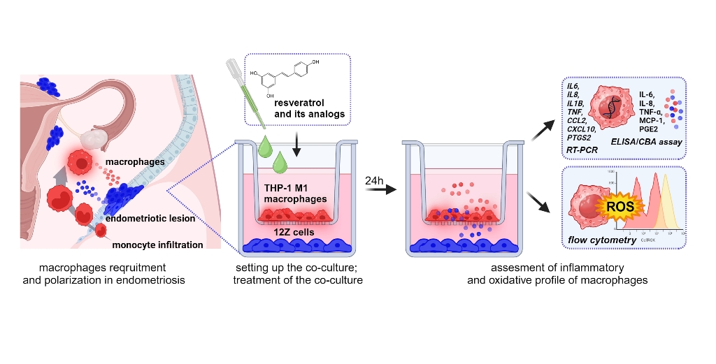
Monocytes were stimulated by 24 h incubation with 10 ng/ml with phorbol-12 myristate 13-acetate (PMA) followed by washing with RPMI 1640 medium to eliminate the effect of PMA as described by Smith et al. [13]. Morphological changes, such as an increase in cell size, formation of pseudopodia, and adhesion, were observed to assess the acquisition of a macrophage-like phenotype. The co-culture system was performed using a 6-well hanging cell culture inserts (0.4-µm porous; Millicell, Billerica, MA, USA). First, the THP-1 monocytes were seeded (2×10⁵ cells/cm2) into the upper chamber of the transwell system in 2 ml of RPMI 1640 medium and 10% of FBS and placed into 6- well plate containing the 3 ml of medium. Once seeded THP-1 cells were treated immediately with 10 ng/ml of PMA for 24 h to stimulate differentiation. A new 6-well plate was prepared the same day with 12Z cells, which were seeded at 0.3×10⁵ cells/cm2 in DMEM/F12 media with 10% FBS. The THP-1 cells placed into the insert and activated with PMA as well as 12Z on the 6-well plate were incubated separately for 24 h to allow for attachment. The next day, differentiated THP-1 cells on the inserts were washed with the medium. Macrophages were polarized in M1 macrophages by incubation with 50 ng/ml of IFN-γ (PeproTech, USA) and 10 pg/ml of LPS (PeproTech, USA) in RPMI 1640 medium and 10% of FBS according to the method described by Smith et al. and Genin et al. [13,14]. Simultaneously, medium on the 6-well plate with 12Z was changed to RPMI 1640 and 2% of FBS. After 24 h the chambers containing the THP-1-derived macrophages were directly placed on top of the 6-well plates with the 12Z cells, and the resulting co-culture system cells. Macrophages and 12Z cells were co-cultured in RPMI 1640 supplemented with 2% of FBS treated with resveratrol and its derivatives for 24 h. Figure 1 provides a schematic representation of the experimental design consisting of macrophage differentiation, macrophage polarization, co-culture setup, treatment with analyzed compounds and further analysis.
2.4. MTT Assay
THP-1 cells were grown in 24-well plates at an initial density of 1×10⁵/cm2 and stimulated by 24-hour incubation with 10 ng/ml PMA. Cultures were then treated with resveratrol at concentrations ranging from 1 µM to 50 µM and incubated for 24 h under standard culture conditions in RPMI 1640 medium and 2% of FBS. 12Z cells were grown in 24-well plates at an initial density of 0.25 ×10⁵/cm2 and were treated with resveratrol at the same range concentrations under standard culture conditions in RPMI 1640 medium and 2% of FBS for 24 h. We applied the same culture conditions for both cell lines as for the co-cultures, such as the same cell media, concentration of FBS in the media and time of incubation. Cell viability and metabolic activity in both cultures were determined using the 3-(4,5-dimethylthiazol-2-yl)-2,5-diphenyltetrazolium bromide (MTT) colorimetric assay according to the protocol described in previous studies [15]. The absorbance was measured using a Tecan M200 Infinite microplate reader (Tecan Group Ltd., Männedorf, Switzerland).
2.5. RNA Extraction and Real-Time PCR
Total RNA from the THP-1-derived macrophages co-cultured with 12Z was extracted using Trizol reagent (Invitrogen, USA) according to the previously described protocol [16]. Complementary deoxyribonucleic acids (cDNAs) were synthesized using a cDNA Transcriptor First-Strand kit (Roche Diagnostics GmbH, Mannheim, Germany). Quantitative real-time polymerase chain reaction (qRT-PCR) reactions were carried out using SYBR® Select Master Mix (Life Technologies, Carlsbad, CA, USA). The relative expression was calculated using the 2−ΔΔCt method, where Ct represents the threshold cycle. GAPDH was used as the internal control. The primers used for the amplification of cDNAs are listed in Table 1.
2.6. Quantitation of Cytokine Levels by Cytometric Bead Array
The conditioned media (CM) from macrophages and 12Z cells co-culture were collected 24 h after treatment with compounds, centrifuged to remove cellular debris, and applied for subsequent experiment or stored at −80°C until use. The concentrations of Interleukin-6 (IL-6) and Interleukin 8 (IL-8) in the conditioned media were quantitatively determined immediately with the CBA Human Inflammatory Cytokine Cytometric Bead Array (BD Biosciences, San Jose, CA, USA), as previously described [17]. A standard calibration curve was established for each kit with a maximum and minimum detection limit (1.0-5000 pg/mL). Samples were analyzed using a FACSAriaII flow cytometer (BD Biosciences) and FACSDiva v6.1.2 software (BD Biosciences).
2.7. Quantitation of Cytokine Levels by ELISA
The monocyte chemoattractant protein-1 (MCP-1) and tumor necrosis factor alpha (TNF-α) levels were determined using enzyme-linked immunosorbent assay (ELISA) according to the standard protocols of the supplier (R&D Systems, Minneapolis, MN, USA). The prostaglandin E2 (PGE2) concentration was detected using respective ELISA kits (Cayman Chemical, Ann Arbor, MI, USA) according to the manufacturer’s instructions. Results are expressed in pg of cytokine normalized per μg of proteins quantified using Pierce® BCA Protein Assay Kit (Thermo Fisher Scientific).
2.8. Assessment of Intracellular Reactive Oxygen Species (ROS) by Flow Cytometry
Reactive oxygen species (ROS) generation was determined using a CellROX® Deep Red Flow Cytometry Assay Kit (Life Technologies, Carlsbad, CA, USA). The cell-permeable CellROX Deep Reagent is non-fluorescent in a reduced state and exhibits a strong fluorogenic signal upon oxidation. The fluorescence intensity of CellROX® Deep Red reflects the ROS levels in live cells. Dead cells were measured in each sample using the Sytox® Green Dead Cell Stain.
Macrophages were co-cultured with 12Z cells according to standard protocol. After treatment of the co-cultures with resveratrol or its derivatives, macrophages were gently detached from the surface of the flask, rinsed, and centrifuged. Cells were resuspended in phenol red-free RPMI medium, and CellROX® Deep Red reagent was added to the samples at a final concentration of 500 nM and incubated for 60 min at 37 °C in the dark. During the final 15 minutes of staining, Sytox Green Dead Stain in an optimized concentration of 45 nM was added. Cells were subjected to imaging Amnis™ FlowSight™ flow cytometer (Luminex Corp., Austin, TX, USA) equipped with three lasers for excitation (405 nm, 488 nm, and 642 nm), five fluorescence channels (acquisition by a multi-channel CCD camera), and side scatter detector (SSC). Post-acquisition data analysis was performed using ImageStream Data Exploration and Analysis Software (IDEAS® 6.2.187, Luminex, Austin, TX, USA). Approximately 0.5 × 10⁴ cells were analyzed in each of the samples. Single-color compensation controls for CellROX® Deep Reagent and Sytox® Green Dead Stain were also acquired using the integrated software INSPIRE® (Luminex, Austin, TX, USA) for data collection. The analysis detected of oxidatively stressed (live cells), non-stressed cells (live cells), and dead cells representing the discrete subpopulations.
2.9. Statistical Analysis
The obtained results were expressed as means ± SD from three independent replications. The Shapiro–Wilk test was used to assess distributional assumptions, and the equality of variances hypothesis was verified with Levene’s test. A Student’s t-test was used to compare two groups of data. One-way analysis of variance (ANOVA) followed by Tukey’s post hoc test was performed to determine the differences between the mean values of multiple groups. p<0.05 was considered to be statistically significant and asterisks were used to indicate different levels of significance (*p<0.05, **p<0.01, ***p<0.001). Statistical analysis was performed using STATISTICA version 13.3 software (Statsoft, Inc., Tulsa, OK, USA).
3. Results
3.1. Differentiation, Polarization of Macrophages, and a Co-Culture of Endometriotic 12Z Cells and M1-Polarized THP-1 Cells
Human THP-1 monocytes were seeded into the upper chamber of the transwell vessel and differentiated into macrophages by incubation in the presence of PMA. As shown in Figure 2A, THP-1 monocytes displayed a suspended culture before induction and were characterized as small, round, and transparent. THP-1 cells exposed to PMA became adherent and larger, a characteristic of M0 cells (Figure 2B). Cells acquired a flatter, more spread, and branching morphology M1 phenotype after exposure to LPS and IFN- γ (Figure 2C). This result suggests that THP-1 cells were successfully polarized into M1-like after PMA, LPS plus IFN-γ induction.
To explore the effects of resveratrol and its analog on the co-culture of polarized macrophages and endometriotic 12Z cells, each population was cultured in indirect contact using transwell inserts. Monocytes were seeded on inserts with a membrane with 0.4 μm pores, which allowed the exchange of soluble factors, avoiding the trans-migration of cells. The monocyte differentiation and then polarization were launched as optimized for mono-culture. Polarized macrophages were co-cultured with 12Z endometriotic cells in the presence of resveratrol and its analog at different concentrations. At the end of incubation, RNA was extracted from macrophages to measure M1 marker expression, conditioned media was collected to quantify the secreted factors, and cells were harvested to measure ROS generation. Macrophages after co-culture with 12Z were presented in Figure 2D.
3.2. Effect of Resveratrol and Its Derivatives on 12Z Cell and THP-1-Derived Macrophage Proliferation
The effect of resveratrol and its natural analogs on the proliferation of endometriotic epithelial 12Z cells and macrophages differentiated from THP-1 monocytes was assessed using the MTT assay. Increasing resveratrol concentration in the endometriotic and macrophage cultures did not cause a suppression of the cell proliferation. The compound dosage responsible for a 20% reduction in viability of 12Z cells was estimated at 50 µM of compound (Figure 3). The analyzed resveratrol analogs exhibited cytotoxic effects on 12Z cells and THP-1 macrophages comparable to the parent compound (data not shown). Consequently, we have chosen three dosages: 5, 10, and 25 µM for further studies in the co-culture system.
3.3. Effect of Resveratrol and Its Derivatives on the Expression of Genes Related to the Inflammatory and Oxidative Profile of Macrophages Co-Cultured with Endometriotic Cells
To examine the effect of resveratrol and its derivatives on the macrophages co-cultured with endometriotic cells, qRT-PCR assays were used to analyze the RNA levels of proinflammatory markers CCL2, CXCL10, IL1B, IL6, IL8, NFKB1 and PTGS2 and endogenous antioxidant enzymes SOD1 and GPX1 (Figure 4). Compared to non-treated inflamed co-culture model of macrophages and endometriotic cells, in response to resveratrol we observed the decreased expression of inflammatory markers as IL6, IL8, IL1B, TNF, CCL2, CXCL10, and PTGS2 mostly at 10 µM and 25 µM concentration. Especially CCL2 and IL1B showed a significant dose-dependent decrease, reaching 3.5-fold and 5-fold levels at 25 µM concentration, respectively. Pterostilbene and piceatannol revealed a similar capacity to modify the expression of pro-inflammatory markers such as IL6, IL8, IL1B and CCL2. The expression of CCL2 and IL1B also followed the dose-dependent decrease after treatment with pterostilbene and piceatannol. The observed effect of pterostilbene on the down-expression of CXCL10 was notably pronounced, with the maximum dose reaching 7.7-fold. IL8 did not follow the pattern of concentration-dependent decrease of expression after treatment with pterostilbene and piceatannol, whereas only resveratrol induced a significant transcript reduction (up to 3.7-fold). Changes in inflammatory genes related to the treatment under polydatin were not evident, with a reduction only for IL6 and PTGS2 at the maximum dose. In the case of the oxidative stress response, the genes encoding SOD2 showed significant up-regulation at the highest dose of resveratrol, pterostilbene, and piceatannol. The macrophages in the co-culture model treated with the same compounds showed GPX1 being slightly induced throughout. However, polydatin displayed a high activation pattern and the highest induction (up to 3.7-fold).
3.4. Effect of Resveratrol and Its Derivatives on the Cytokine/Chemokine Secretion in an Inflamed Co-Culture Model of Macrophages and Endometriotic Cells
In this experiment, flow cytometry analysis CBA Human Inflammatory Cytokine Cytometric Bead Array and ELISA assay were used to quantify inflammatory cytokines and chemokines to verify the effect of resveratrol and its analogs on the inflamed co-culture model of macrophages and endometriotic cells. The results for soluble factors quantity calculated per µg of the cell protein are summarized in Figure 5. Compared with the non-treated co-culture model, resveratrol significantly decreased protein levels of IL-6, IL-8, TNF-α, and PGE2 at 10 µM and 25 µM. We detected increased released soluble factors in co-culture supernatants in response to pterostilbene. The increase in IL-6 to 56.8 pg/ug of cell protein was most striking. Only the generation of PGE2 under pterostilbene was significantly reduced at all doses. When piceatannol or polydatin was included in the exposure, the level of IL-6 was markedly decreased. The release of MCP-1 was reduced marginally but significantly in response to piceatannol or polydatin. A noticeable elevated level of PGE2 was detected in the 25 µM piceatannol-exposed co-culture. The exposure to polydatin induced in turn the significant reduction of PGE2 quantity in the co-culture supernatant at the highest dose.
3.5. Effect of Resveratrol and Its Derivatives on the Intracellular Reactive Oxygen Species (ROS) Generation in an Inflamed Co-Culture Model of Macrophages and Endometriotic Cells
Intracellular ROS production by macrophages co-cultured with endometriotic cells was exanimated using CellROX® Deep Red Reagent, a non-fluorescent dye that becomes fluorescent upon oxidation by ROS. Cell viability was monitored using Sytox® Green Dead Stain, which is impermeable to living cells. Double-stained cells were analyzed by flow cytometry to detect two specified parameters: oxidative stress and monitoring of cell viability. ROS-dependent oxidation and viability of cells was followed in real-time during quantifying by flow cytometry (Figure 6). The CellROX® signal was found on the internal cell membrane and in the cytosol. The ROS signal was distributed in clusters across different parts of the cell, eventually merging until the cytosol was fully occupied. Using multiplexed reagents, we differentiated live stressed cells from dead cells; therefore, three distinct sub-populations of cells were specified: live non-stressed cells, live oxidatively stressed cells, and dead cells. Examples of the single-cell images from these sub-populations are presented in Figure 6A. The results in Figures 6B and 6C show that resveratrol and its derivatives dose-dependently increased the population of non-stressed live cells and simultaneously decreased the sub-population of macrophages producing ROS. Even while exposed to the lowest concentration (5 µM) of all analyzed compounds a significant rise in the non-stressed population was observed. Pterostilbene was the most effective in reducing the ROS-positive fraction of cells. We assessed the ROS levels by determining the CellROX® Deep Red fluorescence intensity via flow cytometry to investigate the effect of resveratrol and its analog on ROS generation. Using fluorescence intensity as a read-out of radical production, the highest dose of piceatannol and pterostilbene was found to most effectively prevent ROS production by macrophages, by 6- and 7.6-fold respectively (Figure 6D). Quantification of the fluorescence signal confirmed that all analyzed compounds significantly diminished the accumulation of free radicals.
4. Discussion
The clinical presentation of endometriosis is largely identified as chronic inflammation in the pelvic cavity [18]. The increased number of monocytes and macrophages in the peritoneal fluid of endometriosis compared to normal women has been well documented in the last decades. They may release a wide list of components, such as prostaglandins, cytokines, growth factors, iron, hormones and other enzymes, presumed to be the essence of disease initiation and progression [19]. Endometriosis shares a common inflammatory pathogenesis with cancers, involving TNF-α, IL-1, IL-6, IL-8, and PGE2, associated with the activation of macrophages, retrograde menstruation, iron overload, and triggering aberrant inflammatory signaling [20]. In actuality, the dualistic concept of macrophage polarization has been discussed because it may not represent the dynamic in vivo immune environment [21]. Mixtures of recruited resident and differentiated macrophages exist in the peritoneum lesions and present a wide spectrum of phenotypes, thus the classification of M1/M2 macrophages may be over simplified. As it is challenging to replicate the complexity of macrophages using an in vitro cell-line-based culture tool, proinflammatory phenotypes of monocyte-derived macrophages were used for the present study. Regarding the disease’s inflammatory hallmark, the proinflammatory cytokines’ pivotal role, and oxidative stress in its pathophysiology, the study aimed to investigate the effect of stilbenes in the co-culture inflamed model of endometriosis. To mimic the condition of the inflamed state, a co-culture of endometriotic 12Z cells and macrophages derived from THP-1 was established. Within this study, our objective was to assess potential differences between resveratrol and its analogs concerning the anti-inflammatory curative potential and resolution of oxidative stress.
Immune cells are the key cellular components of the microenvironment in endometriotic tissue with diverse functions. Thus, the focus on the crosstalk and interaction between endometriotic cells and macrophages should be included in evaluating the effect of anti-inflammatory and antioxidant therapy. So far, only several in vitro co-culture models have been developed to mimic endometriosis with representation of the immune system. For the establishment of these immuno-active co-cultures, human endometrial epithelial and stromal cells (HES, HESC), human endometriotic epithelial and stromal cells (12Z, 22B), as well as THP-1 cells differentiated into macrophages have been used. The endometriosis-associated macrophages were the macrophages stimulated by the conditioned medium of human endometriotic cells [22]. To represent the macrophage–epithelial cell interaction studies, THP-1 cells were differentiated, polarized and then 12Z cells were cultured in the conditioned media from M0, M1, M2 macrophages [23]. The experimental design based on the in vitro direct co-culture of proinflammatory macrophages and endometriotic cells, which was applied in this work, allowed for the creation of a reciprocal activation of macrophages and endometriotic cells via the soluble factors and the ability to obtain more physiologically relevant findings regarding the response of endometriotic microenvironment to anti-inflammatory substances.
The differentiation and polarization protocol for macrophages was optimized, and favorable growth conditions were selected to establish their co-culture with endometriotic cells. The co-culture setup was chosen depending on the doubling rate of cell line used and the planned length of the experiment, according to the recommendations previously reported [13]. As highlighted, several factors that can impact the outcomes of co-cultures with macrophages between research groups like PMA concentration and differentiation period, LPS and IFN-γ concentration, polarization time, cells’ seeding density and time of simultaneous co-culture [14,24]. These variabilities might explain different results in the co-culture establishment and systemic response to the treatment.
Multiple lines of evidence from research on in vivo and in vitro have shown that the anti-inflammatory properties of resveratrol may be explained through the regulation of a variety of signaling pathways, such as arachidonic pathway, nuclear factor kappa B (NF-κb), mitogen-activated protein kinase (MAPK), and activator protein-1 (AP-1) [7]. In studies on tumors, resveratrol has been proven to downregulate the production of pro-inflammatory cytokines, such as TNF-α, IL-6, and IL-1β. Mitigating the inflammatory milieu within the tumor microenvironment hinders the development of angiogenesis, tissue invasion, and metastasis processes [25]. In addition, resveratrol promotes antioxidant defense pathways in several cancer cell lines by eliminating free radicals, enhancing the activities of SOD, CAT, and GPX [7]. As endometriosis behaves malignantly, the study was undertaken to unravel the effect of resveratrol and its analogs in the inflamed model of this disease. The results demonstrate that resveratrol and its analogs can suppress the expression of transcript copies of key pro-inflammatory regulators. As already mentioned, not all induced genes followed the same pattern of downregulation depending on the analyzed compound. While some genes, such as IL6, IL1B, and CCL2, displayed particular responsiveness to resveratrol, pterostilbene, and piceatannol. IL-6 is the most widely studied pro-inflammatory cytokine in the endometriosis. Macrophages in endometriosis produced a significantly higher level of IL-6, which is implicated in monocyte recruitment and, together with its receptor (IL-6R), regulates endometrial stromal cells (ESC) growth in vitro [26]. The inhibitory effect on the IL-6 concentration in the co-culture supernatant was observed after treatment with resveratrol, piceatannol, and polydatin. The standing out result of the IL-6 concentration after treatment with pterostilbene may be associated with the stimulation effect of endometriotic cells to produce high amount of IL-6 by 12Z-associated macrophages, described in the similar model based on the conditioned media experiments [22]. The immunological role of IL-1β is to stimulate macrophages for the synthesis of Il-6 and activation of COX2 and enhancement of PGE2 levels, contributing to the synthesis of estradiol by binding of steroidogenic factor 1 (SF-1) to promoters of steroidogenic and aromatase genes [27]. However, the content of this cytokine in the peritoneal fluid of women with endometriosis is still under discussion [26]. The exposure of co-culture to the resveratrol has a prominent effect on reducing IL-1β expression. The general response of CCL2 expression remained the same for the resveratrol, pterostilbene, and piceatannol, whereas the piceatannol and polydatin noticeably reduced its concentration. MCP-1 (CCL2) is a well-known chemoattractant cytokine for monocytes/macrophages. It is elevated in the peritoneal fluid in women with endometriosis, which can act in a paracrine and autocrine on macrophages [26]. The elevated level of MCP-1 in the co-culture supernatant is probably related to its secretion by epithelial endometriotic cells 12Z. It was previously reported that MCP-1 secreted from endometriotic epithelial cells (12Z) recruits macrophages in endometriotic lesions [22]. Another key chemoattractant in endometriosis is IL-8, which has been proven by many observations of elevated levels of this chemokine in patients’ peritoneal fluid and sera. There was also a significantly high correlation between the Il-8 concentration and angiogenic pathophysiology because it stimulates the adhesion of endometrial cells to fibronectin [27]. In our study, IL8 gene expression and IL-8 protein secretion into co-culture supernatant were reduced only after incubation with resveratrol. The cyclooxygenase-2 (COX-2)/prostaglandin E2 (PGE2) pathway is showing its direct involvement in the pathophysiology of endometriosis. COX-2, a rate-limiting enzyme in the PGE2 compound, is overexpressed in endometriotic tissues inducible by inflammation and other pathogenic stimuli. PGE2 is a master modulator in estrogen biosynthesis and the promotion of cellular proliferation, angiogenesis, chronic pain generation, and immune surveillance [28,29]. The therapeutic inhibitors of COX-2, PGE2, and their receptors may contribute to inhibiting the proliferative potential, migration and invasion of endometriotic cells. COX-2 is an essential therapeutic target for anti-inflammatory drugs, such as nonsteroidal anti-inflammatory drugs (NSAIDs) and selective COX-2 inhibitors, called COXIBS, used in treating endometriosis-affected women. Indeed, both of the inhibitors represent severe side effects and must be used cautiously, as gastrointestinal, renal, disorders and cardiovascular toxicity were reported. In light of this, polyphenols may provide alternative NSAIDs because they often are recognized as COX-2 selective [30,31]. The obtained results demonstrate that the resveratrol, pterostilbene, and polydatin notably reduced expression of COX-2 (PTGS2) in macrophages co-cultured with endometriotic cells, and a qualitatively similar response was observed for the PGE2 concentration in co-culture supernatant. TNF-α, as a product of macrophages, was transcriptionally decreased by the treatment with resveratrol and pterostilbene in our co-culture model. The release of this cytokine to the media was blocked by a maternity compound – resveratrol. Even though TNF-α is a physiological cytokine in proliferative- and secretory-phase endometrium, scientific data indicates the correlation between the concentration of TNF-α in peritoneal fluid and the stage of endometriosis [32]. The scientific data confirmed a statistically significant higher proportion of NF-κB nuclear translocation in peritoneal macrophages from women with endometriosis than non-affected group [33]. There is also reported crosstalk between peritoneal macrophages and endometriotic cells, which secrete CCL17 on JNK signaling, induce NF-κB activation in macrophages, and then promote IL-6 production by them, and form a positive loop [34]. We did not observe the effect of stilbenes on the expression of NF-κB transcription factor, a central regulator of the LPS, cytokine, and stress responses in many cell types, including macrophages. In vitro studies on the cell line THP-1 indicated that LPS rapidly induces NF-κB DNA-binding, persisting until 12 h of incubation and being lost entirely after 24 hours [35]; thus, the effect of analyzed compounds was not noticeable.
Several promising, consistent in vitro studies reported the beneficial effects of resveratrol on the mono-culture of endometriotic cells in the context of anti-inflammatory properties. Kolahdouz-Mohammadi et al. were the first to describe the impact of resveratrol treatment on MCP-1, IL-6, and IL-8 gene expression and protein production and RANTES protein expression in ectopic and eutopic endometrial stromal cells of endometriotic women. Their studies have found that resveratrol effectively diminished the expression of MCP-1, IL-6, IL-8, and RANTES in ectopic endometrial stromal cells [36]. Similar studies with endometriotic stromal cells demonstrated that resveratrol treatment significantly suppressed TNF-α -induced IL-8 release through endogenous SIRT1 activation [37]. However, the significant effects of resveratrol and its analogs on the inflammatory pathology in endometriosis have been described for the first time in the present work. The use of endometriotic cells together with macrophages underlined the importance of establishing a complex in vitro model, which addresses interactions between cells in the endometriotic microenvironment. Obtained results expand current knowledge about the anti-inflammatory properties of the stilbenes, exanimated on the mono-culture of endometriotic cells or immune cells.
Macrophages, erythrocytes, and apoptotic endometriotic cells are found to be inducers of oxidative stress in endometriosis. There is a link between ROS and pro-inflammatory factors contributing to pain and the failure to detoxify lipid peroxidase products under oxidative stress. High levels of malondialdehyde (MDA), pro-inflammatory cytokines (IL-6, TNF-α, and IL-1β), angiogenic factors (IL-8 and VEGF), monocyte chemoattractant proteins (MCP-1), and oxidized LDL were found in the peritoneal fluid of endometriosis patients. Pro-inflammatory and chemotactic cytokines play a significant role in the recruitment and activation of phagocytic cells, which are the leading producers of ROS [38]. With the deepening of oxidative stress in the pelvic cavity, deleterious damage is beginning to appear, like tissue injury and chronic inflammation, leading to the proliferation and fibrosis of ectopic endometrial lesions [27]. Our findings indicate that all analyzed stilbenes significantly reduced ROS accumulation in macrophages co-cultured with 12Z cells. A reduced fraction of ROS-positive cells was detected even with the treatment of the lowest concentration of the stilbenes (5 µM). It is worth emphasizing that an excessive reduction of cells producing macrophages was observed under pterostilbene treatment by about 50%. These findings are consistent with the increasing expression of the antioxidant enzyme SOD1 in macrophages. According to this result, lines of evidence suggest that aberrant changes in endogenous antioxidant enzymes SOD as the first line of defense against oxygen free radicals and GPX – a major peroxide-scavenging enzyme, can contribute to the oxidative damage in endometriosis and be proposed as a biomarker for endometriosis [39,40].
Like other model in vitro experiments, our study has some limitations, which should be considered when interpreting the findings. First, in this study, we included two types of cell lines. However, it is known that the endometriotic niche exists in a complex environment with a dynamic population of epithelial, stromal, immune, endothelial, and glandular cells [41]. However, validating the specific pathology, like altered inflammatory processes in the disease, mostly requires the use of cells, which are most committed to. Secondly, only one phenotype of macrophages was used to form co-culture with endometriotic cells. For more significant validation of the present results, follow-up studies with naive (M0) and alternatively activated (M2) macrophage populations are needed to represent a more comprehensive immunodeficiency manifestation of the disease. Besides, which phenotype of macrophages is responsible for disease progression is still debated. Some analysis highlights that the pro-inflammatory population is typical of the early stages of the disease, whereas the switch to M2-polarization appears in advanced stages together with pro-fibrotic activity [42,43]. The point, which is often emphasized, is that many physiological or pathological macrophages do not show a clear M1 or M2 phenotype, and using the terms M1 and M2 with a specific phenotypic scoring criterion is confusing. Moreover, in that type of study, the verification of properties of natural compounds should be addressed to improve the bioaccessibility of the drug by considering their gastrointestinal digestion and metabolism by gut microorganisms [5]. Therefore, in our experiments, we investigated the immunomodulatory effects of resveratrol and its derivatives at low physiologically attainable concentrations, based on the last work of Feng et al., who observed in vitro and in vivo physiological (5 µM) and pharmacological (>10 µM) effects of resveratrol on THP-1 cells and rodent model [44,45].
5. Conclusions
Summarizing our findings and considering the conclusions from the reported studies on inflamed co-culture of pro-inflammatory macrophages and endometriotic cells, we can state that resveratrol and its analogs can target immune dysregulation and oxidative imbalance in endometriosis. Most of the observed changes in gene expression, cytokine/chemokine concertation in media, and real-time production of ROS support this, with some notable exceptions. Differences between the effectiveness of the compounds confirmed that each stilbene can act preferentially on specific pathways in the cell. Our observation may open the way to the evaluation of alternative therapy and regulation of particular pathways in complex in vitro co-cultures of cells, representing dynamic niches in endometriosis. Further studies are urgently needed to comprehensively understand the immune pathophysiological mechanisms in endometriosis and optimize immunological therapeutic strategies.
Author Contributions
Conceptualization, A.G-G. and A.O.; methodology, A.G-G. and A.O.; validation, A.G-G., W.J., and M.K.; formal analysis, A.O.; investigation, A.G-G., W.J., and M.K.; data curation, A.G-G.; writing—original draft preparation, A.G-G.; writing—review and editing, A.O.; visualization, A.G-G.; supervision, A.O.; project administration, A.G-G.; funding acquisition, A.G-G. All authors have read and agreed to the published version of the manuscript.
Funding
This research was partially financed by the Ministry of Education and Science (Poland) within the project for young scientists at Poznan University of Life Sciences.
Institutional Review Board Statement
Not applicable
Data Availability Statement
The raw data supporting the conclusions of this article will be made available by the authors on request.
Conflicts of Interest
The authors declare no conflicts of interest. The funders had no role in the design of the study; in the collection, analyses, or interpretation of data; in the writing of the manuscript; or in the decision to publish the results.
References
- Critchley, H.O.D.; Babayev, E.; Bulun, S.E.; Clark, S.; Garcia-Grau, I.; Gregersen, P.K.; Kilcoyne, A.; Kim, J.-Y.J.; Lavender, M.; Marsh, E.E.; et al. Menstruation: Science and Society. Am J Obstet Gynecol 2020, 223, 624–664. [Google Scholar] [CrossRef] [PubMed]
- Chambers, M.; Rees, A.; Cronin, J.G.; Nair, M.; Jones, N.; Thornton, C.A. Macrophage Plasticity in Reproduction and Environmental Influences on Their Function. Front Immunol 2021, 11, 607328. [Google Scholar] [CrossRef] [PubMed]
- Greaves, E.; Temp, J.; Esnal-Zufiurre, A.; Mechsner, S.; Horne, A.W.; Saunders, P.T.K. Estradiol Is a Critical Mediator of Macrophage-Nerve Cross Talk in Peritoneal Endometriosis. Am J Pathol 2015, 185, 2286–2297. [Google Scholar] [CrossRef]
- Yang, H.-L.; Zhou, W.-J.; Chang, K.-K.; Mei, J.; Huang, L.-Q.; Wang, M.-Y.; Meng, Y.; Ha, S.-Y.; Li, D.-J.; Li, M.-Q. The Crosstalk between Endometrial Stromal Cells and Macrophages Impairs Cytotoxicity of NK Cells in Endometriosis by Secreting IL-10 and TGF-β. Reproduction 2017, 154, 815–825. [Google Scholar] [CrossRef] [PubMed]
- Gołąbek, A.; Kowalska, K.; Olejnik, A. Polyphenols as a Diet Therapy Concept for Endometriosis-Current Opinion and Future Perspectives. Nutrients 2021, 13, 1347. [Google Scholar] [CrossRef]
- Malaguarnera, L. Influence of Resveratrol on the Immune Response. Nutrients 2019, 11, 946. [Google Scholar] [CrossRef]
- Meng, T.; Xiao, D.; Muhammed, A.; Deng, J.; Chen, L.; He, J. Anti-Inflammatory Action and Mechanisms of Resveratrol. Molecules 2021, 26, 229. [Google Scholar] [CrossRef]
- Donnez, J.; Binda, M.M.; Donnez, O.; Dolmans, M.-M. Oxidative Stress in the Pelvic Cavity and Its Role in the Pathogenesis of Endometriosis. Fertility and Sterility 2016, 106, 1011–1017. [Google Scholar] [CrossRef]
- Murias, M.; Jäger, W.; Handler, N.; Erker, T.; Horvath, Z.; Szekeres, T.; Nohl, H.; Gille, L. Antioxidant, Prooxidant and Cytotoxic Activity of Hydroxylated Resveratrol Analogues: Structure–Activity Relationship. Biochemical Pharmacology 2005, 69, 903–912. [Google Scholar] [CrossRef]
- Yan, W.; Ren, D.; Feng, X.; Huang, J.; Wang, D.; Li, T.; Zhang, D. Neuroprotective and Anti-Inflammatory Effect of Pterostilbene Against Cerebral Ischemia/Reperfusion Injury via Suppression of COX-2. Frontiers in Pharmacology 2021, 12. [Google Scholar] [CrossRef]
- Ravagnan, G.; De Filippis, A.; Cartenì, M.; De Maria, S.; Cozza, V.; Petrazzuolo, M.; Tufano, M.A.; Donnarumma, G. Polydatin, A Natural Precursor of Resveratrol, Induces β-Defensin Production and Reduces Inflammatory Response. Inflammation 2013, 36, 26–34. [Google Scholar] [CrossRef] [PubMed]
- Potdar, S.; Parmar, M.S.; Ray, S.D.; Cavanaugh, J.E. Protective Effects of the Resveratrol Analog Piceid in Dopaminergic SH-SY5Y Cells. Arch. Toxicol. 2018, 92, 669–677. [Google Scholar] [CrossRef] [PubMed]
- Smith, M.P.; Young, H.; Hurlstone, A.; Wellbrock, C. Differentiation of THP1 Cells into Macrophages for Transwell Co-Culture Assay with Melanoma Cells. Bio Protoc 2015, 5, e1638. [Google Scholar] [CrossRef]
- Genin, M.; Clement, F.; Fattaccioli, A.; Raes, M.; Michiels, C. M1 and M2 Macrophages Derived from THP-1 Cells Differentially Modulate the Response of Cancer Cells to Etoposide. BMC Cancer 2015, 15, 577. [Google Scholar] [CrossRef]
- Olejnik, A.; Rychlik, J.; Kidoń, M.; Czapski, J.; Kowalska, K.; Juzwa, W.; Olkowicz, M.; Dembczyński, R.; Moyer, M.P. Antioxidant Effects of Gastrointestinal Digested Purple Carrot Extract on the Human Cells of Colonic Mucosa. Food Chem. 2016, 190, 1069–1077. [Google Scholar] [CrossRef] [PubMed]
- Kowalska, K.; Dembczyński, R.; Gołąbek, A.; Olkowicz, M.; Olejnik, A. ROS Modulating Effects of Lingonberry (Vaccinium Vitis-Idaea L.) Polyphenols on Obese Adipocyte Hypertrophy and Vascular Endothelial Dysfunction. Nutrients 2021, 13, 885. [Google Scholar] [CrossRef]
- Golabek, A.; Kaczmarek, M.; Dondajewska, E.; Sakrajda, K.; Mackiewicz, A.; Dams-kozlowska, H. Application of a Three-dimensional (3D) Breast Cancer Model to Study Macrophage Polarization. Experimental and Therapeutic Medicine 2021, 21, 1–11. [Google Scholar] [CrossRef]
- Hogg, C.; Horne, A.W.; Greaves, E. Endometriosis-Associated Macrophages: Origin, Phenotype, and Function. Front. Endocrinol. 2020, 11. [Google Scholar] [CrossRef]
- Scutiero, G.; Iannone, P.; Bernardi, G.; Bonaccorsi, G.; Spadaro, S.; Volta, C.A.; Greco, P.; Nappi, L. Oxidative Stress and Endometriosis: A Systematic Review of the Literature. Oxid Med Cell Longev 2017, 2017, 7265238. [Google Scholar] [CrossRef]
- Oală, I.E.; Mitranovici, M.-I.; Chiorean, D.M.; Irimia, T.; Crișan, A.I.; Melinte, I.M.; Cotruș, T.; Tudorache, V.; Moraru, L.; Moraru, R.; et al. Endometriosis and the Role of Pro-Inflammatory and Anti-Inflammatory Cytokines in Pathophysiology: A Narrative Review of the Literature. Diagnostics 2024, 14, 312. [Google Scholar] [CrossRef]
- Ding, S.; Guo, X.; Zhu, L.; Wang, J.; Li, T.; Yu, Q.; Zhang, X. Macrophage-Derived Netrin-1 Contributes to Endometriosis-Associated Pain. Annals of Translational Medicine 2021, 9, 29–29. [Google Scholar] [CrossRef]
- Woo, J.-H.; Yang, Y.-I.; Ahn, J.-H.; Choi, Y.S.; Choi, J.-H. Interleukin 6 Secretion from Alternatively Activated Macrophages Promotes the Migration of Endometriotic Epithelial Cells. Biol Reprod 2017, 97, 660–670. [Google Scholar] [CrossRef] [PubMed]
- Sekulovski, N.; Whorton, A.E.; Shi, M.; MacLean, J.A.; Hayashi, K. Endometriotic Inflammatory Microenvironment Induced by Macrophages Can Be Targeted by Niclosamide†. Biol Reprod 2019, 100, 398–408. [Google Scholar] [CrossRef] [PubMed]
- Kämpfer, A.A.M.; Urbán, P.; Gioria, S.; Kanase, N.; Stone, V.; Kinsner-Ovaskainen, A. Development of an in Vitro Co-Culture Model to Mimic the Human Intestine in Healthy and Diseased State. Toxicology in Vitro 2017, 45, 31–43. [Google Scholar] [CrossRef]
- Talib, W.H.; Alsayed, A.R.; Farhan, F.; Al Kury, L.T. Resveratrol and Tumor Microenvironment: Mechanistic Basis and Therapeutic Targets. Molecules 2020, 25, 4282. [Google Scholar] [CrossRef]
- Ramírez-Pavez, T.N.; Martínez-Esparza, M.; Ruiz-Alcaraz, A.J.; Marín-Sánchez, P.; Machado-Linde, F.; García-Peñarrubia, P. The Role of Peritoneal Macrophages in Endometriosis. Int J Mol Sci 2021, 22, 10792. [Google Scholar] [CrossRef] [PubMed]
- Chen, S.; Liu, Y.; Zhong, Z.; Wei, C.; Liu, Y.; Zhu, X. Peritoneal Immune Microenvironment of Endometriosis: Role and Therapeutic Perspectives. Front. Immunol. 2023, 14. [Google Scholar] [CrossRef]
- Sinreih, M.; Anko, M.; Kene, N.H.; Kocbek, V.; Rižner, T.L. Expression of AKR1B1, AKR1C3 and Other Genes of Prostaglandin F2α Biosynthesis and Action in Ovarian Endometriosis Tissue and in Model Cell Lines. Chemico-Biological Interactions 2015, 234, 320–331. [Google Scholar] [CrossRef]
- Lai, Z.-Z.; Yang, H.-L.; Ha, S.-Y.; Chang, K.-K.; Mei, J.; Zhou, W.-J.; Qiu, X.-M.; Wang, X.-Q.; Zhu, R.; Li, D.-J.; et al. Cyclooxygenase-2 in Endometriosis. Int J Biol Sci 2019, 15, 2783–2797. [Google Scholar] [CrossRef]
- Connolly, T.P. Cyclooxygenase-2 Inhibitors in Gynecologic Practice. Clin Med Res 2003, 1, 105–110. [Google Scholar] [CrossRef]
- Tassinari, V.; Smeriglio, A.; Stillittano, V.; Trombetta, D.; Zilli, R.; Tassinari, R.; Maranghi, F.; Frank, G.; Marcoccia, D.; Di Renzo, L. Endometriosis Treatment: Role of Natural Polyphenols as Anti-Inflammatory Agents. Nutrients 2023, 15, 2967. [Google Scholar] [CrossRef] [PubMed]
- Ziętek, A.; Futyma, K.; Nowakowski, Ł.; Gogacz, M.; Rechberger, T. Progress on Macrophage’s Proinflammatory Products as Markers of Acute Endometriosis. Journal of Acute Disease 2015, 4, 169–172. [Google Scholar] [CrossRef]
- Lousse, J.-C.; Van Langendonckt, A.; González-Ramos, R.; Defrère, S.; Renkin, E.; Donnez, J. Increased Activation of Nuclear Factor-Kappa B (NF-kappaB) in Isolated Peritoneal Macrophages of Patients with Endometriosis. Fertil Steril 2008, 90, 217–220. [Google Scholar] [CrossRef]
- Liu, Y.; Wang, J.; Zhang, X. An Update on the Multifaceted Role of NF-kappaB in Endometriosis. Int J Biol Sci 2022, 18, 4400–4413. [Google Scholar] [CrossRef] [PubMed]
- Sharif, O.; Bolshakov, V.N.; Raines, S.; Newham, P.; Perkins, N.D. Transcriptional Profiling of the LPS Induced NF-κB Response in Macrophages. BMC Immunol 2007, 8, 1. [Google Scholar] [CrossRef]
- Kolahdouz-Mohammadi, R.; Shidfar, F.; Khodaverdi, S.; Arablou, T.; Heidari, S.; Rashidi, N.; Delbandi, A. Resveratrol Treatment Reduces Expression of MCP-1, IL-6, IL-8 and RANTES in Endometriotic Stromal Cells. J. Cell. Mol. Med. 2021, 25, 1116–1127. [Google Scholar] [CrossRef] [PubMed]
- Taguchi, A.; Wada-Hiraike, O.; Kawana, K.; Koga, K.; Yamashita, A.; Shirane, A.; Urata, Y.; Kozuma, S.; Osuga, Y.; Fujii, T. Resveratrol Suppresses Inflammatory Responses in Endometrial Stromal Cells Derived from Endometriosis: A Possible Role of the Sirtuin 1 Pathway. J Obstet Gynaecol Res 2014, 40, 770–778. [Google Scholar] [CrossRef]
- Didziokaite, G.; Biliute, G.; Gudaite, J.; Kvedariene, V. Oxidative Stress as a Potential Underlying Cause of Minimal and Mild Endometriosis-Related Infertility. Int J Mol Sci 2023, 24, 3809. [Google Scholar] [CrossRef]
- Mulgund, A.; Doshi, S.; Agarwal, A. Chapter 25 - The Role of Oxidative Stress in Endometriosis. In Handbook of Fertility; Watson, R.R., Ed.; Academic Press: San Diego, 2015; pp. 273–281. ISBN 978-0-12-800872-0. [Google Scholar]
- Ekarattanawong, S.; Tanprasertkul, C.; Somprasit, C.; Chamod, P.; Tiengtip, R.; Bhamarapravatana, K.; Suwannarurk, K. Possibility of Using Superoxide Dismutase and Glutathione Peroxidase as Endometriosis Biomarkers. Int J Womens Health 2017, 9, 711–716. [Google Scholar] [CrossRef]
- Gołąbek-Grenda, A.; Olejnik, A. In Vitro Modeling of Endometriosis and Endometriotic Microenvironment – Challenges and Recent Advances. Cellular Signalling 2022, 110375. [Google Scholar] [CrossRef]
- Laganà, A.S.; Salmeri, F.M.; Ban Frangež, H.; Ghezzi, F.; Vrtačnik-Bokal, E.; Granese, R. Evaluation of M1 and M2 Macrophages in Ovarian Endometriomas from Women Affected by Endometriosis at Different Stages of the Disease. Gynecological Endocrinology 2020, 36, 441–444. [Google Scholar] [CrossRef] [PubMed]
- Kobayashi, H.; Imanaka, S. Understanding the Molecular Mechanisms of Macrophage Polarization and Metabolic Reprogramming in Endometriosis: A Narrative Review. Reproductive Medicine and Biology 2022, 21, e12488. [Google Scholar] [CrossRef] [PubMed]
- Wang, T.T.Y.; Hudson, T.S.; Wang, T.-C.; Remsberg, C.M.; Davies, N.M.; Takahashi, Y.; Kim, Y.S.; Seifried, H.; Vinyard, B.T.; Perkins, S.N.; et al. Differential Effects of Resveratrol on Androgen-Responsive LNCaP Human Prostate Cancer Cells in Vitro and in Vivo. Carcinogenesis 2008, 29, 2001–2010. [Google Scholar] [CrossRef]
- Feng, L.; Yasmeen, R.; Schoene, N.W.; Lei, K.Y.; Wang, T.T.Y. Resveratrol Differentially Modulates Immune Responses in Human THP-1 Monocytes and Macrophages. Nutr Res 2019, 72, 57–69. [Google Scholar] [CrossRef] [PubMed]
Figure 1.
Schematic representation of research design workflow.
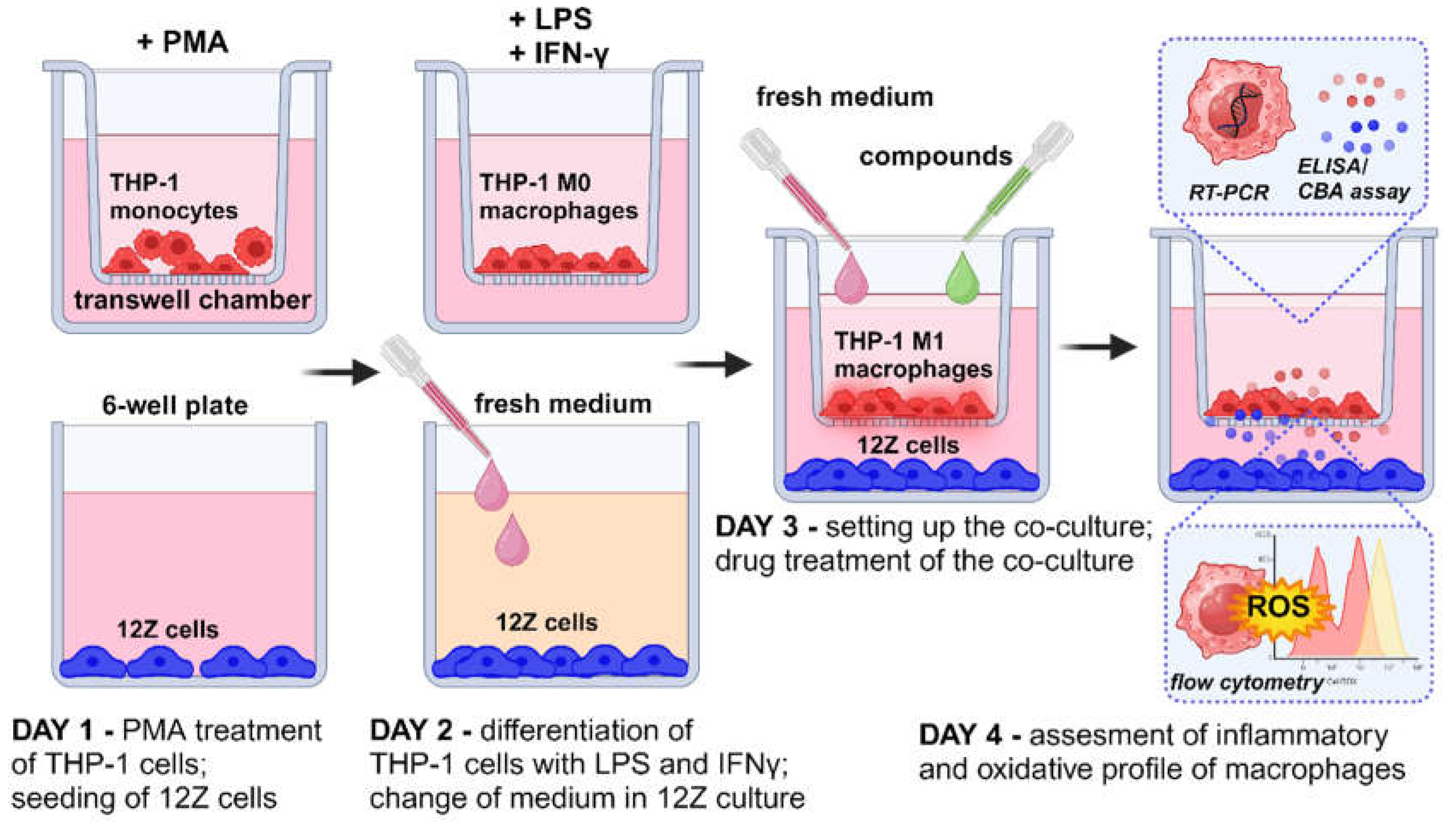
Figure 2.
Morphological comparison of THP-1 cells before (A) and after PMA induction (B), after M1 polarization (C) and co-culture with 12Z (D) using light microscope (magnification × 100).
Figure 2.
Morphological comparison of THP-1 cells before (A) and after PMA induction (B), after M1 polarization (C) and co-culture with 12Z (D) using light microscope (magnification × 100).
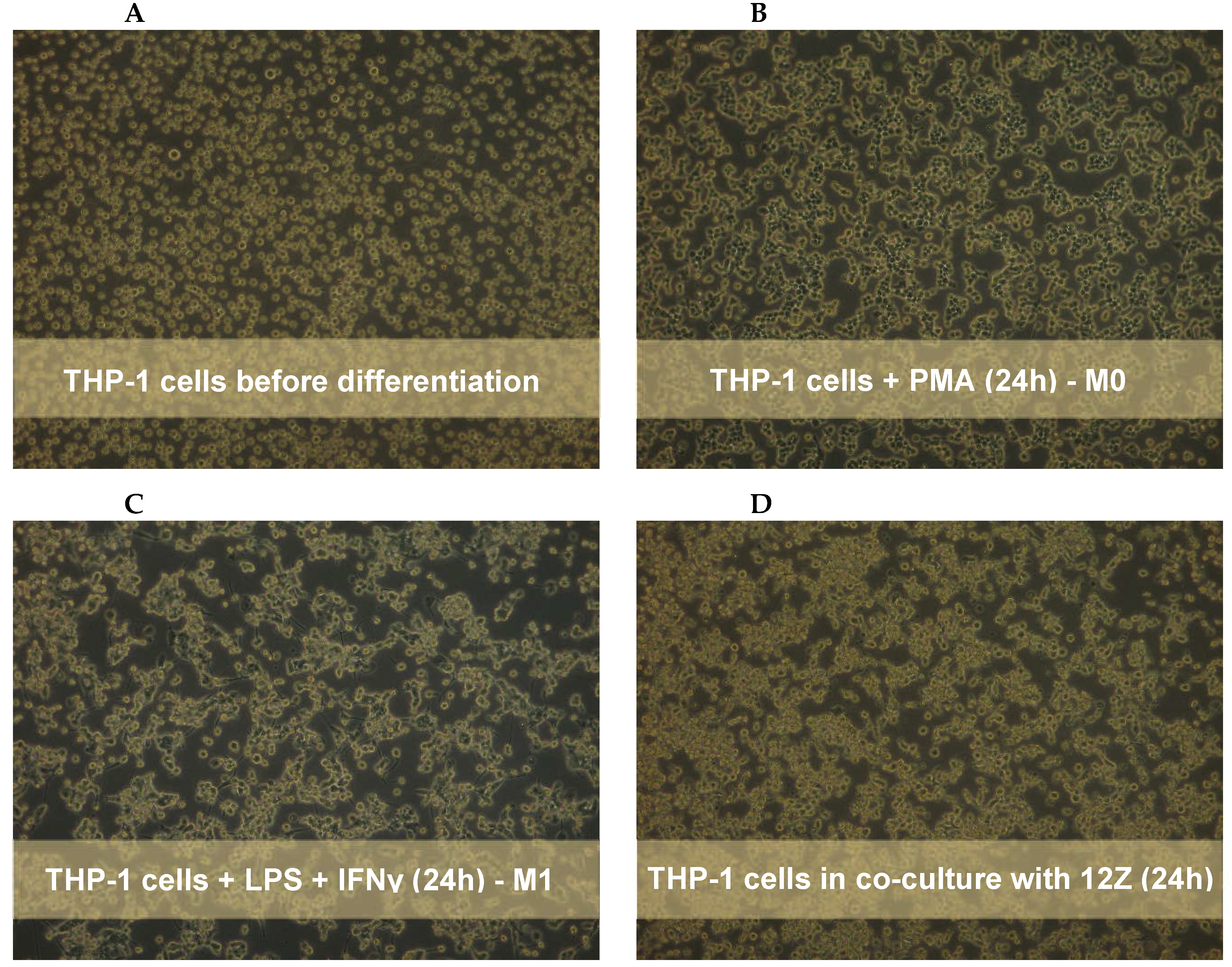
Figure 3.
The effect of resveratrol on endometriotic epithelial cell (12Z) and differentiated macrophage (THP-1) proliferation analyzed by MTT reduction assay. The values represent the means (n = 6) ± SD. The post hoc Tukey test determined the significance of the effects of resveratrol concentration (*p≤0.05; **p≤0.01; ***p≤0.001).
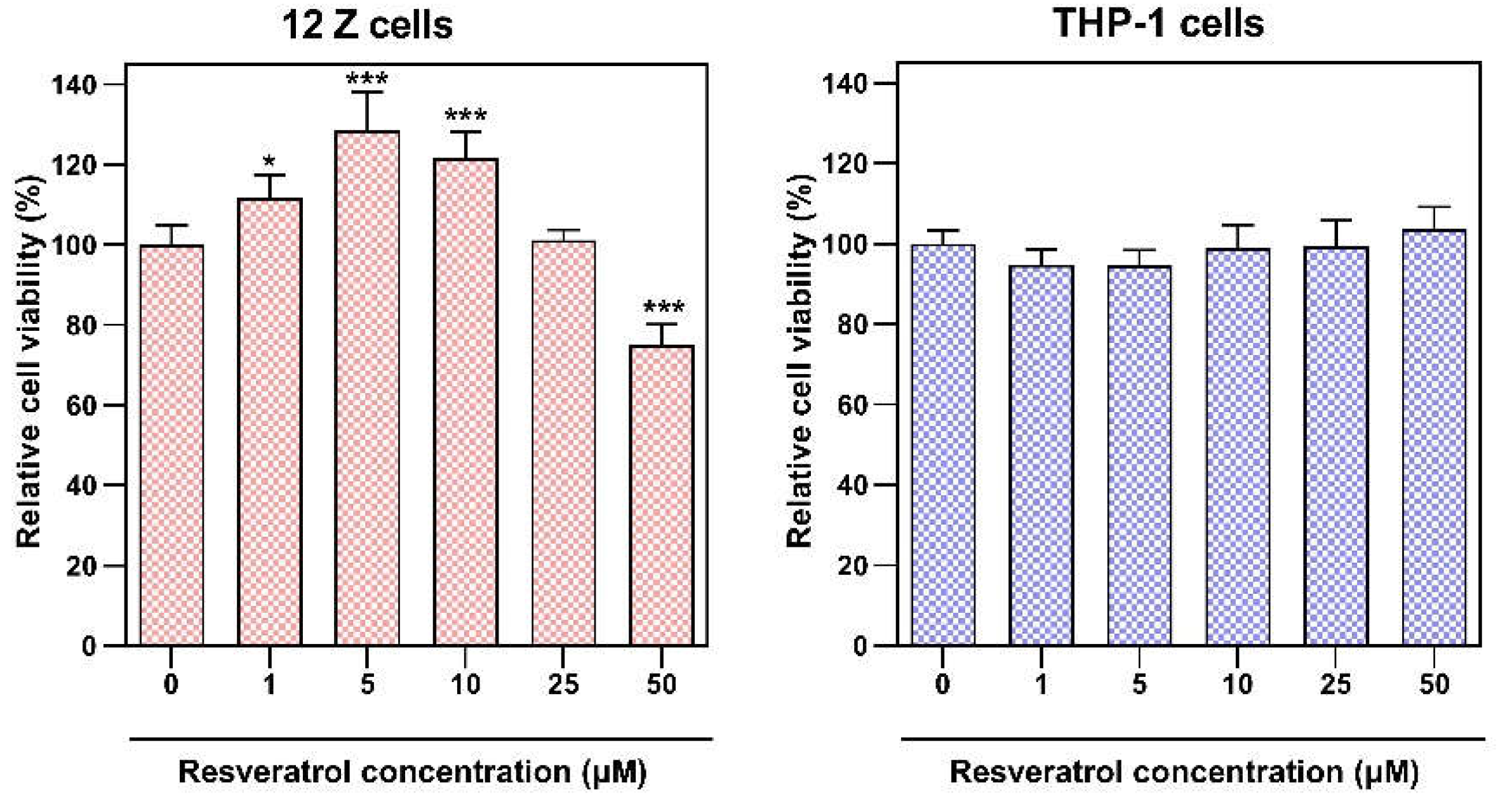
Figure 4.
Effect of resveratrol and its analogs on the mRNA expression of CCL2, CXCL10, GPX1, IL1B, IL6, IL8, NFKB1, PTGS2, SOD1, TNF genes in macrophages co-cultured with endometriotic cells, assessed by real-time PCR. Data were normalized against GAPDH and expressed as relative mRNA expression (-fold of control). The means ± SD of three independent experiments performed in triplicates are displayed. Statistical analysis assessed differences between the exposed cells and the negative control with significant differences *p ≤ 0.05; **p ≤ 0.01; ***p ≤ 0.001.
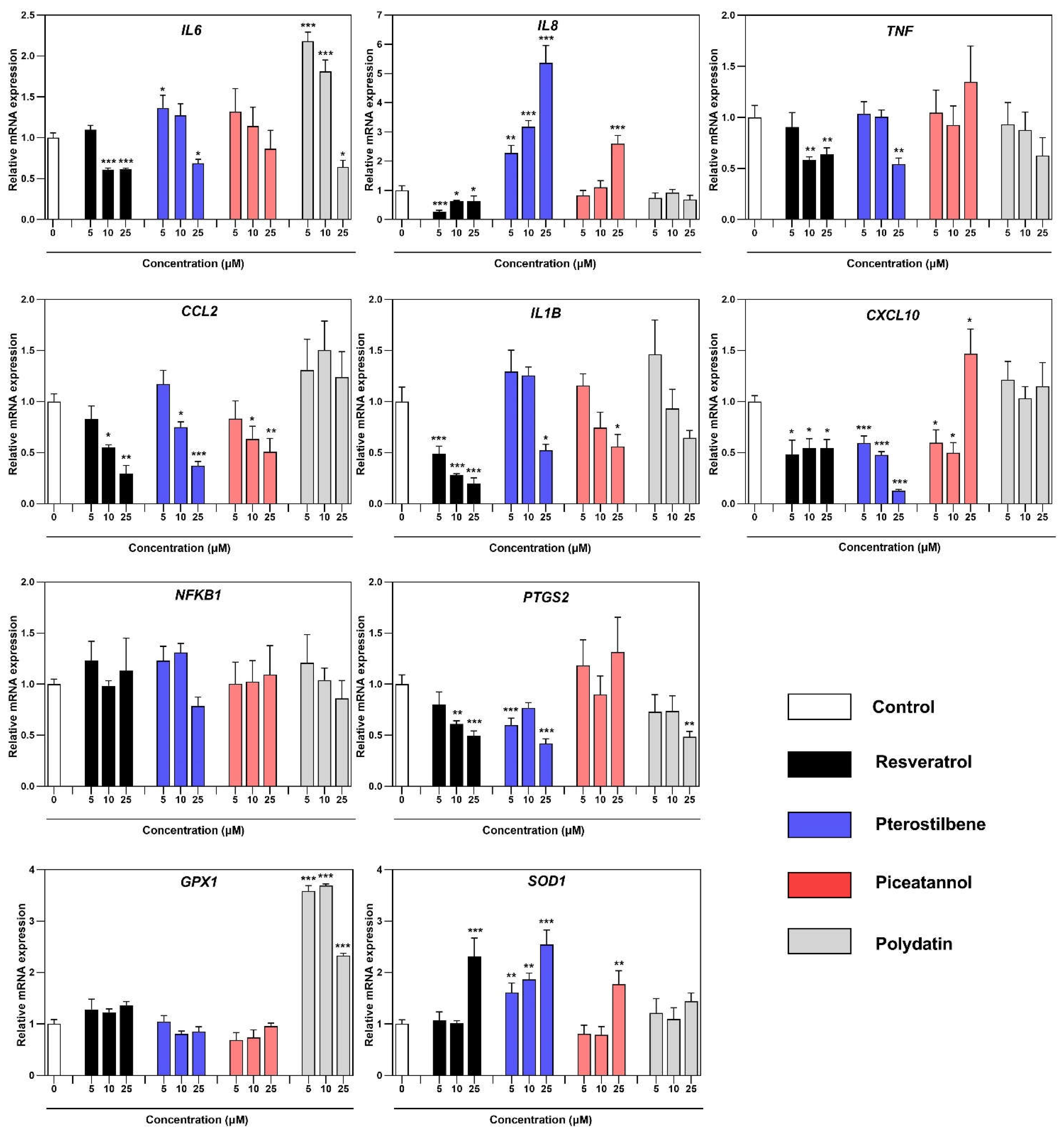
Figure 5.
Effect of resveratrol and its analogs on the cytokines/chemokines release in a co-culture model of macrophages and endometriotic cells, assessed by CBA Human Inflammatory Cytokine Cytometric Bead Array and ELISA. Data was normalized per μg of proteins in each cell sample. The means ± SD of three independent experiments performed in triplicates are displayed. Statistical analysis assessed differences between the exposed cells and the negative control with significant differences *p ≤ 0.05; **p ≤ 0.01; ***p ≤ 0.001.
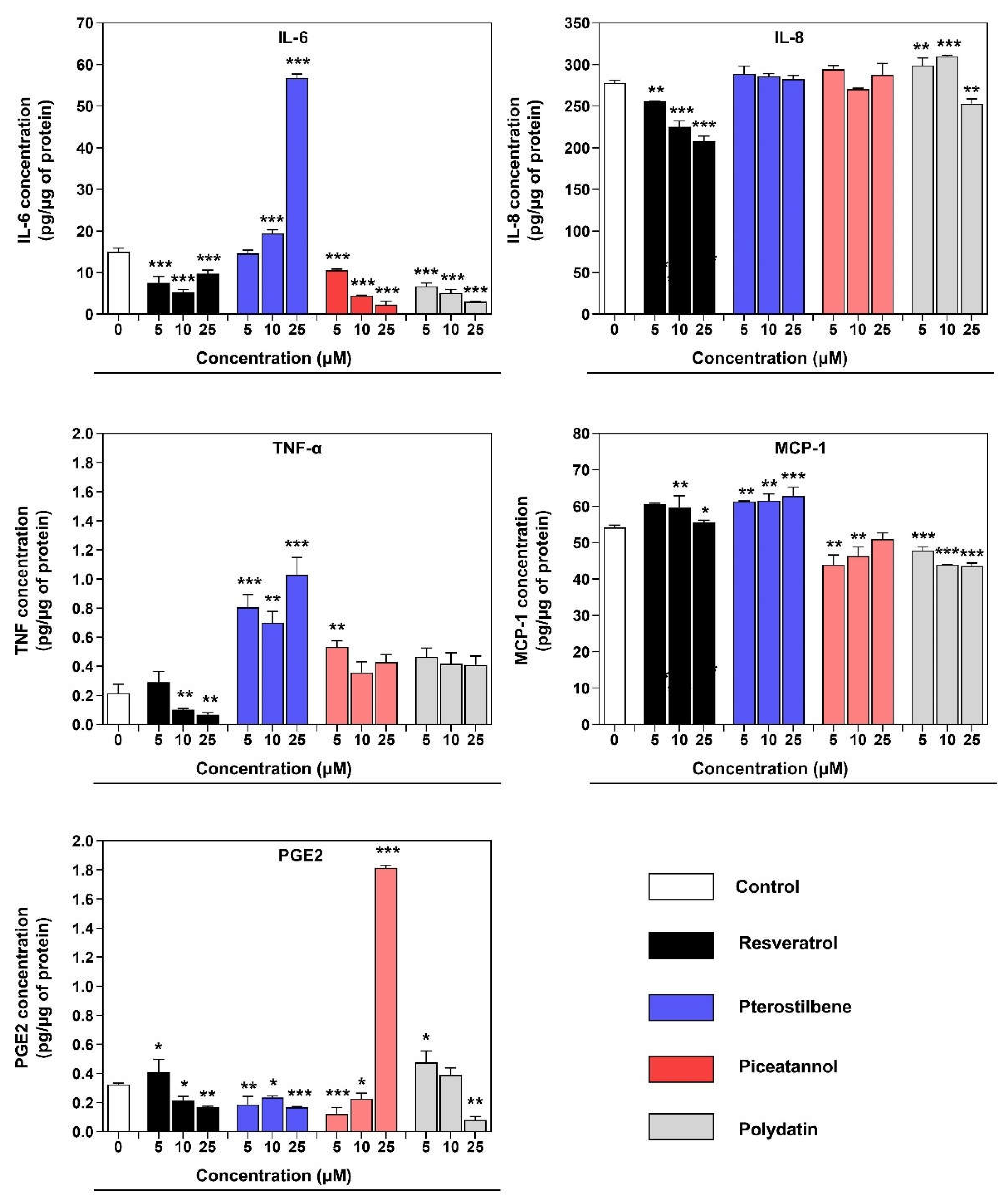
Figure 6.
Flow cytometry analysis of macrophages co-cultured with 12Z cells stained with CellROX® Deep Red Reagent and Sytox® Green Dead stain. Cells were treated with resveratrol, pterostilbene, piceatannol, polydatin, and their vehicle (0.5% ethanol) for 24 h. (A) Representative images of each distinct cellular subset. n = 4; scale bars = 20 μm. (B) Representative flow cytometry dot plots. (C) Summary of CellROX® Deep Red Reagent and Sytox® Green Dead stained populations in macrophages. Data show the percentage distribution of two fractions: live non-stressed cells and live oxidatively stressed cells. (D) Quantitative analysis of fluorescence intensity of CellROX® Deep Red. Statistical analysis assessed differences between the exposed cells and the vehicle control with significant differences *p ≤ 0.05; **p ≤ 0.01; ***p ≤ 0.001.
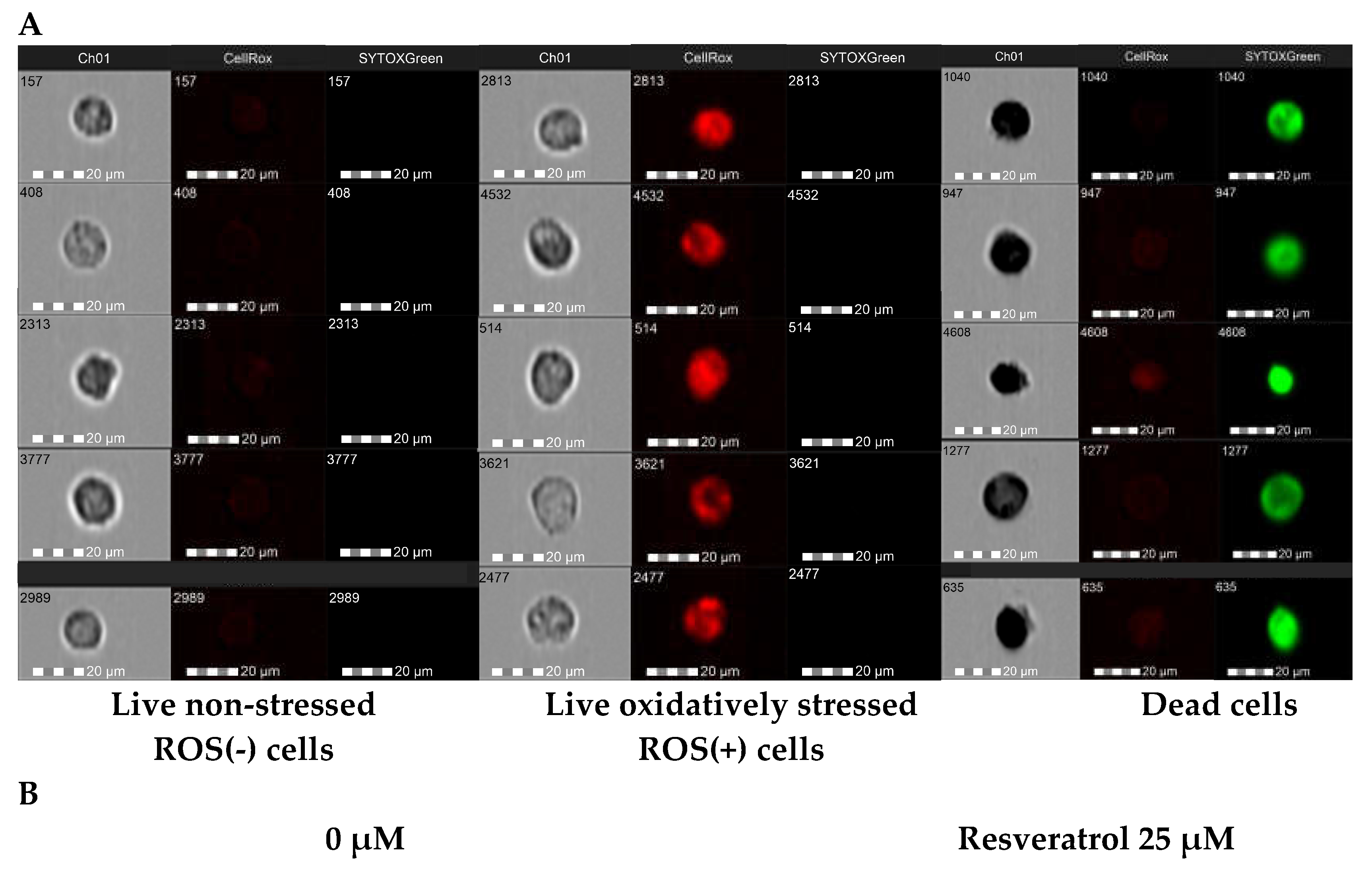
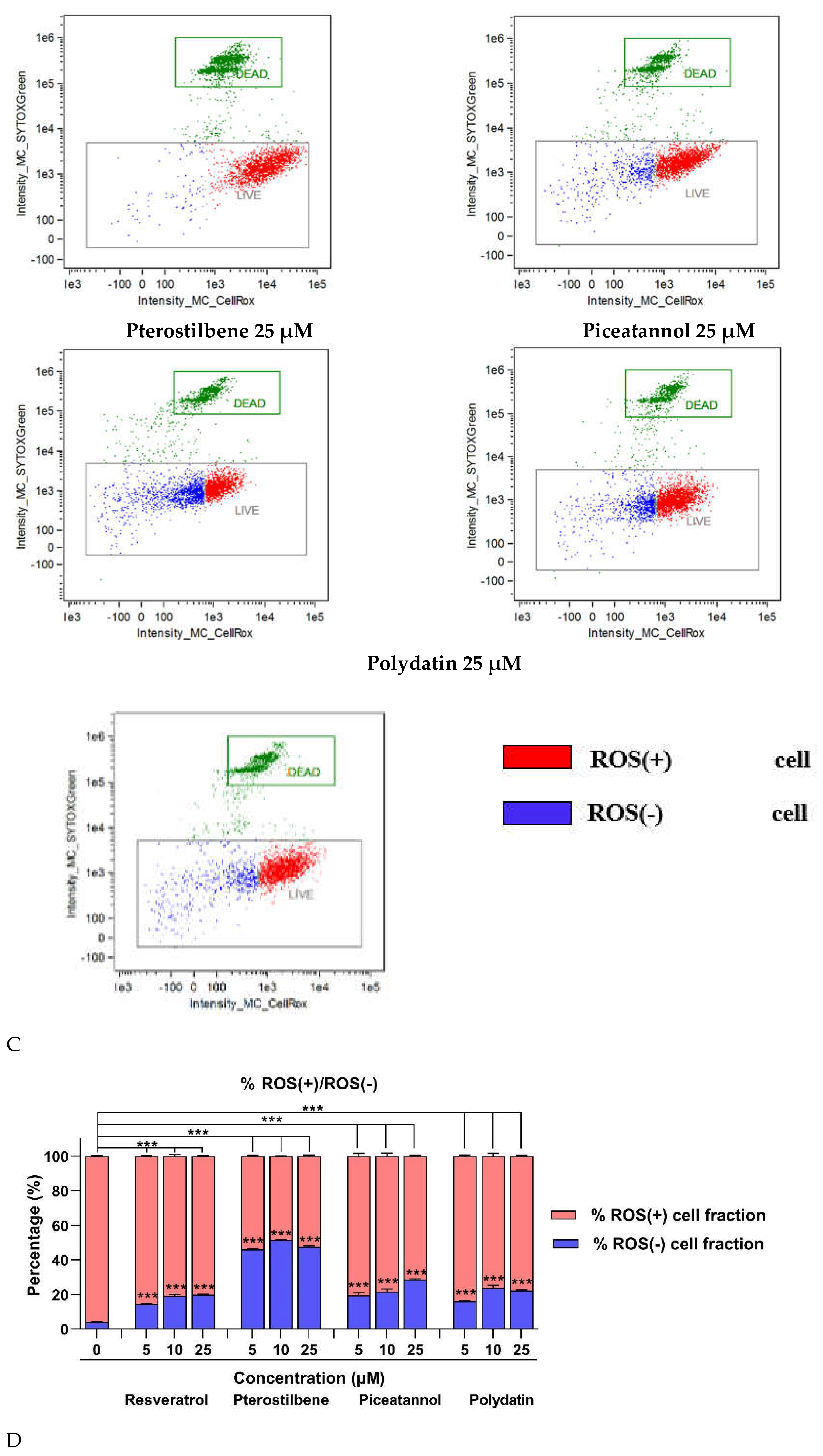
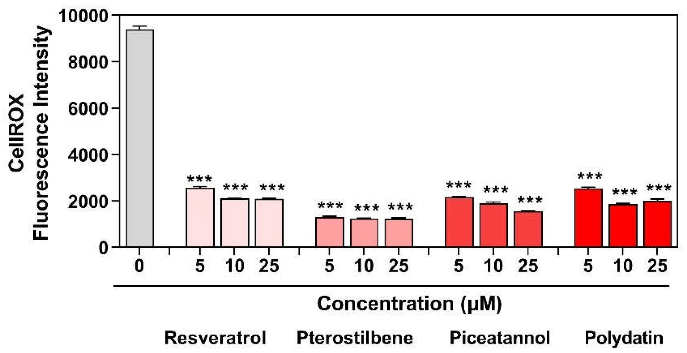

Table 1.
The primers sequence used for real-time PCR.
| Gene | Accession | No. sequence (5’-3’) | Amplicon (bp) |
|---|---|---|---|
| CCL2 | NM_002982.4 | F: AGAATCACCAGCAGCAAGTGTCC R: TCCTGAACCCACTTCTGCTTGG |
98 |
| CXCL10 | NM_001565.4 | F: GAACTGTACGCTGTACCTGCA R: TTGATGGCCTTCGATTCTGGA |
172 |
| GAPDH | NM_002046.7 | F: GTCTCCTCTGACTTCAACAGCG R:ACCACCCTGTTGCTGTAGCCAA |
131 |
| GPX1 | NM_000581.4 | F: GTGCTCGGCTTCCCGTGCAAC R: CTCGAAGAGCATGAAGTTGGGC |
123 |
| IL1B | NM_000576.3 | F: CCACAGACCTTCCAGGAGAATG R: GTGCAGTTCAGTGATCGTACAGG |
131 |
| IL6 | NM_000600.5 | F: AGACAGCCACTCACCTCTTCAG R: TTCTGCCAGTGCCTCTTTGCTG |
132 |
| IL8 | NM_001354840.3 | F: GAGAGTGATTGAGAGTGGACCAC R: CACAACCCTCTGCACCCAGTTT |
112 |
| NFKB1 | NM_003998.4 | F: GCAGCACTACTTCTTGACCACC R: TCTGCTCCTGAGCATTGACGTC |
130 |
| PTGS2 | NM_000963.4 | F: CGGTGAAACTCTGGCTAGACAG R: GCAAACCGTAGATGCTCAGGGA |
156 |
| SOD1 | NM_000454.5 | F: CTCACTCTCAGGAGACCATTGC R: CCACAAGCCAAACGACTTCCAG |
129 |
| TNF | NM_000594.4 | F: CTCTTCTGCCTGCTGCACTTTG R: ATGGGCTACAGGCTTGTCACTC |
135 |
Disclaimer/Publisher’s Note: The statements, opinions and data contained in all publications are solely those of the individual author(s) and contributor(s) and not of MDPI and/or the editor(s). MDPI and/or the editor(s) disclaim responsibility for any injury to people or property resulting from any ideas, methods, instructions or products referred to in the content. |
© 2024 by the authors. Licensee MDPI, Basel, Switzerland. This article is an open access article distributed under the terms and conditions of the Creative Commons Attribution (CC BY) license (http://creativecommons.org/licenses/by/4.0/).
Copyright: This open access article is published under a Creative Commons CC BY 4.0 license, which permit the free download, distribution, and reuse, provided that the author and preprint are cited in any reuse.


