Preprint
Article

This version is not peer-reviewed.

Research on the Improvement Effects and Mechanisms of Magnolia sieboldii Essential Oils on Insomnia in Mice

Submitted:

27 January 2025

Posted:

05 February 2025

Read the latest preprint version here

Abstract

(1) Background: Insomnia is a common sleep disorder that is difficult to cure due to its long duration of influence. Magnolia sieboldii essential oils (MSEOs) have been shown to have antidepressant effects, but there are few studies on treating insomnia. Therefore, this study aimed to investigate the therapeutic effects of MSEOs and to elucidate the molecular and neurophysiological mechanisms by which they alleviate insomnia. (2) Methods: The main components of MSEOs extracted by steam distillation were analyzed by gas chromatography-mass spectrometry (GC-MS). To establish a p-chlorophenylalanine (PCPA) -induced insomnia model in mice, the levels of GAD65, GABAARα1, 5HT-2A, and 5HT-1A were detected by immunohistochemistry and ELISA. The normal neurons in the mouse brain were counted by Nissl staining. The relative mRNA expression levels of related genes in mice were detected by RT- qPCR. (3) Results: A total of 69 components were identified by MSEOs, and the main components were β-elemene (19.94%), (Z)-β-ocimene (14.87%), and Germacrene D (7.05%). Both low and high concentrations of MSEOs can successfully prolong the total sleep time and shorten the sleep latency of mice. GAD65, GABAARα1, 5HT-2A, and 5HT-1A levels still increased to varying degrees after treatment with different concentrations of MSEOs. The results of Nissl staining showed that MSEOs could attenuate PCPA-induced neuronal death. The RT- qPCR results showed that MSEOs enhanced the mRNA expression of 5HT-2A, GABAARα1, and GABAARγ2. (4) Conclusions: MSEOs effectively improved sleep by prolonging total sleep time and shortening latency, potentially through upregulating GAD65, GABAARα1, 5HT-1A, and 5HT-2A levels, protecting neurons, and enhancing mRNA expression of GABAARα1, GABAARγ2, and 5HT-2A, suggesting their potential as a therapeutic for insomnia.

Keywords: 
;  ;  ;  ;  ;  

1. Introduction

Insomnia is marked by difficulties in starting to sleep, recurrent awakenings at night, and a diminished quality of life, a widespread epidemic [1]. The significance of adequate sleep is widely recognized, but due to increasing life pressures, the rate of insomnia continues to rise [2]. Numerous factors are intricately associated with the prevalence of insomnia, including gender, diet, constitution, environment, and age, although these possible influencing factors have not yet been definitively reported. Additionally, insomnia is recognized as a significant contributor to multiple systemic diseases. An epidemiological investigation has indicated that insomnia frequently coexists with depression and anxiety [3]. Meta-analyses have shown that individuals exhibiting symptoms of disruptions in sleep continuity or insomnia disturbances have a significantly increased incidence of hypertension [4]. Benzodiazepines and non-benzodiazepines are currently effective drugs for the treatment of insomnia [5,6,7]. However, prolonged administration of these agents, especially among the elderly, is highly likely to lead to potential side effects, examples include cognitive impairment, dependence, and addiction [8]. Therefore, we believe that natural products emerge as a supplementary alternative, characterized by the synergistic action of multiple chemical components to exert pharmacological effects on numerous targets, thereby effectively reducing the adverse effects associated with single-target pharmacological agents [9,10]. Therefore, herbal medicine, as an alternative therapy, can exert its effects with a lower risk of side effects in the treatment of insomnia.
The use of aromatherapy as a remedy for insomnia is globally acknowledged, encompassing the utilization of aromatic herbs within the realm of Traditional Chinese Medicine (TCM) [11,12]. Aromatic herbs can activate brain functions and regulate the central nervous system [12,13]. For example, Lavandula angustifolia Mill. and Rosa rugosa Thunb. had been used in clinical treatment [14]. Their essential oils exhibit a limited incidence of adverse reactions and exert sedative-hypnotic properties [15]. Recent evidence highlights that plant extracts can enhance the operation of neurotransmitter systems and aid in sleep promotion. Existing research has demonstrated that Magnolia sieboldii essential oils (MSEOs) have sleep-promoting and antidepressant effects. However, the literature on this type of research is minimal, and the mechanism by which MSEOs promote sleep is not fully defined [16,17].
The hypothalamus is the most important brain region in mammals involved in the regulation of the sleep-wake cycle. The currently recognized central neurotransmitters involved in the regulation of the sleep-wake cycle include serotonin, norepinephrine, dopamine, acetylcholine, and γ-aminobutyric acid (GABA), among others. 5-hydroxytryptamine (5-HT) plays a key role in regulating mood and sleep, and its deficiency is often associated with anxiety and depression; GABA, as the main inhibitory neurotransmitter in the central nervous system, helps to reduce neuronal excitability, promoting sleep onset and the maintenance of deep sleep [18]. GABA is an inhibitory neurotransmitter, while glutamate is an excitatory neurotransmitter; the two are the main components of the GABAergic system pathway, and an imbalance in their levels can cause disorders of the sleep-wake cycle rhythm. The levels of glutamate and GABA are influenced by glutamate decarboxylase (GAD) during the synthesis process. Glutamate can be converted into GABA through a decarboxylation reaction under the action of GAD, therefore, the content and activity of GAD can determine the conversion rate of glutamate to GABA [19,20].
In the field of sleep disorder research, the p-chlorophenylalanine (PCPA)-induced insomnia model is a typical pharmacological method used to evaluate the sedative and hypnotic effects of drugs. The core mechanism of this model lies in the inhibition of 5-HT synthesis by PCPA, which leads to a significant decrease in 5-HT levels in the peripheral and central nervous systems of experimental animals, thereby disrupting their circadian rhythms [21]. Injection of PCPA causes damage to the hippocampus, which in turn affects the levels of monoamine neurotransmitters (such as 5-HT, norepinephrine, dopamine) and amino acid neurotransmitters (such as GABA) [22,23]. Compared with other modeling techniques, the PCPA-induced insomnia model has the characteristics of a short modeling cycle and pronounced insomnia symptoms, making it a valuable tool for evaluating potential treatments for insomnia [24].
Therefore, the main objective of this study is to analyze the primary components of MSEOs, their efficacy in the treatment of insomnia, and the mechanisms of action in terms of neurotransmitters and neurophysiology.In this investigation, we employed a PCPA-induced insomnia model to assess the sleep-enhancing properties of MSEOs an analysis will be conducted focusing on the expression levels of 5-HT, GAD65, and GABA, along with normal neuronal levels in brain tissue. The objective of this research is to offer a scientific basis for further understanding the mechanism of MSEOs in the management of insomnia, as well as to promote more research into their clinical application, hoping to provide more solid support for clinicians’ treatment plans.

2. Methods and Materials

2.1. Plant Essential Oil

Dried leaves of Magnolia sieboldii (5 kg) were purchased from Tong Ren Tang Company (Beijing, China). All plant samples were identified by Professor Nian Liu (Zhongkai University of Agriculture and Engineering, Guangzhou, China). As voucher specimens, part of the collected sample has been deposited for safekeeping at the Institute of Natural Medicine and Green Chemistry (Guangdong University of Technology, Guangzhou, China) (Table 1). Use a pulverizer to grind the leaves of Magnolia sieboldii into fine powder. Mix them with a little water in a beaker to form lumps. Put them in a microwave oven and heat for 2 minutes. Mix them with water in a ratio of 1:8 in an efficient essential oil distillation equipment (model: TX05-02, Chengdu, China). Add two spoons of sodium chloride and collect the essential oil for 4 consecutive hours. Add anhydrous sodium sulfate to remove the water and store it at 4°C for experiments.

2.2. Conditions for GC-MS Determination

Essential oils were detected by a DSQ-II Ultra gas chromatography-mass spectrometry (GC-MS) (Thermo, USA) which was equipped with a DB-5MS capillary gas chromatographic column (0.25m 0.33μm) (Agilent, USA). Temperature program: 100°C (10 min) 3°C/min 250°C. The vaporization temperature is 250°C. The volume of the injection was 1.0 µl; Helium flow was 1.0 ml/min. Injection method: shunt; The split ratio was 20:1. The bar of mass spectrometry: ion source is electron blast; the temperature is 200°C; Energy: 70 e V, scanning range: m/z 30 ~ 550 amu.

2.3. PCPA-Induced Insomnia

24 healthy adult male KM mice, SPF grade, aged 6-8 weeks were from Southern Medical University (license number: SCXK (Guangdong)2016-0041). All the animal experiments were performed under the Guidelines for Care and Use of Laboratory Animals of Guangdong University of Technology (Guangzhou, China), and experiments were approved by the Animal Ethics Committee of Guangdong University of Technology (Guangzhou, China). Experimental mice were adaptively fed for a week under conditions of 24±1°C temperature and a 12-hour light/dark cycle. A total of 24 male Kunming mice were stochastically divided into six groups, including the MSEOs-L group (25 mg/kg), MSEOs-M group (50 mg/kg), MSEOs-H group (100 mg/kg), control group, PCPA group, and diazepam (DZP group (0.2 mg/ml). Filter paper is cut into the same size and soaked in different concentrations of essential oil solutions. Then filter paper was placed in four corners of the cage so that each mouse could sniff it for an hour. The weight of each mouse was recorded daily before inhalation. The cage where the control group mice lived was furnished with filter paper soaked in normal saline containing 1%Tween80 for 1h. Four days into the animal study, except for the control group of mice injected with saline containing 1% Tween 80 (PH7-8), the other group of mice was administered with PCPA at a dosage of 0.1 mL per 10 grams of the animal’s body mass for two days. Behavior observation was carried out in mice after the injection. The mice exhibited continuous activity throughout the day, displaying heightened levels of excitability and irritability. Additionally, they experienced a disruption in their circadian rhythm, along with elevated water and food consumption, while their sleep duration was markedly reduced. The group injected with PCPA was significantly different from the control group.

2.4. Pentobarbital-Induced Sleep Test

Half an hour following the final dose, sodium pentobarbital was administered intraperitoneally to the mice at a dose of 50 mg/kg to induce sleep. The number of turning over and sleep state, the time falling asleep (TS) the timing of the drug administration (TR), the latency to sleep onset, and the time of subsequent arousal were documented (TW).
Latency = TS – TR, Total Sleep Time = TW- TS

2.5. BCA Assay

Following the above experiment, the mice were subjected to euthanasia, and blood samples were obtained through the excision of the ocular globes. Blood samples were subjected to centrifugation at a rotational speed of 2500 rpm for a period of 5 minutes at 4°C in order to isolate the serum. Protein levels in serum were measured by a Bicinchoninic Acid (BCA) protein assay Kit (Shanghai, China).

2.6. ELISA Assay

The level of 5-HT in the hypothalamus of mice was measured using an ELISA assay kit (Shanghai, China). After the experiment, the mice were euthanized, and the hypothalamic tissue was quickly removed and then rinsed with cold saline to eliminate residual blood. Hypothalamic tissues were washed with phosphate-buffered saline (PBS), homogenized, and centrifuged, and the supernatant was collected. The procedure was carried out in line with the instruction manual provided in the ELISA kit, and absorbance was assessed at a wavelength of 450 nm, a standard curve was established to calculate the concentration of each detection index.

2.7. Immunohistochemistry Assay

After the mice were killed, their brains were isolated on an ice pad. The remaining blood on the tissues was irrigated with saline solution and blotted with filter paper. Apply 4% paraformaldehyde to the brain tissue samples for a 24-hour fixation period and then embed the processed brain tissue in paraffin sections that were sliced at 4 µm using a slicing machine (CUT 5062, Shanghai, China). The slides were dried for 2 h at 60°C. Afterward, the specimens were soaked in absolute ethanol solutions I and II for 10 minutes each, this was followed by 5-minute immersions in 95%, 90%, 85%, and 70% ethanol. Afterward, then wash the slices with PBS and immerse them in a buffer solution of sodium citrate adjusted to a pH of 6.0 under elevated temperatures for antigen repair. Subsequently, mouse brain tissue sections underwent treatment with 3% H₂O₂ for 25 minutes under dark conditions so that endogenous peroxide was blocked, tissues were blocked using 1.5% normal goat serum. Tissue blocks were incubated with a 15000-fold dilution of rabbit antiserotonin (Incstar, Stillwater, MN) at 37°C for 2 hours, subsequently, the sample was treated with a 12500-fold dilution of rabbit anti-serotonin transporter (Inc-star) for 15 min, 5000-fold dilution of rabbit anti-tyrosine hydroxylase (Pel-Freez Biologicals) for 15 min. Color reaction was performed using 3, 3′-diaminobenzidine (DAB).

2.8. Nissl Staining Assay

The embedded paraffin tissues were sliced at 5 µm and soaked in xylene I and xylene II for 10 minutes each, subsequently in 95%, 90%, 85%, and 70% ethanol with each stage lasting 5 minutes. The sections were subjected to staining with a 0.1% solution of cresyl violet for 20 minutes, followed by rinsing with distilled water. Dehydration and clearing of paraffin sections were performed as follows: First, paraffin-embedded tissue sections were subjected to primary dehydration in 70% ethanol solution for 2 minutes. Subsequently, the sections were transferred to 80% ethanol solution for intermediate dehydration, again maintained for 2 min. Further, the sections were subjected to advanced dehydration in 95% ethanol solution and the treatment time was also 2 min. Immediately following, complete dehydration with absolute ethanol, and the sections were immersed in 100% ethanol for 5 minutes each time for a total of two sessions. Then, after dewaxing and clearing, the sections were immersed in xylene for 10 minutes each time to complete two clearing steps. Finally, the paraffin sections were sealed with resin.

2.9. Immunofluorescence Assays

After the test was completed, the mice were subjected to euthanasia, following which the hypothalamic tissue was excised. The isolated animal tissues were preserved using 4% paraformaldehyde and subsequently sectioned into 4-micrometer-thick paraffin slices. The slides were placed in a citrate antigen retrieval solution contained within a repair box and heated in a microwave for 8 minutes to bring them to a boil. The heat was paused for 8 minutes to maintain warmth, followed by 7 minutes at medium-low temperature. After natural cooling, PBS (pH 7.4) was used to wash the slides and washed three times using a decolorization shaker (5 minutes each time). Overnight incubation was performed on the tissue at 4°C with a GABAAR antibody at a dilution ratio of 1:60. Next, the washed sections were incubated for one hour at 25°C with a secondary antibody (1:800 dilution) while being protected from light. Ultimately, the sections were subjected to staining with DAPI dye was diluted 100-fold for 10 minutes. Image analysis software (Image-Pro 6.0) was used to calculate the results.

2.10. RT-qPCR

Upon conclusion of the two-week treatment regimen, the hypothalamic and hippocampal regions were swiftly excised from the mouse skulls. These excised tissues were subjected to a delicate rinsing process with chilled physiological saline before being flash-frozen on dry ice. Following this, they were preserved at a temperature of −80°C. A sample weighing 50 milligrams from either the hypothalamus or hippocampus was processed in Trizol for a comprehensive homogenization to isolate total RNA. The synthesis of cDNA was carried out by the protocols provided with the cDNA synthesis kits. After this, the produced cDNA was subjected to amplification using primer sequences as detailed in Table 2.

2.11. Statistical Analysis

Data experiments were analyzed by GraphPad Prism software 8.0.2 (GraphPad Software, USA). Statistical results were expressed as mean ± standard error. One-way analysis of variance and the Tukey test were employed to conduct the statistical analysis, with *p < 0.05 demonstrating significant differences relative to the PCPA group.

3. Results

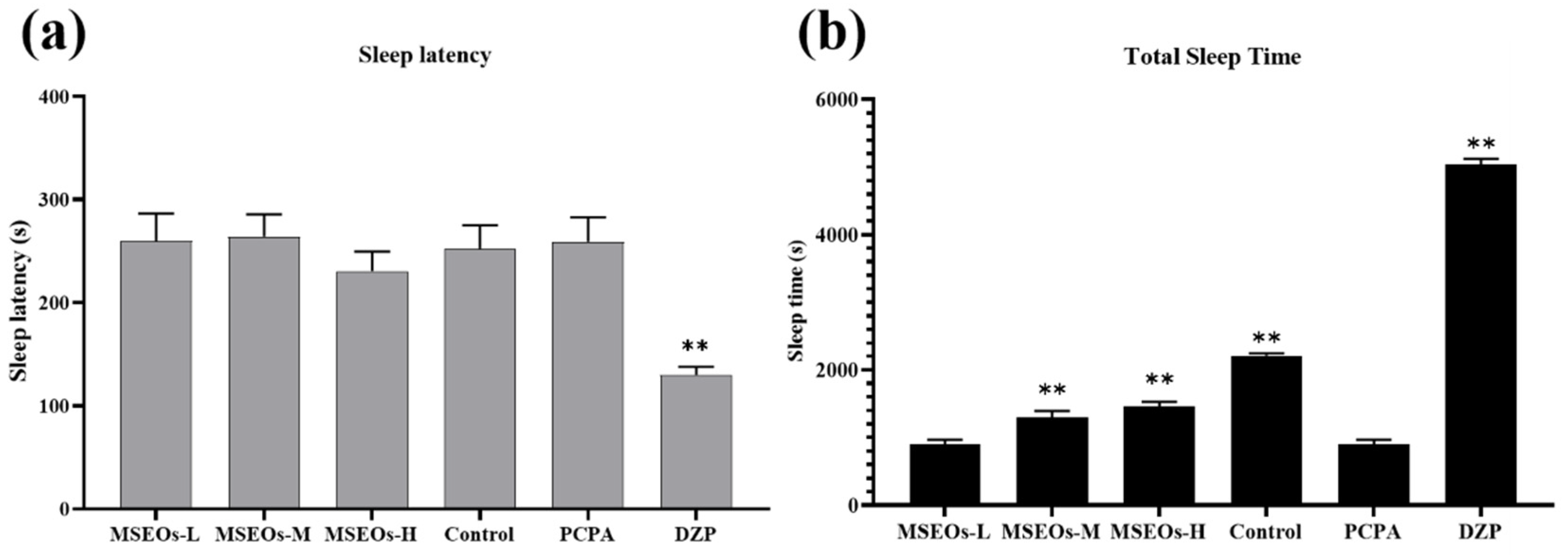
3.1. Impact of MSEOs on Sleep Duration and Latency to Sleep Onset in Insomniac Mice

The experimental results are shown in Figure 1. The sleep latency of PCPA group mice was longer than that of the control group (Figure 1a). Additionally, the total sleep duration of the PCPA group mice was markedly shorter than that of the control group (P<0.01) (Figure 2b). It can be seen that the PCPA can reduce the overall duration of sleep while extending the duration of sleep latency, causing insomnia in mice. This indicates that the insomnia mice model has been successfully established. After treatment with MSEOs, the sleep latency in MSEOs-H-treated mice decreased in comparison to the insomnia model group (Figure 1a), additionally, the duration of sleep for mice in the medium and high-concentration treatment groups of MSEOs was found to be increased relative to the PCPA group (P<0.01) (Figure 1b). In summary, the administration of PCPA via intraperitoneal injection is an effective method for creating a mouse model of insomnia, and treatment with MSEOs can effectively enhance sleep quality in mice with insomnia caused by PCPA.

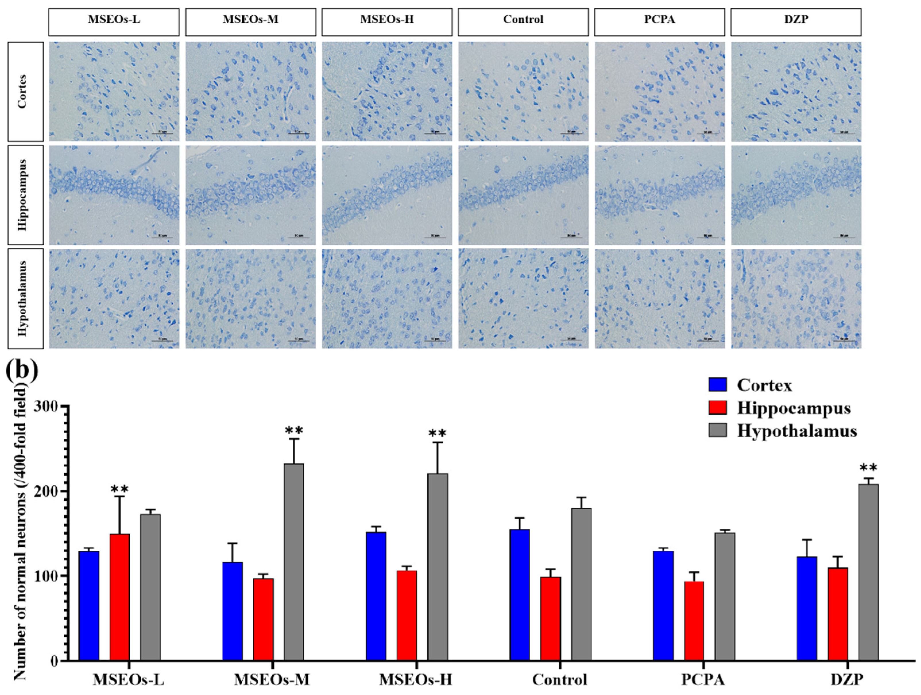
3.2. Neuroprotective Effects of MSEOs on PCPA-Induced Insomnia Mice

Insomnia is intricately linked to neuronal cell damage in brain tissue. When neurons within the hippocampal region sustain damage or undergo atrophy, these alterations adversely impact rapid eye movement (REM) sleep [25]. Additionally, more severe instances of insomnia may arise from significant damage to the hypothalamus [26,27]. Normal neuronal cell bodies contain a large amount of Nissl bodies, which are basophilic clumps or granules primarily involved in protein synthesis [28]. Nissl bodies can be labeled using toluidine blue staining, which also highlights the neuronal contour and nucleus. From the Nissl staining images in Figure 2a, it can be observed that, compared to the PCPA group, the three MSEOs groups exhibit normal morphology and a greater number of neuronal cell bodies in the hypothalamus, hippocampus, and cerebral cortex, with deeper staining of Nissl bodies. Compared to the control group statistical analysis of the count of neurons in the hypothalamus, hippocampus, and cerebral cortex revealed that the PCPA group had fewer uninjured neurons located within the cerebral cortex, hypothalamus, and hippocampus. However, after administration of MSEOs, the count of normal neurons located within the hippocampus, cerebral cortex, and hypothalamus increased, especially with MSEOs-M and MSEOs-H in the hypothalamus and MSEOs-L in the hippocampus showing the most pronounced effects (p < 0.01) (Figure 2b). This indicates that MSEOs have a protective effect on neurons, which may be related to their function in treating sleep disorders.

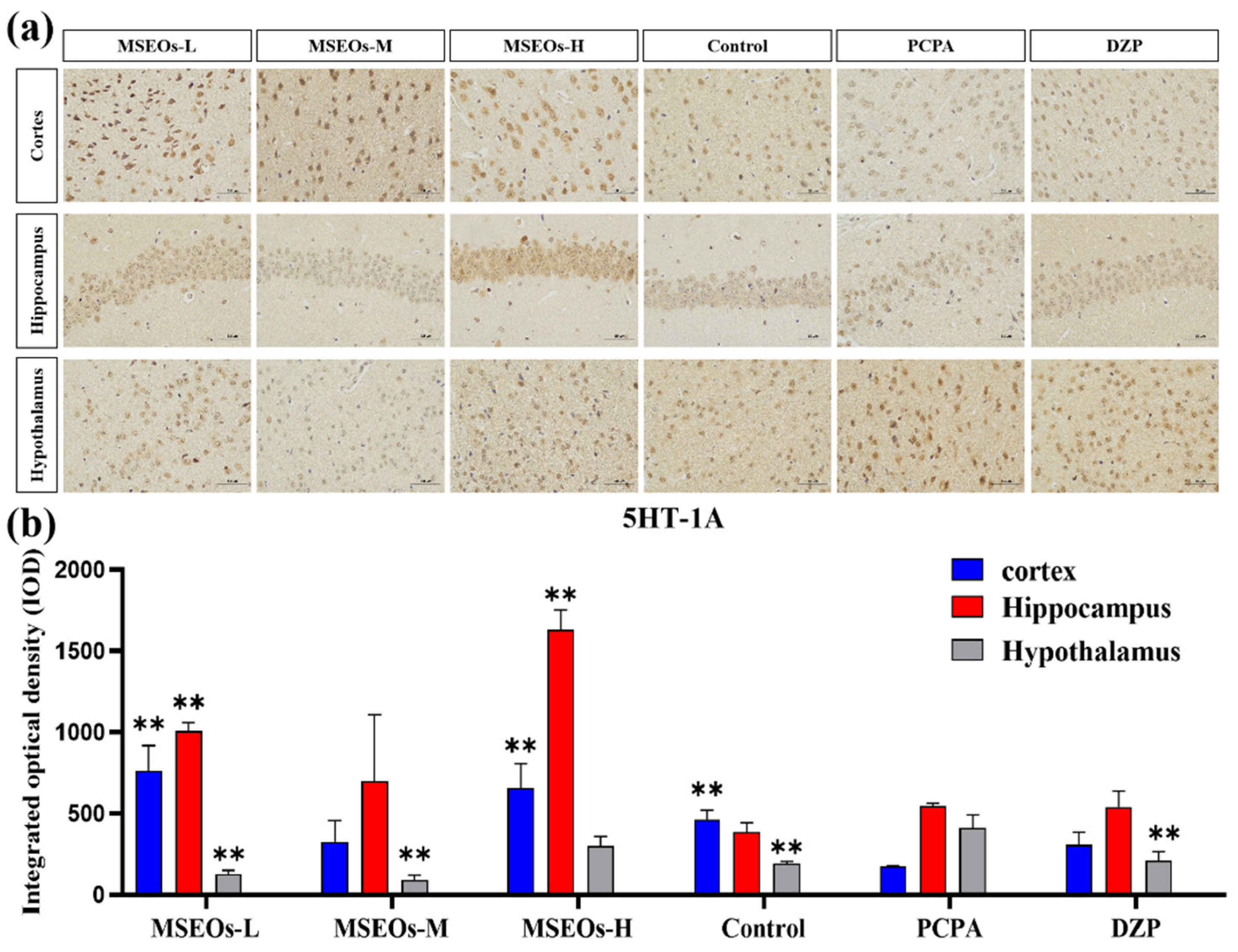
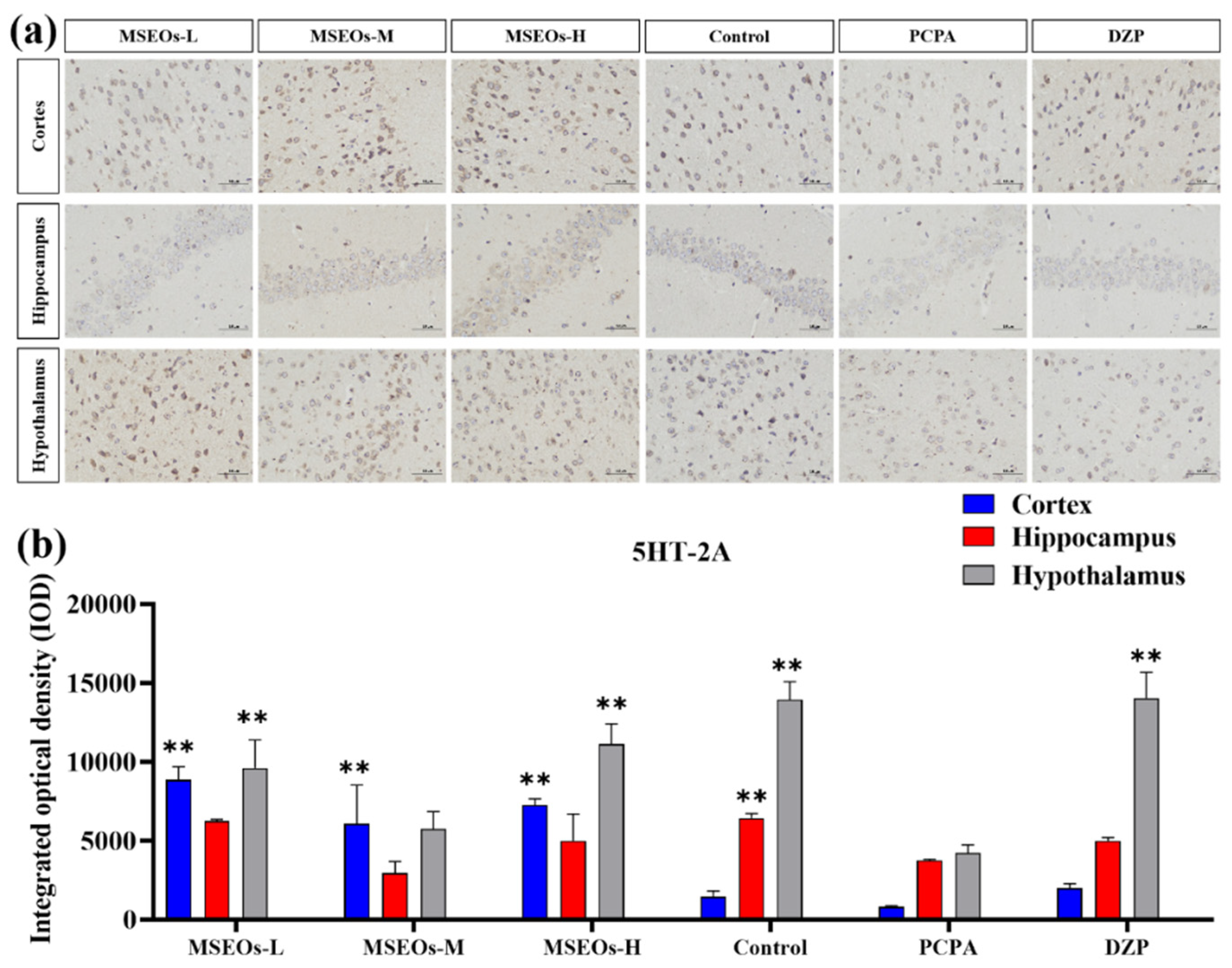
3.3. Expression of 5HT-2A and 5HT-1A

Serotonin, also widely recognized as 5-HT, is a classical neurotransmitter in the central nervous system that significantly influences the regulation of sleep and wakefulness. Serotonin is acknowledged as one of the key mechanisms contributing to insomnia [32]. Its two important subtypes are 5HT-1A and 5HT-2A receptors, respectively. The 5HT-2A and 5HT-1A proteins are extensively distributed throughout the brain, and play a role in the modulation of sleep [29,30], anxiety, and emotions [31]. Previous research has indicated that 5HT-1A is densely present in the hippocampus, with evidence suggesting that the activation of these receptors is effective in alleviating anxiety symptoms. In general, low-dose agonists of the 5HT-1A receptor have been found to enhance both deep sleep and light sleep [33,34,35]. PCPA can cause insomnia by inhibiting presynaptic 5HT-1A autoreceptors and reducing 5-HT levels [36,37].
The test results show (Figure 3) that compared to the normal control group the 5HT-1A expression in mice’s cortex within the PCPA group is notably lower (P<0.01) (Figure 3b), and in the cortex, hippocampus, and hypothalamus, the expression level of 5HT-2A was also decreased (P<0.01) (Figure 4b). This indicates that PCPA can result in reduced expression of both 5HT-2A and 5HT-1A, confirming the insomnia model was successfully induced. In contrast to the insomnia model group, treatment with MSEOs led to a marked elevation in the 5HT-1A levels within the hippocampus and cortex of mice across all three dosage groups (P<0.01), the MSEOs-H group demonstrated the most favorable outcome (Figure 3b). The levels of expression of 5HT-2A in the cortex, hypothalamus, and hippocampus of mice in all three MSEOs dosage groups are also elevated, with MSEOs-L and MSEOs-H showing better effects (Figure 4b). It can be seen that treatment with MSEOs effectively inhibited the decrease in 5HT-2A and 5HT-1A levels in mice exhibiting PCPA-induced insomnia, maintaining the levels of 5HT-2A and 5HT-1A in the mice at normal levels.

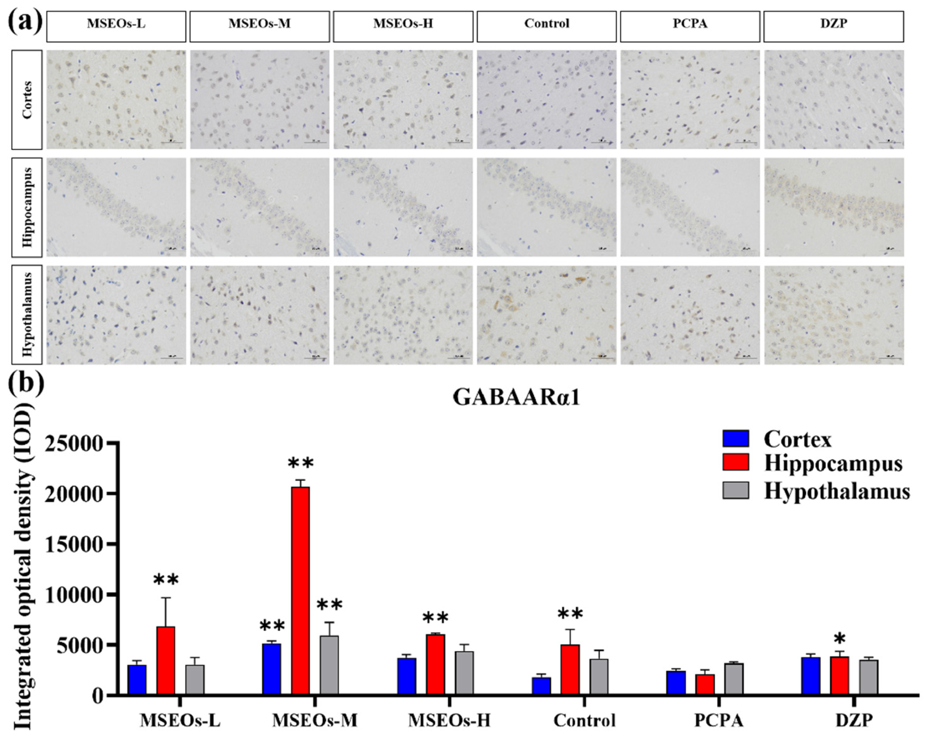
3.4. Effect of MSEOs on GABAARα1 Levels in Mice with Insomnia

GABA is an important inhibitory neurotransmitter, and it also effectively inhibits the activity of most neurons in the suprachiasmatic nucleus [38], its inhibitory effect is intricately connected to circadian rhythms in animals [39,40]. GABA is commonly used as an important indicator for evaluating central nervous system (CNS) function and is also a common metric in insomnia research [41,42], with the A receptor subtype GABAARα1 being the most prevalent [43]. The GABA signaling pathway is primarily mediated by GABAA and GABAB receptors. GABAARα1 and GABAARγ1 in GABAA receptors may be key targets for multi-target sleep-enhancing agents in improving insomnia. Studies have shown that the expression levels of GABAARα1 and GABA were significantly decreased in the PCPA-induced insomnia mouse model [44]. To verify whether MSEOs can exert their insomnia-improving effects through the modulation of the expression of GABAARα1 in the animals, we quantified the levels of GABAARα1 in the mouse brain tissue.
Compared with the control group, the expression of GABAARα1 in the hippocampus and hypothalamus of the insomnia model group was decreased (Figure 5). It can be seen that PCPA can cause a decrease in the GABAARα1 content in mice exhibiting insomnia. However, after intervention with MSEOs, the GABAARα1 levels in brain tissues of MSEOs mice were higher than those of PCPA mice, especially in the MSEOs-M group (p < 0.01). This indicates that MSEOs can inhibit the decrease in GABAARα1 levels in PCPA-induced sleep disorders in mice, maintaining the GABAARα1 content in the mice at normal levels.

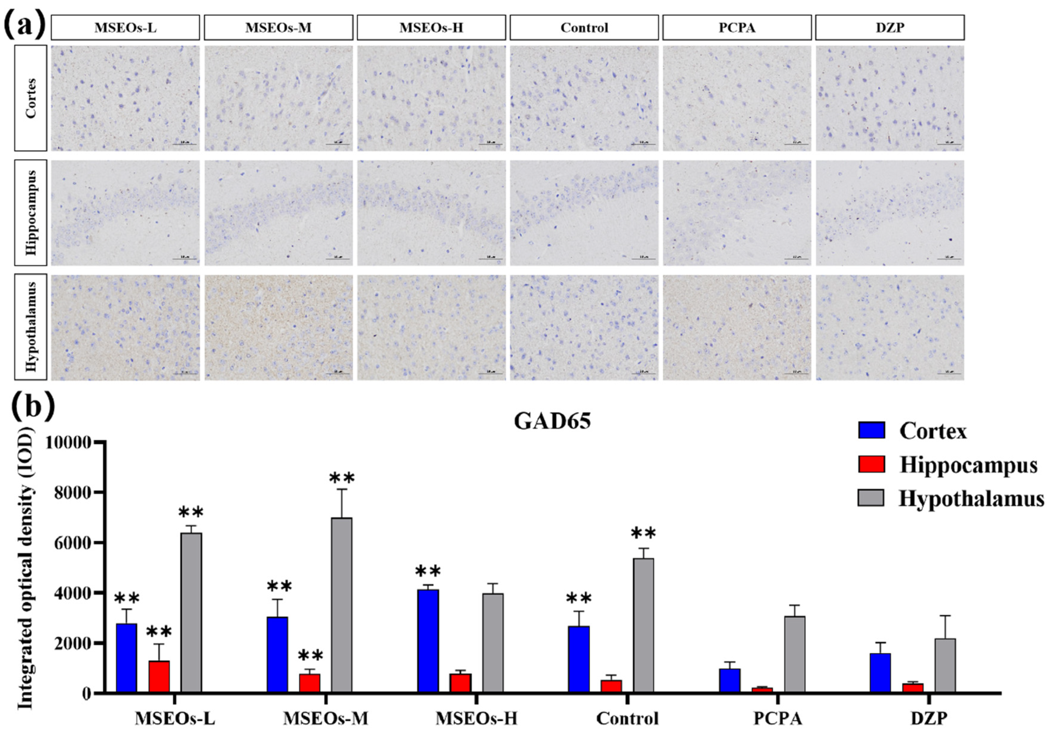
3.5. Impact of MSEOs on GAD65 Protein Levels in the Brain of Mice Subjected to Sleep Deprivation

GABA in the brain mainly catalyzes glutamate synthesis through the GAD enzyme, which converts glutamate into GABA [19,20]. There are two isoforms of glutamate decarboxylase, namely GAD67 and GAD65 [45,46]. Research shows that GAD65 is sensitive to changes in GABA and is commonly found in the cytoplasm of GABAergic neurons, mainly responsible for the synthesis and release of GABA in the brain [47,48]. Previous research has indicated a decrease in GAD65 levels within the cerebral tissue of mice experiencing PCPA-induced sleep disorder treatment [49]. Consequently, the impact of hypnosis of the respective pharmacological agents can be ascertained through the quantification of GAD65 protein expression within the cerebral tissue of murine subjects after their administration.
The GAD65 content in the cortex, hypothalamus, and hippocampus of the PCPA group was markedly lower than that of the control group(P<0.01) (Figure 6b), indicating that intraperitoneal injection of PCPA results in a reduction of GAD65 levels within the brain tissue. In contrast, GAD65 levels in the hypothalamus, cortex, and hippocampus of mice administered with MSEOs were elevated compared to both the blank and the PCPA groups, with the MSEOs-L and MSEOs-M groups showing the most significant effect (P<0.01). The result indicates that treatment with MSEOs is capable of effectively preventing the decline in GAD65 protein levels in the cerebral tissue of mice experiencing PCPA-induced sleep disturbance, maintaining the GAD65 content in their bodies at normal levels.

3.6. RT-qPCR

The results of RT-qPCR (Figure 7) indicated that relative to the control, the mRNA expression of 5HT-2A, GABAARα1, and GABAARγ2 in the insomnia PCPA-treated mice was diminished. Following treatment, the mRNA content of GABAARα1, GABAARγ2, and 5HT-2A in brain tissues of MSEOs groups was markedly higher compared to the levels observed in the insomnia model group (P<0.01), the MSEOs group and the normal control group exhibited similar levels, with no significant variance. These results strongly validated the therapeutic effect of MSEOs on insomnia.

3.7. Essential Oil Component Analysis

The GC-MS analysis yielded results (Table 3) identifying 69 compounds, which comprised 95.71% of the overall essential oil composition. There were 24 Sesquiterpene hydrocarbonszn, 13 Oxygenated sesquiterpenes, 11 Total monoterpenoids, and Oxygenated monoterpenes 6 and 15 other substances, accounting for 60.47%, 10.94%, 29.36%, 6.85% and 5.61% of the total mass, respectively. The content of β-elemene (19.94%) was the highest. Followed by (Z)-β-ocimene (14.87%), Germacrene D (7.05%), cis-(+)Nerolidol (4.51%), isocaryophyllene (3.63%), etc. β-elemene has ameliorative effects on inflammation in the CNS [50] and is found as a major component of many plant essential oils that promote hypnosis [51,52]. Several studies have shown that Nerolidol is an important component capable of inducing sedation in mice [53], as indicated by decreased motility and prolonged sleep duration induced by pentobarbital [51]. Nerolidol has a protective effect against neuroinflammation [54] and the anti-inflammatory activity is associated with the gabaergic system [55], Nerolidol also potentiates neuronal effects by activating the activation of 5-HT receptors, especially the 5-HT1A and 5-HT4 isoforms [56]. Isocaryophyllene has good anticancer activity [57,58], but its effect on the nervous system has not been reported. The effect of Magnolia sieboldii essential oil on improving sleep disorders may be related to the effects of β-elemene and Nerolidol on the nerve center, as well as the results of the interaction with other components.

4. Discussion

In recent years, MSEOs have gradually demonstrated their potential in the therapeutic approach to insomnia [16]. The findings from this research indicate that Magnolia sieboldii can improve sleep quality in mice with PCPA-induced insomnia and modulate the concentrations of pertinent neurotransmitters. This finding provides experimental evidence for its potential therapeutic application in the management of insomnia. At present, the primary therapeutic approaches for insomnia mostly rely on chemical drugs, such as benzodiazepines and zolpidem. These drugs can indeed improve insomnia symptoms in the short term. Nevertheless, prolonged utilization of such medications may result in the development of drug dependency and a series of adverse reactions. In contrast, MSEOs, as a natural plant product, have a mild curative effect and may have fewer adverse reactions, offering a novel therapeutic approach for patients suffering from insomnia. Findings from the behavioral studies suggest that in contrast to the PCPA-treated group, the MSEOs-M treatment abbreviated the latency to sleep onset in mice, and the MSEOs-L and MSEOs-H groups could prolong the total sleep time, suggesting that it has an improving effect on regulating the sleep-wake cycle. This could be associated with its potential to modulate the levels of diverse neurotransmitters through the hypothalamus. Studies have shown that the abnormal regulation of neurotransmitters like DA and 5-HT in the insomnia state can cause excessive arousal reactions and disrupt the normal sleep-wake cycle [18,67,68,69]. 5-HT is vital for modulating mood and sleep patterns, with insufficiencies frequently correlating with anxiety and depressive disorders. GABA, as the inhibitory transmitter, helps to decrease neuronal excitability and promote the onset of sleep and the maintenance of deep sleep [70,71,72,73,74]. Our findings indicate that, in contrast to the control group, the expression of 5HT-1A, 5HT-2A, GABAARα1, and GAD65 in the hypothalamus of mice in the model group showed decreased levels. After treatment with MSEOs-L, MSEOs-M, and MSEOs-H the levels of these neurotransmitters increased to different extents, and it could also contribute to the protection of neurons. The findings of this research further confirm that MSEOs improve sleep quality by promoting the restoration of imbalanced neurotransmitters and protecting neurons. The increase in the level of GABAARα1 was particularly obvious in the MSEOs medium-dose group. It is speculated that it promotes the release of GABAARα1, thereby alleviating the high excitatory state of the CNS and improving sleep quality. In conclusion, as a natural product, MSEOs provide experimental support for its application in addressing insomnia through the adjustment of neurotransmitter equilibrium and its effects on the improvement of GABAARα1, GAD65, 5HT-1A, and 5HT-2A, and is expected to is anticipated to offer a potent alternative for managing insomnia. In the current academic literature, there is relatively scarce research on the impact of MSEOs on insomnia, and the research perspectives are rather limited, mainly focusing on exploring the effects of MSEOs on two neurotransmitters, 5HT-1A and GABAARα1 [16,17]. Based on this, this study has expanded and deepened the research, not only examining the changes of the above neurotransmitters but also further analyzing the expression levels of 5HT-2A, GAD65, and GABAARγ2. Through this comprehensive research method, we aim to more comprehensively clarify the potential role of MSEOs in alleviating insomnia symptoms in mice. The results of this study have enriched the existing research scope and provided a more solid experimental basis for the application of MSEOs in the field of insomnia treatment. Subsequent investigations are needed to validate its efficacy under different doses and time courses and explore its molecular mechanisms in depth, with the expectation of delivering more trusted treatment plans based on Chinese herbal medicine for clinical insomnia.

Author Contributions

Conceptualization, Guofeng Shi; Methods, Guofeng Shi, Shuanghe Wang; Validation, Wenyu Cao, Xiaoyan Li; Formal analysis, Jiajing Ding; Investigation, Shanshan Luo; Resources, Zixuan Liang; Data management, Yixi Zeng; Writing and manuscript preparation, Guofeng Shi, Shuanghe Wang; Writing review and editing, Yixi Zeng, Yanqing Ma; Visualization, Yanqing Ma; Supervising Lanyue Zhang, Hui Li; Project Management, Lanyue Zhang, Hui Li. All authors have read and agreed to the published version of the manuscript.

Funding

This work was supported by grants from National Natural Science Foundation of China (No. 22408058 and 52273104), Guangdong Basic and Applied Basic Research Foundation (No. 2023A1515220145, KTP20240675).

Institutional Review Board Statement

All animals in this study were from Southern Medical University (License number: SCXK (Guangdong) 2016-0041). All animal experiments were performed by the Guide for the Care and Use of Laboratory Animals of Guangdong University of Technology (Guangzhou, China) and approved by the Animal Ethics Committee of Guangdong University of Technology (Guangzhou, China).

Informed Consent Statement

Not applicable.

Data Availability Statement

All data used for the analyses in this report are available from the the corresponding author on reasonable request.

Acknowledgments

We would like to thank the authors for their support and the National Natural Science Foundation of China (No.22408058 and 52273104) and Guangdong Basic and Applied Basic Research Foundation (No.2023A1515220145, KTP20240675).

Conflicts of Interest

The authors declare no conflicts of interest.

References

  1. Cao, Q.; Jiang, Y.; Cui, S.-Y.; Tu, P.-F.; Chen, Y.-M.; Ma, X.-L.; Cui, X.-Y.; Huang, Y.-L.; Ding, H.; Song, J.-Z.; et al. Tenuifolin, a saponin derived from Radix Polygalae, exhibits sleep-enhancing effects in mice. Phytomedicine 2016, 23, 1797–1805. [Google Scholar] [CrossRef] [PubMed]
  2. Si, Y.; Wang, L.; Lan, J.; Li, H.; Guo, T.; Chen, X.; Dong, C.; Ouyang, Z.; Chen, S.-q. Lilium davidii extract alleviates p-chlorophenylalanine-induced insomnia in rats through modification of the hypothalamic-related neurotransmitters, melatonin and homeostasis of the hypothalamic-pituitary-adrenal axis. Pharmaceutical Biology 2020, 58, 915–924. [Google Scholar] [CrossRef] [PubMed]
  3. La, Y.K.; Choi, Y.H.; Chu, M.K.; Nam, J.M.; Choi, Y.-C.; Kim, W.-J. Gender differences influence over insomnia in Korean population: A cross-sectional study. PLOS ONE 2020, 15, e0227190. [Google Scholar] [CrossRef] [PubMed]
  4. Ramos, A.R.; Weng, J.; Wallace, D.M.; Petrov, M.R.; Wohlgemuth, W.K.; Sotres-Alvarez, D.; Loredo, J.S.; Reid, K.J.; Zee, P.C.; Mossavar-Rahmani, Y.; et al. Sleep Patterns and Hypertension Using Actigraphy in the Hispanic Community Health Study/Study of Latinos. Chest 2018, 153, 87–93. [Google Scholar] [CrossRef] [PubMed]
  5. Ebert, B.; Wafford, K.A.; Deacon, S. Treating insomnia: Current and investigational pharmacological approaches. Pharmacology & Therapeutics 2006, 112, 612–629. [Google Scholar] [CrossRef]
  6. Ramakrishnan, K.; Scheid, D.C. Treatment options for insomnia. American Family Physician 2007, 76, 517–526. [Google Scholar]
  7. Zisapel, N. Drugs for insomnia. Expert Opinion on Emerging Drugs 2012, 17, 299–317. [Google Scholar] [CrossRef]
  8. Choi, J.-W.; Lee, J.; Jung, S.J.; Shin, A.; Lee, Y.J. Use of Sedative-Hypnotics and Mortality: A Population-Based Retrospective Cohort Study. Journal of Clinical Sleep Medicine 2018, 14, 1669–1677. [Google Scholar] [CrossRef]
  9. Zhong, R.-F.; Shen, C.-Y.; Hao, K.-X.; Jiang, J.-G. Flavonoids from Polygoni Multiflori Caulis alleviates p-chlorophenylalanine-induced sleep disorders in mice. Industrial Crops and Products 2024, 218, 119002. [Google Scholar] [CrossRef]
  10. Liu, X.; Yuan, Z.; Zeng, C.; Huang, Y.; Xu, X.; Guo, W.; Zheng, H.; Zhan, R. Role of the Volatile Components in the Anti-insomnia Effect of Jiao-Tai-Wan in PCPA-induced Insomnia Rats. Clinical Complementary Medicine and Pharmacology 2022, 2, 100023. [Google Scholar] [CrossRef]
  11. Cheong, M.J.; Kim, S.; Kim, J.S.; Lee, H.; Lyu, Y.-S.; Lee, Y.R.; Jeon, B.; Kang, H.W. A systematic literature review and meta-analysis of the clinical effects of aroma inhalation therapy on sleep problems. Medicine 2021, 100. [Google Scholar] [CrossRef] [PubMed]
  12. Li, X.; He, C.; Shen, M.; Wang, M.; Zhou, J.; Chen, D.; Zhang, T.; Pu, Y. Effects of aqueous extracts and volatile oils prepared from Huaxiang Anshen decoction on p-chlorophenylalanine-induced insomnia mice. Journal of Ethnopharmacology 2024, 319, 117331. [Google Scholar] [CrossRef] [PubMed]
  13. Oh, D.-R.; Kim, Y.; Jo, A.; Choi, E.J.; Oh, K.-N.; Kim, J.; Kang, H.; Kim, Y.R.; Choi, C.y. Sedative and hypnotic effects of Vaccinium bracteatum Thunb. through the regulation of serotonegic and GABAA-ergic systems: Involvement of 5-HT1A receptor agonistic activity. Biomedicine & Pharmacotherapy 2019, 109, 2218–2227. [Google Scholar] [CrossRef]
  14. Chien, L.-W.; Cheng, S.L.; Liu, C.F. The Effect of Lavender Aromatherapy on Autonomic Nervous System in Midlife Women with Insomnia. Evidence-Based Complementary and Alternative Medicine 2012, 2012, 740813. [Google Scholar] [CrossRef]
  15. Wu, M.Q.; Xu, H.H.; Hu, P.Y.; Yue, P.F.; Yi, J.F.; Yang, M.; Zheng, Q. Research progress on safety of essential oils from traditional Chinese medicine. Chinese Traditional and Herbal Drugs 2022, 53, 6626–6635. [Google Scholar]
  16. Ai, Y.; Tang, M.; Xu, X.; Zhu, S.; Rong, B.; Zheng, X.; Song, N.; He, J.; Zhang, L.; He, T. Volatile Oil from Magnolia sieboldii Improve Neurotransmitter Disturbance and Enhance the Sleep-Promoting Effect in p-Chlorophenylalanine-Induced Sleep-Deprived Mice. Revista Brasileira de Farmacognosia 2023, 33, 1303–1308. [Google Scholar] [CrossRef]
  17. Ai, Y.; Tang, M.; Ai, Y.; Song, N.; Ren, L.; Zhang, Z.; Rong, B.; Chen, X.; Xu, X.; Geng, L.J.R.o.N.P. Antidepressant-Like Effect of Aromatherapy with Magnolia sieboldii Essential Oils on Depression Mice. 2023, 17.
  18. Dong, Y.-J.; Jiang, N.-H.; Zhan, L.-H.; Teng, X.; Fang, X.; Lin, M.-Q.; Xie, Z.-Y.; Luo, R.; Li, L.-Z.; Li, B.; et al. Soporific effect of modified Suanzaoren Decoction on mice models of insomnia by regulating Orexin-A and HPA axis homeostasis. Biomedicine & Pharmacotherapy 2021, 143, 112141. [Google Scholar] [CrossRef]
  19. Bak, L.K.; Schousboe, A.; Waagepetersen, H.S. The glutamate/GABA-glutamine cycle: aspects of transport, neurotransmitter homeostasis and ammonia transfer. Journal of Neurochemistry 2006, 98, 641–653. [Google Scholar] [CrossRef]
  20. Qiao, T.; Wang, Y.; Liang, K.; Zheng, B.; Ma, J.; Li, F.; Liu, C.; Zhu, M.; Song, M. Effects of the Radix Ginseng and Semen Ziziphi Spinosae drug pair on the GLU/GABA-GLN metabolic cycle and the intestinal microflora of insomniac rats based on the brain–gut axis. Frontiers in Pharmacology 2022, 13. [Google Scholar] [CrossRef]
  21. LIU Xue-ping, Z.M., HAO Yue-wei, HOU Liang, ZHANG Su-ming. Effects of Pioglitazone on cognition function and AGEs-RAGE system in insulin resistance in rats[J]. JOURNAL OF SHANDONG UNIVERSITY (HEALTH SCIENCES) 2008, 46, 954–958.
  22. Díaz, A.; De Jesús, L.; Mendieta, L.; Calvillo, M.; Espinosa, B.; Zenteno, E.; Guevara, J.; Limón, I.D. The amyloid-β25–35 injection into the CA1 region of the neonatal rat hippocampus impairs the long-term memory because of an increase of nitric oxide. Neuroscience Letters 2010, 468, 151–155. [Google Scholar] [CrossRef] [PubMed]
  23. Guo, J.; Yin, X. Effects of Suanzaoren decoction on learning memory ability and neurotransmitter content in the brain of rats with senile insomnia model. Zhong Guo Yao Fang 2016, 27, 3085–3087. [Google Scholar]
  24. Xia, X.; Yang, H.; Au, D.W.-Y.; Lai, S.P.-H.; Lin, Y.; Cho, W.C.-S. Membrane Repairing Capability of Non-Small Cell Lung Cancer Cells Is Regulated by Drug Resistance and Epithelial-Mesenchymal-Transition. 2022, 12, 428.
  25. Forouzanfar, F.; Gholami, J.; Foroughnia, M.; Payvar, B.; Nemati, S.; Khodadadegan, M.A.; Saheb, M.; Hajali, V. The beneficial effects of green tea on sleep deprivation-induced cognitive dficits in rats: the involvement of hippocampal antioxidant defense. Heliyon 2021, 7, e08336. [Google Scholar] [CrossRef]
  26. Yamagata, T.; Kahn, M.C.; Prius-Mengual, J.; Meijer, E.; Šabanović, M.; Guillaumin, M.C.C.; van der Vinne, V.; Huang, Y.G.; McKillop, L.E.; Jagannath, A.; et al. The hypothalamic link between arousal and sleep homeostasis in mice. Proceedings of the National Academy of Sciences of the United States of America 2021, 118. [Google Scholar] [CrossRef]
  27. Bajaj, P.; Kaur, G. Acute Sleep Deprivation-Induced Anxiety and Disruption of Hypothalamic Cell Survival and Plasticity: A Mechanistic Study of Protection by Butanol Extract of Tinospora cordifolia. Neurochemical research 2022, 47, 1692–1706. [Google Scholar] [CrossRef]
  28. Leng, F.; Edison, P. Neuroinflammation and microglial activation in Alzheimer disease: where do we go from here? Nature Reviews Neurology 2021, 17, 157–172. [Google Scholar] [CrossRef]
  29. Brown, J.W.; Sirlin, E.A.; Benoit, A.M.; Hoffman, J.M.; Darnall, R.A. Activation of 5-HT1A receptors in medullary raphé disrupts sleep and decreases shivering during cooling in the conscious piglet. American Journal of Physiology-Regulatory, Integrative and Comparative Physiology 2008, 294, R884–R894. [Google Scholar] [CrossRef]
  30. Watson, C.J.; Baghdoyan, H.A.; Lydic, R. Neuropharmacology of Sleep and Wakefulness. Sleep Medicine Clinics 2010, 5, 513–528. [Google Scholar] [CrossRef]
  31. Celada, P.; Puig, M.V.; Amargós-Bosch, M.; Adell, A.; Artigas, F. The therapeutic role of 5-HT&lt;sub&gt;1A&lt;/sub&gt; and 5-HT&lt;sub&gt;2A&lt;/sub&gt; receptors in depression. Journal of Psychiatry and Neuroscience 2004, 29, 252. [Google Scholar]
  32. Elahee, S.F.; Mao, H.-j.; Zhao, L.; Shen, X.-y. Meridian system and mechanism of acupuncture action: A scientific evaluation. World Journal of Acupuncture - Moxibustion 2020, 30, 130–137. [Google Scholar] [CrossRef]
  33. Mestre, T.A.; Zurowski, M.; Fox, S.H. 5-Hydroxytryptamine 2A receptor antagonists as potential treatment for psychiatric disorders. Expert Opinion on Investigational Drugs 2013, 22, 411–421. [Google Scholar] [CrossRef] [PubMed]
  34. Monti, J.M.; Jantos, H. The Role of 5-HT2A/2C Receptors in Sleep and Waking. In 5-HT2C Receptors in the Pathophysiology of CNS Disease; Di Giovanni, G., Esposito, E., Di Matteo, V., Eds.; Humana Press: Totowa, NJ, 2011; pp. 393–412. [Google Scholar]
  35. Zhao, Q.; Luo, L.; Qiao, Y.; Zhang, J. Effects of Evodia rutaecarpa Acupoint Sticking Therapy on Rats with Insomnia Induced by Para-Chlorophenylalanine in 5-HT1Aand 5-HT2A Gene Expressions. Brazilian Archives of Biology and Technology 2022, 65. [Google Scholar] [CrossRef]
  36. Zhang, C.; Zhao, X.; Mao, X.; Liu, A.; Liu, Z.; Li, X.; Bi, K.; Jia, Y. Pharmacological evaluation of sedative and hypnotic effects of schizandrin through the modification of pentobarbital-induced sleep behaviors in mice. European Journal of Pharmacology 2014, 744, 157–163. [Google Scholar] [CrossRef]
  37. Bordukalo-Niksic, T.; Mokrovic, G.; Stefulj, J.; Zivin, M.; Jernej, B.; Cicin-Sain, L. 5HT-1A receptors and anxiety-like behaviours: Studies in rats with constitutionally upregulated/downregulated serotonin transporter. Behavioural Brain Research 2010, 213, 238–245. [Google Scholar] [CrossRef] [PubMed]
  38. Servando, J.; Matus, M.; Cortijo, X.; Gasca, E.; Violeta, P.; P.V, S.; Pérez-Palacios, A.; Ramos-Morales, F. Receptor GABA A: implicaciones farmacológicas a nivel central. Archivos de Neurociencias 2011, 16, 40–45. [Google Scholar]
  39. Cho, S.-M.; Shimizu, M.; Lee, C.J.; Han, D.-S.; Jung, C.-K.; Jo, J.-H.; Kim, Y.-M. Hypnotic effects and binding studies for GABAA and 5-HT2C receptors of traditional medicinal plants used in Asia for insomnia. Journal of Ethnopharmacology 2010, 132, 225–232. [Google Scholar] [CrossRef]
  40. Rudolph, U.; Knoflach, F. Beyond classical benzodiazepines: novel therapeutic potential of GABAA receptor subtypes. Nature Reviews Drug Discovery 2011, 10, 685–697. [Google Scholar] [CrossRef]
  41. Zhong, Y.; Zheng, Q.; Hu, P.; Huang, X.; Yang, M.; Ren, G.; Li, J.; Du, Q.; Liu, S.; Zhang, K.; et al. Sedative and hypnotic effects of Perilla frutescens essential oil through GABAergic system pathway. J Ethnopharmacol 2021, 279, 113627. [Google Scholar] [CrossRef]
  42. Shen, C.Y.; Li, X.Y.; Ma, P.Y.; Li, H.L.; Xiao, B.; Cai, W.F.; Xing, X.F. Nicotinamide mononucleotide (NMN) and NMN-rich product supplementation alleviate p-chlorophenylalanine-induced sleep disorders. JOURNAL OF FUNCTIONAL FOODS 2022, 91. [Google Scholar] [CrossRef]
  43. Roth, F.C.; Draguhn, A. GABA Metabolism and Transport: Effects on Synaptic Efficacy. Neural Plasticity 2012, 2012, 805830. [Google Scholar] [CrossRef]
  44. He, F.; Yan, Y.; Peng, M.; Gao, M.; Zhou, L.; Chen, F.; Yang, L.; Li, L.; Yang, X. Therapeutic potential of folium extract in insomnia treatment: a comprehensive evaluation of behavioral and neurochemical effects in a PCPA-induced mouse model. Journal of the Science of Food and Agriculture 2024, n/a. [Google Scholar] [CrossRef] [PubMed]
  45. Tochitani, S.; Kondo, S. Immunoreactivity for GABA, GAD65, GAD67 and Bestrophin-1 in the Meninges and the Choroid Plexus: Implications for Non-Neuronal Sources for GABA in the Developing Mouse Brain. PLOS ONE 2013, 8, e56901. [Google Scholar] [CrossRef] [PubMed]
  46. Brown, R.E.; McKenna, J.T.; Winston, S.; Basheer, R.; Yanagawa, Y.; Thakkar, M.M.; McCarley, R.W. Characterization of GABAergic neurons in rapid-eye-movement sleep controlling regions of the brainstem reticular formation in GAD67–green fluorescent protein knock-in mice. European Journal of Neuroscience 2008, 27, 352–363. [Google Scholar] [CrossRef]
  47. Obata, K.; Hirono, M.; Kume, N.; Kawaguchi, Y.; Itohara, S.; Yanagawa, Y. GABA and synaptic inhibition of mouse cerebellum lacking glutamate decarboxylase 67. Biochemical and Biophysical Research Communications 2008, 370, 429–433. [Google Scholar] [CrossRef]
  48. Mason, G.F.; Martin, D.L.; Martin, S.B.; Manor, D.; Sibson, N.R.; Patel, A.; Rothman, D.L.; Behar, K.L. Decrease in GABA synthesis rate in rat cortex following GABA-transaminase inhibition correlates with the decrease in GAD67 protein. Brain Research 2001, 914, 81–91. [Google Scholar] [CrossRef]
  49. Shen, C.Y.; Wan, L.; Zhu, J.J.; Jiang, J.G. Targets and underlying mechanisms related to the sedative and hypnotic activities of saponin extracts from semen Ziziphus jujube. Food & function 2020, 11, 3895–3903. [Google Scholar] [CrossRef]
  50. Zhang, R.; Tian, A.; Zhang, H.; Zhou, Z.; Yu, H.; Chen, L. Amelioration of Experimental Autoimmune Encephalomyelitis by β-elemene Treatment is Associated with Th17 and Treg Cell Balance. Journal of Molecular Neuroscience 2011, 44, 31–40. [Google Scholar] [CrossRef]
  51. Xu, Y.; Ma, L.; Liu, F.; Yao, L.; Wang, W.; Yang, S.; Han, T. Lavender essential oil fractions alleviate sleep disorders induced by the combination of anxiety and caffeine in mice. Journal of Ethnopharmacology 2023, 302, 115868. [Google Scholar] [CrossRef]
  52. Komiya, M.; Takeuchi, T.; Harada, E. Lemon oil vapor causes an anti-stress effect via modulating the 5-HT and DA activities in mice. Behavioural Brain Research 2006, 172, 240–249. [Google Scholar] [CrossRef]
  53. Buchbauer, G.; Jirovetz, L.; Jäger, W.; Plank, C.; Dietrich, H. Fragrance Compounds and Essential Oils with Sedative Effects upon Inhalation. Journal of Pharmaceutical Sciences 1993, 82, 660–664. [Google Scholar] [CrossRef]
  54. Javed, H.; Azimullah, S.; Abul Khair, S.B.; Ojha, S.; Haque, M.E. Neuroprotective effect of nerolidol against neuroinflammation and oxidative stress induced by rotenone. BMC Neuroscience 2016, 17, 58. [Google Scholar] [CrossRef] [PubMed]
  55. Fonsêca, D.V.; Salgado, P.R.R.; de Carvalho, F.L.; Salvadori, M.G.S.S.; Penha, A.R.S.; Leite, F.C.; Borges, C.J.S.; Piuvezam, M.R.; Pordeus, L.C.d.M.; Sousa, D.P.; et al. Nerolidol exhibits antinociceptive and anti-inflammatory activity: involvement of the GABAergic system and proinflammatory cytokines. Fundamental & Clinical Pharmacology 2016, 30, 14–22. [Google Scholar] [CrossRef]
  56. Singh, A.; Singh, L. Acyclic sesquiterpenes nerolidol and farnesol: mechanistic insights into their neuroprotective potential. Pharmacological Reports 2024. [Google Scholar] [CrossRef] [PubMed]
  57. Legault, J.; Pichette, A.J.J.o.P. Potentiating effect of β-caryophyllene on anticancer activity of α-humulene, isocaryophyllene and paclitaxel. Pharmacology 2007, 59, 1643–1647. [Google Scholar]
  58. Fidyt, K.; Fiedorowicz, A.; Strządała, L.; Szumny, A.J.C.m. β-caryophyllene and β-caryophyllene oxide—natural compounds of anticancer and analgesic properties. 2016, 5, 3007-3017.
  59. Adams, R.P.; González Elizondo, M.S.; Elizondo, M.G.; Slinkman, E. DNA fingerprinting and terpenoid analysis of Juniperus blancoi var. huehuentensis (Cupressaceae), a new subalpine variety from Durango, Mexico. Biochemical Systematics and Ecology 2006, 34, 205–211. [Google Scholar] [CrossRef]
  60. Lucero, M.E.; Fredrickson, E.L.; Estell, R.E.; Morrison, A.A.; Richman, D.B. Volatile Composition of Gutierrezia sarothrae (Broom Snakeweed) as Determined by Steam Distillation and Solid Phase Microextraction. Journal of Essential Oil Research 2006, 18, 121–125. [Google Scholar] [CrossRef]
  61. Emile, M. Gaydou, Robert Randriamiharisoa, and Jean-Pierre Bianchini;Composition of the Essential Oil of Ylang-Ylang (Cananga odorata Hook Fil. et Thomson forma genuina) from Madagascar. J. Agrie. Food Chem. 1986, 34, 481–487. [Google Scholar]
  62. Sabulal, B.; Dan, M.; J, A.J.; Kurup, R.; Chandrika, S.P.; George, V. Phenylbutanoid-rich rhizome oil of Zingiber neesanum from Western Ghats, southern India. Flavour and Fragrance Journal 2007, 22, 521–524. [Google Scholar] [CrossRef]
  63. Menichini, F.; Tundis, R.; Bonesi, M.; de Cindio, B.; Loizzo, M.R.; Conforti, F.; Statti, G.A.; Menabeni, R.; Bettini, R.; Menichini, F. Chemical composition and bioactivity of Citrus medica L. cv. Diamante essential oil obtained by hydrodistillation, cold-pressing and supercritical carbon dioxide extraction. Natural Product Research 2011, 25, 789–799. [Google Scholar] [CrossRef]
  64. Pinheiro1, P.F.; Queiroz, V.T.d.; Rondelli, V.M.; Costa3, A.V.; Marcelino, T.d.P.; Pratissoli, D. Insecticidal activity of citronella grass essential oil on Frankliniella schultzei and Myzus persicae. Agricultural Sciences 2013, 37(2). [Google Scholar] [CrossRef]
  65. A.R. Sardashti, J. Valizadeh and Y. Adhami. Variation in the Essential Oil Composition of Perovskia Abrotonoides of Different Growth Stage in Baluchestan. World Applied Sciences Journal 2012, 19, 1259–1262.
  66. Bendiabdellah, A.; El Amine Dib, M.; Djabou, N.; Allali, H.; Tabti, B.; Muselli, A.; Costa, J. Biological activities and volatile constituents of Daucus muricatus L. from Algeria. Chemistry Central Journal 2012, 6, 48. [Google Scholar] [CrossRef] [PubMed]
  67. Fang, X.S.; Hao, J.F.; Zhou, H.Y.; Zhu, L.X.; Wang, J.H.; Song, F.Q. Pharmacological studies on the sedative-hypnotic effect of Semen Ziziphi spinosae (Suanzaoren) and Radix et Rhizoma Salviae miltiorrhizae (Danshen) extracts and the synergistic effect of their combinations. Phytomedicine 2010, 17, 75–80. [Google Scholar] [CrossRef]
  68. Feng, J.; Cai, X.; Zhao, J.; Yan, Z. Serotonin Receptors Modulate GABA&lt;sub&gt;A&lt;/sub&gt; Receptor Channels through Activation of Anchored Protein Kinase C in Prefrontal Cortical Neurons. The Journal of Neuroscience 2001, 21, 6502. [Google Scholar] [CrossRef]
  69. Abramowski, D.; Rigo, M.; Duc, D.; Hoyer, D.; Staufenbiel, M. Localization of the 5-hydroxytryptamine2C receptor protein in human and rat brain using specific antisera. Neuropharmacology 1995, 34, 1635–1645. [Google Scholar] [CrossRef]
  70. Wang, H.; Qin, X.; Gui, Z.; Chu, W. The effect of Bailemian on neurotransmitters and gut microbiota in p-chlorophenylalanine induced insomnia mice. Microbial Pathogenesis 2020, 148, 104474. [Google Scholar] [CrossRef]
  71. Mayer, G.; Happe, S.; Evers, S.; Hermann, W.; Jansen, S.; Kallweit, U.; Muntean, M.-L.; Pohlau, D.; Riemann, D.; Saletu, M.; et al. Insomnia in neurological diseases. Neurological research and practice 2021, 3, 15. [Google Scholar] [CrossRef]
  72. Lv, Y.B.; Zhou, Q.; Yan, J.X.; Luo, L.S.; Zhang, J.L. Enzymolysis peptides from Mauremys mutica plastron improve the disorder of neurotransmitter system and facilitate sleep-promoting in the PCPA-induced insomnia mice. Journal of Ethnopharmacology 2021, 274, 9. [Google Scholar] [CrossRef] [PubMed]
  73. Wang, M.Y.; Li, N.; Jing, S.; Wang, C.M.; Sun, J.H.; Li, H.; Liu, J.L.; Chen, J.G. Schisandrin B exerts hypnotic effects in PCPA-treated rats by increasing hypothalamic 5-HT and gamma-aminobutyric acid levels. Exp. Ther. Med. 2020, 20, 7. [Google Scholar] [CrossRef]
  74. Gottesmann, C. GABA mechanisms and sleep. Neuroscience 2002, 111, 231–239. [Google Scholar] [CrossRef]
Figure 1. Impact of MSEOs on latency to sleep onset and total sleep duration in mice with PCPA-induced insomnia. (a) Comparison of sleep latency between different groups of mice. (b) Comparison of sleep duration between different groups. ( **p < 0.01 highlight notable distinctions from the PCPA-treated model group) Data showed mean ± SD (n = 4).
Preprints 147411 g001
Figure 2. A comparative analysis of neuronal quantities across various brain tissues. (a) Nissl’s staining. (b) Quantity of uninjured neurons in each region of mouse brain tissue. (**p < 0.01 highlight notable distinctions from the PCPA-treated model group) Data showed mean ± SD (n = 4).
Preprints 147411 g002
Figure 3. Expression of 5HT-1A protein. (a)Image of immunofluorescently labeled 5HT-1A protein; (b) Quantitative assessment of 5HT-1A expression in various mouse brain regions. ( **p < 0.01 highlight notable distinctions from the PCPA-treated model group) Data showed mean ± SD (n = 4).
Preprints 147411 g003
Figure 4. Expression of 5HT-2A protein. (a) Image of immunofluorescently labeled 5HT-2A protein; (b) Quantitative assessment of 5HT-2A expression in various mouse brain regions. (**p < 0.01 highlight notable distinctions from the PCPA-induced model group) Data showed mean ± SD (n = 4).
Preprints 147411 g004
Figure 5. Expression of GABAARα1 protein. (a) Images of GABAARα1 protein staining in various regions of mouse brain tissue; (b) Quantitative assessment of GABAARα1 proteins in various brain tissues. (**P < 0.01,*P < 0.05 highlight notable distinctions from the PCPA-induced model group) Data showed mean ± SD (n = 4).
Preprints 147411 g005
Figure 6. Expression of GAD65 protein. (a) Images of GAD65 protein staining in various regions of mouse brain tissue; (b) The combined optical density across various brain tissue sections was assessed. (**P < 0.01 highlights notable distinctions from the PCPA-induced model group) Data showed mean ± SD (n = 4).
Preprints 147411 g006
Figure 7. The comparative expression levels of mRNA for GABAARα1, GABAARγ2, 5HT-2A, and 5HT-1A. (**P < 0.01, *P < 0.05 highlight notable distinctions from the PCPA-induced model group) Data showed mean ± SD (n = 4).
Preprints 147411 g007
Table 1. The name, voucher specimen number, and storage location of the plant sample.
Latin name Local name Voucher number Collection time Storage location
Magnolia sieboldii Tiannvmulan 2020-112A 2020.09 Institute of Natural Medicine & Green Chemistry, School of Biomedical and Pharmaceutical Sciences, Guangdong University of Technology
Table 2. Primer sequences information.
Genes Forward (5′-3′) Reverse (3′-5′)
5HT-1A CCAACTATCTCATCGGCTCCTT CTGACCCAGAGTCCACTTGTTG
5HT-2A TATGCTGCTGGGTTTCCTTGT GTTGAAGCGGCTATGGTGAAT
GABAARα1 ATGACAGTGCTCCGGCTAAAC AGTGCATTGGGCATTCAGCT
GABAARγ2 GCAGTTCTGTTGAAGTGGGTGA GCAGGGAATGTAAGTCTGGATGG
Table 3. Relative content (%) and Retention index (RI) of each compound identified from Magnolia sieboldii essential oils.
No Compounds1 RI2 Exp RI3 Ref 4 Relative content (%)
Magnolia sieboldii
1 β-pinene 861 980 A 0.13
2 α-terpinene 877 1018 C 0.87
3 (Z)-β-ocimene 895 1041 B 14.87
4 γ-terpinene 941 1062 C 1.72
5 2-Carene 959 1002 D 0.61
6 fenchone 966 - - 0.17
7 Linalool 971 1096 D 0.8
8 2,6-Dimethyl-2,4,6-octatriene 984 1131 E 2.91
9 (±)-Camphor 1025 - - 0.12
10 Terpinine-4-ol 1051 1177 D 2.99
11 1-(3-methylenecyclopentyl)-Ethanone 1110 - - 0.1
12 Citronellol 1132 1223 D 0.28
13 CIS-3-NONEN-1-OL 1171 1157 D 0.2
14 Decan-1-ol 1184 1266 D 0.16
15 O-Benzyllinalool 1205 - - 0.11
16 delta-elemene 1246 1338 D 0.25
17 α-Terpinyl acetate 1262 1349 D 0.1
18 dl-Citronellol acetate 1264 1352 D 0.5
19 α-Copaene 1278 1376 D 0.3
20 β-elemene 1293 1390 D 19.94
21 isocaryophyllene 1307 1408 D 3.63
22 β-cubebene 1313 1388 D 0.22
23 α-guaiene 1333 - - 0.76
24 alloaromadendrene 1341 1460 D 0.13
25 α-caryophyllene 1345 1454 D 1.84
26 (E)-β-Farnesene 1349 1456 D 1.43
27 valencene 1353 - - 0.22
28 trans-Caryophyllene 1357 1419 D 0.1
29 β-chamigrene 1362 1477 D 0.57
30 Germacrene D 1366 1481 D 7.05
31 α-Curcumene 1372 1483 F 0.76
32 α-bergamotene 1380 1403 F 3.49
33 α-muurolene 1384 1500 H 0.69
34 β-Bisabolene 1390 1505 D 1.15
35 Muurolene 1395 1479 D 0.45
36 β-cadinene 1398 1513 H 2.84
37 α-selinene 1429 1498 D 0.58
38 cis-(+)Nerolidol 1434 1525 J 4.51
39 (-)-β-Bourbonene 1443 1388 D 0.83
40 Caryophyllene oxide 1447 1583 D 0.39
41 α-patchoulene 1454 1456 D 0.5
42 4,7,7-trimethyl-3-phenylbicyclo[2.2.1]heptan-3-ol 1459 - - 0.1
43 juniper camphor 1474 1700 D 0.32
44 α-bulnesene 1482 1509 D 1.3
45 α-muurolol 1487 1646 D 0.17
46 T-cadinol 1490 1663 I 0.55
47 (+)-Viridiflorol 1492 1592 D 0.83
48 δ-Cardinol 1495 1644 D 0.21
49 α-Cadinol 1495 1653 C 2.07
50 Eicosapentaenoic Acid methyl ester 1497 - - 2.26
51 (-)-spathulenol 1505 - - 0.29
52 Spathulenol 1512 1578 E 0.43
53 Isovellerdiol 1515 - - 0.11
54 Andrographolide 1536 - - 0.4
55 cembrene 1543 1938 D 0.2
56 1-Heptatriacotanol 1547 - - 0.43
57 dl-Perillaldehyde 1550 1271 D 0.1
58 p-Menth-8-en-2-ol acetate 1557 - - 0.1
59 artemisia triene 1571 - - 0.1
60 9,10-Dibromo-(+)-camphor 1581 - - 0.13
61 safranal 1599 1196 D 2.71
62 α-santalol 1616 1675 E 0.95
63 Kaur-16-en-18-yl acetate 1647 - - 0.39
64 1,5,5-trimethyl-6-methylidenecyclohexene 1683 - - 0.63
65 3,3,6,6-Tetramethyl-1,4-cyclohexadiene 1700 - - 0.67
66 cyclopentadeca-1,8-diyne 1711 - - 0.5
67 Shizukanolide 1717 - - 0.11
68 Oxacyclotetradeca-4-11-diyne 1730 - - 0.1
69 (1R,2R)-2-methyl-1-(4-methylphenyl)but-3-en-1-ol 1737 - - 0.28
Total identified/% 95.71
Sesquiterpene hydrocarbons/% 60.47
Oxygenated sesquiterpenes/% 10.94
Total monoterpenoids/% 29.36
Oxygenated monoterpenes/% 6.85
Others/% 5.61
1 Compounds are arranged according to their elution sequence from a methyl silicone capillary column measuring 30 m in length and 0.25 mm in diameter, featuring a film thickness of 0.25 μm. 2 Retention indices (RI) were determined about n-alkanes (C6-C40) using the identical methyl silicone capillary column. 3 Literature indices. 4 Numbered Reference. C Adams, et al., 2006 [59]; B Lucero, et al., 2006 [60]; A Emile M. Gaydou, et al. [61]; E Sabulal, et al., 2007 [62]; F Menichini, et al., 2011 [63]; H Pinheiro1, et al., 2013 [64]; J A.R. Sardashti, et al., 2012 [65]; I Bendiabdellah, et al., 2012 [66]; D Robert P.Adams. ldentification of Essential Oil Components by Gas chromatoaraphylMl. Carol Stream: Allured Publishing Corporation 2007:1-698.
Disclaimer/Publisher’s Note: The statements, opinions and data contained in all publications are solely those of the individual author(s) and contributor(s) and not of MDPI and/or the editor(s). MDPI and/or the editor(s) disclaim responsibility for any injury to people or property resulting from any ideas, methods, instructions or products referred to in the content.
Copyright: This open access article is published under a Creative Commons CC BY 4.0 license, which permit the free download, distribution, and reuse, provided that the author and preprint are cited in any reuse.
Prerpints.org logo

Preprints.org is a free preprint server supported by MDPI in Basel, Switzerland.

Subscribe

Disclaimer

Terms of Use

Privacy Policy

Privacy Settings

© 2025 MDPI (Basel, Switzerland) unless otherwise stated