Preprint
Article

This version is not peer-reviewed.

Morpholine-Substituted Tetrahydroquinoline Derivatives as Potential mTOR Inhibitors: Synthesis, Computational Insights, and Cellular Analysis

A peer-reviewed article of this preprint also exists.

Submitted:

26 January 2025

Posted:

27 January 2025

You are already at the latest version

Abstract

This study explores the design and synthesis of substituted tetrahydroquinoline (THQ) derivatives as potential mTOR inhibitors for targeted cancer therapy. Inspired by the structural characteristics of known mTOR inhibitors, eight novel derivatives were synthesized, characterized using advanced spectroscopic techniques, and evaluated for anticancer activity. Computational studies, including molecular docking and molecular dynamics (MD) simulations, highlighted the derivative's strong binding affinity and stability within the mTOR active site. In-vitro cytotoxicity assays demonstrated potent and selective anticancer activity against A549, MCF-7, and MDA-MB-231 cancer cell lines, with minimal toxicity toward normal cells. Compound 10e emerged as the most promising candidate, displaying exceptional activity against A549 cells (IC₅₀ = 0.033 µM) and inducing apoptosis in a dose-dependent manner, surpassing standard agents like Everolimus and 5 Flurouracil. Structure-activity relationship analysis revealed that incorporating trifluoromethyl and morpholine moieties significantly enhanced selectivity and potency. MD simulations further validated these findings, confirming stable protein-ligand interactions and favorable dynamics over a 100-ns simulation period. Collectively, this study underscores the therapeutic potential of THQ derivatives, particularly compound 10e, as promising mTOR inhibitors with potential applications in lung cancer treatment.

Keywords: 
;  ;  ;  

1. Introduction

Cancer remains one of the leading causes of mortality worldwide, with lung cancer standing as a significant contributor to cancer-related deaths [1,2]. The mechanistic target of the rapamycin (mTOR) pathway has become a crucial regulator of cellular growth, metabolism, and survival among the molecular pathways linked to the advancement of cancer [3,4]. Targeting mTOR has become an emphasis for developing novel cancer therapies due to dysregulation of the mTOR pathway is often seen in lung cancer, contributing to tumorigenesis, resistance to apoptosis, and metastasis [5]. mTOR is a serine/threonine kinase that functions within two distinct complexes, mTORC1 and mTORC2, which regulate protein synthesis, autophagy, and cellular metabolism [6,7].
The search for strong and specific mTOR inhibitors has accelerated in recent years [8,9,10]. Despite their therapeutic utility, existing mTOR inhibitors, such as rapamycin and its analogs (rapalogs), are typically limited by their inability to effectively inhibit mTORC1 and their lack of efficacy against mTORC2 [11,12]. Furthermore, new resistance mechanisms emphasize the necessity of creative small compounds that can successfully target mTOR and get around these restrictions. The potential for improved efficacy, selectivity, and bioavailability makes small molecule inhibitors with particular structural and pharmacophoric characteristics appealing options for cancer treatment [13].
In medicinal chemistry, tetrahydroquinoline derivatives are a flexible scaffold that exhibits a range of biological actions, such as antibacterial [14], anti-inflammatory [15], and anticancer effects [16]. It has been demonstrated that adding functional groups, like morpholine, improves the pharmacokinetic and pharmacodynamic characteristics of potential medications [17]. Morpholine's ability to improve solubility, membrane permeability, and target interactions makes it a valuable pharmacophore in the design of novel therapeutic agents [18,19]. Understanding the therapeutic potential of these structural patterns, we set out to synthesize several tetrahydroquinoline derivatives that were replaced with morpholine as possible mTOR inhibitors.
The structural and functional characteristics of mTOR as well as earlier research on its inhibitors served as a reference for the design of the novel THQ derivatives [20,21,22,23]. The binding affinity, stability, and interactions of the proposed derivatives with the mTOR active site (PDB ID: 4JT6) were predicted using in silico methods such as molecular docking and molecular dynamics (MD) simulations [24]. Computational predictions provided a strong rationale for synthesizing these compounds, which were subsequently characterized using advanced spectroscopic techniques, 1H NMR, 13C NMR, and mass spectrometry, to confirm their chemical structures and HPLC for purity.

2. Methodologies

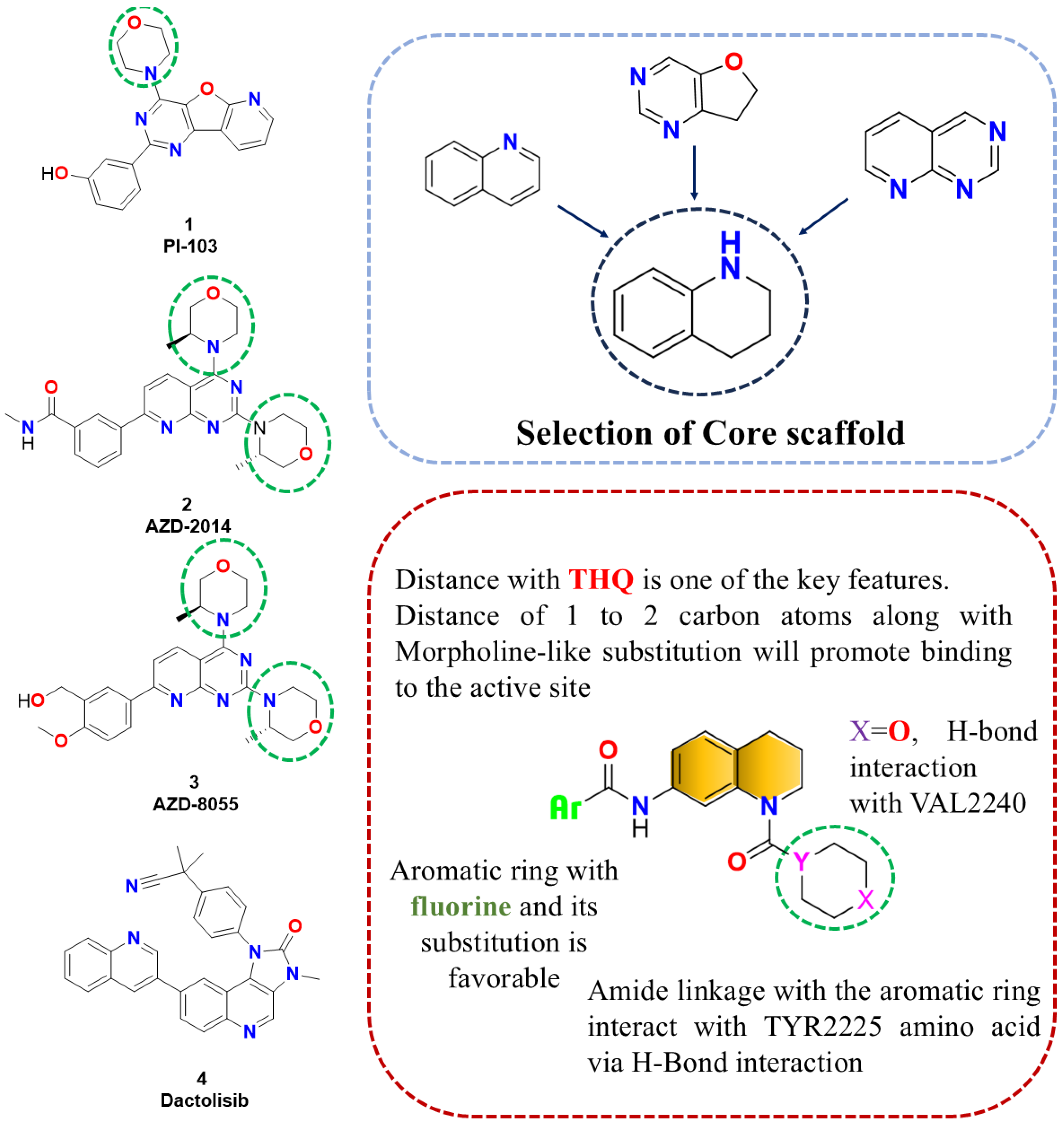
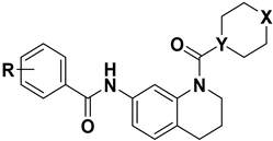
2.1. Designing rationale of Morpholine substituted THQ derivatives

The design strategy for morpholine-substituted tetrahydroquinoline (THQ) derivatives as potential mTOR inhibitors focused on optimizing key molecular features to enhance active site interactions. In the core scaffold, various fluorine/trifluoromethyl-substituted aromatic ring was incorporated, which is favorable for activity [20], with a morpholine-like substitution positioned at a distance of 1–2 carbon atoms from the THQ core to promote binding [19]. An amide linkage connects the aromatic ring, enabling hydrogen bond interactions with the TYR2225 residue, while the oxygen atom (X = O) facilitates additional hydrogen bond interactions with VAL2240 [25]. These structural modifications were strategically chosen to improve binding affinity and specificity toward the mTOR active site.
Tetrahydroquinoline was chosen as the core scaffold based on bioisosteric principles and structural characteristics with significant mTOR inhibitors such as PI-103 (1), AZD-2014 (2), AZD-8055 (3), and dactolisib (4) [26,27,28,29]. PI-103 (1) features a dihydro benzofuran moiety, AZD2014 (2), and AZD8055 (3) incorporate quinazoline cores, respectively, while dactolisib (4) contains a fused quinoline structure. These scaffolds contribute to critical hydrogen bond interactions and π-stacking with residues in the mTOR active site [23]. However, quinoline, quinazoline, and 2,3-dihydrobenzofuran scaffolds were systematically replaced with tetrahydroquinoline due to their ability to maintain similar electronic and spatial properties while improving metabolic stability and reducing off-target effects [30,31]. Furthermore, the inclusion of a morpholine ring, which is typical in most clinical and marketed mTOR inhibitors, was critical for its role in increasing water solubility and facilitating specific interactions with mTOR's ATP-binding pocket [32]. These structural modifications align with established pharmacophoric features, enabling potent and selective mTOR inhibition. Building on our earlier research that established a significant relationship between mTOR and tetrahydroquinoline [19,20,21,33,34], we designed a new set of derivatives to further explore and optimize this interaction. Specifically, a series of substituted N-(1-(morpholine-4-carbonyl) derivatives were synthesized, incorporating the tetrahydroquinoline scaffold for its established mTOR-binding potential.

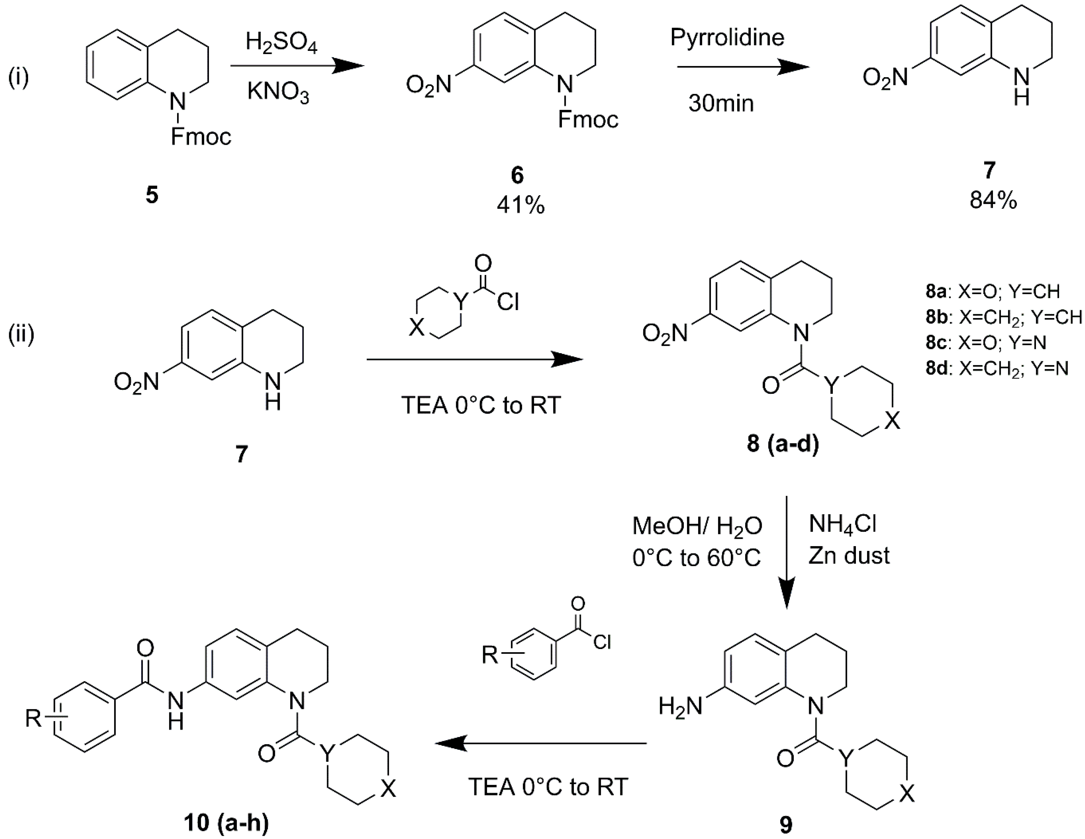
2.2. Synthesis and spectral characterization of designed THQ derivatives

For the synthesis of these designed THQ derivatives, a suitable synthetic scheme was designed which is depicted in Figure 2. Borosilicate glassware and dry solvents were utilized to synthesize all the intermediates and the final desired compounds. There were certain steps in the synthetic scheme where heating and stirring were required which was done with the aid of DIPALI Rota-mantle. Solvents were evaporated with the help of a BUCHI type of rotary vacuum evaporator. Synthesized products were dried under the IR lamp for an appropriate time duration. After drying all synthesized compounds were taken for melting point determination with the help of VEEGO corporation melting point apparatus and are uncorrected. All the THQ derivatives were purified using column chromatography and further their purity was confirmed with the help of the JASCO HPLC instrument and all compounds gave > 95% purity. These tetrahydroquinoline derivatives were characterized with Mass Spectroscopy (WATERS Mass Spectrometer), 1H NMR, and 13C NMR (BRUKER AVANCE-II). For the accurate confirmation and validation of spectral data.

2.2.1. Synthesis of 7-nitro-1,2,3,4-tetrahydroquinoline

The nitrating mixture was prepared using KNO3 (1000 mmol, 0.142 gm) and H2SO4 (1000 mmol, 0.074 mL) and stirred at 0°C for 10 to 15 minutes. At 0 °C, dichloromethane was added to the reaction mixture, stirring for 15 minutes. While keeping the temperature at 0 °C, Fmoc-protected THQ (5) (1000 mmol, 0.5 gm) was added dropwise to the reaction mixture above after being dissolved in dichloromethane. At RT, the reaction mixture was continuously stirred for two hours and thirty minutes. The reaction mixture was poured over crushed ice, and the product was extracted using dichloromethane and then washed with brine. Bright yellow, yield: 41%; mp: 146–148 °C. The crude mixture was then reacted with pyrrolidine to deprotect and remove the Fmoc at room temperature. After 30 min the reaction mixture was extracted with DCM and washed several times with water and brine [35,36].
6-nitro-tetrahydroquinoline and 7-nitro-tetrahydroquinoline (7) were formed with yields of 41% and 25%, respectively. The reaction products were analyzed by HPLC, where the retention times (RT) were 9.33 minutes for 7 nitro-THQ and 9.82 minutes for 6 nitro-THQ, indicating close elution profiles Chromatogram attached as supplementary figure SF-01. The regioisomers were successfully separated using preparative HPLC [37,38].

2.2.2. Synthesis of (7-nitro-3,4-dihydroquinolin-1(2H)-yl)(tetrahydro-2H-pyran-4-yl)methanone (8a)

After dissolving Compound 7 (1000 mmol, 0.2 gm) in dichloromethane, trimethylamine (1000 mmol, 0.16 mL) was added dropwise to the reaction mixture above while being constantly stirred. After 30 minutes of stirring, tetrahydro-2H-pyran-4-carbonyl chloride (1000 mmol, 0.14 mL) was added to the reaction mixture. After being stirred for 24 hours at room temperature, the reaction mixture was put to crushed ice and extracted using dichloromethane. The light-yellow powder was obtained by further washing it with the bicarbonate solution, brine, and sodium sulfate. 66% yield, mp: 177–179 °C.

2.2.3. Synthesis of cyclohexyl(7-nitro-3,4-dihydroquinolin-1(2H)-yl)methanone (8b)

The procedure is the same as 8a only instead of tetrahydro-2H-pyran-4-carbonyl chloride, cyclohexanecarbonyl chloride was used in the reaction. 78% yield, mp: 161-163 °C.

2.2.4. Synthesis of morpholino(7-nitro-3,4-dihydroquinolin-1(2H)-yl)methanone (8c)

The procedure is the same as 8a only instead of tetrahydro-2H-pyran-4-carbonyl chloride, morpholine-4-carbonyl chloride was used in the reaction. 39% yield, mp: 188-190 °C.

2.2.5. Synthesis of (7-nitro-3,4-dihydroquinolin-1(2H)-yl)(piperidin-1-yl)methanone (8d)

The procedure is the same as 8a only instead of tetrahydro-2H-pyran-4-carbonyl chloride, piperidine-1-carbonyl chloride was used in the reaction. 46% yield, mp: 175-176 °C.

2.2.6. Synthesis of (7-amino-3,4-dihydroquinolin-1(2H)-yl)(tetrahydro-2H-pyran-4-yl)methanone (9a)

A 7000 mmol, 0.5 gm combination of ammonium chloride was made with a 3:7 water: methanol ratio. An appropriate amount of methanol was mixed with compound 8a (1000 mmol, 0.3 gm) at the same time, and then zinc dust (10000 mmol, 0.43 gm) was added. This reaction mixture of compound and zinc dust was filled with the ammonium chloride combination mentioned before. The nitro group was reduced to amine after this reaction mixture was stirred for six hours at 60 °C. The mixture was dissolved in ethyl acetate and extracted using bicarbonate after the remaining methanol was removed. The extract was then placed on top of the sodium sulfate and dried, purified by column chromatography
Yield: 68%; mp: 196-198 °C.

2.2.7. Synthesis of (7-amino-3,4-dihydroquinolin-1(2H)-yl)(cyclohexyl)methanone (9b)

The procedure of synthesis of this compound 9b remained the same as that of the synthesis of compound 9a, only difference was that the compound 8b was used instead of 8a.
Yield: 73%; mp: 200-202 °C.

2.2.8. Synthesis of morpholino(7-amino-3,4-dihydroquinolin-1(2H)-yl)methanone (9c)

The procedure for synthesizing compound 9c remained the same as that for synthesizing compound 9a; the only difference was that compound 8c was used instead of 8a.
Yield: 81%; mp: 160-162 °C.

2.2.9. Synthesis of (7-amino-3,4-dihydroquinolin-1(2H)-yl)(piperidin-1-yl)methanone (9d)

The procedure for synthesizing compound 9d remained the same as that for synthesizing compound 9a; the only difference was that compound 8d was used instead of 8a.
Yield: 77%; mp: 177-178 °C.

2.2.10. Synthesis of N-(1-(tetrahydro-2H-pyran-4-carbonyl)-1,2,3,4-tetrahydroquinolin-7-yl)-4-(trifluoromethoxy)benzamide (10a)

Compound 9a (1.7 mmol, 0.5 gm) was dissolved in the DCM. Dropwise trimethylamine (4.9 mmol, 0.46 mL) was added to the above reaction mixture and stirred for 30 min at room temperature. 4-(trifluoromethoxy)benzoyl chloride (1.6 mmol, 0.3 gm) was added to the above reaction mixture, and stirring was continued for 1 h. The reaction mixture was transferred to the crushed ice and extracted with DCM dried over the sodium sulfate resulting in yellow colored compound. The product was purified via column chromatography.
Light yellow Solid Yield: 45.54%; mp: 217-219 °C; 1H NMR (400 MHz, CDCl3) δ PPM: 1.30-1.20 (m, 4H, -CH2), 1.57-1.51 (m, 2H, -CH2); 1.81-1.79 (m, 4H, -CH2), 1.96-1.91 (p, 2H, -CH2), 2.65-2.61 (t, 2H, -CH2); 3.74-3.71 (t, 2H, -CH2) 7.07 (s, 1H, Ar-CH); 7.66 (s, 2H, Ar-CH), 8.01 (s, 1H, Ar-CH), 8.35 (s, 2H, Ar-CH), 8.48 (s, 1H, Ar-CH), 9.03 (s, 1H, NH-CO); 13C NMR (100 MHz, CDCl3) δ PPM: 24.19, 26.26, 29.38, 29.67, 38.64, 67.11, 116.87, 117.23, 119.00, 120.74, 124.14, 128.94, 129.02, 133.24, 135.70, 151.68, 164.45, 174.78; MS (EI) m/z calculated for C23H23F3N2O4 448.44; found 449.47 (M+1); HPLC Purity: 96.13%.

2.2.11. Synthesis of N-(1-(cyclohexanecarbonyl)-1,2,3,4-tetrahydroquinolin-7-yl)-4-(trifluoromethoxy)benzamide (10b)

The reaction process is identical to compound 10a; however, 9b was used to synthesize compound 10b rather than 9a.
Pale yellow Solid Yield: 61.45%; mp: 210-212 °C; 1H NMR (400 MHz, CDCl3) δ PPM: 1.35-1.27 (m, 4H, -CH2), 1.68-1.57 (m, 2H, -CH2); 1.83-1.80 (d, 4H, -CH2), 2.1-1.98 (p, 2H, -CH2), 2.73-2.70 (t, 2H, -CH2); 2.88 (s, 1H, -CH), 3.79-3.76 (t, 2H, -CH2) 7.15-7.13 (d, 1H, Ar-CH2); 8.15-8.13 (d, 1H, Ar-CH); 13C NMR (100 MHz, CDCl3) δ PPM: 24.23, 25.59, 25.70, 26.18, 29.67, 41.59, 116.94, 117.38, 119.01, 120.20, 120.59, 121.59, 128.07, 128.93, 129.15, 132.08, 133.43, 135.77, 151.52, 164.49, 168.56, 176.48; MS (EI) m/z calculated for C24H25F3N2O3 446.47; found 447.46 (M+1); HPLC Purity: 99.06%.

2.2.12. Synthesis of 3,5-difluoro-N-(1-(tetrahydro-2H-pyran-4-carbonyl)-1,2,3,4-tetrahydroquinolin-7-yl)benzamide (10c)

The reaction process is identical to compound 10a; where 9c is reacted with 3,5-difluorobenzoyl chloride in the presence of TEA.
Light yellow crystals Yield: 73.77%; mp: 205-207 °C; 1H NMR (400 MHz, CDCl3) δ PPM: 1.34-1.27 (m, 1H, -CH), 2.05-1.93 (p, 2H, -CH2); 2.77-2.74 (t, 2H, -CH2), 3.36-3.34 (t, 4H, -CH2), 3.60-3.57 (t, 2H, -CH2); 3.80-3.65 (m, 4H, -CH2), 7.04-6.96 (m, 3H, Ar-CH); 7.08-7.06 (d, 1H, Ar-CH), 7.44-7.37 (m, 1H, Ar-CH), 7.65 (d, 1H, Ar-CH), 8.008 (s, 1H, NH); 13C NMR (100 MHz, CDCl3) δ PPM: 23.39, 26.59, 45.54, 46.27, 111.50, 111.98, 112.18, 114.65, 124.23, 129.58, 131.89, 131.99, 132.09, 135.93, 140.91, 158.33, 158.61, 151.68, 159.84, 161.12, 161.19; MS (EI) m/z calculated for C22H22F2N2O3 401.41; found 402.42 (M+1); HPLC Purity: 96.35%.

2.2.12. Synthesis of 3-fluoro-N-(1-(morpholine-4-carbonyl)-1,2,3,4-tetrahydroquinolin-7-yl)-5-(trifluoromethyl)benzamide (10d)

The reaction procedure and reagents are the same as those given for 10a, except 9c, is coupled with 3-fluoro-5-(trifluoromethyl)benzoyl chloride.
White to off white amorphous powder Yield: 41.71%; mp: 218-220 °C; 1H NMR (400 MHz, CDCl3) δ PPM: 1.34-1.27 (m, 1H, -CH), 1.82-1.76 (p, 2H, -CH2); 2.64-2.61 (t, 2H, -CH2), 3.42-3.40 (t, 4H, -CH2), 3.51-3.48 (t, 2H, -CH2); 3.71-3.69 t, 4H, -CH2), 7.02-7.00 (d, 1H, Ar-CH); 7.26-7.21 (d, 1H, Ar-CH), 7.28 (s, 1H, Ar-CH), 7.31 (s, 1H, Ar-CH), 8.12-810. (m, 1H, Ar-CH), 8.20-8.18 (d, 1H, Ar-CH), 8.88 (s, 1H, NH); 13C NMR (100 MHz, CDCl3) δ PPM: 23.13, 26.29 29.70, 46.41, 112.59, 115.10, 117.10, 117.36, 118.54, 118.67, 120.88, 123.58, 123.98, 126.92, 129.58, 131.51, 131.54, 133.68, 136.49, 140.34, 160.19, 160.70, 162.78, 163.36 MS (EI) m/z calculated for C22H21F4N3O3 452.41; found 453.42 (M+1); HPLC Purity: 99.35%.

2.2.13. Synthesis of N-(1-(morpholine-4-carbonyl)-1,2,3,4-tetrahydroquinolin-7-yl)-3,5-bis(trifluoromethyl)benzamide (10e)

The reaction procedure and reagents are the same as those given for 10a, except 9c, is coupled with 3,5-bis(trifluoromethyl)benzoyl chloride.
Yellow amorphous powder Yield: 57.67%; mp: 208-209 °C; 1H NMR (400 MHz, CDCl3) δ PPM: 1.31-1.24 (m, 1H, -CH), 1.48-1.42 (p, 2H, -CH2); 2.43-2.39 (t, 2H, -CH2), 3.46-3.38 (t t, 6H, -CH2), 3.75-3.72 (t, 4H, -CH2), 6.98-6.96 (d, 1H, Ar-CH); 7.38-7.28 (d, 1H, Ar-CH), 8.00 (s, 1H, Ar-CH), 8.37 (s, 2H, Ar-CH), 9.61 (s, 1H, NH); 13C NMR (100 MHz, CDCl3) δ PPM: 14.12, 22.69, 22.79, 25.96, 29.37, 30.18, 31.43, 31.93, 46.58, 46.73, 53.43, 66.58, 113.69, 115.71, 119.00, 121.71, 123.98, 124.54, 127.13, 128.26, 129.53, 131.59, 131.92, 132.25, 136.74, 137.44, 139.81, 161.71, 162.63; MS (EI) m/z calculated for C23H21F6N3O3 501.43; found 502.39 (M+1); HPLC Purity: 97.12%.

2.2.14. Synthesis of 3,5-difluoro-N-(1-(piperidine-1-carbonyl)-1,2,3,4-tetrahydroquinolin-7-yl)benzamide (10f)

The reaction procedure and reagents are the same as those given for 10a, except 9d, is coupled with 3,5-difluorobenzoyl chloride.
Bright yellow powder Yield: 71.20%; mp: 221-223 °C; 1H NMR (400 MHz, DMSO-d6) δ PPM: 1.51-1.49 (m, 6H, -CH2), 1.87-1.84 (p, 2H, -CH2); 2.72-2.68 (t, 2H, -CH2), 3.26 (s, 4H, -CH2), 3.44-3.41 (t, 2H, -CH2), 7.14-7.05 (d, 1H, Ar-CH); 7.25-7.21 (d, 1H, Ar-CH), 7.37-7.36 (t, 2H, Ar-CH), 7.37 (s, 1H, Ar-CH), 7.59-7.54 (p, 1H, Ar-CH), 10.61 (s, 1H, NH); 13C NMR (100 MHz, DMSO-d6) δ PPM: 23.03, 24.55, 25.57, 26.50, 46.16, 46.54, 110.24, 112.37, 112.85, 116.06, 122.50, 129.77, 132.28, 132.48, 137.12, 141.49. 157.95, 158.03, 158.30, 159.71, 160.42, 160.50; MS (EI) m/z calculated for C22H23F2N3O2 399.42; found 400.55 (M+1); HPLC Purity: 95%.

2.2.15. Synthesis of 3-fluoro-N-(1-(piperidine-1-carbonyl)-1,2,3,4-tetrahydroquinolin-7-yl)-5-(trifluoromethyl)benzamide (10g)

The reaction procedure and reagents are the same as those given for 10a, except 9d, is coupled with 3-fluoro-5-(trifluoromethyl)benzoyl chloride.
Off white powder Yield: 44.32%; mp: 213-215 °C; 1H NMR (400 MHz, CDCl3) δ PPM: 1.63-1.59 (m, 6H, -CH2), 1.74-1.71 (p, 2H, -CH2); 2.61-2.59 (t, 2H, -CH2), 3.37-3.36 (s, 4H, -CH2), 3.44-3.42 (t, 2H, -CH2), 6.98-6.96 (d, 1H, Ar-CH); 7.15 (d, 1H, Ar-CH), 7.36-7.24 (m, 1H, Ar-CH), 7.38-7.36 (d, 1H, Ar-CH), 8.15-8.12 (m, 1H, Ar-CH), 8.24-8.22 (d, 1H, Ar-H), 9.18 (s, 1H, NH); 13C NMR (100 MHz, CDCl3) δ PPM: 23.34, 24.56, 25.12, 26.35, 46.22, 46.56, 110.35, 112.43, 112.54, 116.78, 122.23, 129.65, 132.28, 132.48, 137.12, 141.49. 157.95, 158.03, 158.30, 159.71, 160.42, 160.50; MS (EI) m/z calculated for C23H23F4N3O2 449.45; found 450.53 (M+1); HPLC Purity: 99.36%.

2.2.16. Synthesis of N-(1-(piperidine-1-carbonyl)-1,2,3,4-tetrahydroquinolin-7-yl)-3,5 bis(trifluoromethyl)benzamide (10h)

The reaction procedure and reagents are the same as those given for 10a, except 9d, is coupled with 3-5-(trifluoromethyl)benzoyl chloride.
Yellow powder Yield: 65.23 %; mp: 207-208 °C; 1H NMR (400 MHz, CDCl3) δ PPM: 1.30-1.27 (s, 2H, CH2); 1.54-1.51 (p, 2H, -CH2); 1.67-1.63 (m, 6H, -CH2), 2.51-2.47 (t, 2H, -CH2), 3.42-3.40 (m, 6H, -CH2), 7.00-6.98 (d, 1H, Ar-CH); 7.18 (d, 1H, Ar-CH), 7.43-7.40 (dd, 1H, Ar-CH), 8.01 (s, 1H, Ar-CH), 8.24-8.22 (d, 1H, Ar-H), 8.39 (s, 2H, Ar-CH), 9.33 (s, 1H, NH); 13C NMR (100 MHz, CDCl3) δ PPM: 22.65, 24.60, 25.85, 26.06 29.07, 30.17 31.43, 47.15, 113.63, 115.32, 121.75, 123.76, 124.59, 124.76, 127.18, 128.41, 129.44, 131.17, 131.51, 132.17, 136.73, 137.64, 140.16, 161.99, 162.77 ; MS (EI) m/z calculated for C24H23F6N3O2 449.46; found 500.52 (M+1); HPLC Purity: 97.18%.

2.3. In-vitro antiproliferative MTT assay

In order to assess the antiproliferative efficacy of all the synthesized Tetrahydroquinoline derivatives, a panel of three distinct human cancer cell lines breast cancer (MCF-7), lung cancer (A-549), and triple-negative breast cancer (MDA-MB-231) was used [39,40,41]. Additionally, these compounds were evaluated against the VERO cell line to anticipate their toxicity. To detect the VERO toxin, a VERO cell line is typically extracted from the kidney of an adult green monkey [42]. Therefore, in order to determine the toxicity of the synthesized compounds, we employed this cell line in the current study. The RPMI-1650 was used to cultivate the VERO cell lines [43]. The F-12 Ham's medium was used to cultivate the lung cancer (A-549) cell lines [44]. DMEM was used to cultivate the cell lines for breast cancer (MCF-7) and triple-negative breast cancer (MDA-MB-231). Because it feeds the cell line, 5% fatal bovine serum (FBS) was used to make the whole medium. To further protect the cell line from contamination, 0.5% pen-strep (Penicillin + Streptomycin) was added to all the mediums. All of these cell lines were cultivated in sterile, vented T-75 and/or T-25 flasks that were kept at 37 °C and supplied with 5% CO2. All of the compounds' stock solutions were prepared in DMSO, and dilutions were made in the appropriate media so that the 96-well plates' final DMSO content was not greater than 0.1%. First, each drug was tested in triplicate against every cell line at a 25 µM dose. To prepare the serial dilutions and calculate the IC50 values, the compounds exhibiting greater than 50% inhibition were selected. On the first day, 1 × 10⁴ cells were planted, and 24 hours later, the chemicals were added. The MTT dye was added after the compounds and cells had been cultured for 48 hours. After four hours, the MTT dye was taken out of the wells. Each well received 100 µL of DMSO to dissolve the formazan crystals that had formed as a result of the living cells, and absorbance at 570 nm was measured [45].

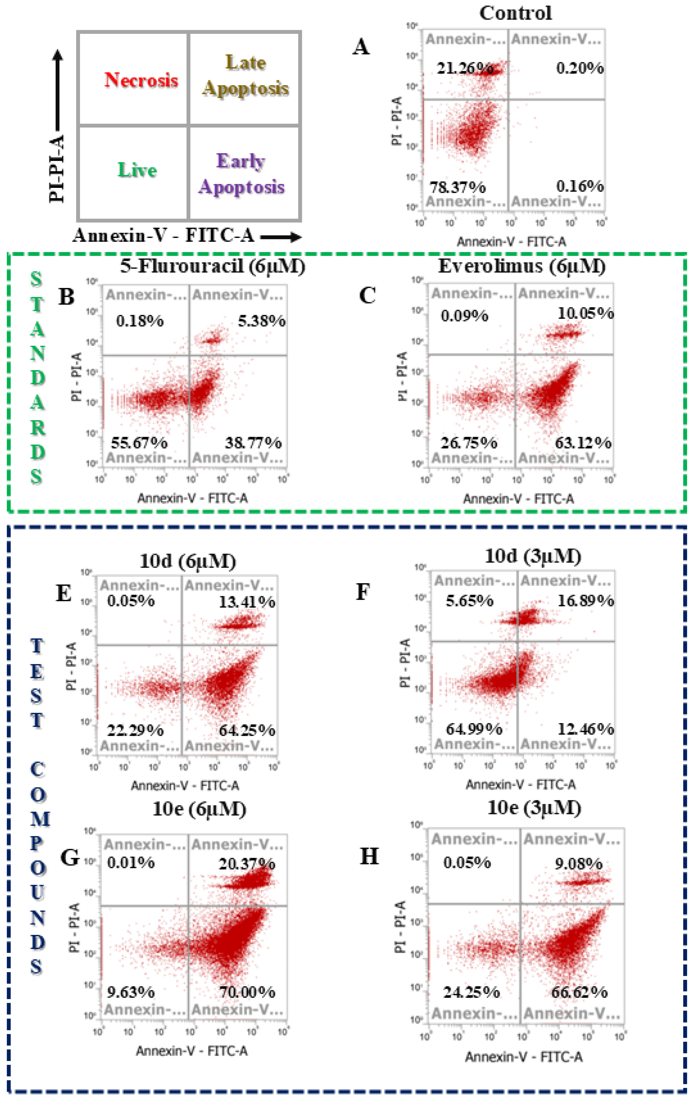
2.4. Apoptosis assay

A549 cells of the lung cancer cell line were seeded in 6 well plates which were supplemented with F-12 Ham’s media, 5% CO2, 5% FBS, and 0.5% pen-strep at 37 ◦C for 24 h. Later, the best 3 compounds, in terms of IC50 values, were taken in two concentrations i.e. 3 µM and 6 µM, respectively. The six wells were maintained in duplicate for the DMSO control and for the positive control 5-fluorouracil and Everolimus were taken. The treated cells, along with the DMSO control group, were incubated for 48 hours. Following the incubation period, the media was aspirated from each well, and 1 mL of trypsin was added to each well for approximately one minute. This was then neutralized with 4 mL of media. Subsequent steps were performed according to the instructions provided in the Annexin V-FITC apoptosis detection kit (Ref Number: BMS500FI, Lot No: 264043; Invitrogen). The contents of each well were collected and transferred into centrifuge tubes, followed by centrifugation at 1000 RPM for 8 minutes. The media was removed, and the cell pellets were washed with PBS. After another centrifugation step, 1 mL of the 1X binding buffer supplied with the Annexin V-FITC apoptosis detection kit was added to the pellets. For 15 to 20 minutes, these tubes were left in the dark. Each plastic flow cytometric tube received 500 µL of this cell suspension of the 1X binding buffer. Each tube was filled with 10 µL of propidium iodide (PI) and 5 µL of Annexin-V FITC conjugate. After these tubes were shielded from the light and incubated for ten minutes, flow cytometry was used to examine the cell apoptosis [46,47,48].

2.5. Molecular Docking

Docking studies were performed using AutoDock Vina software version 4.2.6 [49,50]. The 3D structure of the core protein mTOR (PDB ID: 4JT6) was obtained from the Protein Data Bank (https://www.rcsb.org/). The preparation process included dehydration, hydrogenation, and charge modification, which were carried out using AutoDock software version 4.2.6. This resulted in the modified target protein and all ligands being exported as PDBQT files. Docking simulations were conducted using AutoDock Vina software version 1.2.0, and the results were analyzed and visualized with Discovery Studio 2021 Client software.

2.6. Molecular Dynamics Simulation Study

Molecular dynamics (MD) simulation was conducted using GROMACS 2021.4 (https://manual.gromacs.org/2021.4/index.html) to model the dynamic interactions between the protein and ligand, thereby validating the accuracy of the drug design and supporting the study's rationale [51,52,53]. Protein topology files were generated with the OPLS force field (Optimized Potential for Liquid Simulations, version 15) using the 'pdb2gmx' command. Ligand files compatible with GROMACS were prepared via the LigParagen web server (https://traken.chem.yale.edu/ligpargen/). The protein was solvated in a dodecahedron box with a three-point water model and neutralized using sodium (Na⁺) and chloride (Cl⁻) ions as counterions. Energy minimization of the protein-ligand system was performed using the 'gmx grompp' and 'gmx mdrun' commands. Equilibration was achieved under NVT (Canonical) and NPT (Isobaric) ensembles, with a V-rescale thermostat maintaining a constant temperature of 300 K and a fixed volume. The system underwent a 100-ns MD simulation, during which stability analyses were conducted. These included calculations of root mean square deviation (RMSD), root mean square fluctuation (RMSF), radius of gyration (Rg), solvent-accessible surface area (SASA), and hydrogen bond count (HBond) using the 'gmx rms' command.

3. Results and Discussion

3.1. Chemistry

A strategic synthetic approach was used to synthesize 8 new tetrahydroquinoline (THQ) derivatives, as shown in Figure 2. To protect the THQ molecule's nitrogen atom, 9-fluorenemethoxycarbonyl (Fmoc) was first applied to it. When THQ reacted with sulfuric acid and potassium nitrate, this protection allowed for selective nitration at the 7th position. Pyrrolidine was used to deprotect the Fmoc-protected THQ after nitration. The resulting compound was then reacted with various acid chlorides, including tetrahydro-2H-pyran-4-carbonyl chloride, cyclohexanecarbonyl chloride, morpholine-4-carbonyl chloride, and piperidine-1-carbonyl chloride, as outlined in the synthetic scheme. Subsequently, these intermediates were treated with ammonium chloride and zinc to reduce the nitro group to an amine. The resulting primary amine was then subjected to reactions with different aromatic acid chlorides (denoted as R in the scheme), culminating in the synthesis of the desired eight novel THQ derivatives. The final compounds were characterized using 1H and 13C NMR spectroscopy and mass spectrometry, and their purity was assessed via HPLC analysis. All synthesized derivatives exhibited >95% purity, as confirmed by the HPLC data. Detailed spectra and chromatograms are provided in the supporting information section.

3.2. In-vitro antiproliferative MTT assay

The MTT assay results revealed that morpholine-substituted tetrahydroquinoline derivatives exhibited potent and selective cytotoxicity against A549, MCF-7, and MDA-MB-231 cancer cell lines, with minimal toxicity towards Vero cells. Compound 10e demonstrated the highest activity against A549 cells (IC₅₀ = 0.033 ± 0.003 µM), while 10h was most effective against MCF-7 cells (IC₅₀ = 0.087 ± 0.007 µM). Compound 10d showed broad-spectrum activity with IC₅₀ values of 0.062 ± 0.01 µM, 0.58 ± 0.11 µM, and 1.003 ± 0.008 µM for A549, MCF-7, and MDA-MB-231, respectively. Structure-activity relationship analysis indicated that compounds with X = O and Y = N (e.g., 10d and 10e) exhibited superior activity compared to those with X = CH2 or Y = CH, likely due to enhanced mTOR binding affinity facilitated by the electronegative oxygen.

3.3. Structure-Activity Relationship

Structure-activity relationship (SAR) research of the morpholine-substituted tetrahydroquinoline derivatives provides important information about how certain structural characteristics and functional groups affect the cytotoxic activity against MDA-MB-231, MCF-7, and A549 cancer cell lines. The cytotoxic action is greatly influenced by the substituents on the benzamide molecule. Two trifluoromethyl groups that are highly electron-withdrawing increased the cytotoxicity of all investigated cell lines. With these groups, compound 10e was the most potent derivative, showing the lowest IC₅₀ values for MDA-MB-231 (0.63 ± 0.02 µM) and A549 (0.033 ± 0.003 µM). Similarly, 10h, which also features 3,5-bis(trifluoromethyl) substitution, showed excellent activity against MCF-7 cells (IC₅₀ = 0.087 ± 0.007 µM). The potency of these derivatives can be attributed to enhanced interactions with the mTOR active site, likely through halogen bonding and hydrophobic interactions mediated by the trifluoromethyl groups. The combination of a single fluorine atom at position 3 and a trifluoromethyl group at position 5 also resulted in high activity. Compound 10d displayed IC₅₀ values of 0.062 ± 0.01 µM for A549 and 0.58 ± 0.11 µM for MCF-7. This suggests that the strategic placement of fluorine and trifluoromethyl groups is optimal for maintaining high activity while reducing toxicity. Substitution with two fluorine atoms at positions 3 and 5 resulted in moderate activity. Compound 10c showed IC₅₀ values of 3.73 ± 0.17 µM across the lung cancer cell lines, while compound 10f displayed selective activity against MCF-7 (IC₅₀ = 4.47 ± 0.013 µM). The decreased potency compared to trifluoromethyl derivatives suggests that the bulkier and more electronegative trifluoromethyl groups provide stronger interactions and better activity.
The oxygen atom (X = O) plays a pivotal role in enhancing the cytotoxic activity. The electronegative oxygen enhances the interaction potential of the molecule with mTOR binding residues, possibly by forming hydrogen bonds or improving electronic complementarity. Compounds with X = O consistently outperformed their counterparts with X = CH₂ in terms of potency. For example, 10e (X = O) was significantly more active than 10h (X = CH₂), despite having identical R and Y groups. Replacing the oxygen with a methylene group reduced activity across all cell lines. For instance, 10f (X = CH₂, R = 3,5-difluoro) exhibited no activity against A549 or MDA-MB-231, in contrast to its X = O counterpart, 10c, which showed moderate activity. This highlights the importance of the oxygen atom in maintaining mTOR binding interactions. The nitrogen atom enhances the electronic complementarity of the molecule and contributes to better binding with mTOR residues. Compounds with Y = N consistently showed higher activity compared to their Y = CH counterparts. For instance, 10a (Y = CH, X = O) had an IC₅₀ of 1.06 ± 0.02 µM against A549, whereas 10c (Y = N, X = O) displayed improved potency with an IC₅₀ of 3.73 ± 0.17 µM across all cancer lines. All derivatives exhibited minimal cytotoxicity towards Vero cells (IC₅₀ >25 µM for most compounds), indicating good selectivity for cancer cells. Compounds with strong electron-withdrawing substituents (e.g., 10e and 10d) showed the best selectivity index (SI), demonstrating their potential as targeted mTOR inhibitors.

3.4. Apoptosis Assay

The FACS analysis results, illustrated in Figure 4A–F, provide a detailed examination of the apoptotic potential of the studied compounds, including the standard chemotherapeutic agents 5-FU and Everolimus, alongside four novel derivatives. This characterisation offers critical insights into their mechanisms of action, apoptotic pathways, and potential as therapeutic agents.
Treatments of A549 cells with the synthesised compounds 10d and 10e at the concentrations of 3 µM and 6 µM for 48 hours demonstrated a significant induction of apoptosis (early and last phase combined). Specifically, compound 10d induced 29.35% apoptosis at 3 µM and 77.66% at 6 µM, while compound 10e exhibited even greater efficacy, with apoptosis levels reaching 75.7% and 90.37% at the same concentrations. In contrast, the control group treated with DMSO showed only 0.36% apoptosis in 44.15% of cells, serving as a comparative benchmark. A closer examination of the effects of 10e at 6 µM revealed profound changes in cell viability and apoptotic induction. The percentage of live cells decreased drastically from 7837% (Figure 4A) in the control group to 24.25% (Figure 4H) in the treated group. This reduction in cell viability was accompanied by a sharp increase in apoptotic cells, rising from a negligible 0.36% in the control group to an impressive 90.37%. Notably, the apoptotic population consisted of 70.00% early apoptotic cells and 20.37% late apoptotic cells, indicating that compound 10e primarily drives cells into the early stages of programmed cell death. This mode of action suggests a targeted apoptotic mechanism rather than necrosis, as evidenced by the negligible necrotic cell population observed (0.05% at 3µM & 0.01% at 6µM).
The comparison with 5-FU highlights the remarkable efficacy of compound 10e. While 5-FU is a well-established chemotherapeutic agent, 10e suggests a more controlled and potentially less inflammatory cell death pathway, which is a desirable characteristic in anticancer agents to minimise adverse effects on surrounding tissues. The observed apoptotic effects of compounds 10d and 10e were dose-dependent, with higher concentrations yielding greater efficacy. This dose-response relationship emphasises the potential for fine-tuning therapeutic regimens to maximise efficacy while minimising side effects. Additionally, the significant early apoptotic activity observed with 10e could be explored further to understand its impact on downstream apoptotic signalling pathways, such as caspase activation and mitochondrial membrane potential disruption.
This comprehensive analysis not only underscores the potent apoptotic properties of compound 10e but also establishes its potential as a lead compound for the development of novel anticancer therapies. The findings pave the way for future studies, including mechanistic investigations into its molecular targets, in-vivo efficacy in animal models, and optimisation of its pharmacokinetic and pharmacodynamic profiles. With its demonstrated apoptotic potency, minimal necrosis, and favourable comparison to 5-FU, compound 10e represents a promising candidate for clinical development, offering hope for improved therapeutic options in the treatment of lung cancer.

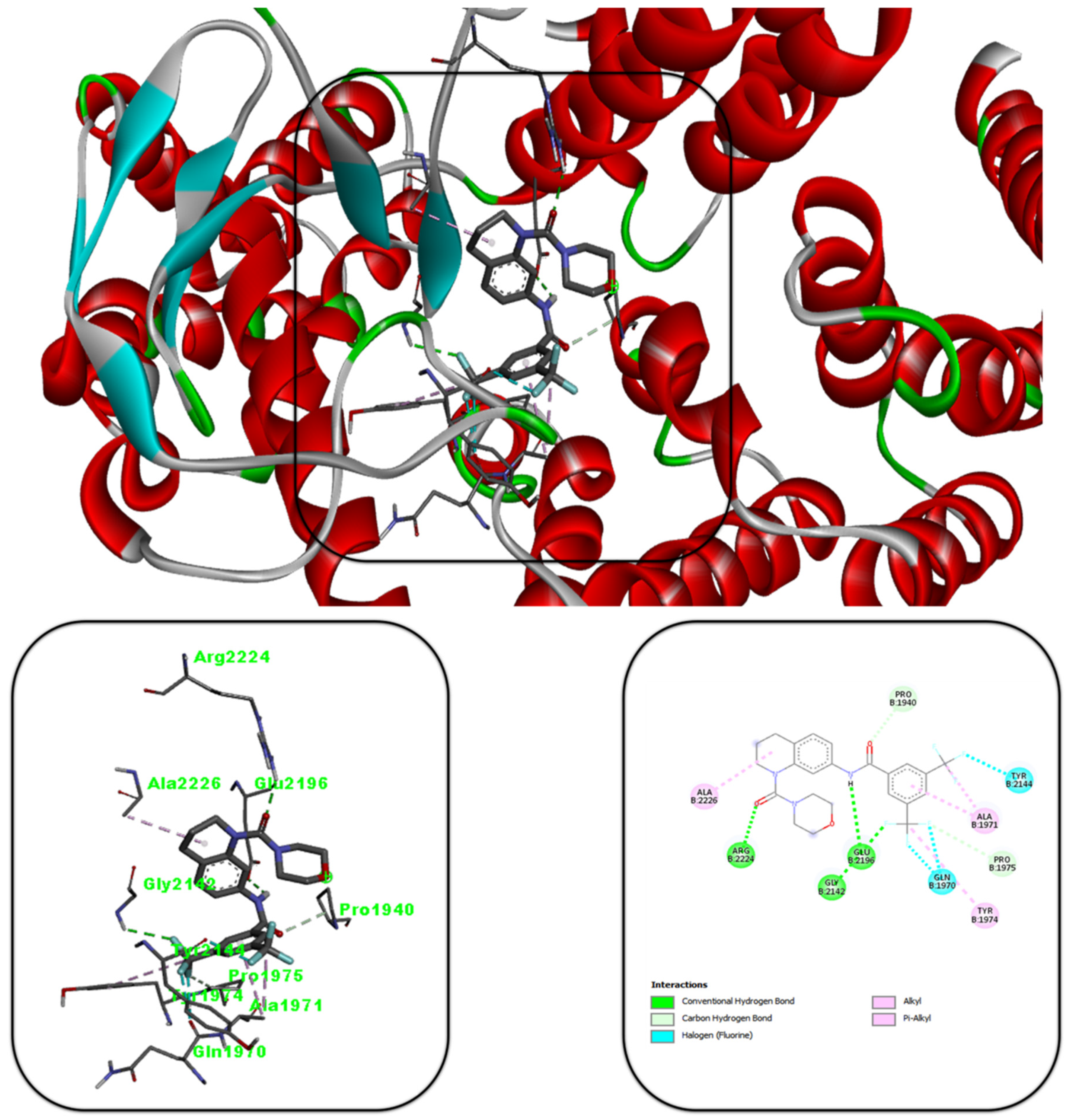
3.5. Molecular Docking Studies
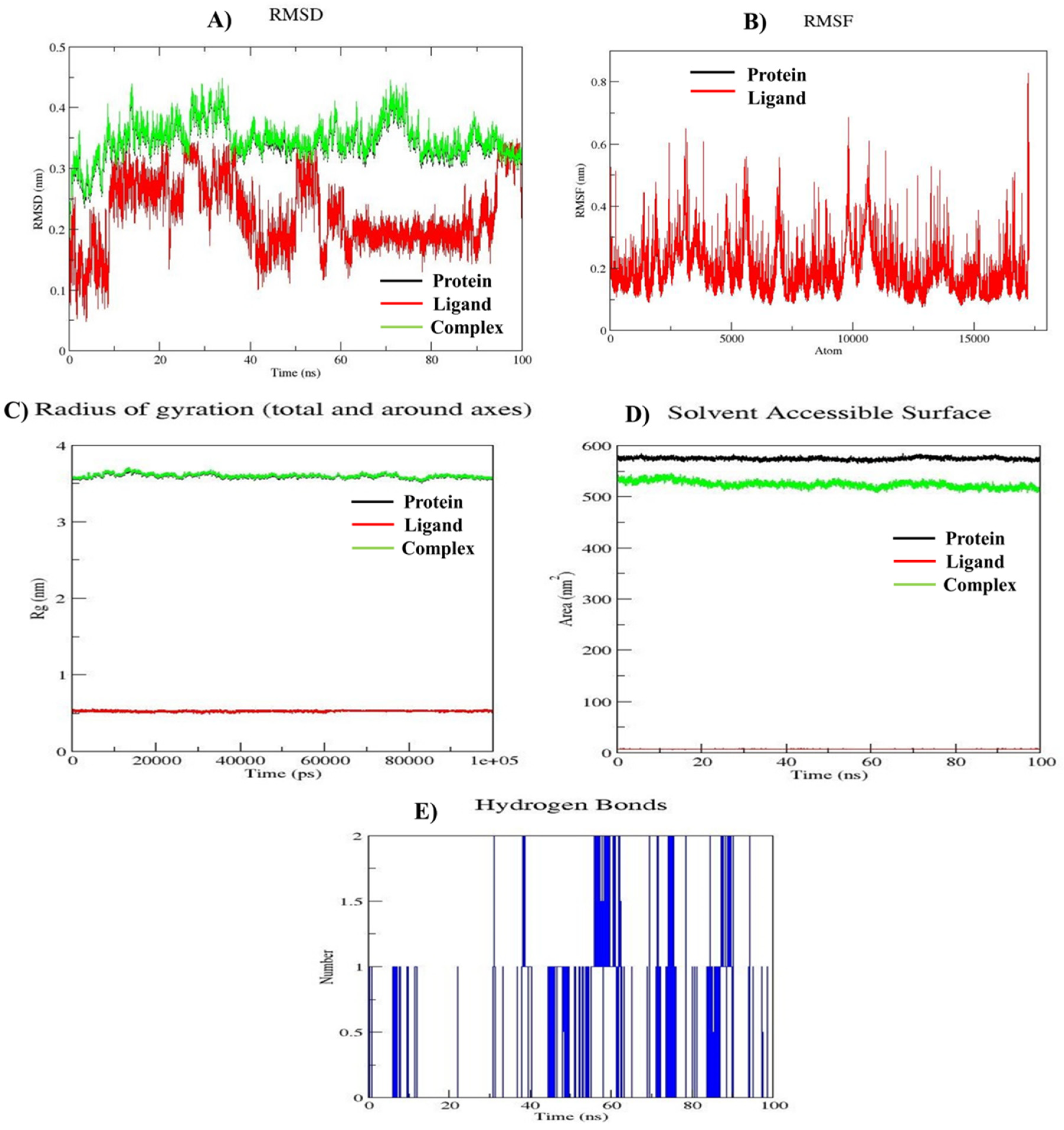
To explore the potential mode of action, binding mode analysis was conducted for all the synthetic compounds, with compound 11 demonstrating the most favorable results compared to the co-crystal ligand (X6K). The docking score of compound 10e (-10.6 kcal/mol) indicated a lower binding energy than the co-crystal ligand (-10.1 kcal/mol), highlighting its enhanced binding efficiency. The binding interactions of compound 10e are summarized in Figure 5. The carbonyl group (C=O) forms a hydrogen bond with Arg2224, while the fluorine group establishes a hydrogen bond with Glu2142. Additionally, the -NH group forms a hydrogen bond with Glu2196. These interactions suggest that the synthesized compound binds stably to the mTOR protein and exhibits cytotoxic effects.

3.6. MD Simulation

The molecular dynamics simulation results for the mTOR protein (PDB ID: 4JT6) in complex with compound 10e, as depicted in Figure 5, demonstrate the stability of the protein-ligand complex throughout the 100 ns simulation period. This is supported by the root mean square deviation (RMSD) graph, which shows consistent stability over time. The root mean square fluctuation (RMSF) analysis, shown in Figure 5(B), reveals moderate fluctuations ranging from 0.1 nm to 0.8 nm. This indicates dynamic behavior of residues at the protein-ligand interface while maintaining overall stability. Additionally, the compactness and structural integrity of the complex are confirmed by the radius of gyration (Rg) values, which remain close to 3.5 nm. The solvent-accessible surface area (SASA), as illustrated in Figure 5(D), highlights stable folding and compactness of the protein structure, further confirming the system's stability throughout the simulation. Hydrogen bonds, which play a critical role in the complex's stability and rigidity, were also analyzed. As shown in Figure 5(E), the hydrogen bonds formed between the ligand and the protein persist consistently, restricting protein movement and enhancing stability. These results collectively indicate that the mTOR-compound 10e complex is structurally stable and dynamically active, offering valuable insights into its potential as a therapeutic target. These findings warrant further investigation and support its promise in drug development efforts.

4. Discussion

This study successfully demonstrates the synthesis, characterization, and evaluation of morpholine-substituted tetrahydroquinoline (THQ) derivatives as potential mTOR inhibitors. The strategic inclusion of morpholine and trifluoromethyl groups in the THQ scaffold was pivotal in achieving enhanced selectivity and potency against cancer cell lines. Molecular docking and dynamics simulations corroborated the in vitro results, establishing the compounds' potential as therapeutic agents for targeting mTOR in lung cancer.
The synthetic strategy yielded eight novel THQ derivatives, with each compound characterized by advanced spectroscopic techniques, including 1H NMR, 13C NMR, and mass spectrometry. High-performance liquid chromatography (HPLC) confirmed the purity of all derivatives (>95%). This meticulous characterization ensured the structural integrity and quality of the compounds, which is critical for correlating their structural features with biological activity.
The MTT assay results highlighted the significant antiproliferative activity of the synthesized compounds against A549, MCF-7, and MDA-MB-231 cancer cell lines. Compound 10e emerged as the most potent derivative, exhibiting an IC₅₀ value of 0.033 ± 0.003 µM against A549 cells. The structure-activity relationship (SAR) analysis revealed that electronegative substituents, such as trifluoromethyl groups, significantly enhance binding affinity to the mTOR active site. Additionally, the inclusion of oxygen (X = O) in the scaffold improved cytotoxic activity, emphasizing the importance of electronic and steric factors in mTOR inhibition.
Flow cytometry results demonstrated that compounds 10d and 10e induced apoptosis in a dose-dependent manner, with compound 10e achieving over 90% apoptosis at 6 µM in A549 cells. The predominance of early apoptosis suggests a specific and controlled mechanism of action, distinguishing these derivatives from necrosis-inducing agents. This targeted apoptotic effect aligns with the desired therapeutic profile for anticancer agents, minimizing off-target effects.
Molecular docking studies revealed strong binding interactions between the derivatives and key residues in the mTOR active site, such as Arg2224 and Glu2142. The docking score of compound 10e (-10.6 kcal/mol) surpassed that of the co-crystal ligand (-10.1 kcal/mol), indicating superior binding efficiency. Molecular dynamics simulations further validated these findings, showing stable protein-ligand interactions over a 100-ns simulation period. Parameters such as root mean square deviation (RMSD), radius of gyration (Rg), and hydrogen bonding confirmed the structural stability of the complexes.

5. Conclusion

This research underscores the potential of morpholine-substituted tetrahydroquinoline derivatives as promising mTOR inhibitors for targeted cancer therapy, particularly against lung cancer. Among the synthesized compounds, 10e exhibited the most potent cytotoxic activity against A549 cells, with an exceptional IC₅₀ value of 0.033 ± 0.003 µM, highlighting its potential as a lead candidate. Compound 10h demonstrated remarkable activity against MCF-7 cells (IC₅₀ = 0.087 ± 0.007 µM), and 10d showed broad-spectrum efficacy across A549, MCF-7, and MDA-MB-231 cells. These derivatives not only exhibited high selectivity indices but also demonstrated a dose-dependent induction of apoptosis, particularly through early apoptotic pathways, thereby minimizing necrotic and off-target effects.
The molecular docking and dynamics simulations further confirmed the experimental observations. Compound 10e, with a docking score of -10.6 kcal/mol, demonstrated superior binding efficiency and stability within the mTOR active site compared to the co-crystal ligand. The structural integrity and stability of the protein-ligand complexes over the simulation period reinforce the therapeutic promise of these derivatives.
These findings advocate for further preclinical evaluations, including in-vivo efficacy studies and pharmacokinetic profiling. The incorporation of trifluoromethyl groups and morpholine moieties in the THQ scaffold emerges as a critical design strategy for developing effective and selective mTOR inhibitors. Compound 10e, in particular, represents a compelling candidate for advancing lung cancer therapeutics.

Financial and Competing Interests Disclosure

This work was supported by the Minor Research Project IPNU/2024-26/144 funded by Nirma University, Ahmedabad, India. The authors have no other relevant affiliations or financial involvement with any organization or entity with a financial interest in or financial conflict with the subject matter or materials discussed in the manuscript apart from those disclosed. No writing assistance was utilized in the production of this manuscript.

Declaration of Interest Statement

The authors declare that there are no competing financials.

Author Contributions

RD & UC conceptualized the work. RD and SS performed all synthetic work under the guidance of UC. The in vitro studies were conducted by SS and RY under the guidance of AJ. RY performed all the in-silico studies. RD, SS, and RY prepare the manuscript overviewed critically by BP, and HB, and reviewed by UC & AJ. BP, HB, GN, UC, and AJ did the final review and editing of the manuscript. All authors have approved the final version of the manuscript.

Acknowledgement

All the authors are thankful to Nirma University, Ahmedabad, India for providing financial assistance to carry out the Minor Research Project IPNU/2024-26/144.

References

  1. Vishwakarma, K.; Dey, R.; Bhatt, H. Telomerase: A Prominent Oncological Target for Development of Chemotherapeutic Agents. European Journal of Medicinal Chemistry 2023, 249, 115121. [CrossRef]
  2. Dey, R.; Vishwakarma, K.; Patel, B.; Vyas, V.K.; Bhatt, H. Evolution of Telomerase Inhibitors: A Review on Key Patents from 2015 to 2023. ChemistrySelect 2024, 9, e202404444. [CrossRef]
  3. Soliman, G. The Role of Mechanistic Target of Rapamycin (mTOR) Complexes Signaling in the Immune Responses. Nutrients 2013, 5, 2231–2257. [CrossRef]
  4. Zou, Z.; Tao, T.; Li, H.; Zhu, X. mTOR Signaling Pathway and mTOR Inhibitors in Cancer: Progress and Challenges. Cell Biosci 2020, 10, 31. [CrossRef]
  5. Bouyahya, A.; El Allam, A.; Aboulaghras, S.; Bakrim, S.; El Menyiy, N.; Alshahrani, M.M.; Al Awadh, A.A.; Benali, T.; Lee, L.-H.; El Omari, N.; et al. Targeting mTOR as a Cancer Therapy: Recent Advances in Natural Bioactive Compounds and Immunotherapy. Cancers 2022, 14, 5520. [CrossRef]
  6. Saxton, R.A.; Sabatini, D.M. mTOR Signaling in Growth, Metabolism, and Disease. Cell 2017, 168, 960–976. [CrossRef]
  7. Panwar, V.; Singh, A.; Bhatt, M.; Tonk, R.K.; Azizov, S.; Raza, A.S.; Sengupta, S.; Kumar, D.; Garg, M. Multifaceted Role of mTOR (Mammalian Target of Rapamycin) Signaling Pathway in Human Health and Disease. Sig Transduct Target Ther 2023, 8, 375. [CrossRef]
  8. Qaderi, S.; Javinani, A.; Blumenfeld, Y.J.; Krispin, E.; Papanna, R.; Chervenak, F.A.; Shamshirsaz, A.A. Mammalian Target of Rapamycin Inhibitors: A New-possible Approach for In-utero Medication Therapy. Prenatal Diagnosis 2024, 44, 88–98. [CrossRef]
  9. Mao, B.; Zhang, Q.; Ma, L.; Zhao, D.-S.; Zhao, P.; Yan, P. Overview of Research into mTOR Inhibitors. Molecules 2022, 27, 5295. [CrossRef]
  10. Dey, R.; Chaube, U.; Bhatt, H.; Patel, B. Small Molecule Drug Design. In Reference Module in Life Sciences; Elsevier, 2024; p. B9780323955027002621 ISBN 978-0-12-809633-8.
  11. Meng, L.; Zheng, X.S. Toward Rapamycin Analog (Rapalog)-Based Precision Cancer Therapy. Acta Pharmacol Sin 2015, 36, 1163–1169. [CrossRef]
  12. Li, J.; Kim, S.G.; Blenis, J. Rapamycin: One Drug, Many Effects. Cell Metabolism 2014, 19, 373–379. [CrossRef]
  13. Zhong, L.; Li, Y.; Xiong, L.; Wang, W.; Wu, M.; Yuan, T.; Yang, W.; Tian, C.; Miao, Z.; Wang, T.; et al. Small Molecules in Targeted Cancer Therapy: Advances, Challenges, and Future Perspectives. Sig Transduct Target Ther 2021, 6, 201. [CrossRef]
  14. Liu, J.; Li, H.; He, Q.; Chen, K.; Chen, Y.; Zhong, R.; Li, H.; Fang, S.; Liu, S.; Lin, S. Design, Synthesis, and Biological Evaluation of Tetrahydroquinoline Amphiphiles as Membrane-Targeting Antimicrobials against Pathogenic Bacteria and Fungi. European Journal of Medicinal Chemistry 2022, 243, 114734. [CrossRef]
  15. Manolov, S.; Ivanov, I.; Bojilov, D. Synthesis of New 1,2,3,4-Tetrahydroquinoline Hybrid of Ibuprofen and Its Biological Evaluation. Molbank 2022, 2022, M1350. [CrossRef]
  16. Fathy, U.; Azzam, M.A.; Mahdy, F.; El-Maghraby, S.; Allam, R.M. Synthesis and in Vitro Anticancer Activity of Some Novel Tetrahydroquinoline Derivatives Bearing Pyrazol and Hydrazide Moiety. Journal of Heterocyclic Chem 2020, 57, 2108–2120. [CrossRef]
  17. Gangapuram, M.; Eyunni, S.; Zhang, W.; Redda, K.K. Design and Synthesis of Tetrahydroisoquinoline Derivatives as Anti-Angiogenesis and Anti-Cancer Agents. ACAMC 2021, 21, 2505–2511. [CrossRef]
  18. Kumari, A.; Singh, R.K. Morpholine as Ubiquitous Pharmacophore in Medicinal Chemistry: Deep Insight into the Structure-Activity Relationship (SAR). Bioorganic Chemistry 2020, 96, 103578. [CrossRef]
  19. Shaw, S.; Dey, R.; Bhatt, H.; Patel, B.; Patel, S.; Dixit, N.; Chaube, U. Designing of Target-specific N -substituted Tetrahydroquinoline Derivatives as Potent mTOR Inhibitors via Integrated Structure-guided Computational Approaches. ChemistrySelect 2024, 9, e202303291. [CrossRef]
  20. Dey, R.; Shaw, S.; Bhatt, H.; Patel, B.; Yadav, R.; Chaube, U. Synthesis, Biological Screening, and Binding Mode Analysis of Some N-Substituted Tetrahydroquinoline Analogs as Apoptosis Inducers and Anticancer Agents. Journal of Molecular Structure 2024, 1318, 139330. [CrossRef]
  21. Chaube, U.J.; Rawal, R.; Jha, A.B.; Variya, B.; Bhatt, H.G. Design and Development of Tetrahydro-Quinoline Derivatives as Dual mTOR-C1/C2 Inhibitors for the Treatment of Lung Cancer. Bioorganic Chemistry 2021, 106, 104501. [CrossRef]
  22. Chhatbar, D.M.; Chaube, U.J.; Vyas, V.K.; Bhatt, H.G. CoMFA, CoMSIA, Topomer CoMFA, HQSAR, Molecular Docking and Molecular Dynamics Simulations Study of Triazine Morpholino Derivatives as mTOR Inhibitors for the Treatment of Breast Cancer. Computational Biology and Chemistry 2019, 80, 351–363. [CrossRef]
  23. Chaube, U.; Bhatt, H. 3D-QSAR, Molecular Dynamics Simulations, and Molecular Docking Studies on Pyridoaminotropanes and Tetrahydroquinazoline as mTOR Inhibitors. Mol Divers 2017, 21, 741–759. [CrossRef]
  24. Yang, H.; Rudge, D.G.; Koos, J.D.; Vaidialingam, B.; Yang, H.J.; Pavletich, N.P. mTOR Kinase Structure, Mechanism and Regulation. Nature 2013, 497, 217–223. [CrossRef]
  25. Chaube, U.; Chhatbar, D.; Bhatt, H. 3D-QSAR, Molecular Dynamics Simulations and Molecular Docking Studies of Benzoxazepine Moiety as mTOR Inhibitor for the Treatment of Lung Cancer. Bioorganic & Medicinal Chemistry Letters 2016, 26, 864–874. [CrossRef]
  26. Park, S.; Chapuis, N.; Bardet, V.; Tamburini, J.; Gallay, N.; Willems, L.; Knight, Z.A.; Shokat, K.M.; Azar, N.; Viguié, F.; et al. PI-103, a Dual Inhibitor of Class IA Phosphatidylinositide 3-Kinase and mTOR, Has Antileukemic Activity in AML. Leukemia 2008, 22, 1698–1706. [CrossRef]
  27. Guichard, S.M.; Curwen, J.; Bihani, T.; D’Cruz, C.M.; Yates, J.W.T.; Grondine, M.; Howard, Z.; Davies, B.R.; Bigley, G.; Klinowska, T.; et al. AZD2014, an Inhibitor of mTORC1 and mTORC2, Is Highly Effective in ER+ Breast Cancer When Administered Using Intermittent or Continuous Schedules. Molecular Cancer Therapeutics 2015, 14, 2508–2518. [CrossRef]
  28. Zhao, L.; Teng, B.; Wen, L.; Feng, Q.; Wang, H.; Li, N.; Wang, Y.; Liang, Z. mTOR Inhibitor AZD8055 Inhibits Proliferation and Induces Apoptosis in Laryngeal Carcinoma. Int J Clin Exp Med 2014, 7, 337–347.
  29. Shi, F.; Zhang, J.; Liu, H.; Wu, L.; Jiang, H.; Wu, Q.; Liu, T.; Lou, M.; Wu, H. The Dual PI3K/mTOR Inhibitor Dactolisib Elicits Anti-Tumor Activity in Vitro and in Vivo. Oncotarget 2018, 9, 706–717. [CrossRef]
  30. Silva, C.F.M.; Pinto, D.C.G.A.; Fernandes, P.A.; Silva, A.M.S. Evolution of the Quinoline Scaffold for the Treatment of Leishmaniasis: A Structural Perspective. Pharmaceuticals 2024, 17, 285. [CrossRef]
  31. Van Voorhis, W.C.; Rivas, K.L.; Bendale, P.; Nallan, L.; Hornéy, C.; Barrett, L.K.; Bauer, K.D.; Smart, B.P.; Ankala, S.; Hucke, O.; et al. Efficacy, Pharmacokinetics, and Metabolism of Tetrahydroquinoline Inhibitors of Plasmodium Falciparum Protein Farnesyltransferase. Antimicrob Agents Chemother 2007, 51, 3659–3671. [CrossRef]
  32. Ali, E.S.; Mitra, K.; Akter, S.; Ramproshad, S.; Mondal, B.; Khan, I.N.; Islam, M.T.; Sharifi-Rad, J.; Calina, D.; Cho, W.C. Recent Advances and Limitations of mTOR Inhibitors in the Treatment of Cancer. Cancer Cell Int 2022, 22, 284. [CrossRef]
  33. Thaker, A.; Patel, S.; Dey, R.; Shaw, S.; Patel, B.; Bhatt, H.; Chaube, U.J. Advancements in mTOR Inhibitors for Non-Small Cell Lung Cancer: Mechanisms, Efficacy, and Future Perspectives. Synlett 2024, a-2507-3598. [CrossRef]
  34. Chaube, U.; Dey, R.; Shaw, S.; Patel, B.D.; Bhatt, H.G. Tetrahydroquinoline: An Efficient Scaffold as mTOR Inhibitor for the Treatment of Lung Cancer. Future Medicinal Chemistry 2022, 14, 1789–1809. [CrossRef]
  35. Zhou, Y.; Zhou, Z.; Chan, D.; Chung, P.Y.; Wang, Y.; Chan, A.S.C.; Law, S.; Lam, K.H.; Tang, J.C.O. The Anticancer Effect of a Novel Quinoline Derivative 91b1 through Downregulation of Lumican. IJMS 2022, 23, 13181. [CrossRef]
  36. Guan, Y.-F.; Liu, X.-J.; Yuan, X.-Y.; Liu, W.-B.; Li, Y.-R.; Yu, G.-X.; Tian, X.-Y.; Zhang, Y.-B.; Song, J.; Li, W.; et al. Design, Synthesis, and Anticancer Activity Studies of Novel Quinoline-Chalcone Derivatives. Molecules 2021, 26, 4899. [CrossRef]
  37. Gu, J.-M.; Hu, X.-R.; Xu, W.-M. 7-Nitro-1,2,3,4-Tetrahydroquinoline. Acta Crystallogr E Struct Rep Online 2006, 62, o62–o63. [CrossRef]
  38. Kaye, B.; Woolhouse, N.M. The Metabolism of a New Schistosomicide 2-Isopropylaminomethyl-6-Methyl-7-Nitro-1,2,3,4-Tetrahydroquinoline (UK 3883). Xenobiotica 1972, 2, 169–178. [CrossRef]
  39. Bachmeier, B.; Fichtner, I.; Killian, P.H.; Kronski, E.; Pfeffer, U.; Efferth, T. Development of Resistance towards Artesunate in MDA-MB-231 Human Breast Cancer Cells. PLoS ONE 2011, 6, e20550. [CrossRef]
  40. Ahmad, S.; Ahmad, A.; Schneider, B.K.; White, C.W. Cholesterol Interferes with the MTT Assay in Human Epithelial-Like (A549) and Endothelial (HLMVE and HCAE) Cells. Int J Toxicol 2006, 25, 17–23. [CrossRef]
  41. Senthilraja, P.; Kathiresan, K. In Vitro Cytotoxicity MTT Assay in Vero, HepG2 and MCF -7 Cell Lines Study of Marine Yeast. J App Pharm Sci 2015, 080–084. [CrossRef]
  42. Norrby, E.; Marusyk, H.; Örvell, C. Morphogenesis of Respiratory Syncytial Virus in a Green Monkey Kidney Cell Line (Vero). J Virol 1970, 6, 237–242. [CrossRef]
  43. Sekar, T.; Grace, B.E.; N., K.; Nivetha K.; Parveen, M.H.; Mohan, G.C. Comparative Analysis of MEM, RPMI 1640 Media on Rabies Virus Propagation in Vero Cells and Virus Quantification by FAT. AJOB 2023, 19, 46–53. [CrossRef]
  44. Al Saqr, A.; Khafagy, E.-S.; Aldawsari, M.F.; Almansour, K.; Abu Lila, A.S. Screening of Apoptosis Pathway-Mediated Anti-Proliferative Activity of the Phytochemical Compound Furanodienone against Human Non-Small Lung Cancer A-549 Cells. Life 2022, 12, 114. [CrossRef]
  45. Kumar, P.; Nagarajan, A.; Uchil, P.D. Analysis of Cell Viability by the MTT Assay. Cold Spring Harb Protoc 2018, 2018, pdb.prot095505. [CrossRef]
  46. Bossy-Wetzel, E.; Green, D.R. Detection of Apoptosis by Annexin V Labeling. In Methods in Enzymology; Elsevier, 2000; Vol. 322, pp. 15–18 ISBN 978-0-12-182223-1.
  47. Chen, S.; Cheng, A.-C.; Wang, M.-S.; Peng, X. Detection of Apoptosis Induced by New Type Gosling Viral Enteritis Virus in Vitro through Fluorescein Annexin V-FITC/PI Double Labeling. WJG 2008, 14, 2174. [CrossRef]
  48. Miller, E. Apoptosis Measurement by Annexin V Staining. In Cancer Cell Culture; Humana Press: New Jersey, 2003; Vol. 88, pp. 191–202 ISBN 978-1-59259-406-1.
  49. Eberhardt, J.; Santos-Martins, D.; Tillack, A.F.; Forli, S. AutoDock Vina 1.2.0: New Docking Methods, Expanded Force Field, and Python Bindings. J. Chem. Inf. Model. 2021, 61, 3891–3898. [CrossRef]
  50. Trott, O.; Olson, A.J. AutoDock Vina: Improving the Speed and Accuracy of Docking with a New Scoring Function, Efficient Optimization, and Multithreading. J Comput Chem 2010, 31, 455–461. [CrossRef]
  51. Qiu, L.; Buie, C.; Reay, A.; Vaughn, M.W.; Cheng, K.H. Molecular Dynamics Simulations Reveal the Protective Role of Cholesterol in β-Amyloid Protein-Induced Membrane Disruptions in Neuronal Membrane Mimics. J. Phys. Chem. B 2011, 115, 9795–9812. [CrossRef]
  52. Abraham, M.J.; Murtola, T.; Schulz, R.; Páll, S.; Smith, J.C.; Hess, B.; Lindahl, E. GROMACS: High Performance Molecular Simulations through Multi-Level Parallelism from Laptops to Supercomputers. SoftwareX 2015, 1–2, 19–25. [CrossRef]
  53. Páll, S.; Abraham, M.J.; Kutzner, C.; Hess, B.; Lindahl, E. Tackling Exascale Software Challenges in Molecular Dynamics Simulations with GROMACS. In Solving Software Challenges for Exascale; Markidis, S., Laure, E., Eds.; Lecture Notes in Computer Science; Springer International Publishing: Cham, 2015; Vol. 8759, pp. 3–27 ISBN 978-3-319-15975-1.
Figure 1. Strategic design of Tetrahydroquinoline analogs.
Preprints 147317 g001
Figure 2. Synthetic Scheme and Procedure: Reagents and conditions: (i) KNO3/H2SO4, DCM, stirring at 0 to RT for 2 hr. there after pyrrolidine at RT for deprotection, (ii) Triethylamine, DCM, stirring for 24 hr.; (iii) Zn/NH4Cl, Methanol, Heating at 60 °C for 6 hr. (iv) Triethylamine, DCM, stirring for 1 to 2 hr.
Preprints 147317 g002
Figure 3. (A) DMSO control cells of lung cancer (A549); (B) Effect of 5-FU at 6µM; (C) Effect of Everolimus at 6µM; (D) Effect of compound 10d at 6µM; (E) Effect of compound 10d at 3µM; (F) Effect of compound 10d at 6µM; (G) Effect of compound 10d at 3µM.
Preprints 147317 g003
Figure 4. Molecular Docking view of compound 10e in 4JT6 binding site.
Preprints 147317 g004
Figure 5. Plots of MD simulations of the complex of Compound 10e with mTOR protein for 100 ns. A. RMSD; B. RMSF; C. Rg; D. SASA plot, E. Number of Hydrogen bonds.
Preprints 147317 g005
Table 1. Compounds along with their IC50 values in different cell lines.
Preprints 147317 i001
Sl No Compound No X Y R A549
(IC50 µM)
MCF-7
(IC50 µM)
MDA-MB-231 (IC50 µM) VERO
(IC50 µM)
1 5-FU - - - 0.28
± 0.008
0.72
± 0.03
3.39
± 0.37
>25
2 Everolimus - - - 0.09
± 0.01
5.86
± 0.07
7.76
± 0.37
>25
3 10a O CH 4-(trifluoromethoxy) 1.06
± 0.02
4.34
± 0.12
8.16
± 0.33
>25
4 10b CH2 CH 4-(trifluoromethoxy) 4.72
± 0.11
>25 6.37
± 0.19
9.82
± 0.08
5 10c O N 3,5-difluoro 3.73
± 0.17
8.31
± 0.43
>25 >25
6 10d O N 3-fluoro-5-(trifluoromethyl) 0.062
± 0.01
0.58
± 0.11
1.003
± 0.008
>25
7 10e O N 3,5-bis(trifluoromethyl) 0.033
± 0.003
2.89
± 0.013
0.63
± 0.02
8.86
± 0.03
8 10f CH2 N 3,5-difluoro >25 4.47
± 0.013
>25 >25
9 10g CH2 N 3-fluoro-5-(trifluoromethyl) 0.68
± 0.13
2.50
± 0.16
>25 >25
10 10h CH2 N 3,5-bis(trifluoromethyl) 3.36
± 0.71
0.087
± 0.007
1.29
± 0.032
>25
Disclaimer/Publisher’s Note: The statements, opinions and data contained in all publications are solely those of the individual author(s) and contributor(s) and not of MDPI and/or the editor(s). MDPI and/or the editor(s) disclaim responsibility for any injury to people or property resulting from any ideas, methods, instructions or products referred to in the content.
Copyright: This open access article is published under a Creative Commons CC BY 4.0 license, which permit the free download, distribution, and reuse, provided that the author and preprint are cited in any reuse.
Prerpints.org logo

Preprints.org is a free preprint server supported by MDPI in Basel, Switzerland.

Subscribe

Disclaimer

Terms of Use

Privacy Policy

Privacy Settings

© 2025 MDPI (Basel, Switzerland) unless otherwise stated