
Submitted:
16 January 2025
Posted:
16 January 2025
You are already at the latest version
Abstract
Euglena gracilis is a microalga that has great promise for the production of biofuels, functional foods, and bioactive compounds, and mutagenesis and effective screening methods are required to develop Euglena strains that have industrial use. Ethyl methanesulfonate (EMS) is widely used mutagen, but is highly lethal to Euglena at typical concentrations. In the present study, low-concentration, long-time EMS exposure combined with serial treatment was introduced for generating Euglena mutants. We then used screening protocols to select cells with altered motility or pigmentation, and isolated two distinct strains of Euglena: Mutant 333 and Mutant 335. Mutant 335, which had a deficiency of chlorophyll, had a high paramylon content (31.62%) and a mild and pleasant odor profile due to decreased concentrations of certain volatile compounds, with confirmation by GC-MS analysis. In contrast, Mutant 333, it was found that an increase in Euglena's motility leads to a decrease in differentiation rate and paramylon content (13.5%), making it unsuitable for industrial applications.
Keywords:
Euglena gracilis
; EMS mutagenesis
; Mutant selection
; Paramylon
; Volatile compounds
1. Introduction
Euglena gracilis is a green microalga known as a potential candidate for production of commercial products including food, fiber, feed, fertilizer, and fuel from biomass [1]. These cells can produce many high-value metabolic products, including vitamins, amino acids, pigments, unsaturated fatty acids, and carbohydrates [1,2,3]. Paramylon (a β-1,3 polymer of glucose) is a storage carbohydrate in Euglena that has potential as a functional product due to its immune-enhancing, cholesterol-reducing, and antioxidant activity [4,5,6,7]. Euglena also converts paramylon into wax esters in the cytoplasm under anaerobic conditions, and these wax esters can be used as biofuel [8,9].
Various mutagenesis techniques and screening methods can be used to develop strains of Euglena with increased biomass productivity, harvesting efficiency, or metabolic flux, and these strains may be suitable for various industrial uses [4,10]. Targeted genetic engineering and random mutation methods can be used to alter the traits of cells [11]. For example, a recent study used the CRISPR-Cas9 method to create a non-motile mutant of Euglena that enabled increased harvest efficiency due to natural precipitation [12]. On the other hand, random mutagenesis has been widely used to generate microalgae with different properties. Random mutagenesis, which can be achieved using ultraviolet (UV) radiation or exposure to ethyl methanesulfonate (EMS), has the advantages of being simple, fast, and inexpensive [10,13]. Thus, previous studies have induced random mutagenesis in Euglena using Fe ions, UV radiation, and heavy particles and then screened for mutants with improved growth rate, increased lipid content, and increased paramylon content [4,12,14,15]. However, very few previous studies have used EMS mutagenesis to develop useful strains of Euglena.
In this study, we introduced a series of EMS treatment processes in Euglena gracilis and then screened for mutants that exhibited differences phenotypic characteristics. This procedure led to the identification of one mutant that was completely deficient in chlorophyll and another mutant that had greatly increased motility. We then characterized the growth rate, paramylon content, and odor analysis.
2. Material and Methods
2.1. Cultivation of Cells
Euglena gracilis UTEX367 (henceforth wild type, WT) was obtained from the UTEX Culture Collection of Algae (University of Texas, Austin) and was cultivated in modified YM liquid medium with cool white fluorescent bulbs at a photon flux density of 50 μmol photons m−2 s−1 (photosynthetically active radiation) at room temperature with agitation at 100 rpm. The modified YM liquid medium consisted of the commercial YM broth (yeast extract 1.5 g L-1, malt extract 1.5 g L-1, peptone 2.5 g L-1, dextrose 5 g L-1) and 1 mL L−1 mineral stock (50 mg L−1 Na2EDTA∙2H2O, 50 mg L−1 ZnSO4∙7H2O, 5 mg L−1 MnCl2∙4H2O, 5 mg L−1 FeSO4∙7H2O, 2 mg L−1 CuSO4∙5H2O, and 1 mg L−1 [NH4]6Mo7O24∙4H2O).
2.2. EMS Treatment, Determination of Cell Survival, and Screening for Mutants
For EMS mutagenesis, Euglena cells in the early-exponential growth phase (3.17×10⁵ cells mL−1) in a 50 mL T-flask were exposed to 0.008, 0.012, 0.016, 0.024, 0.032, and 0.040 M EMS (Sigma-Aldrich), and then observed under a microscope at 24-h intervals for 5 days. The percentage of motile of cells was scored from “–” (0%) to “+++++” (100%), based on these daily observations. The survival rate of cells was determined by spreading a 10 μL sample onto YM agar plate, using the same light and temperature conditions as above. After 2 weeks, the number of colonies was calculated, and cell viability was expressed relative to a control group (no EMS). A concentration of EMS that yielded a survival rate below 5% was considered suitable for mutagenesis and screening [16].
Then, colonies that were lighter green than the WT were selected, transferred into a 50 mL T-flask, and cultured in modified YM liquid medium. During the screening process, the standard YM agar and a modified YM agar (which contained half the amount of agar powder) were prepared to characterize cell movement. Colonies that were colorless were screened on the modified YM agar, transferred into a 50 mL T-flask, and cultured in the modified YM liquid medium for determination of growth rate.
Under the selected conditions, a living colony from the first EMS treatment that had no phenotypic differences from the WT was chosen and re-cultivated for a second EMS treatment. This serial EMS treatment was repeated until a colony exhibited the desired trait: decreased pigmentation (subsequently named Euglena Mutant 335) increased motility (subsequently named Euglena Mutant 333).
2.3. Characterization of Euglena Mutants
To evaluate characteristics related to cell mobility and heterotrophic cell division, a 10 µL drop of the two types of mutant cells and WT cells (each with the same cell count), was added to a modified YM agar plate. To measure the cell mobility, the average diameter of each colony was measured after 24 h. To measure the rate of cell division, all cells derived from one spot were collected using 5 mL of distilled water after 6 days, and the total number of cells was counted using a hemocytometer. To measure cell growth rate in liquid culture, the screened mutants were cultivated in modified YM liquid medium in 50 mL T-flasks under continuous illumination as above. Cell growth was then monitored by measuring optical density at 680 nm (OD680nm).
To evaluate photosynthetic growth, the two mutants and WT cells were cultivated in culture flasks with medium containing 0.5 g L−1 NH4Cl, 0.1 g L−1 MgSO4∙7H2O; 0.1 g L−1 CaCl2∙2H2O; 1 mL L−1 phosphate stock consisting of 0.1 g L−1 NaH2PO4 and 50 mg L−1 K2HPO4; and 1 mL L−1 of a mineral stock (described above). Continuous illumination was provided from a flat LED panel as above. All growth media were sterilized by autoclaving, with maintenance of a temperature of 121°C for at least 20 min.
2.4. Measurement of Chlorophylls
The chlorophyll a (Chl a) and chlorophyll b (Chl b) levels of cells were determined using a dimethyl sulfoxide (DMSO) extraction method [17]. First, 1 mL of cells was centrifuged at 13,000 rpm for 5 min, and the pellet was suspended in 1 mL of DMSO and incubated at 60°C for 40 min. The samples were then centrifuged at 13,000 rpm for 5 min, and the optical density of the supernatant was measured at three wavelengths (OD480nm, OD649nm, OD665nm) using a UV–Vis spectrophotometer (JASCO V-730) [16]. The concentrations were then determined using the following two equations:
Chl a [μg mL-1] = 12.19 ⅹ OD665 – 3.45 ⅹ OD665
Chl b [μg mL-1] = 21.99 ⅹ OD665 – 3.45 ⅹ OD649
2.5. Measurement of Paramylon Using Gravimetry
Paramylon content was measured using a gravimetric method. First, 50 mg of a freeze-dried sample was weighed and suspended in 7 mL of acetone to remove chlorophyll, and the cells were then lysed by sonication for 10 min. This acetone and sonication process was then repeated, the cells were centrifuged at 4000 rpm for 5 min, the sample was suspended in 1% sodium dodecyl sulfate (SDS) solution to remove substances other than paramylon, and the sample was then boiled at 100°C for 30 min. The sample was then cooled to room temperature, boiled again in SDS, and then washed twice using distilled water. Finally, the sample was transferred to a 1.5 mL microtube and centrifuged at 4000 rpm for 5 min. The supernatant was discarded, the sample was dried overnight at 80°C, and the weight was measured. Paramylon content (%) was calculated by dividing the paramylon weight by the dry cell weight [4].
2.6. Measurement of Paramylon Using Aniline Blue Staining
Paramylon was also measured using aniline blue staining. First, 20 mg of a paramylon standard was dissolved in 40 mL of 1M NaOH to make a standard solution. Then, the standard was heated at 80°C for 5 min and mixed with 0.2 M glycine in a ratio of 1:7. Then, 120 µL of a standard containing 0.2 M glycine and 80 µL of 0.1% aniline blue was added to a black microplate. The microplate was heated at 80°C for 15 min, cooled for 15 min (at room temperature in a dark room), and fluorescence was determined using a microplate reader (Infinite 200 Pro, TECAN, Austria), with excitation at 400 nm and an emission cutoff at 505 nm.
For analysis of samples, lyophilized cells were placed in a conical tube (10 mg), 1 M NaOH was added (10 mL), and the sample was then heated at 80°C for 5 min and vortexed. Then, 1 mL of the heated sample was mixed with 7 mL of a 0.2 M glycine solution and vortexed. Next, 120 µL of the sample was mixed with glycine and added to a 96-well black microplate. Then, 80 µL of 0.1% aniline blue was added to each well and the sample was maintained in darkness. The sample was then heated at 80°C for 15 min, cooled for 15 min at room temperature, in a dark room, and fluorescence was determined as described above [4].
2.7. Analysis of Volatile Components
The volatile components of each sample were analyzed using GC-MS (Agilent 7890A & 5975C, Agilent Technologies). These experiments were performed using the headspace analysis method and 50/30 µm, divinylbenzene/carboxen/polydimethylsiloxane (DVB/CAR/PDMS)-coated solid-phase microextraction (SPME) fibers (Supelco Inc.). A 2 g sample was placed in a vial, sealed with an aluminum cap, and equilibrated at 60°C for 20 min. The SPME fiber was then exposed to the inside of the vial for 25 min. The SPME fiber was then adsorbed and analyzed using a system equipped with an HP-5MS column (30 m × 0.25 mm, id: 0.25 um film thickness, Supelco, Inc.). The injector temperature was 220℃, the flow rate was 1.0 mL min-1, the carrier gas was helium, and the analysis was performed under splitless analysis conditions. The oven temperature was maintained at 40°C for 5 min, then increased to 200°C at a rate of 5°C min-1. Volatile compounds isolated from the total ion chromatogram (TIC) were identified by reference to a mass spectrum library. For quantitative analysis the peak area of each volatile component was determined and converted to µg/kg after comparison with the internal standard (C15:0, pentadecane). The separated volatile substances were also analyzed using an olfactory detection port (ODP-Ⅲ, Gerstel, Inc.) that was equipped on the GC-MS. The intensity of the perceived odor was scored from 1 to 4, with a higher number indicating a stronger odor. The intensity and duration of the fragrance were also recorded.
3. Results and Discussion
3.1. EMS Mutagenesis
EMS is widely used in the development of microalgae strains due to its advantages in simple and efficient mutant generation. There is increasing attention devoted to microalgae an eco-friendly feedstock for biodiesel production, because their photosynthesis can increase biomass and lead to the accumulation of abundant lipids. For example, EMS mutations in Chlorella vulgaris resulted with enhanced photosynthetic efficiency under high light conditions (200 μmol photons m² s⁻¹) and a 44.5% increase in biomass production [16]. In another study, the application of EMS mutation to Botryococcus braunii AARL G036 enhanced biomass, lipid, and hydrocarbon productivity by maximizing lipid (up to 48.6%) and hydrocarbon (up to 71.6%) accumulation levels in biomass [18]. Meanwhile, various studies are actively being conducted to develop oxygen-resistant hydrogenase for biohydrogen production [19].
However, very few studies have used EMS mutagenesis with Euglena. The typical concentration of EMS used for mutagenesis of microalgae is 0.5 to 3 M, and EMS is then typically washed off after 1 to 4 h [20,21,22,23]. But our preliminary application of this standard method with 0.5 to 3 M EMS completely killed all Euglena cells. Therefore, we aimed to develop a method for transforming and screening Euglena by treating it with EMS that a ten-fold lower concentration for 72 h, which resulted in survival rate to below 5%. In addition, we also applied a serial EMS treatment, in which the surviving colonies were re-cultured after one EMS treatment, and then re-treated with the same level of EMS until the desired phenotype (altered motility or pigment content) was observed. This method increased the number of mutant cells that could be used for experiments.
Thus, we first treated Euglena cells with different concentrations of EMS (0, 0.008, 0.012, or 0.016 M) and then observed cell motility under a microscope for the following 5 days (Table 1). A 2 h treatment at all tested concentrations had no effect on motility. However, beginning at 24 h, the cells had decreased motility according to the concentration of EMS and time of treatment. After 48 hours, Euglena activity (expressed as its motility) was not observed in the 0.016 M EMS condition, and almost all cells were rounded and coiled. In the condition treated with 0.012 M EMS, activity began to decrease rapidly after 48 hours, and no activity was observed at this concentration after 96 hours. Cells treated with 0.008 M EMS for 96 h had slightly lower activity than the control, but more than 20% of these cells were still actively moving.
In addition to monitoring changes in cell activity, we also measured cell survival by determining CFUs (Figure 1). As a result, treatment with 0.008 EMS led to a survival rate of 85.19% at 2 h, but the survival rate was 11.11% on the 4th day. Treatment with 0.016 M EMS led to complete cell mortality on day 2, and treatment with 0.012 M EMS led to survival rate of 3.7% on day 3. Other studies that examined EMS mutagenesis of microalgae reported survival rates from 0.142% to 26% [21,22,23]. Based on this, we used a 72 h treatment with 0.012 M EMS to induce mutagenesis. Interestingly, EMS concentration and duration of treatment had similar effects on survival and motility. Thus, treatment with 0.012 M EMS for 72 h led to low motility (+) and a low survival rate (3.7%). Therefore, it is concluded that the optimal conditions required for mutation induction using EMS can be sufficiently estimated through comparative observation of Euglena activity via microscopy, even without verifying survival rates through CFU test.
3.2. Screening EMS Mutants
Colonies that survived the 72-h treatment with 0.012 M EMS were isolated, re-cultured, and subjected to identical EMS treatments until the resulting strains exhibited distinct changes in color, morphology, or motility. After sequence of three EMS treatments ultimately led to the isolation of one strain with increased motility (Mutant 333) and another strain with chlorophyll deficiency (Mutant 335).
To select strains with increased motility, we used a YM agar plates that only contained 7 g/L of agar powder, leading to a soft solid agar region that did not restrict cell movement. To compare the motility of mutant strains and the WT, we dropped 10 μL of the same number of mutant and WT Euglena cells (OD680nm = 0.5) at the center of an agar plate and observed the cells every 2 days (Figure 2). After 3 days the colony of Mutant 333 was much larger than that of WT, and after 7 days Mutant 333 had a colony diameter of 7 cm, nearly twice that of the WT (3.8 cm).
To investigate the relationship between mobility and cell division, the two strains (WT and Mutant 333) were grown on plates for 7 days, collected, and cell count was measured under a microscope. The WT cells had a concentration of 1.76 × 10⁵ cells mL-1 and the Mutant 333 cells had a concentration of 0.74 × 10⁵ cells mL-1. Thus, Mutant 333 had increased motility, but decreased production of biomass. Because Mutant 333 was generated through random mutagenesis, its unique phenotype (increased motility and decreased growth) could be attributed to multiple mutations. We also isolated Mutant 335, which had a notable chlorophyll deficiency, after three successive EMS treatments (Figure 3). This mutant had the lowest motility and rate of cell division.
Liquid culture is commonly used for the large-scale production of valuable compounds in Euglena. If the results of liquid culture can be inferred from growth rate on agar plates, it would save significant time and effort in selection of strains capable of rapidly accumulating biomass in liquid medium among the many mutants generated under EMS treatment conditions. Thus, we used modified YM liquid medium to grow WT, Mutant 333, and Mutant 335 cells under identical conditions (Figure 4). The WT cells began to grow rapidly after about 10 days, and reached a maximum absorbance at 20 days (OD680nm = 4.3). In contrast, Mutant 333 reached only about 50% of the WT value after 30 days. Mutant 335 exhibited a lower accumulation of biomass, presumably related to chlorophyll deficiency. Our measurements also demonstrated that the chlorophyll content was slightly greater in Mutant 333 than in WT, but that Mutant 335 had extremely low or undetectable levels of chlorophyll (Figure 5).
Despite the slightly greater chlorophyll content of Mutant 333, it had the lowest growth rate under mixotrophic conditions. However, under phototrophic conditions in static culture (no mixing), Mutant 333 had a slightly better growth rate than WT during the later stages of cultivation (Figure 6). In particular, WT had greater growth during the initial 16 days, but Mutant 333 had greater growth after 16 days. We attribute these results to the greater motility of Mutant 333 under phototrophic conditions without mixing. This enhanced motility likely led to increased overall absorbance of light by chlorophyll at high cell densities (OD680nm > 0.2), resulting in increased photosynthesis. These results demonstrate that increased cell motility on solid media corresponds to increased motility in liquid media. Our examination of Mutant 335 indicated no growth under phototrophic conditions in static culture (data not shown).
3.3. Paramylon Production
We used two methods to measure the accumulation of paramylon in Euglena cells gravimetry and aniline blue staining (Figure 7) [4]. The paramylon content of WT was 20% using gravimetry and 18% using aniline blue staining. The paramylon content of Mutant 333 was approximately 15% using gravimetry 12% using aniline blue staining, lower values than WT. Notably, Mutant 335 had about 1.72-fold more paramylon than WT: approximately 30% by gravimetry and 35% by aniline blue staining. The lower paramylon content in Mutant 333 is likely a consequence of its enhanced motility, because the greater energy consumption used for motility may have reduced the accumulation of paramylon, a carbohydrate storage compound. A previous study described a Euglena mutant generated by Fe ion irradiation that accumulated approximately 1.6-times more paramylon than the WT under autotrophic cultivation [24]. As reduced motility led to increased paramylon accumulation, an increase in motility could similarly result in decreased paramylon content.
Another study examined a naturally occurring mutant of Euglena gracilis (WZSL) that lacked chloroplasts and cannot perform photosynthesis. Under dark conditions and using glucose as a carbon source, this mutant accumulated approximately three-times more paramylon than the WT [25]. A similar study conducted under the same conditions reported the WZSL strain accumulated approximately 1.8-times more paramylon than WT [26]. In the present study, the pigment-deficient Mutant 335 accumulated approximately 1.72-times more paramylon than WT.
3.4. Volatile Compounds
Microorganisms that are cultivated for industrial applications often contain unique aromatic compounds, and the presence or absence of these compounds can affect the application and commercialization of microalga-derived products in the food and cosmetic industries [27]. Particularly, some microalgae can emit strong ‘fishy’ odors under certain growth conditions, and this limits their commercial utilization [28]. Thus, we compared the composition and content of volatile and aromatic compounds in WT and the pigment-deficient mutant (335).
Our initial assessment indicated that Mutant 335 had a less ‘fishy’ odor then WT. This difference is presumably related to metabolic changes associated with the mutant’s lack of chlorophyll [29,30,31,32,33]. To verify this and analyze the aroma component profiles of the mutants, we conducted aroma analysis that specifically compared these two strains. We first identified all volatile compounds using GC/MS (Table 2). The results indicate that Mutant 335 produced fewer types of volatile compounds than WT, and that the concentrations of these compounds were generally lower. Notably, the levels of aldehydes and sulfur compounds, which are known to cause ‘fishy’ odors, are much lower in Mutant 335. In contrast, the WT contained high concentrations of many volatile compounds such as alcohols, aldehydes, sulfur compounds, and hydrocarbons, and this was likely responsible for its strong and complex ‘fishy’ aroma.
Table 3 presents the results of the volatile organic compound (VOCs) analysis for Mutant 335 and WT based GC-olfactory analysis of the 127 volatile compounds listed in Table 2. Notably, Mutant 335 had a ‘mugwort’ scent (1-octen-3-ol, 2-methyl-1-heptene, styrene), and lacked the ‘barley tea’ and ‘green tea’ scent (hexanal, hexyl formate, 4-ethylbenzoic acid) that was present in WT. However, compounds associated with an ‘herbal’ scent (2-methyl-1-undecanol) and a ‘green’ scent (2,4,6-Trimethyldecane) had higher concentrations in Mutant 335. This suggests that the decrease or modification of specific aroma compounds in Mutant 335 may make it suitable for various food products, such as tea, salad dressings, and herb-based foods. Furthermore, the VOC profile of Mutant 335 suggests it may also be useful for other industrial applications, such as cosmetics and perfumes [34].
4. Conclusions
This study successfully established an EMS-based mutagenesis method for Euglena gracilis by comparing the effect of different mutagenesis conditions. We induced mutations using three sequential treatments with EMS at a concentration of 0.012 M over 72 h, and isolated one strain with increased motility (Mutant 333) and another strain with chlorophyll deficiency (Mutant 335). In addition, the changes of cell division in the mutants were similar on agar plates and liquid medium. This demonstrates the feasibility of isolating strains of Euglena that have altered motility and growth rate on plates, without the need for liquid culture. Mutant 333 had 1.4-times lower paramylon content than the WT, and Mutant 335 (which lacks chlorophyll) had 1.72-times more than WT. Moreover, our analysis of volatile substances showed that Mutant 335 had lower levels of aldehydes and sulfur compounds than the WT, so that it had a less ‘fishy’ smell and was therefore more suitable for applications in the food and cosmetics industries.
Data Availability Statement
No new data were created or analyzed in this study. Data sharing is not applicable to this article.
Acknowledgments
This research was supported by the Basic Science Research Program through the National Research Foundation of Korea (NRF) funded by the Ministry of Education (2021R1I1A3055799).
Conflicts of Interest
The authors declare no conflict of interest.
References
- He, J.; Liu, C.; Du, M.; Zhou, X.; Hu, Z.; Lei, A.; Wang, J. Metabolic responses of a model green microalga Euglena gracilis to different environmental stresses. Frontiers in Bioengineering and Biotechnology 2021, 9, 662655. [Google Scholar] [CrossRef] [PubMed]
- Kitaoka, S.; Hosotani, K. Studies on culture conditions for the determination of the nutritive value of Euglena gracilis protein and the general and amino acid compositions of the cells. 1977.
- Baker, E.R.; McLaughlin, J.J.; Hutner, S.H.; DeAngelis, B.; Feingold, S.; Frank, O.; Baker, H. Water-soluble vitamins in cells and spent culture supernatants of Poteriochromonas stipitata, Euglena gracilis, and Tetrahymena thermophila. Archives of Microbiology 1981, 129, 310–313. [Google Scholar] [CrossRef]
- Kim, K.; Kang, J.; Seo, H.; Kim, S.; Kim, D.Y.; Park, Y.; Yu, J.; Lee, T. A novel screening strategy utilizing aniline blue and calcofluor white to develop paramylon-rich mutants of Euglena gracilis. Algal Research 2024, 78, 103408. [Google Scholar] [CrossRef]
- Carballo, C.; Chronopoulou, E.G.; Letsiou, S.; Maya, C.; Labrou, N.E.; Infante, C.; Power, D.M.; Manchado, M. Antioxidant capacity and immunomodulatory effects of a chrysolaminarin-enriched extract in Senegalese sole. Fish & shellfish immunology 2018, 82, 1–8. [Google Scholar]
- Del Cornò, M.; Gessani, S.; Conti, L. Shaping the innate immune response by dietary glucans: any role in the control of cancer? Cancers 2020, 12, 155. [Google Scholar] [CrossRef]
- Stier, H.; Ebbeskotte, V.; Gruenwald, J. Immune-modulatory effects of dietary Yeast Beta-1, 3/1, 6-D-glucan. Nutrition journal 2014, 13, 38. [Google Scholar] [CrossRef] [PubMed]
- Teerawanichpan, P.; Qiu, X. Fatty acyl-CoA reductase and wax synthase from Euglena gracilis in the biosynthesis of medium-chain wax esters. Lipids 2010, 45, 263–273. [Google Scholar] [CrossRef]
- Bakku, R.K.; Yamamoto, Y.; Inaba, Y.; Hiranuma, T.; Gianino, E.; Amarianto, L.; Mahrous, W.; Suzuki, H.; Suzuki, K. New insights into raceway cultivation of Euglena gracilis under long-term semi-continuous nitrogen starvation. Scientific Reports 2023, 13, 7123. [Google Scholar] [CrossRef]
- Yi, Z.; Su, Y.; Xu, M.; Bergmann, A.; Ingthorsson, S.; Rolfsson, O.; Salehi-Ashtiani, K.; Brynjolfsson, S.; Fu, W. Chemical mutagenesis and fluorescence-based high-throughput screening for enhanced accumulation of carotenoids in a model marine diatom Phaeodactylum tricornutum. Marine drugs 2018, 16, 272. [Google Scholar] [CrossRef]
- Harada, R.; Nomura, T.; Yamada, K.; Mochida, K.; Suzuki, K. Genetic engineering strategies for Euglena gracilis and its industrial contribution to sustainable development goals: A review. frontiers in Bioengineering and Biotechnology 2020, 8, 556462. [Google Scholar] [CrossRef]
- Ishikawa, M.; Nomura, T.; Tamaki, S.; Ozasa, K.; Suzuki, T.; Toyooka, K.; Hirota, K.; Yamada, K.; Suzuki, K.; Mochida, K. CRISPR/Cas9-mediated generation of non-motile mutants to improve the harvesting efficiency of mass-cultivated Euglena gracilis. Plant biotechnology journal 2022, 20, 2042. [Google Scholar] [CrossRef] [PubMed]
- Kumar, S.D.; Sojin, K.; Santhanam, P.; Dhanalakshmi, B.; Latha, S.; Park, M.S.; Kim, M.-K. Triggering of fatty acids on Tetraselmis sp. by ethyl methanesulfonate mutagenic treatment. Bioresource Technology Reports 2018, 2, 21–28. [Google Scholar] [CrossRef]
- Yamada, K.; Suzuki, H.; Takeuchi, T.; Kazama, Y.; Mitra, S.; Abe, T.; Goda, K.; Suzuki, K.; Iwata, O. Efficient selective breeding of live oil-rich Euglena gracilis with fluorescence-activated cell sorting. Scientific reports 2016, 6, 26327. [Google Scholar] [CrossRef] [PubMed]
- Imamura, S.; Yamada, K.; Takebe, H.; Kiuchi, R.; Iwashita, H.; Toyokawa, C.; Suzuki, K.; Sakurai, A.; Takaya, K. Optimal conditions of algal breeding using neutral beam and applying it to breed Euglena gracilis strains with improved lipid accumulation. Scientific Reports 2024, 14, 14716. [Google Scholar] [CrossRef] [PubMed]
- Shin, W.-S.; Lee, B.; Jeong, B.-r.; Chang, Y.K.; Kwon, J.-H. Truncated light-harvesting chlorophyll antenna size in Chlorella vulgaris improves biomass productivity. Journal of Applied Phycology 2016, 28, 3193–3202. [Google Scholar] [CrossRef]
- Wellburn, A.R. The spectral determination of chlorophylls a and b, as well as total carotenoids, using various solvents with spectrophotometers of different resolution. Journal of plant physiology 1994, 144, 307–313. [Google Scholar] [CrossRef]
- Thurakit, T.; Pathom-Aree, W.; Pumas, C.; Brocklehurst, T.W.; Pekkoh, J.; Srinuanpan, S. High-efficiency production of biomass and biofuel under two-stage cultivation of a stable microalga Botryococcus braunii mutant generated by ethyl methanesulfonate-induced mutation. Renewable Energy 2022, 198, 176–188. [Google Scholar] [CrossRef]
- Friedrich, B.; Fritsch, J.; Lenz, O. Oxygen-tolerant hydrogenases in hydrogen-based technologies. Current opinion in biotechnology 2011, 22, 358–364. [Google Scholar] [CrossRef]
- Beacham, T.; Macia, V.M.; Rooks, P.; White, D.; Ali, S. Altered lipid accumulation in Nannochloropsis salina CCAP849/3 following EMS and UV induced mutagenesis. Biotechnology reports 2015, 7, 87–94. [Google Scholar] [CrossRef] [PubMed]
- Rumin, J.; Carrier, G.; Rouxel, C.; Charrier, A.; Raimbault, V.; Cadoret, J.-P.; Bougaran, G.; Saint-Jean, B. Towards the optimization of genetic polymorphism with EMS-induced mutagenesis in Phaeodactylum tricornutum. Algal Research 2023, 74, 103148. [Google Scholar] [CrossRef]
- Tharek, A.; Yahya, A.; Salleh, M.M.; Jamaluddin, H.; Yoshizaki, S.; Hara, H.; Iwamoto, K.; Suzuki, I.; Mohamad, S.E. Improvement and screening of astaxanthin producing mutants of newly isolated Coelastrum sp. using ethyl methane sulfonate induced mutagenesis technique. Biotechnology Reports 2021, 32, e00673. [Google Scholar] [CrossRef]
- Patel, V.K.; Maji, D.; Pandey, S.S.; Rout, P.K.; Sundaram, S.; Kalra, A. Rapid budding EMS mutants of Synechocystis PCC 6803 producing carbohydrate or lipid enriched biomass. Algal research 2016, 16, 36–45. [Google Scholar] [CrossRef]
- Muramatsu, S.; Atsuji, K.; Yamada, K.; Ozasa, K.; Suzuki, H.; Takeuchi, T.; Hashimoto-Marukawa, Y.; Kazama, Y.; Abe, T.; Suzuki, K. Isolation and characterization of a motility-defective mutant of Euglena gracilis. PeerJ 2020, 8, e10002. [Google Scholar] [CrossRef]
- Barsanti, L.; Vismara, R.; Passarelli, V.; Gualtieri, P. Paramylon (β-1, 3-glucan) content in wild type and WZSL mutant of Euglena gracilis. Effects of growth conditions. Journal of applied phycology 2001, 13, 59–65. [Google Scholar] [CrossRef]
- Barsanti, L.; Gualtieri, P. Paramylon, a potent immunomodulator from WZSL mutant of Euglena gracilis. Molecules 2019, 24, 3114. [Google Scholar] [CrossRef] [PubMed]
- Cen, C.; Zhang, K.; Fu, J.; Wu, X.; Wu, J.; Zheng, Y.; Zhang, Y. Odor-producing response pattern by four typical freshwater algae under stress: Acute microplastic exposure as an example. Science of The Total Environment 2022, 821, 153350. [Google Scholar] [CrossRef]
- Nunes, M.C.; Ferreira, J.; Raymundo, A. Volatile fingerprint impact on the sensory properties of microalgae and development of mitigation strategies. Current Opinion in Food Science 2023, 51, 101040. [Google Scholar] [CrossRef]
- Moffat, C.; Ellen Harper, M. Metabolic functions of AMPK: aspects of structure and of natural mutations in the regulatory gamma subunits. IUBMB life 2010, 62, 739–745. [Google Scholar] [CrossRef] [PubMed]
- Du, J.; Yanagida, A.; Knight, K.; Engel, A.L.; Vo, A.H.; Jankowski, C.; Sadilek, M.; Tran, V.T.B.; Manson, M.A.; Ramakrishnan, A. Reductive carboxylation is a major metabolic pathway in the retinal pigment epithelium. Proceedings of the National Academy of Sciences 2016, 113, 14710–14715. [Google Scholar] [CrossRef]
- Shi, S.; Tang, R.; Hao, X.; Tang, S.; Chen, W.; Jiang, C.; Long, M.; Chen, K.; Hu, X.; Xie, Q. Integrative Transcriptomic and Metabolic Analyses Reveal That Flavonoid Biosynthesis Is the Key Pathway Regulating Pigment Deposition in Naturally Brown Cotton Fibers. Plants 2024, 13, 2028. [Google Scholar] [CrossRef]
- Zheng, H.; Jiao, J.; Niu, Q.; Zhu, N.; Huang, Y.; Ke, L.; Tang, S.; Liu, H.; Sun, Y. Cloning and functional analysis of GhDFR1, a key gene of flavonoid synthesis pathway in naturally colored cotton. Molecular Biology Reports 2023, 50, 4865–4873. [Google Scholar] [CrossRef] [PubMed]
- Peng, Z.; Gao, Q.; Luo, C.; Gong, W.; Tang, S.; Zhang, X.; Song, W.; Wang, Z.; Liu, H.; Du, X. Flavonoid biosynthetic and starch and sucrose metabolic pathways are involved in the pigmentation of naturally brown-colored cotton fibers. Industrial Crops and Products 2020, 158, 113045. [Google Scholar] [CrossRef]
- Sales, A.; Felipe, L.d.O.; Bicas, J.L. Production, properties, and applications of α-terpineol. Food and bioprocess technology 2020, 13, 1261–1279. [Google Scholar] [CrossRef]
Figure 1.
Survival of Euglena gracilis UTEX367 (WT) after treatment with different concentrations of EMS (0.008, 0.012, 0.016 M) for different durations.
Figure 1.
Survival of Euglena gracilis UTEX367 (WT) after treatment with different concentrations of EMS (0.008, 0.012, 0.016 M) for different durations.
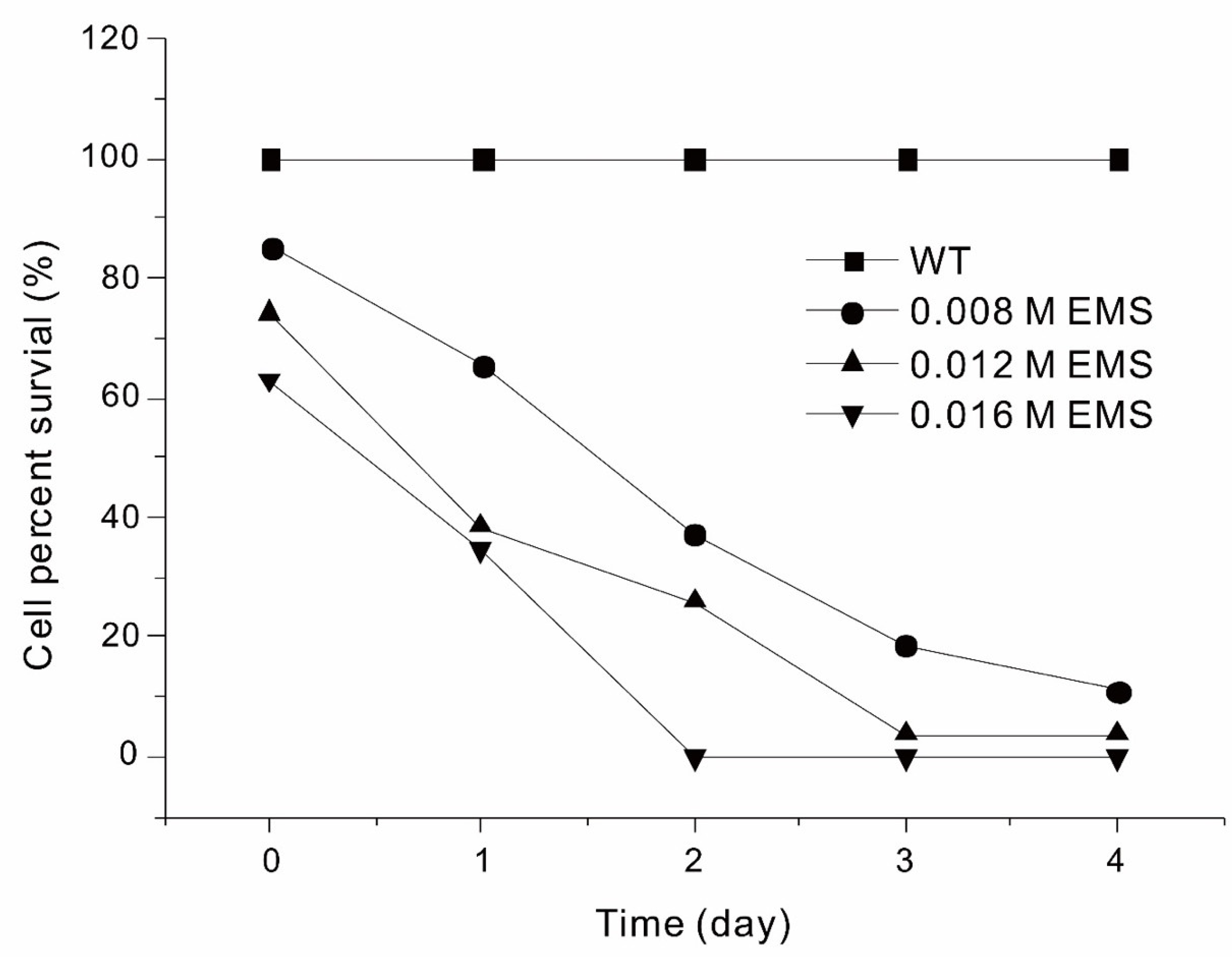
Figure 2.
Motility of Euglena Mutant 333 (A) and WT (B) on agar plates after cultivation for 1, 3, 5, and 7 days (left to right).
Figure 2.
Motility of Euglena Mutant 333 (A) and WT (B) on agar plates after cultivation for 1, 3, 5, and 7 days (left to right).
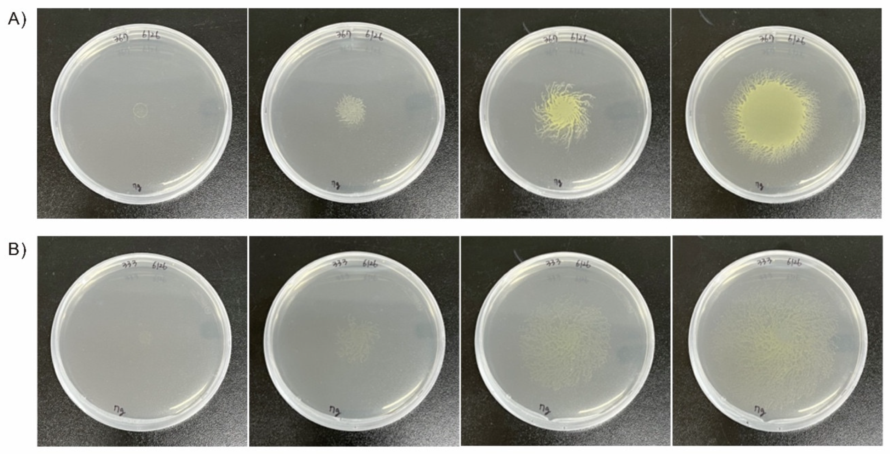
Figure 3.
Appearance of Euglena WT (A) and Mutant 335 after growth on agar plates for 30 days.
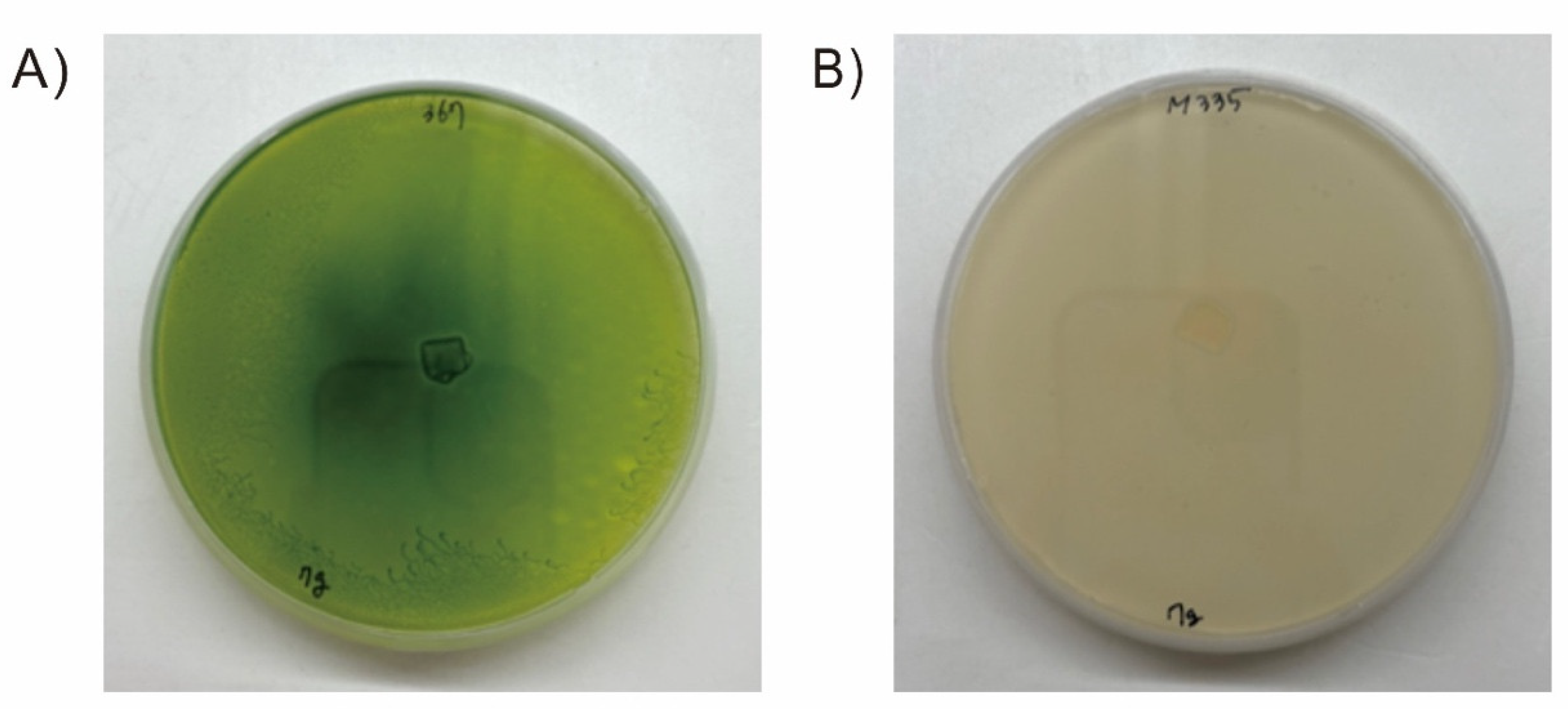
Figure 4.
Growth of Euglena WT, Mutant 333, and Mutant 335 in modified YM liquid medium.
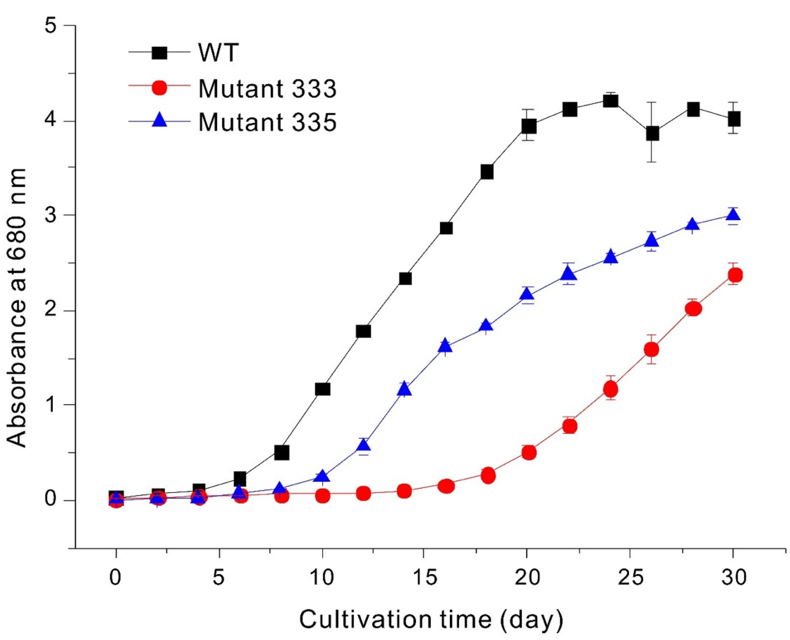
Figure 5.
Levels of chlorophyll a and chlorophyll b in Euglena WT, Mutant 333, and Mutant 335.
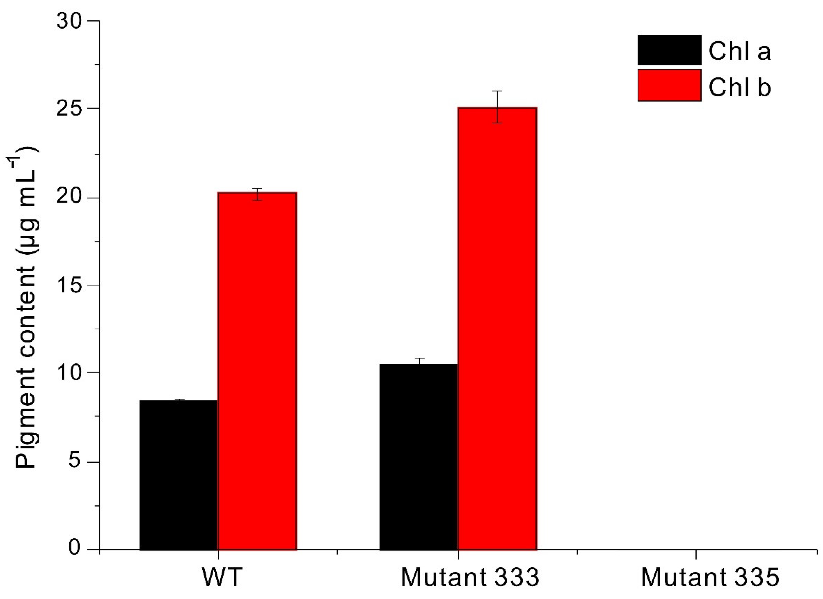
Figure 6.
Growth of Euglena WT and Mutant 333 in glucose-free liquid medium.
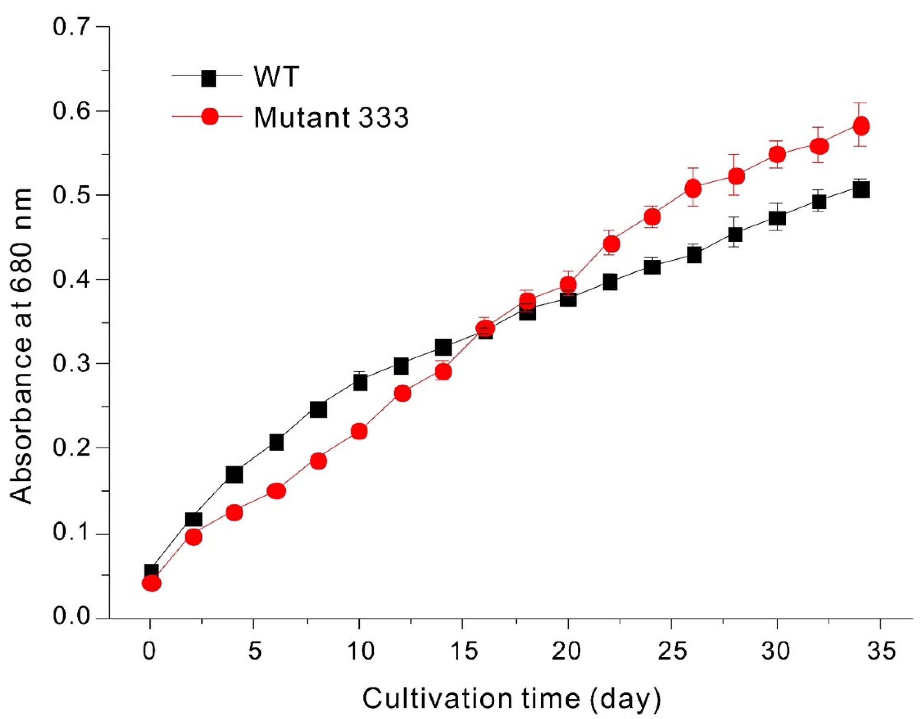
Figure 7.
Paramylon content of Euglena WT, Mutant 333, and Mutant 335 determined by gravimetry and aniline blue staining.
Figure 7.
Paramylon content of Euglena WT, Mutant 333, and Mutant 335 determined by gravimetry and aniline blue staining.
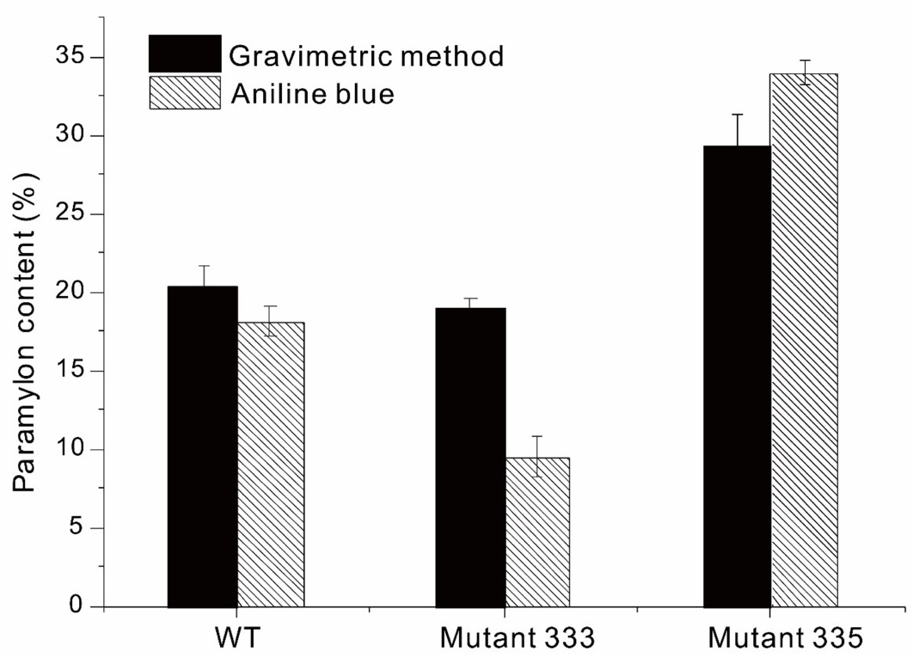

Table 1.
Effect of EMS concentration and duration of treatment on motility of Euglena gracilis.*.
| EMS concentration | 2 h | 24 h | 48 h | 72 h | 96 h |
| 0 M (Control) | +++++ | +++++ | +++++ | +++++ | +++++ |
| 0.008 M | +++++ | +++++ | ++++ | ++ | ++ |
| 0.012 M | +++++ | ++++ | ++ | + | – |
| 0.016 M | +++++ | +++ | – | – | – |
*, + indicates percentage of motile cells (+++++: 100%; ++++: 80%; +++: 60%; ++: 40%; +: 20%; –: 0%).
Table 2.
Concentrations (µg/100 g) of volatile compounds in Euglena WT and Euglena Mutant 335 based on GC/MS.
Table 2.
Concentrations (µg/100 g) of volatile compounds in Euglena WT and Euglena Mutant 335 based on GC/MS.
| Volatile compound |
RT(1) (min) |
RI(2) | Mean±SD | I.D.(3) | |
| Mutant 335 | WT (367) | ||||
| Alcohols (19) | |||||
| 2,2-Dimethyl-3-(1-octylamino)-4-nonanol | 9.25 | 850 | ND(4) | 33.09±46.79 | MS |
| 1-Hexanol | 10.29 | 879 | ND | 80.01±113.15 | MS |
| 1-Octen-3-ol | 13.83 | 984 | ND | 362.54±512.71 | MS |
| 2-Ethylhexanol | 15.39 | 1032 | 148.42±91.16 | 546.36±414.41 | MS/RI(5) |
| 2,2-Dimethyl-1-octanol | 17.44 | 1096 | ND | 102.56±70.91 | MS |
| 1-Hexadecanol | 18.85 | 1145 | 78.01±58.64 | 50.64±46.75 | MS |
| 1-Nonanol | 19.64 | 1172 | ND | 98.97±139.97 | MS |
| 2-Methyl-1-undecanol | 20.23 | 1191 | 51.70±73.11 | ND | MS |
| 1-Decanol | 22.43 | 1271 | 210.94±134.27 | ND | MS |
| 2-Propylheptanol | 24.37 | 1344 | ND | 10.78±15.25 | MS |
| 1-Undecanol | 25.06 | 1370 | 35.14±49.69 | 165.37±233.87 | MS |
| Pentadecanol | 28.41 | 1503 | 123.05±174.02 | ND | MS |
| 1-Docosanol | 28.45 | 1505 | 51.48±72.81 | ND | MS |
| 2,5-Bis(1,1-dimethylethyl)-phenol | 28.49 | 1507 | ND | 78.46±110.96 | MS |
| 1-Heptadecanol | 29.07 | 1532 | 75.86±107.28 | ND | MS |
| Tridecanol | 29.92 | 1568 | 686.01±970.17 | 1,342.40±396.45 | MS |
| Isoheptadecanol | 31.35 | 1630 | 64.10±90.66 | 57.52±81.35 | MS |
| (E)-3-Nonen-1-ol | 31.80 | 1650 | 7.95±11.25 | ND | MS |
| 1-Octadecanol | 33.69 | >1700 | 28.24±39.93 | 100.94±142.75 | MS |
| Aldehydes (7) | |||||
| Hexanal | 7.99 | 810 | ND | 231.23±327.01 | MS/RI |
| 2,2-Dideutero heptadecanal | 10.04 | 872 | ND | 73.52±103.97 | MS |
| Benzaldehyde | 13.24 | 967 | ND | 106.47±150.57 | MS/RI |
| Tetradecanal | 26.00 | 1406 | 145.74±206.11 | 149.41±211.29 | MS |
| 3-Methyl-3-cyclohexen-1-carboxaldehyde | 28.91 | 1525 | ND | 38.06±53.82 | MS |
| Tetradecanal | 30.78 | 1604 | 107.67±117.11 | ND | MS |
| 4-Octadecenal | 33.53 | >1700 | ND | 9.68±13.69 | MS |
| Sulfur-containing compounds (3) | |||||
| Di-tert-dodecyldisulfide | 17.81 | 1108 | 39.69±56.14 | ND | MS |
| Dihexylsulfide | 18.57 | 1136 | ND | 4.43±6.27 | MS |
| Benzothiazole | 21.26 | 1228 | 80.73±34.47 | 282.74±162.22 | MS/RI |
| Acids & esters (18) | |||||
| 2-Propenoic acid | 5.55 | <800 | ND | 525.59±743.30 | MS |
| Methyl methacrylate | 5.59 | <800 | ND | 118.93±168.20 | MS |
| Hexyl formate | 10.21 | 877 | ND | 454.78±643.16 | MS |
| 4-Ethylbenzoic acid | 11.31 | 907 | ND | 393.45±556.42 | MS |
| Sulfurous acid, butyl nonyl ester | 16.08 | 1055 | ND | 50.38±71.24 | MS |
| Octyl chloroformate | 16.68 | 1073 | ND | 275.03±388.96 | MS |
| Acetic acid, octyl ester | 16.69 | 1074 | ND | 96.28±136.16 | MS |
| Methyl salicylate | 20.41 | 1196 | ND | 236.47±334.42 | MS |
| Decyl ether | 20.96 | 1217 | ND | 26.67±0.05 | MS |
| Cyclohexyl isothiocyanate | 21.49 | 1237 | 35.79±50.61 | 252.89±106.97 | MS |
| Didecyl sebacate | 26.97 | 1446 | ND | 26.82±37.93 | MS |
| Heptadecyl heptadecanoate | 27.95 | 1485 | 47.75±67.53 | ND | MS |
| Octadecyl bromoacetate | 29.08 | 1533 | ND | 32.61±46.12 | MS |
| Chloroacetic acid, octadecyl ester | 34.29 | >1700 | ND | 48.87±69.11 | MS |
| Dodecyl fluoroacetate | 34.92 | >1700 | 9.68±13.69 | ND | MS |
| Undec-2-enylester dichloroacetic acid | 35.09 | >1700 | 6.17±8.73 | ND | MS |
| 8,11,14-Eicosatrienoic acid, methyl ester | 35.89 | >1700 | ND | 9.89±13.99 | MS |
| Hexadecyl bromoacetate | 36.34 | >1700 | ND | 47.08±66.57 | MS |
| Heterocyclic compounds (6) | |||||
| Pyridine | 6.52 | <800 | ND | 180.37±89.56 | MS |
| Pyridiniumfluorosulfate | 6.79 | <800 | ND | 6.07±8.58 | MS |
| 2-Pentylfuran | 14.24 | 995 | ND | 340.05±480.91 | MS |
| Camphor | 19.00 | 1150 | ND | 15.84±22.40 | MS/RI |
| 2-Chloro-3,4-diphenylbenzofuro [2,3-b] pyridine | 25.34 | 1381 | ND | 30.83±43.60 | MS |
| Hydrocarbons (67) | |||||
| 1,2-Bis(trimethylsilyl)-benzene | 7.91 | 807 | ND | 155.45±219.84 | MS |
| 2,4-Dimethylheptane | 8.34 | 821 | 12.15±17.18 | ND | MS |
| 4-Methyloctane | 9.78 | 865 | 170.19±240.69 | 392.21±554.66 | MS |
| 2,3,4-Trimethylhexane | 10.09 | 874 | 24.48±34.62 | ND | MS |
| 2-Methyl-1-heptene | 10.34 | 881 | ND | 30.58±43.25 | MS |
| Styrene | 10.98 | 897 | ND | 201.00±284.26 | MS/RI |
| 2,7-Dimethyloctane | 12.18 | 936 | ND | 43.37±61.34 | MS |
| 2,6-Dimethyloctane | 12.33 | 940 | ND | 31.72±44.85 | MS |
| Decane | 13.00 | 960 | 473.20±214.07 | 3,814.51±4,302.46 | MS |
| 2-Methylnonane | 13.29 | 969 | 96.32±136.21 | ND | MS |
| Nonadecane | 13.33 | 970 | 63.78±15.33 | 478.92±345.75 | MS |
| 3-Methylhexane | 13.54 | 976 | ND | 134.60±190.35 | MS |
| 2,4-Dimethylhexane | 13.73 | 981 | ND | 243.84±344.85 | MS |
| 1-Hexyl-3-methylcyclopentane | 14.08 | 990 | 92.92±131.41 | ND | MS |
| 3,6-Dimethylundecane | 14.78 | 1012 | 40.20±56.85 | 288.22±239.30 | MS |
| 2,7-Dimethylundecane | 14.94 | 1017 | 32.06±45.34 | 184.92±261.51 | MS |
| 2,5-Dimethylnonane | 15.11 | 1023 | 50.27±71.09 | 521.16±416.32 | MS |
| 4-Methyldecane | 15.19 | 1026 | 202.41±169.29 | 1,352.80±994.61 | MS |
| Pentyl-cyclopentane | 15.61 | 1040 | ND | 109.91±155.43 | MS |
| 3-Ethyl-5-(2-ethylbutyl)-octadecane | 15.75 | 1044 | 6.79±9.60 | 19.85±28.08 | MS |
| Dodecane | 16.15 | 1057 | 1,230.86±37.26 | 3,786.44±2624.40 | MS |
| 2,6,11-Trimethyldodecane | 16.20 | 1058 | ND | 53.89±76.21 | MS |
| Octadecane | 16.46 | 1066 | 107.69±152.30 | ND | MS |
| 2,4-Dimethylundecane | 16.47 | 1067 | 79.67±40.12 | 80.65±27.23 | MS |
| 4-Ethyl-1,2-dimethyl-benzene | 17.18 | 1088 | ND | 40.07±56.67 | MS |
| Undecane | 17.57 | 1100 | 23.69±33.50 | 229.53±85.60 | MS/RI |
| 4-Methylundecane | 17.64 | 1102 | 242.59±59.44 | 421.89±32.85 | MS |
| Heneicosane | 17.81 | 1109 | ND | 304.15±304.11 | MS |
| 1,2,3,4-Tetramethylbenzene | 18.23 | 1123 | ND | 41.85±59.18 | MS |
| 2-Methylundecane | 18.39 | 1129 | ND | 123.24±145.29 | MS |
| 2,6,10-Trimethyltetradecane | 18.57 | 1135 | ND | 23.03±32.57 | MS |
| (E)-1-Butyl-2-methylcyclopropane | 19.07 | 1153 | ND | 16.07±22.73 | MS |
| 6-Ethyl-2-methyloctane | 19.31 | 1161 | ND | 56.13±79.38 | MS |
| Squalane | 19.44 | 1165 | ND | 54.53±77.12 | MS |
| Cyclododecane | 20.24 | 1191 | 8,955.69±8,032.29 | 17,120.77±5,004.40 | MS |
| Tritetracontane | 20.61 | 1204 | ND | 123.01±173.96 | MS |
| 5-Methyloctadecane | 20.77 | 1210 | 17.02±24.07 | 55.07±77.87 | MS |
| 2,6-Dimethylundecane | 20.86 | 1213 | ND | 240.77±96.84 | MS |
| 2,4-Dimethylicosane | 20.96 | 1217 | ND | 25.64±36.26 | MS |
| 3-Ethyltetracosane | 21.08 | 1221 | ND | 120.26±170.08 | MS |
| Tetracosane | 21.69 | 1244 | ND | 140.68±0.59 | MS |
| 2,4,6-Trimethyldecane | 21.84 | 1249 | 39.19±21.94 | 64.26±90.88 | MS |
| 1,3-Bis(1,1-dimethylethyl)-benzene | 22.03 | 1257 | 181.02±90.18 | 655.22±220.60 | MS |
| 4-Ethylundecane | 22.22 | 1263 | 51.18±72.38 | 10.07±14.24 | MS |
| Nonacosane | 22.23 | 1264 | ND | 126.54±178.95 | MS |
| Tetradecane | 22.67 | 1279 | 116.96±100.04 | 1,545.25±1,439.78 | MS |
| 1,3-Dichloro-2-methoxybenzene | 22.97 | 1290 | ND | 16.32±23.09 | MS |
| Tridecane | 23.04 | 1292 | 18.01±25.47 | 77.50±109.60 | MS/RI |
| 2,3,5,8-Tetramethyldecane | 23.18 | 1297 | 9.06±12.82 | ND | MS |
| Hexacosane | 23.25 | 1299 | ND | 32.14±45.45 | MS |
| 2,3,5-Trimethyldecane | 23.41 | 1306 | ND | 35.18±49.75 | MS |
| 4-Methyltetradecane | 24.31 | 1341 | ND | 10.17±14.39 | MS |
| 2,6,10-Trimethyltridecane | 24.32 | 1342 | ND | 6.31±8.93 | MS |
| 10-Methylnonadecane | 24.82 | 1361 | ND | 6.62±9.36 | MS |
| 1,1-Bis(dodecyloxy)-hexadecane | 26.68 | 1434 | 12.28±17.36 | 87.10±72.05 | MS |
| 2-Methyltetradecane | 27.29 | 1459 | ND | 112.17±21.79 | MS |
| Docosane | 28.10 | 1491 | ND | 108.51±55.08 | MS |
| 1-Chlorooctadecane | 28.85 | 1523 | ND | 7.81±11.05 | MS |
| 3-Methyltridecane | 29.15 | 1535 | ND | 38.21±54.03 | MS |
| (E)-3-Octadecene | 29.25 | 1540 | ND | 49.04±69.36 | MS |
| 2-Methyloctadecane | 29.81 | 1564 | ND | 58.60±82.87 | MS |
| Hexadecane | 30.46 | 1591 | 32.71±46.25 | 756.99±541.19 | MS/RI |
| 2-Methyltetracosane | 31.86 | 1652 | ND | 21.11±29.86 | MS |
| (E)-2-Tetradecene | 32.16 | 1665 | 4,124.93±4,333.01 | 8,371.63±1,937.56 | MS |
| 1-Fluorododecane | 32.64 | 1686 | 61.35±86.77 | ND | MS |
| Cyclotetradecane | 34.92 | >1700 | 80.22±113.44 | 77.86±8.47 | MS |
| 1,3-Cyclooctadiene | 40.67 | >1700 | ND | 50.93±72.03 | MS |
| Ketones (5) | |||||
| 2-(4′-Chloro) styrylchromone | 14.48 | 1002 | ND | 1,822.18±2,576.95 | MS |
| Acetophenone | 16.60 | 1071 | 10.63±15.04 | 119.97±63.45 | MS |
| 2-Nonanone | 17.35 | 1093 | ND | 41.34±58.46 | MS |
| 1,4-Hexadecansultone | 33.70 | >1700 | ND | 15.22±21.52 | MS |
| 2,11-Dodecanedione | 35.53 | >1700 | ND | 13.01±18.39 | MS |
(1) RT: retention time. (2) RI: retention index. (3) I.D.: identification. (4) ND: not detected. (5) MS/RI: identification using both MS and RI. RI: identified using MS.
Table 3.
Odor intensity in Euglena WT and Euglena Mutant 335 based on GC olfactometry.
| Volatile compound | RT(1) | Relative Intensity | Odor description | |
| (min) | Mutant 335 | WT (367) | ||
| Alcohols | ||||
| 1-Hexanol | 10.29 | 0 | 1 | Green |
| 1-Octen-3-ol | 13.83 | 0 | 2 | Mugwort |
| 2-Methyl-1-undecanol | 20.23 | 2 | 0 | Herbaceous |
| Aldehydes | ||||
| Hexanal | 7.99 | 0 | 1 | Green tea |
| Acid & esters | ||||
| Hexyl formate | 10.21 | 0 | 2 | Barley tea |
| 4-Ethylbenzoic acid | 11.31 | 0 | 2 | Green tea |
| Hydrocarbons | ||||
| 2-Methyl-1-heptene | 10.34 | 0 | 1 | Mugwort |
| Styrene | 10.98 | 0 | 1 | Mugwort |
| 2,6-Dimethyloctane | 12.33 | 0 | 2 | Roasted |
| 2,4,6-Trimethyldecane | 21.84 | 2 | 1 | Green |
| 1,3-Bis(1,1-dimethylethyl)-benzene | 22.03 | 1 | 1 | Herbaceous |
(1) RT: retention time.
Disclaimer/Publisher’s Note: The statements, opinions and data contained in all publications are solely those of the individual author(s) and contributor(s) and not of MDPI and/or the editor(s). MDPI and/or the editor(s) disclaim responsibility for any injury to people or property resulting from any ideas, methods, instructions or products referred to in the content. |
© 2025 by the authors. Licensee MDPI, Basel, Switzerland. This article is an open access article distributed under the terms and conditions of the Creative Commons Attribution (CC BY) license (http://creativecommons.org/licenses/by/4.0/).
Copyright: This open access article is published under a Creative Commons CC BY 4.0 license, which permit the free download, distribution, and reuse, provided that the author and preprint are cited in any reuse.


