
Submitted:
06 January 2025
Posted:
07 January 2025
You are already at the latest version
Abstract
Branch dieback and canker diseases caused by Cytospora species negatively affect health of woody plants worldwide. In this study, 59 Cytospora isolates were obtained from symptomatic trees and shrubs growing in southwest Ontario and Saskatchewan, Canada. DNA barcoding approach combined with morphological (culture) characterization identified 15 known species of Cytospora associated with the diseases: C. chrysosperma, C. curvata, C. euonymina, C. hoffmannii, C. kantschavelii, C. leucosperma, C. leucostoma, C. nitschkeana, C. piceae, C. populina, C. pruinopsis, C. pruinosa, C. ribis, C. schulzeri, and C. sorbina. The obtained results contribute to the study of diversity, host affiliation, geographical distribution, and pathogenicity of Cytospora species occurring on woody plants in both natural habitats and agricultural systems. The findings also support the effectiveness of using sequence barcodes (ITS, act1) in fungal taxonomy and plant pathology studies.
Keywords:
branch dieback
; canker disease
; Cytospora
; DNA barcoding
; pathogen
1. Introduction
DNA barcoding is a standardized approach that can be applied for correct identification and recognition of fungi while overcoming the issues with traditional criteria used for description of fungal species [1]. DNA barcode is a relatively short gene region of the highly variable parts of a genome, unique for the species identification. The main advantage of this approach is that a single specimen can provide information about the species, regardless of its morphology or lifestage characteristics [2]. Currently, the internal transcribed spacer (ITS) region of the nuclear ribosomal RNA gene cluster is employed as the main fungal barcode [3].
Cytospora species are well known as causal agents of branch dieback and canker diseases on different woody plants [4,5,6]. Considered as weak pathogens, the species of Cytospora can reduce longevity and productivity of related hosts in the long term [7]. Disease symptoms can initially be observed on twigs and branches in a form of sunken areas with dark discoloration. As disease progresses, cankers form in other tree parts allowing for pathogen overwintering. Cytospora fruiting structures (pycnidia) appear on infected wood tissues during spring and fall seasons mainly. Under favorable conditions (e.g. high moisture), conidia or spores eject from pycnidia and disseminate on nearby hosts with rain splash or wind infecting plant tissue through bark wounds [8].
Traditional approach for Cytospora identification was primarily based on species morphological and ecological characteristics [9,10]. These criteria seem to be insufficient for Cytospora species delimitation because of significant overlap in morphological traits and lack of host specificity within the genus. Besides, different species of Cytospora can co-occur on the same host [7]. However, occurrence of some species can be restricted to a single host genus or family [11,12]. Currently, identification of Cytospora species includes both morphological and molecular (sequence) data analyses. ITS region was initially employed to resolve the phylogeny of Cytospora [7]. The partial protein-coding genes (PCG), such as actin (act1), RNA polymerase II subunit (rpb2), translation elongation factor 1-alpha (tef1-α), and beta-tubulin (tub2) were further applied to phylogenetic analyses of the genus to address the issues with species recognition. Combined multi-gene (ITS+PCGs) sequence data have been used to correctly identify or describe Cytospora species in the most recent studies [13,14,15]. Therefore, a DNA-based approach including multi-locus phylogenetic analysis is critical to uncovering Cytospora species diversity associated with plant diseases.
Trees and shrubs with symptoms of branch dieback and canker diseases (Figure 1) have been observed during the surveys in southwest Ontario and Saskatchewan (Canada) in 2020-23. Since Cytospora are among the main causal agents of these diseases, exhaustive knowledge on the identity of the fungi isolated from symptomatic plants is needed to develop proper control strategies in case of disease outbreaks. Thus, this study was aimed to identify Cytospora species associated with diseased woody plants by applying DNA barcoding approach and morphological (culture) characterization.
2. Materials and Methods
2.1. Sample Collection and Fungus Isolation
Branches and twigs with symptoms of dieback and canker were sampled for isolation and identification of Cytospora spp. associated with the diseases. The isolates were mainly obtained using the single spore isolation technique [16]. To isolate pathogen species from necrotic plant tissue, small wood pieces (0.5-1cm) were surface sterilized with 70% ethanol for 30s, following sterilization with 0.5% sodium hypochlorite for 2m, rinsed three times with sterile water, and plated on malt extract agar (MEA). Cytospora-like colonies were further purified by transferring hyphal-tips to new MEA plates. The obtained isolates were grouped in morphotypes and representative isolates for each morphotype were further selected for morphological (culture) characterization and sequencing.
2.2. Morphological Characterization
Fungus fruiting structures (conidio- or ascomata) were observed and sectioned under a dissecting microscope (AmScope SE306R-PZ). To confirm sample affiliation with Cytospora species, microstructures (conidia, spores) were examined using a compound microscope (AmScope B120C-E5). Radial colony growth and color (both adverse and reverse sides) were accessed after 7d of incubation at room temperature in the dark on MEA. Growth rate was defined as either slow growing (up to 4.5cm) or fast growing (up to 9cm). Pycnidia formation was checked after 21d of incubation.
2.3. DNA Extraction, PCR Amplification and Sequencing
Total genomic DNA (gDNA) was extracted from 7-11d old pure cultures using DNeasy Plant ProKit (Qiagen, Hilden, Germany) following the manufacturer’s instructions. The primers ITS1/ITS4 [17] were used to amplify internal transcribed spacer (ITS) region. Additionally, the partial actin (act1) gene region was amplified with the primer pair ACT-512F and ACT-783R [18] for the selected strains to improve species resolution. The quality of PCR products was examined using electrophoresis in 1% agarose gel. Sanger sequencing were carried out at the Genome Quebec Innovation Centre (Montreal, QC, Canada).
2.4. Sequence Alignment and Phylogenetic Analysis
The initial identification was performed using the BLASTn tool against the GenBank nucleotide database of the National Center for Biotechnology Information (NCBI). Sequence data of the related reference strains [6] were downloaded from the GenBank database. The sequences were initially aligned employing CLUSTAL-X2 v.2.1 [19] and manually edited with MEGA-X [20]. Phylogenetic analyses were executed using randomized accelerated maximum likelihood (RAxML) v. 8.0 method [21] for maximum likelihood (ML) analysis. Bayesian posterior (BP) probabilities were defined with MrBayes v.3.2.7 [22] using the TrEase web server [23]. The ML analysis was performed using the transition (TIM) substitution model with gamma-distributed rate of heterogeneity selected with ModelTest-NG v.0.1.7 [24]. The statistical support values were estimated with bootstrapping of 1,000 replicates [25]. The general time reversible (GTR) model was chosen for the BI analysis. The Markov chain Monte Carlo (MCMC) algorithm was used to estimate Bayesian posterior probabilities (BPP). Six simultaneous Markov chains were run for 1,000,000 generations. A burn-in was implemented with discarding the first 30% of generated trees. The phylograms were visualized using FigTree v. 1.4.4 [26]. The newly generated sequences were deposited in GenBank (Table 1). The final alignment used in the analysis was submitted to TreeBase (www.treebase.org; ID: S31906).
3. Results
3.1. Phylogenetic Analysis
The ML and BP analyses of the combined ITS and act1 sequence data produced phylogenetic trees with similar topologies. The best-scoring ML tree with a log-likelihood value of −3511.991652 is depicted in Figure 2. Estimated base frequencies were as follows: A, C, G, T = 0.250000; substitution rates: AC = 2.518389, AG = 4.873235, AT = 2.518389, CG = 1.000000, CT = 8.755476, GT = 1.000000.
The obtained strains clustered into 15 clades with high support values were assigned to the following known species: C. chrysosperma, C. curvata, C. euonymina, C. hoffmannii, C. kantschavelii, C. leucosperma, C. leucostoma, C. nitschkeana, C. piceae, C. populina, C. pruinopsis, C. pruinosa, C. ribis, C. schulzeri, and C. sorbina.
3.2. Taxonomy
Cytospora chrysosperma (Pers.) Fr., Syst. Mycol. (Lundae) 2(2): 542. 1823. Figure 3A.
Description: See [7].
Culture characteristics: Colonies on MEA initially white, relatively fast growing, with thin, regular texture and sparce aerial mycelium, becoming brownish in adverse and beige in reverse. Sterile mycelium, withouth fruiting structures after 21d of incubation.
Material examined: Canada, Southwest Ontario: 43°13'15.8"N 79°13'32.2"W, from fallen branches of unknown tree, May 2020, E. Ilyukhin (EI-101); 43°03'51.1"N 79°17'10.1"W, from branches of Rhus sp., September 2020, E. Ilyukhin (EI-351); 43°08'21.7"N 79°10'09.3"W, from fallen branches of Populus tremuloides, July 2021, E. Ilyukhin (EI-437). Southwest Saskatchewan: 50°16'59.6"N 107°45'49.4"W, from fallen branches of Populus deltoides, July 2023, E. Ilyukhin (EI-SK-71); 50°38'50.8"N 108°00'18.2"W, from twigs of Salix bebbiana, August 2023, E. Ilyukhin (EI-SK-90); 50°39'35.2"N 108°02'55.5"W, from branches of Populus tremula, August 2023, E. Ilyukhin (EI-SK-93(A)); 50°40'24.6"N 107°57'16.2"W, from fallen branches of Populus sp., August 2023, E. Ilyukhin (EI-SK-127); 50°16'35.1"N 108°24'47.8"W, from twigs of Salix alba, September 2023, E. Ilyukhin (EI-SK-196).
Cytospora curvata Norph., Bulgakov, T.C. Wen & K.D. Hyde, Mycosphere 8 (1): 57. 2017. Figure 3B.
Description: See [27].
Culture characteristics: Colonies on MEA initially white, relatively slow growing, with thick, irregular texture without aerial mycelium, becoming dark green in adverse and dark grey in reverse. Abundant pycnidia appear after 21d of incubation.
Material examined: Canada, Southwest Ontario: 43°06'36.3"N 79°15'04.7"W, from branches of Aronia sp., May 2020, E. Ilyukhin (EI-132); 43°11'59.5"N 79°13'37.1"W, from twigs of Syringa reticulata, May 2020, E. Ilyukhin (EI-203).
Cytospora euonymina X.L. Fan & C.M. Tian, Persoonia 45: 21. 2019. Figure 3C.
Description: See [28].
Culture characteristics: Colonies on MEA initially white, relatively fast growing, with thick, irregular texture without aerial mycelium, becoming brown in adverse and light brown in reverse. Rare pycnidia appear after 21d of incubation.
Material examined: Canada, Southwest Ontario: 43°11'27.3"N 79°15'53.2"W, from twigs of Salix sp., June 2020, E. Ilyukhin (EI-250); 43°06'23.2"N 79°13'39.0"W, from twigs of Berberis vulgaris, August 2020, E. Ilyukhin (EI-316).
Cytospora hoffmannii L. Lin, X.L. Fan & Crous, Stud. Mycol. 109: 354. 2024. Figure 3D.
Culture characteristics: Colonies on MEA initially white, relatively slow growing, with thin, irregular texture and sparce aerial mycelium, becoming olivaceous in adverse and greyish in reverse. Sterile mycelium, withouth fruiting structures after 21d of incubation.
Material examined: Canada, Southwest Saskatchewan: 50°59'43.0"N 106°25'45.1"W, from twigs of Salix bebbiana, October 2023, E. Ilyukhin (EI-SK-231); 50°59'34.9"N 106°25'33.5"W, from branches of Salix sp., October 2023, E. Ilyukhin (EI-SK-237).
Cytospora kantschavelii Gvrit., Mikol. Fitopatol. 7: 547. 1973. Figure 3E.
Culture characteristics: Colonies on MEA initially white, relatively fast growing, with thin, irregular texture without aerial mycelium, becoming light brown in both adverse and reverse. Sterile mycelium, withouth fruiting structures after 21d of incubation.
Material examined: Canada, Southwest Saskatchewan: 50°17'05.5"N 107°47'06.0"W, from twigs of Ulmus glabra (plant tissues plated), September 2022, E. Ilyukhin (EI-SK-33); 50°16'00.8"N 107°46'53.5"W, from twigs of Acer spicatum, October 2022, E. Ilyukhin (EI-SK-44).
Cytospora leucosperma (Pers.) Fr., Syst. Mycol. (Lundae) 2(2): 543. 1823. Figure 3F.
Culture characteristics: Colonies on MEA initially white, relatively slow growing, with thin, regular texture without aerial mycelium, becoming light brown in both adverse and reverse. Sterile mycelium, withouth fruiting structures after 21d of incubation.
Material examined: Canada, Southwest Ontario: 43°12'40.4"N 79°14'31.0"W, branches of Bereberis vulgaris, April 2020, E. Ilyukhin (EI-54(A)).
Cytospora leucostoma (Pers.) Sacc., Michelia 2(7): 264. 1881. Figure 3G.
Culture characteristics: Colonies on MEA initially white, relatively slow growing, with thick, regular texture and sparce aerial mycelium, becoming dark green in adverse and greyish green in reverse. Abundant pycnidia appear after 21d of incubation.
Material examined: Canada, Southwest Ontario: 43°13'07.3"N 79°13'42.3"W, from twigs of Malus sp., May 2020, E. Ilyukhin (EI-78); 43°11'05.2"N 79°13'23.8"W, from branches of Vaccinium sp., May 2020, E. Ilyukhin (EI-223).
Cytospora nitschkeana L. Lin, X.L. Fan & Crous, Stud. Mycol. 109: 367. 2024. Figure 3H.
Description: See [6].
Culture characteristics: Colonies on MEA initially white, relatively fast growing, with thick, regular texture and aerial mycelium, becoming brown in both adverse and reverse. Rare pycnidia appear after 21d of incubation.
Material examined: Canada, Southwest Ontario: 43°12'03.0"N 79°14'25.6"W, from twigs of Salix babylonica, May 2020, E. Ilyukhin (EI-170); 43°12'59.7"N 79°12'51.7"W, from branches of Berberis vulgaris, May 2020, E. Ilyukhin (EI-193); 43°08'29.3"N 79°10'09.0"W, from twigs of Fraxinus americana, July 2021, E. Ilyukhin (EI-429); 43°05'33.3"N 79°18'13.1"W, from branches of Syringa sp., October 2021, E. Ilyukhin (EI-454); 43°05'29.4"N 79°18'19.6"W, from branches of Fraxinus nigra, October 2021, E. Ilyukhin (EI-470). Southwest Saskatchewan: 50°16'35.1"N 108°24'47.8"W, from twigs of Salix alba, September 2023, E. Ilyukhin (EI-SK-195).
Note: The reference strain Cytospora (Valsa) salicina CBS 118.22 is currently designated as C. nitschkeana [6].
Cytospora piceae X.L. Fan, Phytotaxa 383 (2): 188. 2018. Figure 3I.
Description: See [30].
Culture characteristics: Colonies on MEA initially white, relatively fast growing, with thick, irregular texture and dense aerial mycelium, becoming grey with brownish tint in both adverse and reverse. Sterile mycelium, withouth fruiting structures after 21d of incubation.
Material examined: Canada, Southwest Ontario: 43°11'36.8"N 79°16'02.4"W, from branches of Picea pungens (plant tissue plated), August 2020, E. Ilyukhin (EI-273). Southwest Saskatchewan: 50°16'50.6"N 107°47'09.1"W, from branches of Rhus sp., September 2022, E. Ilyukhin (EI-SK-36); 50°40'43.3"N 107°56'38.9"W, from branches of Picea sp. (plant tissue plated), August 2023, E. Ilyukhin (EI-SK-110); 50°16'30.5"N 108°25'02.6"W, from twigs of Fraxinus sp., September 2023, E. Ilyukhin (EI-154(A)).
Cytospora populina (Pers.) Rabenh., Deutschl. Krypt.-Fl. (Leipzig) 1: 148. 1844. Figure 3J.
Description: See [31].
Culture characteristics: Colonies on MEA initially white, relatively fast growing, with thick, irregular texture without aerial mycelium, becoming dark brown in adverse and light brown in reverse. Rare pycnidia appear after 21d of incubation.
Material examined: Canada, Southwest Ontario: 43°08'29.0"N 79°10'23.0"W, from branches of Magnolia sp., July 2021, E. Ilyukhin (EI-434); 43°05'23.6"N 79°18'25.1"W, from branches of Sorbus sp., October 2021, E. Ilyukhin (EI-477(B)); 43°05'26.5"N 79°18'21.7"W, from twigs of Acer platanoides, October 2021, E. Ilyukhin (EI-478). Southwest Saskatchewan: 50°16'36.6"N 108°24'52.3"W, from branches of Aronia sp., September 2023, E. Ilyukhin (EI-SK-184).
Cytospora pruinopsis C.M. Tian & X.L. Fan, Mycol. Progr. 14: 74. 2015. Figure 3K.
Description: See [32].
Culture characteristics: Colonies on MEA initially white, relatively fast growing, with thin, regular texture and dense aerial mycelium, becoming light grey in adverse and grey in reverse. Sterile mycelium, withouth fruiting structures after 21d of incubation.
Material examined: Southwest Saskatchewan: 50°16'44.5"N 107°47'10.0"W, from branches of Populus deltoides, October 2022, E. Ilyukhin (EI-SK-38); 50°16'51.8"N 107°45'37.2"W, from twigs of Ulmus americana, June 2023, E. Ilyukhin (EI-SK-75); 50°40'00.5"N 108°02'59.7"W, from twigs of Malus sp., August 2023, E. Ilyukhin (EI-SK-94(A)); 51°00'01.3"N 106°25'59.3"W, from branches of unknown shrubs, October 2023, E. Ilyukhin (EI-SK-223).
Cytospora pruinosa (Fr.) Sacc., Michelia 1(5): 519. 1879. Figure 3L.
Description: See [6].
Culture characteristics: Colonies on MEA initially white, relatively fast growing, with thin, regular texture and sparce aerial mycelium, becoming bright yellow in adverse and brownish in reverse. Sterile mycelium, withouth fruiting structures after 21d of incubation.
Material examined: Southwest Saskatchewan: 50°38'58.5"N 107°56'52.1"W, from twigs of Aronia sp., August 2023, E. Ilyukhin (EI-SK-133(A)); 50°16'34.8"N 108°24'45.6"W, from twigs of Syringa reticulata, September 2023, E. Ilyukhin (EI-SK-152); 50°16'38.4"N 108°25'01.1"W, from twigs of unknown shrubs, September 2023, E. Ilyukhin (EI-SK-155); 50°59'42.2"N 106°25'22.6"W, from branches of Syringa sp., October 2023, E. Ilyukhin (EI-SK-211); 50°59'37.6"N 106°25'35.4"W, from twigs of Fraxinus sp., October 2023, E. Ilyukhin (EI-SK-215); 50°59'43.8"N 106°25'53.2"W, from branches of unknown shrubs, October 2023, E. Ilyukhin (EI-SK-221).
Cytospora ribis Ehrenb., Sylv. mycol. berol. (Berlin): 28. 1818. Figure 3M.
Description: See [6].
Culture characteristics: Colonies on MEA initially white, relatively fast growing, with thin, regular texture and sparce aerial mycelium, becoming beige in both adverse and reverse. Rare pycnidia appear after 21d of incubation.
Material examined: Southwest Ontario: 43°08'01.3"N 79°22'50.9"W, from twigs of Fraxinus americana, May 2021, E. Ilyukhin (EI-396). Southwest Saskatchewan: 50°16'51.8"N 107°45'40.6"W, from twigs of Syringa vulgaris, June 2023, E. Ilyukhin (EI-SK-60).
Cytospora schulzeri Sacc. & P. Syd., Syll. fung. (Abellini) 14: 918. 1899. Figure 3N.
Culture characteristics: Colonies on MEA initially white, relatively fast growing, with thin, regular texture and dense aerial mycelium, becoming greyish in both adverse and reverse. Rare pycnidia appear after 21d of incubation.
Material examined: Southwest Ontario: 43°04'00.5"N 79°17'06.3"W, from twigs of Sorbaria sorbifolia, September 2020, E. Ilyukhin (EI-378); 43°08'21.5"N 79°22'26.2"W, from branches of Malus sp., May 2021, E. Ilyukhin (EI-414); 43°05'26.5"N 79°18'21.0"W, from twigs of Acer platanoides, October 2021, E. Ilyukhin (EI-480). Southwest Saskatchewan: 50°40'16.2"N 108°00'11.3"W, from twigs of Malus sp., August 2023, E. Ilyukhin (EI-SK-101).
Cytospora sorbina M. Pan & X.L. Fan, Adverseiers in Plant Science 11 (no. 690): 13. 2020. Figure 3N.
Description: See [34].
Culture characteristics: Colonies on MEA initially white, relatively fast growing, with thick, regular texture and sparce aerial mycelium, becoming light orange in adverse and brownish in reverse. Sterile mycelium, withouth fruiting structures after 21d of incubation.
Material examined: Southwest Saskatchewan: 50°17'26.2"N 107°47'17.3"W, from branches of Malus sp., September 2022, E. Ilyukhin (EI-SK-25); 50°17'11.5"N 107°47'24.6"W, from branches of Aronia sp., September 2022, E. Ilyukhin (EI-SK-31); 50°16'55.4"N 107°45'43.9"W, from twigs of Aronia sp., June 2023, E. Ilyukhin (EI-SK-67); 50°16'51.8"N 107°45'37.2"W, from twigs of Prunus sp., June 2023, E. Ilyukhin (EI-SK-76); 50°38'45.8"N 107°58'42.0"W, from branches of Sorbus aucuparia, August 2023, E. Ilyukhin (EI-SK-82); 50°38'50.8"N 108°00'18.2"W, from branches of Prunus padus, August 2023, E. Ilyukhin (EI-SK-92); 50°39'52.4"N 108°02'38.0"W, from branches of Aronia sp., August 2023, E. Ilyukhin (EI-SK-95(A)); 50°38'59.6"N 107°56'52.0"W, from twigs of unknown shrubs, August 2023, E. Ilyukhin (EI-SK-131(A)); 50°16'42.4"N 108°24'36.0"W, from twigs of Sorbus sp., September 2023, E. Ilyukhin (EI-SK-148); 50°16'38.2"N 108°24'52.3"W, from twigs of Viburnum cf. trilobum, September 2023, E. Ilyukhin (EI-SK-157(A)).
4. Discussion
Cytospora species associated with branch dieback and canker diseases of economically important fruit trees have been recently reported in North America [35,36]. In case of disease outbreaks, it can lead to significant yield losses for the growers. Since Cytospora is not host-specific [7,28], the fungus may switch from hosts occurring in natural habitats to fruit trees growing in agricultural systems.
This study was conducted to reveal Cytospora species associated with diseased woody plants in non-agricultural terrains in Canada. DNA barcoding approach and morphological (culture) characterization were applied to properly identify the obtained Cytospora isolates. The analysis based on combined ITS and act1 sequence data resolved phylogenies of the selected Cytospora strains. The results highlighted the relatively rich species diversity of Cytospora isolated from symptomatic plants (15 species amongst 59 isolates). But only four species (C. leucostoma, C. pruinopsis, C. schulzeri and C. sorbina) were isolated from affected Malus spp. in the surveyed areas while 24 species of Cytospora were found to be related to apple tree diseases worldwide [12,34]. It indicates that more studies employing different techniques (e.g., metabarcoding) should be conducted to fully uncover pathogenic species in such a diverse genus. Most of the identified Cytospora have previously been reported as causal agents of tree diseases. The species of C. chrysosperma, C. kantchavelii, C. nivea, C. piceae, C. populina, and C. sorbina were found to be associated with canker disease of common forest-forming tree species such as Juglans nigra, Picea crassifolia, Populus alba, Salix spp., Sorbus tianschanica, and Ulmus pumila [5,30,32,34,37,38]. Host specificity can be attributed to a group of Cytospora species (incl., C. piceae) affiliated with conifers [4,30]. Meantime, C. piceae was isolated from symptomatic deciduous trees (ash) and shrubs (staghorn) in this study. This evidence additionally supports a lack of specific host affiliations among Cytospora spp. Other tree species widely cultivated in agricultural systems (e.g., Malus spp., Prunus persica, Olea europaea) can also be significantly affected by C. leucostoma, C. parasitica, C. pruinosa, and C. pruinopsis [14,15,39] found in the surveyed areas.
Multiple in-vivo pathogenicity assays have been recently conducted to show that Cytospora spp. are able to cause canker symptoms on related hosts [14,40,41]. It was also shown that the species of Cytospora may infect healthy (not stressed) trees maintained under proper growing conditions [12]. It points out that regular monitoring of trees (incl., asymptomatic) growing in the surroundings of fruit tree orchards should be implemented for early detection of pathogenic Cytospora species.
Overall, the obtained results revealed a strong association of Cytospora species with diseased woody plants, as well as the emergence of new hosts in southwestern Ontario and Saskatchewan, Canada. This study will contribute to the further research of fungal tree pathogens and help to develop effective disease prevention and control strategies.
Author Contributions
Conceptualization, E.I. and S.M.; methodology, E.I. and S.M.; validation, E.I. and S.M.; formal analysis, E.I.; investigation, E.I.; data curation, S.M.; writing—original draft preparation, E.I.; writing—review and editing, S.M.; visualization, E.I. All authors have read and agreed to the published version of the manuscript.
Funding
Not applicable.
Institutional Review Board Statement
Not applicable.
Informed Consent Statement
Not applicable.
Data Availability Statement
The ITS and act1 sequences and alignments generated in this study have been deposited in the GenBank database (https://www.ncbi.nlm.nih.gov/genbank/) and TreeBASE (https://treebase.org/treebase-web/), respectively. The accession numbers were provided in the paper.
Acknowledgments
The authors thank Simon Iliukhin for his help with sample collection and data management.
Conflicts of Interest
The authors declare no conflicts of interest.
References
- Casiraghi, M.; Labra, M.; Ferri E, Galimberti, A.; De Mattia, F. DNA Barcoding: A Six-Question Tour to Improve Users' Awareness about the Method. Brief Bioinform. 2010, 11, 440–453.
- Ćelepirović, N.; Novak Agbaba, S.; Karija Vlahović, M. DNA Barcoding of Fungi in the Forest Ecosystem of the Psunj and Papuk Mountains in Croatia. SEEFOR 2020, 11, 145–152.
- Schoch, C.L.; Seifert, K.A.; Huhndorf, S.; Robert, V.; Spouge, J.L.; Levesque, C.A.; Chen, W. Fungal Barcoding Consortium; Fungal Barcoding Consortium Author List. Nuclear Ribosomal Internal Transcribed Spacer (ITS) Region as a Universal DNA Barcode Marker for Fungi. Proc Natl Acad Sci U S A. 2012, 109, 6241–6246.
- Pan, M.; Zhu, H.Y.; Tian, C.M.; Huang, M.R.; Fan, X.L. Assessment of Cytospora Isolates from Conifer Cankers in China, with the Descriptions of Four New Cytospora species. Front. Plant Sci. 2021, 12, 636460.
- Lin, L.; Pan, M.; Tian, C.; Fan, X. Fungal Richness of Cytospora Species Associated with Willow Canker Disease in China. J. Fungi 2022, 8, 377.
- Lin, L.; Fan, XL.; Groenewald, J.Z.; Jami, F.; Wingfield, M.J.; Voglmayer, H., Jaklitsch, W., Castlebury, L.A.; Tian, C.M.; Crous, P.W. Cytospora: an Important Genus of Canker Pathogens. Studies in Mycology 2024, 109, 323–401.
- Adams, G.C.; Wingfield, M.J.; Common, R.; Roux, J. Phylogenetic Relationships and Morphology of Cytospora Species and Related Teleomorphs (Ascomycota, Diaporthales, Valsaceae) from Eucalyptus. Stud. Mycol. 2005, 52, 1–147.
- Biggs, A.R. Integrated Approach to Controlling Leucostoma Canker of Peach in Ontario. Plant Dis. 1989, 73, 869–874.
- Adams, G.C.; Surve-Iyer, R.S.; Iezzoni, A.F. Ribosomal DNA Sequence Divergence and Group I Introns within the Leucostoma species L. cinctum, L. persoonii, and L. parapersoonii sp. nov., Ascomycetes that Cause Cytospora Canker of Fruit Trees. Mycologia 2002, 94, 947–967.
- Fan, X.L.; Tian, C.M.; Yang, Q.; Liang, Y.M.; You, C.J.; Zhang, Y.B. Cytospora from Salix in Northern China. Mycotaxon 2015, 2, 303–315.
- Adams, G.C.; Roux, J.; Wingfield, M.J. Cytospora species (Ascomycota, Diaporthales, Valsaceae): Introduced and Native Pathogens of Trees in South Africa. Australasian Plant Pathology 2006, 35, 521–548.
- Hanifeh, S.; Zafari, D.; Soleimani, M.J.; Arzanlou, M. Multigene Phylogeny, Morphology, and Pathogenicity Trials Reveal Novel Cytospora Species Involved in Perennial Canker Disease of Apple Trees in Iran. Fungal Biol. 2022, 126, 707–726.
- Wang, S.; Jiang, N.; Ma, R. Morphology and Phylogeny Reveal Three New Species of Cytospora Associated with Tree Cankers in China. J. Fungi 2024, 10, 139.
- He, Z.; Abeywickrama, P.D.; Wu, L.; Zhou, Y.; Zhang, W.; Yan, J.; Shang, Q.; Zhou, Y.; Li, S. Diversity of Cytospora Species Associated with Trunk Diseases of Prunus persica (Peach) in Northern China. J. Fungi 2024, 10, 843.
- Li, J.-T.; Li, J.-R.; Jiang, N. Identification of Cytospora Species Isolated from Branch Canker Diseases of Woody Plants in Tibet, China. Forests 2024, 15, 121.
- Phookamsak, R.; Norphanphoun, C.; Tanaka, K.; Dai, D.-Q.; Luo, Z.-L.; Liu, J.-K.; Su, H.-Y.; Bhat, D.J.; Bahkali, A.H.; Mortimer, P.E.; et al. Towards a Natural Classification of Astrosphaeriella-like Species; Introducing Astrosphaeriellaceae and Pseudoastrosphaeriellaceae fam. nov. and Astrosphaeriellopsis, gen. nov. Fungal Divers. 2015, 74, 143–197.
- White, T.J.; Bruns, T.D.; Lee, S.; Taylor, J.W. Amplification and Direct Sequencing of Fungal Ribosomal RNA Genes for Phylogenetics. In PCR Protocols: A Guide to Methods and Applications; Innis, M.A., Gelfand, D.H., Sninsky, J., White, T.J., Eds.; Academic Press: New York, NY, USA, 1990; pp. 312–322.
- Carbone, I.; Kohn, L.M. A Method for Designing Primer Sets for Speciation Studies in Filamentous Ascomycetes. Mycologia 1999, 3, 553–556.
- Thompson, J.D.; Higgins, D.G.; Gibson, T.J. CLUSTAL W: Improving the Sensitivity of Progressive Multiple Sequence Alignment through Sequence Weighting, Position–Specific Gap Penalties and Weight Matrix Choice. Nucleic Acids Res. 1994, 22, 4673–4680.
- Tamura, K.; Stecher, G.; Peterson, D.; Filipski, A.; Kumar, S. MEGA6: Molecular Evolutionary Genetics Analysis Version 6.0. Mol. Biol. Evol. 2013, 30, 2725–2729.
- Stamatakis, S. RAxML Version 8: A Tool for Phylogenetic Analysis and Post-Analysis of Large Phylogenies. Bioinformatics 2014, 30, 1312–1313.
- Huelsenbeck, J.P.; Ronquist, F. MRBAYES: Bayesian Inference of Phylogenetic Trees. Bioinformatics 2001, 17, 754–755.
- Mishra, B.; Ploch, S.; Weiland, C.; Thines, M. The TrEase Web Service: Inferring Phylogenetic Trees with Ease. Mycological Progress 2023, 22, 84.
- Darriba, D.; Posada, D.; Kozlov, A.M.; Stamatakis, A.; Morel, B.; Flouri, T. ModelTest–NG: A New and Scalable Tool for the Selection of DNA and Protein Evolutionary Models. Mol. Biol. Evol. 2020, 37, 291–294.
- Hillis, D.M.; Bull, J.J. An Empirical Test of Bootstrapping as a Method for Assessing Confidence in Phylogenetic Analysis. Syst. Biol. 1993, 42, 182–192.
- Rambaut, A. FigTree, version 1.4.4; Institute of Evolutionary Biology, University of Edinburgh: Edinburgh, UK, 2018.
- Norphanphoun, C.; Wen, T.C.; Hyde, K.D.; Doilom, M.; Daranagama, D.A.; Phookamsak, R.; Bulgakov, T.S. Revisiting the Genus Cytospora and Allied Species. Mycosphere 2017, 8, 51–97.
- Fan, X.L.; Bezerra, J.D.P.; Tian, C.M.; Crous, P.W. Cytospora (Diaporthales) in China. Persoonia 2020, 45, 1–45.
- Shang, Q.J.; Hyde, K.D.; Camporesi, E.; Maharachchikumbura, S.S.N.; Norphanphoun, C.; Brooks, S.; Liu, J.K. Additions to the Genus Cytospora with Sexual Morph in Cytosporaceae. Mycosphere 2020, 11, 189–224.
- Pan, M.; Zhu, H.Y.; Tian, C.M.; Alvarez, L.V.; Fan, X.L. Cytospora piceae sp. nov. Associated with Canker Disease of Picea crassifolia in China. Phytotaxa 2018, 383, 181–196.
- Li, J.; Li, J.; Jiang, N. Morphology and Phylogeny of Cytospora (Cytosporaceae, Diaporthales) Species Associated with Plant Cankers in Tibet, China. MycoKeys 2024, 104, 51-70.
- Yang, Q.; Fan, X.L.; Crous, P.W.; Liang, Y.M.; Tian, C.M. Cytospora from Ulmus pumila in Northern China. Mycol. Prog. 2015, 14, 74.
- Ariyawansa, H.A.; Hyde, K.D.; Jayasiri, S.C.; Buyck, B.; Chethana, K.T.; Dai, D.Q.; Dai, Y.C.; Daranagama, D.A.; Jayawardena, R.S.; Lücking, R.; et al. Fungal Diversity Notes 111–252-Taxonomic and Phylogenetic Contributions to Fungal Taxa. Fungal Divers. 2015, 75, 27–274.
- Pan, M.; Zhu, H.; Bonthond, G.; Tian, C.M.; Fan, X.L. High Diversity of Cytospora Associated with Canker and Dieback of Rosaceae in China, with 10 New Species Described. Front. Plant Sci. 2020, 11, 690.
- Úrbez-Torres, J.R.; Lawrence, D.P.; Hand, F.P.; Trouillas, F.P. Olive Twig and Branch Dieback in California Caused by Cytospora oleicola and the Newly Described Species Cytospora olivarum sp. nov. Plant Dis. 2020, 104, 1908–1917.
- Nouri, M.T.; Li, S.; Travadon, R.; Holtz, B.A.; Maguvu, T.E.; Trouillas, F. First Report of Cytospora azerbaijanica Causing Cytospora Canker and Shoot Dieback on Peach (Prunus persica) in California, U.S.A. Plant Dis. 2023. [CrossRef]
- Sijin, B.; Fenghui, S.; Yannan, Z.; Yanjiang, S.; Hongwei, H.; Jianyu, B. Cytospora chrysosperma Causing Branch Dieback and Canker of Black Walnut in China. Canadian Journal of Plant Pathology, 2020, 42, 203–209.
- Madar, Z.; Solel, Z.; Kimchi, M.; First Report of Cytospora Canker Caused by Cytospora chrysosperma on White Poplar in Israel. Plant Dis. 2004, 88, 220.
- Petrović, E.; Vrandečić, K.; Ivić, D.; Ćosić, J.; Godena S. First Report of Olive Branch Dieback in Croatia Caused by Cytospora pruinosa Défago. Microorganisms, 2023, 11, 1679.
- Dudley, M.M.; Tisserat, N.A.; Jacobi, W.R.; Negron, J.; Stewart, J.E. Pathogenicity and Distribution of Two Species of Cytospora on Populus tremuloides in Portions of the Rocky Mountains and Midwest in the United States. For. Ecol. Manag. 468, 118168.
- Lin, L.; Pan, M.; Bezerra, J.D.P.; Tian, C.; Fan, X. Re-evaluation of the Fungal Diversity and Pathogenicity of Cytospora Species from Populus in China. Plant Dis. 2023, 107, 83–96.
Figure 1.
Symptoms of branch dieback and canker diseases observed in: (a) Ulmus glabra; (b) Sorbus aucuparia; (c) Syringa vulgaris; (d) Acer ginnala; (e) Salix alba; and (f) Picea glauca.
Figure 1.
Symptoms of branch dieback and canker diseases observed in: (a) Ulmus glabra; (b) Sorbus aucuparia; (c) Syringa vulgaris; (d) Acer ginnala; (e) Salix alba; and (f) Picea glauca.
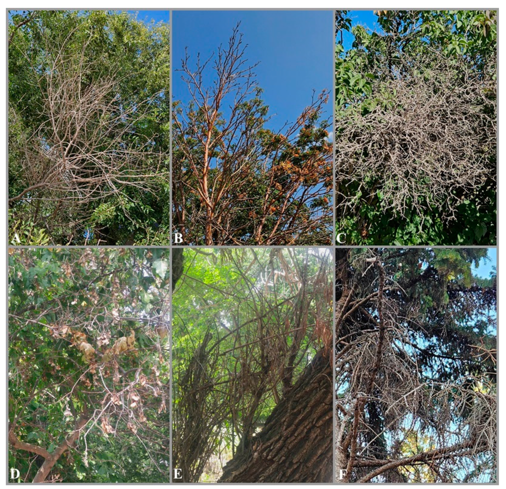
Figure 2.
Phylogram of RAxML tree generated based on the analysis of ITS and act1 sequence data of selected Cytospora strains. Bootstrap support values for ML ≥ 50% and BP ≥ 0.90 are shown as ML/ BP above or below the nodes. Reference strains are marked in bold. The tree is rooted to Diaporthe vaccinii (CBS 160.32).
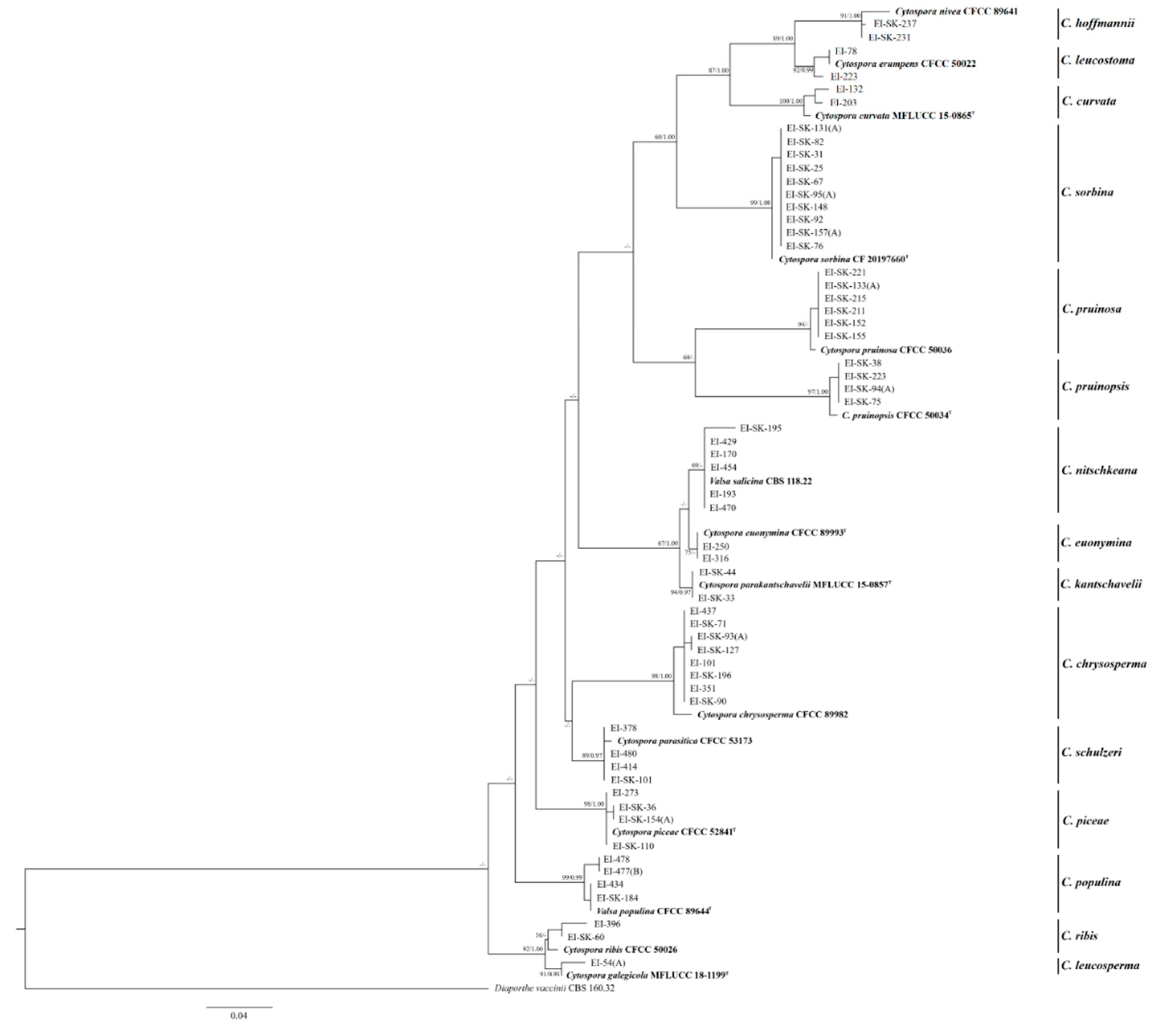
Figure 3.
7-11d old pure cultures of (a) C. chrysosperma; (b) C. curvata; (c) C. euonymina; (d) C. hoffmannii; (e) C. kantschavelii; (f) C. leucosperma; (g) C. leucostoma; (h) C. nitschkeana; (i) C. piceae; (j) C. populina; (k) C. pruinopsis; (l) C. pruinosa; (m) C. ribis; (n) C. schulzeri; (o) C. sorbina.
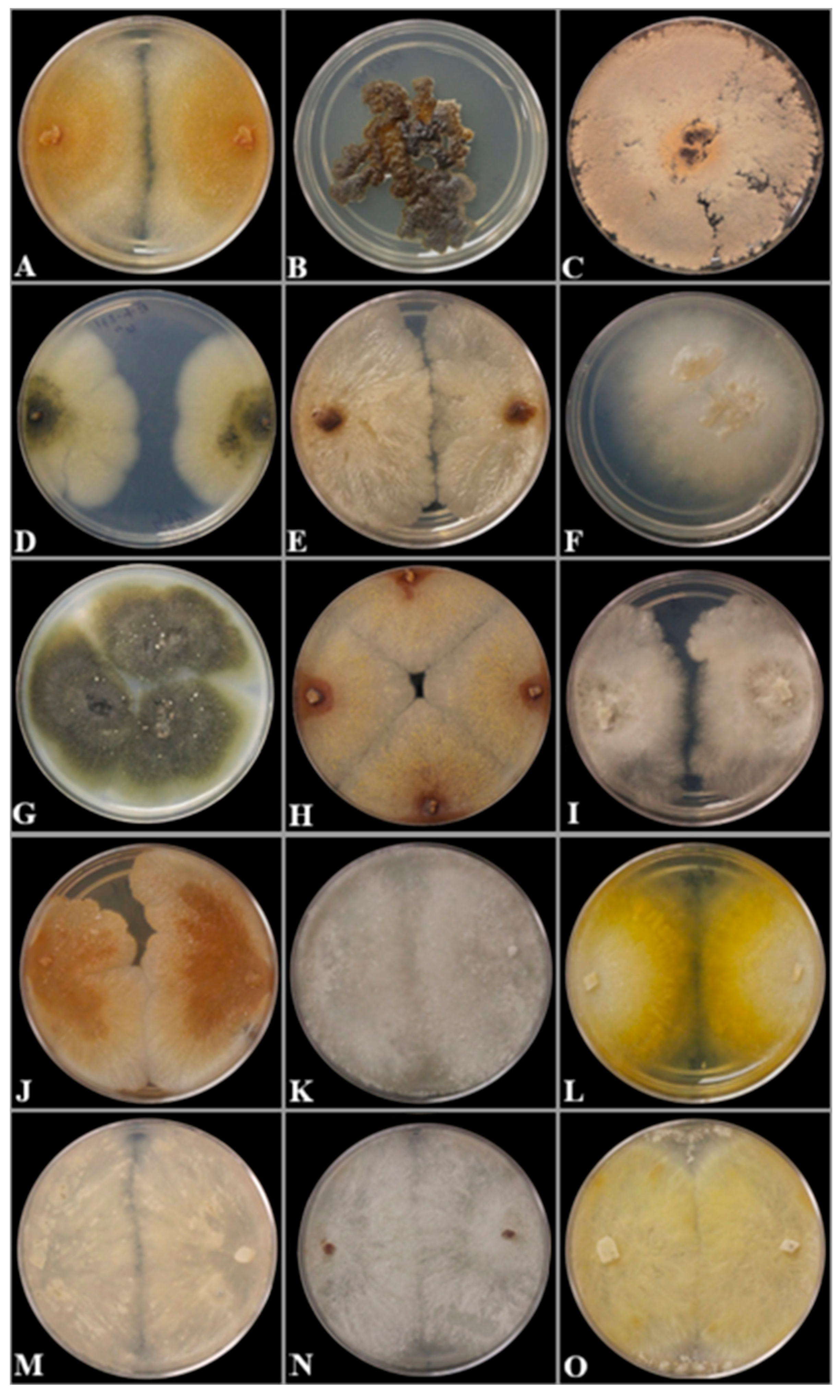

Table 1.
Strains of Cytospora species used in phylogenetic analysis with their GenBank accession numbers. Reference strains are marked in bold. Ex-type strains are marked withT. NA - data not available.
Table 1.
Strains of Cytospora species used in phylogenetic analysis with their GenBank accession numbers. Reference strains are marked in bold. Ex-type strains are marked withT. NA - data not available.
| GenBank accession numbers | |||||
|---|---|---|---|---|---|
| Species | Strain | Country | Host | ITS | act1 |
| Cytospora chrysosperma | CFCC 89982 | China | Ulmus pumila | KP281261 | KP310835 |
| EI-101 | Canada | Unknown tree | PQ356607 | PQ728056 | |
| EI-351 | Canada | Rhus sp. | PQ385601 | NA | |
| EI-437 | Canada | Populus tremuloides | PQ678997 | NA | |
| EI-SK-71 | Canada | Populus deltoides | PQ678998 | NA | |
| EI-SK-90 | Canada | Salix bebbiana | PQ679047 | NA | |
| EI-SK-93(A) | Canada | Populus tremula | PQ679032 | NA | |
| EI-SK-127 | Canada | Populus sp. | PQ679040 | NA | |
| EI-SK-196 | Canada | Salix alba | PQ679042 | NA | |
| Cytospora curvata | MFLUCC 15-0865T | Russia | Salix alba | KY417728 | KY417694 |
| EI-132 | Canada | Aronia sp. | PQ677273 | PQ728060 | |
| EI-203 | Canada | Syringa vulgaris | PQ677316 | PQ728061 | |
| Cytospora euonymina | CFCC 89993T | China | Euonymus kiautschovicus | MH933630 | MH933537 |
| EI-250 | Canada | Salix sp. | PQ678925 | NA | |
| EI-316 | Canada | Berberis vulgaris | PQ383411 | NA | |
| Cytospora hoffmannii | CFCC 89641 | China | Elaeagnus angustifolia | KF765683 | KU711006 |
| EI-SK-231 | Canada | Salix bebbiana | PQ677104 | PQ728057 | |
| EI-SK-237 | Canada | Salix sp. | PQ677112 | PQ728058 | |
| Cytospora kantschavelii | MFLUCC 15-0857T | Russia | Populus × sibirica | KY417738 | KY417704 |
| EI-SK-33 | Canada | Ulmus glabra | PQ678995 | PQ728067 | |
| EI-SK-44 | Canada | Acer spicatum | PQ678996 | PQ728068 | |
| Cytospora leucosperma | MFLUCC 18-1199T | Russia | Galega officinalis | MK912128 | MN685810 |
| EI-54(A) | Canada | Berberis vulgaris | PQ281438 | NA | |
| Cytospora leucostoma | CFCC 50022 | China | Prunus padus | MH933627 | MH933534 |
| EI-78 | Canada | Malus sp. | PP751512 | NA | |
| EI-223 | Canada | Vaccinium sp. | PQ368601 | PQ728059 | |
| Cytospora nitschkeana | CBS. 118.22 | Netherlands | Salix alba | MH854712 | KX964746 |
| EI-170 | Canada | Salix babylonica | PQ356735 | NA | |
| EI-193 | Canada | Berberis vulgaris | PQ362651 | NA | |
| EI-429 | Canada | Fraxinus americana | PQ421750 | NA | |
| EI-454 | Canada | Unknown tree | PQ425077 | NA | |
| EI-470 | Canada | Fraxinus nigra | PQ678921 | NA | |
| EI-SK-195 | Canada | Salix alba | PQ678915 | NA | |
| Cytospora piceae | CFCC 52841T | China | Picea crassifolia | MH820398 | MH820406 |
| EI-273 | Canada | Picea pungens | ON352565 | NA | |
| EI-SK-36 | Canada | Rhus sp. | PQ671332 | NA | |
| EI-SK-110 | Canada | Picea sp. | PQ671333 | NA | |
| EI-SK-154(A) | Canada | Fraxinus sp. | PQ666762 | NA | |
| Cytospora populina | CFCC 89644T | China | Salix psammophila | KF765686 | KU711007 |
| EI-434 | Canada | Magnolia sp. | PQ422182 | NA | |
| EI-477(B) | Canada | Sorbus sp. | PQ683229 | PQ728055 | |
| EI-478 | Canada | Acer platanoides | PQ425482 | NA | |
| EI-SK-184 | Canada | Aronia sp. | PQ683280 | NA | |
| Cytospora pruinopsis | CFCC 50034T | China | Ulmus pumila | KP281259 | KP310836 |
| EI-SK-38 | Canada | Populus deltoides | PQ678306 | PQ728066 | |
| EI-SK-75 | Canada | Ulmus americana | PQ678839 | NA | |
| EI-SK-94(A) | Canada | Malus sp. | PQ678815 | NA | |
| EI-SK-223 | Canada | Unknown shrubs | PQ678307 | NA | |
| Cytospora pruinosa | CFCC 50036 | China | Syringa oblata | KP310800 | KP310832 |
| EI-SK-133(A) | Canada | Aronia sp. | PQ677817 | PQ728065 | |
| EI-SK-152 | Canada | Syringa reticulata | PQ677975 | NA | |
| EI-SK-155 | Canada | Unknown shrubs | PQ680075 | NA | |
| EI-SK-211 | Canada | Syringa sp. | PQ677872 | NA | |
| EI-SK-215 | Canada | Fraxinus sp. | PQ677818 | NA | |
| EI-SK-221 | Canada | Unknown shrubs | PQ680079 | NA | |
| Cytospora ribis | CFCC 50026 | China | Ulmus pumila | KP281267 | KP310843 |
| EI-396 | Canada | Fraxinus americana | PQ393077 | PQ728070 | |
| EI-SK-60 | Canada | Syringa vulgaris | PQ683333 | NA | |
| Cytospora schulzeri | CFCC 53173 | China | Berberissp. | MK673070 | MK673040 |
| EI-378 | Canada | Sorbaria sorbifolia | PQ392014 | NA | |
| EI-414 | Canada | Malus sp. | PQ421083 | NA | |
| EI-480 | Canada | Acer platanoides | PQ432426 | PQ728069 | |
| EI-SK-101 | Canada | Malus sp. | PQ728069 | PQ683213 | |
| Cytospora sorbina | CF 20197660T | China | Sorbus tianschanica | MK673052 | MK673022 |
| EI-SK-25 | Canada | Malus sp. | PQ677620 | PQ728063 | |
| EI-SK-31 | Canada | Aronia sp. | PQ677471 | PQ728062 | |
| EI-SK-67 | Canada | Aronia sp. | PQ677674 | PQ728064 | |
| EI-SK-76 | Canada | Prunus sp. | PQ736325 | NA | |
| EI-SK-82 | Canada | Sorbus aucuparia | PQ677675 | NA | |
| EI-SK-92 | Canada | Prunus padus | PQ677676 | NA | |
| EI-SK-95(A) | Canada | Aronia sp. | PQ680078 | NA | |
| EI-SK-131(A) | Canada | Unknown shrubs | PQ677688 | NA | |
| EI-SK-148 | Canada | Sorbus sp. | PQ680076 | NA | |
| EI-SK-157(A) | Canada | Viburnum cf. trilobum | PQ680077 | NA | |
Disclaimer/Publisher’s Note: The statements, opinions and data contained in all publications are solely those of the individual author(s) and contributor(s) and not of MDPI and/or the editor(s). MDPI and/or the editor(s) disclaim responsibility for any injury to people or property resulting from any ideas, methods, instructions or products referred to in the content. |
© 2025 by the authors. Licensee MDPI, Basel, Switzerland. This article is an open access article distributed under the terms and conditions of the Creative Commons Attribution (CC BY) license (http://creativecommons.org/licenses/by/4.0/).
Copyright: This open access article is published under a Creative Commons CC BY 4.0 license, which permit the free download, distribution, and reuse, provided that the author and preprint are cited in any reuse.


