
Submitted:
02 January 2025
Posted:
08 January 2025
You are already at the latest version
Abstract
Biofilms are communities of microorganisms attached to surfaces, embedded within self-produced extracellular polymeric substances (EPS). They play a dual role in human and environmental contexts. Beneficial biofilms are widely utilized in bioreactors for applications such as energy production and pollutant immobilization, while harmful biofilms are associated with severe infections, including lung infections and urinary tract infections. Understanding the biofilm cycle, encompassing attachment, maturation, and dispersal phases, is critical for optimizing beneficial biofilms and mitigating harmful ones. For example, identifying the optimal late exponential or early maturation phases can enhance biofilm utility, while predicting dispersal timing can inform strategies for disrupting harmful biofilms, safeguarding human health. This study employs a bibliometric approach to analyze the biofilm cycle, extracting core keywords, leading organizations, and active countries/regions. By synthesizing data from the Web of Science, this analysis highlights key trends and knowledge gaps in the field, providing a comprehensive overview of biofilm cycle dynamics. The study also identifies future opportunities for integrating big data and machine learning, which have the potential to revolutionize biofilm research. These technologies could enable precise predictions of biofilm behavior, fostering advancements in healthcare, environmental protection, and industrial processes, and ensuring more effective biofilm management strategies.
Keywords:
Biofilm Cycle
; Bibliography
; VOSviewer
; Big Data
; Machine Learning
1. Introduction
Biofilms are complex communities of microorganisms that attach to surfaces and are encased in self-produced extracellular polymeric substances (EPS) [1,2]. This structure provides a protective and stable environment for the microorganisms, enabling them to thrive under various conditions [3,4]. The study of biofilms is critical due to their dual nature: beneficial biofilms can be harnessed for environmentally friendly applications like bioreactors for energy production [5] or pollutant immobilization [6], while harmful biofilms can lead to serious medical and environmental problems [7,8]. For instance, pathogenic biofilms are a significant cause of infections, including lung infections [9] and urinary tract infections [10], presenting challenges in healthcare and sanitation.
Understanding the biofilm cycle is central to both utilizing beneficial biofilms [5] and mitigating harmful ones [9,10]. The biofilm cycle encompasses various phases, including the attachment phase, maturation phase, and dispersal phase [11]. Each phase involves specific physiological and structural changes within the biofilm community. For example, knowing the duration of the attachment phase can help optimize the conditions for beneficial biofilm growth [12], while understanding the maturation phase timeline aids in exploiting biofilms at their most productive stage, such as during the late exponential or early maturation phase [13]. Similarly, insights into the dispersal phase can inform strategies to disrupt harmful biofilms before they become more resilient or infectious, helping to protect human health by anticipating critical periods in biofilm development.
This study employs a bibliographic approach [14,15] to analyze the biofilm cycle comprehensively, identifying core keywords, leading organizations, and influential countries/regions in this field. By examining the literature, the research maps out the dominant trends and patterns in biofilm cycle studies [16,17], providing a clearer understanding of how these microbial communities develop and behave. The analysis not only summarizes the current knowledge base but also highlights critical gaps and opportunities for innovation. Through this systematic evaluation, the study aims to deepen our understanding of biofilm dynamics and provide actionable insights for both researchers and practitioners.
2. Materials and Methods
The method followed previous studies with slightly modifications [18,19]. This study conducted a bibliometric analysis focusing on the term "Biofilm Cycle" by searching the Web of Science database [20,21] on December 6, 2024. The search yielded 4,385 results, reflecting the significant attention this topic has garnered in recent years. To ensure manageable and insightful analysis, we selected the default 1,000 most relevant articles for further investigation using VOSviewer (Version 1.6.20) [22,23]. This subset represents a comprehensive but concise dataset that aligns with the study’s aim of identifying dominant trends, key contributors, and geographical focus in biofilm research. The decision to limit the dataset to 1,000 records ensures that the analysis remains focused and visually interpretable while retaining enough data for meaningful insights.
For the keyword co-occurrence analysis, we set the minimum occurrence threshold at "5," which means only keywords that appeared at least five times across the selected articles were included. This threshold was chosen to strike a balance between comprehensiveness and clarity, allowing us to highlight prominent themes without cluttering the visualization with less relevant or infrequently occurring terms. This approach revealed recurring topics and concepts in biofilm research, emphasizing their importance in understanding biofilm cycles. Too low a threshold would have overwhelmed the visualization with minor terms, while a higher threshold risked overlooking potentially critical but less frequent keywords.
In the organizational collaboration analysis, we applied a similar logic, selecting organizations with at least five published documents. This criterion was designed to highlight institutions with consistent contributions to biofilm research while filtering out organizations with minimal involvement. The resulting analysis revealed leading research institutions driving advancements in this field and their collaborative networks. Such insights are crucial for identifying centers of excellence and potential partnerships in biofilm studies, offering a roadmap for researchers seeking collaborators or understanding institutional priorities.
For the country and regional analysis, a threshold of ten documents was established to focus on countries with substantial contributions to the field. This choice enabled the identification of regions with robust research activity while minimizing the noise from countries with only marginal contributions. The analysis highlighted global trends in biofilm research, showing a strong concentration of activity in developed nations with well-funded scientific programs. It also provided a comparative perspective on regional expertise and resources in biofilm studies, offering valuable context for future international collaborations and funding strategies. These thresholds and methodological decisions collectively ensured that our analysis remained both manageable and impactful.
3. Results
Figure 1 highlights the results of the keyword analysis, offering a comprehensive view of the research focus within the biofilm cycle domain. A significant group of keywords pertains to microbial strains, including “Escherichia coli,” “Pseudomonas aeruginosa,” and “Staphylococcus aureus.” These bacteria are prominent in biofilm studies due to their roles in both beneficial and harmful biofilm activities. For example, Escherichia coli is commonly associated with biofilm formation in industrial and medical contexts, while Pseudomonas aeruginosa is a well-known pathogen that thrives in biofilm states, complicating infection treatment. The frequent appearance of these keywords reflects the centrality of these microorganisms in advancing biofilm-related knowledge, particularly in understanding biofilm behavior, resistance mechanisms, and applications.
Another set of keywords is closely linked to environmental contexts, such as “sediments,” “wastewater,” “sludge,” and “treatment plants.” These terms underline the critical role biofilms play in ecological and engineered systems. In wastewater treatment, biofilms are essential for nutrient removal and pollutant degradation, often forming on surfaces within treatment plants to enhance efficiency. Similarly, the association with sediments and sludge points to the natural occurrence of biofilms in aquatic environments, where they contribute to ecosystem stability and nutrient cycling. These keywords underscore the importance of biofilm studies in addressing environmental challenges and improving sustainable management practices.
A third cluster of keywords relates to pollutants, including “copper,” “nitrite,” “nitrate,” and “phosphorus.” These terms reflect a growing focus on biofilms’ interactions with contaminants, particularly their ability to immobilize, transform, or degrade these substances. For instance, biofilms can sequester heavy metals like copper or reduce nitrates and nitrites in water systems, offering potential solutions for pollution mitigation. The presence of phosphorus as a keyword suggests interest in biofilms’ role in nutrient dynamics, especially in preventing eutrophication. This cluster highlights the applied significance of biofilm research, particularly in environmental remediation and industrial wastewater treatment.
Lastly, several keywords pertain to biological processes fundamental to the biofilm cycle, including “adhesion,” “infections,” “dispersal,” and “resistance.” These terms represent key phases and characteristics of biofilm formation and development. For instance, adhesion marks the initial attachment phase, which is critical for biofilm establishment, while dispersal reflects the phase where cells leave the biofilm, spreading to new locations. Keywords like “infections” and “resistance” are particularly relevant in medical contexts, highlighting the challenges biofilms pose in healthcare, such as their role in persistent infections and their heightened resistance to antibiotics. Together, these process-related keywords emphasize the complexity of biofilm behavior and the necessity of understanding these dynamics for both harnessing biofilm benefits and mitigating their risks.
Figure 2 illustrates the primary research organizations contributing to the field of biofilm cycle studies, with several institutions occupying central positions in the network. At the core are renowned organizations such as the Chinese Academy of Sciences, Technical University of Denmark, Ohio State University, Montana State University, University of California, Berkeley, Oak Ridge National Laboratory, Harvard University, Tsinghua University, and Nanyang Technological University. These institutions are distinguished by their extensive contributions to biofilm research, encompassing areas like microbial behavior, biofilm formation, and environmental applications. Their central positioning reflects their critical role in driving innovation, disseminating knowledge, and shaping the field's research trajectory.
Beyond these core institutions, numerous other organizations also play significant roles in advancing biofilm cycle research. These include Nanjing Tech University, Delft University of Technology, Shanghai Jiao Tong University, Zhejiang University, Harbin Institute of Technology, Chongqing University, Sun Yat-sen University, Arizona State University, University of New South Wales, University of São Paulo, Hohai University, University of Southampton, Sichuan University, University of Amsterdam, University of Minho, University of Barcelona, Max Planck Institute for Marine Microbiology, and CNRS. These organizations contribute through diverse research efforts, from investigating biofilm mechanics to exploring its applications in environmental and medical sciences. Their geographic distribution also highlights the global interest and collaborative nature of this field, with contributions spanning continents.
Collaboration among these institutions is a defining feature of the biofilm cycle research landscape. By leveraging shared expertise, resources, and datasets, these organizations have collectively advanced our understanding of biofilm dynamics. The partnerships between institutions, both domestic and international, have facilitated groundbreaking discoveries, fostered interdisciplinary research, and accelerated the development of practical applications. The network depicted in Figure 2 underscores the importance of such collaborations in addressing complex scientific questions, demonstrating how collective efforts can propel the biofilm cycle research field forward. This interconnected framework not only enhances scientific productivity but also encourages innovation through diverse perspectives and shared objectives.
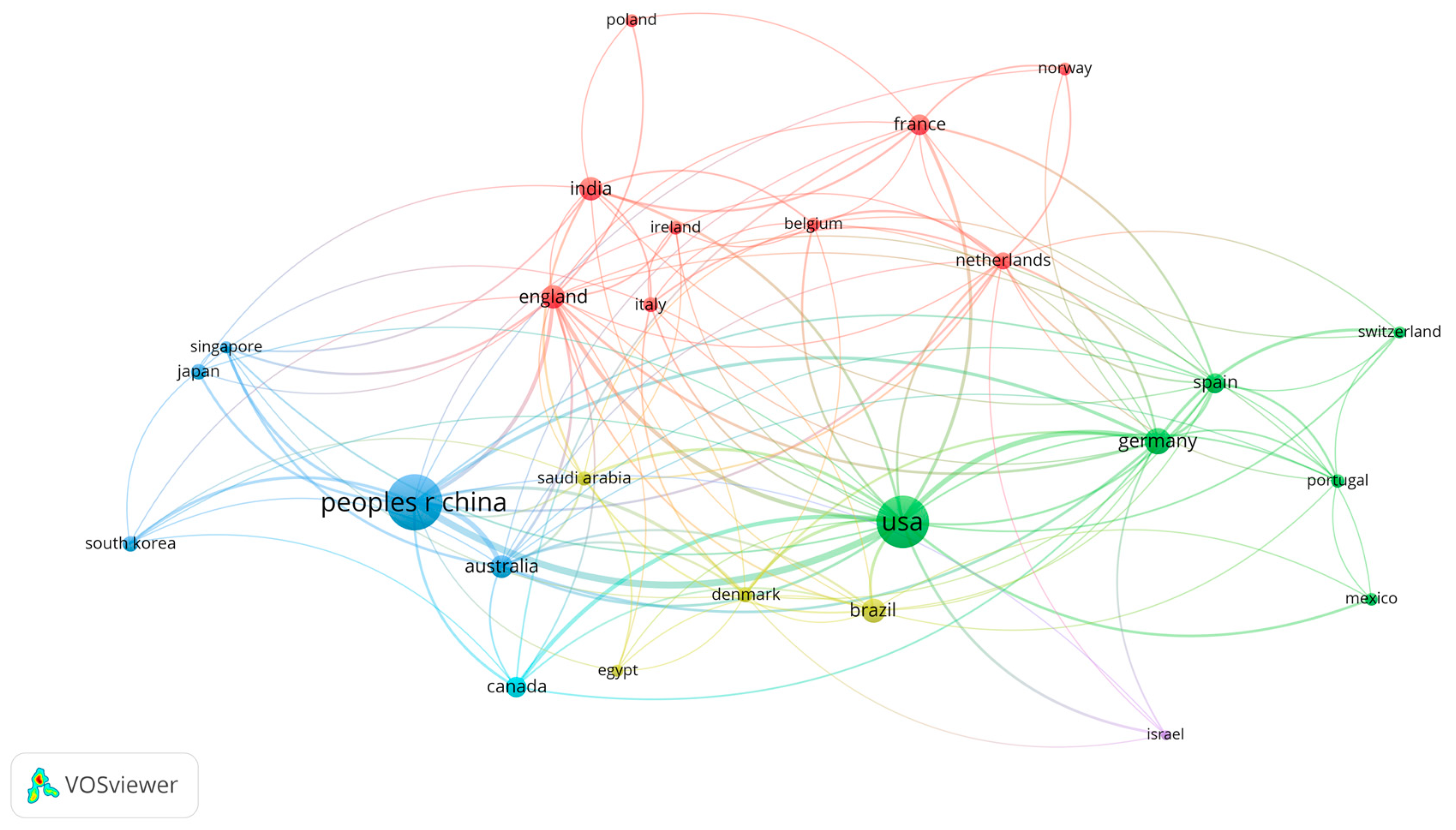
Figure 3 highlights the major countries and regions contributing to the field of biofilm cycle research, emphasizing the global nature of scientific collaboration in this domain. At the forefront are China and the United States, which dominate as the two most influential countries in biofilm research. These nations lead in terms of publication volume, research funding, and the establishment of cutting-edge facilities. Their contributions span a wide array of topics, from fundamental studies on biofilm formation and dispersal phases to practical applications in healthcare, environmental remediation, and industrial processes. China’s rapid rise in research output, supported by initiatives like the Chinese Academy of Sciences, and the United States’ strong academic and industrial partnerships, have solidified their positions as the primary drivers of innovation in this field.
Beyond China and the United States, several other countries have established themselves as significant contributors to biofilm cycle research. In Asia, nations such as South Korea, Japan, and Singapore have made substantial advancements, often focusing on technological innovations and interdisciplinary research approaches. Similarly, Canada and Australia have contributed extensively, particularly in environmental and medical biofilm studies. In Europe, countries like the United Kingdom, Germany, France, Italy, Belgium, Denmark, Ireland, Portugal, and Spain have long-standing traditions in microbiology and biotechnology, enriching the field through diverse research perspectives. Emerging players like Saudi Arabia, Egypt, India, Brazil, and Mexico reflect a growing global interest, bringing unique regional challenges and solutions to biofilm research.
International collaboration among these nations is a hallmark of the biofilm cycle research field, fostering the exchange of knowledge, methodologies, and resources. For instance, partnerships between developed and emerging economies facilitate capacity building and enable access to diverse datasets, enhancing the scope and applicability of biofilm studies. Cross-border initiatives have resulted in breakthroughs in understanding biofilm-related health challenges and environmental issues, demonstrating the value of a collective global effort. Figure 3 underscores the importance of such collaborations, illustrating how nations, despite their geographical and cultural differences, work together to advance the understanding and application of biofilm cycles, ultimately addressing critical global challenges.
4. Discussion
4.1. Analysis of the Biofilm Cycle Stages
Biofilm formation is a complex and dynamic process that occurs in five distinct stages: 1) Bacteria Land on Surface, 2) Irreversible Attachment, 3) Proliferation, 4) Maturation, and 5) Dispersal [24]. These stages represent the lifecycle of biofilm formation, starting with the initial contact of bacteria with a surface and culminating in the release of bacterial cells into the surrounding environment. Bibliographic analysis has been instrumental in understanding the various factors that influence each stage, including microbial interactions, environmental conditions, and the role of extracellular matrix (ECM) components [25].
In the first stage, Bacteria Land on Surface, free-floating bacteria encounter and adhere to a surface [26,27]. This is a reversible attachment, where the bacteria are loosely bound to the surface through weak interactions such as van der Waals forces and electrostatic interactions. The choice of surface material, surface roughness, and environmental factors such as nutrient availability significantly influence this initial phase. Studies have shown that surfaces with hydrophobic properties tend to attract more bacterial adhesion, especially for certain pathogenic strains.
The second stage, Irreversible Attachment, is marked by stronger adhesion of bacteria to the surface [28,29]. This stage is characterized by the production of surface-associated molecules, such as pili and fimbriae, that anchor the bacteria more firmly. Once bacteria irreversibly attach, they begin to produce extracellular polymeric substances (EPS), a key feature of biofilm formation. EPS consists of polysaccharides, proteins, and nucleic acids that contribute to the stability and integrity of the biofilm. This stage is critical because it sets the foundation for further biofilm growth and maturation.
Proliferation, the third stage, involves the growth and division of the attached bacterial cells, leading to an increase in biomass [30,31]. As the bacterial population expands, the biofilm structure becomes more complex. The growing bacteria interact with each other, forming microcolonies that are encased in the EPS matrix. These microcolonies are not homogenous, and gradients of nutrients, oxygen, and waste products often exist within the biofilm, leading to a heterogeneous distribution of microbial activity. This stage is essential for the development of biofilm architecture and the establishment of microbial communities with diverse functions.
The Maturation stage sees the biofilm reaching its full structural complexity [32,33]. The biofilm becomes more organized, with distinct layers and channels formed for nutrient and waste exchange. This stage is characterized by the production of a robust extracellular matrix that provides the biofilm with resistance to antimicrobial agents and physical stress. Maturation enhances the biofilm’s ability to persist in various environments, making it more resilient to external challenges, such as antibiotics and immune responses. At this stage, biofilms can exhibit a high degree of resistance to treatment, which poses challenges in medical and industrial contexts.
Finally, in the Dispersal stage, bacteria are released from the biofilm into the surrounding environment, either as single cells or as clusters [34,35]. This is a crucial stage for biofilm propagation, as it enables bacteria to colonize new surfaces and expand the biofilm. Dispersal can be triggered by environmental changes, such as nutrient depletion, or by the biofilm reaching a certain size. The release of bacteria allows the biofilm to spread, leading to the colonization of new niches. This stage is also associated with the formation of new biofilms in different locations, contributing to the persistence of bacterial populations.

Table 1.
Summary of biofilm cycle stages.
| Stages | Main Characteristics | References |
|---|---|---|
| 1.Bacteria Land on Surface | - Bacteria touch the surface. - Weak, temporary bonds form (e.g., physical forces). |
[26,27] |
| 2.Irreversible Attachment | - Bacteria stick firmly using structures like pili or slime (EPS). - Hard to remove. |
[28,29] |
| 3.Proliferation | - Bacteria grow and produce slime to form a shield. - They communicate and work together. |
[30,31] |
| 4. Maturation | - Biofilm gets thicker and more complex. - High resistance to antibiotics and harsh conditions. |
[32,33] |
| 5. Dispersal | - Parts of the biofilm break off. - Bacteria spread to new places to start over. |
[34,35] |
4.2. Beneficial Biofilm in Biofilm Reactors and Harmful Biofilm in Infections
Biofilms are highly dynamic microbial communities that develop through distinct stages, with the late Proliferation phase and early Maturation phase being particularly important in determining their potential and strength [33,36]. During the late Proliferation phase, the microbial population within the biofilm increases significantly, and the EPS that form the biofilm's matrix begin to solidify. This creates a highly organized structure that is resistant to external environmental stressors, such as antibiotics and immune responses. The early Maturation phase marks the start of biofilm's structural development, where cells continue to organize into complex, multi-layered structures that contribute to the biofilm’s increased resilience. These phases enhance the biofilm’s stability and resistance, making it difficult to remove and giving it its maximum potential strength. Studies have shown that biofilms formed during these stages are particularly effective in both beneficial and harmful applications, as their growth and development lead to a more resilient and long-lasting structure
In terms of beneficial applications, biofilms have been utilized in various fields with impressive results. For instance, biofilms play an important role in environmental remediation by immobilizing heavy metals in water treatment processes [6]. Microorganisms within the biofilm matrix are able to break down and detoxify harmful substances, improving the quality of water and reducing environmental pollution. Biofilms are also integral to microbial fuel cells [5,37], where they facilitate electricity generation by harnessing the metabolic activity of microorganisms to convert organic matter into energy [38]. This provides a sustainable energy solution with potential applications in waste treatment and energy production. Additionally, biofilms are being explored in the field of construction, particularly in self-healing concrete [39]. In this application, bacteria embedded in the biofilm matrix react to cracks in the concrete by precipitating calcium carbonate, which fills the cracks and helps to prolong the lifespan of the material [40,41]. These advancements demonstrate how biofilms can be harnessed for sustainable practices across diverse industries.
However, biofilms can also have significant negative impacts, particularly when pathogenic microorganisms form biofilms on medical devices or within the human body. For example, Pseudomonas aeruginosa forms biofilms in the lungs of individuals with cystic fibrosis, leading to chronic lung infections that are difficult to treat [42,43]. Similarly, Staphylococcus aureus is a common pathogen in urinary tract infections [44,45], and Escherichia coli biofilms can cause severe gastrointestinal diseases such as diarrhea [46,47]. The resilience of these pathogenic biofilms makes them particularly challenging to eradicate, requiring advanced treatments. To prevent the formation of harmful biofilms, researchers have developed various strategies, such as incorporating titanium dioxide (TiO2) into surface coatings like concrete and medical devices [48,49]. TiO2 has antimicrobial properties that prevent bacterial adhesion and biofilm formation, thus reducing the risk of infections [50,51]. This strategy is particularly effective in reducing the impact of biofilms in healthcare settings and promoting hygiene in environments where bacterial contamination is a concern.
4.3. Future Direction: Big Data and Machine Learning on Biofilm Cycle
The future of biofilm cycle research holds immense promise, especially with the integration of big data and machine learning technologies [52,53]. These tools offer powerful capabilities for analyzing and predicting complex biological processes, such as biofilm formation and behavior. In recent years, big data and machine learning have already made significant strides in fields such as facial recognition [54,55], autonomous driving [56,57], species distribution prediction [58], and educational outcome forecasting [59]. Their success in these domains illustrates the potential for similar breakthroughs in biofilm research. Machine learning algorithms can analyze vast datasets to uncover patterns and correlations that would be difficult or impossible to detect through traditional methods. As biofilm research advances, leveraging these technologies could lead to more precise and comprehensive models of biofilm formation, maturation, and detachment processes, helping to improve disease prevention and treatment strategies.
One of the key innovations that big data and machine learning [60] can bring to biofilm research is the development of large-scale databases that track various factors influencing biofilm dynamics. Such a database could include data on bacterial species, surface materials to which bacteria attach, nutrient media composition, biofilm growth duration, and detachment times. By compiling vast amounts of experimental and observational data, researchers can create a comprehensive framework for understanding the multiple variables that govern biofilm behavior. This database would provide a solid foundation for developing predictive models that could forecast the progression of biofilm-related phenomena under different conditions. For instance, machine learning models could analyze this data to predict when a specific biofilm might become most virulent, such as in the case of human infections, enabling timely interventions and personalized medical treatments.
Building upon the foundation of large databases, the use of neural networks [61,62] and other machine learning techniques could further refine predictions and diagnostics in biofilm cycle research. By training neural networks on extensive datasets, researchers could develop models capable of accurately predicting biofilm formation stages, identifying risk factors for infection, and determining when biofilms reach their most dangerous state. For example, a trained neural network model could assess factors such as bacterial species, patient health conditions, and environmental variables to predict when biofilm formation would peak, giving healthcare providers a better understanding of when patients might be at their most vulnerable to biofilm-related infections. This predictive capability would be especially valuable in clinical settings, where timely detection and intervention are critical to patient outcomes. By combining big data with machine learning, the potential to revolutionize biofilm research and enhance both clinical and environmental management is vast.
5. Conclusions
In conclusion, this study utilized bibliographic analysis to explore key aspects of the biofilm cycle, including prominent keywords, organizations, and countries/regions involved in the research. The five stages of biofilm development—bacteria landing on surfaces, irreversible attachment, proliferation, maturation, and dispersal—were summarized from essential literature. The study also highlighted biofilm's significant industrial applications and its potential health risks. Looking ahead, the integration of big data and machine learning is presented as a promising opportunity for enhancing biofilm cycle research, offering new methods for prediction and management in both industrial and medical contexts.
References
- Flemming, H.-C.; Wingender, J. The biofilm matrix. Nature reviews microbiology 2010, 8, 623–633. [Google Scholar] [CrossRef] [PubMed]
- Xiao, R.; Zheng, Y. Overview of microalgal extracellular polymeric substances (EPS) and their applications. Biotechnology advances 2016, 34, 1225–1244. [Google Scholar] [CrossRef] [PubMed]
- Siddharth, T.; Sridhar, P.; Vinila, V.; Tyagi, R.D. Environmental applications of microbial extracellular polymeric substance (EPS): a review. Journal of Environmental Management 2021, 287, 112307. [Google Scholar] [CrossRef] [PubMed]
- Flemming, H.-C.; Neu, T.R.; Wozniak, D.J. The EPS matrix: the “house of biofilm cells”. Journal of bacteriology 2007, 189, 7945–7947. [Google Scholar] [CrossRef]
- Zhao, C.-e.; Chen, J.; Ding, Y.; Wang, V.B.; Bao, B.; Kjelleberg, S.; Cao, B.; Loo, S.C.J.; Wang, L.; Huang, W. Chemically functionalized conjugated oligoelectrolyte nanoparticles for enhancement of current generation in microbial fuel cells. ACS Applied Materials & Interfaces 2015, 7, 14501–14505. [Google Scholar]
- Ding, Y.; Peng, N.; Du, Y.; Ji, L.; Cao, B. Disruption of putrescine biosynthesis in Shewanella oneidensis enhances biofilm cohesiveness and performance in Cr (VI) immobilization. Applied and environmental microbiology 2014, 80, 1498–1506. [Google Scholar] [CrossRef]
- Costerton, J.W.; Stewart, P.S. Battling biofilms. Scientific American 2001, 285, 74–81. [Google Scholar] [CrossRef]
- Rather, M.A.; Gupta, K.; Bardhan, P.; Borah, M.; Sarkar, A.; Eldiehy, K.S.H.; Bhuyan, S.; Mandal, M. Microbial biofilm: A matter of grave concern for human health and food industry. Journal of Basic Microbiology 2021, 61, 380–395. [Google Scholar] [CrossRef]
- Maurice, N.M.; Bedi, B.; Sadikot, R.T. Pseudomonas aeruginosa biofilms: host response and clinical implications in lung infections. American journal of respiratory cell and molecular biology 2018, 58, 428–439. [Google Scholar] [CrossRef]
- Trautner, B.W.; Darouiche, R.O. Role of biofilm in catheter-associated urinary tract infection. American journal of infection control 2004, 32, 177–183. [Google Scholar] [CrossRef]
- Sauer, K.; Stoodley, P.; Goeres, D.M.; Hall-Stoodley, L.; Burmølle, M.; Stewart, P.S.; Bjarnsholt, T. The biofilm life cycle: expanding the conceptual model of biofilm formation. Nature Reviews Microbiology 2022, 20, 608–620. [Google Scholar] [CrossRef] [PubMed]
- Teughels, W.; Van Assche, N.; Sliepen, I.; Quirynen, M. Effect of material characteristics and/or surface topography on biofilm development. Clinical oral implants research 2006, 17, 68–81. [Google Scholar] [CrossRef] [PubMed]
- Lappin-Scott, H.M.; Bass, C. Biofilm formation: attachment, growth, and detachment of microbes from surfaces. American journal of infection control 2001, 29, 250–251. [Google Scholar] [CrossRef] [PubMed]
- Chen, S.; Ding, Y. From bibliography to understanding: water microbiology and human health. Journal of Water and Health 2024, 22, 1911–1921. [Google Scholar] [CrossRef]
- Chen, S.; Ding, Y. A bibliography study of Shewanella oneidensis biofilm. FEMS Microbiology Ecology 2023, 99, fiad124. [Google Scholar] [CrossRef]
- Ma, R.; Hu, X.; Zhang, X.; Wang, W.; Sun, J.; Su, Z.; Zhu, C. Strategies to prevent, curb and eliminate biofilm formation based on the characteristics of various periods in one biofilm life cycle. Frontiers in Cellular and Infection Microbiology 2022, 12, 1003033. [Google Scholar] [CrossRef]
- Rice, S.A.; Tan, C.H.; Mikkelsen, P.J.; Kung, V.; Woo, J.; Tay, M.; Hauser, A.; McDougald, D.; Webb, J.S.; Kjelleberg, S. The biofilm life cycle and virulence of Pseudomonas aeruginosa are dependent on a filamentous prophage. The ISME journal 2009, 3, 271–282. [Google Scholar] [CrossRef]
- Chen, S.; Ding, Y. Tackling heavy metal pollution: evaluating governance models and frameworks. Sustainability 2023, 15, 15863. [Google Scholar] [CrossRef]
- Chen, S.; Ding, Y. Bibliographic Insights into Biofilm Engineering. Acta Microbiologica Hellenica 2024, 69, 3–13. [Google Scholar] [CrossRef]
- Zhu, J.; Liu, W. A tale of two databases: the use of Web of Science and Scopus in academic papers. Scientometrics 2020, 123, 321–335. [Google Scholar] [CrossRef]
- Mongeon, P.; Paul-Hus, A. The journal coverage of Web of Science and Scopus: a comparative analysis. Scientometrics 2016, 106, 213–228. [Google Scholar] [CrossRef]
- Van Eck, N.J.; Waltman, L. Citation-based clustering of publications using CitNetExplorer and VOSviewer. Scientometrics 2017, 111, 1053–1070. [Google Scholar] [CrossRef] [PubMed]
- Bukar, U.A.; Sayeed, M.S.; Razak, S.F.A.; Yogarayan, S.; Amodu, O.A.; Mahmood, R.A.R. A method for analyzing text using VOSviewer. MethodsX 2023, 11, 102339. [Google Scholar] [CrossRef] [PubMed]
- Hall-Stoodley, L.; Stoodley, P. Biofilm formation and dispersal and the transmission of human pathogens. Trends in microbiology 2005, 13, 7–10. [Google Scholar] [CrossRef]
- Toyofuku, M.; Roschitzki, B.; Riedel, K.; Eberl, L. Identification of proteins associated with the Pseudomonas aeruginosa biofilm extracellular matrix. Journal of proteome research 2012, 11, 4906–4915. [Google Scholar] [CrossRef]
- Palmer, J.; Flint, S.; Brooks, J. Bacterial cell attachment, the beginning of a biofilm. Journal of Industrial Microbiology and Biotechnology 2007, 34, 577–588. [Google Scholar] [CrossRef]
- Feng, G.; Cheng, Y.; Wang, S.-Y.; Borca-Tasciuc, D.A.; Worobo, R.W.; Moraru, C.I. Bacterial attachment and biofilm formation on surfaces are reduced by small-diameter nanoscale pores: how small is small enough? npj Biofilms and Microbiomes 2015, 1, 1–9. [Google Scholar] [CrossRef]
- Hinsa, S.M.; Espinosa-Urgel, M.; Ramos, J.L.; O'Toole, G.A. Transition from reversible to irreversible attachment during biofilm formation by Pseudomonas fluorescens WCS365 requires an ABC transporter and a large secreted protein. Molecular microbiology 2003, 49, 905–918. [Google Scholar] [CrossRef]
- Caiazza, N.C.; O'Toole, G.A. SadB is required for the transition from reversible to irreversible attachment during biofilm formation by Pseudomonas aeruginosa PA14. 2004.
- Krsmanovic, M.; Biswas, D.; Ali, H.; Kumar, A.; Ghosh, R.; Dickerson, A.K. Hydrodynamics and surface properties influence biofilm proliferation. Advances in Colloid and Interface Science 2021, 288, 102336. [Google Scholar] [CrossRef]
- He, W.; Liu, H.; Wang, Z.; Tay, F.R.; Shen, Y. The dynamics of bacterial proliferation, viability, and extracellular polymeric substances in oral biofilm development. Journal of Dentistry 2024, 143, 104882. [Google Scholar] [CrossRef]
- Otto, M. Staphylococcal infections: mechanisms of biofilm maturation and detachment as critical determinants of pathogenicity. Annual review of medicine 2013, 64, 175–188. [Google Scholar] [CrossRef] [PubMed]
- Reisner, A.; Haagensen, J.A.J.; Schembri, M.A.; Zechner, E.L.; Molin, S. Development and maturation of Escherichia coli K-12 biofilms. Molecular microbiology 2003, 48, 933–946. [Google Scholar] [CrossRef] [PubMed]
- Kaplan, J.á. Biofilm dispersal: mechanisms, clinical implications, and potential therapeutic uses. Journal of dental research 2010, 89, 205–218. [Google Scholar] [CrossRef] [PubMed]
- Rumbaugh, K.P.; Sauer, K. Biofilm dispersion. Nature Reviews Microbiology 2020, 18, 571–586. [Google Scholar] [CrossRef]
- Kierek-Pearson, K.; Karatan, E. Biofilm development in bacteria. Advances in applied microbiology 2005, 57, 79–111. [Google Scholar]
- Zhao, C.e.; Wu, J.; Ding, Y.; Wang, V.B.; Zhang, Y.; Kjelleberg, S.; Loo, J.S.C.; Cao, B.; Zhang, Q. Hybrid conducting biofilm with built-in bacteria for high-performance microbial fuel cells. ChemElectroChem 2015, 2, 654–658. [Google Scholar] [CrossRef]
- Yang, Y.; Ding, Y.; Hu, Y.; Cao, B.; Rice, S.A.; Kjelleberg, S.; Song, H. Enhancing bidirectional electron transfer of Shewanella oneidensis by a synthetic flavin pathway. ACS synthetic biology 2015, 4, 815–823. [Google Scholar] [CrossRef]
- Zhang, Z.; Ding, Y.; Qian, S. Influence of bacterial incorporation on mechanical properties of engineered cementitious composites (ECC). Construction and Building Materials 2019, 196, 195–203. [Google Scholar] [CrossRef]
- Zhang, Z.; Weng, Y.; Ding, Y.; Qian, S. Use of genetically modified bacteria to repair cracks in concrete. Materials 2019, 12, 3912. [Google Scholar] [CrossRef]
- Zhang, Z.; Liu, D.; Ding, Y.; Wang, S. Mechanical performance of strain-hardening cementitious composites (SHCC) with bacterial addition. Journal of Infrastructure Preservation and Resilience 2022, 3, 3. [Google Scholar] [CrossRef]
- Reynolds, D.; Kollef, M. The epidemiology and pathogenesis and treatment of Pseudomonas aeruginosa infections: an update. Drugs 2021, 81, 2117–2131. [Google Scholar] [CrossRef] [PubMed]
- Bisht, K.; Baishya, J.; Wakeman, C.A. Pseudomonas aeruginosa polymicrobial interactions during lung infection. Current opinion in microbiology 2020, 53, 1–8. [Google Scholar] [CrossRef] [PubMed]
- Saenkham-Huntsinger, P.; Hyre, A.N.; Hanson, B.S.; Donati, G.L.; Adams, L.G.; Ryan, C.; Londoño, A.; Moustafa, A.M.; Planet, P.J.; Subashchandrabose, S. Copper resistance promotes fitness of methicillin-resistant Staphylococcus aureus during urinary tract infection. Mbio 2021, 12, 10–1128. [Google Scholar] [CrossRef] [PubMed]
- Selim, S.; Faried, O.A.; Almuhayawi, M.S.; Saleh, F.M.; Sharaf, M.; El Nahhas, N.; Warrad, M. Incidence of vancomycin-resistant Staphylococcus aureus strains among patients with urinary tract infections. Antibiotics 2022, 11, 408. [Google Scholar] [CrossRef]
- Robins-Browne, R.M.; Hartland, E.L. Escherichia coli as a cause of diarrhea. Journal of gastroenterology and hepatology 2002, 17, 467–475. [Google Scholar] [CrossRef]
- Cabrera-Sosa, L.; Ochoa, T.J. Escherichia coli diarrhea. In Hunter's Tropical Medicine and Emerging Infectious Diseases; Elsevier: 2020; pp. 481–485.
- Hamdany, A.H.; Ding, Y.; Qian, S. Graphene-Based TiO2 Cement Composites to Enhance the Antibacterial Effect of Self-Disinfecting Surfaces. Catalysts 2023, 13, 1313. [Google Scholar] [CrossRef]
- Hamdany, A.H.; Ding, Y.; Qian, S. Visible light antibacterial potential of graphene-TiO2 cementitious composites for self-sterilization surface. Journal of Sustainable Cement-Based Materials 2023, 12, 972–982. [Google Scholar] [CrossRef]
- Hamdany, A.H.; Ding, Y.; Qian, S. Cementitious Composite Materials for Self-Sterilization Surfaces. ACI Materials Journal 2022, 119, 197–210. [Google Scholar] [CrossRef]
- Hamdany, A.H.; Ding, Y.; Qian, S. Mechanical and antibacterial behavior of photocatalytic lightweight engineered cementitious composites. Journal of Materials in Civil Engineering 2021, 33, 04021262. [Google Scholar] [CrossRef]
- Zhou, L.; Pan, S.; Wang, J.; Vasilakos, A.V. Machine learning on big data: Opportunities and challenges. Neurocomputing 2017, 237, 350–361. [Google Scholar] [CrossRef]
- L’heureux, A.; Grolinger, K.; Elyamany, H.F.; Capretz, M.A.M. Machine learning with big data: Challenges and approaches. Ieee Access 2017, 5, 7776–7797. [Google Scholar] [CrossRef]
- Almeida, D.; Shmarko, K.; Lomas, E. The ethics of facial recognition technologies, surveillance, and accountability in an age of artificial intelligence: a comparative analysis of US, EU, and UK regulatory frameworks. AI and Ethics 2022, 2, 377–387. [Google Scholar] [CrossRef] [PubMed]
- Kaur, P.; Krishan, K.; Sharma, S.K.; Kanchan, T. Facial-recognition algorithms: A literature review. Medicine, Science and the Law 2020, 60, 131–139. [Google Scholar] [CrossRef] [PubMed]
- Atakishiyev, S.; Salameh, M.; Yao, H.; Goebel, R. Explainable artificial intelligence for autonomous driving: A comprehensive overview and field guide for future research directions. IEEE Access 2024. [Google Scholar] [CrossRef]
- Ning, H.; Yin, R.; Ullah, A.; Shi, F. A survey on hybrid human-artificial intelligence for autonomous driving. IEEE Transactions on Intelligent Transportation Systems 2021, 23, 6011–6026. [Google Scholar] [CrossRef]
- Chen, S.; Ding, Y. Machine learning and its applications in studying the geographical distribution of ants. Diversity 2022, 14, 706. [Google Scholar] [CrossRef]
- Chen, S.; Ding, Y. A machine learning approach to predicting academic performance in Pennsylvania’s schools. Social Sciences 2023, 12, 118. [Google Scholar] [CrossRef]
- Yang, C.-T.; Kristiani, E.; Leong, Y.K.; Chang, J.-S. Big data and machine learning driven bioprocessing–recent trends and critical analysis. Bioresource technology 2023, 372, 128625. [Google Scholar] [CrossRef]
- Kaveh, A. Applications of artificial neural networks and machine learning in civil engineering. Studies in computational intelligence 2024, 1168, 472. [Google Scholar]
- Yang, X.; Ye, J.; Wang, X. Factorizing knowledge in neural networks. 2022; pp. 73–91.
Figure 1.
Keyword analysis by VOSviewer. The lines indicate the keyword connection.
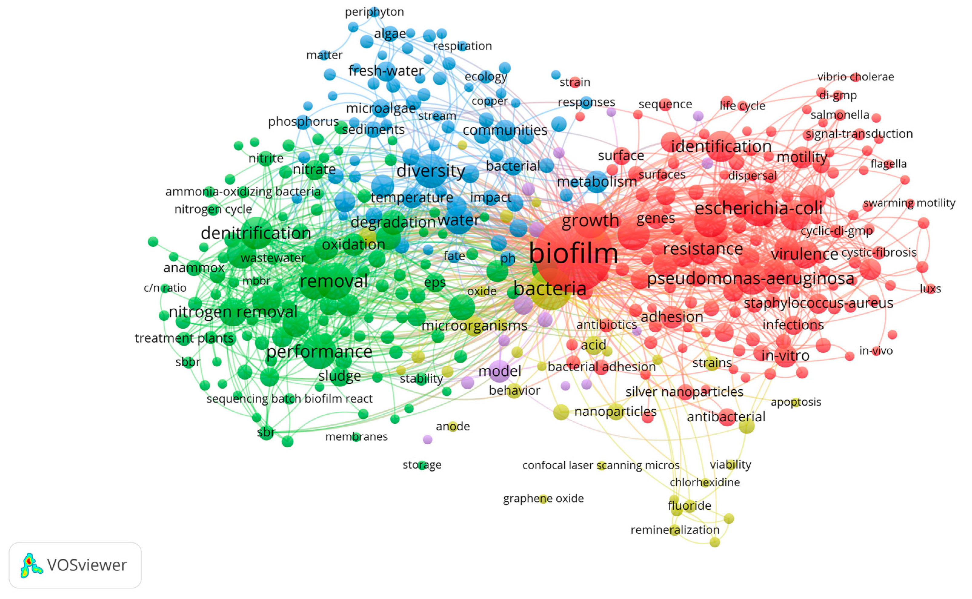
Figure 2.
Organizational analysis by VOSviewer. The lines indicate the research collaboration.
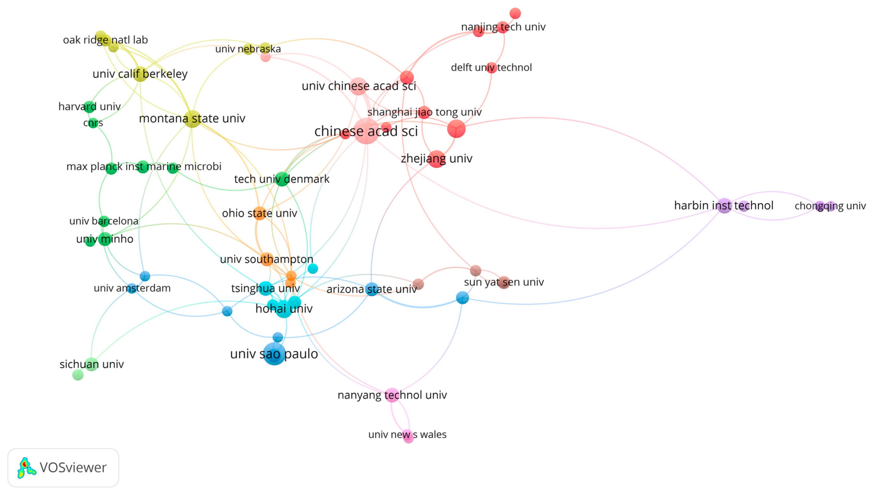
Figure 3.
Country/region analysis by VOSviewer. The lines indicate the research collaboration.
Disclaimer/Publisher’s Note: The statements, opinions and data contained in all publications are solely those of the individual author(s) and contributor(s) and not of MDPI and/or the editor(s). MDPI and/or the editor(s) disclaim responsibility for any injury to people or property resulting from any ideas, methods, instructions or products referred to in the content. |
© 2025 by the author. Licensee MDPI, Basel, Switzerland. This article is an open access article distributed under the terms and conditions of the Creative Commons Attribution (CC BY) license (https://creativecommons.org/licenses/by/4.0/).
Copyright: This open access article is published under a Creative Commons CC BY 4.0 license, which permit the free download, distribution, and reuse, provided that the author and preprint are cited in any reuse.


