
Submitted:
01 January 2025
Posted:
02 January 2025
You are already at the latest version
Abstract
In this research, we have studied the head of the six-banded armadillo, applying advanced imaging techniques such as CT and MRI. Furthermore, by combining the images obtained through these techniques with anatomical cross sections, we present an adequate description of the structures that constitute the head of this species. This anatomical information could provide a valuable diagnostic tool for the clinical evaluation of different desorders in six-banded armadillo including skull malformations, fractures, and neoplasia.
Keywords:
computed tomography
; magnetic resonance imaging
; anatomy
; head
; central nervous system
; six-banded armadillo
1. Introduction
Euphractus sexcinctus, commonly known as the six-banded armadillo, is a member of the Chlamyphoridae family [1]. It is one of the largest armadillo species, reaching approximately 40 centimetres. This terrestrial and solitary mammal inhabits open areas, such as plains or savannahs, as well as forests and jungles. This animal can be found predominantly in the eastern regions of South America, particularly in countries like Argentina, Brazil, Uruguay, Paraguay, Bolivia and certain areas of Suriname [2,3].
The six-banded armadillo is characterized by its dermal plates that cover most of its back, including the head and tail. The carapace comprises six to eight flexible bands and shows a light colouration that varies from yellow to reddish brown [2]. The triangular, pointed-shaped and flat head of this animal features a distinctive mosaic-like structure, and it has up to twenty-five teeth, which are practical for his omnivorous diet that includes invertebrates to carrion and plants [2]. Both its front and hind legs have five toes, used to dig inverted U-shaped burrows and small tunnels [2]. The gestation period for this species lasts about two months, during which they typically have one to three babies. These young armadillos quadruple their weight within the first month and reach adulthood by nine months [1,2,3].
The six-banded armadillo is considered a Least Concern (LC) by the IUCN. However, it faces several risks, primarily due to extensive hunting for local use, including as a protein source and for handicraft and medicinal purposes [3]. With the increasing presence of exotic companion animals and the need to preserve wildlife, biologists, veterinarians and conservationists must understand the complexities of these animals’ anatomy, physiology and lifestyle to be able to diagnose faster and more accurately [4].
The significant anatomical differences among terrestrial mammals, along with the increasing interest in exotic pets as new companion animals, have created challenges for veterinary clinicians in interpreting diagnostic imaging studies. Diagnostic imaging techniques are crucial in enhancing clinical practice and facilitating data collection. Thanks to technological advancements, obtaining anatomical and diagnostic information has become quicker and more efficient, making these tools invaluable in medical practice. Traditional methods like radiology and ultrasound [4], along with modern imaging technologies such as CT and MRI [5,6,7,8,9,10,11], offer superior image resolution, rapid image acquisition, and an improved ability to differentiate overlapping structures. These techniques provide detailed anatomical and functional insights, with an increased capacity to distinguish between bone and soft tissue structures.
Several notable studies have explored the anatomy, physiology, and pathology of these species, particularly focusing on the nine-banded armadillo [12,13,14,15,16,17,18,19,20]. Research has examined various aspects, including the anatomy and functional morphology of xenarthrous vertebrae, spinal development, the skeleton and skull structures, the ear, the nasal cavity and paranasal sinuses, the morphology of the laryngeal cartilages, salivary glands, masticatory adaptations, and the visual cortex [21,22,23,24,25,26,27,28,29,30,31,32,33,34,35,36,37,38,39,40,41,42,43]. Additionally, other studies have addressed pathological conditions, such as fractures [22] and osteoderm lesions [23]. However, to date, only a few papers have investigated the head in these animals [18,19,24,25,38]. Therefore, this research aimed to describe the normal central nervous system (CNS) and its associated structures in the six-banded armadillo using cross-sectional anatomy, CT and MRI. The use of CT, MRI, and detailed anatomical sections can provide pivotal information for enhancing anatomical understanding in education and clinical practice. This approach not only enriches teaching but also significantly improves patient care.
2. Materials and Methods
2.1. Animals
Three adult six-banded armadillo carcasses (two males and one female) from the species Euphractus sexcintus were collected from the “Rancho Texas Lanzarote Park” zoological park (Lanzarote, Canary Islands, Spain). These animals succumbed to natural causes, sparking an opportunity for valuable research.
2.2. CT Technique
To obtain detailed CT images, we employed a 16-slice helical CT scanner (Toshiba Astelion, Canon Medical System, Tokyo, Japan) at the Veterinary Hospital of Las Palmas de Gran Canaria University. The armadillos were carefully positioned in ventral recumbency, symmetrically on the CT table to ensure optimal imaging. We captured sequential CT images with a 1 mm slice thickness using a standard clinical protocol (100 kVp, 80 mA, 512 × 512 acquisition matrix, 1809 × 858 field of view, a spiral pitch factor of 0.94, and a gantry rotation time of 1.5 seconds). To enhance the visibility of the head, three CT windows with varying widths and levels were applied: a bone window (WW = 1500; WL = 300), a lung window (WW = 1400; WL = −500), and a soft tissue window (WW = 350; WL = 40) focusing on the bone window images for clarity. Furthermore, dorsal and sagittal multiplanar reconstructed (MPR) images were generated to better visualize the armadillo’s intricate anatomical head structures. All CT images were subsequently uploaded to an advanced image viewer (OsiriX MD, Apple, Cupertino, CA, USA) for further manipulation and analysis, ensuring a comprehensive evaluation of these unique specimens.
2.3. MRI Technique
A magnetic resonance imaging (MRI) study was conducted on the three six-banded armadillos using a 1.5-Tesla magnet (Toshiba, Vantage Elan, Japan) while the animals were positioned in ventral recumbency. A standard MRI protocol was followed to acquire spin-echo (SE) T1-weighted and T2-weighted images in transverse, sagittal and dorsal planes. For SE T1-weighted transverse images, the parameters were: echo time (TE) of 10 ms, repetition time (TR) of 800 ms, acquisition matrix size of 536 × 384, and a slice thickness of 4.5 mm with 4 mm spacing between slices. The SE T2-weighted transverse images were obtained with a TE of 120 ms, TR of 10,541 ms, acquisition matrix size of 624 × 448, and a slice thickness of 3 mm with 3 mm interslice spacing. The SE T2-weighted sagittal images had a TE of 120 ms, TR of 7529 ms, acquisition matrix size of 512 × 804, and a slice thickness of 2.8 mm with 2 mm interslice spacing. For SE T2-weighted dorsal images, the parameters were: TE of 120 ms, TR of 8282 ms, acquisition matrix size of 468 × 512, and a slice thickness of 3.4 mm with 3 mm interslice spacing. A medical imaging viewer (OsiriX MD, Geneva, Switzerland) was used to analyze the acquired images.
2.4. Anatomical Sections
After the scanning procedure, one of the three six-banded armadillos was placed in dorsal recumbency within expanded polystyrene containers and rapidly frozen at −80 °C for 72 hours. Then, serial sections, each half a centimetre thick, were cut using an electric band saw to produce sequential transverse anatomical cross-sections. The slices were immediately cleaned with water, numbered, and photographed on both sides.
2.5. Anatomic Evaluation
The anatomical cross-sections that most closely matched the CT and MR images were chosen to aid in identifying key structures of the six-banded armadillo’s head. Additionally, consultation with textbooks and relevant references on the anatomy of armadillos and other mammals was required. [21,24,25,26,27,28,29,30,31,32,33,34,35,36,37,38,39,40].
3. Results
Eight representative transverse sections of the armadillo head were selected (Figure 1). Figure 2, Figure 3, Figure 4, Figure 5, Figure 6, Figure 7, Figure 8 and Figure 9 comprise four images: A) anatomic cross-section. B) CT bone window, C) T1-weighted, and D) T2-weighted MR images. These images are presented in a rostrocaudal progression from the olfactory bulb (Figure 2) to the brainstem levels (Figure 9). In addition, Figure 10 contains three sagittal images: a sagittal CT bone window image (B), a sagittal MR T1W image (C), and a parasagittal MR T2W image (D). Finally, Figure 11 comprises a bone-window CT dorsal (B) and a dorsal MRI in T2W (D) images at the level of the tympanic cavity.
3.1. Anatomical Sections
Relevant formations of the central nervous system were identified by anatomical cross-sections. These structures were named in compliance with the International Committee on Veterinary Gross Anatomical Nomenclature. Hence, we could observe the cerebrum divided into a left and right telencephalic hemisphere by a deep longitudinal fissure (Figures 2A–8A). Most cranial sections showed an excellent depiction of the two large olfactory bulbs found on the inferior side of the cerebral hemispheres (Figure 2A). Here, we also distinguished enlarged olfactory recesses bounded medially by the perpendicular plate of the ethmoidal bone (Figures 2A and 3A). The following sections helped distinguish the connexion of both brain hemispheres by fibres of the white matter forming the corpus callosum (Figures 5A and 6A). Moreover, these sections allowed the identification of the lateral ventricles in each hemisphere (Figures 5A–8A).
Additionally, several structures of the diencephalon could be depicted, including the interthalamic adhesion crossing the third ventricle between the two thalami, the optic nerve, the pituitary gland and the lateral and medial geniculate bodies (Figures 5A, 7A and 8A), which were limited laterally by the internal capsule. The ventral part of this capsule and the piriform lobes were essential remarks to identify the amygdala and the globus pallidus (Figure 6A). On the most caudal sections, identifiable formations of the mesencephalon, such as the rostral colliculus, the mesencephalic aqueduct and the cerebral peduncles were distinct (Figure 8A). From the metencephalon, we could identify the cerebellar hemispheres, the vermis, the nodule covering part of the fourth ventricle and prominent ventral structures, including the middle cerebellar peduncles and the pons (Figure 9A).
Concerning the bony formations, different bones comprising the neurocranium were identified, including the frontal, temporal (with its squamous, petrous, and tympanic components), the zygomatic arch, the sphenoid, the maxilla, the mandible and the occipital bones (Figures 2A–9A). In addition, these transverse sections facilitated the visualization of structures of the oral cavity including the tongue, the palatine plexus and the soft palate (Figures 2A–8A), and other important components of the auditory system, such as the external ear with the acoustic meatus and the auricular cartilage (Figure 9A). Besides, the tympanic cavity and bulla were also depicted in these sections (Figures 7A–9A). Furthermore, we distinguished relevant muscles related to masticatory function, such as the temporalis, the medial and lateral pterygoid muscles, and the masseter muscles (Figures 4A–8A).
3.2. Computed Tomography Study
Concerning the nervous system, the CT images facilitated the distinction of relevant formations, such as the olfactory bulbs, which appeared symmetrical bilateral prominences with intermediate attenuation (Figures 2B, 10B, 11B). However, we could not identify the olfactory recesses with this imaging technique due to its low resolution for soft tissues. Despite this limitation, we successfully identified the longitudinal cerebral fissure separating the two telencephalic hemispheres (Figures 2B and 3B).
Additionally, we defined other nervous structures with similar attenuation, including the piriform lobes which were recognized thanks to their globose shape and ventrolateral position (Figure 6B). Moreover, these sections depicted significant parts of the diencephalon, such as the pituitary gland, which was observed resting in the pituitary fossa (Figure 8B). More caudally, the dorsal, transverse and sagittal CT images identified the cerebellar vermis and the adjacent cerebellar hemispheres (Figures 9B–11B).
Regarding the different bony structures, the CT images revealed pivotal skull bones showing high CT attenuation. Therefore, we distinguished the frontal, zygomatic process, the vomer, occipital, basioccipital, sphenoid, temporal and maxillary bones (Figures 2B–10B). Moreover, we identified different parts of the mandible, including the ramus and the mandibular condyle, the temporomandibular joint, and some components of the larynx and the hyoid apparatus (Figures 2B–11B). It is important to emphasize that the most notable bony structure of this animal, due to its species exclusivity, corresponds to the osteoderms, located across the entire dorsal portion of the head as a row of ossified geometric plates (Figures 2B–11B)
These CT images were also helpful in depicting essential elements of the auditory system, such as the external ear and the tympanic bullae recognizable on both sides of the midline of the skull, laterally followed by the external acoustic meatus (Figures 8B, 9B and 11B). Besides, other anatomical structures with intraluminal content, such as the nasopharyngeal and the oral cavities, were displayed with this technique, showing a vacuum effect (Figures 2B–10B).
In the ventral parts of the CT images, several muscles were recognized with the corresponding attenuation, such as the omohyoid and sternohyoid muscles, the medial and lateral pterygoid muscles, the masseter muscle, and the temporal muscles (Figures 2B–8B).
3.3. Magnetic Resonance Imaging (MRI)
No consistent anatomical differences were observed in the studied six-banded armadillos, and the anatomical sections satisfactorily matched with the structures shown in the MRI images. Furthermore, it is remarkable that compared to CT, the MRI technique portrayed a better view of the CNS structures. Therefore, the use of this technique allowed the identification of the olfactory bulbs (Figures 2C,D, 10C,D and 11D) as triangular, rostral structures with a moderate to hyperintense uniform signal intensity, divided by the longitudinal cerebral fissure (Figures 2C,D, 3C,D, 4C,D, 5D, 6D, 7C,D and 8D). Their identification was crucial to the olfactory recess observation (Figures 2D, 3D, 4D, 10D and 11D), which appears as hyperintense in the T2W images, although they were not visible in the T1W images. These more rostral images also facilitated the identification of the dorsal sagittal sinus (Figures 2C,D, 3C,D, 4C,D, 5C,D, 6C,D, 7C,D and 8C,D), represented as a moderate intense and hyperintense in T1W and T2W images, respectively. Furthermore, the telencephalic hemispheres were observed in both T1W and T2W, with moderate and regular intensity signals (Figures 3C,D, 4C,D, 5C,D, 6C,D, 7C,D, 8C,D, 9C,D, 10C,D and 11D).
Moreover, the transverse, sagittal and dorsal images depicted relevant components of the ventricular system, including the lateral ventricles, the dorsal and ventral part of the third ventricle, the mesencephalic aqueduct and the fourth ventricle displaying moderate intense and hyperintense signals in T1W and T2W images, respectively (Figures 5C,D, 6C,D, 7C,D, 8C,D, 9D, 10C,D and 11D). The lateral ventricles facilitated the observation of the caudate nucleus, shown with a moderate intensity and an oval shape (Figures 5C,D, 10D and 11D) and the hippocampus, a large bean-shaped structure with a moderate intensity signal in both T1W and T2W images (Figures 6C,D, 7C,D, 8C,D, 10D and 11D). In addition, the two parts of the third ventricle enclosed the interthalamic adhesion, limited laterally by the right and left sides of the thalamus (Figures 6D, 7D, and 10C,D). Pivotal white matter identifiable structures were the corpus callosum, the fornix and the internal capsule, with a lower intensity than other structures in the CNS, but only visible in T2W (Figures 5D, 6D, 7D, 8D, 10D and 11D). A slight hypointense layer of white matter was essential to demarcate the amygdala, showing a moderate intensity signal (Figure 6C,D).
On the more caudal transverse and sagittal MR images, we distinguished the colliculus and the cerebral peduncles with adequate resolution (Figures 8D and 10C,D). Close to these peduncles, we could visualize the genicular bodies, showing intermediate signals (Figures 7C,D, 8D and 10D). Other relevant formations of the nervous system, including the vermis and hemispheres of the cerebellum, as well as the middle cerebellar peduncles were displayed in the T1 and T2W images (Figures 9C,D, 10C,D and 11D).
MRI images were also pivotal to appreciate diverse eyeball components, such as the vitreous humour and lens, and the extraocular muscles (Figures 2C,D and 3C,D), displaying moderate or high intensity signals. Additionally, we accurately pinpointed different muscles responsible for the chewing movement of the mandible and the temporomandibular joint, including the lateral and medial pterygoid muscles, the masseter and temporal muscles, which presented moderate intensity signals in T1 and T2W transverse images (Figures 4C,D–-8C,D).
4. Discussion
Modern diagnostic imaging techniques have greatly enhanced anatomical knowledge and pathology detection in veterinary medicine [5,7,22,23]. Compared to traditional methods such as radiology and ultrasounds, advanced imaging techniques provide superior resolution of anatomical structures, more precise definitions of lesion extent and characteristics, faster image acquisition, and eliminating superimposition. These advantages have revolutionized research, veterinary practice, and education [6,7,8,9,10,11,12,13,14,17]. However, the use of these technologies in exotic animal medicine remains limited due to their high cost, availability, and logistical challenges in obtaining images from certain species. While CT and MRI studies of the CNS have been conducted in both exotic and domestic species, including dogs, horses, iguanas, porcupines, guinea pigs, rabbits, and members of the Dasypodidae family such as the nine-banded armadillo [10,25,26,27,28,29,30,31], there is a lack of detailed anatomical descriptions of these images for the normal six-banded armadillo. Therefore, this study provided a comprehensive anatomical analysis of the six-banded armadillo’s CNS and associated structures through the combination of anatomical cross-section and CT and MRI images.
In this investigation, transverse anatomical sections were quite helpful in adequately identifying the main formations of the six-banded armadillo head. Hence, these images facilitated the identification of pivotal structures of its encephalon and formations belonging to the oral cavity and the auditory system. However, its eyes were not discernible because of its small size and position. Despite this limitation, they were clearly defined in the CT and MR images.
Concerning CT images, the sagittal and dorsal sections provide clear images of the triangular, pointed-shaped and flat head of this animal. Interestingly, these CT images were also pivotal in visualizing the notable development of the tympanic portion of the temporal bone, which could be an important finding to explain its auditory capacities. In addition, other formations of the armadillo head were also defined. Thus, the CT bone window was especially useful in identifying various bony structures of the head, such as the osteoderms, the zygomatic arch, the frontal, the squamous and petrous parts of the temporal, the occipital bone, the maxilla and the mandible. In contrast, it exhibited a low capacity for identifying soft tissue like muscles or nervous tissue due to the minor density differences in CT images. Similar findings have been displayed in studies performed on other exotic species, including the rhinoceros iguana, the guinea pig, the six-banded armadillo, rabbits and the crested porcupine [24,28,30,32].
In this study, T1 and T2W MR images displayed a better definition of the nervous system and its associated structures than those obtained by CT. Moreover, T2W was crucial to visualize the great development of the rhinencephalic formations, including the olfactory bulbs and recesses, as well as the lateral and intermediate olfactory tracts, which has been related to a well-developed sense of smell by improving olfactory airflow and residence time of odorants within the olfactory regions [45,46]. These findings have been identified in the seven-banded armadillo [47] and to a lesser degree in a variety of animals, such as carnivores [48], rodents [49], and ungulates [50]. Other pivotal nervous formations were also pinpointed, including the hippocampus, caudate nucleus, the caudal colliculus and the amygdala. Regarding this structure, subjective image analysis and objective measurements identified a large amygdala. Studies performed on Dasypus hybridus have also identified a relevant development of its amygdala [47], mainly referring to the cortical and medial nuclei of the amygdaloid complex. They suggest that this finding could be associated with the notable development of the rhinencephalon, which has been demonstrated in other animals [47].
It is also crucial to note that our study, which focuses on the nervous system, is limited by including only three armadillo specimens. However, we consider that is possible to use this information as an initial reference for clinicians, biologist and researchers. Furthermore, to gain a more comprehensive understanding of these findings and to expand upon the information presented in studies like this, additional research involving a greater number of specimens and a deeper understanding of the species’ natural history is necessary.
5. Conclusions
The use of anatomical cross-sections combined with modern diagnostic techniques played a crucial role in accurately identifying the main structures that make up the armadillo head. Hence, this study revealed specific morphological characteristics of the armadillo’s head. The findings are valuable for assessing various pathological conditions, including skull fractures, malformations, and tumours. Additionally, these techniques can enhance veterinary anatomy education by providing a clear view of structures without the interference of overlapping parts, making it easier to visualize the extent of different types of lesions.
Author Contributions
Conceptualization, J.R.J. and D.M.B.; methodology, J.R.J., P. P.-O., D.M.B., I.M., A.M.-E., M.E., and M.M.; formal analysis, J.R.J., D.M.B., A.R., N.R.M.,A.M.-E., G.R., M.C.F.; investigation, J.R.J., G.R., P.P.-O., D.M.B., N.R-M., A.R., I.M., and A.M.-E.; resources, D.M.B.; writing—original draft preparation, J.R.J., M.C.F. and D.M.B.; writing—review and editing, J.R.J., M.M., I.M., P.P.-O., D.M.B., N.R-M., G.R., A.M.-E., and M.E. All authors have read and agreed to the published version of the manuscript.
Funding
This research received no external funding.
Institutional Review Board Statement
Not applicable.
Informed Consent Statement
An informed consent from the owner, allowed us to carry out this study. Therefore, Rancho Texas Lanzarote Park was informed that all the information obtained from this study was treated as confidential to the extent permitted by law, and only used for research or teaching purposes.
Data Availability Statement
The information is available at https://accedacris.ulpgc.es.
Acknowledgments
In loving memory of Alvaro Domingo and Honorio Rodriguez Garcia. We thank Ayesh Mohamad, Nicolasa Rodríguez, Carmen Mingot, Emilia Mingot, Concepción Mingot, Nicolas Aquino, Marisa Mohamad, and Jamal Jaber for their support and constructive comments. Moreover, we thank Rancho Texas Lanzarote Park (Lanzarote, Canary Islands, Spain) for providing the animals for this study.
Conflicts of Interest
The authors declare no conflicts of interest.
References
- Gardner, A.L. Order Cingulata. In Mammal Species of the World: A Taxonomic and Geographic Reference, 3rd ed.; Wilson, D.E., Reeder, D.M., Eds.; Johns Hopkins University Press: Baltimore, MD, USA, 2005; p. 97. ISBN 978-0-8018-8221-0.
- Redford, K.H.; Wetzel, R.M. Euphractus sexcinctus. Mamm. Species 1985, 252, 1–4.
- Brittany, B. Euphractus sexcinctus. Available online: https://animaldiversity.org/accounts/Euphractus_sexcinctus/ (accessed on 20 November 2024).
- Farrow, C.S. Veterinary Diagnostic Imaging: Birds, Exotic Pets and Wildlife; Mosby Elsevier: St. Louis, MO, USA, 2009.
- Lauridsen, H.; Hansen, K.; Wang, T.; Agger, P.; Andersen, J.L.; Knudsen, P.S.; Rasmussen, A.S.; Uhrenholt, L.; Pedersen, M. Inside out: Modern Imaging Techniques to Reveal Animal Anatomy. PLoS ONE 2011, 6, e17879.
- Behroozi, M.; Billings, B.K.; Helluy, X.; Manger, P.R.; Güntürkün, O.; Ströckens, F. Functional MRI in the Nile Crocodile: A New Avenue for Evolutionary Neurobiology. Proc. R. Soc. B Biol. Sci. 2018, 285, 20180178.
- Knipe, M.F. Principles of Neurological Imaging of Exotic Animal Species. Vet. Clin. North Am. Exot. Anim. Pract. 2007, 10, 893–907.
- Banzato, T.; Hellebuyck, T.; Van Caelenberg, A.; Saunders, J.H.; Zotti, A. A Review of Diagnostic Imaging of Snakes and Lizards. Vet. Rec. 2013, 173, 43–49.
- Morales Bordon, D.; Suárez-Cabrera, F.; Ramírez, G.; Paz-Oliva, P.; Morales-Espino, A.; Arencibia, A.; Encinoso, M.; Ventura, M.R.; Jaber, J.R. Study of the Normal Crested Porcupine (Hystrix Cristata) Nasal Cavity and Paranasal Sinuses by Cross-Sectional Anatomy and Computed Tomography. Vet. Sci. 2024, 11, 611.
- Morales-Bordon, D.; Encinoso, M.; Arencibia, A.; Jaber, J.R. Cranial Investigations of Crested Porcupine (Hystrix cristata) by Anatomical Cross-Sections and Magnetic Resonance Imaging. Animals 2023, 13, 2551.
- Fumero-Hernández, M.; Encinoso, M.; Melian, A.; Nuez, H.A.; Salman, D.; Jaber, J.R. Cross Sectional Anatomy and Magnetic Resonance Imaging of the Juvenile Atlantic Puffin Head (Aves, Alcidae, Fratercula arctica). Animals 2023, 13, 3434.
- Gaudin, T.J.; Biewener, A.A. The functional morphology of xenarthrous vertebrae in the armadillo Dasypus novemcinctus (Mammalia, Xenarthra). J. Morphol. 1992, 214, 63–81.
- Oliver, J.D.; Jones, K.E.; Pierce, S.E.; Hautier, L. Size and shape regional differentiation during the development of the spine in the nine-banded armadillo (Dasypus novemcinctus). Evol. Dev. 2021, 23, 496–512.
- Alves, L.S.; Babicsak, V.R.; Charlier, M.G.S.; Vulcano, L.C. Radiography, computed tomography and 3D reconstruction of the pelvis in the nine-banded armadillo, Dasypus novemcinctus. In Proceedings of the 40th World Small Animal Veterinary Association Congress, Bangkok, Thailand, 15–18 May 2015; Volume 40, pp. 66–67.
- Vizcaıno, S.F.; Milne, N. 2002: Structure and function in armadillo limbs (Mammalia: Xenarthra: Dasypodidae). J. Zool. 2002, 257, 117–127.
- Wible, J.R. Petrosal anatomy of the nine-banded armadillo, Dasypus novemcintus Linnaeus, 1758 (Mammalia, Xenarthra, Dasypodidae). Ann. Carnegie Mus. 2010, 79, 1–28.
- Alves, L.S.; Midon, M.; Filadelpho, A.L.; Vulcano, L.C.; Knotek, Z. Gross Osteology, Radiographic and Computed Tomographic Morphology of the Axial Skeleton of the Nine-Banded Armadillo (Dasypus novemcinctus). Anat. Histol. Embryol. 2017, 46, 162–177.
- Silva, A.B.; Sousa, M.M.; Silva, J.V.; Oliveira, I.M.; Barbosa, C.M.; Santos, M.; Conde, A.M. Anatomy of the nasal cavity of nine-banded armadillo (Dasypus novemcinctus, Linnaeus, 1758). J. Interdiscip. Biocienc. 2016, 1, 1–4.
- Billet, G.; Hautier, L.; de Thoisy, B.; Delsuc, F. The hidden anatomy of paranasal sinuses reveals biogeographically distinct morphotypes in the nine-banded armadillo (Dasypus novemcinctus). PeerJ 2017, 5, e3593.
- Fonseca, C.M.B.; da Silva, A.B.S.; Cavalcante, M.M.A.S.; de Oliveira, I.M.; Moura, S.M.S.; Cunha, B.M.; Leite, C.M.C.; de Carvalho, M.A.M.; Moura, W.R.A.; Rizzo, M.D.S.; et al. Morphology of laryngeal cartilage of the nine-banded armadillo (Dasypus novemcinctus) Linnaeus, 1758. Microsc. Res. Tech. 2017, 80, 1089–1095.
- Vizcaíno, S.F.; Fariña, R.A.; Bargo, M.S.; De Iuliis, G. Functional and phylogenetic assessment of the masticatory adaptations in Cingulata (Mammalia, Xenarthra). Rev. Asos. Paleontol. Argent. 2004, 41, 651–664.
- Alves, L.S.; Oliva, L.R.; Charlier, M.G.S.; Bonatelli, S.P.; Inamassu, L.R.; Vulcano, L.C.; Teixeira, C.R. Fratura em seguimento lombar da coluna vertebral em um tatugalinha (Dasypus novemcinctus). An. Simp. Intern. Diag. Imag. SINDIV 2013, 3, 57–58.
- Boyde, A.; Mills, D.; Abba, A.M.; Ezquiaga, M.C. Fleas and lesions in armadillo osteoderms. J. Anat. 2023, 242, 1029–1036.
- Jaber, J.R.; Morales Bordon, D.; Arencibia, A.; Corbera, J.A.; Conde-Felipe, M.; Ayala, M.D.; Encinoso, M. Correlation between Cross-Sectional Anatomy and Computed Tomography of the Normal Six-Banded Armadillo (Euphractus Sexcintus) Nasal Cavity and Paranasal Sinuses. Animals (Basel) 2024, 14, 1135.
- Scholl B Rylee, J.; Luci, J.J.; Priebe, N.J.; Padberg, J. Orientation selectivity in the visual cortex of the nine-banded armadillo. J. Neurophysiol 2017, 117, 1395–1406.
- Jacqmot, O.; Van Thielen, B.; Hespel, A.-M.; Luijten, P.R.; de Mey, J.; Van Binst, A.; Provyn, S.; Tresignie, J. T2-Weighted Turbo Spin-Echo Magnetic Resonance Imaging of Canine Brain Anatomy at 1.5T, 3T, and 7T Field Strengths. Anat. Rec. (Hoboken) 2022, 305, 222–233.
- Araújo, J.V.S.; Cavalcante, M.M.A. de S.; Gonçalves, P.C. de J.; Guerra, S.P.L.; Da Silva, A.B.S.; Conde Júnior, A.M. Descriptive macroscopic anatomy of the central nervous system six-banded armadillo (Euphractus sexcintus, Linnaeus, 1758) and nine-banded armadillo (Dasypus novemcinctus, Linnaeus, 1758). J. Interdiscip. Biociênc. 2015, 1, 13.
- González Rodríguez, E.; Encinoso Quintana, M.; Morales Bordon, D.; Garcés, J.G.; Artiles Nuez, H.; Jaber, J.R. Anatomical Description of Rhinoceros Iguana (Cyclura cornuta cornuta) Head by Computed Tomography, Magnetic Resonance Ima-ging and Gross-Sections. Animals 2023, 13.
- Arencibia, A.; Vazquez, J.M.; Rivero, M.; Latorre, R.; Sandoval, J.A.; Vilar, J.M.; Ramirez, J.A. Computed tomography of normal cranioencephalic structures in two horses. Anat. Histol. Embryol. 2000, 29, 295–299.
- Mahdy, M. Correlation between computed tomography, magnetic resonance imaging and cross-sectional anatomy of the head of the guinea pig (Cavia porcellus, Linnaeus 1758). Anat. Histol. Embryol. 2022, 51, 51–61.
- De Rycke, L.M.; Saunders, J.H.; Gielen, I.M.; van Bree, H.J.; Simoens, P.J. Magnetic resonance imaging, computed tomography, and cross-sectional views of the anatomy of normal nasal cavities and paranasal sinuses in mesaticephalic dogs. Am. J. Vet. Res. 2003, 64, 1093–1098.
- Van Caelenberg, A.I.; De Rycke, L.M.; Hermans, K.; Verhaert, L.; Van Bree, H.J.; Gielen, I.M. Low-field magnetic resonance imaging and cross-sectional anatomy of the rabbit head. Vet. J. 2011, 188, 83–91.
- Ferrari, C.C.; Carmanchahi, P.D.; Aldana Marcos, H.J.; Affanni, J.M. Ultrastructural Characterisation of the Olfactory Mucosa of the Armadillo Dasypus Hybridus (Dasypodidae, Xenarthra). J. Anat. 2000, 196, 269–278.
- Carmanchahi, P.D.; Aldana Marcos, H.J.; Ferrari, C.C.; Affanni, J.M. The Vomeronasal Organ of the South American Armadillo Chaetophractus Villosus (Xenarthra, Mammalia): Anatomy, Histology and Ultrastructure. J. Anat. 1999, 195, 587–604.
- Basso, A.P.; Sidorkewicj, N.S.; Casanave, E.B.; Mason, M.J. The Middle Ear of the Pink Fairy Armadillo Chlamyphorus Truncatus (Xenarthra, Cingulata, Chlamyphoridae): Comparison with Armadillo Relatives Using Computed Tomography. J. Anat. 2020, 236, 809–826.
- Ruby, J.R.; Allen, E.R. Ultrastructure of the Salivary Bladder of the Nine-Banded Armadillo. Cell Tissue Res. 1976, 169, 383–394.
- Le Verger, K.; González Ruiz, L.R.; Billet, G. Comparative Anatomy and Phylogenetic Contribution of Intracranial Osseous Canals and Cavities in Armadillos and Glyptodonts (Xenarthra, Cingulata). J. Anat. 2021, 239, 1473–1502.
- Phillips, J.A.; Harlow, H.J.; McArthur, N.H.; Ralph, C.L. Epithalamus of the Nine-Banded Armadillo, Dasypus Novemcinctus. Comp. Biochem. Physiol. A Comp. Physiol. 1986, 85, 477–481.
- Squarcia, S.M.; Sidorkewicj, N.S.; Camina, R.; Casanave, E.B. Sexual dimorphism in the mandible of the armadillo Chaetophractus villosus (Desmarest, 1804) (Dasypodidae) from northern Patagonia, Argentina. Braz. J. Biol. 2009, 69, 347–352.
- Sigurdsson, B.; Hauglund, N.L.; Lilius, T.O.; Mogensen, F.L.-H.; Mortensen, K.N.; Beschorner, N.; Klinger, L.; Bærentzen, S.L.; Rosenholm, M.P.; Shalgunov, V.; et al. A SPECT-Based Method for Dynamic Imaging of the Glymphatic System in Rats. J. Cereb. Blood Flow Metab. 2023, 43, 1153–1165.
- Mrzílková, J.; Patzelt, M.; Gallina, P.; Wurst, Z.; Šeremeta, M.; Dudák, J.; Krejčí, F.; Žemlička, J.; Musil, V.; Karch, J.; et al. Imaging of Mouse Brain Fixated in Ethanol in Micro-CT. Biomed Res. Int. 2019, 2019, 2054262.
- Boscaini, A.; Iurino, D.A.; Billet, G.; Hautier, L.; Sardella, R.; Tirao, G.; Gaudin, T.J.; Pujos, F. Phylogenetic and Functional Implications of the Ear Region Anatomy of Glossotherium Robustum (Xenarthra, Mylodontidae) from the Late Pleistocene of Argentina. Sci. Nat. 2018, 105, 28.
- Hautier, L.; Billet, G.; de Thoisy, B.; Delsuc, F. Beyond the Carapace: Skull Shape Variation and Morphological Systematics of Long-Nosed Armadillos (Genus Dasypus). PeerJ 2017, 5.
- Feijó, A.; Patterson, B.D.; Cordeiro-Estrela, P. Taxonomic Revision of the Long-Nosed Armadillos, Genus Dasypus Linnaeus, 1758 (Mammalia, Cingulata). PLoS One 2018, 13.
- Craven, B.A.; Paterson, E.G.: Settles, G.S. The fluid dynamics of canine olfaction: unique nasal airflow patterns as an explanation of macrosmia. J. Roy. Soc. Interface 2010, 7, 933-943.
- Eiting, T.P.; Smith, T.D.; Perot, J.B.; Dumont, E.R. The role of the olfactory recess in olfactory airflow. J Exp Biol 2014, 217, 1799-1803.
- Ferrari, C.; Aldana, H.; Carmanchahi, P.D.; Benítez, I.; Affanni, J.M. The brain of the armadillo Dasypus hybridus. A general view of its most salient features. Biocell 1998, 22, 123-140.
- Sisson, S.; Grossman, J.D. Anatomía de los animales domésticos. 1972, Salvat Editores, S.A, Madrid.
- Köning, J.F.; Klippel, R.A. The rat brain. An stereotaxic atlas. 1967, R. Krieger Publishing Co. Inc, New York.
- Ariëns Kappers, C.U.; Huber, G.C.; Crosby, E.C. The comparative anatomy of the nervous system of vertebrates including the man Vol II. 1960. Hafner Publishing Company, New York.
Figure 1.
parasagittal T2W MR image showing the approximate levels of transverse slices.
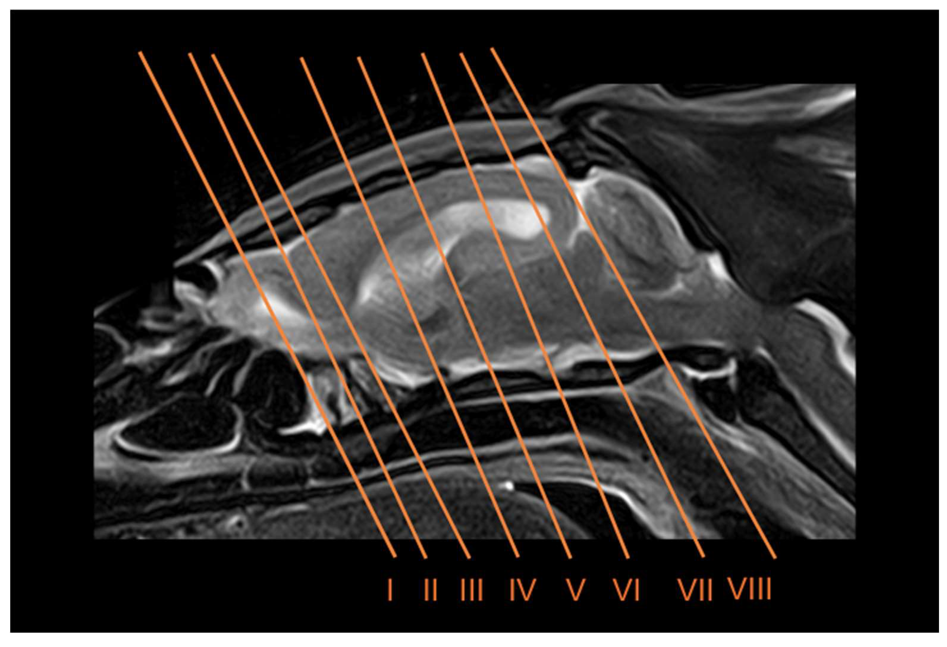
Figure 2.
Transverse anatomical cross-section (A), CT bone window (B), MR T1W (C) and MR T2W (D) images of the head of a six-banded armadillo at the level of the olfactory bulb, corresponding to line I in Figure 1. Dss: dorsal sagittal sinus. Eom: extraocular muscles. Et: ethmoturbinates. F: frontal bone. L: lens. Lcf: longitudinal cerebral fissure. Lp: perpendicular plate (ethmoid bone). M: mandible (body). Mhm: mylohyoid muscle. Mx: maxilla. Mxs: maxillary sinus. Np: nasopharynx. Ob: olfactory bulb. Od: osteoderm. Om+sm: omohyoid+ sternohyoid muscles. Or: olfactory recess. Pp: palatine plexus. Scl: sclera. SphS: sphenoid sinus. T: tongue. V: vomer. Vch: vitreous chamber. Za: zygomatic arch.
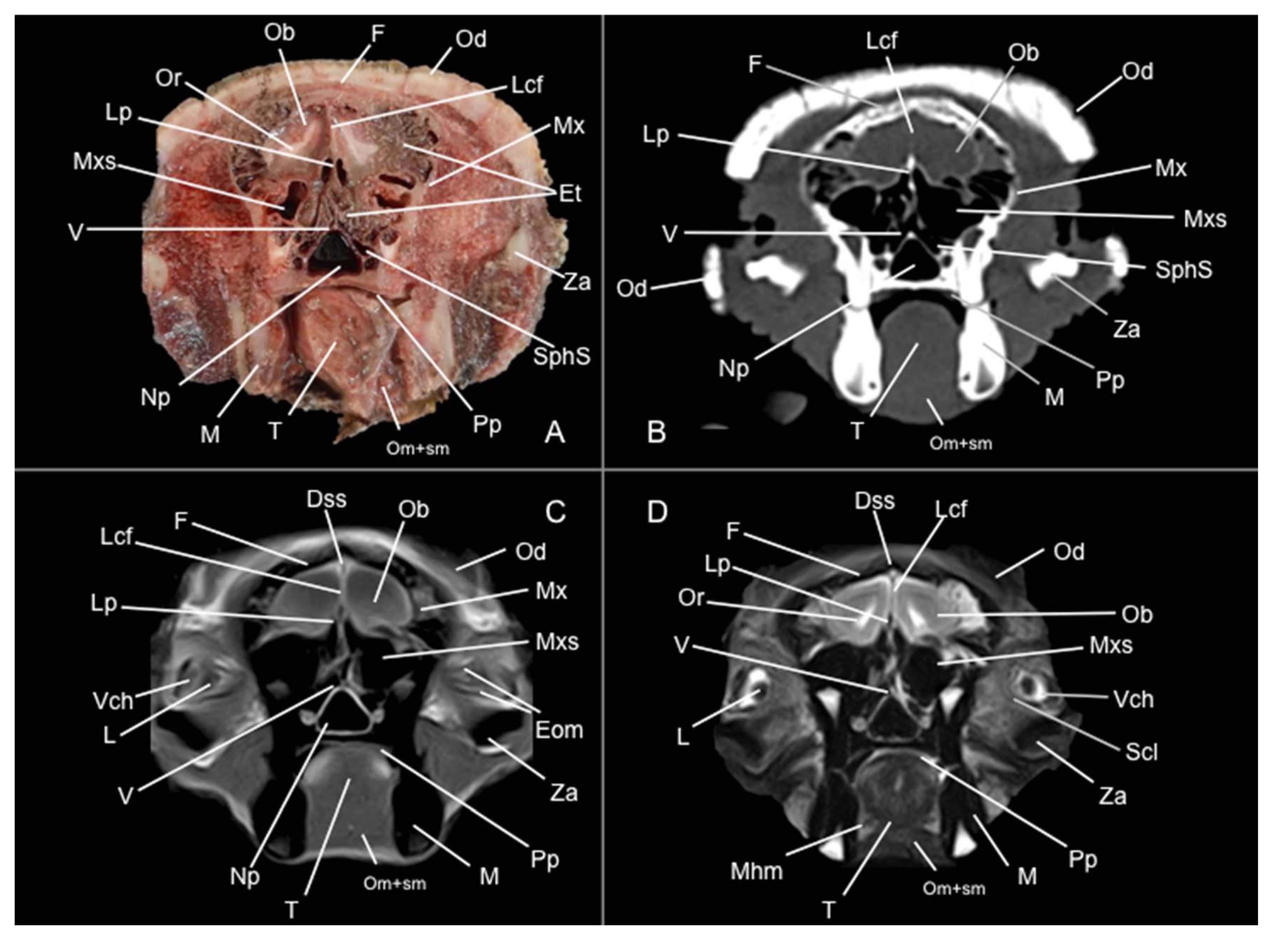
Figure 3.
Transverse anatomical cross-section (A), CT bone window (B), MR T1W (C) and MR T2W (D) images of the head of a six-banded armadillo at the level of the olfactory recess, corresponding to line II in Figure 1. Dss: dorsal sagittal sinus. Eom: extraocular muscles. Et: ethmoturbinates. F: frontal bone. iot: intermedial olfactory tract. Lcf: longitudinal cerebral fissure. Lot: lateral olfactory tract. Lp: perpendicular plate (ethmoid bone). M: mandible (body). M’: mandible (ramus). Mx: maxilla. Np: nasopharynx. Od: osteoderm. Om+sm: omohyoid + sternohyoid muscles. Or: olfactory recess. Pp: palatine plexus. Rc: rostral commissure. Sp: soft palate. T: tongue. Th: telencephalic hemisphere (frontal lobe). V: vomer. Za: zygomatic arch.
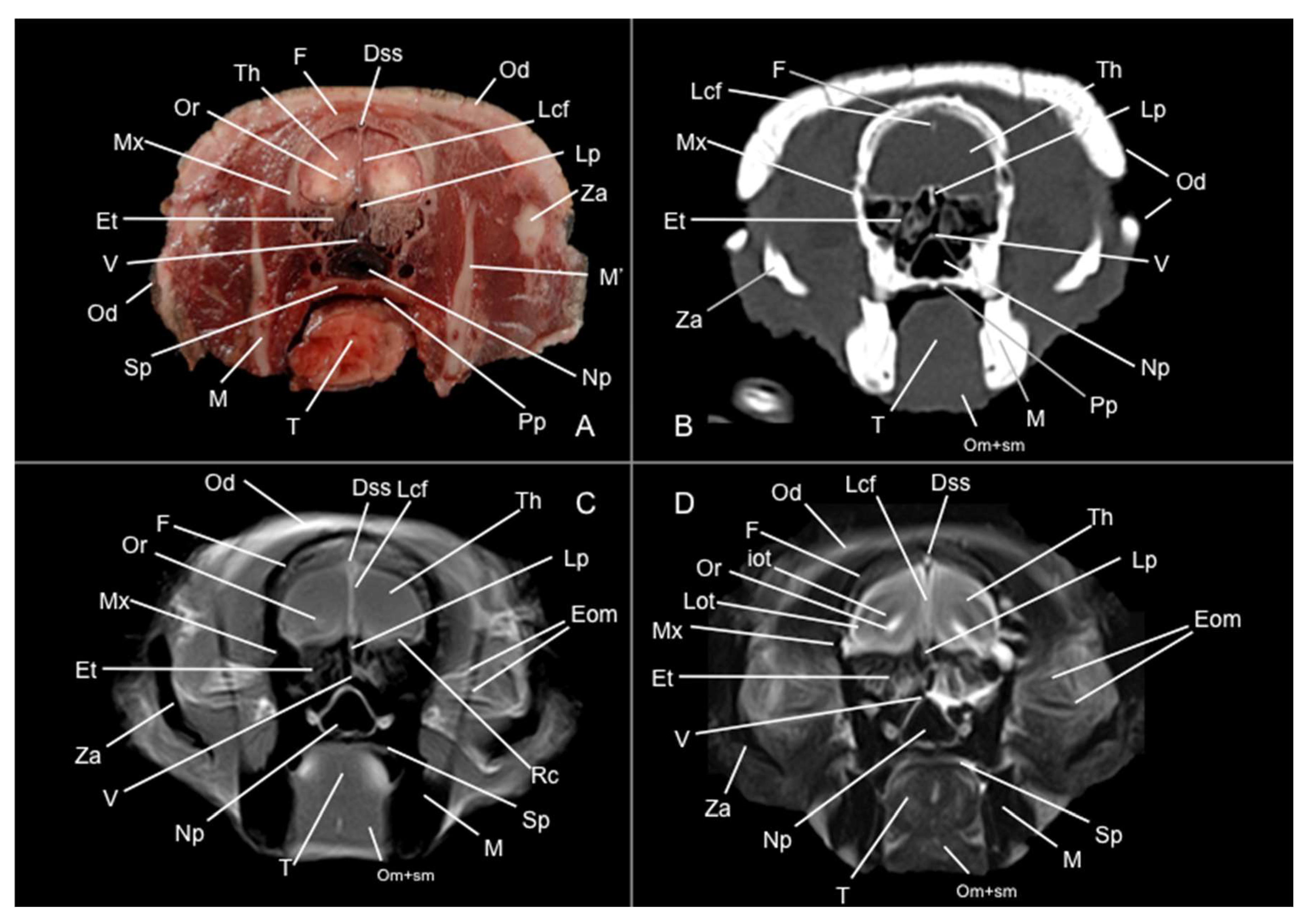
Figure 4.
Transverse anatomical cross-section (A), CT bone window (B), MR T1W (C) and MR T2W (D) images of the head of a six-banded armadillo at the level of the rostral commissure, corresponding to line III in Figure 1. Dss: dorsal sagittal sinus. Et: ethmoturbinates. F: frontal bone. iot: intermedial olfactory tract. Lcf: longitudinal cerebral fissure. Lot: lateral olfactory tract. Lrs: lateral rhinal sulcus. M: mandible (body). M’: mandible (ramus). Mm: masseter muscle. Mx: maxilla. Oc: oral cavity. Od: osteoderm. Om+sm: omohyoid + sternohyoid muscles. Or: olfactory recess. Ptm: medial pterygoid muscle. Rc: rostral commissure. Ron: rostral olfactory nucleus. T: tongue. Th: telencephalic hemisphere (frontal lobe). Za: zygomatic arch.
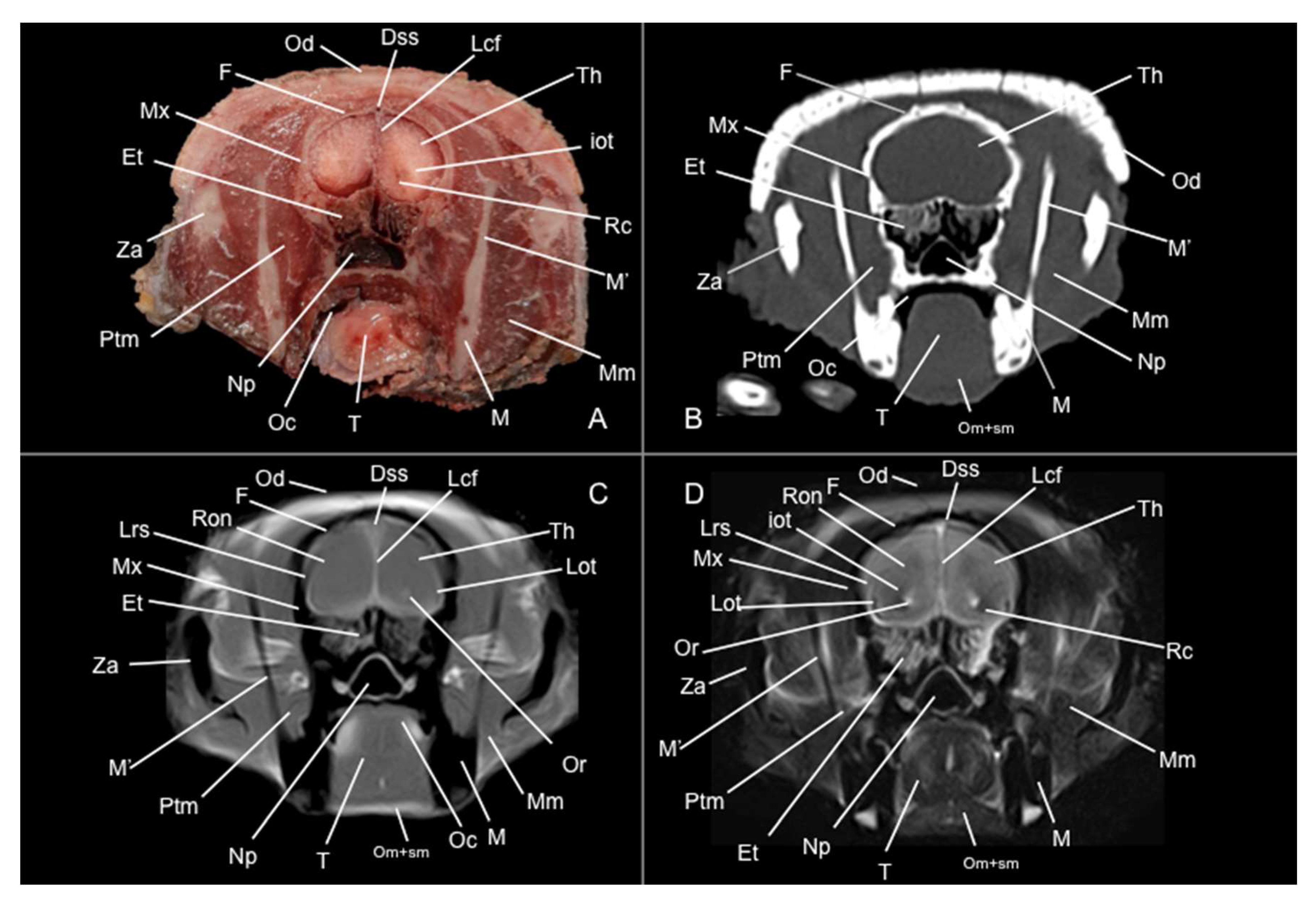
Figure 5.
Transverse anatomical cross-section (A), CT bone window (B), MR T1W (C) and MR T2W (D) images of the head of a six-banded armadillo at the level of the body of the caudate nucleus, corresponding to line III in Figure 1. An: Accumbens nucleus. Cc: corpus callosum. Cn: caudate nucleus (body). CnII: cranial nerve II. Dss: dorsal sagittal sinus. Et: ethmoturbinates. Ec: external capsule. Lcf: longitudinal cerebral fissure. Lv: lateral ventricle. M: mandible (body). M’: mandible (ramus). Mm: masseter muscle. Mx: maxilla. Np: nasopharynx. Oc: oral cavity. Od: osteoderm. Om+sm: omohyoid + sternohyoid muscles. Op: olfactory peduncle. P: parietal bone. Ptm: medial pterygoid muscle. Ptm’: lateral pterygoid muscle. Rc: rostral comissure. Sph: sphenoid bone. T: tongue. Th: telencephalic hemisphere (parietal lobe). Tm: temporal muscle. Ts: telencephalic septum. Za: zygomatic arch.
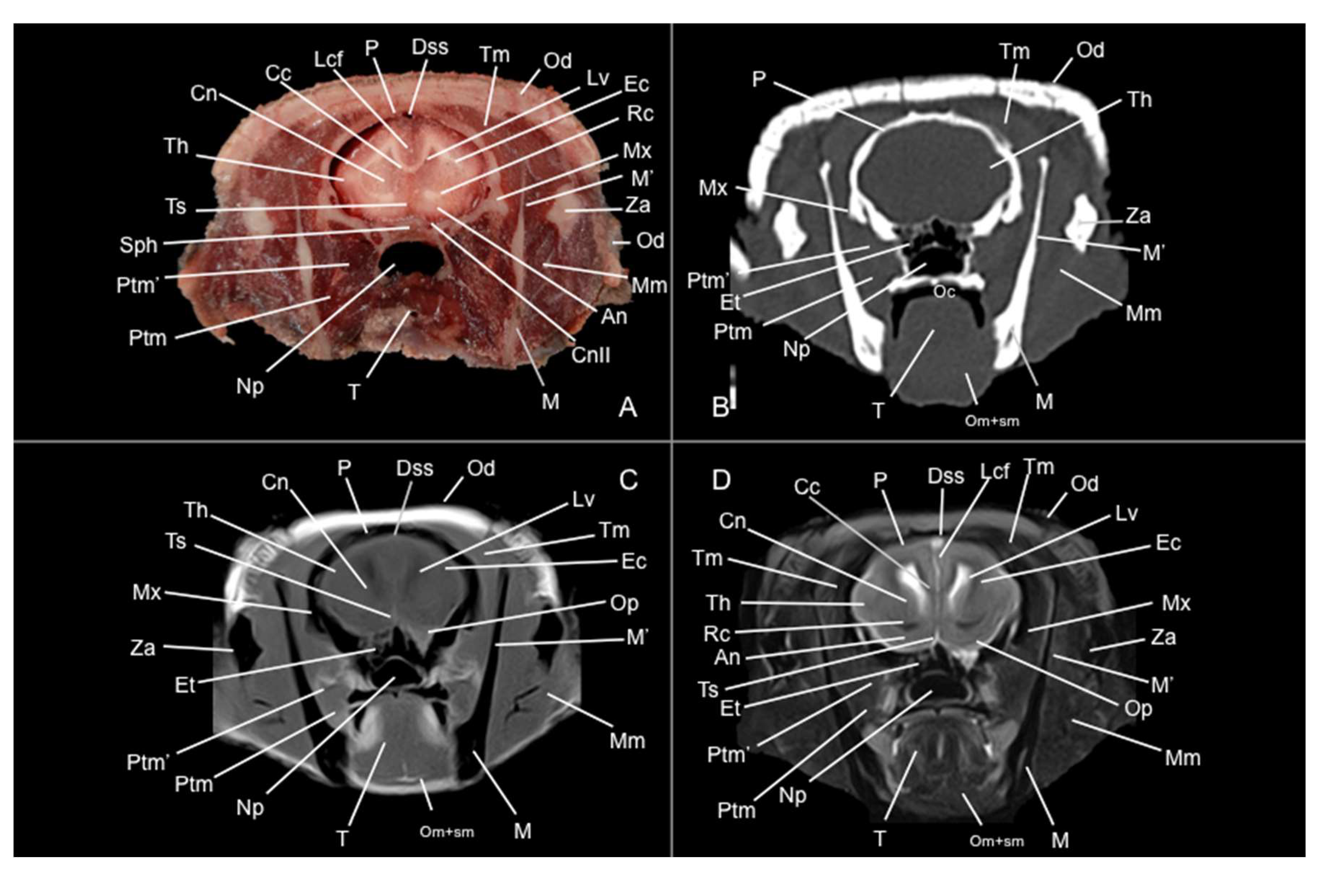
Figure 6.
Transverse anatomical cross-section (A), CT bone window (B), MR T1W (C) and MR T2W (D) images of the head of a six-banded armadillo at the level of the amygdala corresponding to line V in Figure 1. Am: amygdala. Cc: corpus callosum. Cn: caudate nucleus (tail). CnV cranial nerve V. Dss: dorsal sagittal sinus. Fo: fornix. Fo’: pillars of fornix. Ha: habenula. Hp: hippocampus. Ic: internal capsule. III: third ventricle. Ia: interthalamic adhesion. Lcf: longitudinal cerebral fissure. Lv: lateral ventricle. M: mandible (body). M’: mandible (ramus). Mm: masseter muscle. Np: nasopharynx. Oc: oral cavity. Od: osteoderm. Om+sm: omohyoid + sternohyoid muscles. P: parietal bone. Pa: globus pallidus. Pl: piriform lobe. Ptm: medial pterygoid muscle. Ptm’: lateral pteryogid muscle. Sp: soft palate. Sph: sphenoid bone. T: tongue. Te: temporal bone. Th: telencephalic hemisphere (temporal lobe). Tm: temporal muscle. Za: zygomatic arch.
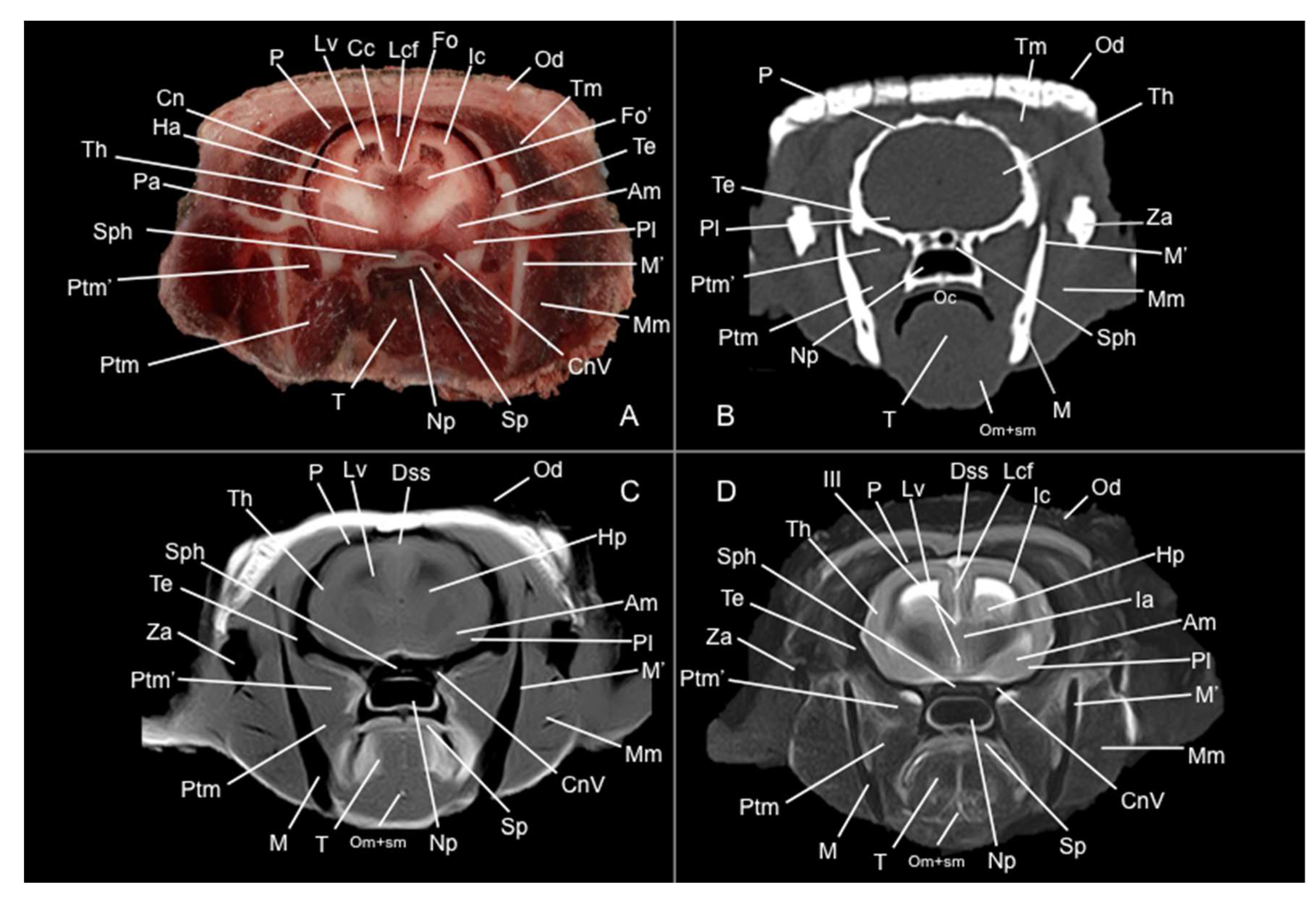
Figure 7.
Transverse anatomical cross-section (A), CT bone window (B), MR T1W (C) and MR T2W (D) images of the head of a six-banded armadillo at the level of the caudal part of the diencephalon, corresponding to line VI in Figure 1. CnV: cranial nerve V. Dss: dorsal sagittal sinus. Hp: hippocampus. Ia: interthalamic adhesion. Ic: internal capsule. III: third ventricle. Lcf: longitudinal cerebral fissure. Lgb: lateral geniculate body. Lr: Lingual root. Lv: lateral ventricle. M’: mandible (ramus). M’’: mandibular condyle. Mgb: medial geniculate body. Mm: masseter muscle. Np: nasopharynx. Oc: oral cavity. Od: osteoderm. Om+sm: omohyoid + sternohyoid muscles. Ot: optic tract. P: parietal bone. Pg: pituitary gland. Ptm: medial pterygoid muscle. Ptm’: lateral pterygoid muscle. Sp: soft palate. Bsph: Basisphenoid bone. T: tongue. Tb: tympanic bulla. Te: temporal bone. Th: telencephalic hemisphere (temporal lobe). Tm: temporal muscle. TMj: temporomandibular joint. Za: zygomatic arch.
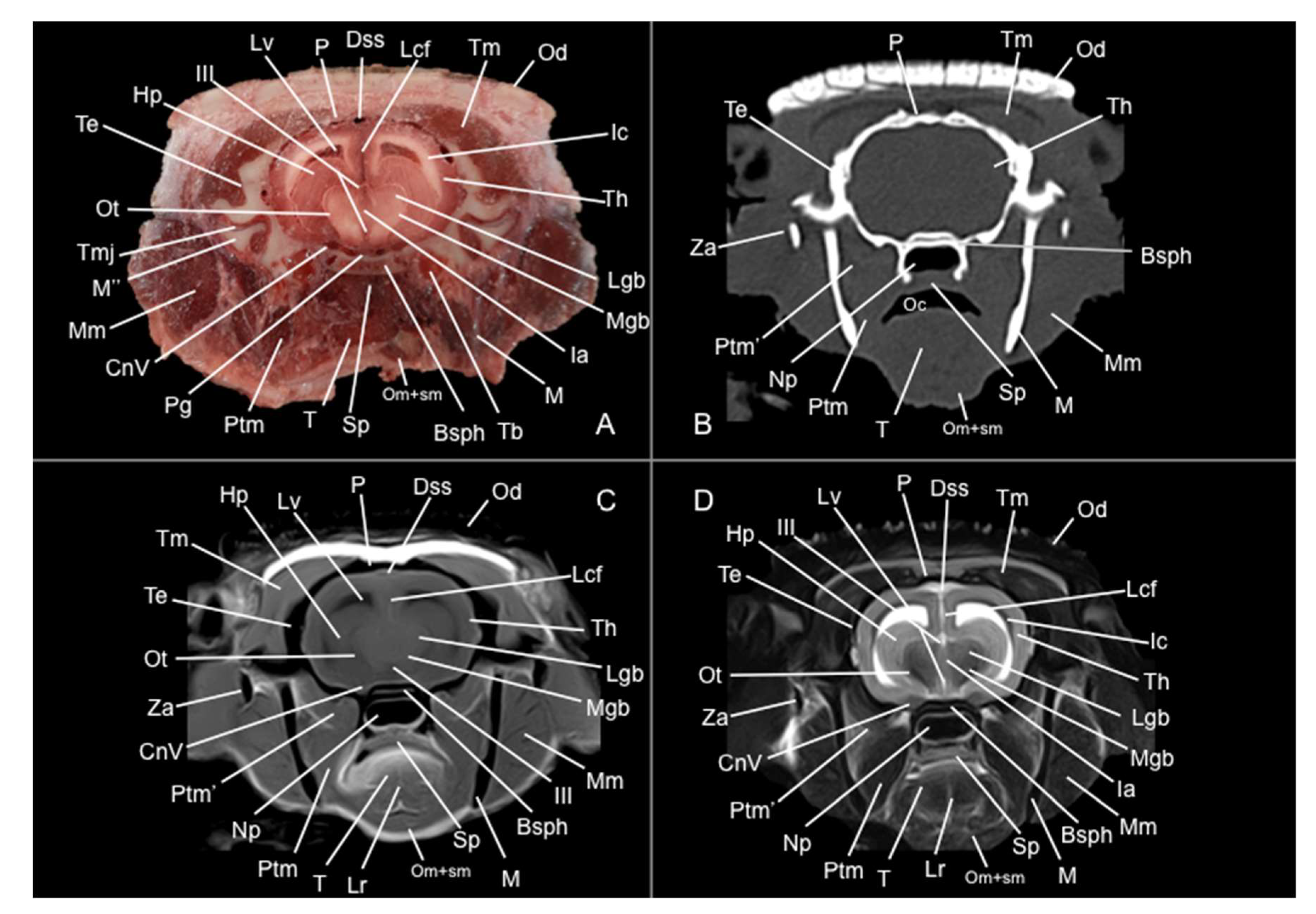
Figure 8.
Transverse anatomical cross-section (A), CT bone window (B), MR T1W (C) and MR T2W (D) images of the head of a six-banded armadillo at the level of the mesencephalon corresponding to line VII in Figure 1. Bo: basioccipital. CnV: craneal nerve V. Cp: cerebral peduncle. Dss: dorsal sagittal sinus. Ea: ear. Eam: external acoustic meatus. Hp: hippocampus. Ic: internal capsule. Lcf: Longitudinal cerebral fissure. Lr: lingual root. Lv: lateral ventricle. M’: mandible (ramus). M’’: mandibular condyle. Ma: mesencephalic aqueduct. Mgb: medial geniculate body. Mm: masseter muscle. Np: nasopharynx. Oc: oral cavity. Od: osteoderm. Om+sm: omohyoid + sternohyoid muscles. P: parietal bone. Po: pons. Ptm: medial pterygoid muscle. Ptm’: lateral pterygoid muscle. Rc: rostral colliculus. Sp: soft palate. T: tongue. Tb: tympanic bulla. Te: temporal bone. Th: telencephalic hemisphere (occipital lobe). Tm: temporal muscle. Tmj: temporomandibular joint.
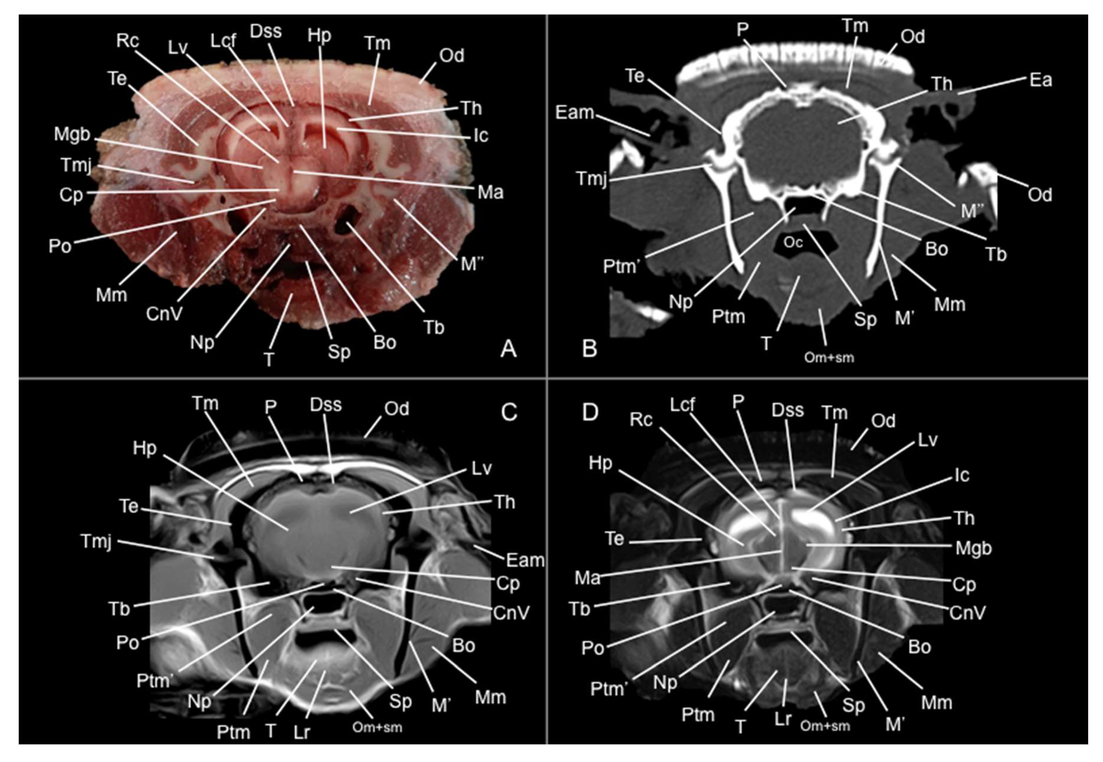
Figure 9.
Transverse anatomical cross-section (A), CT bone window (B), MR T1W (C) and MR T2W (D) images of the head of a six-banded armadillo at the level of the fourth ventricle, corresponding to line VIII in Figure 1. AuC: auricular cartilage. Bo: basioccipital. Cb: cerebellum (body). Cep: cerebellar peduncle (middle). Ch: cerebellar hemisphere. Ea: ear. Ep: epiglottis. Ha: hyoid apparatus (stylohyoid bone). IV: fourth ventricle. M: mandible. Mo: medulla oblongata. No: nodule. Od: osteoderm. Py: pyramids. Sg: salivary gland. Tb: tympanic bulla. Tc: tympanic cavity. Th: telencephalic hemisphere (occipital lobe). ThC: thyroid cartilage.
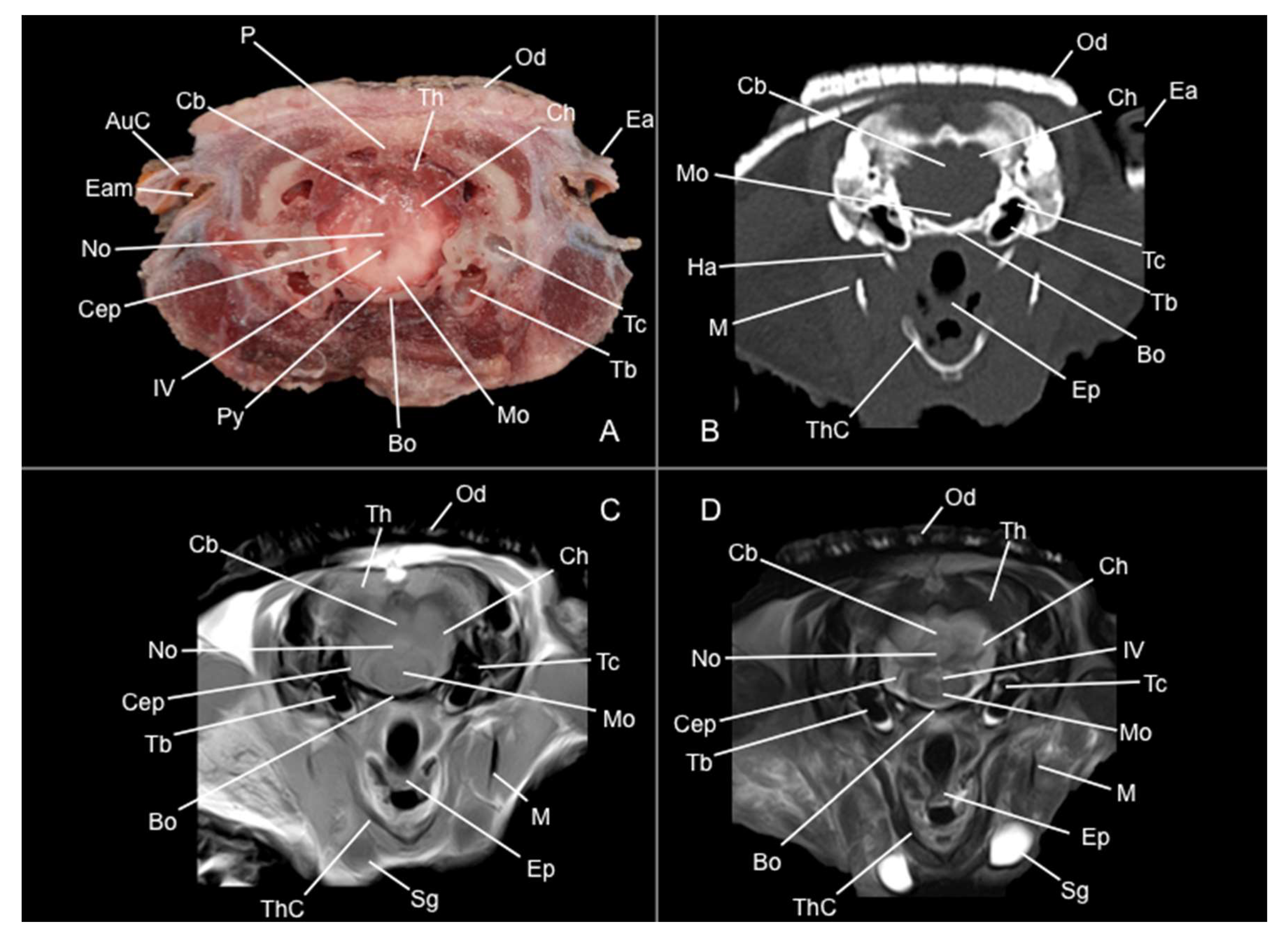
Figure 10.
Sagittal CT bone window (B), sagittal MR T1W (C) and parasagittal MR T2W (D) images of the head of a six-banded armadillo. Bh: basihyoid bone. CaC: caudal colliculus. Cb: cerebellum (vermis). Cc: corpus callosum. Cm: cerebral meninges. Cn: caudate nucleus. Cp: cerebral peduncle. Ep: epiglottis. F: frontal bone. Hap: hard palate. Hp: hippocampus. Ia: interthalamic adhesion. III: third ventricle. Lgb: lateral geniculate body. Lp: perpendicular plate (ethmoid bone). Lv: lateral ventricle. Mgb: medial geniculate body. Mo: medula oblongata. O: occipital bone. Ob: olfactory bulb. Oc: oral cavity. Od: osteoderm. Or: olfactory recess. Pg: pituitary gland. Po: pons. Sp: soft palate. Ta: thalamus. Th: telencephalic hemisphere (parietal lobe). ThC: thyroid cartilage. Tm: tectum of the mesencephalon.
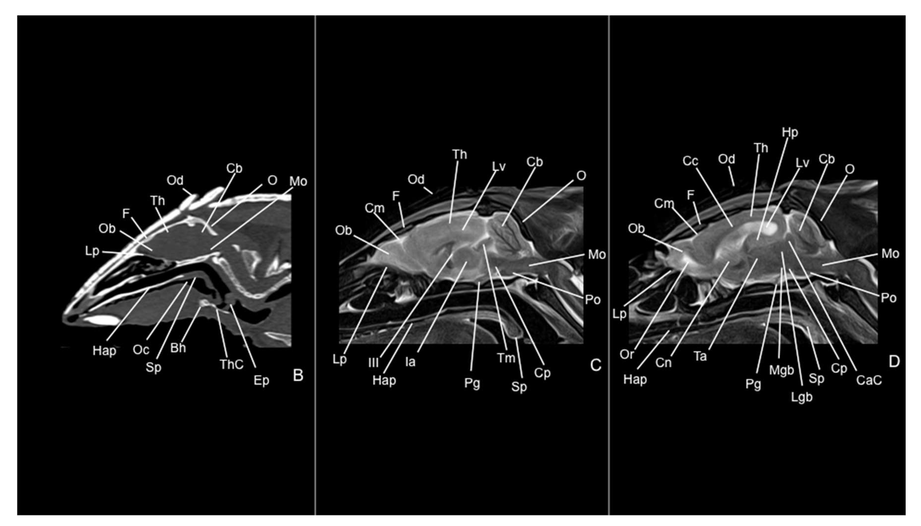
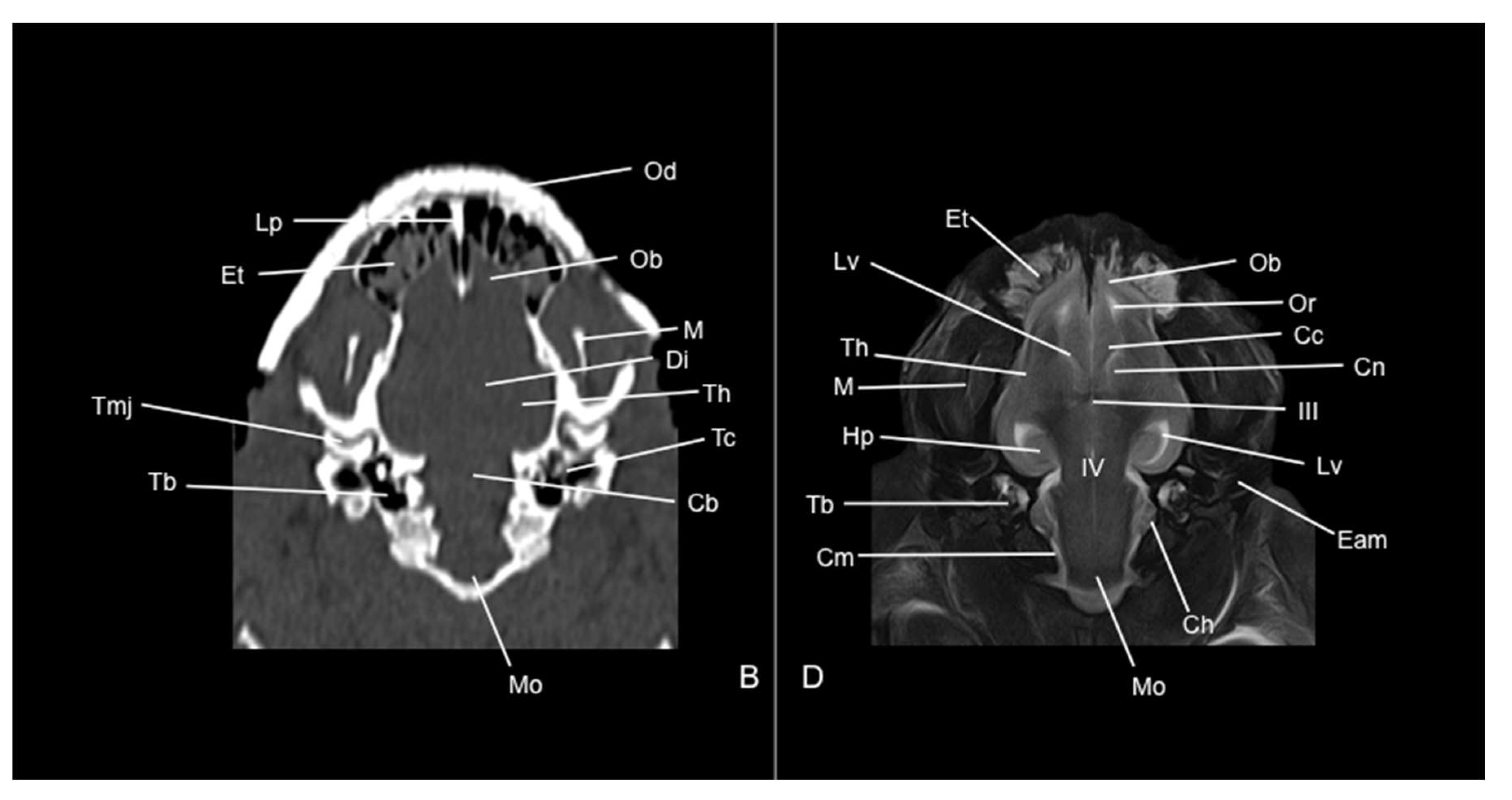
Figure 11.
Dorsal CT bone window (B) and MR T2W (D) images of the head of a six-banded armadillo at the level of the middle ear. Cb: cerebellum (body). Cc: corpus callosum. Ch: cerebellar hemispheres. Cn: caudate nucleus. Cm: cerebral meninges. Di: diencephalon. Eam: external acoustic meatus. Hp: hippocampus. III: third ventricle. IV: fourth ventricle. Lp: perpendicular plate (ethmoid bone). Lv: lateral ventricle. M: mandible. Mo: medulla oblongata. Ob: olfactory bulb. Od: osteoderm. Or: olfactory recess. Tb: tympanic bulla. Tc: tympanic cavity. Th: telencephalic hemisphere. Tmj: temporomandibular joint.
Disclaimer/Publisher’s Note: The statements, opinions and data contained in all publications are solely those of the individual author(s) and contributor(s) and not of MDPI and/or the editor(s). MDPI and/or the editor(s) disclaim responsibility for any injury to people or property resulting from any ideas, methods, instructions or products referred to in the content. |
© 2025 by the authors. Licensee MDPI, Basel, Switzerland. This article is an open access article distributed under the terms and conditions of the Creative Commons Attribution (CC BY) license (http://creativecommons.org/licenses/by/4.0/).
Copyright: This open access article is published under a Creative Commons CC BY 4.0 license, which permit the free download, distribution, and reuse, provided that the author and preprint are cited in any reuse.


