Preprint
Article

This version is not peer-reviewed.

Putrescine Depletion in Leishmania donovani Parasites Causes Immediate Proliferation Arrest Followed by an Apoptosis-Like Cell Death

A peer-reviewed article of this preprint also exists.

Submitted:

19 December 2024

Posted:

23 December 2024

You are already at the latest version

Abstract

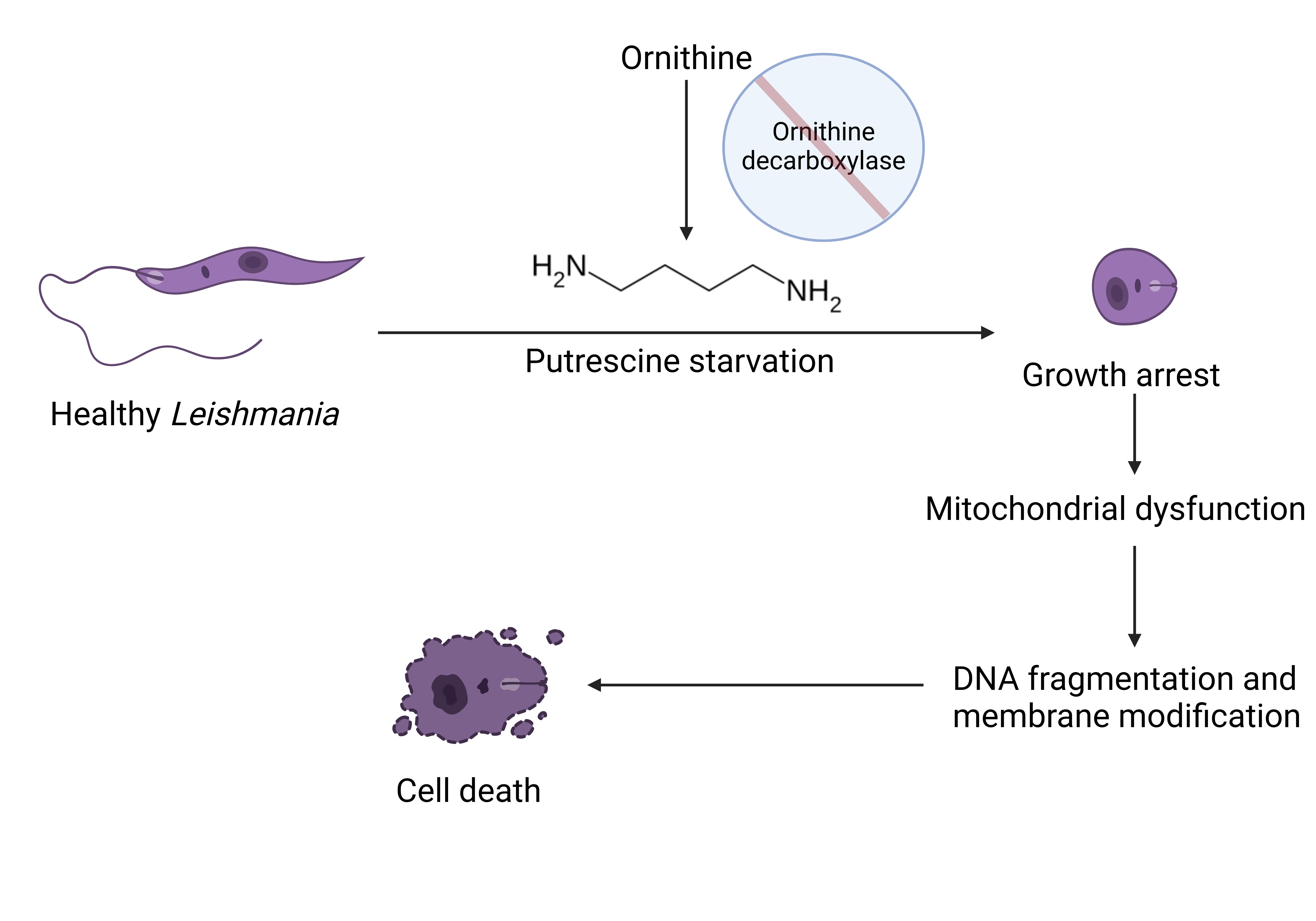
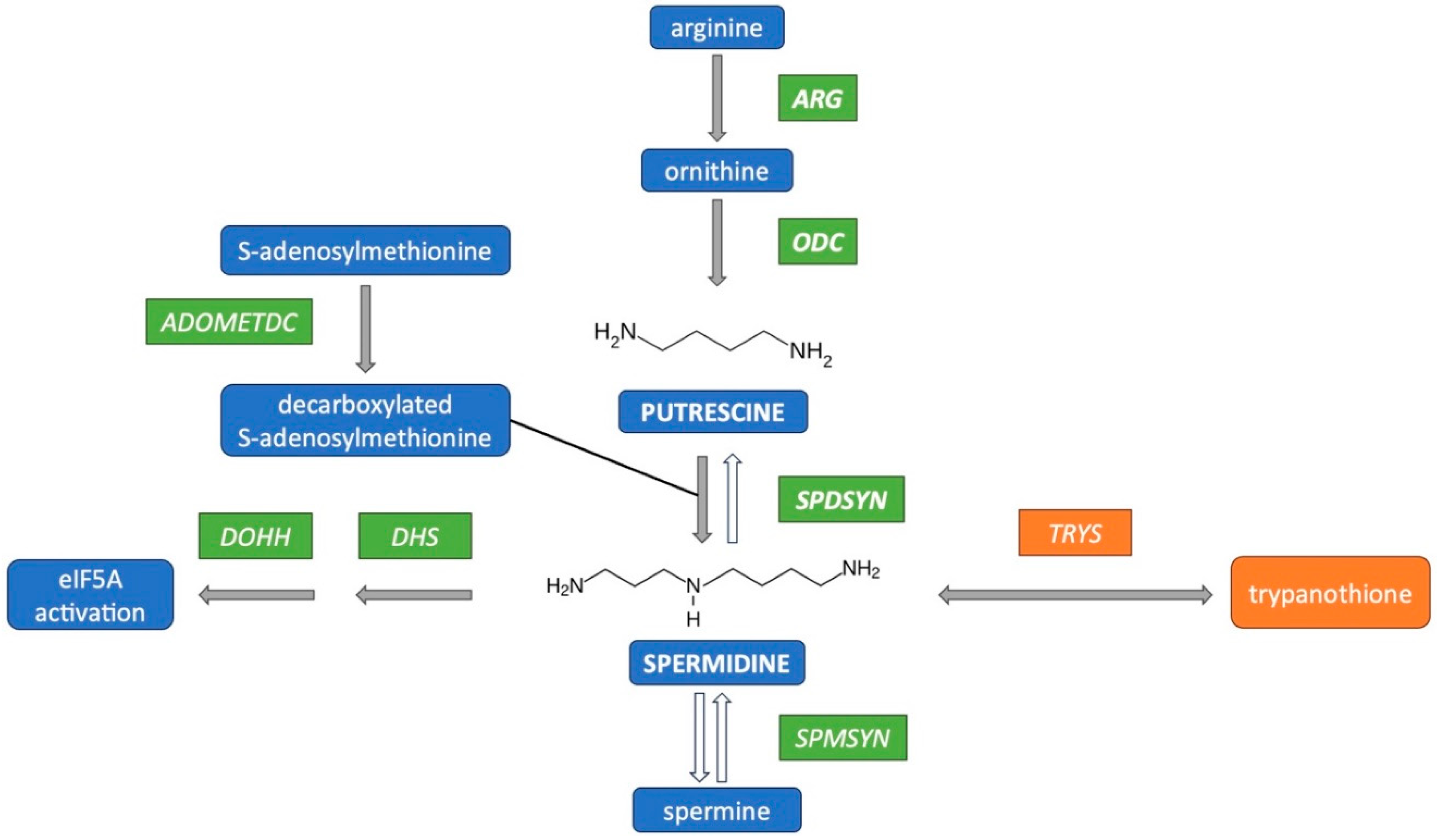
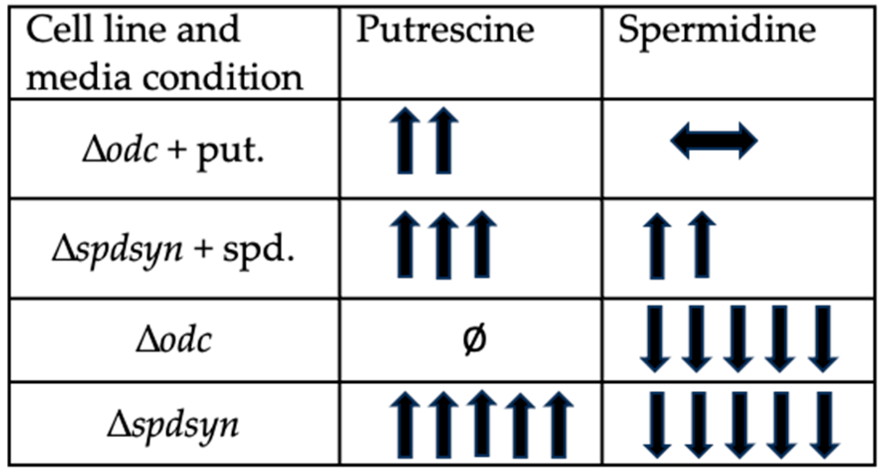
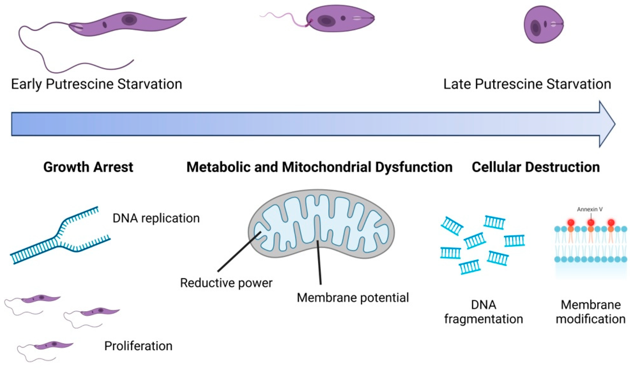
The polyamine pathway in Leishmania parasites has emerged as a promising target for therapeutic intervention, yet the functions of polyamines in parasites remain largely unexplored. Ornithine decarboxylase (ODC) and spermidine synthase (SPDSYN) catalyze the sequential conversion of ornithine to putrescine and spermidine. We previously described that Leishmania donovani Δodc and Δspdsyn mutants exhibit markedly reduced growth in vitro and diminished infectivity in mice, with the effect being most pronounced in putrescine-depleted Δodc mutants. Here, we report that in polyamine-free media, ∆odc mutants arrested proliferation and replication, while ∆spdsyn mutants showed a slow growth and replication phenotype. Starved ∆odc parasites also exhibited a marked reduction in metabolism, which was not observed in the starved ∆spdsyn cells. In contrast, both mutants displayed mitochondrial membrane hyperpolarization. Hallmarks of apoptosis, DNA fragmentation and membrane modifications, were observed in Δodc mutants incubated in polyamine-free media. These results show that putrescine depletion had an immediate detrimental effect on cell growth, replication, and mitochondrial metabolism and caused an apoptosis-like death phenotype. Our findings establish ODC as the most promising therapeutic target within the polyamine biosynthetic pathway for treating leishmaniasis.

Keywords: 
;  ;  ;  ;  ;  ;  ;  

1. Introduction

Leishmaniasis, a neglected tropical disease, affects over one billion people in nearly 90 countries across Africa, South-East Asia, the Middle East, Europe, Central and South America. Annually, approximately 1 million new cases and 70,000 deaths are reported (Grifferty et al., 2021; Mathison & Bradley, 2023; Organization, 2023; Torres-Guerrero et al., 2017). The risk of infection significantly increases in impoverished communities, and recent outbreaks have been fueled by factors such as human migration, civil unrest, and war (Alvar et al., 2006; Bamorovat et al., 2024; Burza et al., 2018; Grifferty et al., 2021; Hotez, 2018). Additionally, environmental issues like deforestation, urbanization, and climate change have contributed to the rising incidence of cases, with leishmaniasis now being endemic in the United States (Curtin & Aronson, 2021; Grifferty et al., 2021; Hotez, 2018; Mann et al., 2021; McIlwee et al., 2018; Montaner-Angoiti & Llobat, 2023; Nepal et al., 2024; Organization, 2023).
Leishmania parasites have a dimorphic life cycle, existing as flagellated promastigotes in sand flies and non-flagellated amastigotes in mammals, primarily residing in macrophages (Mann et al., 2021; Mathison & Bradley, 2023; Torres-Guerrero et al., 2017). Leishmaniasis in humans, caused by over 20 different species of Leishmania, manifests mainly as cutaneous and visceral forms. Cutaneous leishmaniasis (CL) leads to ulcerative skin lesions, with an estimated 600,000 to 1 million new cases each year (Organization, 2023). In contrast, visceral leishmaniasis (VL), caused by L. donovani and L. infantum, is predominantly fatal if untreated and is the second leading cause of mortality among parasitic diseases. An estimated 50,000 to 90,000 new infections occur annually, though underreporting remains significant (Organization, 2023). VL affects internal organs and presents symptoms such as fever and weight loss (Burza et al., 2018; Mann et al., 2021; Mathison & Bradley, 2023; Organization, 2023).
Currently, no vaccines exist to prevent leishmaniasis in humans and treatment options are limited, often with severe side effects and growing drug resistance complicating care (Ikeogu et al., 2020; Kumari et al., 2021; Le Pape, 2008; Mann et al., 2021; Mathison & Bradley, 2023; Pradhan et al., 2022; Saini et al., 2024; Sasidharan & Saudagar, 2021). Furthermore, the persistence of Leishmania parasites post-treatment has spurred ongoing research into the mechanisms behind their resilience (Barrett et al., 2019; Jara et al., 2023; Mandell & Beverley, 2017; Roy et al., 2024). Together, the lack of ideal treatment options, the absence of a vaccine, and the increasing incidence and spread of the disease underscore the urgent need to identify new therapeutic targets.
Notably, polyamine biosynthesis has already been clinically validated as a treatment target in the related pathogen Trypanosoma brucei gambiense (Alvarez-Rodriguez et al., 2022; Hidalgo et al., 2021; LoGiudice et al., 2018; Perez-Pertejo et al., 2024). These ubiquitous and essential cations play a critical role in various cellular processes, including growth, differentiation, and macromolecular synthesis (Carter et al., 2022; Nakanishi & Cleveland, 2021; Sagar et al., 2021; Wallace, 2009; Xuan et al., 2023). A key inhibitor in this pathway, D,L-α-difluoromethylornithine (DFMO, eflornithine), effectively targets ornithine decarboxylase (ODC), the enzyme responsible for synthesizing the polyamine putrescine. DFMO has demonstrated remarkable success in treating African sleeping sickness caused by Trypanosoma brucei gambiense (Alvarez-Rodriguez et al., 2022; Hidalgo et al., 2021; Perez-Pertejo et al., 2024). DFMO is also active against Leishmania in vitro and in murine and hamster infectivity models and recent studies have highlighted the importance of the polyamine biosynthetic pathway as a potential therapeutic target in Leishmania (Abirami et al., 2023; Carter et al., 2022; Carter et al., 2021; Perez-Pertejo et al., 2024; Rodrigues et al., 2022; Santiago-Silva et al., 2022).
The polyamine biosynthetic pathway in Leishmania consists of four enzymes: arginase (ARG), ornithine decarboxylase (ODC), spermidine synthase (SPDSYN), and S-adenosylmethionine decarboxylase (ADOMETDC) (Figure 1). ARG converts the essential amino acid arginine to ornithine, which is directly channeled into polyamine biosynthesis. ODC then converts ornithine to putrescine, and SPDSYN produces spermidine, a vital metabolite involved in hypusination and activation of eukaryotic translation initiation factor 5A in both parasite and host (Chawla et al., 2010; Chawla et al., 2012; Park et al., 1997). Unique to trypanosomatids, spermidine conjugates with glutathione to form trypanothione, which is essential for redox balance and oxidative stress defense (Fairlamb, 1990; Fairlamb & Cerami, 1992). Trypanothione synthetase/amidase (TRYS) catalyzes its synthesis and hydrolysis (Fyfe et al., 2008). Unlike humans, Leishmania neither produces spermine nor has a polyamine back-conversion pathway.
We previously generated gene deletion mutants for ODC (∆odc) and SPDSYN (∆spdsyn) in L. donovani using targeted gene replacement strategies (Boitz et al., 2009; Gilroy et al., 2011; Jiang et al., 1999; Roberts et al., 2001). Characterization of these mutants revealed that both enzymes are essential for polyamine biosynthesis, as the conditionally lethal null mutants depend on supplementation with putrescine or spermidine for growth. The ∆odc mutants exhibit profoundly reduced infectivity compared to wild-type parasites, while the ∆spdsyn mutants show a less pronounced, yet substantial, decrease in infectivity (Boitz et al., 2009; Gilroy et al., 2011). The inability of ∆odc mutants to establish infections suggests that putrescine is unavailable to intracellular parasites, a hypothesis supported by the rapid conversion of arginine to spermine in macrophages (Kropf et al., 2005) and the typically low levels of putrescine in differentiated mammalian cells (Igarashi & Kashiwagi, 2010; Pegg, 2009). Our findings, combined with evidence that DFMO reduces infectivity in mice and hamsters (Gradoni et al., 1989; Mukhopadhyay & Madhubala, 1993; Olenyik et al., 2011), validate ODC as a potential therapeutic target in Leishmania. Notably, the structure of the leishmanial ODC features a unique N-terminal extension not found in the human enzyme (Das et al., 2015) and both computer modeling and inhibitor studies demonstrate that the enzyme is a druggable target (Das et al., 2015; Das et al., 2016; Grover et al., 2012; Hazra et al., 2013; Pandey et al., 2016; Sheikh et al., 2023; Vannier-Santos et al., 2008).
Notable differences between the growth phenotypes of the ∆odc and ∆spdsyn mutants were also observed in vitro (Perdeh et al., 2020). In the ∆odc mutants, putrescine depletion leads to cell rounding, immediate cessation of proliferation, and loss of viability, whereas putrescine-rich ∆spdsyn mutants display an intermediate proliferation phenotype and can persist in a quiescent-like state for five to six weeks before cell death occurs. Contrary to the long-standing belief that putrescine’s sole function is as precursor for spermidine synthesis (Jiang et al., 1999; Reguera et al., 2009), these findings suggest it is also crucial for parasite growth and infectivity. However, the functions of putrescine remain largely unexplored, highlighting the need for further investigation into its role in cellular processes. The ∆odc and ∆spdsyn mutants serve as ideal tools due to their distinct intracellular polyamine dynamics. Specifically, putrescine levels deplete rapidly in ∆odc mutants incubated in polyamine-free media, while they accumulate in ∆spdsyn mutants under the same conditions (Perdeh et al., 2020). In contrast, spermidine levels remain low but stable in both cell lines (Perdeh et al., 2020).
In this study, we investigated the effects of polyamine withdrawal on cell growth, metabolism, and death in L. donovaniodc and ∆spdsyn mutant cell lines. Our findings demonstrate that ∆odc mutants exhibited rapid arrest in proliferation and replication, alongside significant metabolism impairment, while ∆spdsyn mutants displayed a much more moderate phenotype. Both mutants showed hyperpolarization of the mitochondrial membrane but only the ∆odc mutants displayed hallmarks of apoptosis, specifically DNA fragmentation and membrane modifications. These results underscore the critical role of putrescine in cellular function and highlight ODC as a promising therapeutic target in the polyamine biosynthetic pathway for the treatment of leishmaniasis.

2. Materials and Methods

2.1. Materials

Dulbecco’s Modified Eagle Medium and chicken serum were procured from Thermo Fisher Scientific. Antibiotics, including hygromycin, neomycin, and puromycin, were obtained from InvivoGen. Resazurin was purchased from VWR International, putrescine and spermidine were sourced from MilliporeSigma, and 5,5′,6,6′-tetrachloro-1,1′,3,3′-tetraethyl benzimidazolyl carbocyanine iodide (JC-1) was bought from Cayman Chemical. The BrdU Cell Proliferation ELISA Kit (colorimetric) was acquired from Abcam and the In Situ Cell Death Detection Kit, Fluorescein, was purchased from Roche. Propidium iodide (PI) was obtained from Cell Signaling Technology, and FITC Annexin V was purchased from BioLegend.

2.2. Cell Lines and Culture Conditions

Promastigote parasites were cultured at 27°C in a completely defined Dulbecco’s Modified Eagle Medium optimized for Leishmania promastigotes. In this medium, fetal bovine serum was substituted with chicken serum to prevent polyamine oxidase-mediated toxicity (DME-L CS) (Iovannisci & Ullman, 1983; Perdeh et al., 2020; Roberts et al., 2001). All genetically modified parasites originated from the wild-type (WT) LdBob strain of L. donovani (Goyard et al., 2003), which was initially provided by Dr. Stephen M. Beverley (Washington University, St. Louis, MO). The Δodc and Δspdsyn mutants were previously created using targeted gene replacement methods (Boitz et al., 2009; Gilroy et al., 2011) and contain the hygromycin phosphotransferase and neomycin phosphotransferase drug resistance genes (Δodc) or the hygromycin phosphotransferase and puromycin acetyltransferase drug resistance genes (Δspdsyn). The Δodc cell line was routinely grown in the presence of 100 μM putrescine, 50 μg/mL hygromycin, 20 μg/mL neomycin, and the Δspdsyn cell line was cultured in 100 μM spermidine, 50 μg/mL hygromycin, 10 μg/mL puromycin, unless otherwise specified.

2.3. Proliferation Assay

All three cell lines were washed three times in phosphate-buffered saline (PBS) to remove any residual polyamines. For the initial proliferation curve comparing polyamine starvation among cell lines, wild-type cells were incubated in polyamine-free media lacking both drugs and polyamine supplementation. The ∆odc mutants were incubated in either polyamine-free media or media supplemented with 100 µM putrescine, while ∆spdsyn mutants were grown in polyamine-free media or media supplemented with 100 µM spermidine. For the proliferation curve examining polyamine supplementation in wild-type cells, parasites were incubated in polyamine-free media or in media supplemented with 500 µM putrescine, or 500 µM spermidine, or a combination of 500 µM putrescine and 500 µM spermidine. All cell lines were seeded at a density of 5 x 10⁵ cells/mL on day 0, and their growth was monitored over a period of 11 days. Cell counting was performed using a MacsQuant 10 flow cytometer (Miltenyi Biotec).

2.4. Replication Assay

Bromodeoxyuridine incorporation was measured using the BrdU Cell Proliferation ELISA Kit (colorimetric) from Abcam. This assay was used to assess DNA replication in wild-type parasites and in mutant cell lines that were supplemented with polyamines or incubated in polyamine-free media.
Parasites were washed three times in PBS and resuspended at a density of 5 x 10⁶ cells/mL in the following media conditions: wild-type, ∆odc, and ∆spdsyn parasites in polyamine-free media; ∆odc mutants in media supplemented with 100 µM putrescine; and ∆spdsyn mutants in media supplemented with 100 µM spermidine. After overnight incubation at 27°C, 1 x 10⁸ parasites were harvested, centrifuged, and seeded in triplicate at 1 x 10⁶ cells/100 µL in 96-well plates with fresh media corresponding to their initial conditions.
Cells were then treated according to the manufacturer’s protocol. Briefly, 20 µL of 1x BrdU was added to each well, except the wild-type negative control wells, followed by overnight incubation at 27°C. Cells were fixed and incubated with an anti-BrdU monoclonal detector antibody, followed by Peroxidase Goat Anti-Mouse IgG Conjugate, and TMB Peroxidase Substrate incubation. After adding the Stop Solution, absorbance was measured at 450 nm using a BioTek Synergy H1 Multimode Reader (Agilent).

2.5. Metabolism Assay

Metabolic activity was determined by measuring the conversion of resazurin into the fluorescent compound resorufin.
Parasites were washed three times in PBS before incubation in the appropriate media. Wild-type cells were incubated in polyamine-free media. The ∆odc mutants were cultured either in polyamine-free media or media supplemented with 100 µM putrescine, while the ∆spdsyn mutants were incubated in either polyamine-free media or media supplemented with 100 µM spermidine. Wild-type parasites and mutant cells in supplemented media were seeded at a concentration of 5 x 10⁵ cells/mL on day 0. To ensure sufficient cell material for the assay, ∆odc mutants in polyamine-free media were seeded at a higher concentration of 1 x 10⁷ cells/mL and ∆spdsyn mutants in polyamine-free media were seeded at 2 x 10⁶ cells/mL.
Resazurin fluorescence and cell numbers were measured on days 3 and 4 of starvation, during the log phase of cell growth, when metabolic activity is expected to be optimal. Cells (1 x 10⁷) were harvested, centrifuged at 3,000 rpm for 10 minutes, and resuspended in 1 mL of the same media. Cells were then counted in triplicate in a 96-well plate with 100 µL samples per well, using a MACSQuant flow cytometer (Miltenyi Biotec). Following this, 10 µL of 44 µM resazurin was added to each well, and plates were incubated at 27°C for four hours. Resorufin fluorescence was measured at 554Ex–593Em nm using a BioTek Synergy H1 Multimode Reader (Agilent). Metabolic activity per cell was calculated by dividing the resorufin fluorescence by the number of cells in each sample.

2.6. Assessment of Mitochondrial Membrane Potential

The mitochondrial membrane potential (ΔΨm) was assessed using the cationic dye JC-1. This dye aggregates within mitochondria with an intact mitochondrial membrane potential, emitting red fluorescence at 590 nm, whereas in cells with depolarized mitochondrial membranes, JC-1 remains cytosolic and monomeric, displaying green fluorescence at 530 nm.
Parasites were washed three times in PBS before incubation in the appropriate media. Wild-type cells were incubated in polyamine-free media. The ∆odc mutants were cultured either in polyamine-free media or media supplemented with 100 µM putrescine, while the ∆spdsyn mutants were incubated in either polyamine-free media or media supplemented with 100 µM spermidine. Wild-type parasites and mutant cells in supplemented media were seeded at a concentration of 3 x 10⁵ cells/mL on day 0. To ensure sufficient cell material for the assay, ∆odc mutants in polyamine-free media were seeded at a higher concentration of 5 x 10⁶ cells/mL, and ∆spdsyn mutants in polyamine-free media were seeded at 1 x 10⁶ cells/mL.
Samples were collected on days 3, 4, and 8, washed once with PBS supplemented with 158 µg/mL glucose (PSG), and resuspended in 1 mL PSG. As a control, wild-type cells were treated with the mitochondrial uncoupler carbonyl cyanide m-chlorophenyl hydrazone (CCCP) at 75 µM for 10 minutes at 27°C. All samples were stained with 0.22 mM JC-1 (except an unstained wild-type control), incubated for one hour at 27°C, centrifuged and resuspended in 500 µL PSG. Analysis was performed on a MACSQuant flow cytometer (Miltenyi Biotec) using an excitation laser at 488 nm, 30mW. Emission signals were collected using the FITC B1(525/50 nm) and PE B2 (585/40 nm) detectors to capture green and red fluorescence, respectively. Flow cytometer data was analyzed using FlowJoTM v10 (BD Life Sciences) and the 590:530 fluorescence ratio was calculated for each data point.

2.7. DNA Fragmentation Assay

DNA fragmentation was assessed using the In Situ Cell Death Detection Kit (Roche), which is based on the Terminal deoxynucleotidyl transferase (TdT) dUTP Nick End Labeling (TUNEL) method and detects free 3’-hydroxyl ends generated by endonuclease degradation.
Parasites were washed three times in PBS before incubation in the appropriate media. Wild-type cells were incubated in polyamine-free media. The ∆odc mutants were cultured either in polyamine-free media or media supplemented with 100 µM putrescine, while the ∆spdsyn mutants were incubated in either polyamine-free media or media supplemented with 100 µM spermidine. Wild-type parasites and mutant cells in supplemented media were seeded at a concentration of 5 x 10⁵ cells/mL on day 0. To ensure sufficient cell material for the assay, ∆odc mutants in polyamine-free media were seeded at a higher concentration of 5 x 10⁶ cells/mL, and ∆spdsyn mutants in polyamine-free media were seeded at 1 x 10⁶ cells/mL.
Samples were collected on days 4, 7, and 10 and processed according to the manufacturer’s instructions. Briefly, 1 mL cells were harvested and washed in PBS before being fixed in a 2% paraformaldehyde (PFA) solution in PBS for 60 minutes. Following PFA removal, cells were permeabilized with the kit’s permeabilization buffer, washed with PBS and incubated with the TUNEL reaction mixture for 1 hour at 37°C. After incubation, the cells were washed again and analyzed using a MACSQuant flow cytometer (Miltenyi Biotec). Single-channel trace files generated by the flow cytometer were overlaid and compared using FlowJoTM v10 (BD Life Sciences). Overton % positive cell populations were generated by using the “Compare population” function in FlowJoTM v10. Stained populations were compared with a mixed stock of unlabeled control cells.

2.8. Membrane Modifications Assay

Membrane modifications were evaluated using FITC-Annexin V (Biolegend) and PI (Cell Signaling Technology) staining. Annexin V binds to phosphatidylserine exposed on the outer leaflet of the plasma membrane during early apoptosis, while PI penetrates cells with compromised membranes, marking late apoptotic or necrotic cells.
Parasites were washed three times in PBS before incubation in the appropriate media. Wild-type cells were incubated in polyamine-free media. The ∆odc mutants were cultured either in polyamine-free media or media supplemented with 100 µM putrescine, while the ∆spdsyn mutants were incubated in either polyamine-free media or media supplemented with 100 µM spermidine. Samples were collected on days 3 or 4, 7, and 14. Approximately 600 µL of cell culture was harvested and washed twice with 1 mL of PBS before being resuspended in 200 µL of Annexin V binding buffer (10 mM HEPES, 150 mM NaCl, 2.5 mM CaCl₂ in H₂O), containing 5 µL of 90 ug/mL Annexin V and 5 µL of 10 mg/mL PI. The cells were then incubated at room temperature for 30 minutes, during which they were protected from light, and then analyzed using a MACSQuant flow cytometer (Miltenyi Biotec). Appropriate single-stained and unstained controls were prepared and treated identically.
Scatterplots generated from flow cytometry data were analyzed using FlowJoTM v10 (BD Life Sciences) with quadrant gating applied according to the manufacturer’s guidelines. Percentages of cells within each quadrant were exported to GraphPad Prism v10 (GraphPad software) for data analysis.

2.9. Data Visualization and Statistical Analysis

Data visualization and statistical analysis was conducted using GraphPad Prism v10 (GraphPad software). Error bars in the graphs represent standard deviations. Statistical analysis was conducted using ANOVA and statistical comparisons between group means were considered significant at p < 0.05.

3. Results

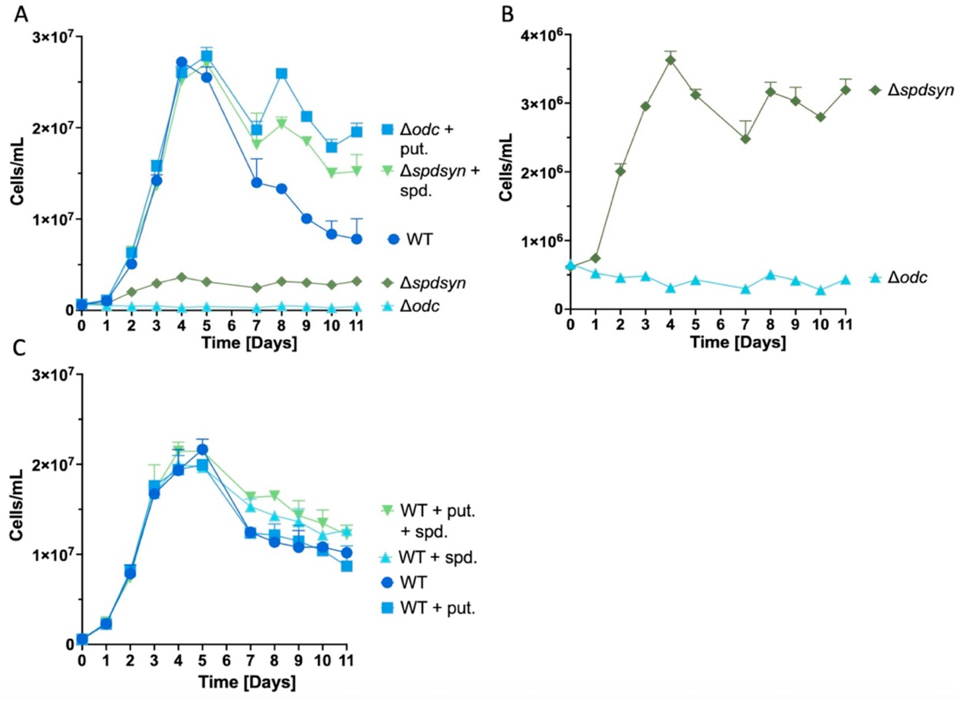
3.1. Extended Polyamine Starvation Exposes Distinct Growth Patterns in Parasites

A prior study demonstrated that ∆odc parasites do not proliferate in polyamine-free media, while ∆spdsyn parasites exhibit only slow growth under these conditions over a 7-day period (Perdeh et al., 2020). In the current study, we extended these observations to 11 days of polyamine-free incubation to monitor the growth phenotype further and assess whether ∆spdsyn mutants eventually achieve cell numbers comparable to wild-type or polyamine-supplemented parasites.
During the first 7 days, all cell lines displayed growth trends consistent with previously published observations (Perdeh et al., 2020) (Figure 2A). The ∆spdsyn mutants incubated in polyamine-free media showed a maximum density of ~4 x 10⁶ cells/mL — notably higher than the ∆odc mutants but almost ten-fold lower than wild-type or supplemented cultures. After reaching this plateau, ∆spdsyn mutants sustained this low maximum cell number without further growth between days 4 - 11 (Figure 2B). In contrast, ∆odc mutants in polyamine-free media showed no proliferation, maintaining a stable cell count around 3–5 x 10⁵ cells/mL throughout the 11-day experiment (Figure 2B).
Both wild-type cells and mutants cultured in polyamine-supplemented media exhibited similar growth patterns through logarithmic and stationary phases (Figure 2A). By day 4 or 5, they peaked at approximately 3 x 10⁷ cells/mL, followed by a gradual decline likely due to general nutrient depletion and overgrowth. However, in week two, notable differences emerged between the wild-type parasites and supplemented mutants (Figure 2A). Wild-type cell counts decreased continuously from a peak of approximately 2.5 x 10⁷ cells/mL to below 1 x 10⁷ cells/mL by day 11 (~60% loss in cell density). In contrast, cell counts for the supplemented mutants dipped slightly but stabilized around 2 x 10⁷ cells/mL between days 7 to 11 (~ 20% loss only in cell density).
Because previous research demonstrated higher intracellular polyamine levels in supplemented ∆odc and ∆spdsyn mutants compared to wild-type parasites (Perdeh et al., 2020), we aimed to investigate whether polyamine supplementation could stabilize cell concentration and prolong survival, as observed in the supplemented mutants during week 2 (Figure 2A). To examine this, wild-type parasites were incubated in media enriched with 500 µM putrescine, 500 µM spermidine, or a combination of both (500 µM putrescine plus 500 µM spermidine). The growth patterns of wild-type parasites were similar regardless of the supplement conditions (Figure 2C) and did not show the distinct differences observed between wild-type parasites and supplemented mutants in Figure 2A.
To summarize, we extended previous findings on the growth behavior of ∆odc and ∆spdsyn mutants under polyamine starvation, showing that ∆spdsyn mutants achieved higher cell densities than ∆odc mutants but plateaued at levels far below wild-type or supplemented cultures. Supplemented mutant cells maintained stable counts through week two, while wild-type cells declined, showing a distinct difference in growth dynamics.

3.2. Putrescine is Essential for DNA Synthesis and Replication

To investigate whether polyamine depletion affects DNA replication and potentially contributes to the growth arrest of ∆odc parasites, we measured BrdU incorporation as a marker of DNA synthesis. Wild-type and supplemented mutant cells exhibited proficient BrdU incorporation, with no significant differences between them (Figure 3). In contrast, ∆odc mutants grown in polyamine-free media showed virtually no BrdU incorporation, which was comparable to the no-cell control, and significantly less than that of ∆odc mutants in 100 µM putrescine (p < 0.0001) (Figure 3). Although ∆spdsyn mutants in polyamine-free media displayed some BrdU incorporation, it was significantly less than that of mutants in 100 µM spermidine (p = 0.0242) (Figure 3). The lack of DNA synthesis in ∆odc mutants and the limited DNA synthesis in ∆spdsyn mutants grown in polyamine-free media corresponded to their respective growth phenotypes illustrated in Figure 2A,B.

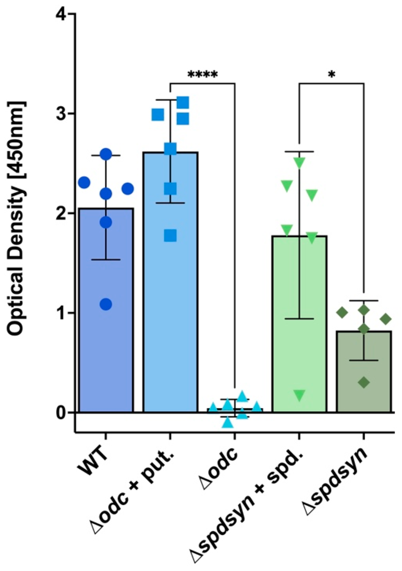
3.3. Putrescine Depletion Reduces Metabolism

To investigate if polyamine deprivation reduces metabolic activity, the conversion of resazurin to the fluorescent compound resorufin via intracellular reductases was measured. Measurements were taken on days 3 and 4, when parasites underwent robust logarithmic growth (Figure 2A). Results showed a significant reduction in metabolism in the ∆odc mutants incubated in polyamine-free media compared to those incubated in 100 µM putrescine (p = 0.0023 for day 3, p = 0.0019 for day 4) as well as to wild-type and ∆spdsyn parasites (Figure 4). In contrast, no significant difference in resazurin conversion was observed in ∆spdsyn mutants incubated in polyamine-free media compared to those grown in 100 µM spermidine (Figure 4). In summary, resazurin conversion showed that wild-type cells, supplemented ∆odc mutants, and ∆spdsyn mutants (in both polyamine-free and supplemented media) exhibited similar metabolic activity on days 3 and 4, while metabolism was significantly reduced in ∆odc mutants under polyamine deprivation.

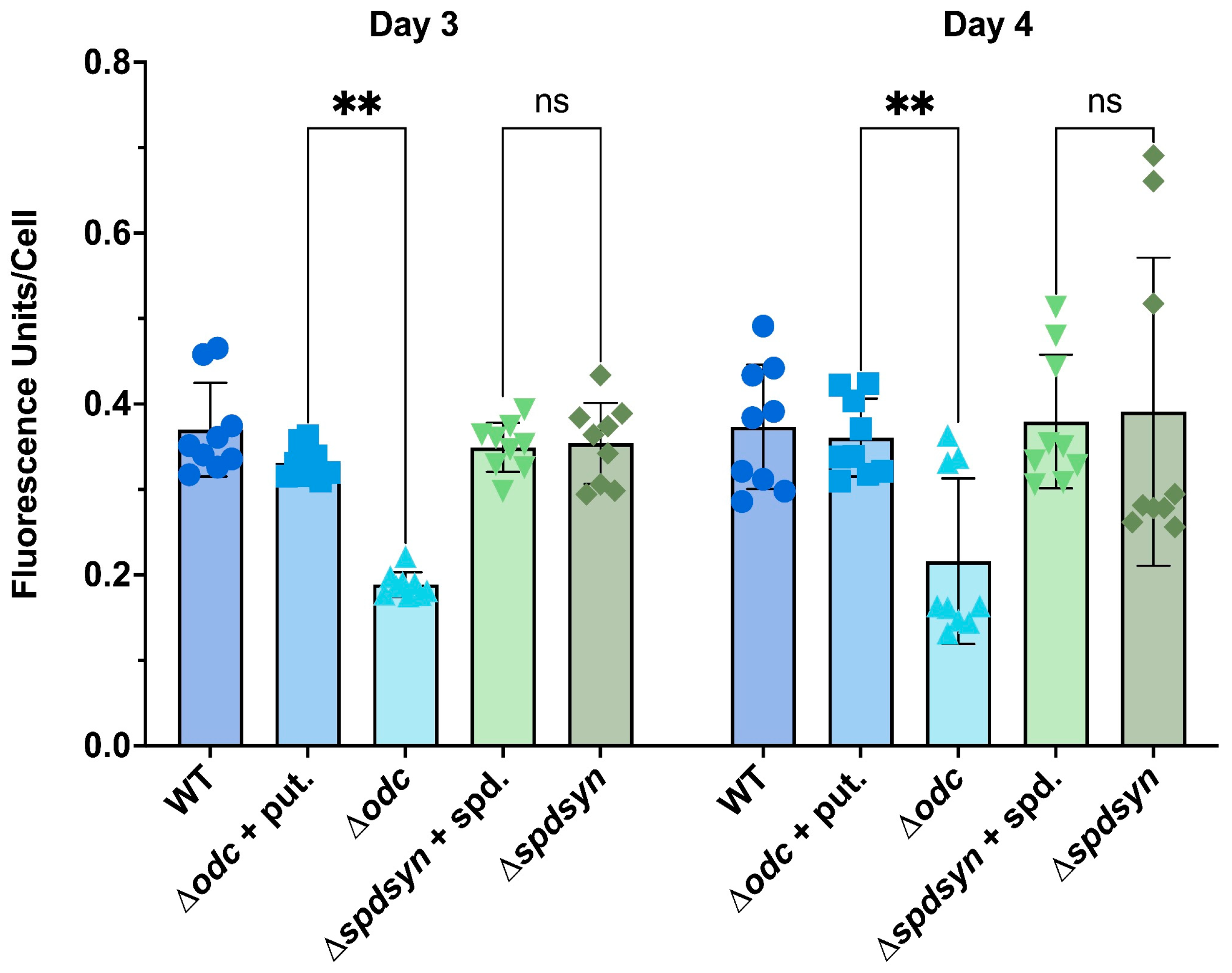
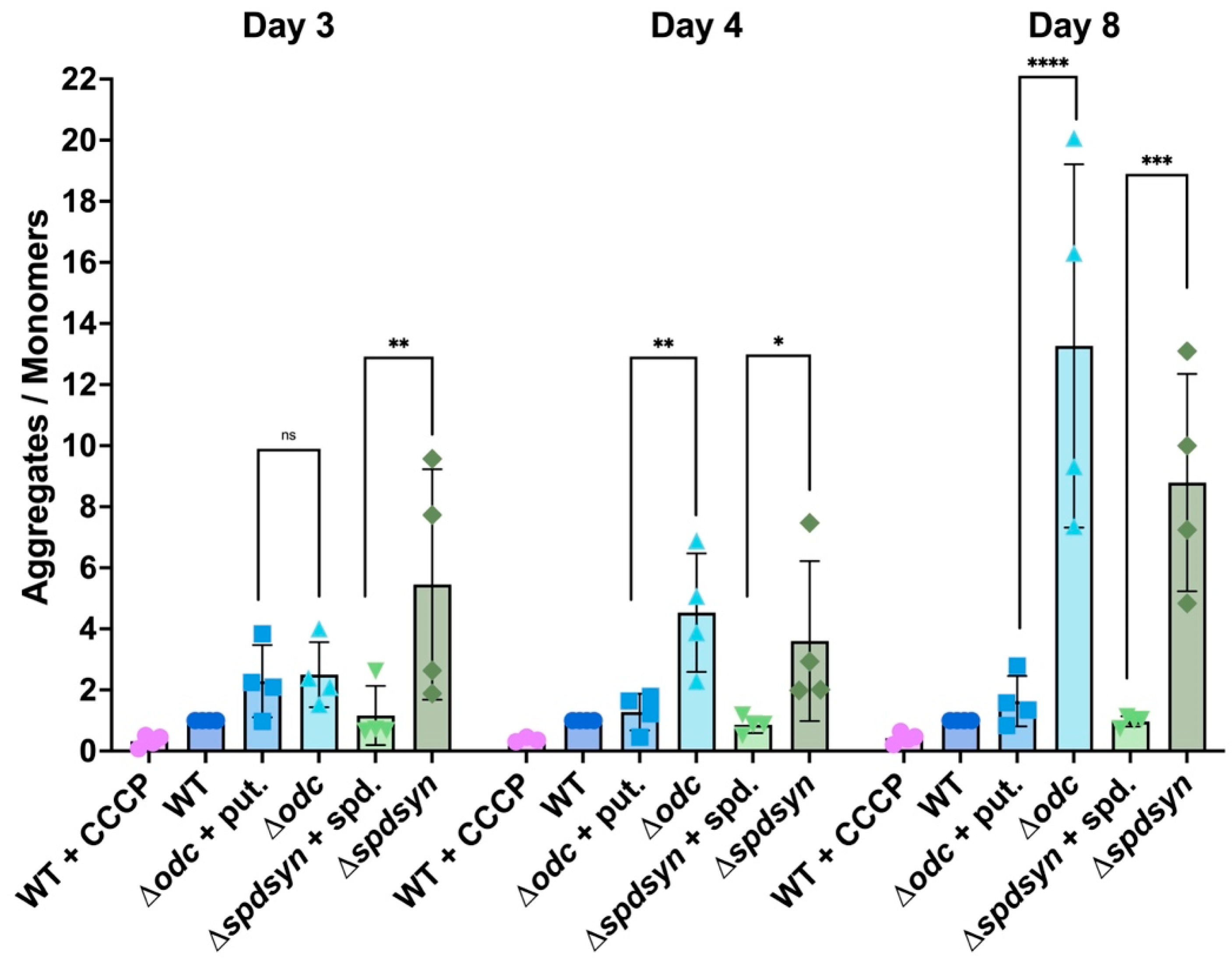
3.4. Polyamine Depletion Affects Mitochondrial Membrane Potential

To assess whether the mitochondrial potential is compromised in polyamine-starved mutant cell lines, we employed JC-1, a commonly used dye for evaluating mitochondrial membrane potentials. JC-1 selectively accumulates and aggregates in mitochondria, shifting its emission color from green to red as the membrane potential increases. A higher red-to-green fluorescence ratio (590:530) indicates a healthy or hyperpolarized mitochondrial membrane potential, whereas a lower ratio signifies depolarization.
We validated our method using CCCP, a known mitochondrial uncoupler. As expected, CCCP treatment caused a shift from red to green fluorescence, resulting in a lower aggregate-to-monomer ratio in wild-type cells (Figure 5). Throughout the experiment, the aggregate-to-monomer ratios of the ∆odc and ∆spdsyn mutants in media supplemented with putrescine or spermidine, respectively, remained similar to that of wild-type parasites, with minor but insignificant variability observed.
In contrast, the ∆odc parasites incubated in polyamine-free media showed an increased aggregate-to-monomer ratio compared to those supplemented with putrescine. Although the ratios were similar between the two groups on day 3, a significant difference appeared on day 4 (p = 0.0038) and became even more pronounced by day 8 (p = <0.0001). A comparable increase in aggregate-to-monomer ratio was observed in ∆spdsyn mutants in polyamine-free media relative to those in spermidine-supplemented media across all sample days (p = 0.0020 on day 3, p = 0.0138 on day 4, and p = 0.0008 on day 8). The observed elevated aggregate-to-monomer ratio in polyamine-starved ∆odc and ∆spdsyn mutants indicates hyperpolarized mitochondrial membranes.
Overall, these results suggest that both mutant cell lines exhibited altered mitochondrial membrane potential in polyamine-free conditions compared to wild-type and supplemented mutants.

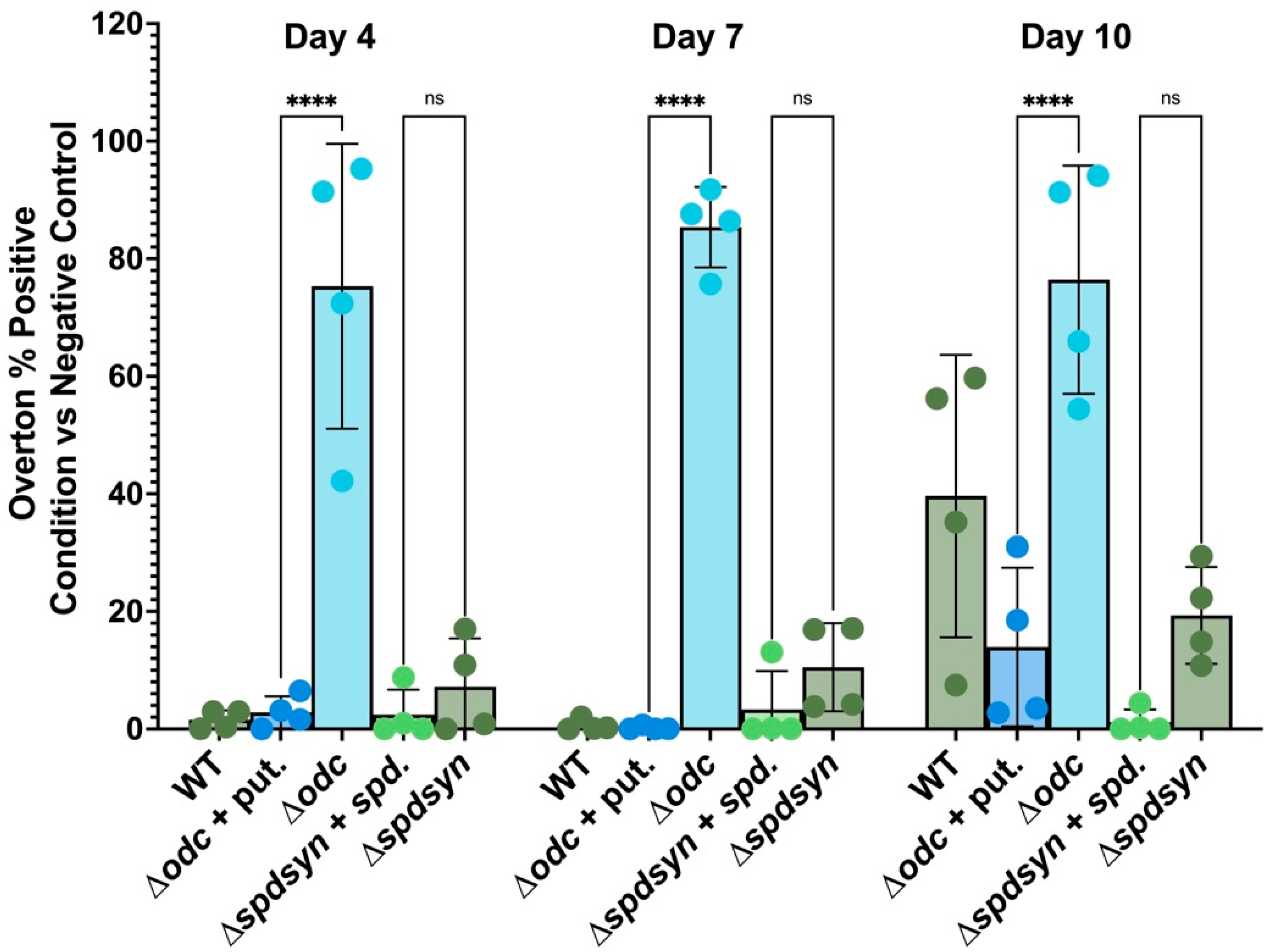
3.5. Putrescine Depletion triggers DNA Fragmentation

Because mitochondrial dysfunction can be a sign of apoptosis, we investigated if DNA fragmentation, a hallmark of apoptosis, occurred in the polyamine-starved cell lines. A TUNEL assay was used to detect free hydroxyl ends produced during DNA degradation by endonucleases.
Wild-type cells showed no DNA degradation on day 4 or 7, but exhibited DNA degradation on day 10 when they experienced general nutrient deprivation. Some degradation was also observed by day 10 in the supplemented ∆odc mutants but surprisingly minimal DNA degradation was seen in the supplemented ∆spdsyn parasites. Notably, on day 10 the percentage of DNA degradation in wild-type parasites was significantly higher compared to that of supplemented ∆spdsyn mutants (p = 0.003).
DNA fragmentation was markedly increased in ∆odc mutants incubated in polyamine-free media throughout the entire incubation period. In comparison to ∆odc mutants supplemented with polyamines, the difference was highly significant, with p < 0.0001 observed on days 4, 7, and 10. In contrast, ∆spdsyn mutants exhibited only a slight, statistically insignificant increase in DNA degradation compared to their counterparts incubated in spermidine-supplemented media.
To summarize, ∆odc mutants incubated in polyamine-free media exhibited consistently high levels of DNA fragmentation throughout the experiment, strikingly exceeding those observed in their supplemented counterparts. Remarkably, the ∆spdsyn mutants supplemented with spermidine demonstrated minimal DNA fragmentation, even at day 10 — a time point by which substantial DNA degradation was evident across all other cell lines and conditions.

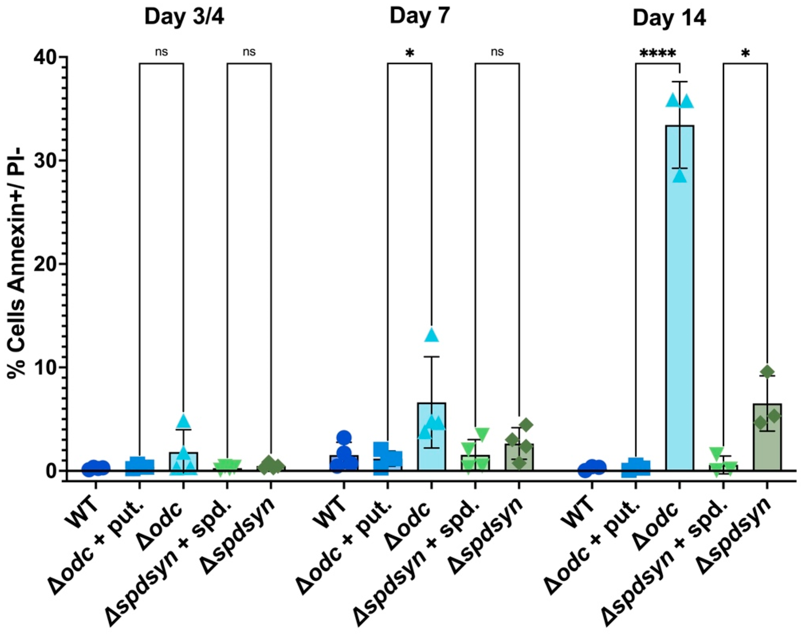
3.6. Polyamine Deprivation Causes Membrane Modifications

We used Annexin V and PI staining in flow cytometry to differentiate live, apoptotic, necrotic, and late apoptotic/necrotic cells based on membrane integrity and phosphatidylserine (PS) exposure. Wild-type cells, ∆odc mutants (with or without putrescine supplementation), and ∆spdsyn mutants (with or without spermidine supplementation) were incubated for 14 days, with samples analyzed on day 3 or 4, day 7, and day 14. On day 3 or 4, less than 2% of cells were Annexin V-positive and PI-negative, indicating minimal apoptosis (Figure 7, Table 1). By day 7, the percentage of apoptotic cells remained low (below 3%) in wild-type cells, putrescine-supplemented ∆odc mutants, and ∆spdsyn mutants, regardless of spermidine supplementation. In contrast, ∆odc mutants grown in polyamine-free media showed a significantly (p = 0.0064) higher rate of apoptosis at 6.62%, compared to 1.19% in putrescine-supplemented ∆odc mutants. This difference was even more profound by day 14 (p < 0.0001), when ∆odc mutants grown in polyamine-free media exhibited 33.43% apoptotic cells, compared to a low percentage of 0.29% in supplemented ∆odc mutants. A more modest but statistically significant (p = 0.0278) increase in apoptosis was observed in ∆spdsyn mutants incubated in polyamine-free media, 6.52%, compared to those with spermidine supplementation, where apoptosis remained at 0.58%.
Analysis of Annexin V-negative PI-negative cells, indicating live parasites, revealed a notably high percentage of live cells, 72.73%, in ∆spdsyn mutants incubated in polyamine-free media at day 14 (Table 1). In contrast, wild-type cells, ∆spdsyn and ∆odc mutants grown with polyamine supplementation showed less than 10% live cells, with ∆odc mutants incubated in polyamine-free media displaying 15.68% PI-negative cells.
Collectively, flow cytometry analysis using Annexin V and PI staining showed that ∆odc and ∆spdsyn mutants incubated in polyamine-free media had significantly higher levels of apoptosis over time compared to mutants with polyamine supplementation and this was more pronounced in the ∆odc parasites. In addition, ∆spdsyn mutants in polyamine-free media displayed a much higher percentage of live cells at day 14 than wild-type cells and supplemented mutants.

4. Discussion

Although recent studies have underscored the critical nature of polyamines in Leishmania parasites (Boitz et al., 2009; Carter et al., 2022; Muxel et al., 2017; Phillips, 2018; Santiago-Silva et al., 2022; Sebastian et al., 2024), their specific functions remain largely unexplored. Putrescine has emerged as a critical metabolite that has essential functions beyond its role as precursor for spermidine formation (Boitz et al., 2017; Perdeh et al., 2020). L. donovani polyamine pathway mutants provide valuable tools to investigate the roles of putrescine, since under polyamine-free conditions, residual spermidine levels remain comparably low in both lines, while putrescine depletes rapidly in ∆odc mutants and accumulates in ∆spdsyn mutants (Perdeh et al., 2020).
Our findings suggest that putrescine depletion has a direct impact on growth, replication, metabolism, and type of cell death. The depletion of putrescine in the ∆odc mutants incubated in polyamine-free media led to an immediate cessation of proliferation and DNA replication upon polyamine withdrawal, while ∆spdsyn mutants grown in polyamine-free media presented a less severe growth and replication impairment (Figure 2 and Figure 3). Notably, ∆odc mutants exhibited a substantial metabolic decline, unlike the ∆spdsyn mutants (Figure 4), suggesting a more critical role for putrescine in cellular metabolism. Although both mutants showed hyperpolarization of the mitochondrial membrane potential in polyamine-free media, only the ∆odc cells exhibited a significant increase in the apoptosis-like indicators, DNA fragmentation and Annexin V staining (Figure 6 and Figure 7).
Figure 6. Percentage of cells with DNA fragmentation assessed by flow cytometry and TUNEL analysis. Wild-type parasites (WT), ∆odc mutants cultured in either 100 µM putrescine-supplemented or polyamine-free media, and ∆spdsyn mutants cultured in either 100 µM spermidine-supplemented or polyamine-free media, were analyzed. Each cell line was analyzed after 4, 7, and 10 days. The experiment was performed four times (n = 4).
Preprints 143557 g006
Figure 7. Percent of apoptotic cells as assessed by Annexin V and propidium iodide (PI) staining. The percentage of parasites that stained positive for Annexin V (indicating early apoptosis) and negative for PI (indicating membrane integrity) is shown. Wild-type parasites (WT), ∆odc mutants in either 100 µM putrescine-supplemented or polyamine-free media, and ∆spdsyn mutants in either 100 µM spermidine-supplemented or polyamine-free media were analyzed. Samples were collected on days 3 or 4, 7, and 14 of the experiment. The experiment was performed four times (n = 4).
Preprints 143557 g007
Cell growth data over 11 days (Figure 2A) insinuate that the elevated polyamine levels present in the supplemented ∆odc and ∆spdsyn mutants (Figure 8) (Perdeh et al., 2020) may provide protection and bolster cell survival during stress conditions. Our results show that in the second week of incubation, the supplemented ∆odc and ∆spdsyn mutants had slightly higher cell numbers compared to wild-type parasites, suggesting greater resilience under nutrient-depleted conditions. This trend was further supported by absence of PI staining (Table 1), which indicated that by day 14, only 0.19% of wild-type cells remained viable, whereas 4.39% and 9.93% of the supplemented ∆odc and ∆spdsyn mutants, respectively, were still alive. However, adding polyamines (either 500 µM putrescine, 500 µM spermidine, or both) to the media did not increase the cell numbers of wild-type parasites (Figure 2C). This might indicate that wild-type parasites, which can synthesize adequate polyamines for their growth and survival, do not readily import additional polyamines. While Leishmania parasites possess polyamine transporters (Balana-Fouce et al., 1989; Basselin et al., 2000; Hasne & Ullman, 2011; Kandpal & Tekwani, 1997), and the polyamine pathway mutants rely on these transport systems for survival (Figure 2) (Boitz et al., 2009; Gilroy et al., 2011; Jiang et al., 1999; Perdeh et al., 2020; Roberts et al., 2001), little is known about the regulation of polyamine transport. An alternative explanation for the enhanced resilience in the supplemented mutants could be other cellular or metabolic adaptations that occurred due to the gene deletion events.
Previous studies have shown that ∆spdsyn parasites can enter a quiescent-like state and survive for up to six weeks in polyamine-free media (Perdeh et al., 2020). Our findings support these observations, as we found that a substantial proportion of ∆spdsyn parasites remained viable even after prolonged incubation in polyamine-free media, as indicated by lack of PI staining (Table 1). Specifically, after 14 days, 72.73% of the cells were alive, a much higher percentage than observed in wild-type parasites, spermidine-supplemented ∆spdsyn mutants, or in ∆odc parasites, regardless of putrescine supplementation.
The persistence or quiescence of Leishmania and related parasites has gained attention due to its links to treatment failure, relapse, and chronic disease (Barrett et al., 2019; Dirkx et al., 2024; Jara et al., 2023; Lodi et al., 2024; Mandell & Beverley, 2017; Pessenda & da Silva, 2020; Roy et al., 2024). Understanding quiescence mechanisms could lead to better treatment paradigms and reduced relapse rates. Both supplemented ∆odc and ∆spdsyn mutants and ∆spdsyn parasites incubated in polyamine-free media exhibited persistence-like traits, characterized by stable cell numbers (Figure 2) and/or higher percentages of viable cells (Table 1). These traits were associated with significantly elevated putrescine levels but variable spermidine pools (Figure 8) (Perdeh et al., 2020), implying that putrescine may play a role in parasite persistence.
The ability of mutant parasites to proliferate in polyamine-free media (Figure 2) closely aligned with their replication profile (Figure 3). The ∆odc mutants were unable to synthesize DNA and showed no cell growth, while the ∆spdsyn mutants exhibited limited DNA replication and low levels of proliferation. These findings suggest that putrescine is important for DNA synthesis. Because DNA replication is required for cell division, this impairment alone could account for the growth deficit observed in the putrescine-depleted ∆odc mutants (Figure 2).
To evaluate metabolic activity in the mutant cell lines, we used resazurin assays, which measure the reduction of resazurin to the fluorescent compound resorufin by cellular dehydrogenases in the presence of NADH or NADPH (Corral et al., 2013; Mikus & Steverding, 2000). This reaction serves as an indirect indicator of mitochondrial health, as the majority of NADH is typically produced in mitochondria through energy-generating pathways such as the TCA cycle and oxidative phosphorylation (Li & Sauve, 2015; Yang & Sauve, 2021). The ∆odc parasites cultured in polyamine-free media exhibited significantly reduced metabolic activity compared to wild-type parasites and supplemented ∆odc mutants (Figure 4). In contrast, ∆spdsyn parasites showed metabolic activity comparable to wild-type parasites, regardless of polyamine supplementation (Figure 4). These findings suggest that reduced spermidine levels in ∆spdsyn mutants do not compromise cellular and mitochondrial metabolism. However, maintaining intracellular levels of putrescine appears to be important for metabolic activity, overall cell health, and potentially mitochondrial function.
A critical role of putrescine in mitochondrial function and integrity in Leishmania parasites has previously been reported. The ODC inhibitor 1, 4-diamino-2-butanone reduces intracellular polyamine levels and causes structural and functional mitochondrial damage in both Leishmania and the related parasite Trypanosoma cruzi (Menezes et al., 2006; Vannier-Santos et al., 2008). Notably, trypanosomatid parasites, like Leishmania, are especially vulnerable to mitochondrial dysfunction because they have only a single mitochondrion per cell, making this organelle critical for parasite survival and a potential target for therapeutic intervention (Menna-Barreto, 2019; Pedra-Rezende et al., 2022).
To further investigate whether putrescine depletion in ∆odc mutants impacts mitochondrial function, we assessed mitochondrial membrane potential using JC-1, a cationic, membrane-permeable dye commonly employed for this purpose (Cossarizza et al., 1993; Reers et al., 1995; Reers et al., 1991; Smiley et al., 1991). Both mutant cell lines exhibited an altered mitochondrial membrane potential under polyamine-free conditions compared to wild-type and supplemented mutants, with a higher aggregate-to-monomer ratio indicative of hyperpolarization (Figure 5). While mitochondrial depolarization is often linked to dysfunction, as it disrupts ion gradients and ATP production, hyperpolarization can also signal stress and contribute to cell death. A recent publication reported that hypericin, whose main mechanism of action is the inhibition of SPDSYN, induced mitochondrial membrane hyperpolarization and cell death in L. donovani (Sebastian et al., 2024). Mitochondrial hyperpolarization, induced by the inhibition of F0-F1 ATP synthase or complex I, has been shown to cause increased reactive oxygen species production and programmed cell death in Leishmania parasites (Mehta & Shaha, 2004; Roy et al., 2008; Sen et al., 2004).
Reduced metabolic activity was observed exclusively in putrescine-depleted ∆odc mutants, but not in ∆spdsyn mutants, when both were incubated in polyamine-free media (Figure 4). In contrast, mitochondrial hyperpolarization was observed in both mutant cell lines under these conditions (Figure 5). This observation suggests that mitochondrial hyperpolarization is likely driven by the low spermidine levels shared by both mutants. Spermidine may play an important role in sustaining the mitochondrial membrane potential through electron transport chain activity, while putrescine appears to be critical for maintaining cellular reducing power, potentially through its influence on NAD(P)H levels and/or NAD(P)H dehydrogenase activity.
In mammalian cells, spermidine has been shown to directly enhance mitochondrial health by improving mitochondrial respiration, membrane potential, and ATP production (Al-Habsi et al., 2022; Fairley et al., 2023; Schroeder et al., 2021; Zimmermann et al., 2023). Additionally, spermidine-mediated hypusination of eIF5A plays a crucial role in maintaining mitochondrial function, as lower levels of hypusinated eIF5A are associated with reduced oxygen consumption and ATP generation (Barba-Aliaga & Alepuz, 2022; Liang et al., 2021; Pereira et al., 2016). While it is unknown to what extent these mechanisms occur in Leishmania parasites, the low spermidine levels in the mutant cell lines likely lead to reduced eIF5A hypusination, which may in turn contribute to impairment of mitochondrial health and respiration in a similar manner to mammalian cells.
Because mitochondrial dysfunction can lead to an apoptosis-like phenotype (Abate et al., 2020; Apostolova et al., 2011; Mukherjee et al., 2002; Sen et al., 2004), we examined other hallmarks of apoptosis. DNA fragmentation and membrane modifications, driven by endonuclease-mediated cleavage of chromosomal DNA and the externalization of phospholipids such as phosphatidylserine, are central features of apoptosis and both of these mechanisms have been previously described in Leishmania parasites (Basmaciyan & Casanova, 2019; Gannavaram & Debrabant, 2012; Jimenez-Ruiz et al., 2010; Proto et al., 2013). While phosphatidylserine itself has been reported to be absent in Leishmania parasites, similar phospholipids appear to perform an analogous role, as Annexin V staining—used to detect phosphatidylserine externalization—has been observed in numerous studies of these organisms (Gannavaram & Debrabant, 2012; Jimenez-Ruiz et al., 2010; Proto et al., 2013; Weingartner et al., 2012).
DNA fragmentation was detected within the first week of incubating ∆odc mutants in polyamine-free media (Figure 6), followed by membrane modifications observed during the second week of putrescine starvation (Figure 7). These findings suggest that putrescine depletion induced an apoptosis-like death phenotype. While both ∆odc and ∆spdsyn mutants incubated in polyamine-free media underwent mitochondrial hyperpolarization, the apoptosis phenotype was only observed in the ∆odc mutants, perhaps indicating that the elevated putrescine levels in the ∆spdsyn mutants (Figure 8) protected the cells from undergoing programmed cell death. Similarly, supplemented ∆spdsyn mutants demonstrated significantly lower levels of DNA fragmentation after 10 days of incubation compared to other cell lines and conditions (Figure 6). This protective effect may have been due to the elevated intracellular polyamine levels (Figure 8).
The concept of programmed cell death in single cell protozoan parasites like Leishmania, remains intriguing and controversial, given that a single-celled organism may not seem to require such a process (Basmaciyan & Casanova, 2019; Gannavaram & Debrabant, 2012; Kaczanowski et al., 2011; Proto et al., 2013). Current hypotheses include that apoptosis may benefit the population as a whole by avoiding hyperparasitism or the immune response in the host (Basmaciyan & Casanova, 2019; Cecilio et al., 2014; Gannavaram & Debrabant, 2012; Kaczanowski et al., 2011; Reece et al., 2011). Although Leishmania lacks classic apoptotic proteins and signaling pathways have not been identified, the parasite shows evidence of apoptosis-like characteristics, including mitochondrial dysfunction, DNA fragmentation, membrane modifications, cell shrinkage and rounding (Basmaciyan & Casanova, 2019; Gannavaram & Debrabant, 2012; Jimenez-Ruiz et al., 2010; Kaczanowski et al., 2011; Proto et al., 2013). This insinuates that apoptosis in Leishmania and other protozoan parasites may represent a rudimentary or primitive evolutionary precursor to the more complex forms of cell death observed in multicellular organisms (Basmaciyan & Casanova, 2019; Reece et al., 2011; Taylor-Brown & Hurd, 2013). Nonetheless, further research into Leishmania apoptosis holds promise for uncovering novel targets for drug development and therapeutic intervention (Basmaciyan & Casanova, 2019; Gannavaram & Debrabant, 2012). The ∆odc cell line, in particular, may serve as a valuable tool for exploring the molecular mechanisms underlying programmed cell death. Notably, most prior studies have focused on apoptosis induced by drugs, whereas gene deletion mutants like ∆odc, which exhibit intrinsic apoptotic features, could provide a more precise and controlled model for understanding these processes.
Limitations of the studies presented here include uncertainty about whether the observed effects are directly linked to putrescine depletion or occur as a result of cellular stress induced by putrescine starvation. Additionally, there was some inherent variability across cell lines and conditions, particularly in the DNA fragmentation and membrane modifications assays. This variability may result from polyamine-starved parasites forming a heterogeneous population, with individual cells exhibiting distinct responses. Furthermore, these studies thus far were conducted in the promastigote stage, highlighting the need for future research in the medically relevant amastigote stage.

5. Conclusions

Our findings, combined with previously published observations (Perdeh et al., 2020), provide a better understanding of the functions of putrescine. A key insight is that putrescine depletion triggers both early and late cellular changes (Figure 9). Early changes, observed within the first two days of starvation, include growth arrest, cessation of DNA replication, and morphological alterations, followed by reduced metabolism, mitochondrial dysfunction, and DNA fragmentation. By the second week of starvation, membrane modifications, another hallmark of an apoptosis-like cell death, emerged. Notably, these effects are specific to putrescine depletion and are not observed with general nutrient starvation. Together, these findings highlight the essential roles of putrescine in DNA replication, cellular proliferation, and metabolism. Moreover, they offer a plausible explanation for the more pronounced effects of the ∆odc gene deletion on in vivo infectivity compared to deletions in other polyamine pathway enzymes (Boitz et al., 2017; Boitz et al., 2009; Gilroy et al., 2011). In conclusion, our studies support the polyamine biosynthetic pathway in Leishmania as a promising therapeutic target, with ODC standing out as a key target for therapeutic development.

Author Contributions

Conceptualization, S.R. and N.C.; methodology, S.R., J.J., J.T., and S.N.; validation, S.R., J.J. and J.T.; formal analysis, S.R., J.J. and J.T.; investigation, J.J, J.T, S.N., A.G., Y.A., S.K., T.P, K.K., J.L.; data curation, S.R., J.J., J.T., and S.N; writing—original draft preparation, S.R., J.J., and J.T.; writing—review and editing, S.R., J.J., J.T, and N.C.; visualization, S.R., J.J., and J.T.; supervision, S.R.; project administration, S.R.; funding acquisition, S.R. All authors have read and agreed to the published version of the manuscript.

Funding

This research was funded by the National Institute of Allergy and Infectious Diseases (NIAID) of the National Institutes of Health (NIH) under award number R15AI151980. AG was funded by an American Society of Pharmacology and Experimental Therapeutics (ASPET) Summer Undergraduate Research Fellowship (SURF).

Institutional Review Board Statement

Not applicable

Informed Consent Statement

Not applicable

Data Availability Statement

The data presented in this study are available on request from the corresponding author due to the large volume or specialized format of the raw data, requiring specialized data sharing.

Acknowledgments

The authors used OpenAI’s ChatGPT to improve the readability and clarity of this manuscript. All final content and responsibility for the manuscript remain with the authors.

Conflicts of Interest

The authors declare no conflicts of interest. The funders had no role in the design of the study; in the collection, analyses, or interpretation of data; in the writing of the manuscript; or in the decision to publish the results.

References

  1. Abate, M., Festa, A., Falco, M., Lombardi, A., Luce, A., Grimaldi, A., Zappavigna, S., Sperlongano, P., Irace, C., Caraglia, M., & Misso, G. (2020). Mitochondria as playmakers of apoptosis, autophagy and senescence. Semin Cell Dev Biol, 98, 139-153. [CrossRef]
  2. Abirami, M., Karan Kumar, B., Faheem, Dey, S., Johri, S., Reguera, R. M., Balana-Fouce, R., Gowri Chandra Sekhar, K. V., & Sankaranarayanan, M. (2023). Molecular-level strategic goals and repressors in Leishmaniasis - Integrated data to accelerate target-based heterocyclic scaffolds. Eur J Med Chem, 257, 115471. [CrossRef]
  3. Al-Habsi, M., Chamoto, K., Matsumoto, K., Nomura, N., Zhang, B., Sugiura, Y., Sonomura, K., Maharani, A., Nakajima, Y., Wu, Y., Nomura, Y., Menzies, R., Tajima, M., Kitaoka, K., Haku, Y., Delghandi, S., Yurimoto, K., Matsuda, F., Iwata, S., … Honjo, T. (2022). Spermidine activates mitochondrial trifunctional protein and improves antitumor immunity in mice. Science, 378(6618), eabj3510. [CrossRef]
  4. Alvar, J., Yactayo, S., & Bern, C. (2006). Leishmaniasis and poverty. Trends Parasitol, 22(12), 552-557. [CrossRef]
  5. Alvarez-Rodriguez, A., Jin, B. K., Radwanska, M., & Magez, S. (2022). Recent progress in diagnosis and treatment of Human African Trypanosomiasis has made the elimination of this disease a realistic target by 2030. Front Med (Lausanne), 9, 1037094. [CrossRef]
  6. Apostolova, N., Blas-Garcia, A., & Esplugues, J. V. (2011). Mitochondria sentencing about cellular life and death: a matter of oxidative stress. Curr Pharm Des, 17(36), 4047-4060. [CrossRef]
  7. Balana-Fouce, R., Ordonez, D., & Alunda, J. M. (1989). Putrescine transport system in Leishmania infantum promastigotes. Mol Biochem Parasitol, 35(1), 43-50. [CrossRef]
  8. Bamorovat, M., Sharifi, I., Khosravi, A., Aflatoonian, M. R., Agha Kuchak Afshari, S., Salarkia, E., Sharifi, F., Aflatoonian, B., Gharachorloo, F., Khamesipour, A., Mohebali, M., Zamani, O., Shirzadi, M. R., & Gouya, M. M. (2024). Global Dilemma and Needs Assessment Toward Achieving Sustainable Development Goals in Controlling Leishmaniasis. J Epidemiol Glob Health, 14(1), 22-34. [CrossRef]
  9. Barba-Aliaga, M., & Alepuz, P. (2022). Role of eIF5A in Mitochondrial Function. Int J Mol Sci, 23(3). [CrossRef]
  10. Barrett, M. P., Kyle, D. E., Sibley, L. D., Radke, J. B., & Tarleton, R. L. (2019). Protozoan persister-like cells and drug treatment failure. Nat Rev Microbiol, 17(10), 607-620. [CrossRef]
  11. Basmaciyan, L., & Casanova, M. (2019). Cell death in Leishmania. Parasite, 26, 71. (La mort cellulaire chez Leishmania.). [CrossRef]
  12. Basselin, M., Coombs, G. H., & Barrett, M. P. (2000). Putrescine and spermidine transport in Leishmania. Mol Biochem Parasitol, 109(1), 37-46. [CrossRef]
  13. Boitz, J. M., Gilroy, C. A., Olenyik, T. D., Paradis, D., Perdeh, J., Dearman, K., Davis, M. J., Yates, P. A., Li, Y., Riscoe, M. K., Ullman, B., & Roberts, S. C. (2017). Arginase Is Essential for Survival of Leishmania donovani Promastigotes but Not Intracellular Amastigotes. Infect Immun, 85(1). [CrossRef]
  14. Boitz, J. M., Yates, P. A., Kline, C., Gaur, U., Wilson, M. E., Ullman, B., & Roberts, S. C. (2009). Leishmania donovani ornithine decarboxylase is indispensable for parasite survival in the mammalian host. Infect Immun, 77(2), 756-763. [CrossRef]
  15. Burza, S., Croft, S. L., & Boelaert, M. (2018). Leishmaniasis. Lancet, 392(10151), 951-970. [CrossRef]
  16. Carter, N. S., Kawasaki, Y., Nahata, S. S., Elikaee, S., Rajab, S., Salam, L., Alabdulal, M. Y., Broessel, K. K., Foroghi, F., Abbas, A., Poormohamadian, R., & Roberts, S. C. (2022). Polyamine Metabolism in Leishmania Parasites: A Promising Therapeutic Target. Med Sci (Basel), 10(2). [CrossRef]
  17. Carter, N. S., Stamper, B. D., Elbarbry, F., Nguyen, V., Lopez, S., Kawasaki, Y., Poormohamadian, R., & Roberts, S. C. (2021). Natural Products That Target the Arginase in Leishmania Parasites Hold Therapeutic Promise. Microorganisms, 9(2). [CrossRef]
  18. Cecilio, P., Perez-Cabezas, B., Santarem, N., Maciel, J., Rodrigues, V., & Cordeiro da Silva, A. (2014). Deception and manipulation: the arms of leishmania, a successful parasite. Front Immunol, 5, 480. [CrossRef]
  19. Chawla, B., Jhingran, A., Singh, S., Tyagi, N., Park, M. H., Srinivasan, N., Roberts, S. C., & Madhubala, R. (2010). Identification and characterization of a novel deoxyhypusine synthase in Leishmania donovani. J Biol Chem, 285(1), 453-463. [CrossRef]
  20. Chawla, B., Kumar, R. R., Tyagi, N., Subramanian, G., Srinivasan, N., Park, M. H., & Madhubala, R. (2012). A unique modification of the eukaryotic initiation factor 5A shows the presence of the complete hypusine pathway in Leishmania donovani. PLoS One, 7(3), e33138. [CrossRef]
  21. Corral, M. J., Gonzalez, E., Cuquerella, M., & Alunda, J. M. (2013). Improvement of 96-well microplate assay for estimation of cell growth and inhibition of Leishmania with Alamar Blue. J Microbiol Methods, 94(2), 111-116. [CrossRef]
  22. Cossarizza, A., Baccarani-Contri, M., Kalashnikova, G., & Franceschi, C. (1993). A new method for the cytofluorimetric analysis of mitochondrial membrane potential using the J-aggregate forming lipophilic cation 5,5’,6,6’-tetrachloro-1,1’,3,3’-tetraethylbenzimidazolcarbocyanine iodide (JC-1). Biochem Biophys Res Commun, 197(1), 40-45. [CrossRef]
  23. Curtin, J. M., & Aronson, N. E. (2021). Leishmaniasis in the United States: Emerging Issues in a Region of Low Endemicity. Microorganisms, 9(3). [CrossRef]
  24. Das, M., Kumar, R., & Dubey, V. K. (2015). Ornithine decarboxylase of Leishmania donovani: biochemical properties and possible role of N-terminal extension. Protein Pept Lett, 22(2), 130-136. [CrossRef]
  25. Das, M., Singh, S., & Dubey, V. K. (2016). Novel Inhibitors of Ornithine Decarboxylase of Leishmania Parasite (LdODC): The Parasite Resists LdODC Inhibition by Overexpression of Spermidine Synthase. Chem Biol Drug Des, 87(3), 352-360. [CrossRef]
  26. Dirkx, L., Van Acker, S. I., Nicolaes, Y., Cunha, J. L. R., Ahmad, R., Hendrickx, R., Caljon, B., Imamura, H., Ebo, D. G., Jeffares, D. C., Sterckx, Y. G., Maes, L., Hendrickx, S., & Caljon, G. (2024). Long-term hematopoietic stem cells trigger quiescence in Leishmania parasites. PLoS Pathog, 20(4), e1012181. [CrossRef]
  27. Fairlamb, A. H. (1990). Trypanothione metabolism and rational approaches to drug design. Biochem Soc Trans, 18(5), 717-720. [CrossRef]
  28. Fairlamb, A. H., & Cerami, A. (1992). Metabolism and functions of trypanothione in the Kinetoplastida. Annu Rev Microbiol, 46, 695-729. [CrossRef]
  29. Fairley, L. H., Lejri, I., Grimm, A., & Eckert, A. (2023). Spermidine Rescues Bioenergetic and Mitophagy Deficits Induced by Disease-Associated Tau Protein. Int J Mol Sci, 24(6). [CrossRef]
  30. Fyfe, P. K., Oza, S. L., Fairlamb, A. H., & Hunter, W. N. (2008). Leishmania trypanothione synthetase-amidase structure reveals a basis for regulation of conflicting synthetic and hydrolytic activities. J Biol Chem, 283(25), 17672-17680. [CrossRef]
  31. Gannavaram, S., & Debrabant, A. (2012). Programmed cell death in Leishmania: biochemical evidence and role in parasite infectivity. Front Cell Infect Microbiol, 2, 95. [CrossRef]
  32. Gilroy, C., Olenyik, T., Roberts, S. C., & Ullman, B. (2011). Spermidine synthase is required for virulence of Leishmania donovani. Infect Immun, 79(7), 2764-2769. [CrossRef]
  33. Goyard, S., Segawa, H., Gordon, J., Showalter, M., Duncan, R., Turco, S. J., & Beverley, S. M. (2003). An in vitro system for developmental and genetic studies of Leishmania donovani phosphoglycans. Mol Biochem Parasitol, 130(1), 31-42. [CrossRef]
  34. Gradoni, L., Iorio, M. A., Gramiccia, M., & Orsini, S. (1989). In vivo effect of eflornithine (DFMO) and some related compounds on Leishmania infantum preliminary communication. Farmaco, 44(12), 1157-1166. https://www.ncbi.nlm.nih.gov/pubmed/2517472.
  35. Grifferty, G., Shirley, H., McGloin, J., Kahn, J., Orriols, A., & Wamai, R. (2021). Vulnerabilities to and the Socioeconomic and Psychosocial Impacts of the Leishmaniases: A Review. Res Rep Trop Med, 12, 135-151. [CrossRef]
  36. Grover, A., Katiyar, S. P., Jeyakanthan, J., Dubey, V. K., & Sundar, D. (2012). Mechanistic insights into the dual inhibition strategy for checking Leishmaniasis. J Biomol Struct Dyn, 30(4), 474-487. [CrossRef]
  37. Hasne, M. P., & Ullman, B. (2011). Genetic and biochemical analysis of protozoal polyamine transporters. Methods Mol Biol, 720, 309-326. [CrossRef]
  38. Hazra, S., Ghosh, S., Das Sarma, M., Sharma, S., Das, M., Saudagar, P., Prajapati, V. K., Dubey, V. K., Sundar, S., & Hazra, B. (2013). Evaluation of a diospyrin derivative as antileishmanial agent and potential modulator of ornithine decarboxylase of Leishmania donovani. Exp Parasitol, 135(2), 407-413. [CrossRef]
  39. Hidalgo, J., Ortiz, J. F., Fabara, S. P., Eissa-Garces, A., Reddy, D., Collins, K. D., & Tirupathi, R. (2021). Efficacy and Toxicity of Fexinidazole and Nifurtimox Plus Eflornithine in the Treatment of African Trypanosomiasis: A Systematic Review. Cureus, 13(8), e16881. [CrossRef]
  40. Hotez, P. J. (2018). The rise of leishmaniasis in the twenty-first century. Trans R Soc Trop Med Hyg, 112(9), 421-422. [CrossRef]
  41. Igarashi, K., & Kashiwagi, K. (2010). Modulation of cellular function by polyamines. Int J Biochem Cell Biol, 42(1), 39-51. [CrossRef]
  42. Ikeogu, N. M., Akaluka, G. N., Edechi, C. A., Salako, E. S., Onyilagha, C., Barazandeh, A. F., & Uzonna, J. E. (2020). Leishmania Immunity: Advancing Immunotherapy and Vaccine Development. Microorganisms, 8(8). [CrossRef]
  43. Iovannisci, D. M., & Ullman, B. (1983). High efficiency plating method for Leishmania promastigotes in semidefined or completely-defined medium. J Parasitol, 69(4), 633-636. https://www.ncbi.nlm.nih.gov/pubmed/6631633.
  44. Jara, M., Arevalo, J., Llanos-Cuentas, A., den Broeck, F. V., Domagalska, M. A., & Dujardin, J. C. (2023). Unveiling drug-tolerant and persister-like cells in Leishmania braziliensis lines derived from patients with cutaneous leishmaniasis. Front Cell Infect Microbiol, 13, 1253033. [CrossRef]
  45. Jiang, Y., Roberts, S. C., Jardim, A., Carter, N. S., Shih, S., Ariyanayagam, M., Fairlamb, A. H., & Ullman, B. (1999). Ornithine decarboxylase gene deletion mutants of Leishmania donovani. J Biol Chem, 274(6), 3781-3788. [CrossRef]
  46. Jimenez-Ruiz, A., Alzate, J. F., Macleod, E. T., Luder, C. G., Fasel, N., & Hurd, H. (2010). Apoptotic markers in protozoan parasites. Parasit Vectors, 3, 104. [CrossRef]
  47. Kaczanowski, S., Sajid, M., & Reece, S. E. (2011). Evolution of apoptosis-like programmed cell death in unicellular protozoan parasites. Parasit Vectors, 4, 44. [CrossRef]
  48. Kandpal, M., & Tekwani, B. L. (1997). Polyamine transport systems of Leishmania donovani promastigotes. Life Sci, 60(20), 1793-1801. [CrossRef]
  49. Kropf, P., Fuentes, J. M., Fahnrich, E., Arpa, L., Herath, S., Weber, V., Soler, G., Celada, A., Modolell, M., & Muller, I. (2005). Arginase and polyamine synthesis are key factors in the regulation of experimental leishmaniasis in vivo. FASEB J, 19(8), 1000-1002. [CrossRef]
  50. Kumari, D., Perveen, S., Sharma, R., & Singh, K. (2021). Advancement in leishmaniasis diagnosis and therapeutics: An update. Eur J Pharmacol, 910, 174436. [CrossRef]
  51. Le Pape, P. (2008). Development of new antileishmanial drugs--current knowledge and future prospects. J Enzyme Inhib Med Chem, 23(5), 708-718. [CrossRef]
  52. Li, W., & Sauve, A. A. (2015). NAD(+) content and its role in mitochondria. Methods Mol Biol, 1241, 39-48. [CrossRef]
  53. Liang, Y., Piao, C., Beuschel, C. B., Toppe, D., Kollipara, L., Bogdanow, B., Maglione, M., Lutzkendorf, J., See, J. C. K., Huang, S., Conrad, T. O. F., Kintscher, U., Madeo, F., Liu, F., Sickmann, A., & Sigrist, S. J. (2021). eIF5A hypusination, boosted by dietary spermidine, protects from premature brain aging and mitochondrial dysfunction. Cell Rep, 35(2), 108941. [CrossRef]
  54. Lodi, L., Voarino, M., Stocco, S., Ricci, S., Azzari, C., Galli, L., & Chiappini, E. (2024). Immune response to viscerotropic Leishmania: a comprehensive review. Front Immunol, 15, 1402539. [CrossRef]
  55. LoGiudice, N., Le, L., Abuan, I., Leizorek, Y., & Roberts, S. C. (2018). Alpha-Difluoromethylornithine, an Irreversible Inhibitor of Polyamine Biosynthesis, as a Therapeutic Strategy against Hyperproliferative and Infectious Diseases. Med Sci (Basel), 6(1). [CrossRef]
  56. Mandell, M. A., & Beverley, S. M. (2017). Continual renewal and replication of persistent Leishmania major parasites in concomitantly immune hosts. Proc Natl Acad Sci U S A, 114(5), E801-E810. [CrossRef]
  57. Mann, S., Frasca, K., Scherrer, S., Henao-Martinez, A. F., Newman, S., Ramanan, P., & Suarez, J. A. (2021). A Review of Leishmaniasis: Current Knowledge and Future Directions. Curr Trop Med Rep, 8(2), 121-132. [CrossRef]
  58. Mathison, B. A., & Bradley, B. T. (2023). Review of the Clinical Presentation, Pathology, Diagnosis, and Treatment of Leishmaniasis. Lab Med, 54(4), 363-371. [CrossRef]
  59. McIlwee, B. E., Weis, S. E., & Hosler, G. A. (2018). Incidence of Endemic Human Cutaneous Leishmaniasis in the United States. JAMA Dermatol, 154(9), 1032-1039. [CrossRef]
  60. Mehta, A., & Shaha, C. (2004). Apoptotic death in Leishmania donovani promastigotes in response to respiratory chain inhibition: complex II inhibition results in increased pentamidine cytotoxicity. J Biol Chem, 279(12), 11798-11813. [CrossRef]
  61. Menezes, D., Valentim, C., Oliveira, M. F., & Vannier-Santos, M. A. (2006). Putrescine analogue cytotoxicity against Trypanosoma cruzi. Parasitol Res, 98(2), 99-105. [CrossRef]
  62. Menna-Barreto, R. F. S. (2019). Cell death pathways in pathogenic trypanosomatids: lessons of (over)kill. Cell Death Dis, 10(2), 93. [CrossRef]
  63. Mikus, J., & Steverding, D. (2000). A simple colorimetric method to screen drug cytotoxicity against Leishmania using the dye Alamar Blue. Parasitol Int, 48(3), 265-269. [CrossRef]
  64. Montaner-Angoiti, E., & Llobat, L. (2023). Is leishmaniasis the new emerging zoonosis in the world? Vet Res Commun, 47(4), 1777-1799. [CrossRef]
  65. Mukherjee, S. B., Das, M., Sudhandiran, G., & Shaha, C. (2002). Increase in cytosolic Ca2+ levels through the activation of non-selective cation channels induced by oxidative stress causes mitochondrial depolarization leading to apoptosis-like death in Leishmania donovani promastigotes. J Biol Chem, 277(27), 24717-24727. [CrossRef]
  66. Mukhopadhyay, R., & Madhubala, R. (1993). Effect of a bis(benzyl)polyamine analogue, and DL-alpha-difluoromethylornithine on parasite suppression and cellular polyamine levels in golden hamster during Leishmania donovani infection. Pharmacol Res, 28(4), 359-365. [CrossRef]
  67. Muxel, S. M., Aoki, J. I., Fernandes, J. C. R., Laranjeira-Silva, M. F., Zampieri, R. A., Acuna, S. M., Muller, K. E., Vanderlinde, R. H., & Floeter-Winter, L. M. (2017). Arginine and Polyamines Fate in Leishmania Infection. Front Microbiol, 8, 2682. [CrossRef]
  68. akanishi, S., & Cleveland, J. L. (2021). Polyamine Homeostasis in Development and Disease. Med Sci (Basel), 9(2). [CrossRef]
  69. Nepal, B., McCormick-Baw, C., Patel, K., Firmani, S., & Wetzel, D. M. (2024). Cutaneous Leishmania mexicana infections in the United States: defining strains through endemic human pediatric cases in northern Texas. mSphere, 9(3), e0081423. [CrossRef]
  70. Olenyik, T., Gilroy, C., & Ullman, B. (2011). Oral putrescine restores virulence of ornithine decarboxylase-deficient Leishmania donovani in mice. Mol Biochem Parasitol, 176(2), 109-111. [CrossRef]
  71. Organization, W. H. (2023). Leishmaniasis. Retrieved 12.5. from https://www.who.int/news-room/fact-sheets/detail/leishmaniasis.
  72. Pandey, R. K., Prajapati, P., Goyal, S., Grover, A., & Prajapati, V. K. (2016). Molecular Modeling and Virtual Screening Approach to Discover Potential Antileishmanial Inhibitors Against Ornithine Decarboxylase. Comb Chem High Throughput Screen, 19(10), 813-823. [CrossRef]
  73. Park, M. H., Lee, Y. B., & Joe, Y. A. (1997). Hypusine is essential for eukaryotic cell proliferation. Biol Signals, 6(3), 115-123. [CrossRef]
  74. Pedra-Rezende, Y., Bombaca, A. C. S., & Menna-Barreto, R. F. S. (2022). Is the mitochondrion a promising drug target in trypanosomatids? Mem Inst Oswaldo Cruz, 117, e210379. [CrossRef]
  75. Pegg, A. E. (2009). Mammalian polyamine metabolism and function. IUBMB Life, 61(9), 880-894. [CrossRef]
  76. Perdeh, J., Berioso, B., Love, Q., LoGiudice, N., Le, T. L., Harrelson, J. P., & Roberts, S. C. (2020). Critical functions of the polyamine putrescine for proliferation and viability of Leishmania donovani parasites. Amino Acids, 52(2), 261-274. [CrossRef]
  77. Pereira, K. D., Tamborlin, L., Meneguello, L., de Proenca, A. R., Almeida, I. C., Lourenco, R. F., & Luchessi, A. D. (2016). Alternative Start Codon Connects eIF5A to Mitochondria. J Cell Physiol, 231(12), 2682-2689. [CrossRef]
  78. Perez-Pertejo, Y., Garcia-Estrada, C., Martinez-Valladares, M., Murugesan, S., Reguera, R. M., & Balana-Fouce, R. (2024). Polyamine Metabolism for Drug Intervention in Trypanosomatids. Pathogens, 13(1). [CrossRef]
  79. Pessenda, G., & da Silva, J. S. (2020). Arginase and its mechanisms in Leishmania persistence. Parasite Immunol, 42(7), e12722. [CrossRef]
  80. Phillips, M. A. (2018). Polyamines in protozoan pathogens. J Biol Chem, 293(48), 18746-18756. [CrossRef]
  81. Pradhan, S., Schwartz, R. A., Patil, A., Grabbe, S., & Goldust, M. (2022). Treatment options for leishmaniasis. Clin Exp Dermatol, 47(3), 516-521. [CrossRef]
  82. Proto, W. R., Coombs, G. H., & Mottram, J. C. (2013). Cell death in parasitic protozoa: regulated or incidental? Nat Rev Microbiol, 11(1), 58-66. [CrossRef]
  83. Reece, S. E., Pollitt, L. C., Colegrave, N., & Gardner, A. (2011). The meaning of death: evolution and ecology of apoptosis in protozoan parasites. PLoS Pathog, 7(12), e1002320. [CrossRef]
  84. Reers, M., Smiley, S. T., Mottola-Hartshorn, C., Chen, A., Lin, M., & Chen, L. B. (1995). Mitochondrial membrane potential monitored by JC-1 dye. Methods Enzymol, 260, 406-417. [CrossRef]
  85. Reers, M., Smith, T. W., & Chen, L. B. (1991). J-aggregate formation of a carbocyanine as a quantitative fluorescent indicator of membrane potential. Biochemistry, 30(18), 4480-4486. [CrossRef]
  86. Roberts, S. C., Jiang, Y., Jardim, A., Carter, N. S., Heby, O., & Ullman, B. (2001). Genetic analysis of spermidine synthase from Leishmania donovani. Mol Biochem Parasitol, 115(2), 217-226. [CrossRef]
  87. Rodrigues, I. A., Garcia, A. R., Paz, M. M., Grilo Junior, R. G. D., Amaral, A. C. F., & Pinheiro, A. S. (2022). Polyamine and Trypanothione Pathways as Targets for Novel Antileishmanial Drugs. In A. B. Vermelho & C. T. Supuran (Eds.), Antiprotozoal Drug Development and Delivery (pp. 143-180). Springer International Publishing. [CrossRef]
  88. Roy, A., Ganguly, A., BoseDasgupta, S., Das, B. B., Pal, C., Jaisankar, P., & Majumder, H. K. (2008). Mitochondria-dependent reactive oxygen species-mediated programmed cell death induced by 3,3’-diindolylmethane through inhibition of F0F1-ATP synthase in unicellular protozoan parasite Leishmania donovani. Mol Pharmacol, 74(5), 1292-1307. [CrossRef]
  89. Roy, K., Ghosh, S., Karmakar, S., Mandal, P., Hussain, A., Dutta, A., & Pal, C. (2024). Inverse correlation between Leishmania-induced TLR1/2 and TGF-beta differentially regulates parasite persistence in bone marrow during the chronic phase of infection. Cytokine, 185, 156811. [CrossRef]
  90. Sagar, N. A., Tarafdar, S., Agarwal, S., Tarafdar, A., & Sharma, S. (2021). Polyamines: Functions, Metabolism, and Role in Human Disease Management. Med Sci (Basel), 9(2). [CrossRef]
  91. Saini, I., Joshi, J., & Kaur, S. (2024). Leishmania vaccine development: A comprehensive review. Cell Immunol, 399-400, 104826. [CrossRef]
  92. Santiago-Silva, K. M., Camargo, P. G., & Bispo, M. L. F. (2022). Promising Molecular Targets Related to Polyamine Biosynthesis in Drug Discovery against Leishmaniasis. Med Chem, 19(1), 2-9. [CrossRef]
  93. Sasidharan, S., & Saudagar, P. (2021). Leishmaniasis: where are we and where are we heading? Parasitol Res, 120(5), 1541-1554. [CrossRef]
  94. Schroeder, S., Hofer, S. J., Zimmermann, A., Pechlaner, R., Dammbrueck, C., Pendl, T., Marcello, G. M., Pogatschnigg, V., Bergmann, M., Muller, M., Gschiel, V., Ristic, S., Tadic, J., Iwata, K., Richter, G., Farzi, A., Ucal, M., Schafer, U., Poglitsch, M., … Madeo, F. (2021). Dietary spermidine improves cognitive function. Cell Rep, 35(2), 108985. [CrossRef]
  95. Sebastian, P., Namdeo, M., Devender, M., Anand, A., Kumar, K., Veronica, J., & Maurya, R. (2024). Polyamine-Enriched Exosomes from Leishmania donovani Drive Host Macrophage Polarization via Immunometabolism Reprogramming. ACS Infect Dis. [CrossRef]
  96. Sen, N., Das, B. B., Ganguly, A., Mukherjee, T., Tripathi, G., Bandyopadhyay, S., Rakshit, S., Sen, T., & Majumder, H. K. (2004). Camptothecin induced mitochondrial dysfunction leading to programmed cell death in unicellular hemoflagellate Leishmania donovani. Cell Death Differ, 11(8), 924-936. [CrossRef]
  97. Sheikh, S. Y., Ansari, W. A., Hassan, F., Faruqui, T., Khan, M. F., Akhter, Y., Khan, A. R., Siddiqui, M. A., Al-Khedhairy, A. A., & Nasibullah, M. (2023). Drug repositioning to discover novel ornithine decarboxylase inhibitors against visceral leishmaniasis. J Mol Recognit, 36(7), e3021. [CrossRef]
  98. Smiley, S. T., Reers, M., Mottola-Hartshorn, C., Lin, M., Chen, A., Smith, T. W., Steele, G. D., Jr., & Chen, L. B. (1991). Intracellular heterogeneity in mitochondrial membrane potentials revealed by a J-aggregate-forming lipophilic cation JC-1. Proc Natl Acad Sci U S A, 88(9), 3671-3675. [CrossRef]
  99. Taylor-Brown, E., & Hurd, H. (2013). The first suicides: a legacy inherited by parasitic protozoans from prokaryote ancestors. Parasit Vectors, 6, 108. [CrossRef]
  100. Torres-Guerrero, E., Quintanilla-Cedillo, M. R., Ruiz-Esmenjaud, J., & Arenas, R. (2017). Leishmaniasis: a review. F1000Res, 6, 750. [CrossRef]
  101. Vannier-Santos, M. A., Menezes, D., Oliveira, M. F., & de Mello, F. G. (2008). The putrescine analogue 1,4-diamino-2-butanone affects polyamine synthesis, transport, ultrastructure and intracellular survival in Leishmania amazonensis. Microbiology (Reading), 154(Pt 10), 3104-3111. [CrossRef]
  102. Wallace, H. M. (2009). The polyamines: past, present and future. Essays Biochem, 46, 1-9. [CrossRef]
  103. Weingartner, A., Kemmer, G., Muller, F. D., Zampieri, R. A., Gonzaga dos Santos, M., Schiller, J., & Pomorski, T. G. (2012). Leishmania promastigotes lack phosphatidylserine but bind annexin V upon permeabilization or miltefosine treatment. PLoS One, 7(8), e42070. [CrossRef]
  104. Xuan, M., Gu, X., Li, J., Huang, D., Xue, C., & He, Y. (2023). Polyamines: their significance for maintaining health and contributing to diseases. Cell Commun Signal, 21(1), 348. [CrossRef]
  105. Yang, Y., & Sauve, A. A. (2021). Assays for Determination of Cellular and Mitochondrial NAD(+) and NADH Content. Methods Mol Biol, 2310, 271-285. [CrossRef]
  106. Zimmermann, A., Hofer, S. J., & Madeo, F. (2023). Molecular targets of spermidine: implications for cancer suppression. Cell Stress, 7(7), 50-58. [CrossRef]
Figure 1. Polyamine biosynthetic pathway in Leishmania parasites. The polyamine biosynthetic pathway in Leishmania is depicted with gray arrows. This pathway illustrates the sequential conversion of arginine to ornithine, putrescine, and spermidine, catalyzed by arginase (ARG), ornithine decarboxylase (ODC), and spermidine synthase (SPDSYN), respectively. S-adenosylmethionine decarboxylase (ADOMETDC) generates decarboxylated S-adenosylmethionine, which serves as the aminopropyl donor for spermidine synthesis. The two polyamines produced in Leishmania, putrescine and spermidine, are shown in uppercase. Unique to trypanosomatids is the reversible formation of trypanothione, catalyzed by the bidirectional enzyme trypanothione synthetase/amidase (TRYS) in Leishmania. The modification and activation of eukaryotic translation initiation factor 5A (eIF5A) by deoxyhypusine synthase (DHS) and deoxyhypusine hydroxylase (DOHH) occur in both Leishmania parasites and the human host. White arrows indicate the spermine synthase (SPMSYN) reaction and the simplified back-conversion pathway present in the mammalian host but absent in Leishmania.
Preprints 143557 g001
Figure 2. Proliferation of wild-type and mutant parasites in response to polyamine availability. Parasites proliferation was monitored over 11 days using a flow cytometer. (A) Proliferation of wild-type (WT) cells in polyamine-free media (dark blue circles), ∆odc mutants in media with 100 µM putrescine (blue squares), ∆spdsyn mutants in media with 100 µM spermidine (green triangles), as well as ∆odc (light blue triangles) and ∆spdsyn mutants (dark green diamonds) in polyamine-free media. (B) Growth of the ∆odc (light blue triangles) and ∆spdsyn (dark green diamonds) mutants grown in polyamine-free media is shown to allow a better comparison of the cellular proliferation rate between the two mutants. (C) Growth of wild-type parasites in polyamine-free media (dark blue circles), in media supplemented with 500 µM putrescine (blue squares), 500 µM spermidine (light blue triangles), or a combination of 500 µM putrescine and 500 µM spermidine (light green triangles). Three experiments were conducted in technical triplicate (n = 3) for each experimental design, as illustrated in panels (A, B) and (C). Consistent results were observed across all experiments and one representative experiment from each design is shown.
Preprints 143557 g002
Figure 3. DNA synthesis in wild-type and mutant cell lines. DNA synthesis was assessed by measuring BrdU incorporation. The optical density, representative of BrdU incorporation levels, is displayed for wild-type cells (WT), ∆odc mutants supplemented with 100 µM putrescine, ∆spdsyn mutants supplemented with 100 µM spermidine, and ∆odc and ∆spdsyn mutants incubated in polyamine-free media. The experiment was performed twice in biological triplicate (n = 6).
Preprints 143557 g003
Figure 4. Metabolic activity of wild-type and mutant cell lines. Fluorescence units per cell, reflecting the conversion of resazurin to resorufin as a measure of metabolic activity, is shown. Fluorescence was measured in wild-type parasites (WT), ∆odc mutants incubated in either 100 µM putrescine-supplemented or polyamine-free media, and ∆spdsyn mutants incubated in either 100 µM spermidine-supplemented or polyamine-free media. The experiment was performed three times in technical triplicate (n = 9).
Preprints 143557 g004
Figure 5. Mitochondrial membrane potential in wild-type and mutant parasites as monitored by JC-1 aggregate-to-monomer ratios. Wild-type parasites (WT), ∆odc mutants cultured in either 100 µM putrescine-supplemented or polyamine-free media, and ∆spdsyn mutants cultured in either 100 µM spermidine-supplemented or polyamine-free media, were analyzed after 3, 4, and 8 days. The 590:530 fluorescence ratio was calculated for each data point as aggregate-to-monomer ratio. The wild-type aggregate-to-monomer ratio was set to 1 as a baseline, allowing the ratios of other cell lines and conditions to be normalized relative to this reference. The experiment was performed four times (n = 4).
Preprints 143557 g005
Figure 8. Schematic presentation of polyamine levels in mutant parasites relative to wild-type parasites. Arrows indicate whether polyamine levels are similar (sideways arrow), increased (upward arrow), or decreased (downward arrow) relative to wild-type levels and the null symbol denotes undetectable levels. The number of arrows corresponds to the magnitude of change (fold increase or decrease). Trends shown are based on previously published findings (Perdeh et al., 2020).
Preprints 143557 g008
Figure 9. Model of cellular impairments due to putrescine depletion over time. Putrescine depletion in ∆odc mutants led to profound cellular health impairments. DNA replication and cell proliferation ceased immediately upon polyamine withdrawal. By day 3, mitochondrial dysfunction was evident, marked by a decrease in reductive capacity and mitochondrial membrane hyperpolarization. DNA fragmentation was observed on day 4 and membrane modifications began on day 7 and progressively worsened, ultimately leading to an apoptosis-like cell death. Created in BioRender. Johnston, J. (2024) https://BioRender.com/q18x404.
Preprints 143557 g009
Table 1. Percentage of parent cell population in each gated quadrant of Annexin V/ propidium iodide (PI) double staining.
Day Cell Line Apoptotic (AnnexinV+/ PI-) Live
(AnnexinV-/ PI-)
Late Apoptosis / Necrotic
(AnnexinV+/ PI+)
Necrotic
(AnnexinV-/ PI+)
Day 3-4 WT 0.25 ± 0.09 96.78 ± 1.85 1.56 ± 0.62 1.43 ± 1.31
Δodc + put. 0.4 ± 0.16 97.48 ± 0.49 1.26 ± 0.64 0.9 ± 0.28
Δodc 1.82 ± 1.87 88.28 ± 9.81 6.73 ± 7.61 3.19 ± 2.39
Δspdsyn + spd. 0.26 ± 0.14 97.8 ± 0.64 1 ± 0.21 0.96 ± 0.59
Δspdsyn 0.47 ± 0.23 96.13 ± 0.34 2.03 ± 0.73 1.38 ± 0.91
Day 7 WT 1.52 ± 1.08 77.1 ± 12.09 16.21 ± 10.54 5.19 ± 1.7
Δodc + put. 1.19 ± 0.63 68.73 ± 16.09 19.34 ± 9.03 10.79 ± 7.84
Δodc 6.62 ± 3.82 74.6 ± 6.03 13.13 ± 3.05 5.69 ± 1.84
Δspdsyn + spd. 1.55 ± 1.28 68.13 ± 15.75 24.7 ± 12.79 5.67 ± 3.94
Δspdsyn 2.64 ± 1.32 82.23 ± 8.03 9.88 ± 7.82 5.27 ± 2.39
Day 14 WT 0.26 ± 0.17 0.19 ± 0.23 87.23 ± 7.64 12.33 ± 7.57
Δodc + put. 0.29 ± 0.2 4.39 ± 6.09 78.53 ± 13.96 16.75 ± 9.29
Δodc 33.43 ± 3.42 15.68 ± 5.97 37.1 ± 11.8 13.75 ± 6.78
Δspdsyn + spd. 0.58 ± 0.7 9.93 ± 13.91 79.3 ± 12.42 10.19 ± 2.51
Δspdsyn 6.52 ± 2.18 72.73 ± 20.88 13.34 ± 13.06 7.45 ± 5.83
*Values presented with standard deviation (n=4). Bolded are the highest percentages within each cell line and condition on every sample day.
Disclaimer/Publisher’s Note: The statements, opinions and data contained in all publications are solely those of the individual author(s) and contributor(s) and not of MDPI and/or the editor(s). MDPI and/or the editor(s) disclaim responsibility for any injury to people or property resulting from any ideas, methods, instructions or products referred to in the content.
Copyright: This open access article is published under a Creative Commons CC BY 4.0 license, which permit the free download, distribution, and reuse, provided that the author and preprint are cited in any reuse.
Prerpints.org logo

Preprints.org is a free preprint server supported by MDPI in Basel, Switzerland.

Subscribe

Disclaimer

Terms of Use

Privacy Policy

Privacy Settings

© 2025 MDPI (Basel, Switzerland) unless otherwise stated