
Submitted:
05 December 2024
Posted:
05 December 2024
You are already at the latest version
Abstract
Remote vital sensing in veterinary medicine is a relatively new area of practice, which involves the acquisition of data without invasion of the body cavities of live animals. This paper aims to review several technologies in remote vital sensing: infrared thermography, remote photoplethysmography (rPPG), radar, wearable sensors, and computer vision and machine learning. In each of these technologies, we outline its concepts, uses, strengths, and limitations in multiple animal species, and its potential to reshape health surveillance, welfare evaluation, and clinical medicine in animals. The review also provides information about the problems associated with applying these technologies, including species differences, external conditions, and the question of the reliability and classification of these technologies. Other considerations we look at include future work, where we look at, for example, using artificial intelligence, multiple sensing modalities, and finding species-specific solutions. This contribution gives a clear understanding of the status and future possibilities of remote vital sensing in veterinary applications and stresses the importance of that technology for the development of the veterinary field in terms of animal health and science.
Keywords:
Remote vital sensing
; veterinary medicine
; infrared thermography
; photoplethysmography
; radar sensing
; wearable sensors
; computer vision
; artificial intelligence
; animal welfare
; non-invasive monitori
1. Introduction
Remote vital sensing in veterinary medicine means monitoring various physiological characteristics of animals without contacting them. This burgeoning discipline has received much attention recently because it can potentially offer a new, valuable tool for animal health surveillance and clinical medicine. Compared to other methods of capturing data, remote sensing technologies have the following benefits: the stress level is low on the animal(s), data can be collected iteratively, and data can be obtained from the animal in its natural environment [1].
The need for remote vital sensing in veterinary science cannot be overemphasized. The benefit is that it promotes timely identification of health problems, extends the possibility of chronic disease tracking, and contributes information for research uses. Similarly, it is suggested that such technologies may also promote improvements in slippery caressing and reduction of handling and restraint during assessment [2].
The goal of this review is to present the current status of remote vital sensing for large animals with a special emphasis on veterinary medicine. This section will highlight the technologies being used and developed in this field as follows; Infrared thermography, Remote photoplethysmography (rPPG), Radar-based sensing, Wearable sensors, Computer vision, and machine learning.
For each of the technologies, we shall outline this information under principles, literature review, and potential applications in veterinary practice. Furthermore, we will discuss the issues arising when implementing these technologies and evaluate possible advancements in their research.
Vital sensing using remote technology can also be a great potential in veterinary surgery rooms. A future enhancement may create the opportunity to depreciate regular monitoring equipment in a manner that eliminates cables and monitors. This might mean less cluttered surgeries and could lessen stress to the animal patients in the process. We will consider this and other innovative uses of the theory in the course of the review.
By synthesizing the latest research and identifying key trends, this review seeks to provide veterinary professionals, researchers, and technology developers with a clear understanding of the current landscape and future potential of remote vital sensing in animal healthcare.
2. Infrared Thermography
2.1. Principles and Technology
Infrared thermography (IRT) is established on the standpoint of all objects with a temperature over absolute zero emit infrared radiation. The radiation emitted is in relation to the temperature of the object and is hence capable of delivering non-contact temperature readings [2]. In veterinary uses, IRT cameras monitor and record this infrared radiation emanating from an animal's skin surface as electrical signals that are worked upon to form a thermograph or thermogram [3].
Current intracutaneous reflectance thermometry cameras employed in veterinary medicine work within the long-wave infrared region (8-14 μm) which is more resistant to absorption by gases in the environment. These cameras can distinguish even the temperatures as small as 0.05°C making susceptible measurements [4].
Figure 1.
Basic Principles of Infrared Thermography.
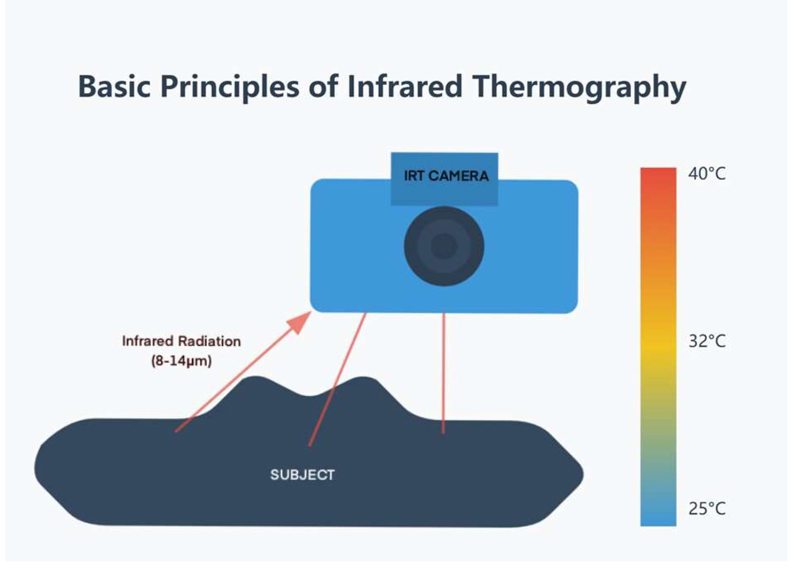
The core components of an IRT system include:
- (1)
- Infrared detector: Usually a focal plane array (FPA) of microbolometers or quantum detectors.
- (2)
- Optical system: Lenses that are cognizant of the infrared radiation onto the detector.
- (3)
- Signal processing unit: Converts detector signals into temperature values.
- (4)
- Display: Presents the thermal image, frequently using a color palette to symbolize temperature variations.
Recent advancements in IRT have led to the development of portable, high-resolution cameras with real-time imaging capabilities. Some systems now incorporate smartphone compatibility, making them more accessible for field use in veterinary practice [5].
It is important to note that IRT measures surface temperature, which can be influenced by various factors, including ambient temperature, airflow, fur or feather cover, and the animal's physiological state. Therefore, proper standardization of imaging conditions and interpretation of results is crucial for accurate diagnoses [6].
2.2. Applications in Various Species
2.2.1. Cattle:
- (1)
- Detection of mastitis: IRT can identify temperature changes in udders associated with clinical and subclinical mastitis, potentially allowing for earlier intervention [7].
- (2)
- Lameness diagnosis: In regard to the issues of hooves and legs, thermal imaging helps find different temperature signs that indicate lameness and foot diseases at an initial stage [8].
2.2.2. Horses:
- (1)
- Musculoskeletal injuries: IRT can aid in revealing inflammation or injury in tendons, ligaments, and muscles before the clinical symptoms manifest [9].
- (2)
- Saddle-fitting assessment: Pressure points and thermal images typical for improper saddle, can be identified with the help of thermal imaging [10].
2.2.3. Small Animals (Dogs and Cats):
2.2.4. Wildlife and Zoo Animals:
- (1)
- Non-invasive physiological monitoring: Companies permit monitoring of the temperature and physiological state of free-ranging or confined wildlife without even capturing or restraining them [4].
- (2)
- Stress assessment: Stress responses have also been assessed using thermal imaging where eye temperature variation has been taken in different animals [12].
2.3. Advantages and Limitations
Advantages:
- (1)
- Non-invasive and non-contact: This relieves pressure and potential harm to the animal, but also to the handler as well [2].
- (2)
- Real-time imaging: IRT offers a tangible means by which the surface temperature distribution can be observed almost instantly [3].
- (3)
- Broad applicability: IRT can be applied to different animal types and for various objectives [13].
- (4)
- Early detection: IRT can establish relationships with fundamental indicators of health with high predictive capability of subclinical states before symptom manifestations [14].
Limitations:
- (1)
- Environmental sensitivity: Expresses a remarkable sensitivity to room temperature, humidity, and airflow conditions [3].
- (2)
- Surface temperature only: This may in fact not read out the internal body temperature or deep tissue conditions perfectly [2].
- (3)
- Fur/feather interference: Heavy pelts of fur or feathers can create a problem for the correct measurement of temperature [4].
- (4)
- Standardization issues: Little consistency between species and conditions [2].
- (5)
- Cost and expertise: Infrared cameras are not cheap while interpreting the images calls for professional experts [13].
2.4. Future Directions
Future research in veterinary infrared thermography will likely focus on several key areas:
- (1)
- Lack of standardization across different species and conditions proposed for these assays [2].
- (2)
- Accomplishments in image processing techniques and artificial intelligence for solving pattern recognition and diagnostic status [5]
- (3)
- Development of more portable devices for field and remote settings.
- (4)
- Interoperability for real-time interpretation with telemedicine platforms.
- (5)
- Standard thermal patterns for species of specific interest and diagnostic criteria for differential diagnoses. [6]
- (6)
- Better description of classes through the development of more precise quantitative methods in order to make the process more objective and sensitive.
- (7)
- Research design participant observation in order to evaluate the utility of IRTs in tracking disease states and responses.
As all these aspects advance, IRT is set to have a wider applicability of being a non-invasive real-time determinant of the physiological and pathological status of all aspects of the animal body. Yet, to capture this potential, further research activities and integrative approaches along with rigorous verification of new approaches and applications are inevitable.
3. Remote Photoplethysmography (RPG)
3.1. Technology Overview and Principles
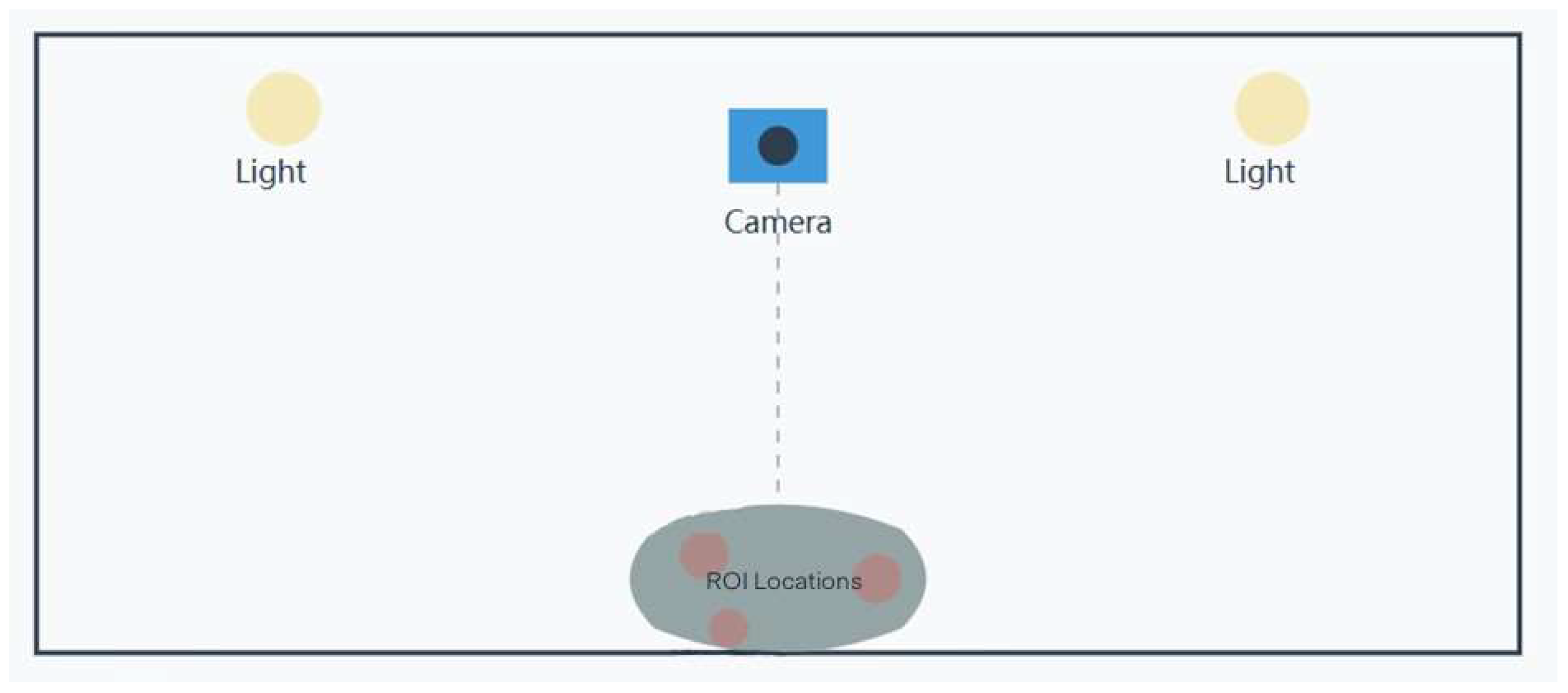
Remote Photoplethysmography (RPG) is a contact-less optical-based technique to measure physiological parameters most often the heart rate and respiration rate based on the color variation of the skin or any visible body part. This is founded on the fact that blood absorption of light is sensitive to the cardiac cycle, thereby triggering slight changes in skin shade, which is revealed by advanced imaging/SPI [15].
The RPG process typically involves the following steps:
- (1)
- Video Acquisition: The subject is recorded using a digital camera or smartphone that has a view of the skin or bodies with fur [16].
- (2)
- Region of Interest (ROI) Selection: Some regions that potentially experience pulsatile variations are also detected and monitored on the subject’s body by algorithms.
- (3)
- Color Channel Separation: It is typically divided into red green blue where green is largely sensitive to the volume change of blood [17].
- (4)
- Signal Extraction: To acquire the rPPG signal, temporal differences of pixel values inside the selected ROI are employed [18].
- (5)
- Signal Processing: Signal processing, noise suppression, spectrum analysis, and other methods, as well as machine learning methods are used to filter out the desired physiological signals from interference [19].
- (6)
- Parameter Estimation: The processed signal affords approximate estimates of various physiological parameters including the heart and respiratory rates of the human body [20].
Figure 2.
rPPG Video Acquisition Setup.
Future development of rPPG has shifted towards enhancing its resilience to movement interference and changing illumination. These conditions are especially difficult in veterinary uses because animals shift around and have different coat kinds and colors. Recent work on machine learning and deep learning techniques has established great potential for improving the accuracy and robustness of rPPG signal measurements in realistic environments [21].
3.2. Application in Various Species
3.2.1. Dogs:
The authors have shown that rPPR can apply both heart and respiratory rates from video recordings even with furred occlusion and interferences that involve movement.
3.2.2. Cats:
Far from being explored as much as dogs, some results of its application to cats seem promising. Another pilot study showed that the correlation coefficient of rPPG-based heart rate with reference was 0.92 with a reference device in the cats during their veterinary examinations.
3.2.3. Horses:
Previous work on rPPG has demonstrated its ability to track a number of physiological parameters during different exercises in equine medicine. When using rPPG for HR monitoring in exercising horses, the mean absolute error was 4.3 bpm compared to a chest strap heart rate monitor.
3.2.4. Cattle:
RPPG has been studied in previous work for stress identification and for healthcare in cattle. The study identified changes in heart rate variability using rPPG with 85% accuracy compared to ECG, which can influence the level of animal welfare without touch [22].
3.2.5. Wildlife:
There exist increasing research engagements in the use of rPPG to monitor wildlife. Early research has indicated that the health and stress of wild animals in captivity may be evaluated without the need to capture or restrain the animals.
3.3. Challenges and Potential Solutions
- (1)
- Motion Artifacts: One of the main problems in the rPPG study is handling motion artifacts, particularly in unencumbered animals. To overcome this problem, novel motion tracking and compensation methods are being worked on.
- (2)
- Fur Occlusion: Fur in most dogs or cats is a strong issue affecting rPPG in most veterinary patients. Attempts are being made to look for other measurement locations and to devise new algorithms that can handle situations when minimal skin is being exposed to an MRI [1].
- (3)
- Species Variability: The multivaried animal species and distinct physiological attributes and skin/fur morphology also act as barriers to the ideal rPPG solutions. Optimal algorithms and calibration techniques for each species are in the course of being established.
- (4)
- Environmental Factors: The fluctuations in light intensity and the background color within veterinary environments may interfere with rPPG signals. Scientists are trying to compute perfect algorithms that take into account lighting conditions [23].
- (5)
- Real-time Processing: Real-time processing of rPPG data for immediate clinical application is technically feasible, but challenging for the development of robust systems. The empowering edge computing and more efficient algorithms are being researched.
3.4. Discussion
The future of rPPG in veterinary medicine presents several key areas for development:
- (1)
- Integration with other sensing modalities to provide more comprehensive health monitoring.
- (2)
- Development of species-specific algorithms and reference ranges to account for the unique physiological characteristics of different animals.
- (3)
- Incorporation into telemedicine platforms to enable more comprehensive remote health assessments [24].
- (4)
- Advanced signal processing and AI techniques to enhance signal quality and interpretation in veterinary rPPG [21].
- (5)
- Exploration of miniaturized, wearable rPPG devices for continuously monitoring unrestrained animals.
- (6)
- Application in non-invasive stress and welfare assessment.
In a later stage of the development of the technology large-scale validation trials in a clinical veterinary context are going to be required to prove the reliability of the technology across different animal species, within different ages and health states. The idea is to bring together a research consortium that will pursue further development of rPPG for veterinary applications by expanding clinical work and collaborating with interdisciplinary specialists in both academia and industry that will validate novel approaches and applications of rPPG.
4. Radar-Based Sensing
4.1. Principles of Operation
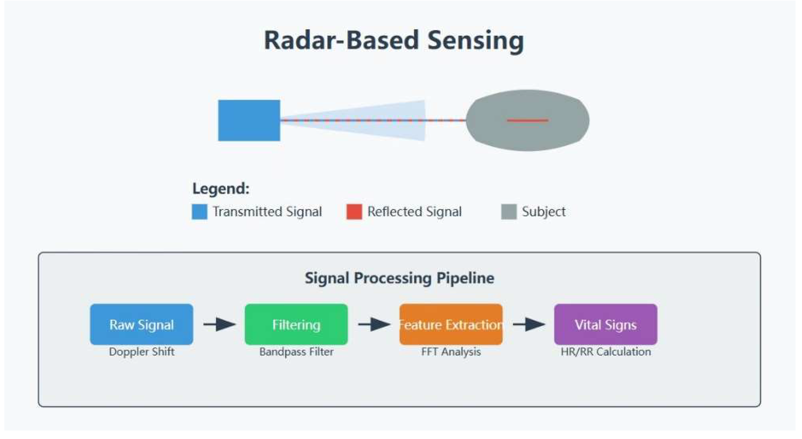
Biophysical monitoring in veterinary medicine involves the use of electromagnetic waves to treat objects in motion, a principle that adapts well to monitoring common signs such as respiratory and cardiac rhythms. This technology is based on the Doppler effect whereby the frequency of the reflected wave varies depending on the motion of the target [25].
The basic principle implies their radio emission at a certain frequency in the direction of the animal. These waves are reflected off the animal’s body, especially the chest wall, and thus are expected to produce an echo. In view of this, variations in breathing and heartbeat lead to small variations in the frequency of the reflected waves by the chest wall. By analyzing these frequency shifts, the system can get detail information on the respiratory rate and cardiac rate of the animal [26].
Several types of radar systems have been explored for vital sign monitoring:
- (1)
- Continuous-Wave (CW) Doppler Radar: This type always transmits a single-frequency signal and determines changes in the frequency of the returned signal. It is non-invasive, time-effective, and affordable although it is vulnerable to patient movement [27].
- (2)
- Frequency-modulated continuous-wave (FMCW) Radar: This type charges and discharges the transmitted signal at different rates, providing finer distance estimation and possibly, enhanced vital sign identification [28].
- (3)
- Ultra-Wideband (UWB) Radar: This type employs extremely brief pulses over a broad spectrum and could provide better penetration through fur and tissue than the previous kind while having a high temporal resolution [29].
Figure 3.
Principles of Radar-Based Sensing.
It is also evident that the radar operating frequency determines penetration depth and radar range. Higher frequencies such as the frequencies of 24 and 60 GHz have enhanced motion sensing but reduced throughput. For instance, although higher frequencies (for example, 60 Hz) are limited in penetration depths (observing only several millimeter thicknesses of the human skin), it is very sensitive to small motions and activities. In contrast, lower frequency (for instance 2.4 GHz) is deeper penetrating [25].
4.2. Applications in Various Species
4.2.1. Horses:
There is a successful attempt using a 24 GHz continuous-wave radar system to monitor the vital signs in horses where respiratory rate assessed while in different postures, standing and lying, was 96.7% compared to the reference method. Nevertheless, it was even more difficult and delicate to detect the heart rate because the range of chest wall movements in connection with respiration conceals the somewhat weaker cardiac movements.
4.2.2. Cattle:
Studies on the application of radar technology in monitoring the respiration rate of cattle as proposed in this paper were found to have achieved an accuracy of 98.5% as compared to visual observation of respiration. This technology can be used for early detection of diseases in large herds [30]. Another research conducted to analyze using ultra-wideband radar for heart rate monitoring without physical contact with the livestock received 95% accuracy compared to electrocardiogram rates [31].
4.2.3. Pigs:
A system applying millimeter-wave radar for respiratory rate measurement in pigs achieving 97.8% accuracy at the comparison of the respiratory rate and the pattern of the respiratory cycle. This technology may be beneficial in disease diagnosis and detection of the health status of the stock in swine production.
4.2.4. Dogs:
Selected case: Recent studies determined that respiratory rate can be assessed in dogs by ultra-wideband radar with fur penetration with a mean absolute error of 1.8 breath/minute compared with the reference technique. GSR was easier to detect while heart rate detection proved a complex task, yet certain developments in signal processing proved ways.
4.2.5. Poultry:
Although not radar, similar to sensing by detecting radio frequencies, has been used before in poultry. RFID technology in tracking bird behaviors and movements may also be applied to vital signs monitoring using radar methodologies [32].
4.3. Advantages and Challenges
Advantages:
- (1)
- Non-contact and Non-invasive: This method makes it possible to monitor the vitals without any contact with the animal.
- (2)
- Penetration through Fur and Light Barriers: It can also pass through fur and lighter colors, which makes it ideal for use on animals of different kinds [25].
- (3)
- Continuous Monitoring: This method also offers an opportunity to monitor continuously rather than restraining animals [30].
- (4)
- Multiparameter Measurement: It potentially monitor a number of biophysical variables at once [33].
- (5)
- Operation in Various Lighting Conditions: This kind of therapy is useful where the blood flow is poor, during nighttime, or in the absence of light [34].
Challenges:
- (1)
- Motion Artifacts: Movement is often unhandy when the recording is made amid any tiny physiological movements due to its sensitivity to motion artifacts.
- (2)
- Signal Separation: It is still difficult to extract the cardiac signals specifically from respiratory signals and other movements of the body [34].
- (3)
- Species-Specific Adaptations: Requires the design of species-specific computations and even, in some cases, specific modifications to molecular hardware.
- (4)
- Limited Research on Long-term Effects: More safety investigations are required on possible chronic impacts of constant radar illumination on the creatures [25].
- (5)
- Environmental Interference: Interference with signals from other devices or the reflection of surfaces in its vicinity [33].
- (6)
- Cost and Complexity: Modern capable radars are sometimes costly, and may require high-level management [35].
4.4. Discussion
The future of radar-based sensing in veterinary medicine presents numerous opportunities for advancement:
- (1)
- (2)
- Multi-sensor fusion to enhance accuracy by combining radar with other sensing modalities.
- (3)
- Miniaturization and portability of radar systems for widespread adoption in veterinary practice [25].
- (4)
- Development of species-specific applications to account for anatomical and physiological variations across different animals.
- (5)
- Extension of long-term monitoring potential to various veterinary applications [30].
- (6)
- Integration with telemedicine platforms to enhance remote health assessment capabilities [35].
- (7)
- Exploration of novel applications beyond vital sign monitoring.
- (8)
- Integration of machine learning and AI to significantly enhance radar system capabilities in veterinary settings.
Most importantly realizing the best of radar-based sensing for veterinary medicine will require more research, interdisciplinarity, and validation across species and contexts. They can potentially transform veterinary diagnosis and health assessment of patients through noninvasive, continuous, and remote monitoring for various applications.
5. Wearable Sensors
5.1. Types of Wearable Sensors for Veterinary Use
Wearable sensors in veterinary medicine should be defined as various types of devices to assess different types of physiological characteristics and behaviors of animals. The main types include:
- (1)
- Accelerometers and Inertial Measurement Units (IMUs): These sensors detect movement and position to give details of the activity, walking style, or even standing post. For example, using collar-mounted accelerometers, in a study, with dogs, the classification accuracy of 91.3% of different activities including, walking, trotting, and galloping was achieved [36].
- (2)
- Heart Rate Monitors: These devices generally employ ECG, or PPG that estimates heart rate and heart rate variability. An ECG worn by dogs produced a 0.99 coefficient when compared with conventional ECG readings [37]. Various technologies have been implemented for heart rate monitoring of livestock comprising chest belt sensors and implantable sensors [38].
- (3)
- Temperature Sensors: Prolonged and consistently high temperatures mirror various diseases early enough, therefore constant temperature checks are essential. Peculiarly, one combining the temperature sensors into a collar-worn device for cattle reached an identification accuracy of ±0.1°C higher than the rectal temperature measurement as a standard [39].
- (4)
- GPS Trackers: Primarily used in tracking locations, GPS devices use other sensors in the monitoring process too. GPS collars were used in dogs and were identified to have an average positional error of less than 5 meters when tracking movement patterns [40].
- (5)
- Respiratory Rate Monitors: These monitors frequently employ strain gauges or impedance pneumography in monitoring breathing patterns. A respiratory rate monitoring wearable device in horses had ± 2 breaths per minute compared to visual counts of respiratory rates leading to the conclusion that the wearable device could be used as the standard for respiratory rate counts.
- (6)
- Blood Oxygen Saturation (SpO2) Sensors: As such although rare pulse oximetry sensors are being modified for veterinary applications. One research comparing reflectance pulse oximetry for constant SpO2 tracking in dogs achieved an accuracy range of ±2% against ABG analysis.
- (7)
- Pressure Sensors: These are particularly helpful in equine medicine for determining the location of a saddle and what it will feel like to the horse. An experimental comparing pressure-sensing pads to assess the fit of saddles in equines reported accuracy of 95% of pads compared to an expert saddle fitter in identifying the areas of concentrated pressure.
- (8)
- Environmental Sensors: These sensors are, more often than not, part of an interconnected system of other wearable devices used to track the planet’s surroundings. It has been applied in live-stock management for detecting a shift in relative humidity and ammonia level with a study showing its 90 percent efficiency in recognizing environmentally damaging conditions [41].
- (9)
- Biochemical Sensors: One subset of wearable that is starting to receive focus is sensors that bear the responsibility of detecting biochemical indicators in body fluids. For instance, a sweat-cooled wearable sensor for horses was able to determine the electrolyte concentration during the exercise with a difference of ±5% from the actual laboratory results [42].
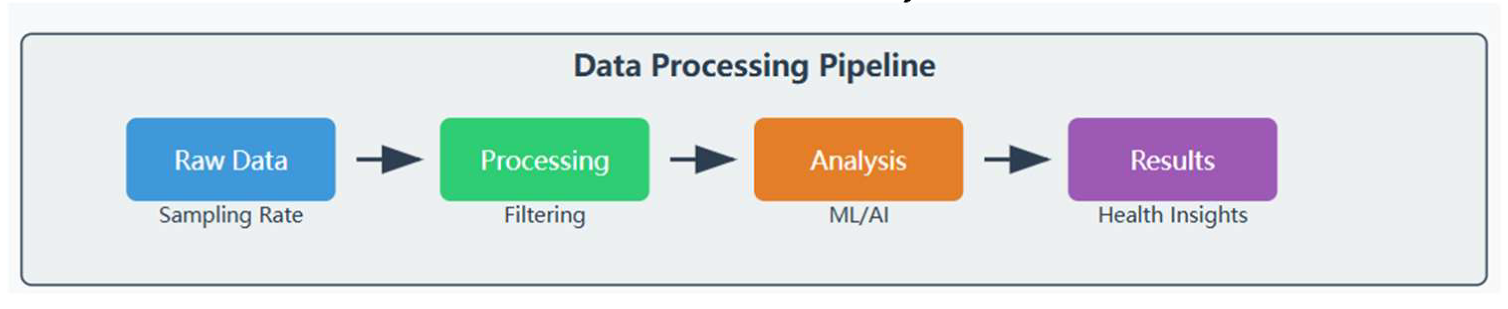
Figure 4.
Data processing pipeline.
5.2. Applications in Various Species
5.2.1. Companion Animals:
Activity, sleep, and biometrics data are collected by interactive and unobtrusive wearable devices. Research assessed a canine activity monitor used for a commercial activity and provided evidence of its applicability for the objective assessment of mobility [36]. Another group created a wireless ECG patch for canines, which yielded a strong positive agreement with conventional ECG readings though the modality was in dogs [37].
Accelerometers of wearable devices have also been applied to investigate activity patterns and energy costs in domestic cats. It has been conducted on indoor cats the physical activity of which parameters were measured by collar-mounted accelerometers to understand their daily behavior patterns.
5.2.2. Livestock:
Wearable sensors have countless applications in dairy and beef cattle, with garment-like devices being the most popular among farmers. Neck-mounted accelerometers were used in the studies to monitor feeding and rumination behaviors and the guild accurately [39].
5.2.3. Equine:
Horses have been the most important subject of wearable sensors used in performance monitoring and health control. Another study showed how IMU could be used for quantitative gait analysis in horses and interpret lameness and performance.
5.2.4. Wildlife:
Wearable sensors have greatly enhanced tracking and studies of wildlife migration and the use of its habitats, for instance, the use of GPS tags to monitor bird migration [43], or physiological characteristics such as depth and acceleration that use time-depth recorders and accelerometers on marine mammals [44].
5.3. Advantages and Challenges
Advantages:
- (1)
- Continuous, Long-term Monitoring: Portable biosensors measure physiological data and activity 24/7 for weeks at a time. This is more informative than mere clinical assessments that are carried out once in a while. Worth noting, that the constant data gathering in these cases could help in the diagnosis of early health complications and enhance the welfare of animals [41].
- (2)
- Non-invasive and Well-tolerated: Noninvasive wearable sensors are used in most animal applications and the use of such sensors is normally scratched off veterinary care problems depending on the level of invasiveness. Specifically, this paper reviewed the case of high compliance with the activity monitor in dogs without significantly altering their natural behaviors [36].
- (3)
- Real-time Data Access: A majority of wearable sensors provide real-time streaming capabilities so that veterinarians and owners can have updated information about the health status of their animals [37]. This may be of special use in emergency situations and in animals that have ongoing health issues.
- (4)
- Objective Measurement: P wearable sensors give factual, measurable information on physiological aspects and activities. This can help in addition to, or maybe even replace subjective judgments in clinical.
- (5)
- Remote Monitoring: Wearable sensors allow for non-stop tracking of animals mostly livestock and wild animals that require time-to-time checkups. This aspect has obtained additional value under conditions of telemedicine solutions [35].
Challenges:
- (1)
- Device Comfort and Interference: Arguably, the most substantial gap relates to guaranteeing that wearables do not cause inconvenience, including by failing to compromise an animal’s usual activities. Body-sized sensors require the unique features of the respective species that will not feel uncomfortable wearing sensors or changing their behavior patterns [45].
- (2)
- Battery Life and Data Transmission: Restricted battery life and data transfer make it challenging to apply wearable sensors for extended periods especially where the animal belongs to the wilds. There are discussions on the current issues and future possibilities of bio-logging devices for monitoring wildlife [46].
- (3)
- Data Interpretation and Management: Data may be collected constantly from worn sensors, and large amounts of information may be produced amenable to only complex techniques of analysis. Researchers have stressed that more sophisticated paradigms and tools are required to analyze these data [39].
- (4)
- Accuracy and Reliability: Promulgating the reliability of these wearable sensors whereby absolute measurement results can be obtained across various animal species, sizes, and conditions still presents some difficulty. Deviation in motion, influence from the surrounding environment, and the variability of an individual subject can also pose a challenge to the sensors. The type of studies like canine ECG monitor validation are very important in determining the accuracy of such devices
- (5)
- Standardization: There are also no identical ways of approaching data aggregation, processing, and evaluating across the different WDGs and animal species; these are some obstacles to popularizing and comparing outcomes [36].
- (6)
- Cost and Accessibility: Exemplar wearable sensors may be expensive especially when considering applying them to large populations of livestock or wildlife. There is debate on whether low-cost technologies are feasible at present in order to enhance the extensive use of wearable technology in veterinary practice [41].
- (7)
- Ethical Considerations: Wearable sensors especially those used for wildlife come with some ethical issues as regards any effects the sensors will have on the behavior of the animals and or their welfare. There are continuing debates over the most appropriate ethical concerns and guidelines on the use of bio-logging in animals [47].
- (8)
- Durability and Environmental Resistance: Wearable sensors when applied in veterinary must withstand different wear and tear such as water, dust, and extreme temperatures. This is especially difficult for gadgets applied in livestock or wildlife tracking [46].
5.4. Discussions
The future of wearable sensors in veterinary medicine presents numerous opportunities for advancement:
- (1)
- (2)
- Enhanced battery life and power management, including energy harvesting technologies.
- (3)
- Advanced data analytics employing sophisticated machine learning and AI algorithms for early disease detection and personalized health monitoring [39].
- (4)
- Integration with other technologies to provide more comprehensive health assessments.
- (5)
- (6)
- Standardization and validation efforts are needed to ensure widespread adoption and comparability [36].
- (7)
- Development of real-time health alert systems and edge computing capabilities.
- (8)
- Creating biodegradable sensors for wildlife applications.
Reaching the greatest potential of wearable sensors for veterinary can also involve further composite work of both academics and industrial sectors, more investment in the research area, and discussion about integrating animal and environmental concerns into veterinary medicine.
6. Computer Vision and Machine Learning in Remote Vital Sensing
6.1. Role of AI in Improving Sensing Accuracy
In the understanding of AI for veterinary care, the elements pertinent in remote vital sensing include computer vision and machine learning. These technologies assist in handling and analyzing the high data from different sensing modalities, eradicating most hindrances of the traditional sensing approaches.
Figure 5.
AI processing pipeline.
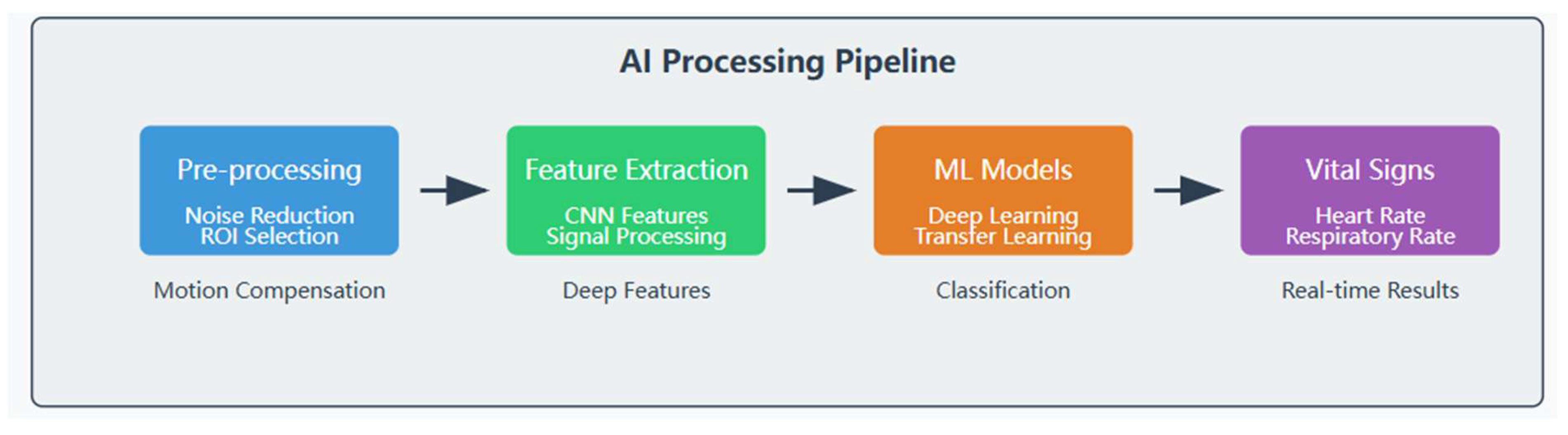
6.1.1. Signal Processing and Noise Reduction:
Deep learning models particularly, have demonstrated their capability of handling raw sensor data and filtering noise. For instance, the accuracy of estimating the heart rates from the video has been shown to be enhanced by using the convolutional attention network and these techniques filter motion artefacts and noise in the environment [21]. Presumably, such approaches may also be useful for analyzing veterinary remote sensing to enhance the signal-to-noise ratio in various species and settings.
6.1.2. Feature Extraction and Pattern Recognition:
In human language processing, machine learning algorithms are valuable since they are capable of locating small and hard-to-discover features in vast data that a human analyst is unlikely to notice or qualitative analysis to discover or capture. In-depth analyses were conducted by deploying deep learning to identify meaningful characteristics on video sequences that could be employed forп predicting cattle respiratory rates Consequently, AI proved valuable for automated vital sign assessment.
6.1.3. Multi-Modal Data Fusion:
It is shown that utilizing data from multiple sensing modalities is possible using AI techniques and enables a more informative and accurate evaluation of animal conditions. Scholars have proposed that through the processing of data from multiple sensors such as infrared, accelerometer, video, etc., it is feasible to boost the precision of clinical observation of the health conditions of cattle [50].
6.1.4. Adaptive Learning and Personalization:
Key health indicators can be calculated based on baseline parameters of the respective individual animal so machine learning models can be applied and adjusted for such. An automated health-monitoring system has been designed for calves using deep learning with an ability to learn animal characteristics therefore enhancing anomaly detection.
6.1.5. Real-Time Analysis and Decision Support:
Consequently, an AI item can analyze data in real-time and offer decisions instantly. The benefit of edge AI in such production systems must be realized; they could help in early disease identification and overall health surveillance of the animals [51].
6.1.6. Handling Large-Scale Data:
In conditions where there are many cows, or data has to be gathered over a long period of time, AI performs the analysis quickly. Investigations have shown the ability of machine learning to predict coccidiosis in intensive livestock production systems via analyzing serves from huge sensors to identify minor signs of health alteration [52].
6.1.7. Overcoming Species-Specific Challenges:
Due to the aspects mentioned above, AI models can be adjusted in accordance with the difficulties of different sorts of animals. Several works examined transfer learning methods for cross-species image classification that can be relevant in applying the sensing algorithms across different veterinary applications.
6.1.8. Enhancing Image-Based Vital Sign Extraction:
Compartment two discusses how computer vision techniques enhanced by deep learning have enhanced the precision of extracting vital signs from images and videos. Researchers proposed a method for understanding the patterns of dogs’ physical activity based on automatically analyzing videos which can be extended to the task of vital sign monitoring.
6.1.9. Improving Wearable Sensor Accuracy:
AI can improve the quality of its wearable sensors due to movement artifacts and external conditions. A paper intensively applied ID on increasing sheep’s wearable precise sensor to classify technologies using machine learning algorithms, showed that AI could maintain wearable sensor analysis efficiently.
6.2. Applications in Various Species
6.2.1. Cattle:
- (1)
- Respiratory Rate Monitoring: Facial landmarks are detected and aligned to track the movement of certain key points in the face, and a deep learning model is trained to estimate respiratory rates in cows taken from video sequences. In terms of performance, their system proved to be invariant to changes in lighting and dynamic environments where its animals move around, detecting breath accurately.
- (2)
- Lameness Detection: Observing cow gait extracted from video using computer vision techniques, a lameness early detection system was established. Their method managed to give over 90% diagnosing accuracy of lame cows [53].
6.2.2. Horses:
- (1)
- Gait Analysis: IMU data is used where deep learning methods are employed in the automatic identification of lameness in horses. Its method produced preliminary signs of success in detecting less obvious gait issues.
- (2)
- Behavior Classification: Research utilized machine learning algorithms to identify horse movement from accelerometer information and antecedently demonstrated high predictive accuracy in differentiating grazing, strolling, and lying down time.
6.2.3. Companion Animals:
- (1)
- Activity Classification: A machine learning approach is used to analyze accelerometer data to classify dog activities, and the model proposes remarkable accuracy rates of activities including walking, running, or even resting.
- (2)
- Heart Rate Estimation: The studied computer vision methods predicted the main heart rates of dogs and cats from videos that pointed to further opportunities for contactless assessment of vital signs at veterinary practices.
6.2.4. Pigs:
Using deep learning techniques, pig vocal communications were classified in terms of respiratory diseases with parameters gathered from coughing.
6.2.5. Poultry:
An assessment of broiler chicken gait is created utilizing computer vision to increase the availability of automated welfare assessment for chickens [54].
6.2.6. Wildlife:
Deep learning is also applied for the identification of species from camera trap images implying a lowered work in surveys and wildlife studies [55].
6.3. Advantages and Challenges
6.3.1. Advantages:
Processing Large Volumes of Data:
AI and machine learning algorithms are also suited for processing large data from remote sensing devices in a short time. This capability is valuable in scenarios where monitoring is to be done continuously. For instance, there was evidence showing that deep learning models can process video data in real-time for respiratory rate estimation in cattle.
Detecting Subtle Patterns:
This feature is especially important because machine learning models can often recognize peculiar features or patterns in data that will not be noticeable to a human being. This sensitivity may result in early diagnosis of disease in the body. A team demonstrated this benefit in their study where machine learning models were used to identify signs of coccidiosis in livestock before their symptoms were visible [52].
Automated and Continuous Monitoring:
Automated solutions can always be on duty unlike human beings who get tired and hence can constantly monitor animal health [51].
Integrating Multiple Data Sources:
This capability of machine learning means that data from all those sensors and modalities can be integrated to produce a larger and more rounded picture of health. A team did this, particularly in the video-based physiological measurement work that incorporated spatial and temporal data [21].
Adaptability to Different Species:
Transfer learning methods The AI models trained in one species can be used on others Methods of AI can be time-saving in developing species-specific monitoring methods. It was investigated in papers focused on cross-modal image classification in agriculture.
6.3.2. Challenges:
Data Requirements:
The solution of many complex AI problems often entails the usage of substantial data sets or rich feature set training data. Such datasets can be difficult to acquire especially in veterinary use cases for rare species or diseases.
Species-Specific Algorithms:
The high number of animal species within the sphere of veterinary medicine requires the creation of species-dependent calculation and modeling algorithms. This is because the versatility of the kind of patients veterinary deals with can make it hard to apply the developed AI solutions from one context to another.
Interpretability and Explainability:
Deep learning models, in fact, are opaque, meaning that one cannot easily understand how such a model is going to make a decision at the end of the process. Such a lack of explainability can be a major limitation for clinical application, however. This concern was also mentioned in medical AI where the need for XAI is crucial in a healthcare environment [56].
Validation in Clinical Settings:
The reliability of such systems and algorithms in clinical practice is critical, but the process can be a lengthy and costly endeavor. The same authors pointed out that in their studies of methods for assessing animal welfare, attention was paid to the need for new technologies’ pilot data [57].
Ethical Considerations:
AI integration in animal monitoring impacts data sharing, the rights of animals, and the risk of employing the all-inclusive technological solution [50].
Technical Infrastructure:
AI-based remote sensing systems may need a large quantity of technical support such as high bandwidth networks and high computing power. This can be difficult to achieve in areas with few resources in veterinary medicine [51].
Handling Environmental Variability:
AI models must be also immune to various light conditions, which are endured in veterinary facilities, from dimly lighted offices to outdoor fluctuating light conditions. One team tackled this issue in their effort toward video-based respiratory rate estimation for cattle across all levels of lighting.
6.4. Discussions
The future of computer vision and machine learning in remote vital sensing for veterinary medicine presents numerous exciting opportunities:
- (1)
- Multi-modal sensing integration for more comprehensive health assessments [50].
- (2)
- (3)
- Transfer learning and few-shot learning techniques address the challenge of limited datasets for various animal species.
- (4)
- Edge AI for remote monitoring, enabling real-time analysis in resource-limited settings [51].
- (5)
- Personalized health monitoring for more precise detection of health deviations.
- (6)
- Automated stress and pain assessment to revolutionize animal welfare management [57].
- (7)
- Advanced computer vision techniques for vital sign monitoring extend beyond current applications.
- (8)
- Federated learning approaches facilitate collaborative research while maintaining data privacy [59].
- (9)
- AI for automated diagnostic support to aid veterinarians in clinical decision-making [60].
Such optimal circumstances will call for interdisciplinary research cooperation, empirical validation of the developed tools, as well as concern for ethical issues. However they have the potential to greatly improve animal health surveillance and wellbeing in all sorts of veterinary contexts.
7. Challenges in Remote Vital Sensing for Veterinary Medicine
Despite the promising advancements in remote vital sensing for veterinary medicine, several significant challenges remain. These challenges must be addressed to ensure these technologies' widespread adoption and efficacy in clinical practice.
7.1. Species Diversity
Indeed, one of the main issues associated with veterinary remote sensing is the large number of animals that differ in terms of their morphology and physiology. In contrast to human medicine, in which technologies can be often tailored for a specific species, veterinary applications need to consider several factors like body size, shape, fur or feather insulation, and physiological parameters differing between animals. Such a variety of surface textures requires either the creation of versatile censoring technologies or the individual approaches to every species; the latter can often prove to be very complicated technically when implemented in reality and more often costly.
7.2. Environmental Factors
Most remote sensing technologies are affected by environmental factors, whereas veterinary environments are quite different. Elements like temperature, relative humidity, and light conditions, as well as background motions, can cause a major difference in the measurements. For instance, while using infrared thermography environment temperature has been known to injure the results, optical sensing for example rPPG is sensitive to light. While the analytical solution to the problem does not present much difficulty, the challenge of creating sound algorithms that behave nicely and are able to predict even these small changes in the ecology of a system represents a more difficult endeavor.
7.3. Motion Artifacts and Animal Behavior
While treating patients, the doctors need to be sure which body part they are dealing with, while humans can easily be ordered to sit or lie still, this is not the same as dealing with animals which may be in constant motion or simply behave in rather unanticipated ways. This movement can inject substantial artifacts in the measurements acquired from remote sensing instruments [37]. In spite of recent advances in motion artifact algorithms, suboptimal sensor measurements in unrestrained animals of different species and activity levels remain a significant issue.
7.4. Validation and Standardization Issues
One of the major issues arising from veterinary remote sensing is that there is no universally acceptable method of data acquisition; processing and analysing data from a distance. Such calibrations of these technologies against gold standard measures are imperative but challenging because many reference techniques are intrusive [2]. Moreover, the development of normal range and clinical threshold of remotely sensed species-specific parameters is still a crude practice and the interpretation of data is often a complex task in operational clinical applications.
7.5. Technical Limitations
Most current remote sensing technologies have profound technical issues that hamper them from being widely applied. These include 1) limited range and/or depth of penetration, especially for systems based on radar technology; 2) vulnerability to interfering signals; and, 3) requirement for sophisticated hardware and/or knowledge in order to implement and decode resultant data. Acquiring efficiencies to overcome these technical obstacles while achieving price and ease of use is a tremendous issue for the researchers and developers in this field.
7.6. Data Management and Integration
While remote sensing is steadily entering many fields, the issues of big data management and integration are increasingly pressing. Thus, creating the means for the data’s processing, storage, and analysis, as well as its combination with the existing veterinary information systems, is critically important for the real-life application of these technologies [1].
To overcome these challenges, interdisciplinary cooperation between veterinarians, engineers, computer scientists, and animal behaviorists will have to be continued. Overcoming these challenges, remote vital sensing technologies will be able to revolutionise veterinary medicine and enhance capabilities in animal care and assessment.
8. Future Directions
As RVSI in veterinary medicine still remains under further development, several lines from further research and development appear to be promising. These directions are intended to respond to present needs and contribute to the more advanced use of remote sensing in veterinary applications.
Speaking of the future of remote vital sensing in veterinary medicine, the following improvements can be expected that will dramatically change animal care. There is still much work to be done to reach this potential, and much of it will require the whole of multidisciplinary cooperative research together with veterinary applications thought-provoking efforts.
8.1. Multimodal Sensing Approaches
Future work will probably aim at addressing issues of how to combine several types of sensors to enhance performance and precision. Thus, it is possible to integrate various technologies, including infrared thermography, rPPG, and radar-based sensing to eliminate the drawbacks of the separate method and cover more extensive health monitoring. For example, combining thermal and visual imaging data could provide a better definition for sensing inflammation while correcting for external conditions.
8.2. Advanced Signal Processing and Machine Learning
Complex signal processing and reinforcement will require learning algorithms in order to improve the effectiveness of remote sensing systems. Specifically, deep learning models appear to be able to dampen noise, remove motion artifacts, and extracting the physiological signal(s) from the noisy data. Future research activities will probably be directed toward designing better and more flexible algorithms for dealing with the variety of veterinary patients and the various conditions under which they exist.
8.3. Species-Specific Solutions
Due to high species diversity, the subsequent future work might focus on the identification of the species-orientated RS techniques and models. This could imply adapting sensor designs, algorithms as well as reference ranges to the physiology of the various animal species [2]. Despite such asymptomatic solutions may be time-consuming and potentially costly, they could reduce errors and enhance the practicality of remote vital sensing in veterinary medicine.
8.4. Miniaturization and Wearable Technologies
The growth of microelectronics technology and material science are expected to prompt enhanced wearable sensors for animalCareful that are smaller, more comfortable, and longer-lasting. Subsequent wearable gadgets may intersect multiple noninvasive sensing strategies so as to substantially track physiological status, other physique characteristics, and indicator measurements during unlabeled living creatures [37].
8.5. Integration with Telemedicine and AI-Driven Diagnostics
There are some expectations as to the future development of the remote vital sensing in veterinary medicine connected with the experience in the development of telemedicine. Specifically, research will apply to the creation of automatic and efficient systems for acquiring, transmitting, and interpreting remote sensing data, and converting the data into timely real-time health information and disease detection. They could be integrated with AI-based diagnostics which might result further in the effective clinical use of these technologies and lead to automated health evaluation as well as tailor-made approaches to treatment [1].
8.6. Standardization and Validation Efforts
Future studies will require further determination of reference standards for remote vital sensing between different species in order to overcome the existing problems of data interpretation and clinical relevance. This will include extensive external validation analyses of the remote sensing techniques against gold standard measurements with different species and clinical conditions. The adoption of these technologies will also require much effort in order to demonstrate their reliability and usefulness in clinical practice.
8.7. Environmental Monitoring and One Health Approaches
Possible future uses of remote vital sensing are not only in individual animals but also in other environmental or population health assessments. For instance, thermal imaging might be used to identify looming disease epidemics among animals like cattle or wild animals. This is in line with One Health principle that considers the health of animals, human beings, and the environment as inter-connected [2].
8.8. Ethical Considerations and Animal Welfare
With the growth of these technologies, there will be a great concern in ethical matters concerning the technologies. Subsequent studies should investigate how the spectrum of ehtnosense can improve animals’ well-being; reduce stress during their preliminary medical examination; and, in general, advance the veterinary services.
9. Conclusion
Vital remote sensing for veterinary care is a relatively new area with the potential for substantial growth in the near future. This review has examined infrared thermography, remote photoplethysmography, radar-based sensing, wearable sensors, and computer vision applications that collectively enhance the solutions for non-invasive animal health surveillance.
The benefits of these technologies are as follows. They can provide the opportunity to monitor animal patients constantly and without stress, the identification of health problems in their early stages, and the control of many various ailments [2]. In addition, it also supports surveillance for research goals where more information about animal physiology and how they behave in their natural environment is needed.
However, there are still many obstacles in this way: There are also challenges that include the versatility of veterinary patients, factors in the environment, patient’s movement, and validation as well as standardization. Furthermore, temporal issues such as technical restriction and data management remain the challenges that researchers and developers need to address.
Nevertheless, the future of remote vital sensing in veterinary medicine seems to be bright. It is expected that developments in multi-modal sensing, machine learning methods, and wearable systems will further improve the precision and relevance of these approaches [37]. Connecting with telemedicine and AI to diagnose could help increase the application and development of remote sensing technology in animal health [1].
Providing further, one has to be concerned with the ethical aspects and guarantee that such technologies are used appropriately and benefit animal life. Remote sensing and its capability to reduce stress related to health checks and enhance the quality of patients serves the veterinary profession's mission and vision of prioritizing animals’ well-being.
Therefore, remote vital sensing in veterinary medicine remains a novel area of application but it has transformational potential to enhance animal health, augment research and reshape veterinary practice. The innovation of these technologies for veterinary practices, as well as deficiencies found in their current form, call for future interdisciplinary work from veterinary professionals, engineers, computer scientists, and animal behaviorists.
In the subsequent years, this field will most probably record great development that may transform how society oversees and manages animal health. These individual animal technologies may improve with time and may complement intra-species population health intervention and One Health frameworks of animal-human environmental interface healthiness [2].
References
- Cugmas, B. , Štruc, E., & Spigulis, J. Photoplethysmography in dogs and cats: a selection of alternative measurement sites for a pet monitor. Physiological Measurement 2019, 40, 01NT02. [Google Scholar] [PubMed]
- Rekant, S. I. , Lyons, M. A., Pacheco, J. M., Arzt, J., & Rodriguez, L. L. Veterinary applications of infrared thermography. American journal of veterinary research 2016, 77, 98–107. [Google Scholar] [PubMed]
- Cilulko, J. , Janiszewski, P., Bogdaszewski, M., & Szczygielska, E. Infrared thermal imaging in studies of wild animals. European Journal of Wildlife Research 2013, 59, 17–23. [Google Scholar]
- McCafferty, D. J. The value of infrared thermography for research on mammals: previous applications and future directions. Mammal Review 2007, 37, 207–223. [Google Scholar] [CrossRef]
- Tattersall, G. J. Infrared thermography: A non-invasive window into thermal physiology. Comparative Biochemistry and Physiology Part A: Molecular & Integrative Physiology 2016, 202, 78–98. [Google Scholar]
- Vainionpää, M. , Raekallio, M., Tuhkalainen, E., Hänninen, H., Alhopuro, N., Savolainen, M.,... & Vainio, O. Comparison of three thermal cameras with canine hip area thermographic images. Journal of Veterinary Medical Science 2012, 74, 1539–1544. [Google Scholar]
- Zaninelli, M. , Redaelli, V., Luzi, F., Bronzo, V., Mitchell, M., Dell'Orto, V.,... & Savoini, G. First evaluation of infrared thermography as a tool for the monitoring of udder health status in farms of dairy cows. Sensors 2018, 18, 862. [Google Scholar]
- Alsaaod, M. , Schaefer, A. L., Büscher, W., & Steiner, A. The role of infrared thermography as a non-invasive tool for the detection of lameness in cattle. Sensors 2015, 15, 14513–14525. [Google Scholar]
- Soroko, M. , & Howell, K. Infrared thermography: current applications in equine medicine. Journal of Equine Veterinary Science 2018, 60, 90–96. [Google Scholar]
- Arruda, T. Z. , Brass, K. E., & De La Corte, F. D. Thermographic assessment of saddles used on jumping horses. Journal of Equine Veterinary Science 2011, 31, 625–629. [Google Scholar]
- Pavelski, M. , Silva, D. M., Leite, N. C., Junior, D. A., de Sousa, R. S., Guérios, S. D., & Dornbusch, P. T. Infrared thermography in dogs with mammary tumors and healthy dogs. Journal of Veterinary Internal Medicine 2015, 29, 1578–1583. [Google Scholar] [PubMed]
- Travain, T. , Colombo, E. S., Heinzl, E., Bellucci, D., Prato Previde, E., & Valsecchi, P. Hot dogs: Thermography in the assessment of stress in dogs (Canis familiaris)—A pilot study. Journal of Veterinary Behavior 2015, 10, 17–23. [Google Scholar]
- Luzi, F. , Mitchell, M., Nanni Costa, L., & Redaelli, V. Thermography: current status and advances in livestock animals and in veterinary medicine. Fondazione Iniziative Zooprofilattiche e Zootecniche, Brescia 2013, 1-192.
- Redaelli, V. , Bergero, D., Zucca, E., Ferrucci, F., Costa, L. N., Crosta, L., & Luzi, F. Use of thermography techniques in equines: principles and applications. Journal of Equine Veterinary Science 2014, 34, 345–350. [Google Scholar]
- Sun, Y. , Hu, S., Azorin-Peris, V., Greenwald, S., Chambers, J., & Zhu, Y. Motion-compensated noncontact imaging photoplethysmography to monitor cardiorespiratory status during exercise. Journal of Biomedical Optics 2015, 16, 077010. [Google Scholar]
- van Gastel, M. , Stuijk, S., & de Haan, G. Motion robust remote-PPG in infrared. IEEE Transactions on Biomedical Engineering 2016, 62, 1425–1433. [Google Scholar]
- Verkruysse, W. , Svaasand, L. O., & Nelson, J. S. Remote plethysmographic imaging using ambient light. Optics express 2008, 16, 21434–21445. [Google Scholar]
- McDuff, D. J. , Estepp, J. B. A survey of remote optical photoplethysmographic imaging methods. In 2015 37th Annual International Conference of the IEEE Engineering in Medicine and Biology Society (EMBC) 2015, 6398–6404. [Google Scholar]
- Wang, W. , den Brinker, A. C., Stuijk, S., & de Haan, G. Algorithmic principles of remote PPG. IEEE Transactions on Biomedical Engineering 2017, 64, 1479–1491. [Google Scholar]
- Poh, M. Z. , McDuff, D. J., & Picard, R. W. Advancements in noncontact, multiparameter physiological measurements using a webcam. IEEE transactions on biomedical engineering 2011, 58, 7–11. [Google Scholar]
- Chen, W. , & McDuff, D. (2018). DeepPhys: Video-Based Physiological Measurement Using Convolutional Attention Networks. In Proceedings of the European Conference on Computer Vision (ECCV) (pp. 349–365). [Google Scholar]
- Jorquera-Chavez, M. , Fuentes, S., Dunshea, F. R., Jongman, E. C., & Warner, R. D. Computer vision and remote sensing to assess physiological responses of cattle to pre-slaughter stress, and its impact on beef quality: A review. Meat Science 2019, 156, 11–22. [Google Scholar]
- Kumar, M. , Veeraraghavan, A., & Sabharwal, A. DistancePPG: Robust non-contact vital signs monitoring using a camera. Biomedical Optics Express 2015, 6, 1565–1588. [Google Scholar] [PubMed]
- Jerem, P. , Jenni-Eiermann, S., McKeegan, D., McCafferty, D. J., & Nager, R. G. Eye region surface temperature dynamics during acute stress relate to baseline glucocorticoids independently of environmental conditions. Physiology & Behavior 2019, 210, 112627. [Google Scholar]
- Li, C. , Lubecke, V. M., Boric-Lubecke, O., & Lin, J. A review on recent advances in Doppler radar sensors for noncontact healthcare monitoring. IEEE Transactions on microwave theory and techniques 2017, 61, 2046–2060. [Google Scholar]
- Wang, J. , Wang, X., Zhu, Z., Huangfu, J., Li, C., & Ran, L. 1-D microwave imaging of human cardiac motion: An ab-initio investigation. IEEE Transactions on Microwave Theory and Techniques 2019, 67, 5334–5342. [Google Scholar]
- Liang, X. , Zhang, H., Ye, S., Fang, G., & Gulliver, T. A. Improved denoising method for through-wall vital sign detection using UWB impulse radar. Digital Signal Processing 2018, 74, 72–93. [Google Scholar]
- Ren, L. , Wang, H., Naishadham, K., Liu, Q., & Fathy, A. E. Non-invasive detection of cardiac and respiratory rates from stepped frequency continuous wave radar measurements using the state space method. IEEE Transactions on Biomedical Engineering 2016, 63, 1906–1918. [Google Scholar]
- Leem, S. K. , Khan, F., & Cho, S. H. Vital sign monitoring and mobile phone usage detection using IR-UWB radar for intended use in car crash prevention. Sensors 2018, 18, 1563. [Google Scholar]
- Chung, Y. , Oh, S., Lee, J., Park, D., Chang, H. H., & Kim, S. Automatic detection and recognition of pig wasting diseases using sound data in audio surveillance systems. Sensors 2019, 19, 1188. [Google Scholar]
- Sakamoto, T. , Muragaki, R., Fujiwara, K., Okada, S., Tsuruta, H., Otsuka, K.,... & Fukuda, K. Measurement of instantaneous heart rate using radar echoes from the human body. Electronics Letters 2018, 54, 864–866. [Google Scholar]
- Fontana, I. , Tullo, E., Scrase, A., & Butterworth, A. Vocalization sound pattern identification in young broiler chickens. Animal 2017, 11, 274–280. [Google Scholar]
- Wang, J. , Wang, X., Chen, L., Huangfu, J., Li, C., & Ran, L. Noncontact distance and amplitude-independent vibration measurement based on an extended DACM algorithm. IEEE Transactions on Instrumentation and Measurement 2020, 69, 3233–3241. [Google Scholar]
- Tran, V. P. , Al-Jumaily, A. A., & Islam, S. M. Doppler radar-based non-contact health monitoring for obstructive sleep apnea diagnosis: A comprehensive review. Big Data and Cognitive Computing 2019, 3, 3. [Google Scholar]
- Loughran, K. A. , Larkin, M., & Meehan, M. Veterinary Telemedicine: A Literature Review. Animals 2020, 10, 2314. [Google Scholar]
- Belda, B. , Enomoto, M., Case, B. C., & Lascelles, B. D. X. Initial evaluation of PetPace activity monitor. The Veterinary Journal 2018, 237, 63–68. [Google Scholar] [PubMed]
- Brugarolas, R. , Latif, T., Dieffenderfer, J., Walker, K., Yuschak, S., Sherman, B. L.,... & Bozkurt, A. Wearable heart rate sensor systems for wireless canine health monitoring. IEEE Sensors Journal 2016, 16, 3454–3464. [Google Scholar]
- Jukan, A. , Masip-Bruin, X., & Amla, N. Smart computing and sensing technologies for animal welfare: A systematic review. ACM Computing Surveys (CSUR) 2017, 50, 1–27. [Google Scholar]
- Vázquez-Diosdado, J. A. , Paul, V., Ellis, K. A., Coates, D., Loomba, R., & Kaler, J. A combined offline and online algorithm for real-time and long-term classification of sheep behaviour: Novel approach for precision livestock farming. Sensors 2019, 19, 3201. [Google Scholar]
- McGreevy, P. , Wilson, B., Starling, M. J., & Serpell, J. A. Behavioural risks in male dogs with minimal lifetime exposure to gonadal hormones may complicate population-control benefits of desexing. PloS one 2017, 12, e0185122. [Google Scholar]
- Neethirajan, S. Recent advances in wearable sensors for animal health management. Sensing and Bio-Sensing Research 2017, 12, 15–29. [Google Scholar] [CrossRef]
- Joonho, K. , Seungmin, C., Inyong, K., Hyunjae, L., Shutao, Q., Jongsu, L.,... & Dae-Hyeong, K. Wearable salivary uric acid mouthguard biosensor with integrated wireless electronics. Biosensors and Bioelectronics 2020, 150, 111902. [Google Scholar]
- Kays, R. , Crofoot, M. C., Jetz, W., & Wikelski, M. Terrestrial animal tracking as an eye on life and planet. Science 2015, 348, aaa2478. [Google Scholar] [PubMed]
- Heylen, B. C. , D. A. Bio-telemetry is an essential tool in movement ecology and marine conservation. In YOUMARES 8–Oceans Across Boundaries: Learning from each other; Springer: Cham, 2018; pp. 83–107. [Google Scholar]
- Hawkins, P. , Morton, D. B., Cameron, D., Cuthill, I., Francis, R., Freire, R.,... & Weary, D. Refinement of the use of non-human primates in scientific research. Part III: refinement of procedures. Laboratory Animals 2020, 54, 323–354. [Google Scholar]
- Wilmers, C. C. , Nickel, B., Bryce, C. M., Smith, J. A., Wheat, R. E., & Yovovich, V. The golden age of bio-logging: how animal-borne sensors are advancing the frontiers of ecology. Ecology 2015, 96, 1741–1753. [Google Scholar]
- Cooke, S. J. , Wilson, A. D., Elvidge, C. K., Lennox, R. J., Jepsen, N., Colotelo, A. H., & Brown, R. S. Ten practical realities for institutional animal care and use committees when evaluating protocols dealing with fish in the field. Reviews in Fish Biology and Fisheries 2017, 27, 501–521. [Google Scholar]
- Wang, C. , Li, X., Hu, H., Zhang, L., Huang, Z., Lin, M.,... & Yang, Y. Monitoring of the central blood pressure waveform via a conformal ultrasonic device. Nature Biomedical Engineering 2020, 2, 687–695. [Google Scholar]
- Føre, M. , Frank, K., Norton, T., Svendsen, E., Alfredsen, J. A., Dempster, T.,... & Berckmans, D. Precision fish farming: A new framework to improve production in aquaculture. Biosystems Engineering 2018, 173, 176–193. [Google Scholar]
- Neethirajan, S. The role of sensors, big data and machine learning in modern animal farming. Sensing and Bio-Sensing Research 2020, 29, 100367. [Google Scholar] [CrossRef]
- Neethirajan, S. , & Kemp, B. Digital Livestock Farming. Sensing and Bio-Sensing Research 2021, 32, 100408. [Google Scholar]
- Borgonovo, F. , Ferrante, V., Grilli, G., Pascuzzo, R., Vantini, S., & Guarino, M. A data-driven prediction method for an early warning of coccidiosis in intensive livestock systems: A preliminary study. Animals 2020, 10, 747. [Google Scholar]
- Zhao, K. , Bewley, J. M., He, D., & Jin, X. Automatic lameness detection in dairy cattle based on leg swing analysis with an image processing technique. Computers and Electronics in Agriculture 2018, 148, 226–236. [Google Scholar]
- Fernandez, A. P. , Norton, T., Tullo, E., van Hertem, T., Youssef, A., Exadaktylos, V., & Berckmans, D. Real-time monitoring of broiler chicken gait using a machine vision-based system. Computers and Electronics in Agriculture 2020, 177, 105675. [Google Scholar]
- Norouzzadeh, M. S. , Nguyen, A., Kosmala, M., Swanson, A., Palmer, M. S., Packer, C., & Clune, J. Automatically identifying, counting, and describing wild animals in camera-trap images with deep learning. Proceedings of the National Academy of Sciences 2018, 115, E5716–E5725. [Google Scholar]
- Holzinger, A. , Langs, G., Denk, H., Zatloukal, K., & Müller, H. Causability and explainability of artificial intelligence in medicine. Wiley Interdisciplinary Reviews: Data Mining and Knowledge Discovery 2019, 9, e1312. [Google Scholar] [PubMed]
- Mota-Rojas, D. , Broom, D. M., Orihuela, A., Velarde, A., Napolitano, F., & Alonso-Spilsbury, M. Effects of human-animal relationship on animal productivity and welfare. Journal of Animal Behaviour and Biometeorology 2020, 8, 196–205. [Google Scholar]
- Liakos, K. G. , Busato, P., Moshou, D., Pearson, S., & Bochtis, D. Machine learning in agriculture: A review. Sensors 2018, 18, 2674. [Google Scholar]
- Rieke, N. , Hancox, J., Li, W., Milletarì, F., Roth, H. R., Albarqouni, S.,... & Cardoso, M. J. The future of digital health with federated learning. NPJ digital medicine 2020, 3, 1–7. [Google Scholar]
- Kahn, C. M. (2020). The Merck Veterinary Manual. Merck & Co., Inc., Kenilworth, NJ, USA.
Disclaimer/Publisher’s Note: The statements, opinions and data contained in all publications are solely those of the individual author(s) and contributor(s) and not of MDPI and/or the editor(s). MDPI and/or the editor(s) disclaim responsibility for any injury to people or property resulting from any ideas, methods, instructions or products referred to in the content. |
© 2024 by the authors. Licensee MDPI, Basel, Switzerland. This article is an open access article distributed under the terms and conditions of the Creative Commons Attribution (CC BY) license (http://creativecommons.org/licenses/by/4.0/).
Copyright: This open access article is published under a Creative Commons CC BY 4.0 license, which permit the free download, distribution, and reuse, provided that the author and preprint are cited in any reuse.


