Preprint
Article

This version is not peer-reviewed.

Core-Shell PLGA Nanoparticles: In Vitro Evaluation of System Integrity

A peer-reviewed article of this preprint also exists.

Submitted:

28 November 2024

Posted:

30 November 2024

You are already at the latest version

Abstract

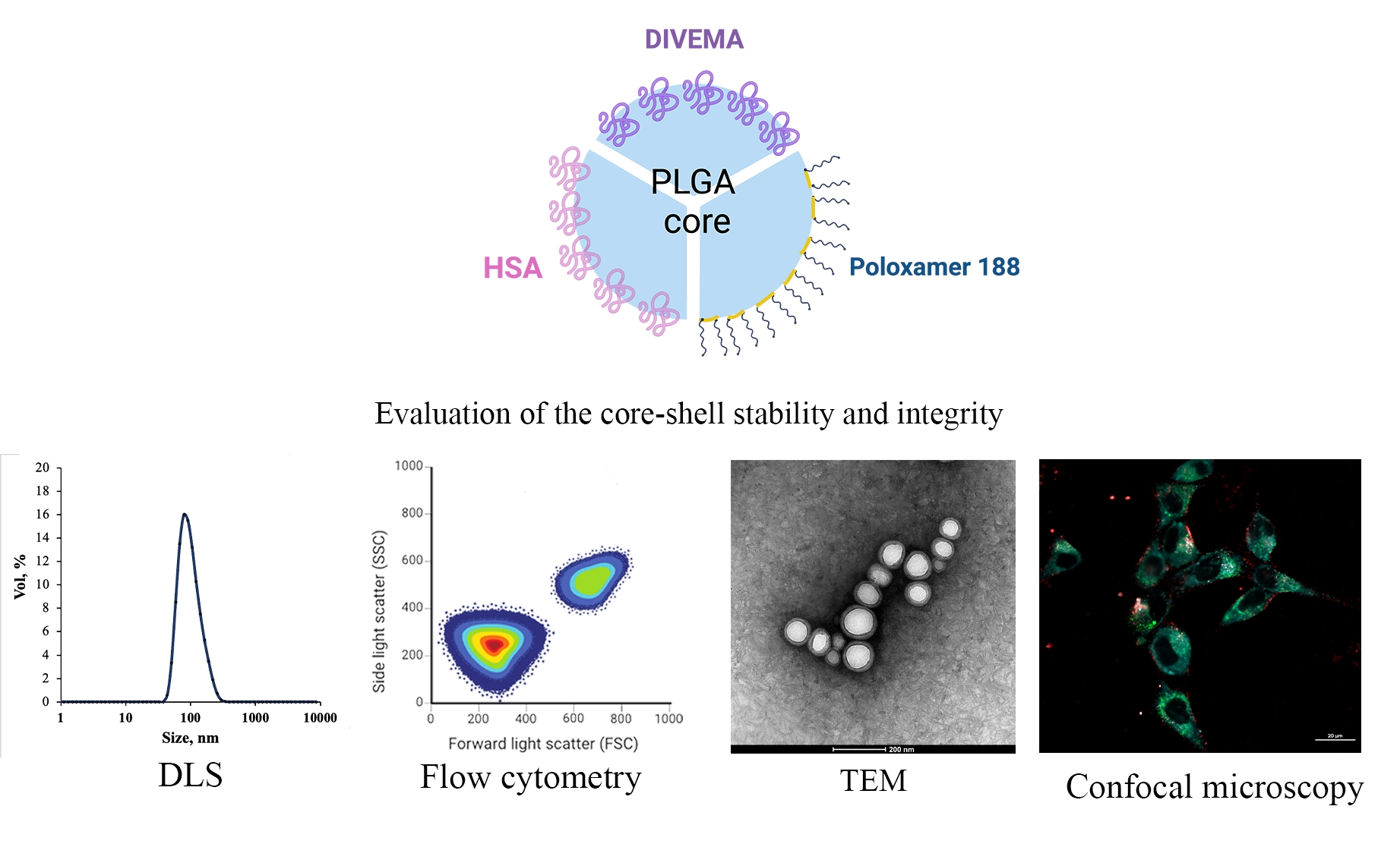
The objective of this study was to compare the properties of core-shell nanoparticles with a PLGA core and shells composed of different types of polymers; focusing on their structural integrity. Core PLGA nanoparticles were prepared by either the high-pressure homogenization — solvent evaporation technique or nanoprecipitation; using poloxamer 188 (P188); copolymer of divinyl ether with maleic anhydride (DIVEMA); and human serum albumin (HSA) as shell-forming polymers. The shells were formed through adsorption; interfacial embedding; or conjugation. For dual fluorescent labeling; the core and shell-forming polymers were conjugated with Cyanine5; Cyanine3; and rhodamine B. The nanoparticles had negative zeta potentials and sizes ranging from 100 to 250 nm (measured by DLS); depending on the shell structure and preparation technique. The core-shell structure was confirmed by TEM and fluorescence spectroscopy; with the appearance of FRET phenomena due to the donor-acceptor properties of the labels. All shells enhanced cellular uptake of the nanoparticles in Gl261 murine glioma cells. Integrity of the core-shell structure upon their incubation with cells was evidenced by intracellular colocalization of the fluorescent labels using Manders’ colocalization coefficients. This comprehensive approach may be useful for selection of the optimal preparation method already at the early stages of the core-shell nanoparticle development.

Keywords: 
;  ;  ;  ;  ;  ;  

1. Introduction

Drug delivery by nanoparticles may considerably improve the safety and selectivity of many drugs by altering their pharmacokinetics and tissue distribution using the endogenous mechanisms. The first success of this strategy was achieved in antibiotic therapy by exploiting the known ability of colloidal particles, especially hydrophobic, to accumulate in the resident macrophages of the macrophage-rich organs, such as the liver, spleen, lungs, etc. (organs of the mononuclear phagocytic system, MPS) [1]. Loading of antibiotics into the nanoparticles improved their penetration into macrophages, which serve as a niche for pathogens, and thus considerably enhanced their efficacy [2]. Later, the discovery of the enhanced permeability and retention (EPR) effect, which enables the extravasation and retention of colloids in the tumor due to the differences between the vascular systems of solid tumors and normal tissues, inspired enormous interest in nanomedicines for cancer chemotherapy. Indeed, the preclinical studies provided numerous instances of the improved antitumor effect of the nanoparticle-bound drugs due to their enhanced trafficking to tumors. However, it was soon realized that the accumulation of nanoparticles in the MPS organs, although useful for drug delivery to these organs, considerably interferes with their ability to target other sites in the body, decreasing blood circulation half-life and targeting capability [3]. Moreover, the large heterogeneity of the EPR effect observed on both intra- and interindividual levels leads to heterogeneous therapeutic outcomes [4,5,6]. Indeed, the advantage of the approved nanoformulations of classical anticancer drugs designed to exploit the EPR effect, such as Caelyx®, Abraxane®, Genexol-PM® or Onivyde®, was achieved more due to their enhanced safety than to any improvement in antitumor effect or patient survival [6].
These disappointing results gave rise to numerous new trends in pharmaceutical nanotechnology, starting from the use of theranostics for more insightful pre-selection of patients who could benefit from nanomedicines [7,8] to the development of delivery systems for the next generations of drugs (immune check-point inhibitors [9], nanoadjuvants [10], RNA-based therapeutics [11], drug combinations, etc.) and new types of nanocarriers [12,13].
Among the latter, core-shell nanoparticles draw considerable attention; over the last few decades, they have been extensively studied for many applications, including in the biomedical field (reviewed in [14,15,16,17,18]). This technology indeed enables the optimization of plain “first-generation” nanoparticles by improving their in vitro and in vivo stability, as well as offering the possibility to load them with both hydrophilic and hydrophobic agents and tune their release profiles. Moreover, hydrophilic shells may shield nanoparticles from macrophages (the stealth effect), thus optimizing their biodistribution profile and enhancing their capability to reach other organs besides the MPS. Shells rich in functional groups could also enhance the feasibility of nanoparticle surface modification and conjugation with drugs or ligands for targeted delivery.
The biodegradable and biocompatible poly(lactide-co-glycolic) acid (PLGA) nanoparticles, one of the most popular nanocarriers in drug delivery [19], appear to be good candidates for this approach. These nanoparticles are relatively hydrophobic, and therefore their inherent disadvantage is the massive accumulation in macrophage-rich organs. Moreover, the attachment of bioligands that could enable active tumor targeting is difficult because the reactive groups on the surface of the PLGA nanoparticles are few (limited to the end carboxylic groups of the polymer) and sterically hindered. The advantages of core-shell PLGA nanocarriers, such as steric stabilization, modulation of drug release profiles, and facilitated surface functionalization, have been demonstrated in numerous studies using various shell-forming agents such as PEG or PEG derivatives, chitosan, alginate, albumin, collagen, silk fibroin, etc. were demonstrated in a number of studies [14,20,21,22,23,24]. The methods used to create the shells primarily involve the adsorption of shell-forming polymers onto nanoparticle surfaces, which can be performed either during their formation or by incubating preformed nanoparticles in a solution of the shell-forming agent [22]. Sometimes, cross-linking of the shell [25] or its conjugation with the PLGA core [26,27] is used to improve system stability.
Clearly, the integrity of the core-shell structure is essential for the successful performance of the system in the biological environment. At the same time, this integrity is not always well elucidated.
The objective of the present study was to evaluate the integrity of nanoparticles with a PLGA core and shells composed of different types of polymers in cell-free media and upon their interaction with cells, using Gl261 murine glioma cells as the model. The shell-forming polymers included the non-ionic polymeric surfactant poloxamer 188 (PLGA/P188 NP), DIVEMA (a copolymer of divinyl ether with maleic anhydride, PLGA/DIVEMA NP), and human serum albumin (PLGA/HSA NP). The methods of preparing the core-shell nanoparticles were developed in our previous studies [28,29,30]. Due to their structure, the selected polymers create a hydrophilic shell on the nanoparticle surface and improve their colloidal stability. A considerable number of functional groups exhibited by HSA (NH2, COOH, and SH) and DIVEMA (COOH) shall also facilitate the binding of ligands to the core-shell nanocarrier.
These polymers are well characterized and appear to be suitable functional excipients. Both poloxamer 188 and HSA are effective surfactants and stabilizers with a long history of application in pharmaceutical products [31,32]. Moreover, coating PLGA nanoparticles with poloxamer 188 altered their biodistribution and enabled brain delivery of encapsulated doxorubicin; importantly, this formulation also improved the doxorubicin safety profile compared to the free drug [33,34,35]. The relatively low toxicity of low-molecular weight DIVEMA used in this study has been demonstrated in clinical studies [36]. A number of studies have revealed the broad spectrum of DIVEMA’s biological activities, such as antiviral and antibacterial activity and induction of immune responses to tumors [37,38].
Apart from the physicochemical evaluation, the core-shell system integrity was monitored by fluorescent spectroscopy using PLGA nanoparticles, the core and shell of which were labeled with pairs of fluorescent dyes with donor-acceptor properties (Cyanine5/Cyanine3 and Cyanine5/rhodamine B). These dye pairs are capable of exhibiting the Förster resonance energy transfer (FRET) phenomenon. This distance-dependent phenomenon enables the evaluation of core and shell integrity based on changes in donor and acceptor fluorescence intensity, which are observed only when the molecules are localized in close proximity [39,40]. Internalization of the core-shell nanoparticles in Gl261 cells was investigated using confocal laser fluorescent microscopy.

2. Materials and Methods

2.1. Materials

PLGA (Purasorb® PDLG 5004A, 50/50, η=0.4 dL/g, acid terminated) was purchased from Corbion Biomaterials (the Netherlands). N-(3-dimethylaminopropyl)-N′-ethylcarbodiimide hydrochloride (EDC), N,N′-dicyclohexylcarbodiimide (DCC), 4-(dimethylamino)pyridine (DMAP), N–hydroxysuccinimide (NHS), diisopropylethylamine (DIPEA), human serum albumin (HSA), polyvinyl alcohol (PVA, 9-10 kDa, 80% hydrolyzed), poloxamer 188 (P188), 2,2’-azobisisobutyronitrile (AIBN), rhodamine B (RhB) and rhodamine B isothiocyanate (RhBITC) were purchased from Sigma-Aldrich (USA). The reactive derivatives of the fluorescent dyes Cyanine5 (Cy5) amine and Cyanine3 (Cy3) amine were purchased from Lumiprobe (Russia). D-mannitol was purchased from Dia-M (Russia). All other chemicals were of analytical grade.

2.2. Preparation of Core-Shell PLGA Nanoparticle

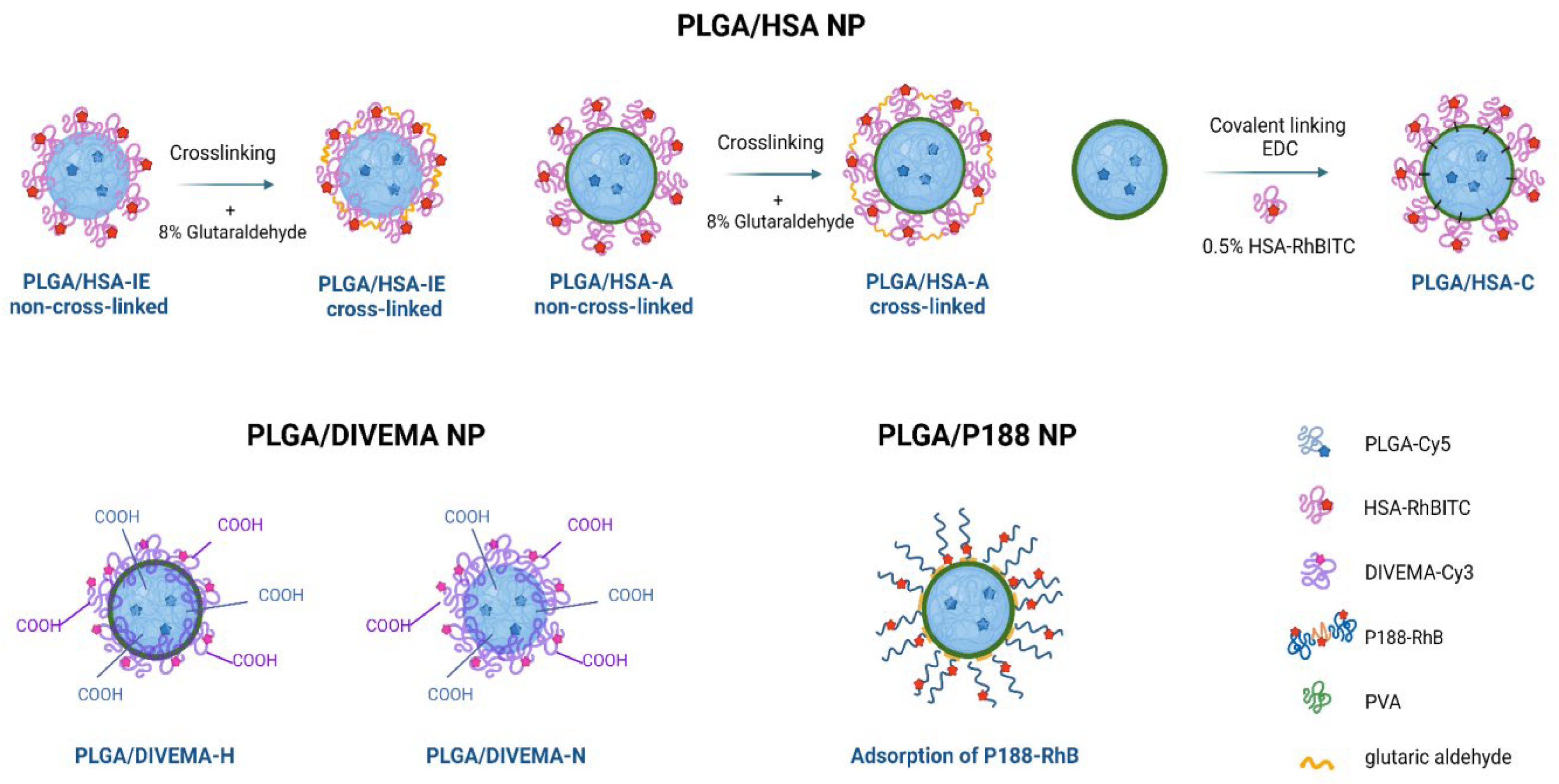
2.2.1. Preparation of Dual-Labeled Nanoparticles with a PLGA Core and HSA Shell (PLGA-Cy5/HSA-RhBITC NP)

For fluorescence labeling, conjugates of PLGA with Cyanine5 amine (PLGA-Cy5) and HSA with rhodamine B isothiocyanate (HSA-RhBITC) were synthesized. PLGA-Cy5 was synthesized by conjugating the PLGA acid terminal group with the water-soluble amine derivative of the Cyanine5 dye (Cy5, λex 651 nm, λem 670 nm) using NHS/EDC coupling reaction in the presence of DIPEA, as previously described in [28,41,42]. Briefly, solutions of Cy5 amine, DIPEA, EDC, NHS, and PLGA in dichloromethane (DCM) were combined, and the reaction mixture was stirred in the dark (48 h, ambient temperature). The resulting solution was washed three times with water and a water/methanol (1:1) mixture. The organic phase was then dried over anhydrous sodium sulfate, and the solvent was removed by evaporation. The residue was dissolved in ethyl acetate, and the polymer was precipitated by adding hexane (10-fold volume). The Cy5 content in the dried conjugate was measured spectrophotometrically. The PLGA-Cy5 conjugate with a dye-to-polymer ratio of 1:600 (w/w) was used in the experiments.
The PLGA-Cy5 conjugate was analyzed by gel permeation chromatography as described in [28]. Briefly, the analysis was performed using a Waters HPLC system equipped with a set of Styrogel HR5E and HR4E columns, a refractive index detector (Waters 2414 RI Detector) and a UV detector (Milton Roy UV-detector 3100 model, λ 264 nm). Tetrahydrofuran was used as the solvent and eluent at a flow rate of 1.00 mL/min. Data were analyzed using Z-lab software.
HSA was conjugated with rhodamine B isothiocyanate following the method described by Yang et al. [43]. Briefly, rhodamine B isothiocyanate was dissolved in DMSO, and this solution was added dropwise to a 1% HSA solution (0.15 mM) in a 9:1 v/v mixture of saline (0.15 М NaCl) and buffer (0.15 М NaHCO3, pH 9.0). The RhBITC/HSA molar ratio was 5:1. The obtained solution was stirred overnight under cooling. A NH4Cl solution (50 mM) was then added to stop the reaction, followed by an additional hour of stirring. The resulting fluorescently labeled HSA was purified by dialysis against phosphate-buffered saline (PBS, 0.01 М PBS, pH 7.4) at 4 °C in the dark. The concentration of RhBITC was determined by calculating the fluorophore/protein molar ratio (F/P):
C H S A R h B I T C = A 280 C F × A 558 ε H S A × d i l u t i o n f a c t o r ,
F P = A 558 ε R h B I T C × C   H S A R h B I T C   × d i l u t i o n   f a c t o r
where A558 represents the absorbance (A) of RhBITC at 558 nm; A280 is the absorbance (A) of HSA-RhBITC at 280 nm; εHSA is the molar extinction coefficient of HSA; εRhBITC is the molar extinction coefficient of RhBITC; CF is the correction factor to account for dye absorbance at 280 nm; dilution factor accounts for any dilution of the protein:dye sample for absorbance measurement. The HSA-RhBITC conjugate used in further experiments had an F/P ratio of 1.25.
Core-shell nanoparticles were prepared by the high-pressure homogenization — solvent evaporation technique (o/w). The HSA shell was formed using three different techniques: by using HSA as a stabilizer during nanoparticle formation (interfacial embedding of HSA) or by adsorbing or conjugating HSA onto previously prepared PLGA nanoparticles without a shell (Supplementary Figure S1) [29].
Interfacial embedding of HSA. A solution of a 1:1 mixture of PLGA and PLGA-Cy5 (150 mg + 150 mg) in dichloromethane was emulsified with an aqueous 0.5% HSA-RhBITC solution (30 mL) using a high-shear homogenizer (Ultra-Turrax T18 Basic, IKA-Werke, GmbH, Staufen, Germany). This coarse emulsion was further homogenized at high pressure (15,000 psi, Microfluidizer L10, Microfluidics, Newton, MA, USA), and the solvent was removed by vacuum evaporation. The resulting suspension was then filtered through a sintered glass filter and freeze-dried using 2.5% (w/v) D-mannitol as a cryoprotectant. The freeze-dried nanoparticles were stored at 4 °C. Unbound HSA was removed by repeated washing and centrifugation of the nanosuspension (48,060⨯g, 20 °C, 20 min). A portion of the sample was subjected to shell cross-linking with glutaraldehyde to improve shell stability [29]. Based on the findings of Langer et al. [44], 117 µL of an 8% glutaraldehyde solution (~0.1 mmol) per 1 mg of HSA (15 µmol) was added to the reaction mixture to ensure reliable HSA cross-linking.
Adsorption of HSA. To prepare PLGA/HSA nanoparticles by adsorption, PLGA-Cy5 nanoparticles were first prepared using 1% polyvinyl alcohol (PVA, 9–10 kDa, 80% hydrolyzed, Merck, Germany) as a stabilizer, as described in [41]. Then the nanoparticles were washed with water and resuspended in a 0.5% aqueous HSA-RhBITC solution, followed by 30 min of incubation under continuous stirring (37 °C, 130 rpm). Unbound HSA-RhBITC was removed by repeated centrifugation (48,060⨯g, 18 °C, 10 min). The supernatants were successively analyzed by spectrophotometry for the presence of unbound HSA. A portion of the sample was subjected to HSA cross-linking [29] followed by freeze-drying, as described above.
Conjugation of HSA. HSA amino groups were conjugated with the carboxyl end groups of PLGA through a coupling reaction using 1-ethyl-3-(3-dimethylaminopropyl)carbodiimide hydrochloride (EDC) in phosphate buffer (1 h, 0.05 M, pH 6.8) [45]. An indirect acid-base titration in MES buffer (0.05 M, рН 5) revealed that the PLGA-Cy5 nanoparticles contained 2.4 µmol-eq/mL of functional carboxyl groups. A 10-fold molar excess of EDC (24 µmol-eq/mL, 0.46 mg/mL NP) and NHS (10-fold excess: 24 µmol-eq/mL, 0.46 mg/mL NP) was then added to the nanoparticle suspension. The mixture was incubated at 37 °С for 2 hours under stirring (90 rpm), washed by centrifugation (48,060×g, 30 min, 25 °С), and resuspended in MES buffer (0.05M, рН 5). Activated nanoparticles were then incubated in a 1% HSA-RhBITC solution under continuous stirring (90 rpm, 1 h, 37°С). Unconjugated HSA was removed by two-step washing (centrifugation at 48,060×g, 10 min, 18 °С). The precipitated nanoparticles were resuspended in HEPES buffer (0.1 M, pH 7.4). The HSA content in the supernatants was assessed spectrophotometrically based on the fluorescent properties of the labeled polymer. The nanosuspensions were then freeze-dried using 2.5% mannitol as a cryoprotectant.

2.2.2. Preparation of Dual-Labeled Nanoparticles with a PLGA Core and DIVEMA Shell (PLGA-Cy5/DIVEMA-Cy3 NP)

The alternating copolymer of divinyl ether and maleic anhydride (DIVEMA) was synthesized by radical cyclopolymerization of the corresponding monomers in dry acetone, using 2,2’-azobisisobutyronitrile (AIBN) as the initiator and tetrahyfrofuran as the chain transfer agent [28,46]. The polymer structure was confirmed by IR and NMR spectroscopy, as previously described in [28]. A sample of DIVEMA copolymer with a molecular weight (MW) of 23 kDa and a characteristic viscosity of 0.16 dL/g (measured in borate buffer containing 0.2 M NaCl) was used in the following experiments.
Fluorescent labeling of DIVEMA was achieved by conjugating the carboxyl groups with the water-soluble amine derivative of Cyanine3 dye (Cy3, λex 555 nm, λem 570 nm) via a carbodiimide coupling reaction, as described in [28]. First, the DIVEMA carboxyl groups (2.26 mmol DIVEMA solution in acetone [600 mg in 30 mL of acetone]) were activated by NHS (23.4 mg, 0.20 mmol in 1 mL of acetone) for 30 min under continuous stirring in the dark. A dicyclohexylcarbodiimide (DCC) solution (14.1 mg [68.3 μmol] in 0.5 mL of acetone) and a Cy3 amine solution in a mixed organic solvent (18 mg [28.7 μmol] in 1 mL acetone and 0.5 ml ethanol) were then added. The mixture was incubated for 3 hours under constant stirring, after which the conjugate was purified by three-fold precipitation in diethyl ether (ten times the volume). The precipitate was separated and washed with chloroform. The absence of unbound Cy3 in the wash solvent was monitored by the UV spectroscopy at 543 nm. The product was dried under vacuum to a constant weight. The resulting purified DIVEMA-Cy3 conjugate was stored in an evacuated flask or under an inert gas atmosphere. The conjugate formation was confirmed by gel permeation chromatography using a refractive index detector and a UV detector, as previously described [28]. The Cy3 content was quantified by UV spectroscopy at λmax 552 nm. The conjugate containing 2.7% w/w of the dye (dye-to-polymer ratio of 1:40, w/w) was used in the experiments.
Preparation of PLGA-Cy5/DIVEMA-Cy3 nanoparticles by high pressure homogenization—solvent evaporation technique (PLGA-DIVEMA-H). A 1:1 mixture of PLGA and PLGA-Cy5 polymers (2 x 300 mg [2 x 2.3 mmol]) was dissolved in 7.2 mL of DCM. The resulting solution was mixed with a solution of DIVEMA-Cy3 in acetone (71.4 mg [0.268 mmol] in 4.8 mL), and the organic phase was added to a 0.5% aqueous PVA solution (60 mL). The mixture was first emulsified using a high-shear homogenizer (Ultra-Turrax T18, 23,600 rpm) and then subjected to high-pressure homogenization (Microfluidics M-110P, 15000 psi). Afterwards, the organic solvent was removed under vacuum, and the resulting suspension was filtered through a glass sintered filter (pore size 90-150 μm) and freeze-dried using 2.5% (w/v) of D-mannitol as a cryoprotectant. After lyophilization, the nanoparticles were resuspended in purified water and subjected to centrifugation: first at 10,000×g (10 min, 5 °C) to remove agglomerates, and then at 48,060×g (two times, 30 min, 5 °C). The resulting nanosuspension was freeze-dried with 2.5%mannitol as a cryoprotectant and stored at 4 °С. Plain PLGA nanoparticles (PLGA without a shell) were obtained in a similar way, with the addition of 4.8 mL of acetone without DIVEMA to the polymer solution.
Preparation of PLGA-Cy5/DIVEMA-Cy3 nanoparticles by nanoprecipitation (PLGA-DIVEMA-N). The PLGA/PLGA-Cy5 (1:1) mixture (60 mg; 0.46 mmol) was dissolved in a mixture of acetonitrile and acetone (1:2 v/v). Then, 24.7 mg (93 μmol) of DIVEMA-Cy3 was added, and the resulting solution was added dropwise to 60 mL of dH2O under stirring (1,000 rpm) and incubated for 4 hours. The organic solvents were evaporated under vacuum. The resulting suspension was filtered through a sintered glass filter (pore diameter 90-150 μm) and freeze-dried with the addition of 2.5% (w/v) mannitol as a cryoprotectant. The resulting suspension was passed through a glass sintered filter with a pore diameter of 90-150 μm (Por 1); 2.5% mannitol was added as cryoprotectant. Plain PLGA nanoparticles (PLGA without a shell) were obtained similarly, but without DIVEMA and using 0.5% aqueous PVA solution instead of purified water.

2.2.3. Preparation of PLGA Nanoparticles Coated with Fluorescently Labeled Poloxamer 188

Synthesis of poloxamer 188 conjugate with RhB (P188-RhB). The conjugate of poloxamer 188 with rhodamine B (P188-RhB) was synthesized and characterized as reported previously [30]. Briefly, a solution of P188 (1,000 mg, 0.12 mmol), RhB (126 mg, 0.26 mmol), EDC (55 mg, 0.29 mmol), and 4-(dimethylamino)pyridine (DMAP, 59 mg, 0.48 mmol) in dimethylformamide (DMF, 15 mL) was stirred at ambient temperature for 3 days in the dark. Afterwards, the mixture was diluted with Et2O, and the crude product was precipitated overnight in a freezer at −20 °C. The precipitate was washed three times with a mixture of DMF/Et2O (1:1) while cooling. The crude product was then purified by gel filtration using a Sephadex G25 column. The combined fractions containing the product were then lyophilized. The absence of unbound dye was confirmed by thin-layer chromatography using a mixture of i-PrOH/H2O (3:5) as the eluent. The stability of the conjugate in PBS (pH 7.4), DMEM, or human plasma was also evaluated by TLC (incubation for 2 h at 37 °C). The conjugate containing 4.49% w/w of the dye was used in the experiments.
Evaluation of poloxamer 188 adsorption on the PLGA nanoparticles’ surface. The PLGA-Cy5-P188-RhB nanoparticles were prepared by incubating the PLGA-Cy5 NP (prepared by the high-pressure homogenization—solvent evaporation technique as described above) in a 1% aqueous solution of P188-RhB for 30 minutes.
The evaluation of P188-RhB adsorption was carried out as reported previously [30]. To analyze the P188-RhB content in one vial, the nanoparticle lyophilizate was resuspended in 1.5 mL of the 1% P188-RhB solution. The suspension was incubated at room temperature while stirring at 130 rpm. The aliquots (400 μL) collected after 30, 90 and 180 minutes were centrifuged to separate the PLGA NP (48,068×g, 30 min, 5 °C). The precipitated nanoparticles were washed with water (1 mL), dried, and dissolved in DMSO (2 mL). The P188-RhB content in the resulting solution was determined spectrophotometrically at λmax 555 nm (UV-1900i spectrophotometer, Shimadzu Corp, Japan) using the calibration curve. The amount of adsorbed P188-RhB in mg/m2 of the PLGA NP surface was calculated as:
S = c V s ρ P L G A d 6 m ,
where m is the mass of PLGA nanoparticles in a vial, mg; с is the concentration of the P188 RhB conjugate in the analyzed sample, determined from the calibration curve, mg/mL; Vs is the sample volume, 2 mL; ρPLGA is the PLGA density, 1.2 g/cm3; d is the nanoparticle diameter, nm.

2.3. Characterization of Nanoparticles

The mean hydrodynamic size and polydispersity index (PDI) of the nanoparticles were measured by dynamic light scattering (DLS) using the Zetasizer NanoZS (5 mW He-Ne laser, operating wavelength 633 nm, Malvern Instruments, Malvern, UK). The zeta-potential was determined by electrophoretic light scattering (ELS) in a U-shaped disposable folded capillary cell using the same instrument. Measurements of all samples were performed in triplicate. The samples were diluted to a concentration of 0.2 mg/mL polymer in Milli-Q water.
The total PLGA concentration in all samples was quantified using capillary electrophoresis (CAPEL 105M, Russia) as described previously [47]. The amount of PLGA was assessed by the amount of lactate formed after the nanoparticle hydrolysis in 1 M NaOH (37 °C, 24 h, continuous shaking at 200 rpm). The content of the –COOH groups in PLGA nanoparticles was determined by potentiometric titration [47].
The dye content in the nanoparticles was quantified by UV spectroscopy (Shimadzu UV-1800, Japan). The Cy3 and Cy5 content in the nanoparticles was measured at 553 nm and 649 nm for Cy3 and Cy5, respectively, after dissolving the core-shell nanoparticles in DMSO. The HSA-RhBITC shell content in the nanoparticles was calculated indirectly by the difference between the content of HSA-RhBITC (determined by UV spectroscopy at λmax 558 nm in water) added and washed in the process of nanoparticle preparation as:
m ( H S A ) N P =   m ( H S A ) a d d e d m ( H S A ) w a s h e d ,
where m(HSA)NP is the content of HSA-RhBITC on the nanoparticle surface; m(HSA)added is the content of HSA-RhBITC added during the nanoparticle synthesis; m(HSA)washed is the content of HSA-RhBITC washed during the nanoparticle synthesis.
The morphology of the core-shell nanoparticles was studied by transmission electron microscopy (TEM) using a JEOL JEM-1400 electron microscope (JEOL, Japan) at an accelerating voltage of 120 kV. A diluted aqueous nanosuspension was applied to Cu grids coated with a Formvar polymer film, and the solvent was allowed to dry completely. To improve image quality, additional negative contrasting of the nanoparticles was performed using the UranyLess EM Stain (Electron Microscopy Sciences, USA).
The TEM images of the PLGA/HSA-IE cross-linked nanoparticles were also obtained using the Tecnai™ 12 G2 BioTwin Spirit microscope (ThermoFisher Scientific, USA) equipped with Eagle 4K detector (ThermoFisher Scientific, USA) at an accelerating voltage of 120 kV. The samples for the TEM study were prepared using negative staining. The 300 mesh Cu grids coated with a carbon support film were subjected to glow discharge in PELCO easiGlow™ for 30 sec with a 25-mA plasma current. The sample solution (3 μL) was applied to the grid for 30 seconds, after which the grid was washed with a droplet of distilled water and stained with a 1% aqueous solution of uranyl acetate for 60 seconds.

2.4. Evaluation of Fluorescent Properties

The quantum yield and brightness of the fluorescently labeled nanoparticles were assessed as reported previously [42]. The light absorption values and the integrated fluorescence intensities were determined for each nanoparticle type.
The quantum yields of the nanoparticles were calculated as:
φ = φ s t t g   α t g   α s t n n s t 2 ,
where φ and φst are the quantum yield of the dye-labeled nanoparticles and standard, respectively; tg α and tg αst are tangents of the slope of the dependences of the integrated fluorescence intensity on light absorption for the test compound and standard, respectively; n and nst are refractive indices of the media in which the measurements were made.
The brightness of the nanoparticles was calculated as [48]:
B = φ · ε · N ,
where ε is the extinction coefficient of the dye determined via Beer-Lambert law, l∙mol−1∙cm−1; φ is the quantum yield; N is thenumber of fluorescent dye molecules encapsulated inside the nanoparticles.
RhB (φ=0.7, ε =106,000 M-1 cm-1 [49]) was used as a reference to determine the quantum yield and brightness of Cy3-, RhB- or RhBITC-labeled core-shell nanoparticles. Cy5 (φ=0.2, ε =250,000 M-1 cm-1 [50]) was used as a reference to determine the quantum yield and brightness of Cy5-labeled nanoparticles.
Fluorescence spectroscopy FRET studies. The fluorescence spectra were measured for nanoparticles with either one or two fluorescent labels. The following types of nanoparticles were used for the study: PLGA-Cy5/HSA NP, PLGA/HSA-RhBITC NP, PLGA-Cy5/HSA-RhBITC NP, PLGA/DIVEMA-Cy3 NP, PLGA-Cy5/DIVEMA-Cy3 NP, PLGA-Cy5/P188 NP, PLGA/P188-RhB NP, and PLGA-Cy5/P188-RhB. Nanoparticle suspensions in water were prepared so that the fluorescent label content in nanoparticles with one label was equal to the content of the same dye in nanoparticles with two fluorescent labels. The fluorescence spectra of the PLGA/HSA NP and PLGA/P188 NP were recorded at λex = 550 nm over the emission range of λem 565-800 nm, and at λex = 644 nm over of λem 655-800. The fluorescence spectra for the PLGA/DIVEMA NP were taken at λex = 530 nm over the emission range of λem 545-740 nm, and at λex = 630 nm over of λem 640-800 nm.

2.5. Evaluation of Core-Shell Nanoparticle Stability by Physicochemical Methods

The colloidal stability of the nanoparticles was assessed at different time points by measuring their hydrodynamic diameters (DLS) at 37 °C in simulated physiological media: PBS (pH 7.4; 0.15 M) and 4.5% HSA solution in PBS.
The stability of the core–shell structures was also evaluated by assessing the amount of fluorescently labeled shell remaining on the nanoparticle surface through fluorescence measurements. The nanosuspensions (100 μg/mL as PLGA) were incubated in PBS or PBS containing 4.5% HSA. At selected time points (0, 0.5, 1, 2, 4, and 6 h), the 400-μL aliquots were taken, and then the fluorescence spectra of the suspension were recorded. Then the aliquots were centrifuged (48,060×g, 18 °C, 20 min) to precipitate the nanoparticles and the fluorescence intensity of each supernatant was measured. The fluorescence spectra were recorded for HSA-RhBITC and P188-RhB (λex 560 nm, λem 570-800 nm), PLGA-Cy5 (λex 649 nm, λem 660-800 nm), and DIVEMA-Cy3 (λex 530 nm, λem 540-800 nm). The percentage of the detached shell at each time point was calculated as the ratio of the fluorescence intensities in the supernatant to those in the nanosuspension.

2.6. Cells

Gl261 murine glioblastoma cells were purchased from the American Type Culture Collection (ATCC). The cells were cultivated in DMEM supplemented with GlutaMax™ (2 mM, Gibco), 10 % fetal bovine serum (Biowest, US) and penicillin (100 U/mL), and streptomycin 100 g/mL (Gibco). The cells were cultured under standard conditions (37° C, 5% CO2) and used between passages 5-9.

2.7. Investigation of Nanoparticle Internalization by Confocal Microscopy

Images were obtained using a Nikon A1R MP+ multiphoton confocal microscope (Nikon Instr., INC, USA) equipped with four lasers: 405 nm (λem 425-475 nm), 488 nm (λem 500-550 nm), 561 nm (λem 570-620 nm), and 638 nm (λem 663-738 nm), and Plan Apo 20x/0,75 Dic N, Apo IR 60x/1.27 WI, and Apo TIRF 60x/1,49 oil Dic objectives. Data were analyzed using NIS-Elements AR Software. Gl261 cells were seeded onto confocal 35-mm high coverslip dishes (Ibidi, USA) (70x103 cells) 48 hours prior to the study. The cells were incubated with the samples for 15, 30 and 45 min (final concentration 100 μg/mL in serum-containing medium). The cells were then washed three times with DPBS to remove nanoparticles from the cell surface. Lysosomes were stained using LysoTracker Green DND26 (50 nМ, Molecular Probes, USA). Plates were transferred to the microscope stage for live-cell imaging.
To assess the core-shell structure integrity of the nanoparticles as well as their colocalization with lysosomes (for the evaluation of their major NP internalization pathway and intracellular localization), Manders’ overlap coefficient (MOC) values were calculated. MOC values were calculated within the selected ROIs — three random ROIs per image. Colocalization plots were built to compare the core-shell integrity of the nanoparticles (HSA, DIVEMA or P188 shells with PLGA cores) obtained by different techniques.

2.8. Investigation of Nanoparticle Uptake by Flow Cytometry

The Gl261 cells were seeded into 24-well plates (Corning) (100x103 cells /well) and incubated for 24 hours to allow attachment and reach 70-90% confluence. Then the samples were added to a final concentration of 100 μg/mL in cell culture medium (DMEM supplemented with 10% FBS) and incubated for 15, 30 and 45 minutes.
At the specified time points, the cells were washed twice with HBSS buffer, detached using TrypLE solution (ThermoFisher, USA), and centrifuged at 1,000×g (5 min, 18 °C). The cell pellets were resuspended in FACS buffer and analyzed on the FACS MoFlo XDP (Beckman Coulter, USA). The nanoparticle uptake by the cells was determined as the percentage of NP-positive cells: PLGA-Cy5-positive or double-positive (PLGA-Cy5- and HSA-RhBITC//DIVEMA-Cy3/RhB-positive cells).

2.9. Statistics

The data were analyzed using GraphPad Prism 9 software (San Diego, CA, USA) with one- or two-way ANOVA followed by Tukey’s and Dunnett’s multiple comparisons tests. Characterization of particle size, size distribution, zeta potential, and all in vitro measurements were conducted in triplicate.

3. Results and Discussion

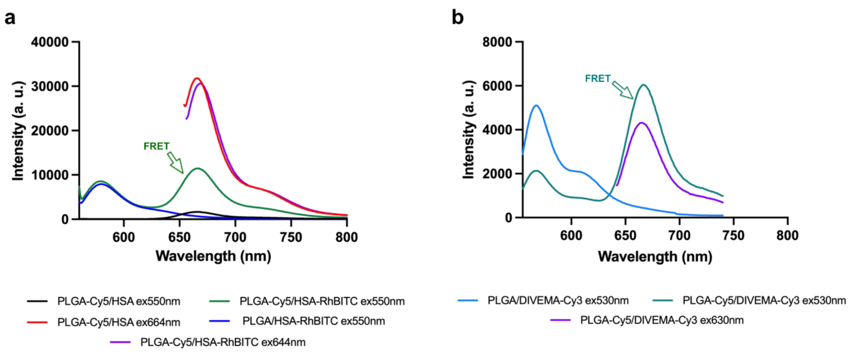
3.1. Core-Shell Nanoparticle Preparation and Characterization

The main objective of this study was to develop an integrated approach for the evaluation of core-shell nanoparticle stability using PLGA nanoparticles with different polymeric shells as model structures. As mentioned above, the following shell-forming polymers were used: 1) poloxamer 188 (P188), a nonionic triblock copolymer of polyethylene oxide and polypropylene oxide; 2) human serum albumin (HSA), one of the most abundant plasma protein with high ligand binding capacity and a natural carrier of many exogenous and endogenous substances [51]; 3) a copolymer of divinyl ether and maleic anhydride (DIVEMA), a polyanion with versatile biologic activity [52]. DIVEMA is a polyanhydride that, upon hydrolysis, turns into a polycarboxylate containing four carboxyl groups per monomeric unit.
To enable visualization of core-shell nanoparticles’ interaction with cells, both the core- and the shell-forming polymers were labeled with covalently bound bright fluorescent dyes. As shown in our previous studies, only covalent linking ensures stable retention of fluorescent labels by nanoparticles, which helps to avoid artifacts in imaging the nanoparticle interactions with biological objects [42]. Therefore, the following dye-polymer conjugates were synthesized using carbodiimide chemistry: PLGA–Cyanine5 (PLGA-Cy5), HSA–rhodamine B (using RhBITC derivative, HSA-RhBITC), poloxamer 188–rhodamine B (P188-RhB), and DIVEMA–Cyanine3 (DIVEMA-Cy3). The covalent binding of the dyes to the polymers was confirmed by GPC or TLC analysis, as described in [28,29,30]. The chemical structures of the fluorescently labeled polymers are schematically presented in Figure 1.
The core PLGA nanoparticles were prepared by the commonly used method of high-pressure homogenization followed by solvent removal. This method involves the emulsification of a PLGA solution in DCM in an aqueous solution of polyvinyl alcohol (PVA), one of the most popular stabilizers used for the preparation of PLGA nanoparticles [31]. The core-shell nanoparticles were prepared using previously established techniques, including adsorption of the shell-forming polymer onto the previously prepared core PLGA nanoparticles (P188, HSA), interfacial embedding achieved by adding the shell-forming polymer in the course of nanoparticle preparation (DIVEMA, HSA), or by covalent linking of the shell to the PLGA carboxyl end groups (HSA) [28,29,30].
The types of dual-labeled core-shell PLGA nanoparticles prepared and investigated in this study are shown schematically in Figure 2 and Supplementary Figure S1. The physicochemical parameters of these nanoparticles are shown in Table 1. The nanoparticles had hydrodynamic diameters of 100-250 nm (volume distribution) depending on the preparation method and shell type, and they were negatively charged –parameters considered optimal for drug delivery [53].
The methods of shell formation were chosen based on the polymer properties. In the case of HSA, three techniques were used for shell formation to reveal the optimal conditions for the use of this versatile and complex molecule. HSA is known to adsorb to various surfaces depending on their charge and hydrophobicity [54]; it can also provide steric stabilization of the colloidal carriers, protecting them from recognition by macrophages [55]. In the case of the adsorption technique, shell formation was achieved by incubating the “bare nanoparticles” in the HSA solution. For the interfacial embedding of HSA, the PLGA NP were also prepared using the high-pressure homogenization—solvent evaporation technique; however, in this case, HSA (0.5% solution) was used as a stabilizer in the outer aqueous phase instead of PVA. Indeed, due to its amphiphilic properties, HSA could be located in the interfacial area between the organic and aqueous phases, with its hydrophilic fragments protruding into the water, while the more hydrophobic fragments are “immersed” into the PLGA core. As shown in Table 1, the method of interfacial embedding was the most effective, producing a considerably more abundant shell on the nanoparticle surface compared to HSA adsorption or conjugation (0.52, 0.11, and 0.48 mg HSA per mg PLGA, respectively). Similar results were obtained in the study by Hyun at el., where the interfacial embedding of HSA yielded nanoparticles with a higher amount of HSA compared to adsorption [56]. For better stability, the HSA shells in the PLGA/HSA NP prepared by adsorption and interfacial embedding were additionally cross-linked by glutaric aldehyde. This approach was shown to improve the stability of the HSA-coated nanoparticles upon dilution and in the presence of other proteins (endogenous albumin) [57].
As seen from Table 1, the parameters of the PLGA/HSA nanoparticles depended on the method of shell formation. Thus, adsorption and conjugation of HSA led to an increase in the hydrodynamic diameter (from ≈ 110 nm to ≈ 140 nm) and partial compensation of the negative surface zeta potential (from ≈ -20 to ≈ -7 mV) compared to the bare PLGA NP, which correlated with the results of other authors [45]. In contrast, the interfacial embedding of HSA (acting as a surfactant in the absence of PVA) led to the formation of 90-100-nm particles with a higher negative zeta potential (≈ -30 mV). These data correlate with the results of Hyun et al., who observed similar tendencies for the PLGA/HSA nanoparticles obtained by interfacial embedding and by physical adsorption of HSA [56] It is noteworthy that cross-linking of the HSA shell did not exert any influence on the nanoparticle parameters compared to the non-cross-linked nanoparticles.
Interfacial embedding was also applicable for the preparation of PLGA/DIVEMA NP. Indeed, the radical copolymerization of maleic anhydride and divinyl ether yields DIVEMA in its polyanhydride form (Figure 1). In this form, DIVEMA is readily soluble in acetone but not in water. When added into the aqueous-organic mixture during nanoparticle formation, the anhydride cycles are gradually hydrolyzed, converting the copolymer into an amphiphilic molecule. This process is rather slow, so the DIVEMA-Cy3 conjugate retains its amphiphilic nature and is capable of interacting with PLGA on one hand and creating a hydrophilic shell on the nanoparticle surface on the other [58]. The presence of DIVEMA in the PLGA/DIVEMA nanoparticles was previously confirmed by FTIR spectroscopy [28]. Moreover, the presence of the outer shell rich in carboxyl groups on the PLGA/DIVEMA NP surface was confirmed by the pH-dependent changes in their size, which decreased upon lowering the pH level or in the presence of salts due to the decreased dissociation of DIVEMA carboxyl groups. Both phenomena are typical for polyelectrolytes [28,58,59].
The fluorescently labeled PLGA-Cy5/DIVEMA-Cy3 NP were prepared by either the high-pressure homogenization—solvent evaporation technique (PLGA/DIVEMA-H NP) or by nanoprecipitation (PLGA/DIVEMA-H) (Figure 2). Dual labeling was achieved by using a (1:1) mixture of PLGA and PLGA-Cy5 conjugate as the PLGA constituent and DIVEMA-Cy3 conjugate. In both cases, the presence of DIVEMA led to a considerable increase in the mean hydrodynamic diameter (measured by DLS) (Тable 2), which suggests the presence of a more pronounced hydration shell and abundant polyanionic groups on the surface of the hybrid nanoparticles. The sizes of the PLGA/DIVEMA-N and PLGA/DIVEMA-H were about 180 nm and 264 nm compared to 130 nm and 178 nm for the bare particles, respectively (Table 1). The content of the –COOH groups in both types of the PLGA/DIVEMA NP was considerably higher compared to the plain PLGA NP: 0.72 ± 0.06 vs 0.09 ± 0.04 mmol/g PLGA and 0.46 ± 0.05 vs 0.04 ± 0.01 mmol/g PLGA for the PLGA/DIVEMA-H and PLGA/DIVEMA-N nanoparticles, respectively. Accordingly, these nanoparticles had higher negative zeta potentials compared to the plain PLGA NP (Table 2). The relative content of the shell (DIVEMA-Cy3) was higher in the case of the nanoprecipitation method, which is probably due to the particle formation in the absence of PVA in the aqueous phase: 0.47 vs 0.10 mg/mg PLGA, respectively (Table 1). Due to this more considerable shell, combined with their more favorable size and size distribution parameters, only the PLGA/DIVEMA-N NP were used in further experiments.
The PLGA-Cy5/P188-RhB nanoparticles were prepared by incubating the plain nanoparticles in a 1% P188-RhB solution [30]. Conjugation of P188 with rhodamine B did not affect its surfactant properties: the amount of P188-RhB adsorbed on the nanoparticle surface was even increased compared to P188 (0.38 vs 0.14 mg/m2 of NP or 0.017 vs 0.006 mg/mg PLGA, respectively). Modification of the PLGA NP with poloxamer 188 and P188-RhB did not induce any changes in their hydrodynamic diameter and surface potential (Table 1), which correlates with our previous observations [61]. It is noteworthy that the P188 shell content is the lowest compared to HSA-RhBITC and DIVEMA-Cy3 (0.017 mg/mg PLGA).
As shown by TEM, all types of the core-shell nanoparticles had a spherical shape (Figure 3). Due to its electron density, the HSA shell was well visualized by TEM [62]. The presence of the hydrated HSA shell was confirmed by the smaller nanoparticle size observed by TEM, compared to the DLS measurements of the same sample: 50-100 nm vs 80-150 nm (Vol, PDI <0.20). This observation correlates with the data obtained by other authors [60,62].
The most prominent shell was observed on the PLGA/HSA NP prepared by interfacial embedding with cross-linking (PLGA/HSA IE cross-linked NP, Figure 3d). These nanoparticles had the highest HSA/PLGA ratio (0.52 mg/mg PLGA, Table 1), compared to the adsorption and conjugation methods (Figure 3a,b); a similar phenomenon was also observed by Hyun H. et al. [45,56]. These authors suggested that HSA introduced onto the nanoparticle surface during the emulsification stage could form a shell consisting of several HSA layers. The high density of HSA on the PLGA/HSA-IE NP surface is also consistent with the relatively high negative surface charge similar to the nanoparticles obtained in [45,56].
In the case of the PLGA/DIVEMA-N, the direct comparison of the sizes measured by TEM and DLS is not possible due to the high polydispersity of the PLGA/DIVEMA-N NP (PDI >0.2); however, it appears that in the TEM micrographs, the majority of the hybrid nanoparticles had smaller diameters than the diameter measured by DLS: ≈ 200 nm vs 280 nm (Volume, Table 1). Accordingly, this phenomenon is most likely explained by the hydration of the hydrophilic shell in the aqueous medium. The P188-RhB shell was not visible in TEM images, which has been noted in a number of other studies [63,64].

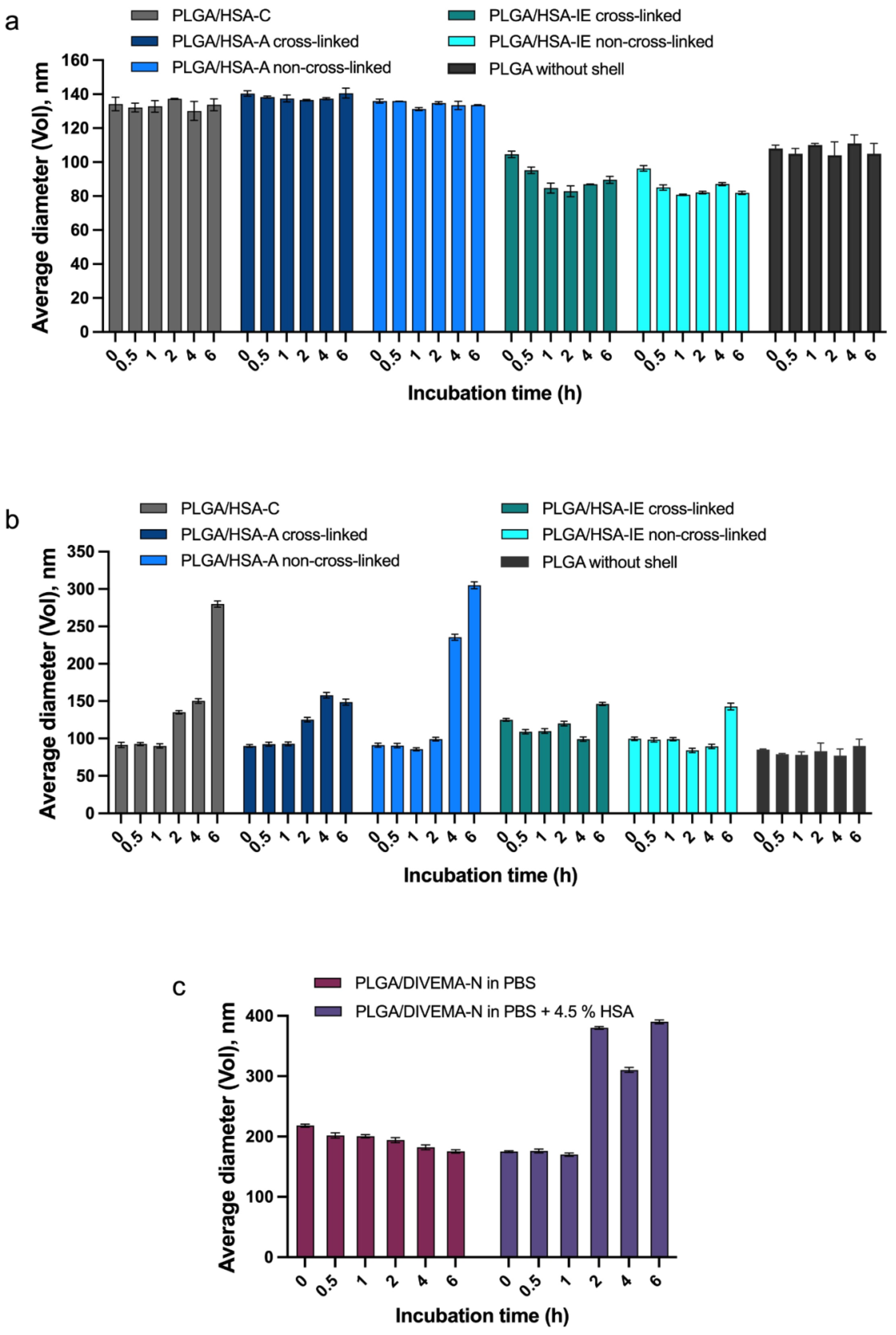
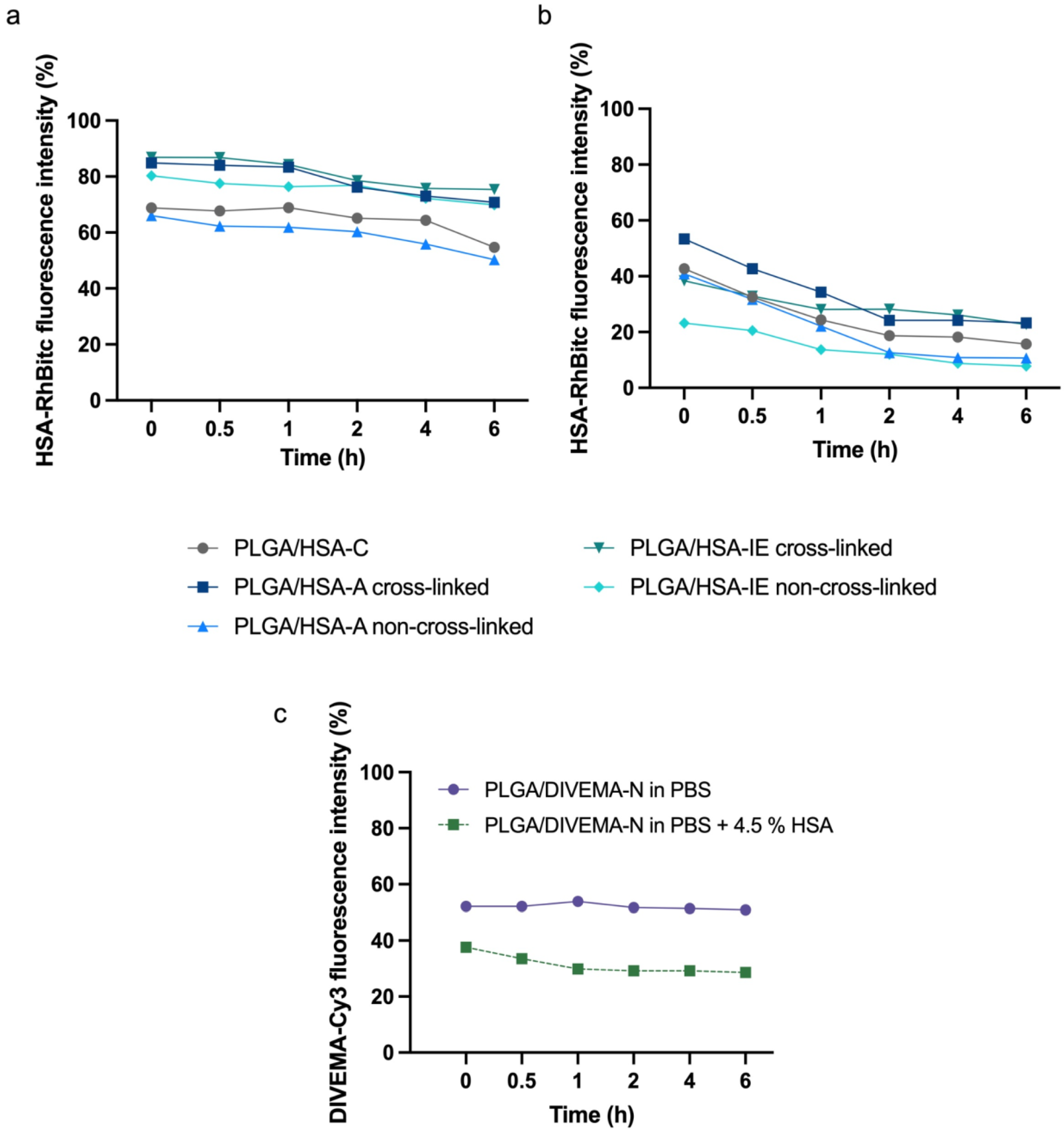
3.2. Evaluation of Integrity by Physicochemical Methods

The stability of the core-shell structures in biorelevant media was initially studied using DLS and spectrofluorimetry. As shown by the DLS measurements, all HSA- and DIVEMA-coated nanoparticles preserved their initial hydrodynamic diameters and polydispersity for at least 1 hour (Supplementary Table S1) in the HSA-containing medium (pH 7.4, PBS buffer with 4.5% of HSA) and for 6 hours (Supplementary Table S2) in the absence of HSA (PBS pH 7.4) (Figure 4). An increase in the average particle size (by volume) in the presence of HSA may indicate the adsorption of HSA on the surface of the particles [66], the replacement of the shell with HSA molecules, loss of stability, and aggregation. The formation of the protein corona on the PLGA/HSA NP (adsorption and interfacial embedding) during nanoparticle incubation in serum was confirmed in [56].
The next step involved evaluating the core-shell structure integrity of the nanoparticles by fluorescence spectroscopy. As mentioned above, the core and the shells of the nanoparticles were labeled with pairs of fluorescent dyes with known donor-acceptor properties, where Cy5 is the acceptor molecule, and Cy3 and rhodamine B are the donors [67]. As shown by the fluorescence measurements (Table 2), all dual-labeled PLGA nanoparticles had sufficient brightness for visualization based on a comparison of the parameters of free dyes and nanoparticles (Supplementary Table S3) [49,50,67].
As mentioned above, donor-acceptor pairs are capable of exhibiting the distance-dependent Förster resonance energy transfer (FRET) phenomenon, observed only in the case of the close proximity (< 10 nm) of both dye molecules, enabling the energy transfer from the donor fluorophore to the acceptor fluorophore [66,68,69]. In the present study, FRET was detected for the Cy5 – Cy3 (PLGA-Cy5/DIVEMA-Cy3 NP) and Cy5 – RhBITC (PLGA-Cy5/HSA-RhBITC) pairs. As shown in Figure 5 (representative images), the fluorescence intensity of the acceptor in double-labeled nanoparticles is detected when the excitation occurs at the donor wavelength (550 nm for RhBITC and 530 nm for Cy3).
The FRET phenomenon was observed for all types of PLGA/HSA NP, indicating the closeness of the shell and PLGA core (interaction between RhBITC and Cy5). Moreover, the most prominent FRET phenomenon was observed for the HSA shell obtained by the interphase embedding method (Supplementary Figure S2). Similarly, the PLGA/DIVEMA NP demonstrated core-shell integrity through a strong FRET signal between Cy3 (DIVEMA) and Cy5 (PLGA), indicating the close proximity of the donor (in the PLGA core) and the acceptor (in the shell) (Figure 5b). No FRET phenomenon was detected for the PLGA/P188 NP (data not shown), which may be due to the large distance between the donor in the P188-PhB shell and the acceptor in the PLGA nanoparticle core. Poloxamer 188 reacts with rhodamine B through terminal hydroxyl groups of polyoxyethylene (Figure 1) [30]. Presumably, when P188-RhB is adsorbed on the surface of PLGA nanoparticles, the hydrophobic chains of polyoxypropylene most likely interact with the hydrophobic core of PLGA, while the hydrophilic chains of polyoxyethylene with the grafted dye are solubilized by water molecules and protrude into the surrounding medium [70,71], which prevents them from approaching the nanoparticle surface and hinders efficient energy transfer from the donor to the acceptor.
To evaluate how the modification method influenced core-shell structure stability, the amount of detached shell was assessed within 6 hours of nanoparticle incubation in PBS and 4.5% HSA solution (imitating HSA concentration in plasma). The amount of detached shell was evaluated based on the fluorescence intensity of the supernatant after sample centrifugation at high acceleration for complete nanoparticle sedimentation (48,060×g). Stability of the core-shell structure was evaluated as the percentage of the shell remaining on the PLGA nanoparticle surface (Figure 6).
As shown in the graphs, considerable fractions of the HSA/RhBITC shell detached immediately upon their addition into the media for all nanoparticle types (Figure 6a). This initial burst effect was more pronounced for the PLGA/HSA NP obtained by conjugation and adsorption without crosslinking (~30%). In contrast, in the case of nanoparticles with cross-linked shells (adsorption-linked and interfacial embedding-linked), above 80% of the shell remained attached to the surface within 1 hour of incubation (Figure 6a).
The HSA-RhBITC shells appeared to be less stable in the serum-containing medium (4.5% HSA in PBS), with more than 50% of the shell detaching from the nanoparticle surface within the first hour (Figure 6b, Supplementary Table S4). Interestingly, the PLGA/HSA-IE non-cross-linked nanoparticles, which were highly stable in albumin-free medium, demonstrated the lowest stability in the presence of 4.5% HSA in the medium. The initial amount of detached HSA-RhBITC in all cases, including the PLGA/HSA-C NP obtained by conjugation, could be explained by the presence of HSA, loosely associated with the surface. The nanoparticle samples prepared with additional cross-linking stage were more stable compared to the non-linked ones. Thus, the cross-linked nanoparticles (PLGA/HSA-IE cross-linked and PLGA/HSA-A cross-linked) were chosen for further in vitro experiments.
A similar trend was observed for PLGA/DIVEMA NP (Figure 6c, Supplementary Table S4). The amount of detached shell was evaluated based on the relative fluorescence intensity (DIVEMA-Cy3) of the supernatant after sample centrifugation. Apparently, the high initial amount of detached DIVEMA-Cy3 resulted from a shell loosely associated with the surface.

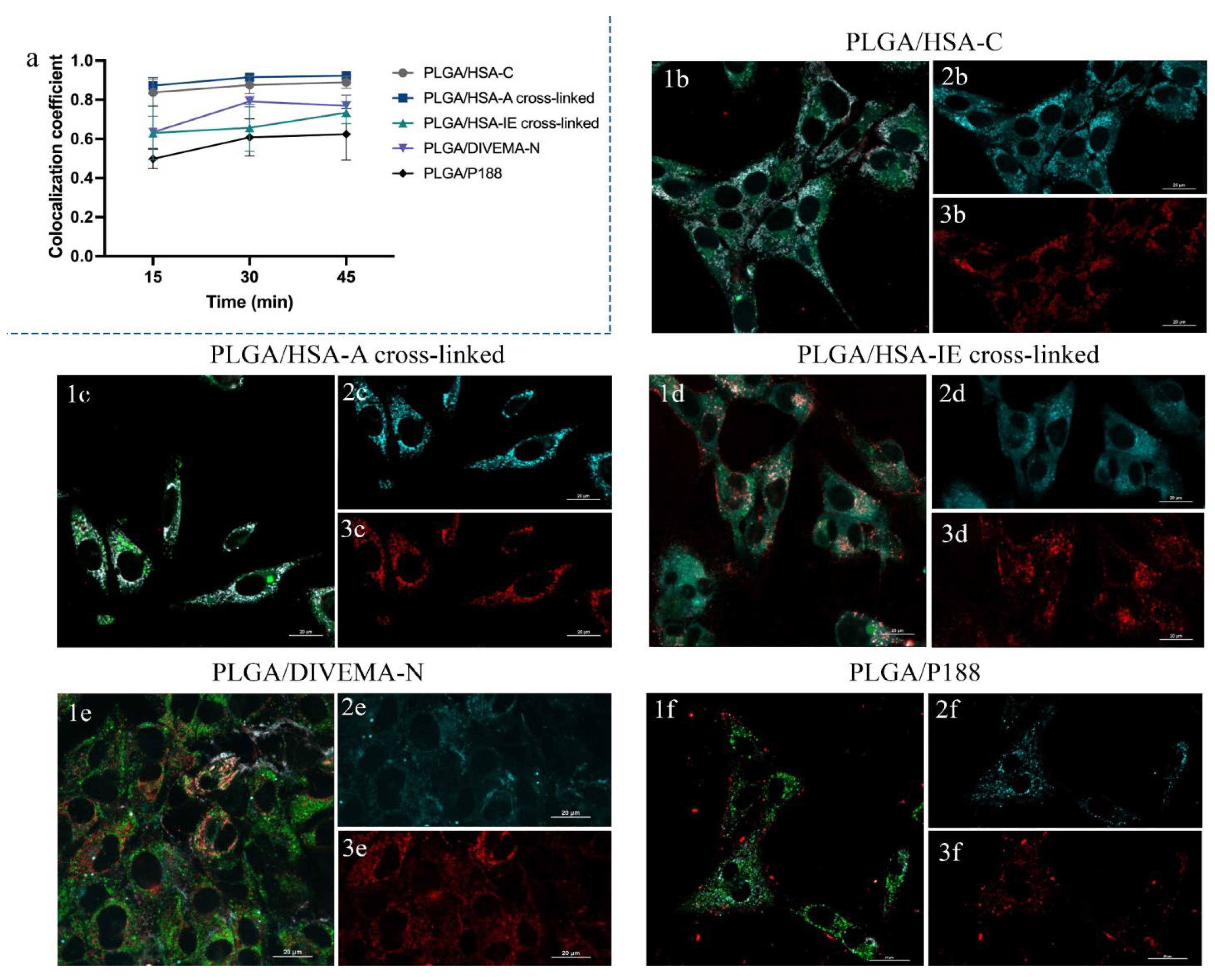
3.3. Investigation of the Integrity of the Core-Shell Nanoparticles Upon Internalization in Gl261 Cells by Confocal Microscopy

For the in vitro experiments, it is essential to track the integrity of the core-shell structures during their incubation with the cell culture media/serum. This study aimed to provide a more comprehensive overview of the nanoparticles’ structural changes (lifecycle) once they were added to the cells. Dual labeling of the nanoparticles allowed for evaluation of core-shell integrity over time by confocal laser scanning microscopy (CLSM). Life cell imaging was performed at different time intervals during nanoparticle incubation with Gl261 cells. The Manders’ Overlap Coefficient (MOC) was chosen to evaluate the core-shell colocalization, as it is widely used in fluorescence microscopy to quantify colocalization and is implemented in most biological image analysis software packages [73]. Unlike Pearson’s correlation, MOC is primarily sensitive to the co-occurrence of signals from two channels, almost independent of the signal level and proportionality.
To investigate how the modification method affected the integrity of the PLGA/HSA NP (i.e., the attachment of surface-bound albumin), colocalization coefficients between the PLGA-Cy5 (core) and HSA-RhBITC (shell) were calculated upon incubation with cells for different time periods (Supplementary Table S5). Only the cross-linked samples were used in these experiments due to their higher stability in model media. All three selected nanoparticle samples were efficiently internalized by the Gl261 cells (Figure 7). Colocalization coefficients above 0.5 indicated a correlation between the nanoparticle core and shell localization in the course of the experiment (Figure 7a). However, the MOC values were significantly lower at all time points for the PLGA/HSA NP obtained via interfacial embedding (0.63 compared to 0.83 for the nanoparticles produced by HSA conjugation or adsorption, Supplementary Table S5). Interestingly, unlike other PLGA/HSA nanoparticles (Figure 7b,c), the PLGA/HSA NP obtained by interfacial embedding (Figure 7d) tended to accumulate on the outer membrane after 15-30 min of incubation with cells, while the HSA-RhBITC signal was detected in the cell cytoplasm (Supplementary Figure S3). Subsequent incubation of this sample (30-45 min) led to increased colocalization between the core and the shell (0.73 after 45 min of incubation). These nanoparticles’ shells were most probably partly detached from the core upon introduction into the cell culture medium, due to dilution, and were internalized faster than the nanoparticles (PLGA-Cy5), suggesting that the PLGA/HSA nanoparticles obtained by interfacial embedding are less stable upon internalization into cells than those obtained by conjugation or adsorption (Supplementary Figure S4, S5).
Similar experiments were performed for the nanoparticles coated with DIVEMA, produced by the nanoprecipitation method (Figure 7e). The MOC values ranged from 0.63 to 0.79, suggesting that these nanoparticles preserved their core-shell structure (Supplementary Table S6). At 15 minutes, the MOC value was 0.42, indicating the presence of unbound or easily detached DIVEMA-Cy3. As a result, a significant amount of free DIVEMA-Cy3 accumulated in the cell cytoplasm during the first minutes of incubation (Supplementary Figure S6).
The P188 coating of the PLGA nanoparticles appeared to be relatively stable (Supplementary Figure S7). During the first 15 minutes of incubation of poloxamer-coated NP with cells, the MOC was 0.48 (Figure 7f, Supplementary Table S7), indicating that the uptake of free poloxamer by the cells occurred faster than the internalization of the PLGA nanoparticles. Further incubation (30-45 min) increased the average values of colocalization coefficients, indicating a high level of overlap between the PLGA-Cy5 and P188-RhB signals. The colocalization coefficients varying from 0.49 to 0.62 during incubation with the cells suggested that the core-shell structure partially preserved its integrity upon nanoparticle internalization and intracellular trafficking.
Colocalization coefficients were also calculated between the PLGA-Cy5 nanoparticle core and lysosomes (Supplementary Tables S5-7). Late endosomes and lysosomes are the main compartments involved in clathrin-mediated endocytosis, which is known to be one of the most common pathways for polymeric nanoparticle internalization in the vast majority of cell cultures [74]. Interestingly, the modification of PLGA nanoparticles with different shells did not significantly alter the colocalization of the PLGA-Cy5 nanoparticle core with lysosomes in Gl261 cells: the MOC values were found to vary from 0.3 to 0.5 for almost all samples, suggesting only partial accumulation of nanoparticles in lysosomes. Thus, it can be hypothesized that clathrin-mediated endocytosis is not the main internalization route for PLGA nanoparticles. These data align with the results of Beigulenko et al., who demonstrated that only a small portion of PLGA nanoparticles coated with fluorescently labeled poloxamer (P188-RhB) was associated with lysosomes [30]. It is also worth mentioning that caveolin-mediated endocytosis plays the primary role in albumin uptake into cells [75]. Therefore, the albumin coating of nanoparticles could be expected to affect the intracellular trafficking of PLGA nanoparticles. However, this was not observed in the present study. One possible reason could be a change in the conformation of albumin molecules on the nanoparticle surface, leading to alterations in the interaction between nanoparticles and cells [56]. These findings, however, require further investigation of the endocytosis routes, as they may also be attributed to the specific characteristics of the cell culture.

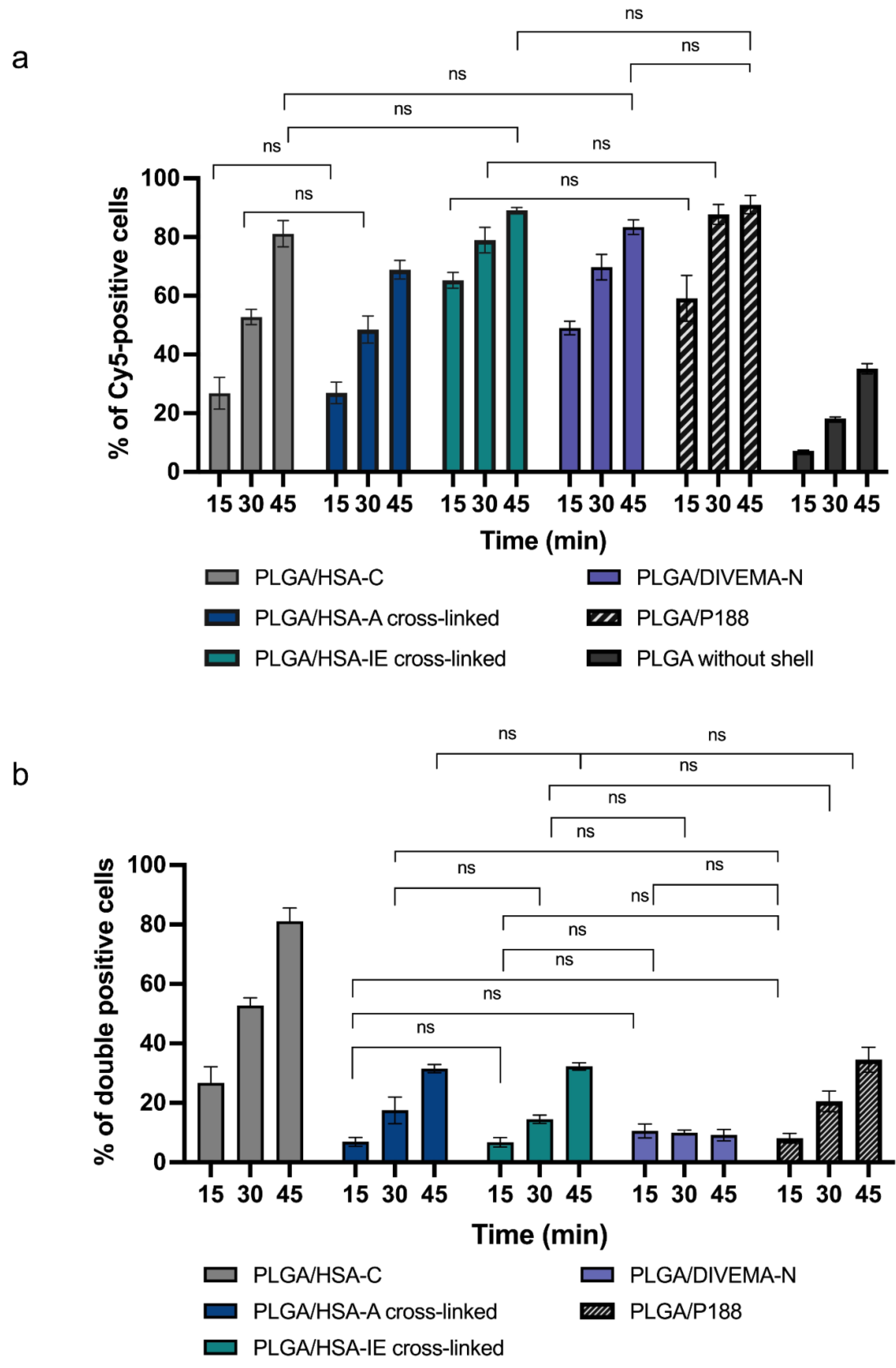
3.4. Investigation of the Dynamics of Nanoparticle Uptake by Gl261 Glioma Cells

To gain a deeper understanding of nanoparticle interactions with cells, the dynamics of their accumulation in Gl261 cells was investigated using flow cytometry. The nanoparticle uptake by the cells was determined as the percentage of NP-positive cells: PLGA-Cy5-positive or double-positive (both PLGA-Cy5- and HSA-RhBITC-, DIVEMA-Cy3- or P188-RhB-positive) cells (Figure 8). First, the percentage of PLGA-Cy5 positive cells was calculated to evaluate the possible influence of nanoparticle surface modification on their interaction with cells. Then, the percentage of cells positive for both PLGA-Cy5 (the core) and HSA-RhBITC, RhB or Cy3 (the shell) was determined to assess the structural integrity of the nanoparticles upon interaction with the cells over time. The nanoparticle uptake by the cells (percentage of nanoparticle-positive cells) tended to increase with time for nanoparticles obtained by all shell-forming techniques.
The presence of the HSA shell increased the number of nanoparticle-positive cells (Figure 8a, Supplementary Table S8). The percentage of PLGA-Cy5 positive cells significantly increased compared to control PLGA-Cy5 nanoparticles without shells (p < 0.0001 for all samples), regardless of the preparation method used (only statistically insignificant data are shown in the graph). This phenomenon could be attributed to the interaction of nanoparticles with albumin-binding proteins on the cell membrane. Interestingly, based on the PLGA-Cy5 fluorescence (nanoparticle core), the maximal percentage of cellular uptake was observed for the PLGA-HSA NP obtained by interfacial embedding: around 65 % of PLGA-Cy5-positive cells were detected after only 15 minutes of incubation, compared to 27% for PLGA/HSA nanoparticles produced by conjugation or adsorption (p <0.0001). However, the percentage of double-positive cells (positive for both PLGA-Cy5 and HSA-RhBITC) was the highest for the PLGA/HSA nanoparticles obtained by conjugation (Figure 8b). The number of double-positive cells was twice as high for PLGA/HSA-C compared to other HSA-coated batches at all investigated time points. These results are consistent with the confocal experiments, which demonstrated the high stability of the core-shell structure for PLGA/HSA-C.
The nanoparticles modified with P188 have been successfully used in several in vivo studies [33,35], suggesting that the P188 surface modification is sufficient to significantly enhance nanoparticle interaction with cells. Indeed, the PLGA nanoparticle coating with poloxamer 188 increased the number of PLGA-Cy5-positive cells by about 45%, compared to bare PLGA-Cy5 nanoparticles after 30 minutes of incubation with the cells (Figure 8a, Supplementary Table S9).
As shown in Figure 8a, the presence of the DIVEMA shell also significantly increased the number of PLGA-Cy5 positive cells (Figure 8a, Supplementary Table S10); however, the percentage of double-positive cells remained below 10% throughout the entire incubation period. These results are consistent with the stability tests performed in a 4.5% HSA solution, where a significant portion of the shell already detached from the nanoparticle surface within the first minutes of incubation in the HSA-containing medium. It is worth mentioning that flow cytometry can measure the number of events but cannot distinguish whether the nanoparticle core or shell is internalized by the cells or merely accumulated on the cell membrane [76]. In contrast, confocal microscopy enables detailed investigation of nanoparticle uptake, intracellular localization, and trafficking. Therefore, it is essential to use several methods, such as flow cytometry in conjunction with confocal microscopy, to fully characterize the behavior of nanoparticle formulations.
Thus, despite the different nature and properties of the shell-forming polymers, all shells enhanced the nanoparticle uptake by Gl261 cells as compared to uncoated nanoparticles. In the case of the PLGA/HSA nanoparticles, the highest stability of the HSA shell upon incubation in the HSA-containing media and with Gl261 cells was achieved by conjugation (PLGA/HSA-C), compared to the adsorption (PLGA/HSA-A) and interfacial embedding methods (PLGA/HSA-IE). Interestingly, the interfacial embedding method provided the highest shell content (HSA, DIVEMA) per polymer weight (~0.5 mg/mg PLGA) and high FRET-effect; however, in both cases low percentage of the core-shell double-positive cells (flow cytometry) and low Manders’ overlap coefficient (CLSM) indicated the loss of the core-shell integrity in the presence of HSA in the incubation medium. The differences between the core-shell systems may become more pronounced in the in vivo conditions, when they will be introduced into a very dynamic multicomponent environment and various blood components will be competing for the space on the particle surface. At the same time, the suggested combination of the evaluation techniques enables thorough analysis of the core-shell structure stability and may be useful for selection of the optimal preparation method already at the early stages of nanoparticle development.

4. Conclusions

In this study, the properties of core-shell nanoparticles with a PLGA core and shells made of polymers with significantly different structures and physicochemical properties were compared, with a focus on their structural integrity. The basic parameters of the nanoparticles, such as their size and zeta potential, depended on the shell composition and preparation technique; however, all particles had hydrodynamic diameters of ≤ 250 nm and were negatively charged — parameters considered optimal for drug delivery. Notable, despite differences in their chemical structures, polarity, molecular mass and hydrophobicity, all shells were largely retained by the core nanoparticles in both biorelevant media and within cells. Moreover, all shells enhanced the cellular uptake of the nanoparticles. The structural integrity of the core-shell PLGA nanoparticles was confirmed using a variety of methods, including physicochemical evaluation, fluorescence spectroscopy, confocal microscopy, and flow cytometry. The latter methods were facilitated by dual labeling of the nanoparticles with dyes possessing donor-acceptor properties. This comprehensive approach provides valuable insights into the behavior of core-shell nanoparticles in different environments and their interactions with tumor cells.

Supplementary Materials

Figure S1: Schematic representation of the preparation of dual labeled nanoparticles with a PLGA-Cy5 core and various shells; Figure S2: Fluorescent spectra of PLGA-Cy5/HSA-RhBITC NP in water; Figure S3: Confocal images of Gl261 cells after incubation with PLGA/HSA-IE cross-linked NP; Figure S4: Confocal images of Gl261 cells after incubation with PLGA/HSA-A-C NP; Figure S5: Confocal images of Gl261 cells after incubation with PLGA/HSA-A cross-linked NP; Figure S6: Confocal images of Gl261 cells after incubation with PLGA/DIVEMA-N NP; Figure S7: Confocal images of Gl261 cells after incubation with PLGA/P188 NP; Table S1: The size and size distribution of core-shell nanoparticles upon 6 hours of incubation in PBS in the presence of 4.5% HSA; Table S2: The size and size distribution of core-shell nanoparticles upon incubation in PBS; Table S3: Fluorescent properties of free dyes; Table S4: Estimation of the percentage of shell remaining on the nanoparticle surface after 6 hours of incubation in PBS in the presence and absence of 4.5% HSA; Table S5: Colocalization between the PLGA-Cy5 core and HSA-RhBITC shell and lysosomes in Gl261 cells; Table S6: Colocalization between the PLGA-Cy5 core and DIVEMA-Cy3 shell and lysosomes in Gl261 cells; Table S7: Colocalization between the PLGA-Cy5 core and P188-RhB shell and lysosomes in Gl261 cells; Table S8: Evaluation of PLGA-Cy5/HSA-RhBITC NP uptake by Gl261 cells at different time points; Table S9: Evaluation of PLGA-Cy5/P188-RhB uptake by Gl261 cells at different time points; Table S10: Evaluation of PLGA-Cy5/DIVEMA-Cy3 NP uptake by Gl261 cells at different time point.

Author Contributions

Conceptualization, Svetlana Gelperina; Formal analysis, Julia Malinovskaya, Julia Kotova and Veronika Vadekhina; Investigation, Tatyana Kovshova, Julia Malinovskaya, Julia Kotova, Marina Gorshkova, Lyudmila Vanchugova, Nadezhda Osipova, Pavel Melnikov, Veronika Vadekhina, Alexey Nikitin and Yulia Ermolenko; Methodology, Julia Malinovskaya, Marina Gorshkova, Pavel Melnikov, Yulia Ermolenko and Svetlana Gelperina; Writing – original draft, Tatyana Kovshova, Julia Malinovskaya and Julia Kotova; Writing – review & editing, Svetlana Gelperina.

Funding

This research was funded by the State Assignment of the Ministry of Science and Higher Education of the Russian Federation, project no. FSSM-2022-0003).

Data Availability Statement

Data will be made available on request.

Acknowledgments

The authors are grateful to the D. I. Mendeleev Center for the Collective Use of Scientific Equipment for performing analytical tests.

Conflicts of Interest

The authors declare no conflicts of interest.

References

  1. Champion, J.A.; Pustulka, S.M.; Ling, K.; Pish, S.L. Protein Nanoparticle Charge and Hydrophobicity Govern Protein Corona and Macrophage Uptake. ACS Appl Mater Interfaces 2020, 12. [Google Scholar] [CrossRef]
  2. Abed, N.; Couvreur, P. Nanocarriers for Antibiotics: A Promising Solution to Treat Intracellular Bacterial Infections. Int J Antimicrob Agents 2014, 43. [Google Scholar] [CrossRef] [PubMed]
  3. Gustafson, H.H.; Holt-Casper, D.; Grainger, D.W.; Ghandehari, H. Nanoparticle Uptake: The Phagocyte Problem. Nano Today 2015, 10. [Google Scholar] [CrossRef] [PubMed]
  4. Venditto, V.J.; Szoka, F.C. Cancer Nanomedicines: So Many Papers and so Few Drugs! Adv Drug Deliv Rev 2013, 65. [Google Scholar] [CrossRef] [PubMed]
  5. Danhier, F. To Exploit the Tumor Microenvironment: Since the EPR Effect Fails in the Clinic, What Is the Future of Nanomedicine? Journal of Controlled Release 2016, 244. [Google Scholar] [CrossRef]
  6. Golombek, S.K.; May, J.N.; Theek, B.; Appold, L.; Drude, N.; Kiessling, F.; Lammers, T. Tumor Targeting via EPR: Strategies to Enhance Patient Responses. Adv Drug Deliv Rev 2018, 130. [Google Scholar] [CrossRef]
  7. Miller, M.A.; Gadde, S.; Pfirschke, C.; Engblom, C.; Sprachman, M.M.; Kohler, R.H.; Yang, K.S.; Laughney, A.M.; Wojtkiewicz, G.; Kamaly, N.; et al. Predicting Therapeutic Nanomedicine Efficacy Using a Companion Magnetic Resonance Imaging Nanoparticle. Sci Transl Med 2015, 7. [Google Scholar] [CrossRef]
  8. Malinovskaya, J.; Salami, R.; Valikhov, M.; Vadekhina, V.; Semyonkin, A.; Semkina, A.; Abakumov, M.; Harel, Y.; Levy, E.; Levin, T.; et al. Supermagnetic Human Serum Albumin (HSA) Nanoparticles and PLGA-Based Doxorubicin Nanoformulation: A Duet for Selective Nanotherapy. Int J Mol Sci 2023, 24. [Google Scholar] [CrossRef]
  9. Kiaie, S.H.; Salehi-Shadkami, H.; Sanaei, M.J.; Azizi, M.; Shokrollahi Barough, M.; Nasr, M.S.; Sheibani, M. Nano-Immunotherapy: Overcoming Delivery Challenge of Immune Checkpoint Therapy. J Nanobiotechnology 2023, 21. [Google Scholar] [CrossRef]
  10. Jin, S.M.; Yoo, Y.J.; Shin, H.S.; Kim, S.; Lee, S.N.; Lee, C.H.; Kim, H.; Kim, J.E.; Bae, Y.S.; Hong, J.H.; et al. A Nanoadjuvant That Dynamically Coordinates Innate Immune Stimuli Activation Enhances Cancer Immunotherapy and Reduces Immune Cell Exhaustion. Nat Nanotechnol 2023, 18. [Google Scholar] [CrossRef]
  11. Ghidini, M.; Silva, S.G.; Evangelista, J.; Do Vale, M.L.C.; Farooqi, A.A.; Pinheiro, M. Nanomedicine for the Delivery of RNA in Cancer. Cancers (Basel) 2022, 14. [Google Scholar] [CrossRef]
  12. Tracey, S.R.; Smyth, P.; Barelle, C.J.; Scott, C.J. Development of next Generation Nanomedicine-Based Approaches for the Treatment of Cancer: We’ve Barely Scratched the Surface. Biochem Soc Trans 2021, 49. [Google Scholar] [CrossRef] [PubMed]
  13. Hare, J.I.; Lammers, T.; Ashford, M.B.; Puri, S.; Storm, G.; Barry, S.T. Challenges and Strategies in Anti-Cancer Nanomedicine Development: An Industry Perspective. Adv Drug Deliv Rev 2017, 108. [Google Scholar] [CrossRef] [PubMed]
  14. Jenjob, R.; Phakkeeree, T.; Crespy, D. Core-Shell Particles for Drug-Delivery, Bioimaging, Sensing, and Tissue Engineering. Biomater Sci 2020, 8. [Google Scholar] [CrossRef] [PubMed]
  15. Kumar, R.; Mondal, K.; Panda, P.K.; Kaushik, A.; Abolhassani, R.; Ahuja, R.; Rubahn, H.G.; Mishra, Y.K. Core-Shell Nanostructures: Perspectives towards Drug Delivery Applications. J Mater Chem B 2020, 8. [Google Scholar] [CrossRef]
  16. Dhas, N.L.; Raval, N.J.; Kudarha, R.R.; Acharya, N.S.; Acharya, S.R. Core-Shell Nanoparticles as a Drug Delivery Platform for Tumor Targeting. In Inorganic Frameworks as Smart Nanomedicines; 2018. [CrossRef]
  17. Rescignano, N.; Kenny, J.M. Stimuli-Responsive Core-Shell Nanoparticles. Core-Shell Nanostructures for Drug Delivery and Theranostics: Challenges, Strategies and Prospects for Novel Carrier Systems 2018, 245–258. [CrossRef]
  18. Mehandole, A.; Walke, N.; Mahajan, S.; Aalhate, M.; Maji, I.; Gupta, U.; Mehra, N.K.; Singh, P.K. Core–Shell Type Lipidic and Polymeric Nanocapsules: The Transformative Multifaceted Delivery Systems. AAPS PharmSciTech 2023, 24. [Google Scholar] [CrossRef] [PubMed]
  19. Rezvantalab, S.; Drude, N.I.; Moraveji, M.K.; Güvener, N.; Koons, E.K.; Shi, Y.; Lammers, T.; Kiessling, F. PLGA-Based Nanoparticles in Cancer Treatment. Front Pharmacol 2018, 9. [Google Scholar] [CrossRef] [PubMed]
  20. Dandamudi, M.; McLoughlin, P.; Behl, G.; Rani, S.; Coffey, L.; Chauhan, A.; Kent, D.; Fitzhenry, L. Chitosan-Coated Plga Nanoparticles Encapsulating Triamcinolone Acetonide as a Potential Candidate for Sustained Ocular Drug Delivery. Pharmaceutics 2021, 13. [Google Scholar] [CrossRef]
  21. Gu, P.; Wusiman, A.; Wang, S.; Zhang, Y.; Liu, Z.; Hu, Y.; Liu, J.; Wang, D. Polyethylenimine-Coated PLGA Nanoparticles-Encapsulated Angelica Sinensis Polysaccharide as an Adjuvant to Enhance Immune Responses. Carbohydr Polym 2019, 223. [Google Scholar] [CrossRef]
  22. Bee, S.L.; Hamid, Z.A.A.; Mariatti, M.; Yahaya, B.H.; Lim, K.; Bee, S.T.; Sin, L.T. Approaches to Improve Therapeutic Efficacy of Biodegradable PLA/PLGA Microspheres: A Review. Polymer Reviews 2018, 58. [Google Scholar] [CrossRef]
  23. Gref, R.; Domb, A.; Quellec, P.; Blunk, T.; Müller, R.H.; Verbavatz, J.M.; Langer, R. The Controlled Intravenous Delivery of Drugs Using PEG-Coated Sterically Stabilized Nanospheres. Adv Drug Deliv Rev 2012, 64. [Google Scholar] [CrossRef]
  24. Kim, S.M.; Patel, M.; Patel, R. PLGA Core-Shell Nano/Microparticle Delivery System for Biomedical Application. Polymers (Basel) 2021, 13. [Google Scholar] [CrossRef] [PubMed]
  25. Qiao, F.; Zhang, J.; Wang, J.; Du, B.; Huang, X.; Pang, L.; Zhou, Z. Silk Fibroin-Coated PLGA Dimpled Microspheres for Retarded Release of Simvastatin. Colloids Surf B Biointerfaces 2017, 158. [Google Scholar] [CrossRef]
  26. Wang, F.; Liu, X.; Yuan, J.; Yang, S.; Li, Y.; Gao, Q. Synthesis and Characterization of Poly(Lactic Acid-Co-Glycolic Acid) Complex Microspheres as Drug Carriers. J Biomater Appl 2016, 31. [Google Scholar] [CrossRef]
  27. Yuan, Y.; Shi, X.; Gan, Z.; Wang, F. Modification of Porous PLGA Microspheres by Poly-L-Lysine for Use as Tissue Engineering Scaffolds. Colloids Surf B Biointerfaces 2018, 161. [Google Scholar] [CrossRef]
  28. Gorshkova, M.; Vanchugova, L.; Osipova, N.; Nikitin, A.; Kotova, J.; Kovalenko, E.; Ermolenko, Y.; Malinovskaya, J.; Kovshova, T.; Gelperina, S. Hybrid DIVEMA/PLGA Nanoparticles as the Potential Drug Delivery System 2024(submitted, preprint). [CrossRef]
  29. Kotova, J.O.; Osipova, N.S.; Malinovskaya, J.A.; Melnikov, P.A.; Gelperina, S.E. Properties of Core–Shell Nanoparticles Based on PLGA and Human Serum Albumin Prepared by Different Methods. Mendeleev Communications 2023, 33. [Google Scholar] [CrossRef]
  30. Beigulenko, D. V.; Semyonkin, A.S.; Malinovskaya, J.A.; Melnikov, P.A.; Medyankina, E.I.; Kovshova, T.S.; Ermolenko, Y. V.; Gelperina, S.E. Internalization of PLGA Nanoparticles Coated with Poloxamer 188 in Glioma Cells: A Confocal Laser Scanning Microscopy Study. Mendeleev Communications 2023, 33. [Google Scholar] [CrossRef]
  31. Bollenbach, L.; Buske, J.; Mäder, K.; Garidel, P. Poloxamer 188 as Surfactant in Biological Formulations – An Alternative for Polysorbate 20/80? Int J Pharm 2022, 620. [Google Scholar] [CrossRef]
  32. Kabanov, A. V.; Alakhov, V.Y. Pluronic® Block Copolymers in Drug Delivery: From Micellar Nanocontainers to Biological Response Modifiers. Crit Rev Ther Drug Carrier Syst 2002, 19. [Google Scholar] [CrossRef]
  33. Pereverzeva, E.; Treschalin, I.; Treschalin, M.; Arantseva, D.; Ermolenko, Y.; Kumskova, N.; Maksimenko, O.; Balabanyan, V.; Kreuter, J.; Gelperina, S. Toxicological Study of Doxorubicin-Loaded PLGA Nanoparticles for the Treatment of Glioblastoma. Int J Pharm 2019, 554. [Google Scholar] [CrossRef]
  34. Gelperina, S.; Maksimenko, O.; Khalansky, A.; Vanchugova, L.; Shipulo, E.; Abbasova, K.; Berdiev, R.; Wohlfart, S.; Chepurnova, N.; Kreuter, J. Drug Delivery to the Brain Using Surfactant-Coated Poly(Lactide-Co-Glycolide) Nanoparticles: Influence of the Formulation Parameters. European Journal of Pharmaceutics and Biopharmaceutics 2010, 74. [Google Scholar] [CrossRef] [PubMed]
  35. Maksimenko, O.; Malinovskaya, J.; Shipulo, E.; Osipova, N.; Razzhivina, V.; Arantseva, D.; Yarovaya, O.; Mostovaya, U.; Khalansky, A.; Fedoseeva, V.; et al. Doxorubicin-Loaded PLGA Nanoparticles for the Chemotherapy of Glioblastoma: Towards the Pharmaceutical Development. Int J Pharm 2019, 572. [Google Scholar] [CrossRef] [PubMed]
  36. Hainsworth, J.D.; Forbes, J.T.; Grosh, W.W.; Greco, F.A. Phase I Study of MVE-2 Evaluating Toxicity and Biologic Response Modification Capability. Cancer Immunology Immunotherapy 1986, 22. [Google Scholar] [CrossRef] [PubMed]
  37. Regelson, W. The Biologic Activity of Polyanions: Past History and New Prospectives. Journal of Polymer Science: Polymer Symposia 1979, 66. [Google Scholar] [CrossRef]
  38. Breslow, D.S. Biologically Active Synthetic Polymers. Pure and Applied Chemistry 1976, 46. [Google Scholar] [CrossRef]
  39. Yang, G.; Liu, Y.; Teng, J.; Zhao, C.X. FRET Ratiometric Nanoprobes for Nanoparticle Monitoring. Biosensors (Basel) 2021, 11. [Google Scholar] [CrossRef]
  40. Kaeokhamloed, N.; Legeay, S.; Roger, E. FRET as the Tool for in Vivo Nanomedicine Tracking. Journal of Controlled Release 2022, 349. [Google Scholar] [CrossRef]
  41. Malinovskaya, Y.; Melnikov, P.; Baklaushev, V.; Gabashvili, A.; Osipova, N.; Mantrov, S.; Ermolenko, Y.; Maksimenko, O.; Gorshkova, M.; Balabanyan, V.; et al. Delivery of Doxorubicin-Loaded PLGA Nanoparticles into U87 Human Glioblastoma Cells. Int J Pharm 2017, 524. [Google Scholar] [CrossRef]
  42. Zhukova, V.; Osipova, N.; Semyonkin, A.; Malinovskaya, J.; Melnikov, P.; Valikhov, M.; Porozov, Y.; Solovev, Y.; Kuliaev, P.; Zhang, E.; et al. Fluorescently Labeled Plga Nanoparticles for Visualization in Vitro and in Vivo: The Importance of Dye Properties. Pharmaceutics 2021, 13. [Google Scholar] [CrossRef]
  43. Yang, T.; Sun, D.; Xu, P.; Li, S.; Cen, Y.; Li, Y.; Xu, Q.; Sun, Y.; Li, W.; Lin, Y.; et al. Stability of Bovine Serum Albumin Labelled by Rhodamine B Isothiocyanate. Biomedical Research (India) 2017, 28, ISSN: 0970938X.
  44. Langer, K.; Balthasar, S.; Vogel, V.; Dinauer, N.; Von Briesen, H.; Schubert, D. Optimization of the Preparation Process for Human Serum Albumin (HSA) Nanoparticles. Int J Pharm 2003, 257. [Google Scholar] [CrossRef]
  45. Manoochehri, S.; Darvishi, B.; Kamalinia, G.; Amini, M.; Fallah, M.; Ostad, S.N.; Atyabi, F.; Dinarvand, R. Surface Modification of PLGA Nanoparticles via Human Serum Albumin Conjugation for Controlled Delivery of Docetaxel. DARU, Journal of Pharmaceutical Sciences 2013, 21. [Google Scholar] [CrossRef] [PubMed]
  46. Volkova, I.F.; Gorshkova, M.Y.; Ivanov, P.E.; Stotskaya, L.L. New Scope for Synthesis of Divinyl Ether and Maleic Anhydride Copolymer with Narrow Molecular Mass Distribution. Polym Adv Technol 2002, 13. [Google Scholar] [CrossRef]
  47. Kumskova, N.; Ermolenko, Y.; Osipova, N.; Semyonkin, A.; Kildeeva, N.; Gorshkova, M.; Kovalskii, A.; Kovshova, T.; Tarasov, V.; Kreuter, J.; et al. How Subtle Differences in Polymer Molecular Weight Affect Doxorubicin-Loaded PLGA Nanoparticles Degradation and Drug Release. J Microencapsul 2020, 37. [Google Scholar] [CrossRef] [PubMed]
  48. Ashoka, A.H.; Aparin, I.O.; Reisch, A.; Klymchenko, A.S. Brightness of Fluorescent Organic Nanomaterials. Chem Soc Rev 2023, 52. [Google Scholar] [CrossRef]
  49. Taniguchi, M.; Lindsey, J.S. Database of Absorption and Fluorescence Spectra of >300 Common Compounds for Use in PhotochemCAD. Photochem Photobiol 2018, 94, 290–327. [Google Scholar] [CrossRef]
  50. Malicka, J.; Gryczynski, I.; Fang, J.; Lakowicz, J.R. Fluorescence Spectral Properties of Cyanine Dye-Labeled DNA Oligomers on Surfaces Coated with Silver Particles. Anal Biochem 2003, 317. [Google Scholar] [CrossRef]
  51. Tao, H. yu; Wang, R. qi; Sheng, W. jin; Zhen, Y. su The Development of Human Serum Albumin-Based Drugs and Relevant Fusion Proteins for Cancer Therapy. Int J Biol Macromol 2021, 187. [Google Scholar] [CrossRef]
  52. Butler, G.B. Synthesis and Antitumor Activity of “Pyran Copolymer. ” Journal of Macromolecular Science, Part C 1982, 22. [Google Scholar] [CrossRef]
  53. Nel, A.E.; Mädler, L.; Velegol, D.; Xia, T.; Hoek, E.M.V.; Somasundaran, P.; Klaessig, F.; Castranova, V.; Thompson, M. Understanding Biophysicochemical Interactions at the Nano-Bio Interface. Nat Mater 2009, 8. [Google Scholar] [CrossRef]
  54. Adamczyk, Z.; Nattich-Rak, M.; Dąbkowska, M.; Kujda-Kruk, M. Albumin Adsorption at Solid Substrates: A Quest for a Unified Approach. J Colloid Interface Sci 2018, 514. [Google Scholar] [CrossRef]
  55. Sato, H.; Nakhaei, E.; Kawano, T.; Murata, M.; Kishimura, A.; Mori, T.; Katayama, Y. Ligand-Mediated Coating of Liposomes with Human Serum Albumin. Langmuir 2018, 34. [Google Scholar] [CrossRef] [PubMed]
  56. Hyun, H.; Park, J.; Willis, K.; Park, J.E.; Lyle, L.T.; Lee, W.; Yeo, Y. Surface Modification of Polymer Nanoparticles with Native Albumin for Enhancing Drug Delivery to Solid Tumors. Biomaterials 2018, 180. [Google Scholar] [CrossRef] [PubMed]
  57. Li, C.; Li, Y.; Gao, Y.; Wei, N.; Zhao, X.; Wang, C.; Li, Y.; Xiu, X.; Cui, J. Direct Comparison of Two Albumin-Based Paclitaxel-Loaded Nanoparticle Formulations: Is the Crosslinked Version More Advantageous? Int J Pharm 2014, 468. [Google Scholar] [CrossRef] [PubMed]
  58. Gorshkova, M.; Lebedeva, T.; Chervina, L.; Stotskaya, L. Kinetics of the Hydrolysis of Divinyl Ether-Maleic Anhydride Copolymer in Water and Stability of the Copolymer in Acetone. Polymer Science, Series A 1995, 37, 1003–1007. [Google Scholar]
  59. Liu, M.; Zheng, Y.; Liu, Y.; Zhang, Z.; Wang, Y.; Li, J.; Chen, Q.; Li, J.; Huang, Y.; Yin, Q. Effects of Poly(Vinyl Alcohol) and Poly(Acrylic Acid) on Interfacial Properties and Stability of Compound Droplets. Int J Hydrogen Energy 2020, 45. [Google Scholar] [CrossRef]
  60. Jha, P.K.; Desai, P.S.; Li, J.; Larson, R.G. PH and Salt Effects on the Associative Phase Separation of Oppositely Charged Polyelectrolytes. Polymers (Basel) 2014, 6. [Google Scholar] [CrossRef]
  61. Kovshova, T.; Osipova, N.; Alekseeva, A.; Malinovskaya, J.; Belov, A.; Budko, A.; Pavlova, G.; Maksimenko, O.; Nagpal, S.; Braner, S.; et al. Exploring the Interplay between Drug Release and Targeting of Lipid-like Polymer Nanoparticles Loaded with Doxorubicin. Molecules 2021, 26. [Google Scholar] [CrossRef]
  62. Wang, X.; Liang, Y.; Fei, S.; He, H.; Zhang, Y.; Yin, T.; Tang, X. Formulation and Pharmacokinetics of HSA-Core and PLGA-Shell Nanoparticles for Delivering Gemcitabine. AAPS PharmSciTech 2018, 19. [Google Scholar] [CrossRef]
  63. Filippov, S.K.; Khusnutdinov, R.; Murmiliuk, A.; Inam, W.; Zakharova, L.Y.; Zhang, H.; Khutoryanskiy, V. V. Dynamic Light Scattering and Transmission Electron Microscopy in Drug Delivery: A Roadmap for Correct Characterization of Nanoparticles and Interpretation of Results. Mater Horiz 2023, 10. [Google Scholar] [CrossRef]
  64. Inyang, E.; Kuriakose, A.E.; Chen, B.; Nguyen, K.T.; Cho, M. Engineering Delivery of Nonbiologics Using Poly(Lactic- Co-Glycolic Acid) Nanoparticles for Repair of Disrupted Brain Endothelium. ACS Omega 2020, 5. [Google Scholar] [CrossRef]
  65. Dong, H.; Tian, L.; Gao, M.; Xu, H.; Zhang, C.; Lv, L.; Zhang, J.; Wang, C.; Tian, Y.; Ma, X. Promising Galactose-Decorated Biodegradable Poloxamer 188-Plga Diblock Copolymer Nanoparticles of Resibufogenin for Enhancing Liver Cancer Therapy. Drug Deliv 2017, 24. [Google Scholar] [CrossRef] [PubMed]
  66. Ural, M.S.; Joseph, J.M.; Wien, F.; Li, X.; Tran, M.A.; Taverna, M.; Smadja, C.; Gref, R. A Comprehensive Investigation of the Interactions of Human Serum Albumin with Polymeric and Hybrid Nanoparticles. Drug Deliv Transl Res 2024, 14, 2188–2202. [Google Scholar] [CrossRef] [PubMed]
  67. Dietrich, A.; Buschmann, V.; Müller, C.; Sauer, M. Fluorescence Resonance Energy Transfer (FRET) and Competing Processes in Donor-Acceptor Substituted DNA Strands: A Comparative Study of Ensemble and Single-Molecule Data. Reviews in Molecular Biotechnology 2002, 82. [Google Scholar] [CrossRef] [PubMed]
  68. Sinigaglia, G.; Magro, M.; Miotto, G.; Cardillo, S.; Agostinelli, E.; Zboril, R.; Bidollari, E.; Vianello, F. Catalytically Active Bovine Serum Amine Oxidase Bound to Fluorescent and Magnetically Drivable Nanoparticles. Int J Nanomedicine 2012, 7. [Google Scholar] [CrossRef]
  69. Wu, L.; Huang, C.; Emery, B.P.; Sedgwick, A.C.; Bull, S.D.; He, X.P.; Tian, H.; Yoon, J.; Sessler, J.L.; James, T.D. Förster Resonance Energy Transfer (FRET)-Based Small-Molecule Sensors and Imaging Agents. Chem Soc Rev 2020, 49. [Google Scholar] [CrossRef]
  70. Sanchez-Gaytan, B.L.; Fay, F.; Hak, S.; Alaarg, A.; Fayad, Z.A.; Pérez-Medina, C.; Mulder, W.J.M.; Zhao, Y. Real-Time Monitoring of Nanoparticle Formation by FRET Imaging. Angewandte Chemie - International Edition 2017, 56. [Google Scholar] [CrossRef]
  71. Kayes, J.B.; Rawlins, D.A. Adsorption Characteristics of Certain Polyoxyethylene-Polyoxypropylene Block Co-Polymers on Polystyrene Latex. Colloid and Polymer Science Kolloid-Zeitschrift & Zeitschrift für Polymere 1979, 257. [Google Scholar] [CrossRef]
  72. Santander-Ortega, M.J.; Csaba, N.; Alonso, M.J.; Ortega-Vinuesa, J.L.; Bastos-González, D. Stability and Physicochemical Characteristics of PLGA, PLGA:Poloxamer and PLGA:Poloxamine Blend Nanoparticles: A Comparative Study. Colloids Surf A Physicochem Eng Asp 2007, 296, 132–140. [Google Scholar] [CrossRef]
  73. Dunn, K.W.; Kamocka, M.M.; McDonald, J.H. A Practical Guide to Evaluating Colocalization in Biological Microscopy. Am J Physiol Cell Physiol 2011, 300. [Google Scholar] [CrossRef]
  74. Mettlen, M.; Chen, P.H.; Srinivasan, S.; Danuser, G.; Schmid, S.L. Regulation of Clathrin-Mediated Endocytosis. Annu Rev Biochem 2018, 87. [Google Scholar] [CrossRef]
  75. Chatterjee, M.; Ben-Josef, E.; Robb, R.; Vedaie, M.; Seum, S.; Thirumoorthy, K.; Palanichamy, K.; Harbrecht, M.; Chakravarti, A.; Williams, T.M. Caveolae-Mediated Endocytosis Is Critical for Albumin Cellular Uptake and Response to Albumin-Bound Chemotherapy. Cancer Res 2017, 77. [Google Scholar] [CrossRef] [PubMed]
  76. Salvati, A.; Nelissen, I.; Haase, A.; Åberg, C.; Moya, S.; Jacobs, A.; Alnasser, F.; Bewersdorff, T.; Deville, S.; Luch, A.; et al. Quantitative Measurement of Nanoparticle Uptake by Flow Cytometry Illustrated by an Interlaboratory Comparison of the Uptake of Labelled Polystyrene Nanoparticles. NanoImpact 2018, 9, 42–50. [Google Scholar] [CrossRef]
Figure 1. Chemical structures of fluorescently labeled polymers used for the preparation of core-shell nanoparticles: (1) PLGA conjugate with Cyanine5 (PLGA-Cy5), (2) DIVEMA in anhydride form, (3) DIVEMA conjugate with Cyanine3 (DIVEMA-Cy3), (4) poloxamer 188 conjugate with rhodamine B (P188-RhB), (5) HSA conjugate with rhodamine B (HSA-RhBITC).
Preprints 141234 g001
Figure 2. Schematic representation of the types of dual-labeled core-shell PLGA nanoparticles.
Preprints 141234 g002
Figure 3. TEM images: (a) PLGA/HSA-С, (b) PLGA/HSA-A cross-linked; (c) PLGA without shell, (d) PLGA/DIVEMA-N (JEOL JEM-1400 electron microscope, negative staining with Uranyless stain); (e) PLGA/HSA-IE cross-linked (TEM Tecnai™ 12 G 2 BioTwin Spirit electron microscope, negative staining with uranyl acetate).
Preprints 141234 g003
Figure 4. Average diameters of core-shell nanoparticles during 6 hours of incubation in model media (37 °C, mean ± SD; n = 3): (a) PLGA-Cy5/HSA-RhB incubation in PBS, (b) PLGA-Cy5/HSA-RhB incubation in PBS in the presence of 4.5% HSA; (c) PLGA-Cy5/DIVEMA-Cy3 incubation in PBS in the presence or absence of 4.5% HSA. PLGA nanoparticles without a shell were used as a control. Size measurements were performed by DLS (volume distribution).
Preprints 141234 g004
Figure 5. Fluorescent spectra of representative nanoparticle samples: (a) PLGA-Cy5/HSA-RhBITC (PLGA/HSA-IE cross-linked); (b) PLGA-Cy5/DIVEMA-Cy3 (PLGA/DIVEMA-N).
Preprints 141234 g005
Figure 6. Evaluation of core-shell structure stability. Percentage of total shell-dye concentration remaining on the nanoparticle surface (based on relative fluorescence intensity values); (a) 6 hours of PLGA-Cy5/HSA-RhB incubation in PBS, (b) 6 hours of PLGA-Cy5/HSA-RhB incubation in PBS in the presence of 4.5% HSA; (c) 6 hours of PLGA-Cy5/DIVEMA-Cy3 incubation in PBS in the presence or absence of 4.5% HSA. Representative data.
Preprints 141234 g006
Figure 7. Confocal images of Gl261 cells after incubation with core-shell nanoparticles. (a) Manders’ overlap coefficient (MOC) showing colocalization between the core and the shell of the nanoparticles at different time points (15, 30, 45 min; n = 3); (b) PLGA/HSA-C; (c) PLGA/HSA-A cross-linked, (d) PLGA/HSA-IE cross-linked; (e) PLGA-DIVEMA-N, (f) PLGA/P188. (1) merged images (green – lysosomes, cyan – shell, red – core); (2) shell (HSA-RhBITC, DIVEMA-Cy3 or P188-RhB); (3) core (PLGA-Cy5). CSLM. Scale bar: 20 μm.
Preprints 141234 g007
Figure 8. Core-shell nanoparticle uptake by Gl261 cells depending on the nanoparticle preparation technique and incubation time (15, 30 and 45 min of incubation). (a) % Cy5-positive cells; (b) % Cy5- and RhBITC/Cy3/RhB double-positive cells for PLGA-Cy5/HSA-RhBITC NP, PLGA-Cy5/DIVEMA-Cy3 NP, and PLGA-Cy5/P188-RhB NP, respectively. Statistical analysis was performed by two-way ANOVA followed by Tukey’s multiple comparisons test (mean ± SD, n =3); ns = not significant, p > 0.05.
Preprints 141234 g008
Table 1. Physicochemical parameters of the nanoparticles (mean ± SD, n=3).
Method of shell formation (method of nanoparticle preparation*) Nanoparticle size and size distribution, nm Zeta potential,
mV
Shell content, mg/mg PLGA
Mean diameter PDI Volume size distribution
PLGA-Cy5/HSA-RhBITC NP
Conjugation
(PLGA/HSA-C)
153 ± 2 0.201 ± 0.017 147 (100%) -7.0 ± 1.2 0.48
Adsorption, cross-linked
(PLGA/HSA-A cross-linked)
148 ± 2 0.183 ± 0.018 143 (100%) -7.6 ± 0.8 0.11
Adsorption, non-cross-linked
(PLGA/HSA-A
non-cross-linked)
135 ± 1 0.118 ± 0.014 133 (100%) -6.2 ± 2.7 0.11
Interfacial embedding,
cross-linked
(PLGA/HSA-IE cross-linked)
103 ± 3 0.138 ± 0.014 92 (100%) -26.3 ± 0.7 0.50
Interfacial embedding,
non-cross-linked
(PLGA/HSA-IE
non-cross-linked)
90 ± 1 0.056 ± 0.022 83 (100%) -31.9 ± 2.7 0.52
PLGA without shell *** 116 ± 2 0.098 ± 0.015 108 (100%) -20.9 ± 1.1 -
PLGA-Cy5/DIVEMA-Cy3 NP
Interfacial embedding
(PLGA/DIVEMA-H)
264 ± 4 0.225 ± 0.009 341 (94.2%)
5118 (5.8%)
-34.9 ± 0.1 0.10
PLGA without shell ** 178 ± 24 0.230 ± 0.050 290 (100%) -20.3 ± 1.7 -
Interfacial embedding (nanoprecipitation)
(PLGA/DIVEMA-N)
180 ± 21 0.274 ± 0.007 284 (100%) -51.3 ± 1.4 0.47
NP without shell (nanoprecipitation) ** 130 ± 15 0.070 ± 0.015 170 (100%) -18.5±3.5 -
PLGA-Cy5/P188-RhB
Adsorption of P188-RhB 97 ± 1 0.100 ± 0.007 85 (100%) -21.8 ± 0.7 0.017
Adsorption of P188 110 ± 2 0.16 ± 0.01 97 (100%) -23.5 ± 1.1 0.006
PLGA without shell *** 100 ± 2 0.078 ± 0.015 100 (100%) -20.9 ± 1.1 -
* The nanoparticles were prepared by the high-pressure homogenization—solvent evaporation technique unless indicated otherwise. **The nanoparticles were prepared with PVA as a surfactant. *** Plain NP were used to obtain core-shell particles by adsorption or conjugation methods.
Table 2. Fluorescent properties of core-shell nanoparticles, representative data.
Sample Dye content, µg/ml Dye to polymer ratio, µg/mg Quantum Yield Brightness,M-1 cm-1
PLGA-Cy5/HSA-RhBITC RhBITC Cy5 RhBITC Cy5 RhBITC Cy5 RhBITC Cy5
PLGA/HSA-С 4.80 1.62 3.93 0.64 0.09 0.12 1.03x10⁸ 3.61x10⁷
PLGA/HSA-A cross-linked 3.62 1.81 12.07 0.68 0.11 0.18 3.78x10⁷ 6.70x10⁷
PLGA/HSA-A non-cross-linked 4.86 2.53 16.20 0.88 0.15 0.23 3.41x10⁷ 6.46x10⁷
PLGA/HSA-IE cross-linked 3.11 1.31 0.98 0.78 0.19 0.31 1.42x10⁸ 1.46x10⁷
PLGA/HSA-IE non-cross-linked 2.64 1.80 0.80 0.85 0.21 0.29 9.53x10⁷ 1.01x10⁷
PLGA-Cy5/DIVEMA-Cy3 Cy3 Cy5 Cy3 Cy5 Cy3 Cy5 Cy3 Cy5
4.12 2.36 11.4 2.03 0.12 0.21 1.66x10⁸ 2.56x10⁸
PLGA-Cy5/P188-RhB RhB Cy5 RhB Cy5 RhB Cy5 RhB Cy5
4.26 4.65 44.8 0.83 0.63 0.37 3.77x10⁷ 4.19x10⁷
Disclaimer/Publisher’s Note: The statements, opinions and data contained in all publications are solely those of the individual author(s) and contributor(s) and not of MDPI and/or the editor(s). MDPI and/or the editor(s) disclaim responsibility for any injury to people or property resulting from any ideas, methods, instructions or products referred to in the content.
Copyright: This open access article is published under a Creative Commons CC BY 4.0 license, which permit the free download, distribution, and reuse, provided that the author and preprint are cited in any reuse.
Prerpints.org logo

Preprints.org is a free preprint server supported by MDPI in Basel, Switzerland.

Subscribe

Disclaimer

Terms of Use

Privacy Policy

Privacy Settings

© 2025 MDPI (Basel, Switzerland) unless otherwise stated