
Submitted:
29 July 2024
Posted:
30 July 2024
You are already at the latest version
Abstract
Magnetic Fe3O4 nanoparticles (MNPs) are becoming more important every day in drug targeting. We prepared here MNPs in a simple one-step reaction by following the solvothermal method assisted by azide and alkyne functionalized polyethylene glycol (PEG400) polymers, as well as by PEG6000 and the polyol -cyclodextrin (CD). The composition, morphology, and structure of the nanospheres were characterized using Transmission Electron Microscopy (TEM), Nuclear Magnetic Resonance (NMR), X-Ray Diffraction Diffractometry (XRD), Atomic Force Microscopy (AFM), Fourier-Transform Infrared Spectroscopy (FT-IR), Matrix-Assisted Laser Desorption/Ionization (MALDI) and Vibrating Sample Magnetometry (VSM). The obtained nanoparticles (@Fe3O4-PEGs and @Fe3O4-CD) showed diameters between 90-250 nm, depending on the polymer used and the Fe3O4·6H2O precursor concentration, typically, 0.13 M, 200 °C, and 24 h of reaction. MNPs exhibited superparamagnetic properties with high saturation of magnetization at room temperature, reaching values of 59.9 emu/g (@Fe3O4-PEG6000), and no ferromagnetism. Likewise, they showed temperature elevation after applying an alternating magnetic field (AMF) in all cases, obtaining Specific Absorption Rate (SAR) values of up to 51.87 ± 2.23 W/g in the case of @Fe3O4-PEG6000. Both PEGs and the βCD played a crucial role as electrostatic stabilizers in the formation of Fe3O4 nanospheres, providing a polymeric/polyol coating around the magnetic cores. Additionally, the formed systems are susceptible for click chemistry, as was demonstrated in the case of the cannabidiol-propargyl derivative (CBD-Pro), which was synthesized and covalently attached to the azide functionalized surface of the system @Fe3O4-PEG400-N3. Prepared MNPs are highly dispersible in water, remaining in suspension for over 3 days, and non-toxic in the T84 human colon cancer cell line, indicating that they are ideal candidates for biomedical applications.
Keywords:
CBD-derivatives
; Magnetite
; Metalloorganic Nanoparticles
; Surface functionalization
; Hyperthermia
; SPION
1. Introduction
It has been theorized that the emergence of novel properties and potential applications in materials with tiny and monodisperse dimensions, would be a significant advancement. Therefore, the development and synthesis of magnetic nanoparticles (MNPs) has gained crucial relevance in research, generating a continuously growing interest, due to their potential applications in areas from chemistry, physics, or electronics [1]. Medical applications have also been strongly developed, ranging from magnetic resonance imaging and medical diagnostics to magnetic hyperthermia and drug delivery technologies [2,3,4]
The main limitation of MNPs is the inherent long-term instability. Degradation occurs through two main pathways: (1) loss of dispersibility, where MNPs tend to aggregate due to Van der Waals forces, overcoming the high surface energy and strong magnetic attraction between particles, and (2) loss of magnetism, when oxidation of MNPs occurs [1,5]. These issues can be addressed by developing a functionalized surface around the MNP cores, providing a protective shell where it can be covalently attached different species, such small molecules, peptides, or even antibodies [6,7]. Moreover, this functionalization can improve biocompatibility and hydrophilicity of the magnetic material. In case of superparamagnetic systems, the response to an external magnetic field is even better, improving individual properties and their application through magnetic hyperthermia therapies.
Several studies have shown that the encapsulation of MNPs prevents agglomeration by reducing the available surface area, and thus indirectly decreasing the surface energy which conduces to agglomeration. Encapsulation also creates a protective layer around naked MNPs, preventing them from oxidizing to maghemite (γ-Fe2O3) after prolonged exposure to air [8]. The introduction of this coating layer on the surface of MNPs ensures the chemical stability of MNPs and preserves their magnetic properties. Coated MNPs also present advantages in biomedical applications compared to naked MNPs, including lower cytotoxicity, higher cytocompatibility, and better bioconjugation capability due to the presence of reactive materials on its surface [9], which, together with its magnetic potential, could be useful for the treatment of certain diseases such as cancer. In order to address this point, it is necessary to functionalize and coat MNPs with specific polymers and macromolecules designed to improve the conjugation. This involves a precise sequence, first, the synthesis of MNPs, second, surface functionalization, and third, polymerization and coating of the magnetic cores, all within a relatively short time frame to prevent agglomeration [10,11,12,13].
One of the most distinguished methods to synthesize magnetite is the solvothermal decomposition, initially reported by Deng et al. for the preparation of monodisperse single-crystal ferrite microspheres, affording size distribution of 90-200 nm [14]. Many studies have applied this methodology to prepare MNPs. The search for different size ranges controlling the reaction time [15], the analysis of the effect of surfactants [16], the use of urea as a homogeneous precipitant [17], or citrate groups as electrostatic stabilizers [18,19], have been studied. PEG coating has also been carried out for the stabilization of magnetite nanoparticles, together with the common use of polyols with various reducing capacities, for MNPs preparation [20,21,22,23]. βCD-Citrate and carboxymethyl-βCD derivatives have also been used for stabilization of gum-arabic modified magnetite and pristine magnetite nanosystems, respectively, via carbodiimide activation [24,25]. More complex derivatives, as mono-6-deoxy-6-(p-tolylsulfonyl)-βCD have been used to surface modify and stabilize Fe3O4 nanoparticles by electrochemical methods [26], and more recently, βCD combined with ionic liquids was used for coating of co-precipitation prepared MNPs [27], while copolymers as βCD-co-(methacryloyloxy)ethyl phosphorylcholine were used for coating Fe3O4 nanoparticles in hemocompatibility studies [28]. All these methods involve the synthesis of MNPs previously to the surface modification.
However, to the best of our knowledge, few has been done towards the synthesis of MNPs using PEGs previously functionalized. Here we present our results on the preparation of MNPs by following the solvothermal method, using polyols or polymers as precursors: βCD, and PEG400 and its alkyne and azide derivatives, and PEG6000, all of them acting as surfactants and stabilizers. This one-step process resulted in obtaining MNPs with a hydrophilic coating, providing a protective layer over the magnetic clusters. In addition to its stabilizing function, the PEG coating opens a unique opportunity for use in target drug delivery systems and hyperthermia treatment. In this sense, we expand the study by introducing a functional coating through the synthesis of PEG derivatives incorporating alkyne and azide groups and tested the magnetic properties and biocompatibility of the nanoformulations in vitro. These functional groups enable the performance of click chemistry reactions, which represents a substantial contribution due to the versatility and potential that these reactions offer [29]. To illustrate this point, we also carried out the preparation of a CBD-alkyne derivative to be incorporated by covalent bonds formation by the click reaction to the azide-modified surface of the MNP. This approach marks a significant advance toward creating multifunctional and versatile systems that can be adapted for a wide range of biomedical applications.
2. Experimental
2.1. Chemicals
Iron(III) chloride hexahydrate, N,N-dimethylformamide (DMF), NaN3, propargyl chloride, propargyl bromide, dichloromethane (DCM), 3-bromopropionyl chloride, ethanol, PEG400, PEG6000, ethylene glycol, sodium acetate, β-cyclodextrin, iron(III) acetylacetonate and triethylamine (TEA) were purchased from Merck. All chemicals were used without further purification. Cannabidiol (CBD) was synthesized according to reference [30].
2.2. Characterization Techniques
The morphology of the MNPs were determined using a JEOL JEM 1400 Transmission Electron Microscope (TEM). Samples were dispersed nanoparticles in ethanol or water onto copper grids with a carbon substrate. After allowing the nanoparticles to dry on the grid, they were visualized for analysis. Atomic Force Microscopy (AFM) was also used for the same purpose, focusing on studying the topography and interactions of the ELR (Electrostatic-Langmuir-Rodin) with molecules. Images were obtained using the AFM equipment (diMultiModeTMV from Veeco Instruments - Nanoscope V) and the tapping technique. During measurement, a resolution of 256 lines/area was used, and scanning was performed over an area of 10 μm x 10 μm.
The chemical structure of the MNPs was characterized using an X-Ray Diffractometer (XRD). Transmission powder X-Ray diffraction data using MoKa1 radiation was recorded using a Bruker D8 ADVANCE diffractometer. The incident beam consists of a primary monochromator + focussing mirror, a 2 mm anti-scatter slit and a 2.5o soller were used in both the incident and transmitted beams. The detection system consists of an EIGER detector (from the commercial company DECTRIS) specially designed and optimized for Mo anodes, the detector was used with an aperture of 4 x 21o, working in VDO mode. Measurements were made from 2 to 40o (2θ) during 120 min. The tube worked at 50 kV and 50 mA. Functional groups and bonds within the MNPs were examined using a Bruker Vertex70 Fourier-Transform Infrared Spectrometer (FT-IR). Measurements were conducted using attenuated total reflectance with the Golden Gate Single Reflection Diamond ATR System accessory, directly exposing the sample to air without the need for dispersion or treatment. Spectra acquisition employed a standard spectral resolution of 4 cm-1 in the spectral range of 4000-400 cm-1 with 64 accumulations. Additionally, Nuclear Magnetic Resonance (NMR) spectra were obtained using a Bruker Advanced III 500 instrument. 1H spectra were recorded at 500 MHz, and 13C spectra at 100 MHz. All compounds were dissolved in deuterated chloroform (CDCl3) or deuterated dimethyl sulfoxide (DMSO-d6). Shift values are expressed in ppm, referenced to TMS. For molecular mass distributions a MALDI-TOF/TOF UltrafleXtreme (Bruker Daltonics) was used. 2,5-Dihydroxybenzoic acid (DHB) or α-cyano-4-hydroxy-cynamic acid (CHCA) were used as matrixes. All spectra were obtained in the positive ion mode using an accelerating voltage of 8 kV for the first source and 15 kV for the second source and a laser intensity of ~10 % greater than threshold. Samples used for the MALDI analyses were prepared as follows: 10 mL of MNPs solution (3 – 4 mg/mL in CHCl3) were mixed with 30 mL of DHB or CHCA solution (0.1 M in CHCl3/THF 90/10 v/v). This solution was added to 1 mL of a 0.01 M solution of CF3COONa salt as cationizating agent, in THF as solvent. Then 1 mL of each analyte/matrix/salt mixture was spotted on the MALDI sample holder and slowly dried to allow analyte/matrix co-crystallization.
The analysis of the magnetization behavior was performed on a Vibrating Sample Magnetometer (8600 Series VSM, Lake Shore Cryotronics, U.S.A.) using magnetic fields ranging from −19000 to 19000 Oe at 300 K, with samples concentration of 1 g/L of Fe (100 µL) dropped over a cotton ball. The diamagnetic component of the cotton was subtracted as background.
2.3. Specific Absorption Rate Analysis
SAR was calculated to determine the magnetic properties and the ability to generate hyperthermia of nanoformulations. Commercial equipment (D5 Series from nB nanoScale Biomagnetics) and a fiber optic sensor were used to measure the temperature rise. All nanoformulations were diluted at 0.5 mg/mL Fe concentration in 0.5 mL of water. They were subjected to an alternating magnetic field (385 kHz, 28 kA/m) for 25 min. The SAR value (W/g) was calculated using equation 1 where CpH2O was the specific heat of water (J/g K), mH2O was the mass of water (g), mBMLs was the mass of Fe (g) present in all nanoformulations and (dT/dt) was the slope of temperature rise within the first 1500 s of the heating curve [31]:
2.4. Synthesis of PEG Derivatives: PEG400-N3 and PEG400-Pro (Scheme 1)
PEG400-N3: In a 250 mL flask with Ar inlet, an amount of PEG400 (3.54 mL) was dissolved in DCM (90 mL) at -5 oC. To this solution, 3-bromopropanoyl chloride (502 μL, 0.854 g, 0.005 mol) in DCM (20 mL) was added along a period of 20 min by using a programmable syringe pump. The solution was stirred for 30 min at -5 °C. After this period, reaction mixture was left to reach room temperature (21 °C). Then, the solvent was removed in vacuum to give a yellowish syrup. Under Ar, over this syrup (PEG-Br, 4.73 g), H2O or DMF (90 mL) and NaN3 (0.45 g, 7 mmol) were subsequently added. The flask containing the mixture of reaction was heated at 70 °C (silicone bath) and magnetically stirred during 12 h. After this period, the residue was drained into diethyl ether (200 mL) to separate the PEG400-N3. Organic mixture washed with brine (3 x 15 mL), dried over anhydrous MgSO4 and filtered off. Filtrates were concentrated to dryness under vacuum to give PEG400-N3 as a colorless syrup (3.94 g, 90 % yield).
Scheme 1.
Two-step synthesis of PEG400-N3 and synthesis of PEG400-Pro.
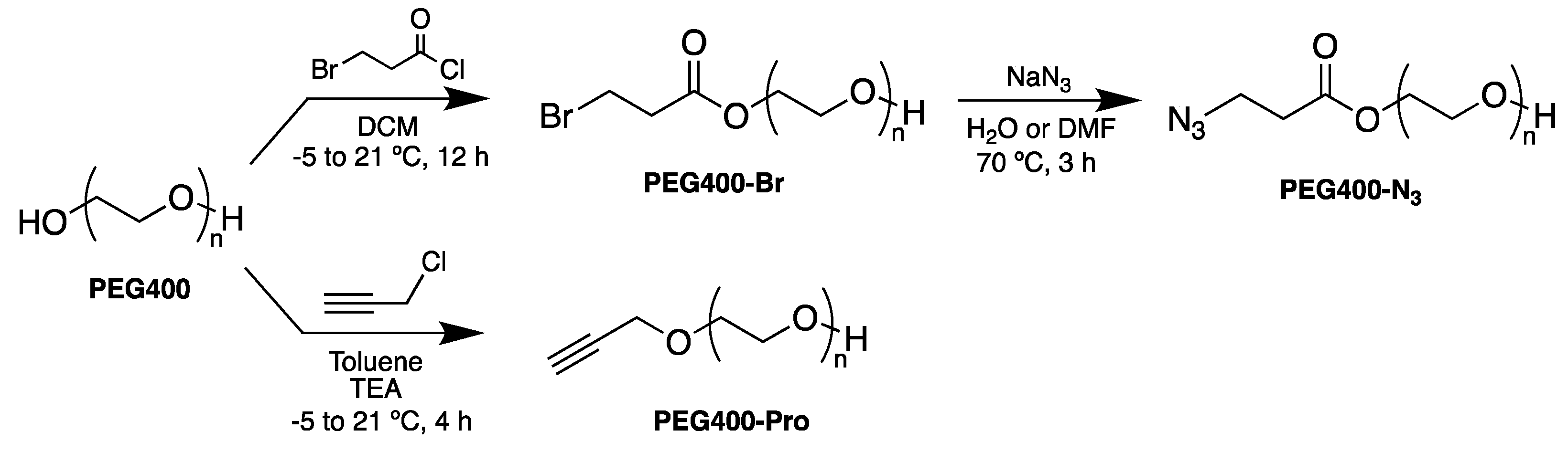
PEG400-Pro: Under Ar, in a 100 mL round bottom flask, PEG400 (0.885 mL) and TEA (3 drops) were mixed with anhydrous toluene (20 mL). The flask was cooled at -5 °C. To this mixture, a solution of propargyl chloride (10 μL, 0.01 mmol) in anhydrous toluene (20 mL) was added dropwise over a period of 20 min, using the programmable syringe pump. The reaction mixture was stirred for 60 min at -5 °C. After this period, mixture was left to warm up to room temperature (21 °C) along a period of about 4 h. Then, the mixture of reaction was washed brine (3 x 5 mL), dried over anhydrous MgSO4 and filtered off. Finally, filtrates were concentrated to dryness under vacuum to obtain PEG400-Pro as a colorless syrup (978 mg, 98 % yield).
NMR, FT-IR and MALDI Spectra for these compounds are included in Supplementary Material (figures S.M.1 to S.M.11)
2.5. Synthesis of MNPs
In a typical procedure, FeCl3·6H2O (1.35 g, 5 mmol) was dissolved in ethylene glycol (40 mL) to form a clear solution, followed by the addition of NaAcO (3.6 g, 44 mmol) and PEG or derivatives (PEG400, PEG6000, PEG400-N3, PEG400-Pro) or βCD (1.0 g). The mixture was stirred vigorously at 21 oC for 30 min and then transferred to sealed tubes. These tubes were heated at 200 oC for 24 h. After this period, mixture of reaction was allowed to cool at 21 oC. The black solid obtained was decanted, washed three times with ethanol, then dried at 70 oC for 3 h, to give @Fe3O4-PEG400, @Fe3O4-PEG6000, @Fe3O4-PEG400-N3, @Fe3O4-PEG400-Pro and @Fe3O4-βCD MNPs (Table 1) systems, which were finally dispersed in ethanol (20 mL, Table 1).
2.6. Synthesis of CBD-Pro Derivatives and @Fe3O4-PEG400-N3 Covalent Surface Functionalization with CBD to Obtain @Fe3O4-PEG400-CBD (Scheme 2)
CBD-Pro-1 and CBD-Pro-2: CBD (200 mg, 0.64 mmol) was dissolved in DMF (6.5 mL). The solution was cooled to 0 oC and then propargyl bromide (0.274 mL, 3.2 mmol) and K2CO3 (131.6 mg, 0.95 mmol) were added. This suspension was stirred at room temperature for 48 h. Upon completion of reaction, the reaction solution was extracted with ethyl acetate (3 × 300 mL). The organic phase was then dried with anhydrous Mg2SO4, and the solvent was removed under vacuum to give the crude product, which was further purified by column chromatography (petroleum ether/ethyl acetate, 90:1) to give CBD-2′-propynyl ether (CBD-Pro-1) and CBD-2′,6′-dipropynyl ether (CBD-Pro-2) [32]. CBD-Pro-1: 50 % yield, 1H-NMR (CDCl3 , 500 MHz) δ(ppm): 6.34 (s, 1H), 6.29 (s, 1H), 6.03 (s, 1H), 5.57 (s, 1H), 4.58-4.53 (m,2H), 4.31 (s, 1H), 4.00 (d, J = 10.1 Hz, 1H), 2.50-2.47 (m, 2H), 1.68 (s, 3H), 0.88 (t, J = 7.0 Hz, 3H). CBD-Pro-2: 45 % yield, 1H-NMR (CDCl3 , 500 MHz) δ(ppm): 6.45 (s, 2H), 5.21 (s, 1H), 4.62-4.55 (m, 4H), 4.43 (dd, J = 2.6 Hz, 2H), 4.00-3.97 (m, 1H), 2.88 (td, J = 10.8 Hz, 1H), 2.54 (t, J = Hz, 2H), 2.45 (t, 2H), 1.97 (d, J = 17.7 Hz, 1H), 1.79-1.70 (m, 2H), 1.61-1.56 (m, 2H), 1.38-1.28 (m, 4H), 0.89 (t, J = 6.9 Hz, 3H). See S.M.12-14.
Scheme 2.
Synthesis of propargyl derivatives of CBD and preparation of @Fe3O4-PEG400-CBD.
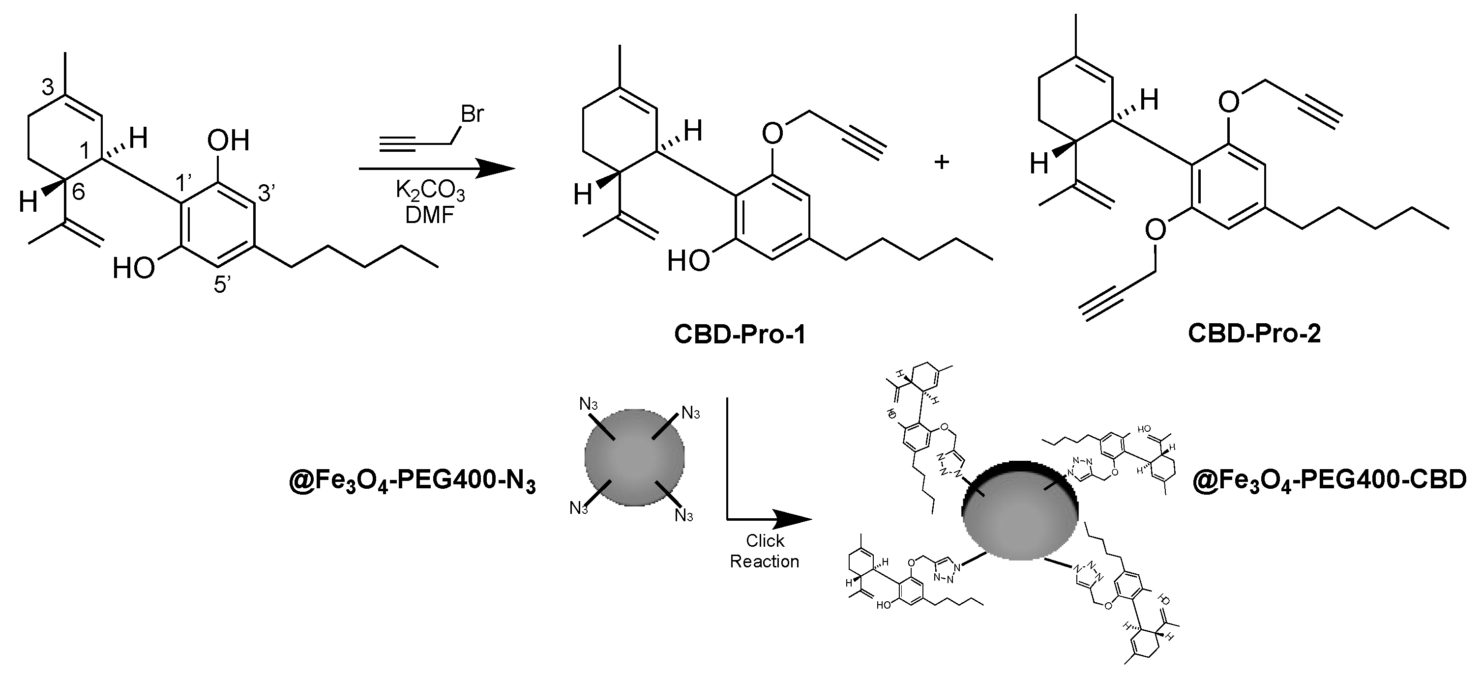
@Fe3O4-PEG400-CBD: in a reaction vial containing CBD-Pro-1, a solution (5 mL) of CuSO4 (2.5 mM) and sodium ascorbate (50 mM), water was added. After 5 min, a solution of @Fe3O4-PEG400-N3 (10 mM) in degassed methanol/water (1:1 v/v) was added. After incubation for 4 h under Ar, the reaction mixture was diluted with water (20 mL) and extracted with DCM (2 x 5 mL). Organic extracts were dried over anhydrous MgSO4, filtered off and concentrated to dryness under vacuum to give @Fe3O4-PEG400-CBD samples, which was analyzed by FT-IR (S.M.15).
2.7. In Vitro Proliferation Assay
The human colon cancer cell line T84 was obtained from the American Type Culture Collection (Rockville, MD, USA). Cells were grown in Dulbecco’s Modified Eagle’s Medium (DMEM), supplemented with 10 % fetal bovine serum (FBS) and 1 % of penicillin-streptomycin (Sigma–Aldrich, Madrid, Spain). The cell line was maintained at 37 °C in an atmosphere containing 5 % CO2. Cells were seeded in 48-well plates at a concentration of 5 x 10³ cells/well and were incubated overnight. MNPs coated with PEG400, PEG6000 and βCD were then administered at a concentration of Fe3O4 ranging from 1 to 100 µg/mL and were incubated for 72 h. Sulforhodamine B (SRB) assay was used to determine the relative proliferation percentage and the optical density of the samples was measured at 492 nm (Titertekmultiscan Colorimeter, Flow, Irvine, CA, USA), as described previously [33].
3. Results and Discussion
3.1. Synthesis of PEG Derivatives
The PEG400 was synthetically modified to incorporate alkyne, through the propargyl moiety (PEG400-Pro), or azide (PEG400-N3) groups, and then used for the preparation of the MNPs. The synthesis of PEG400-N3 was carried out in a two-steps route using PEG400 as starting material. First, PEG400-Br was prepared in good yield by treating PEG400 with 3-bromopropanoyl chloride using DCM as solvent, and second, PEG400-Br was made to react with sodium azide to afford the PEG400-N3 derivative. Last reaction was tested using anhydrous DMF and H2O as solvents, reaching similar yields in both cases, 82.5 % and 78.2 %, respectively. Therefore, subsequent repetitions of the synthesis were performed in H2O due to cost savings and the synthesis speed, as it was not necessary to set up the distillation apparatus to obtain anhydrous DMF. Synthesis of PEG400-Pro was conducted by treating PEG400 with propargyl chloride in dry toluene in presence of TEA, and under moisture-free conditions. We observed that the presence of traces of water, the formation of the product does not occur.
The obtention of the PEG400-Br, PEG400-N3 and PEG400-Pro systems was confirmed by NMR, FT-IR and MALDI (S.M.1-11). The formation of the PEG400-Br was confirmed by the 1H-NMR spectrum, which shows the presence of a signal corresponding to the methylene group attached to the bromine atom, at 4.1 ppm, integral 2H, and another signal at 2.8 ppm (2H) assigned to the methylene moiety vicinal to the carbonyl group. The presence of C=O was also confirmed by the quaternary carbon characteristic peak at 170.3 ppm in the 13C-NMR and the intense IR absorption at 1600 cm-1. For PEG400-N3, in the 1H-NMR can be seen the peaks corresponding to one methylene attached to an azide group at 2.6 ppm (2H) and these assigned to a carbonyl group at 4.2 ppm (2H) are observed, where the methylene chemical shift differs from PEG400-Br. The characteristic peak of the carbonyl carbon is also visible (171.4 ppm, 13C-NMR). FT-IR abortion at 2102 cm-1 confirms the presence of the azide group . In case of PEG400-Pro, the formation of the alkyne terminal group was confirmed by the characteristic signal at 3.0 ppm (1H) in the 1H-NMR, which can be assigned to the alkyne terminal hydrogen. The presence of the alkyne moiety was also confirmed by the infrared analysis (FT-IR), in which was clearly observed a highly intense bending vibration frequency at 737 cm-1 assigned to the triple bond group. MALDI-TOF analyses were also carried out. Analysis of PEG400 standard, with the addition of Na+, a single series of PEG400 ions completely dominate the spectrum. The [H(C2H4O)nOH + Na]+ ions at m/z 365, 409, 453, 497, 541, 585, 629, 673, 717, and 761 correspond to PEG400 oligomers with the number of repeating units with n=8–17. In case of PEG400-N3 it can be seen two characteristics ions at about m/z 613 and 657, attributed to the substitution of the hydroxyl group by the azide group, and corresponding to the mass increase of m/z of 28 with respect to the m/z 585 and 629 in PEG400. Similarly, the substitution of a hydroxyl group was confirmed by the peak at m/z 481, representing an increase of about 30 mass units with respect of the peak at m/z of 453 in PEG400, due to the incorporation of the propargyl moiety.
3.2. Preparation of MNPs Systems
Typical syntheses of Fe3O4 using the co-precipitation or oxidation methods require the addition of cationic surfactants, such as CTAB, for the stabilization and monodispersity of MNPs [11]. It is known that MNPs exhibit a pronounced tendency to agglomerate during their formation in the liquid-phase process [34]. Additionally, for the polymerization with certain polymers like pNIPAM or p4VP, prior functionalization of the magnetic cores is necessary to enable the binding of specific molecules [35]. Conversely, for polymers like PEG, prior functionalization of the magnetic cores is not required due to the inherent chemical and physical affinity between PEG and the surface of MNPs [11]. The hydroxyl groups of PEG can interact through hydrogen bonding and London dispersion forces with the surface groups of magnetite.
To achieve all in situ monodisperse and coated Fe3O4 nanoparticles, we followed the synthetic procedure of the solvothermal method, involving three critical additions. First, NaAcO was added for electrostatic stabilization and to prevent particle agglomeration. In our system, the presence of NaAcO is crucial as it seemed to facilitate the reduction of FeCl3 to Fe3O4 using ethylene glycol. Second, PEG and the different derivatives that we have synthesized were added, in order to act as a surfactants or stabilizers. PEG inherently provided a hydrophilic coating to the MNPs, contributing to preventing particle agglomeration. The third critical feature was the increase in the reaction temperature up to 200 °C, an indispensable requirement for producing Fe3O4.
The solvothermal reduction reaction was carried out by heating the solution at 200 °C and maintaining this temperature during 24 h in sealed flasks (Table 1). The heat provided during the process promotes the reduction reaction, allowing metallic ions to transform into magnetic nanoparticles, in contrast to the thermal decomposition method that produces hydrophobic MNPs in an organic phase [35], this method generates water-dispersible nanoparticles through a thermal decomposition process in hydrophilic polyalcoholic solvents. Polyalcohols serve three functions, high boiling point solvent, reducing agent, and stabilizer to control nanoparticle growth and prevent aggregation. The use of polyols as a surfactant or stabilizer results in MNPs with inherent hydrophilic coating. This simplifies the use of these nanoparticles for subsequent biological applications, as no modification is needed to enhance their dispersion in water, and both the synthesis of the magnetite cores and the polymeric coating occur in a single step.
The size and shape of the prepared MNPs were examined using TEM and AFM. Figure 1 shows representative images of the MNPs structures prepared through the solvothermal reduction method. As it can be seen, well-dispersed spherical clusters containing aggregates of Fe3O4 NPs, with very narrow diameter distributions, are observed. Furthermore, the shape and size of the Fe3O4 products with different polymeric coatings remained unchanged in morphology compared to those of the Fe3O4 precursor with PEG400.
The diameters of the MNPs were influenced by the type of polymeric coating (Table 1). Under the same reaction conditions, with the Fe3O4·6H2O precursor concentration of 0.13 M, temperature of 200 °C and reaction time of 24 h, a diameter of approximately 250 nm was obtained for @Fe3O4-PEG400, while 90 nm was measured for @Fe3O4-PEG400-N3, being 150 nm for @Fe3O4-PEG6000, 155 nm for @Fe3O4-βCD and 200 nm for @Fe3O4-PEG400-Pro (Figure 2, ImageJ Software). In all cases, the diameter for the magnetite cores was about 15 nm.
Chemical composition of MNPs was studied by XRD and FT-IR techniques (Figure 3 and Figure 4, respectively). Samples of Fe3O4 were analyzed by XRD, an effective technique to characterize the crystalline structure of synthesized magnetic nanoparticles. As the polymer and polyol are poorly crystalline material, are not seen in XRD, and only the magnetite can be observed. Figure 3 shows the XRD pattern of @Fe3O4-PEG6000, representative for the prepared systems, showing the six magnetite characteristics peaks at 2θ of 28.7º (2 2 0), 34.8º (3 1 1), 43.5º (4 0 0), 53.8º (4 2 2), 57.5º (5 1 1) and 62.4º (4 4 0). The XRD pattern matches well with the standard XRD data for bulk magnetite [14], with almost no impurity phase (α-Fe).
Samples @Fe3O4-PEG400-Pro, @Fe3O4-PEG400-N3, @Fe3O4-βCD and @Fe3O4-PEG6000 were chemically characterized by FT-IR to identify the coating on the surface of nanoparticles (Figure 4). FT-IR provides evidence for the existence of the different functional groups in the coating.
Figure 4A displays the spectrum for @Fe3O4-PEG400-Pro, where it can be seen the bending deformation band of acetylene at 608 cm-1. In Figure 4B, corresponding to @Fe3O4-PEG400-N3, the azide absorption band is observed at frequencies of 1240 and 2164 cm-1 (red arrows) Additionally, in the four IR spectra, it is observed the vibration (560-590 cm-1) of the Fe-O bond in reference to that of magnetite present. The pure magnetite systems exhibited a broad absorption peak of around 3200 cm−1, which was assigned to the OH stretching vibration from surface hydroxyl groups. This is also one of the most intense bands in @Fe3O4-PEG400-βCD (Figure 4C), where it corresponds to the multiple OH functional groups present, but also to adsorbed water. It is worth to note that OH stretching is almost not detected in @Fe3O4-PEG400-N3 spectrum (Figure 4B), corresponding to a mostly complete surface substitution of OH by azide groups. On the other hand, the bands assigned to the vibrations of the methylene chains, and C–O and C–C groups are observed in the interval 840-1500 cm−1. As expected, the most intense βCD vibrations are observed at 1029, 1153 and 1600 cm-1for the case of the @Fe3O4-βCD sample (Figure 4C, green arrows).
3.3. Preparation of @Fe3O4-PEG400-CBD
As a proof of concept, we carried out the surface functionalization of @Fe3O4-PEG400-N3 by treatment with CBD-Pro-1 (Scheeme 2). After 4 h of reaction the formation of the @Fe3O4-PEG400-CBD system was confirmed by FT-IR, where the charactristics bands of the triazol moiety were observed at 1485, 1529 and 1547 cm-1 (S.M.15)
3.4. Magnetic Measurements
The magnetic properties of the ferrite nanospheres were investigated using a VSM. Superparamagnetism is the reaction to an applied magnetic field without retaining any magnetization after the removal of the applied magnetic field. It denotes re-dispersion of the magnetic nanoparticles in solution without the occurrence of severe aggregation from which ferromagnetic nanoparticles often suffer, which limits their applications. It is highly desirable the superparamagnetic behavior of MNPs due to his high efficiency to absorb the energy of an alternating magnetic field and convert it into heat [36].
Figure 5 shows magnetization curves measured at 300 K for Fe3O4 nanospheres coated with the PEG400, PEG6000 and βCD (@Fe3O4-PEG400, @Fe3O4-PEG6000 and @Fe3O4-βCD samples). Qualitative analysis of the hysteresis curves, where no remanence and coercivity values are observed in the magnetization, indicated that the MNPs are superparamagnetic, usually named as SPION (Superparamagnetic Iron Oxide Nanoparticles), with no observed ferromagnetism. Furthermore, the MNPs were well dispersed in water and could be separated from the solution by the attraction of a magnet.
The measured saturation magnetization values for the prepared MNPs were found to be 59.9 emu/g for @Fe3O4-PEG6000, 47.8 emu/g for @Fe3O4-PEG400 and 30.8 emu/g for @Fe3O4-βCD. Among them, the coated βCD nanoparticles showed lower saturation of magnetization value than that for PEG coated nanoparticles, and all of them lower that non-coated MNPs. This fact is known, and it is attributed to the non-magnetic material over the surface of Fe3O4 nanoparticles, being a proof of the presence of organic molecules coating the surface.
3.3. Hyperthermia and In Vitro Proliferation Analyses
The ability to generate hyperthermia was analyzed after 25 min of application of a high frequency alternating magnetic field. The heating curves obtained are represented in Figure 6A. MNPs coated with PEG400, PEG6000 and βCD were able to increase temperature up to 5.80, 9.27 and 6.03 ºC, leading to SAR values of 33.72 ± 0.85, 51.87 ± 2.23 and 33.65 ± 2.63 W/g, respectively. SAR is a relevant measure to characterize the magnetic capabilities of a nanoformulation since it refers to the amount of energy converted into heat in the unit of time and mass. This ability to produce heat makes it possible to use nanoformulations as hyperthermia generation agents, understood as the ability to generate a temperature increase when an external magnetic field is applied [37]. This characteristic has been used as a treatment for a wide range of cancers, with very promising results both in vitro and in vivo [38,39]. In this regard, a high SAR value would allow an increase in the capacity to generate heat and therefore decrease the dose of nanoformulation necessary to produce cell death. Some parameters that influence the SAR value are the size and shape of the nanoparticle, as well as the magnetic anisotropy and the frequency and amplitude of the magnetic field, among others [40]. In this case, @Fe3O4-PEG6000 presents a higher temperature rise than the rest of nanoformulations, so it could be the one with the best systems for cellular hyperthermia assays.
To determine the biocompatibility of the different fabricated nanoformulations, a toxicity study was performed. The in vitro antiproliferative effect of all the nanoformulations was analyzed in the human colon cancer cell line T84. SRB assay showed relative proliferation percentages close to 100 % for every nanoformulation with no significant differences compared to control at most doses administered (Figure 6B), showing good in vitro biocompatibility.
Finally, to examine the colloidal stability of the ferrite samples, and to know the article durability, magnetic ferrite nanospheres (@Fe3O4-PEG400, 20 mg) were dispersed in doubly distilled water (80 mL) through sonication. The magnetic particles remained in suspension for over 3 days, demonstrating that they can be well dispersed in an aqueous solution. Therefore, with appropriate surface modifications, these nanospheres may be suitable for clinical diagnostics and drug, protein, virus, or bacteria transport.
4. Conclusions
We successfully carried out the synthesis of MNPs with different polymeric hydrophilic coatings in a single step reaction following the solvothermal method. To the best of our knowledge, no other study has documented the use of this method to obtain magnetic spheres with a functionalized polymeric coating. The nanoparticles had diameters in the range of 90 to 250 nm, and each magnetic core composing the cluster had a size of about 15 nm.
The magnetic properties of PEG and βCD coated Fe3O4 systems were investigated at room temperature, and superparamagnetic behavior was confirmed, with a maximum saturation magnetization reaching values of about 60 emu/g for PEG6000 systems. Two of these coatings (PEG400-N3 and PEG400-Pro) allows to perform click chemistry reactions, facilitating the construction of more complex structures from basic units. This fact was illustrated in the case of the cannabidiol-propargyl derivative (CBD-Pro), which was covalently attached to the MNPs functionalized surface (@Fe3O4-PEG400-N3) to obtain @Fe3O4-PEG400-CBD systems.
The synthesized MNPs show great promise for drug inclusion and administration, as the magnetic cores provide a quick and efficient magnet-like function with optimum temperature rise and SAR values, following their exposition to a high frequency alternating magnetic field, and great biocompatibility results in the T84 human colon cancer cell line. The coating produced inherently during synthesis opens a wide range of applications within biomedicine.
Acknowledgments
Authors thanks Andalusian Government for financial support, FQM397 and UMA20 FEDERJA84 Projects.
References
- Ehrmann, A.; Nguyen, T.A.; Ahmadi, M.; Farmani, A.; Nguyen-Tri, P. Magnetic nanoparticle-based hybrid materials, fundamentals, and applications, 1st ed.; Elsevier Ltd. 2021. [Google Scholar]
- Rahmati, S.; David, A.E. A review of design criteria for cancer-targeted, nanoparticle-based MRI contrast agents. Appl. Mater. Today 2024, 37. [Google Scholar] [CrossRef]
- Rezaei, B.; Yari, P.; Sanders, S.M.; Wang, H.; Chugh, V.K.; Liang, S.; Mostufa, S.; Xu, K.; Wang, J.; Gómez-Pastora, J.; et al. Magnetic Nanoparticles: A Review on Synthesis, Characterization, Functionalization, and Biomedical Applications. Small 2023, 20, e2304848. [Google Scholar] [CrossRef]
- Szwed, M.; Marczak, A. Application of Nanoparticles for Magnetic Hyperthermia for Cancer Treatment—The Current State of Knowledge. Cancers 2024, 16, 1156. [Google Scholar] [CrossRef]
- Lucena-Serrano, C.; Lucena-Serrano, A.; Díaz, A.; Valpuesta, M.; Villaverde, G.; López-Romero, J.M.; Sarabia, F.; Laurenti, M.; Rubio-Retama, J.; Contreras-Cáceres, R. SPION nanoparticles for delivery of dopaminergic isoquinoline and benzazepine derivatives. Bioorganic Med. Chem. 2022, 69, 116910. [Google Scholar] [CrossRef]
- Wu, W.; He, Q.; Jiang, C. Magnetic Iron Oxide Nanoparticles: Synthesis and Surface Functionalization Strategies. Nanoscale Res. Lett. 2008, 3, 397–415. [Google Scholar] [CrossRef]
- Ahmad, F.; Salem-Bekhit, M.M.; Khan, F.; Alshehri, S.; Khan, A.; Ghoneim, M.M.; Wu, H.-F.; Taha, E.I.; Elbagory, I. Unique Properties of Surface-Functionalized Nanoparticles for Bio-Application: Functionalization Mechanisms and Importance in Application. Nanomaterials 2022, 12, 1333. [Google Scholar] [CrossRef]
- Unni, M.; Uhl, A.M.; Savliwala, S.; Savitzky, B.H.; Dhavalikar, R.; Garraud, N.; Arnold, D.P.; Kourkoutis, L.F.; Andrew, J.S.; Rinaldi, C. Thermal Decomposition Synthesis of Iron Oxide Nanoparticles with Diminished Magnetic Dead Layer by Controlled Addition of Oxygen. ACS Nano 2017, 11, 2284–2303. [Google Scholar] [CrossRef]
- Ganapathe, L.S.; Mohamed, M.A.; Yunus, R.M.; Berhanuddin, D.D. Magnetite (Fe3O4) nanoparticles in biomedical application: from synthesis to surface functionalization. Magnetochem 2020, 6, 68. [Google Scholar] [CrossRef]
- Guerrini, L.; Alvarez-Puebla, R.A.; Pazos-Perez, N. Surface Modifications of Nanoparticles for Stability in Biological Fluids. Materials 2018, 11, 1154. [Google Scholar] [CrossRef]
- Garcia-Pinel, B.; Ortega-Rodríguez, A.; Porras-Alcalá, C.; Cabeza, L.; Contreras-Cáceres, R.; Ortiz, R.; Díaz, A.; Moscoso, A.; Sarabia, F.; Prados, J.; et al. Magnetically active pNIPAM nanosystems as temperature-sensitive biocompatible structures for controlled drug delivery. Artif. Cells, Nanomedicine, Biotechnol. 2020, 48, 1022–1035. [Google Scholar] [CrossRef]
- Karimzadeh, I.; Aghazadeh, M.; Doroudi, T.; Ganjali, M.R.; Kolivand, P.H. Superparamagnetic Iron Oxide (Fe3O4) Nanoparticles Coated with PEG/PEI for Biomedical Applications: A Facile and Scalable Preparation Route Based on the Cathodic Electrochemical Deposition Method. Adv. Phys. Chem. 2017, 2017, 1–7. [Google Scholar] [CrossRef]
- Kumar, P.; Khanduri, H.; Pathak, S.; Singh, A.; Basheed, G.A.; Pant, R.P. Temperature selectivity for single phase hydrothermal synthesis of PEG-400 coated magnetite nanoparticles. Dalton Trans. 2020, 49, 8672–8683. [Google Scholar] [CrossRef]
- Deng, H.; Li, X.; Peng, Q.; Wang, X.; Chen, J.; Li, Y. Monodisperse Magnetic Single-Crystal Ferrite Microspheres. Angew. Chem. 2005, 44, 2782–2785. [Google Scholar] [CrossRef] [PubMed]
- Ni, X.; Zhang, J.; Zhao, L.; Wang, F.; He, H.; Dramou, P. Study of the solvothermal method time variation effects on magnetic iron oxide nanoparticles (Fe3O4) features. J. Phys. Chem. Solids 2022, 169. [Google Scholar] [CrossRef]
- Li, Y.; Wang, Z.; Ali, Z.; Tian, K.; Xu, J.; Li, W.; Hou, Y. Monodisperse Fe3O4 spheres: Large-scale controlled synthesis in the absence of surfactants and chemical kinetic process. Sci. China Mater. 2019, 62, 1488–1495. [Google Scholar] [CrossRef]
- Su, M.; He, C.; Shih, K. Facile synthesis of morphology and size-controlled α -Fe 2 O 3 and Fe 3 O 4 nano-and microstructures by hydrothermal/solvothermal process: The roles of reaction medium and urea dose. Ceram. Int. 2016, 42, 14793–14804. [Google Scholar] [CrossRef]
- Liu, J.; Sun, Z.; Deng, Y.; Zou, Y.; Li, C.; Guo, X.; Xiong, L.; Gao, Y.; Li, F.; Zhao, D. Highly Water-Dispersible Biocompatible Magnetite Particles with Low Cytotoxicity Stabilized by Citrate Groups. Angew. Chem. Int. Ed. 2009, 48, 5875–5879. [Google Scholar] [CrossRef]
- Jiang, X.; Wang, F.; Cai, W.; Zhang, X. Trisodium citrate-assisted synthesis of highly water-dispersible and superparamagnetic mesoporous Fe3O4 hollow microspheres via solvothermal process. J. Alloy. Compd. 2015, 636, 34–39. [Google Scholar] [CrossRef]
- Cheng, C.; Xu, F.; Gu, H. Facile synthesis and morphology evolution of magnetic iron oxide nanoparticles in different polyol processes. New J. Chem. 2011, 35, 1072–1079. [Google Scholar] [CrossRef]
- Kotoulas, A.; Dendrinou-Samara, C.; Angelakeris, M.; Kalogirou, O. The Effect of Polyol Composition on the Structural and Magnetic Properties of Magnetite Nanoparticles for Magnetic Particle Hyperthermia. Materials 2019, 12, 2663. [Google Scholar] [CrossRef]
- Chen, F.; Liu, R.; Xiao, S.; Zhang, C. Solvothermal synthesis in ethylene glycol and adsorption property of magnetic Fe3O4 microspheres. Mater. Res. Bull. 2014, 55, 38–42. [Google Scholar] [CrossRef]
- Jamshidiyan, M.; Shirani, A.; Alahyarizadeh, G. Solvothermal synthesis and characterization of magnetic Fe3O4nanoparticle by different sodium salt sources. Mater. Sci. 2017, 35, 50–57. [Google Scholar] [CrossRef]
- Banerjee, S.S.; Chen, D.-H. Magnetic Nanoparticles Grafted with Cyclodextrin for Hydrophobic Drug Delivery. Chem. Mater. 2007, 19, 6345–6349. [Google Scholar] [CrossRef]
- Badruddoza, A.; Tay, A.; Tan, P.; Hidajat, K.; Uddin, M. Carboxymethyl-β-cyclodextrin conjugated magnetic nanoparticles as nano-adsorbents for removal of copper ions: Synthesis and adsorption studies. J. Hazard. Mater. 2010, 185, 1177–1186. [Google Scholar] [CrossRef]
- Zhu, J.; He, J.; Du, X.; Lu, R.; Huang, L.; Ge, X. A facile and flexible process of β-cyclodextrin grafted on Fe3O4 magnetic nanoparticles and host–guest inclusion studies. Appl. Surf. Sci. 2011, 257, 9056–9062. [Google Scholar] [CrossRef]
- Sinniah, S.; Mohamad, S.; Manan, N.S. Magnetite nanoparticles coated with β-cyclodextrin functionalized-ionic liquid: Synthesis and its preliminary investigation as a new sensing material. Appl. Surf. Sci. 2015, 357, 543–550. [Google Scholar] [CrossRef]
- Li, S.; Sharaf, M.G.; Rowe, E.M.; Serrano, K.; Devine, D.V.; Unsworth, L.D. Hemocompatibility of β-Cyclodextrin-Modified (Methacryloyloxy)ethyl Phosphorylcholine Coated Magnetic Nanoparticles. Biomolecules 2023, 13, 1165. [Google Scholar] [CrossRef]
- Singh, G.; Majeed, A.; Singh, R.; George, N.; Singh, G.; Gupta, S.; Singh, H.; Kaur, G.; Singh, J. CuAAC ensembled 1,2,3-triazole linked nanogels for targeted drug delivery: a review. RSC Adv. 2023, 13, 2912–2936. [Google Scholar] [CrossRef] [PubMed]
- López-Romero, J.M.; Moya-Utrera, F.; Carrasco, A.R.; Sarabia, F. Cannabinoid synthesis starting out from olivetol and terpene in dichloromethane with FeCl3 as catalyst, WO2024028516A1, 2024.
- Garcés, V.; González, A.; Gálvez, N.; Delgado-López, J.M.; Calvino, J.J.; Trasobares, S.; Fernández-Afonso, Y.; Gutiérrez, L.; Dominguez-Vera, J.M. Magneto-optical hyperthermia agents based on probiotic bacteria loaded with magnetic and gold nanoparticles. Nanoscale 2022, 14, 5716–5724. [Google Scholar] [CrossRef]
- Zi, C.-T.; Xie, Y.-R.; Niu, Y.; Liu, Z.-H.; Yang, L.; Xi, Y.-K.; Li, Z.-J.; Zhang, F.-M.; Xiang, Z.-M.; Sheng, J. New cannabidiol (CBD) derivatives: Synthesis, anti-inflammatory activity, and molecular docking. Phytochem. Lett. 2022, 51, 97–103. [Google Scholar] [CrossRef]
- Ortiz R, R.; Cabeza, L.; Arias, J.L. Poly(butylcyanoacrylate) and Poly(ε-caprolactone) Nanoparticles Loaded with 5-Fluorouracil Increase the Cytotoxic Effect of the Drug in Experimental Colon Cancer. AAPS J. 2015, 17, 918–929. [Google Scholar] [CrossRef] [PubMed]
- Engelmann, U.; Buhl, E.M.; Baumann, M.; Schmitz-Rode, T.; Slabu, I. Agglomeration of magnetic nanoparticles and its effects on magnetic hyperthermia. Curr. Dir. Biomed. Eng. 2017, 3, 457–460. [Google Scholar] [CrossRef]
- Mohanta, S.C.; Saha, A.; Devi, P.S. PEGylated Iron Oxide Nanoparticles for pH Responsive Drug Delivery Application. Mater. Today: Proc. 2018, 5, 9715–9725. [Google Scholar] [CrossRef]
- Alvear-Jiménez, A.; Gutierrez, I.Z.; Shen, Y.; Villaverde, G.; Lozano-Chamizo, L.; Guardia, P.; Tinoco, M.; Garcia-Pinel, B.; Prados, J.; Melguizo, C.; et al. Electrospraying as a Technique for the Controlled Synthesis of Biocompatible PLGA@Ag2S and PLGA@Ag2S@SPION Nanocarriers with Drug Release Capability. Pharmaceutics 2022, 14, 214. [Google Scholar] [CrossRef] [PubMed]
- Jose, J.; Kumar, R.; Harilal, S.; Mathew, G.E.; Parambi, D.G.T.; Prabhu, A.; Uddin, S.; Aleya, L.; Kim, H.; Mathew, B. Magnetic nanoparticles for hyperthermia in cancer treatment: an emerging tool. Environ. Sci. Pollut. Res. 2019, 27, 19214–19225. [Google Scholar] [CrossRef] [PubMed]
- Fernandes, S.; Fernandez, T.; Metze, S.; Balakrishnan, P.B.; Mai, B.T.; Conteh, J.; De Mei, C.; Turdo, A.; Di Franco, S.; Stassi, G.; et al. Magnetic Nanoparticle-Based Hyperthermia Mediates Drug Delivery and Impairs the Tumorigenic Capacity of Quiescent Colorectal Cancer Stem Cells. ACS Appl. Mater. Interfaces 2021, 13, 15959–15972. [Google Scholar] [CrossRef] [PubMed]
- Kwon, S.-H.; Al Faruque, H.; Kee, H.; Kim, E.; Park, S. Exosome-based hybrid nanostructures for enhanced tumor targeting and hyperthermia therapy. Colloids Surfaces B: Biointerfaces 2021, 205, 111915. [Google Scholar] [CrossRef]
- Narayanaswamy, V.; Al-Omari, I.A.; Kamzin, A.S.; Issa, B.; Tekin, H.O.; Khourshid, H.; Kumar, H.; Mallya, A.; Sambasivam, S.; Obaidat, I.M. Specific Absorption Rate Dependency on the Co2+ Distribution and Magnetic Properties in CoxMn1-xFe2O4 Nanoparticles. Nanomaterials 2011, 11, 1231. [Google Scholar] [CrossRef]
Figure 1.
A) TEM image of @Fe3O4-PEG400 nanospheres. B) TEM image of a single @Fe3O4-PEG400 nanosphere. C) AFM image of @Fe3O4-PEG400. D) AFM image of @Fe3O4-PEG400 single nanosphere.
Figure 1.
A) TEM image of @Fe3O4-PEG400 nanospheres. B) TEM image of a single @Fe3O4-PEG400 nanosphere. C) AFM image of @Fe3O4-PEG400. D) AFM image of @Fe3O4-PEG400 single nanosphere.
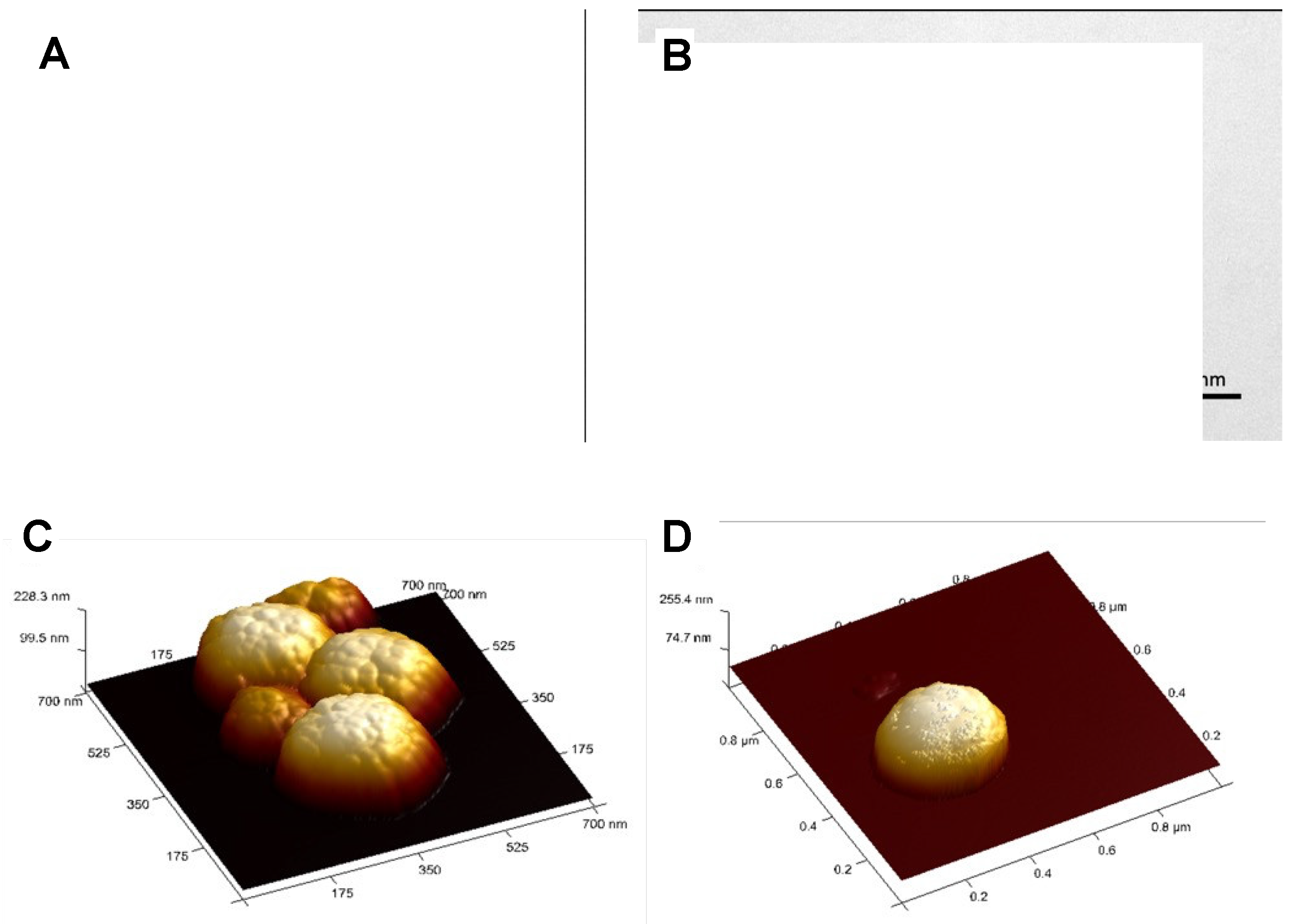
Figure 2.
TEM images of A) @Fe3O4-PEG400-Pro, B) @Fe3O4-PEG400-N3, C) @Fe3O4-βCD, and D) @Fe3O4-PEG6000.
Figure 2.
TEM images of A) @Fe3O4-PEG400-Pro, B) @Fe3O4-PEG400-N3, C) @Fe3O4-βCD, and D) @Fe3O4-PEG6000.
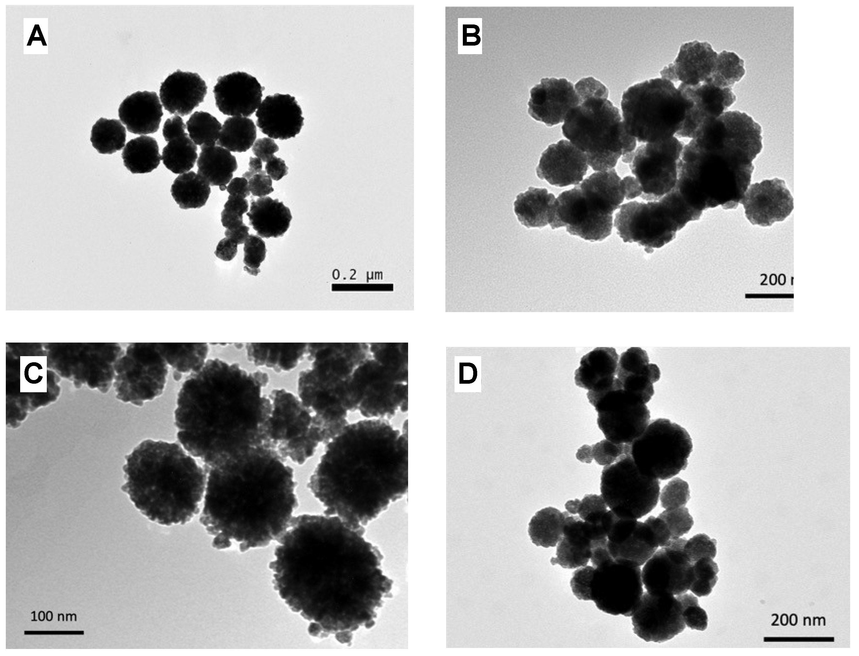
Figure 3.
Representative XRD diffractogram of Fe3O4 nanoparticles coated with PEG (@Fe3O4-PEG6000).
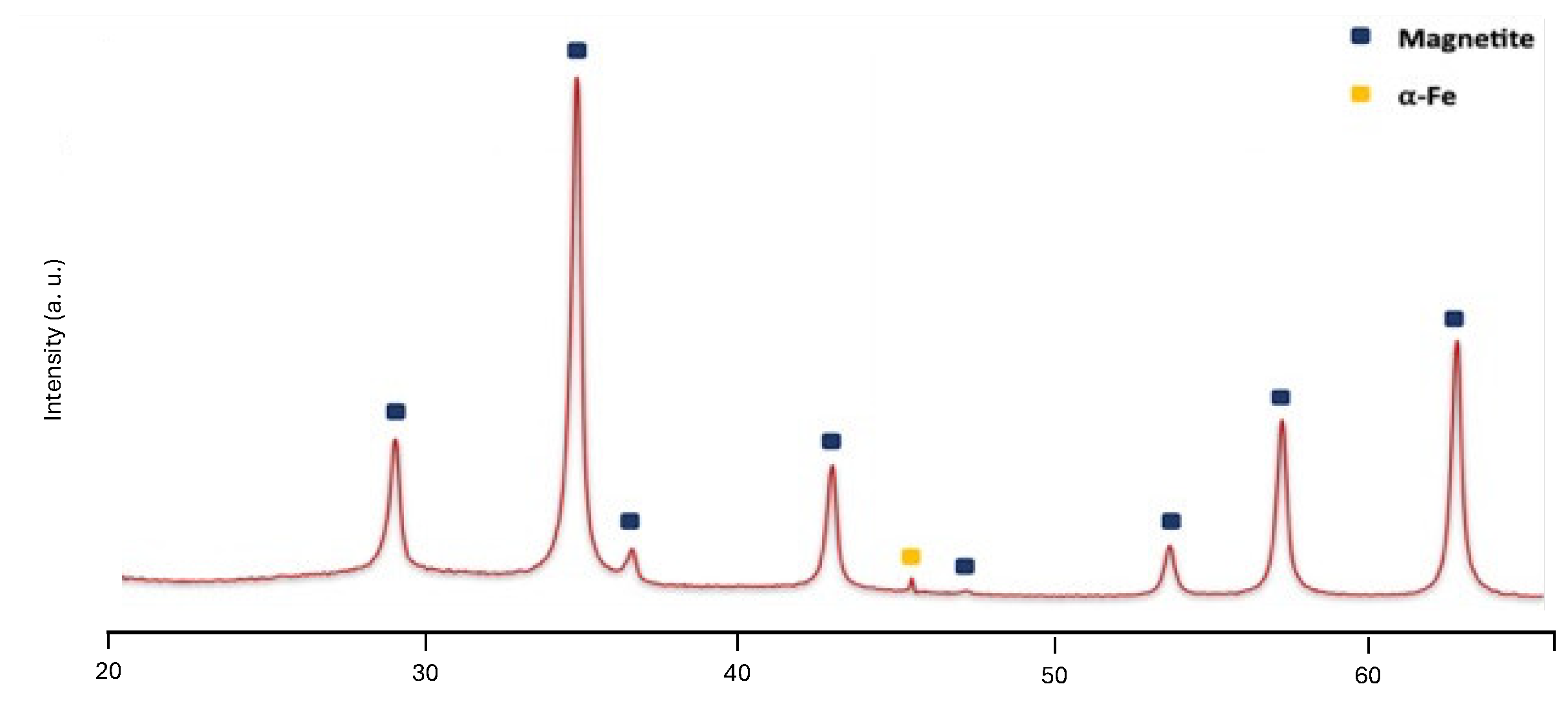
Figure 4.
FT-IR Spectra of A) @Fe3O4-PEG400-Pro, B) @Fe3O4-PEG400-N3, C) @Fe3O4-βCD and D) @Fe3O4-PEG6000 samples.
Figure 4.
FT-IR Spectra of A) @Fe3O4-PEG400-Pro, B) @Fe3O4-PEG400-N3, C) @Fe3O4-βCD and D) @Fe3O4-PEG6000 samples.
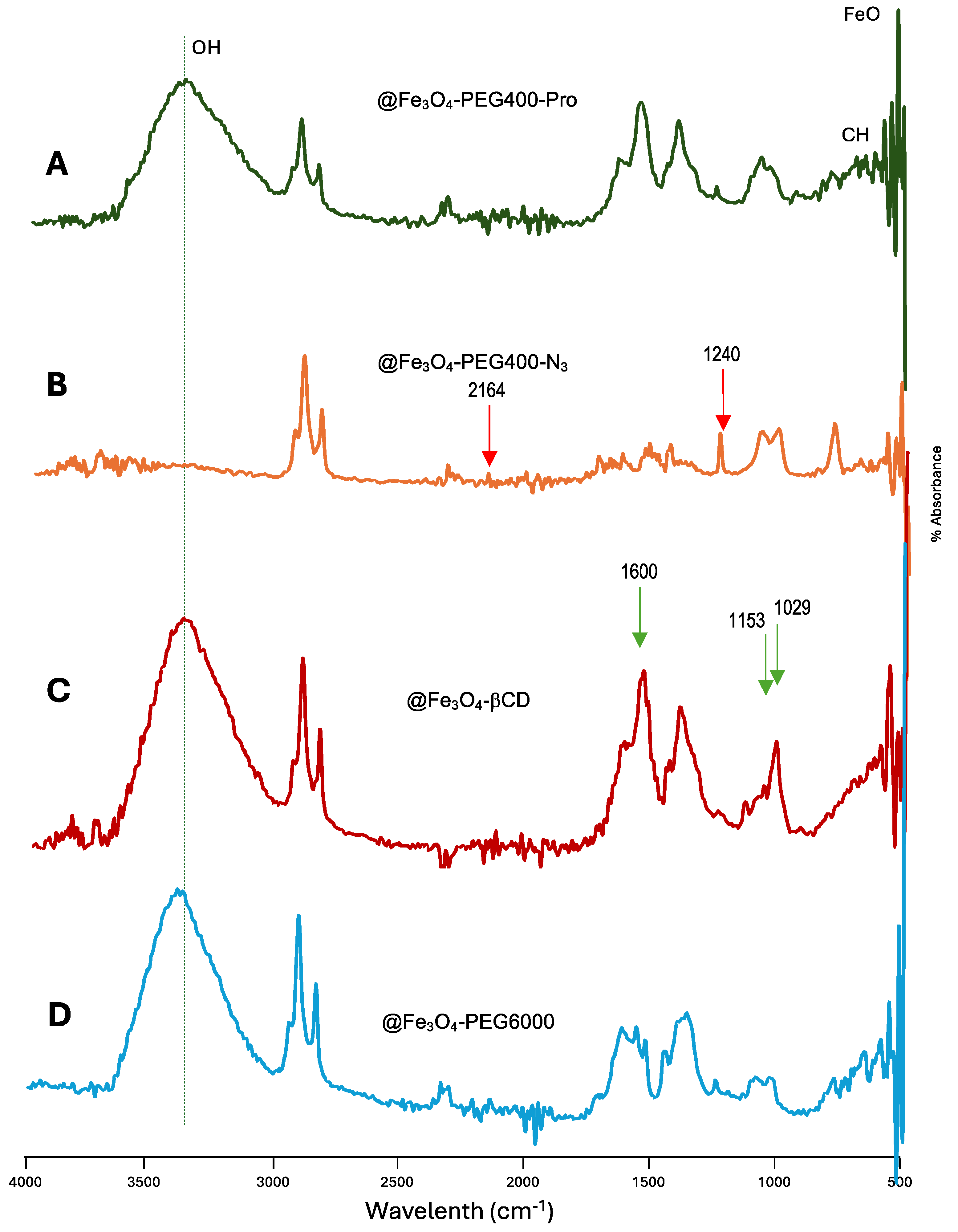
Figure 5.
Magnetization curves of @Fe3O4 coated with βCD, PEG400 or PEG6000.
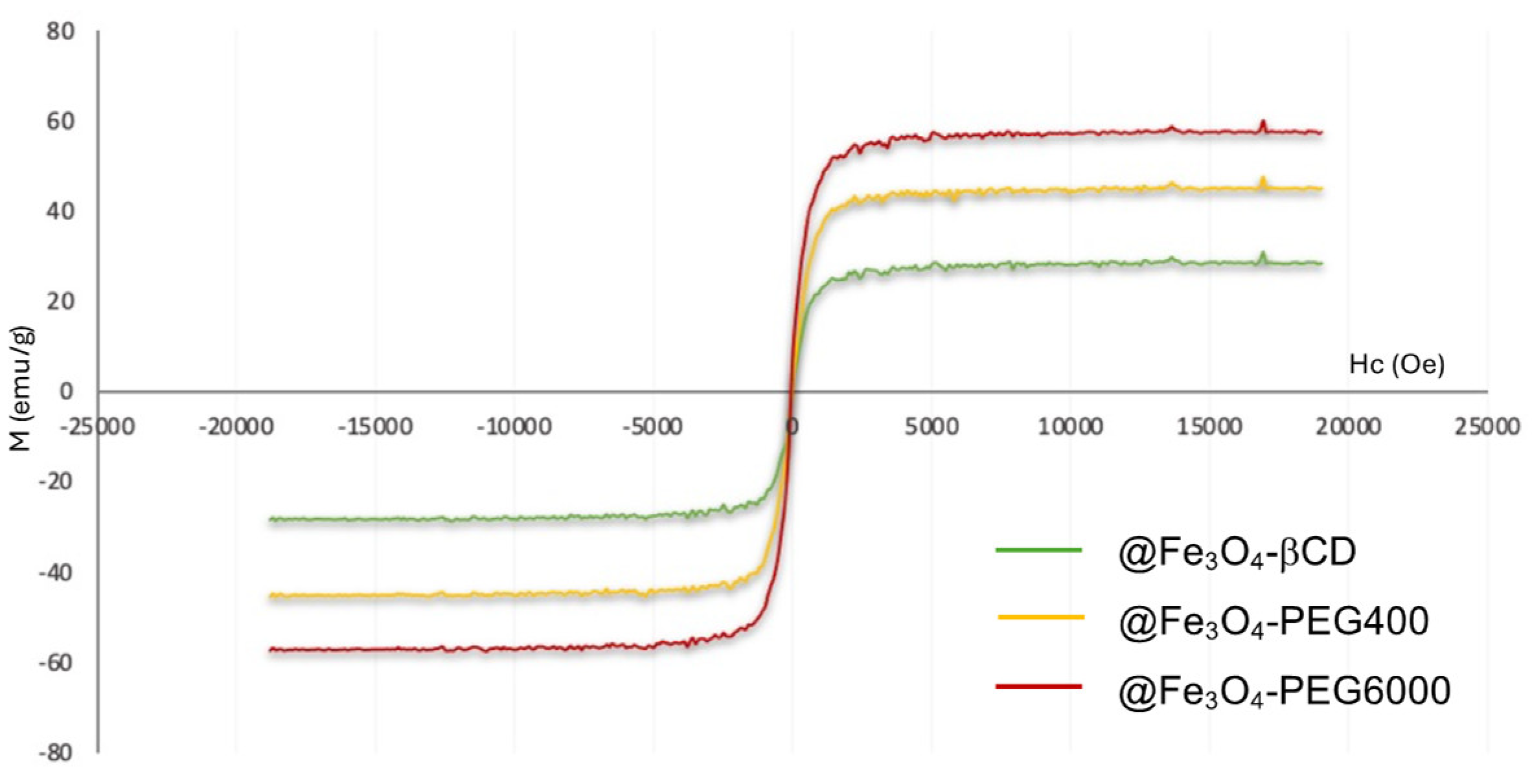
Figure 6.
A) Temperature rise obtained after application of an alternating magnetic field (385 kHz; 28 kA/m) during 25 min for MNPs coated with PEG400, PEG6000 and βCD at a concentration of 0.5 mg/mL of Fe3O4. The data were represented as the mean of 3 measurements ± standard deviation. B) In vitro proliferation assay of MNPs coated with PEG400, PEG6000 and βCD at 72 h of exposition. Graphs show the percentage of proliferation of T84 at doses ranging from 1-100 µg/mL of Fe3O4. Results were expressed as mean ± SD of triplicate cultures.
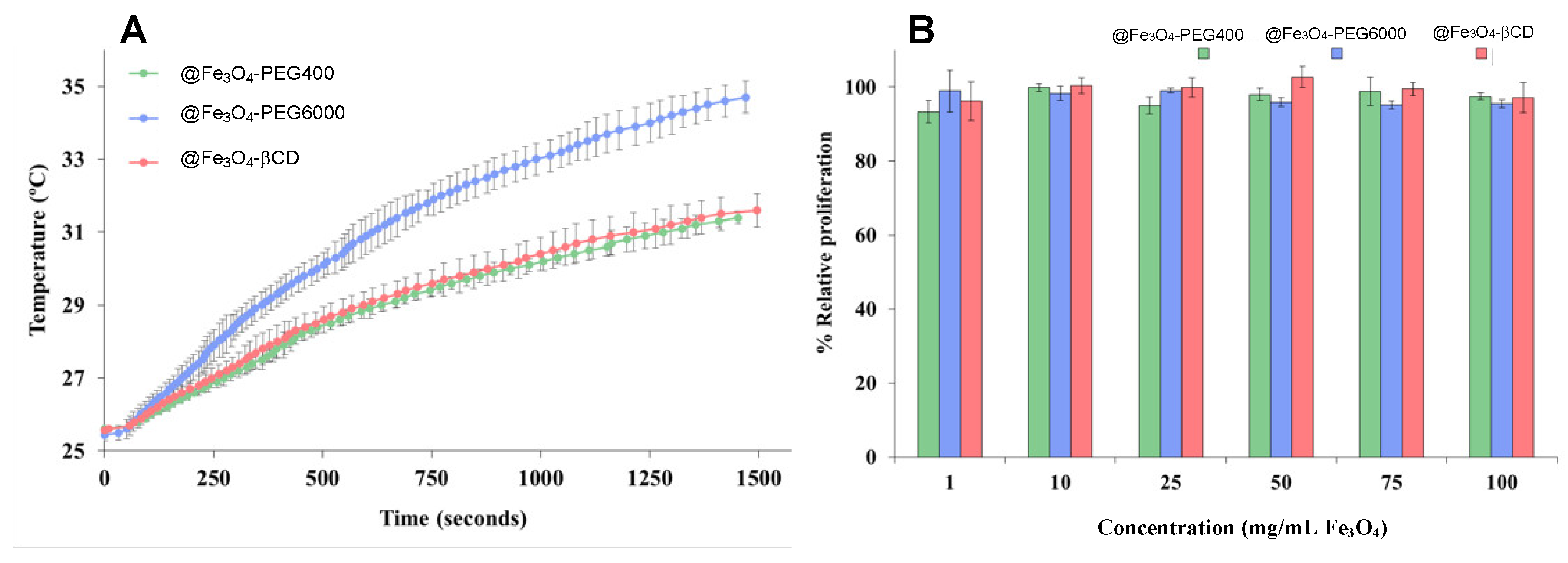

Table 1.
Sizes and concentrations of MNPs with different polymeric coatings.
| MNP | Size (nm) | Concentration (mg/mL) |
| @Fe3O4-PEG400 | 250 | 6.3 |
| @Fe3O4-PEG6000 | 150 | 7.5 |
| @Fe3O4-PEG400-N3 | 90 | 6.7 |
| @Fe3O4-PEG400-Pro | 200 | 6.9 |
| @Fe3O4-βCD | 155 | 7.1 |
Disclaimer/Publisher’s Note: The statements, opinions and data contained in all publications are solely those of the individual author(s) and contributor(s) and not of MDPI and/or the editor(s). MDPI and/or the editor(s) disclaim responsibility for any injury to people or property resulting from any ideas, methods, instructions or products referred to in the content. |
© 2024 by the authors. Licensee MDPI, Basel, Switzerland. This article is an open access article distributed under the terms and conditions of the Creative Commons Attribution (CC BY) license (http://creativecommons.org/licenses/by/4.0/).
Copyright: This open access article is published under a Creative Commons CC BY 4.0 license, which permit the free download, distribution, and reuse, provided that the author and preprint are cited in any reuse.


