
Submitted:
15 November 2024
Posted:
19 November 2024
You are already at the latest version
Abstract
Twelve compounds (1-12): kaempferol (1), luteolin (2), luteolin 4'-O-β-xyloside (3), luteolin 4'-O-β-glucoside (4), quercetin 4'-O-β-xyloside (5), kaempferol-3-O-[6"-O-(E)-p-coumaroyl]-β-D-glucoside (trans-tiliroside) (6), protocatechuic acid (7), gallic acid (8), methyl gallate (9), ethyl gallate (10), shikimic acid-3-O-gallate (11), and 3,3',4'-tri-O-methyl-ellagic acid 4-sulfate (12) were isolated and identified from the aerial parts of Helianthemum cinereum (Cav.) Pers (synonym: Helianthemum rubellum C. Presl. All compounds were isolated by applying different chromatographic procedures, such as silica gel, RP-18 and sephadex LH-20 columns. The structures were elucidated by extensive spectroscopic methods, mainly nuclear magnetic resonance NMR 1D and 2D, and mass spectrometry, as well as by comparison with the reported spectroscopic data. The two organic extracts, ethyl acetate (EtOAc) and butanol (BuOH) were evaluated for their potent phenolic and flavonoid contents using Folin-Ciocalteu and aluminum chloride colorimetric methods. Furthermore, the antioxidant activity of the two extracts was determined using the DPPH, FRAP, and ABTS methods. Pure trans-tiliroside (6), the main isolated compound, and luteolin 4'-O-β-xyloside (3) were evaluated for their antitumor activity against lung cancer (A549), melanoma (A375) and pancreatic cancer (Mia PaCa-2 and Panc-1) cell lines by MTT assay.
Keywords:
Cistaceae
; Helianthemum
; NMR
; Flavonoids
; Antitumor activity
; Antioxidant activity
1. Introduction
The Cistaceae family consists of over 200 species in eight genera (Cistus, Fumana, Halimium, Helianthemum, Tuberaria, Crocanthemum, Hudsonia and Lechea), mostly found in temperate and subtropical regions of the northern hemisphere, especially the western Mediterranean region, with a secondary centre in the eastern United States [1]. The genus Helianthemum is the largest genus in Cistaceae family, with more than 140 spread species [2,3]. Plants of the genus are frequently used in traditional medicine for their various therapeutic properties: healing, gastro-protective, anti-hemorrhoidal, antiseptic, antifungal, anti-inflammatory, analgesic, and antidiarrheal [4,5,6].
The leaves of H. cinereum, H. apenninum, H. marifolium and H. syriacum species are used for burns, wound healing, stomach ache, diarrhea and gastroenteritis. Whole plants of this genus are ingested in decoctions and infusions for gastrointestinal problems. They are also applied to infected wounds or burns as a poultice [7]. In Algerian flora, this genus is presented by more than 40 species, most of them are generally found in western Algeria [8].
The phytochemistry of the genus Helianthemum has been reported recently by Mouffouk et al. [9]. In their minireview, the authors describe the results of the chemical investigations on 21 Helianthemum taxa that are described all over the world. According to these studies, secondary metabolites such as flavonoids, lignans, phenolic acids and sterols are the major constituents. Regarding the Algerian’s flora species: H. lippii, H. sessiliflorum, H. Kahiricum, H. rifocomum, H. gelatum, and H. hirtum are the only species that have been investigated [10,11,12,13,14,15,16]. A careful analysis demonstrated that flavones derived from luteolin and flavanols derived from quercetin and kaempferol are the main metabolites in the studied species. In addition, simple phenolics are also present and dominated by gallate derivatives and phenolic acids.
As part of our ongoing phytochemical research on Algerian Helianthemum species, we examined the ethyl acetate (EtOAc) and butanol (BuOH) extracts derived from the aerial parts of Helianthemum cinereum (Cav.) Pers. (synonym: Helianthemum rubellum C. Presl).
To the best of our knowledge, no advanced chemical analysis has been reported on H. cinereum species and only a screening and quantisation of polyphenols of eleven taxa in south-eastern Spain has been done by Rubio-Moraga et all [7].
According to this study, polyphenols of H. cinereum were quantised from the methanolic and water extracts and luteolin, kaempferol were the most dominated compounds. In addition, the antioxidant and antimicrobial activities of these two extracts has been accessed [7].
Our chemical investigation on the Algerian H. cinereum species has revealed a secondary metabolite pattern dominated by flavonoids and phenolic compounds. Twelve compounds were fully purified and characterised mainly by analysing the NMR 1D and 2D spectra, as well as by comparing them with data reported in the literature. Additionally, the total phenolic and flavonoid contents and antioxidant activity were carried out on the ethyl acetate and butanol extracts. The results revealed that ethyl acetate extract shows high amount of phenolic and flavonoid compounds and exhibits high antioxidant capacity. The main compound trans-tiliroside and luteolin 4'-O-β-xyloside (3) have been tested in vitro for their antitumoral activity against lung cancer (A549), melanoma (A375), and pancreatic cancer (Mia PaCa-2 and Panc-1) cell lines using the MTT assay, where they demonstrate a moderate decline in cell viability.
2. Materials and Methods
2.1. Plant Material
The aerial parts of Helianthemum cinereum (Cav.) Pers were collected in the region of Oued El-ma, Merouana (Batna) during the flowering period in May 2018. The plant was identified by Pr. OUDJEHIH Bachir of the Department of Agronomy of the Institute of Veterinary and Agronomic Sciences from the University of Batna 1. The plant material was air-dried, and ground in a cross-beater mill (Retsch SK 100) from Retsch France Verder S.A.R.L. (Éragny, France), equipped with a 2 mm sieve.
2.2. General Experimental Procedures
The NMR spectra were obtained on a Bruker 500 (500 MHz for 1H and 125 MHz for 13C) and a Bruker 300 (300 MHz for 1H and 75 MHz for 13C) spectrometers. All 1D and 2D NMR experiments were performed using the standard Bruker library of microprograms. The spectra were recorded in deuterated methanol. Chemical shifts (δ) are in part per million (ppm) relative to TMS and Coupling constant (J) are in Hz. referenced solvent peaks of CD3OD: 1H δ 3.31, 13C δ 49.1.
Electrospray ionization mass spectra were acquired with a Micromass Q-Tof 2 (Micromass, Manchester, UK), operating in positive ion mode, equipped with a Z-spray source, an electrospray probe, and a syringe pump. The source and desolvation temperatures were 80 and 150 oC, respectively. The capillary voltage was 3000 V. The spectra were acquired at a nominal resolution of 9000 and at cone voltages of 30 V. Nebulisation and collision gases were N2 and Ar, respectively. Compound solution in methanol were introduced at a 10 μL min-1 flow rate.
2.3. Extraction and Isolation
One kilogram of the dried part of aerial parts of H. cinereum were extracted three times (3 × 10 L) with a mixture of 70% EtOH in water at room temperature. After evaporation of the solvent, the resulted aqueous solution (300 mL) was successively partitioned three times with 200 mL of petroleum ether (PE), 200 mL of ethyl acetate (AcOEt) and 200 mL of butanol (BuOH), to obtain, after removal of the solvents in vacuum, three organic extracts: PE (5.0 g), EtOAc (10.0 g), and BuOH (40.0 g). The AcOEt extract (10.0 g) was first chromatographed on a silica gel column eluting with a gradient solvent of CHCl3/MeOH (100/0 - 0/100) to give nine fractions (F1 to F9).
The fraction F6 (810 mg) was applied to a silica gel column chromatography, eluted with CHCl3/MeOH (100/0-0/100) to give sixteen sub-fractions (F6-1 to F6-16). The sub-fraction F6-8 (92 mg) was then chromatographed over Sephadex LH-20 column, and eluted with isocratic elution system CHCl3/MeOH (1/1) to yield five further sub-fractions (F6-8-1 to F6-8-5). The subfraction F6-8-3 (40 mg) was purified by using RP-18 SPE cartridge eluted with increasing proportions of a gradient MeOH in H2O, to afford compounds (3) (4 mg), (6) (15 mg), (7) (4 mg), and (10) (5 mg). The last sub-fraction F6-8-5 was further repurified on RP-18 SPE cartridge, eluted with gradient of MeOH in H2O (100-0) to yield compounds (1) (1.4 mg), (2) (8 mg), (5) (1.4 mg), (8) (21 mg) and (9) (1.6 mg).
The fraction F7 (2 g) was subjected to SiO2 gel column chromatography and eluting with a gradient of CHCl3 in MeOH (100/0-0/100) to obtain ten fractions (F7-1 to F7-10). The sub-fraction F7-9 (400 mg) was continually purified on sephadex LH-20 column with isocratic system (CHCl3 /MeOH: 1/1) to obtain eight subfractions (F7-9-1 to F7-9-8). The third fraction of this series (F7-9-3) was repurified by using the RP-18 SPE cartridge, eluted with H2O/MeOH (0/100-100/0) to give compound (4) (2 mg).
With the same manner of purification, the polar butanol extract of H. cinereum was firstly passed through RP-18 column chromatography using only MeOH to remove free sugars. The methanolic solution was concentrated and a mass of nine grams was purified by using RP-18 vacuum liquid chromatography (VLC) using gradient of MeOH in H2O. The fraction eluted with water (4g) was subjected to a silica gel column chromatography by using a gradient of CH2Cl2 in MeOH to afford fifteen fractions (F1-F15). The subfraction F9 (90 mg) was further separated on RP-18 SPE cartridge and eluted with a gradient of MeOH in H2O to afford six subfractions (F2-1 to F2-6). The third subfraction F2-3 (25 mg) was purified on Sephadex LH-20 column using an isocratic system MeOH–CHCl3 (1:1) to give compound (11) (3 mg). However, the purification of the fifth subfraction F2-5 (8.1 mg) by using RP-18 SPE cartridge and eluted with a gradient of MeOH in H2O gave compound (12) (0.5 mg).
Compound 3: Luteolin 4'-O-β-xyloside, yellow amorphous solid: 1H-NMR (500 MHz, CD3OD) δH (ppm): aglycone 7.47 (1H, dd, J = 8.0 and 2.2 Hz, H6'), 7.45 (1H, s, H-2'), 7.25 (1H, d, J = 9.1 Hz, H-5'), 6.62 (1H, s, H-3), 6.45 (1H, d, J = 2.0 Hz, H-8), 6.21 (1H, d, J = 2.0 Hz, H-6), xylose: 4.94 (1H, overlap, H-1′′), 4.0 (1H, dd, J = 11.5 and 5.1 Hz, H-5a′′), 3.63 (1H, m, H-4′′), 3.57 (1H, dd, J =7.5 Hz, H-2′′), 3.50 (1H, t, J = 8.3 Hz, H-3′′), 3.43 (1H, d, J = 11.3 Hz, H-5b′′); 13C-NMR (125 MHz, CD3OD) δC (ppm): aglycone 183.7 (C, C-4), 165.0 (C, C-2), 158.8 (C, C-5), 164.1 (C, C-7), 158.7 (C, C-9), 149.9 (C, C-4′), 148.1 (C, C-3′), 105.1(C, C-3), 126.7 (C, C1′), 119.9 (CH, C-6′), 116.5 (CH, C-5′), 113.5 (CH, C-2′), 105.3 (C, C-10), 93.8 (CH, C-8), xylose: 102.3 (CH, C-1′′), 75.8 (CH, C-3′′), 73.2 (CH, C-2′′), 69.5 (CH, C-4′′), 65.5 (CH, C-5′′).
Compound 4: Luteolin 4'-O-β-glucoside, yellow amorphous solid: 1H-NMR (500 MHz, CD3OD) δH (ppm) aglycone: 7.49 (1H, dd, J = 8.6 and 2.1 Hz, H-6'), 7.47(1H, d, J = 2.1Hz, H-2'), 7.34 (1H, d, J = 8.5 Hz, H-5'), 6.64 (1H, s, H-3), 6.48 (1H, d, J = 2.1 Hz, H-8), 6.23 (1H, d, J = 2.1 Hz, H-6), glucose: 4.96 (1H, d, J = 8.0 Hz, H-1′′), 3.94 (1H, dd, J = 12.2 and 2,0 Hz, H-6a′′), 3.75 (1H, dd, J = 12.2 and 5.6 Hz, H-6b′′), 3.56 (1H, t , J = 7.2 Hz, H-2′′), 3.52 (1H, m, H-5′′), 3.50 (1H, m, H-3′′), 3.45 (1H, t, J = 9.4 Hz, H-4′′); 13C-NMR (125 MHz, CD3OD) δC (ppm) aglycone: 182.5 (C, C-4), 164.8 (C, C-2), 164.2(C, C-7), 161.8(C, C-5), 158.1(C, C-9), 148.6 (C, C-4′), 147.2 (C, C-3′), 125.8 (C, C-1′), 119.5 (CH, C-6′), 117.5 (CH, C-5′), 114.5 (CH, C-2′), 104.1 (C, C-10), 104.8 (CH, C-3), 99.8 (CH, C-6), 94.8 (CH, C-8), glucose: 102.7 (CH, C-1′′), 76.1 (CH, C-5′′), 77.1 (CH, C-3′′), 73.3 (CH, C-2′′), 69.8 (CH, C-4′′), 62.2 (CH, C-6′′).
Compound 5: Quercetin 4'-O-β-xyloside, yellow crystal: 1H-NMR (500 MHz, CD3OD) δH (ppm) aglycon: δH 7.80 (1H, d, J=2.0 Hz, H-2′), 7.73 (1H, dd, J = 8.8, 2.0 Hz, H-6′), 7.23 (1H, d, J=8.8 Hz, H-5′), 6.42 (1H, d, J = 2.0 Hz, H-8), 6.21 (1H, d, J = 2.0 Hz, H-6), xylose : 4.91 (1H, d, J = 7.8 Hz, H-1′′), 3.99 (1H, dd, J = 11.3 and 5.3 Hz, H-5′′a), 3.63 (1H, m, H-4′′), 3.55 (1H, m, H-2′′), 3.48 (1H, m, H-3′′), 3.42 (1H, m, H-5b′′); 13C NMR (125 MHz, CD3OD) δC (ppm) aglycone: 176.2 (C, C-4), 164.7 (C, C-7), 161.9 (C, C-9), 158.8 (C, C-5), 146.5 (C, C-4′), 146.4 (C, C-3′), N.D (C, C-3), 126.4 (C, C-1′), 119.8 (CH, C-6′), 116.0 (CH, C-5′), 115.5 (CH, C-2′), 103.1 (C, C-10), 98.0 (CH, C-6), 93.1 (CH, C-8), xylose: 102.5 (CH, C-1′′), 75.9 (CH, C-3′′), 73.2 (CH, C-2′′), 69.6 (CH, C-4′′), 65.6 (CH2, C-5′′).
Compound 11: Shikimic acid 3-O-gallate, white powder: 1H NMR (500 MHz, CD3OD): δH 6.84 (1H, s, H-2), 5.32 (1H, dt, J = 5.2 Hz, H-5), 4.51 (1H, t, J = 3.8 Hz, H-3), 4.04 (1H, dd, J = 3.5, H-4), 2.88 (1H, dd, J = 5.2 and 18.5 Hz, H-6a), 2.40 (1H, dd, J = 5.2 and 18.5 Hz, H-6b); galloyl-H: 7.12 (2H, s, H-2'/H-6'), 13C NMR (125 MHz, CD3OD): 171.4 (C, C-7), 137.5 (CH, C-2), 131.2 (C, C-1), 71.6 (CH, C-5), 69.4 (CH, C-4), 66.9 (CH, C-3), 28.7 (CH2, C-6), galloyl: 168.1 (-OCO-), 121.2 (C, C-1'), 110.4 (2CH, C-2'/C-6'), 145.9 (2CH, C-3'/C-5'), 139.5( CH, C-4').
Compound 12: 3,3',4'-tri-O-methylellagic acid sulphate, amorphous: 1H NMR (500 MHz, CD3OD): δ 8.36 (1H, s, H-5), 7.78 (1H, s, H-5′), 4.30 (3H, s, OCH3-3), 4.17 (3H, s, OCH3-3'), 4.08 (3H, s, OCH3-4'), 13C NMR (125 MHz, CD3OD): 159,1 (C-9), 155.2 (C, C-4'), 145.0 (C, C-3), 141.8 (C, C-3'), 119.3 (CH, C-5), 115.8 (C, C-1), 112.9 (C, C-1'), 108.6 (CH, C-5'), 62.2 (OCH3, C-3), 61.8 (OCH3, C-3'), 56.9 (OCH3, C-4').
2.4. Determination of Total Phenolic Compounds (TPC)
The total phenolic compounds (TPC) amount in H. cinereum extract was determined by the Folin Ciocalteu method as described by [46]. Firstly, extract was diluted (50 times in Ethanol) due to its high concentration. Then, aliquot of 0.5 mL was mixed with 2.5 mL of 10% (v/v) Folin-Ciocalteu reagent. After 8 min in the dark, 2 mL of sodium carbonate Na2CO3 (7.5%, w/v) were added, and the reaction was carried out in the dark for 1 h. Then, the absorbance at 765 nm was measured using a UV-Vis spectrophotometer (UV 1700, Shimadzu, Japan). Gallic acid standard curve was built by the preparation of five concentrations of gallic acid (10, 50, 75, 100 and 250 µg/g), starting from a stock solution of 5 mg/g. TPC concentration was obtained from the calibration curve and expressed as mg gallic acid equivalent per gram of dried sample weight (mg GAE/g dry weight).
2.5. Determination of Total Flavonoids Compounds (TFC)
Total flavonoids content of H. cinereum extract was determined by the aluminum chloride colorimetric method as described by [47] with some modifications, 0.5 ml of extract in methanol was mixed with 0.1 ml of aluminum chloride solution (10 %), 0.1 ml of potassium acetate (1M) and 4.3 ml of distilled water. The mixture was incubated at room temperature for 30 min. Then, the absorbance was measured at 415 nm using a UV-Vis spectrophotometer (UV 1700, Shimadzu, Japan), and the flavonoid content was calculated based on a quercetin calibration curve, obtained by preparation of a serial concentrations (5 to 60 µg/g). All experiments were performed in triplicate and averaged; the results are expressed in mg of quercetin equivalent per gram of dry extract (mg QE/g).
2.6. Antioxidant Activity
2.6.1. DPPH Radical Scavenging Capacity
The free radical scavenging activity of H. cinereum extracts was investigated by the DPPH method as described by [48] with some modifications. Different concentrations of H. cinereum extract were prepared in methanol and aliquots of 100 μL were added to 3.5 ml of DPPH solution (30 μg/g in methanol). A blank consisting of DPPH solution without extract was prepared. All samples were kept in darkness for 15 minutes. Then the absorbance was measured against a blank (Methanol) at 515 nm in a UV-Vis spectrophotometer (UV 1700, Shimadzu, Japan). The DPPH solution was daily prepared and the concentration was checked using a calibration curve obtained by preparation of serial concentrations (4,8, 16, 32 and 64 μg/g).
The antioxidant capacity of the samples was expressed as DPPH percentage of inhibition (I%), calculated by the following formula:
where:
A0 and A represent the absorbance values of blank (DPPH with methanol) and the plant extract sample (DPPH with extract), respectively. The values of the inhibition percentage after 30 minutes were plotted versus the extract concentration to obtain the antioxidant capacity curve, and a linear regression was designed to obtain the IC50 value (concentration giving a reduction of 50% of DPPH concentration). The obtained IC50 value is inversely proportional to the antioxidant activity.
2.6.2. Ferric Reducing Antioxidant Power (FRAP) Assay
The iron reducing capacity of the H. cinereum extracts was determined by the method described by [49], with some modifications. 1 mL aliquots consisting on various dilutions of extract in methanol were added to 2.5 ml of phosphate buffer (0.2 M, pH 6.6), and 2.5 ml of potassium ferricyanide (1 %). Then, the mixture was incubated at 50°C for 20 min and 2.5 ml of trichloroacetic acid (10 %) were added to the sample and centrifuged at 3000 rpm for 10 min. After that, a volume of 2.5 mL of the mixture supernatant was added to 2.5 mL of distilled water and 0.5 mL ferric chloride (0.1%) and vigorously mixed. Then the absorbance of the sample is measured using a UV-Vis spectrophotometer (UV 1700, Shimadzu, Japan) at 700 nm against a blank similarly prepared by replacing the plant extract with methanol.
Ascorbic acid calibration curve was built by preparation of a serial concentrations (0.001, 0.025, 0.05, 0.1 and 0.2 mg/g), the results are expressed in milligrams equivalent of ascorbic acid per gram of extract.
2.6.3. ABTS Assay
The ABTS radical scavenging activity of H. cinereum extracts was carried out according to the method described by [50]. Firstly, 1 mL of ABTS stock solution (7 mM) was mixed with 88 μL of potassium persulfate solution (140 mM) and kept overnight protected from light and at room temperature. Next, the mixture was diluted with potassium buffer saline (0.2 M, pH 7.4) to obtain an ABTS solution with an absorbance of 0.70 ± 0.02 at 734 nm. Next, a volume of 3 ml of this ABTS solution was mixed with 1 ml of the sample at different concentrations (25, 50, 75, 100 and 250 µg/mL). The reaction took place in the dark for 6 minutes, and the absorbance at 734 nm was measured. Samples containing the same volume of methanol and the same dilution of ascorbic acid served as white and positive controls, respectively. ABTS free radical scavenging capacity was determined using the following Formula (3):
where A1 is ABTS radical and sample mixture absorbance; A2 is the solvent and sample mixture absorbance and A0 is the absorbance of the solvent and ABTS radical mixture.
2.7. Antitumoral Activity
2.7.1. Cell Culture
The A549 and A375 cell lines were acquired from the European Collection of Authenticated Cell Cultures (ECACC), and Mia PaCa-2 and Panc-1 cell lines were kindly provided by Dr. Sónia Melo (i3s, Porto, Portugal). All cell lines were maintained in High-Glucose Dulbecco’s Modified Eagle’s Medium (DMEM, PAN-Biotech, Aidenbach, Germany), supplemented with 10% (v/v) fetal bovine serum (FBS, PAN-Biotech, Aidenbach, Germany), 2 mM L-glutamine, and 1% pen/strep (100 U/mL penicillin, 100 ug/mL streptomycin (Grisp, Porto, Portugal). Cells were cultured at 5% CO2 and at 37 ºC.
2.7.2. Cell Viability Assessment
A stock solution was made by dissolving trans-tiliroside or luteolin 4'-O-xyloside in dimethyl sulfoxide (DMSO, Sigma-Aldrich, St. Louis, Missouri, USA). All cell lines were seeded in 96-well plates at a density of 12 500 cells/mL and incubated for 24 hours for adhesion. Next, cells were exposed to a range of concentrations of trans-tiliroside (10, 25, 50, 75, 100, 150 and 200 μM) or of luteolin (10, 25, 50, 75, 100, 150, 200, 250 and 300 μM) for 72 h. After the exposure, cell viability was assessed by 3-(4,5-dimethyl-2-thiazolyl)-2,5-diphenyl-2H-tetrazolium bromide (MTT, Sigma-Aldrich, St. Louis, Missouri, USA) assay. Briefly, the exposure medium was replaced by fresh medium and 50 μL of MTT (1.0 mg/mL dissolved in phosphate-buffered saline, PBS, PAN-Biotech, Aidenbach, Germany) and plates were incubated for 3 hours. Then, the medium was removed and 150 μL of DMSO were added and plates were shaken in the dark for 1 h. The absorbance was read in a microplate reader BioTek Synergy HT plate reader at 570 nm. Cells with only medium without compounds were used as control. Three independent assays were completed with three replicates each.
2.8. Statistical Analysis
For the TPC and TFC determinations and antioxidant activity, each sample was analyzed three times. The data presented are the means of the results obtained. The errors are expressed as standard deviations. A Student's t-test was used to determine significant differences (p ≤ 0.05) between the samples analyzed. The null hypothesis - that the samples are identical - was first considered. The experimental t values were compared with the theoretical t values. If the two values were equal or the experimental value was lower than the theoretical value, the null hypothesis was accepted. Conversely, if the experimental value was higher than the theoretical value, the null hypothesis was rejected, leading to the conclusion that the samples were significantly different.
In addition, the Pearson correlation matrix was performed using the Data Analysis Tools of Microsoft Excel version 2408. The Pearson correlation matrix is a table showing the Pearson correlation coefficients between pairs of variables. The Pearson correlation coefficient, denoted as r, measures the linear relationship between two continuous variables, indicating how strongly they are related and in which direction (positive or negative). Values of r range from -1 to 1.
Perfect positive correlation, meaning that as one variable increases, the other also increases in a perfectly linear manner, is obtained when r = 1. A perfect negative correlation, which means that as one variable increases, the other decreases in a perfectly linear manner, is obtained when r = -1.
Finally, when r = 0, there is no linear correlation, indicating that there is no linear relationship between the variables.
For the antitumor activity, the results are represented as the mean ± standard deviation. SigmaPlot version 14.0 (Systat Software, San Jose, CA, USA) for Windows was used to for statistical analysis. Data were analyzed by one-way ANOVA (p < 0.05) followed by Dunnett’s test (p < 0.05).
3. Results and Discussions
3.1. Phytochemical Study
The chemical composition of ethyl acetate (EtOAc) and butanol (BuOH) organic extracts of the aerial parts of Helianthemum cinereum is compared by TLC in different eluent systems. The composition and the relative distribution of the metabolites were characterized by a plethora of UV-visible metabolites in both extracts. However, rich metabolite pattern was observed in the ethyl acetate extract, it showed less polar compounds in comparison with the butanol extract. Thus, both extracts were subjected to a series of chemical purifications starting by silica gel columns chromatography as a first step following by Sephadex LH-20 columns and ending with C18-SPE cartridges. After separation (described in experimental part), nine compounds were identified from the ethyl acetate extract and three compounds from the butanol extract (Figure 1): kaempferol (1) [17], luteolin (2) [18], luteolin 4'-O-β-xyloside (3) [19], luteolin 4'-O-β-glucoside (4) [20], quercetin 4'-O-β-xyloside (5) [21], kaempferol-3-O-[6"-O-(E)-p-coumaroyl]-β-D-glucoside (trans-tiliroside) (6) [11,16,22,23], protocatechuic acid (7) [24], gallic acid (8) [14], methyl gallate (9) [25], ethyl gallate (10)[26], shikimic acid 3-O-gallate (11) [27], 3,3',4'-tri-O-methyl-ellagic acid 4-sulfate (12) [28,29].
To the best of our knowledge, compounds: luteolin 4'-O-β-xyloside (3), luteolin 4'-O-β-glucoside (4), quercetin 4'-O-β-xyloside (5), ethyl gallate (phyllemblin) (10), shikimic acid 3-O-gallate (11), and 3,3',4'-tri-O-methyl-ellagic acid 4-sulfate (12) are isolated here for the first time in Cistaceae family. Luteolin 4'-O-β-xyloside (3), luteolin 4'-O-β-glucoside (4), and quercetin 4'-O-β-xyloside (5) are uncommon flavonoid 4'-O-β-glycosides, with limited data available in the literature [19,20,21]. Additionally, kaempferol (1) and luteolin (2) are reported from some species of Cistus genus[18,30]. However, the other compounds (6), (7), (8) and (9) are reported from the genus Helianthemum, in particular, from the Algerian species: H. lippii, H. sessiliflorum, H. Kahiricum, H. rifocomum, H. gelatum and H. hirtum [10,11,12,13,14,15,16].
It is very interesting to mention here that we have isolated ellagic acid compound (12) as a sulphate derivative. The occurrence of ellagic acid derivatives is very common in Cistaceae family [31], and their identification here is further supported by the occurrence of quercetin 3-sulphate and isorhamnetin 3-sulphate in the species Helianthemum squamatum [31,32,33].
3.2. Total Phenolic and Flavonoids Contents
It is known that phenolic compounds are very important secondary metabolites of plants with redox properties responsible for antioxidant activity [34,35]. The phenolic compounds content was measured by a colorimetric method using Folin-Ciocalteu reagent for each plant extract. Results were derived from a calibration curve (y = 9.7205x + 0.0325, R2 = 0.991) for gallic acid at different concentrations (10-250 µg/mL) and expressed as gallic acid equivalents (GAE) per gram of dry extract weight (Table 1). The total flavonoids content of the two extracts of H. cinereum (Table 1) are measured by using aluminum chloride colorimetric method and the results were obtained from quercetin calibration curve (y = 0,0145x + 0,0099, R2 = 0.998).
The phenolic and flavonoid contents of the ethyl acetate extract showed a higher values (361.51 mg GAE/g; 148.23 ± 0.51 mg QE/g) than the butanol extract (145.88 mg GAE/g; 94.89 ± 0.29 mg QE/g). The total phenolic content (TPC) values observed in this study are higher than those reported by Benabdelaziz et al. for H. sessiliflorum, where the ethyl acetate and butanol extracts showed TPC values of 42.51 ± 1.01 mg GAE/g and 40.02 ± 2.81 mg GAE/g, respectively [12]. Moreover, the TPC values in this study exceed those found for the hydro-methanolic extract of H. canum (284.13 ± 0.30 mg GAE/g) [36] . Furthermore, total phenolic content values found in this study are higher than those of the ethyl acetate (46.70 ± 0.22 mg QE/g) and butanol (35.48 ± 0.36 mg QE/g) extracts of H. sessiliflorum and the hydro-methanolic extract of H. canum (13.13 ± 0.10 mg QE/g) [12,36].
3.3. Antioxidant Activities
Due to the complex activity of the phytoconstituents, the antioxidant activity of the extracts cannot be assessed by a single method, and it is recommended to use several methods, as each technique provides different and complementary information about the activity and the mechanism of action [37].
In this study, the antioxidant activity of H. cinereum extracts was evaluated using three different methods, as presented in Table 2. Across all methods (DPPH, FRAP, and ABTS), the ethyl acetate extract demonstrated stronger antioxidant capacity compared to the butanol extract. The antioxidant capacity showed by DPPH method is notably higher than that reported by Benabdelaziz et al., for the ethyl acetate and butanol extracts of H. sessiliflorum, which had IC50 values of 23.75 ± 2.07 μg/mL and 94.03 ± 1.52 μg/mL, respectively [12]. Additionally, the IC50 values in this study were lower than those found for the hydro-methanolic extract of H. canum (0.19 mg/mL) [36], thus indicating the higher antioxidant activity.
The Pearson correlation results presented in Figure 2 show exceptionally strong relationships between total phenolic content (TPC), total flavonoid content (TFC) and various measures of antioxidant activity (DPPH, FRAP and ABTS assays) in the analysed samples. Analysis of the data shows that there is a perfect positive correlation between total phenolic content and total flavonoid content. This means that as the phenolic content in the samples increases, the flavonoid content increases proportionally. Phenolics and flavonoids, both subclasses of polyphenolic compounds, often co-occur and exhibit similar variations in plant materials. In addition, both TPC and TFC show a perfect positive correlation with the results of the Ferric Reducing Antioxidant Power (FRAP) assay. This suggests that higher levels of phenolics and flavonoids in samples are directly related to increased antioxidant power as measured by the FRAP assay. However, the total phenolic and flavonoid contents are perfectly negatively correlated with DPPH radical scavenging capacity and ABTS radical scavenging activity. If the DPPH and ABTS values represent IC₅₀ values (the concentration required to inhibit 50% of the free radicals), a lower IC₅₀ indicates higher antioxidant activity. Therefore, as the phenolic and flavonoid contents increase, the IC₅₀ values decrease, indicating increased antioxidant activity. This is consistent with the positive correlation between DPPH and ABTS. This means that samples with higher (or lower) DPPH radical scavenging capacity also have correspondingly higher (or lower) ABTS radical scavenging activity. Both assays measure the ability of antioxidants to scavenge free radicals, so they are expected to be highly correlated.
Finally, the FRAP values are perfectly negatively correlated with the DPPH and ABTS assay results. This indicates that samples with higher antioxidant power as measured by FRAP have lower IC₅₀ values in the DPPH and ABTS assays, which in turn indicates higher antioxidant activity. These findings from our study are supported by the previous studies that have shown that the capacity of the antioxidant is highly associated with the total flavonoid content and total phenolic compounds of the plant extract [38].
3.4. Antitumor Activity
The antitumoral activity of trans-tiliroside (6) and luteolin 4'-O-β-xyloside (3) was investigated against lung cancer (A549), melanoma (A375) and pancreatic cancer (Mia PaCa-2 and Panc-1) cell lines by MTT assay. The MTT assay is a widely used method that reduces a tetrazolium compound (MTT) into formazan. This colorimetric assay assesses the cellular metabolic activity, which is an indicator of toxicity [39]. As shown in Figure 3, all cell lines exhibited a decline in cell viability in a concentration dependent manner. In the case of trans-tiliroside, Mia PaCa-2 and Panc-1 had a similar response, with Panc-1 having a decline of cell viability with the lowest tested concentration (10 μM), while Mia PaCa-2 was only affected by concentrations equal or above 50 μM. Notwithstanding, the determined IC50 was slightly lower to Mia PaCa-2 (46.2 ± 0.89 μM) than to Panc-1 (54.0 ± 1.95 μM). These results are consistent with a previous work that showed that trans-tiliroside can inhibit the growth of Panc-1 cells and determined a IC50 of 68.48 μM for 72 h of exposure [40]. Both A549 and A375 cell lines showed to be more resistant to trans-tiliroside, having a significant decrease in cell viability when exposed to concentrations equal or superior to 75 μM and an IC50 of 108 ± 2.80 and 102 ± 2.56 μM, respectively. The work of Lu et al., in which they observed a decline of 80% in cell viability of B16-F10, another melanoma cell line, when exposed to 168 μM of trans-tiliroside for 72 h [41].
Luteolin 4'-O- β-xyloside (3), on the other hand, had a more subtle effect on the four cell lines. The A375 cell line had a significant decrease in cell viability when exposed to concentrations up to 75 μM, contrary to the other three cell lines. Nevertheless, literature reveals that luteolin aglycone has a strong antitumoral effect. In fact, studies suggest an IC50 between 7 and 17 μM for melanoma cell lines [42], and an IC50 values of 32.6 μM and 40 μM for A549 cell line [43,44]. Pancreatic cancer cell lines also showed to be strongly affected by concentrations above 160 μM [45]. However, in our work, Mia PaCa-2 was the only cell line that showed a cell viability lower than 50% when exposed to the tested concentrations and the calculated IC50 was 229 ± 9.09 μM.
5. Conclusions
The present study focused on the phytochemical study and the biological activities of the Algerian species Helianthemum cinerum, which allowed us to isolate twelve compounds from the ethyl acetate (EtOAc) and butanol (BuOH) extracts. All the compounds were identified by spectral analysis, mainly through NMR experiments (¹H, ¹³C, COSY, HSQC, and HMBC) and mass spectrometry, as well as by comparing their spectroscopic data with those reported in the literature. To the best of our knowledge, this is the first report of six compounds being isolated from the Cistaceae family, including luteolin 4'-O-β-xyloside (3), luteolin 4'-O-β-glucoside (4), quercetin 4'-O-β-xyloside (5), ethyl gallate (phyllemblin) (10), shikimic acid 3-O-gallate (11), and 3,3',4'-tri-O-methyl-ellagic acid 4-sulfate (12). Although, the isolated compounds trans-tiliroside and luteolin 4'-O- β-xyloside were assessed for their antitumor activity against lung cancer (A549), melanoma (A375) and pancreatic cancer (Mia PaCa-2 and Panc-1) cell lines by MTT assay where they showed an important activity.
In addition to determining total flavonoid and phenolic contents (TFC and TPC), the extracts were evaluated for their antioxidant capacity using in vitro free radical scavenging assays (DPPH, ABTS) and the FRAP assay. The results of antioxidant capacity revealed that the ethyl acetate (EtOAc) extract exhibited stronger activity than the butanol (BuOH) extract; however, considering the available data, the potential of both extracts remains higher compared to extracts from other species of the same genera. Correspondingly, a positive correlation between total phenolic and flavonoid contents and the antioxidant activity of the investigated extracts indicated that these phytoconstituents are the major contributors to the antioxidant capacities of this plant. These findings suggest that H. cinereum could be a promising source of natural antioxidants.
Author Contributions
Methodology, A. B.; and H. O.; Investigation, A. B., A. S., H. B.; D.S. and G.G-L.; Project administration, A. M. S. S.; Resources, E. O-K. and C. N.; Software, M. W. and A.S.; Supervision, F. B.; Writing – original draft, A. S.; Writing – review & editing; D. C. G. A. P.
Funding
This research was funded by the Romanian Ministry of Research, Innovation and Digitization (MCID) through Program 1—Development of the National R&D System, Subprogram 1.2-Institutional Performance-Projects for Excellence Financing in RDI–grant number 2PFE/2021 and by Core Program within the National Research, Development and Innovation Plan 2022–2027, carried out with the support of MCID—grant number 7 N/2022-23020101(SIA-PRO).
Acknowledgments
The authors thank the University of Aveiro, FCT/MCTES for their support to the LAQV-REQUIMTE (LA/P/008/2020 DOI https://doi.org/10.54499/LA/P/0008/2020, UIDP/50006/2020/ DOI https://doi.org/10.54499/UIDP/50006/2020 and UIDB/50006/2020 DOI https://doi.org/10.54499/UIDB/50006/2020 through national funds and, where applicable, co-financed by the FEDER, within the PT2020 Partnership Agreement, and to the Portuguese NMR Network. The authors acknowledge the financial support to CESAM by FCT/MCTES (UIDP/50017/2020 & UIDB/50017/2020 & LA/P/0094/2020), through national funds, and when appropriate co-financed by FEDER under the PT2020 Partnership Agreement. H. O. thanks FCT for the research contract under the Scientific Employment Stimulus (DOI 10.54499/CEECIND/04050/2017/CP1459/CT0023). D. S. thanks FCT for the PhD grant (2022.11049.BD). The authors gratefully acknowledge the financial support provided by the Algerian universities: University of Batna 1, University of Batna 2, and Abbes Laghrour Khenchela University.
Conflicts of Interest
The authors declare no conflicts of interest.
References
- J.M. Arrington and K. Kubitzki Flowering Plants · Dicotyledons; Verlag Berlin Heidelberg, Ed.; Springer, 2003.
- Albaladejo, R.G.; Martín-Hernanz, S.; Reyes-Betancort, J.A.; Santos-Guerra, A.; Olangua-Corral, M.; Aparicio, A. Reconstruction of the Spatio-Temporal Diversification and Ecological Niche Evolution of Helianthemum (Cistaceae) in the Canary Islands Using Genotyping-by-Sequencing Data. Ann Bot 2021, 127, 597–611. [Google Scholar] [CrossRef] [PubMed]
- Mabberly, D.J. The Plant-Book: A Portable Dictionary of the Vascular Plants; Cambridge University press. England, 1997.
- Başlar, S.; Doǧan, Y.; Mert, H.H. A Study on the Soil-Plant Interactions of Some Cistus L. Species Distributed in West Anatolia. Turk J Botany 2002, 26. [Google Scholar]
- Ustun, O.; Gurbuz, I.; Kusmenoglu, S.; Turkoz, S. Fatty Acid Content of Three Cistus Species Growing in Turkey. Chem Nat Compd 2004, 40, 526–528. [Google Scholar] [CrossRef]
- Wang, B.; Qiu, Y.L. Phylogenetic Distribution and Evolution of Mycorrhizas in Land Plants. Mycorrhiza 2006, 16, 299–363. [Google Scholar] [CrossRef] [PubMed]
- Rubio-moraga, Á.; Argandoña, J.; Mota, B.; Pérez, J.; Verde, A. Screening for Polyphenols, Antioxidant and Antimicrobial Activities of Extracts from Eleven Helianthemum Taxa ( Cistaceae ) Used in Folk Medicine in South-Eastern Spain. J Ethnopharmacol 2013, 1–10. [Google Scholar] [CrossRef]
- Quézel, P.; Santa, S. Nouvelle Flore de l’Algérie et Des Régions Désertiques Méridionales. In; 1962; p. 502.
- Mouffouk, S.; Mouffouk, C.; Mouffouk, S.; Haba, H. Medicinal, Pharmacological and Biochemical Progress on the Study of Genus Helianthemum: A Review. Curr Chem Biol 2023, 17, 147–159. [Google Scholar] [CrossRef]
- Djemam, N.; Lassed, S.; Gül, F.; Altun, M.; Monteiro, M.; Menezes-Pinto, D.; Benayache, S.; Benayache, F.; Zama, D.; Demirtas, I.; et al. Characterization of Ethyl Acetate and N-Butanol Extracts of Cymbopogon Schoenanthus and Helianthemum Lippii and Their Effect on the Smooth Muscle of the Rat Distal Colon. J Ethnopharmacol 2020, 252, 112613. [Google Scholar] [CrossRef]
- Benabdelaziz, I.; Haba, H.; Lavaud, C.; Harakat, D.; Benkhaled, M. Lignans and Other Constituents from Helianthemum Sessiliflorum Pers. Records of Natural Products 2015, 9, 342–348. [Google Scholar]
- Benabdelaziz, I.; Marcourt, L.; Benkhaled, M.; Wolfender, J.-L.; Haba, H. Antioxidant and Antibacterial Activities and Polyphenolic Constituents of Helianthemum Sessiliflorum Pers. Nat Prod Res 2017, 31, 686–690. [Google Scholar] [CrossRef] [PubMed]
- Bouzergoune, F.; Bitam, F.; Aberkane, M.C.; Mosset, P.; Fetha, M.N.H.; Boudjar, H.; Aberkane, A. Preliminary Phytochemical and Antimicrobial Activity Investigations on the Aerial Parts of Helianthemum Kahiricum. Chem Nat Compd 2013, 49. [Google Scholar] [CrossRef]
- Chemam, Y.; Benayache, S.; Marchioni, E.; Zhao, M.; Mosset, P.; Benayache, F.; McPhee, D.J. On-Line Screening, Isolation and Identification of Antioxidant Compounds of Helianthemum Ruficomum. Molecules 2017, 22, 1–14. [Google Scholar] [CrossRef] [PubMed]
- Terfassi, S.; Dauvergne, X.; Cérantola, S.; Lemoine, C.; Bensouici, C.; Fadila, B.; Christian, M.; Marchioni, E.; Benayache, S. First Report on Phytochemical Investigation, Antioxidant and Antidiabetic Activities of Helianthemum Getulum. Nat Prod Res 2022, 36, 2806–2813. [Google Scholar] [CrossRef]
- Wassila, B. Constituants Chimiques Des Espèces Helianthemum Hirtum Ssp. Ruficomum (Cistaceae) et Onobrychis Crista-Galli (Fabaceae), Batna 1, 2020.
- Barbosa, E.; Calzada, F.; Campos, R. Antigiardial Activity of Methanolic Extracts from Helianthemum Glomeratum Lag. and Rubus Coriifolius Focke in Suckling Mice CD-1. J Ethnopharmacol 2006, 108, 395–397. [Google Scholar] [CrossRef] [PubMed]
- Vogt, T.; Gerhard Gul, P. Accumulation of Flavonoids during Leaf Development in Cistus Laurifolius. Phytochemistry 1994, 36, 591–597. [Google Scholar] [CrossRef]
- Yuan, S.; Yin, S.; Liu, M.; Kong, J.Q. Isolation and Characterization of a Multifunctional Flavonoid Glycosyltransferase from Ornithogalum Caudatum with Glycosidase Activity. Sci Rep 2018, 8, 1–13. [Google Scholar] [CrossRef]
- Nawwar, M.A.M.; Hussein, S.A.M.; Merfort, I. Leaf Phenolics of Punica Granatum. Phytochemistry 1994, 37, 1175–1177. [Google Scholar] [CrossRef]
- Jambor, J.; Skrzypczak, L. Flavonoids from the Flowers of Nymphaea Alba L. Acta Societatis Botanicorum Poloniae 1991, 60, 119–125. [Google Scholar] [CrossRef]
- Calzada, F.; Lopéz, R.; Meckes, M.; Cedillo-Rivera, R. Flavonoids of the Aerial Parts of Helianthemum Glomeratum. International Journal of Pharmacognosy 1995, 33, 351–352. [Google Scholar] [CrossRef]
- Toan Phan, N.H.; Dieu Thuan, N.T.; Duy, N. Van; Mai Huong, P.T.; Cuong, N.X.; Nam, N.H.; Thanh, N. Van; Minh, C. Van Flavonoids Isolated from Dipterocarpus Obtusifolius. Vietnam Journal of Chemistry 2015, 53, 131–136. [Google Scholar] [CrossRef]
- An, L.J.; Guan, S.; Shi, G.F.; Bao, Y.M.; Duan, Y.L.; Jiang, B. Protocatechuic Acid from Alpinia Oxyphylla against MPP+-Induced Neurotoxicity in PC12 Cells. Food and Chemical Toxicology 2006, 44, 436–443. [Google Scholar] [CrossRef]
- Boudermine, S.; Malafronte, N.; Mencherini, T.; Esposito, T.; Aquino, R.P.; Beghidja, N.; Benayache, S.; D’Ambola, M.; Vassallo, A. Phenolic Compounds from Limonium Pruinosum. Nat Prod Commun 2015, 10, 319–321. [Google Scholar] [CrossRef] [PubMed]
- Ooshiro, A.; Hiradate, S.; Kawano, S.; Takushi, T.; Fujii, Y.; Natsume, M.; Abe, H. Identification and Activity of Ethyl Gallate as an Antimicrobial Compound Produced by Geranium Carolinianum. Weed Biol Manag 2009, 9, 169–172. [Google Scholar] [CrossRef]
- Gen-Ichiro Nonaka, Masayuki Ageta, and I. N. Tannins and Related Compounds. XXV. A New Class of Gallotannins Possessing a (-)-Shikimic Acid Core from Castanopsis Cuspidata Var. Sieboldii NAKAI. (1). Chem Pharm Bull (Tokyo) 1985, 33, 96–101. [Google Scholar] [CrossRef]
- Manurung, J.; Kappen, J.; Schnitzler, J.; Frolov, A.; Wessjohann, L.A.; Agusta, A.; Muellner-Riehl, A.N.; Franke, K. Analysis of Unusual Sulfated Constituents and Anti-Infective Properties of Two Indonesian Mangroves, Lumnitzera Littorea and Lumnitzera Racemosa (Combretaceae). Separations 2021, 8. [Google Scholar] [CrossRef]
- Owczarek, A.; Rózalski, M.; Krajewska, U.; Olszewska, M.A. Rare Ellagic Acid Sulphate Derivatives from the Rhizome of Geum Rivale L.-Structure, Cytotoxicity, and Validated HPLC-PDA Assay. Applied Sciences (Switzerland) 2017, 7. [Google Scholar] [CrossRef]
- Tomás-Lorente, F.; Garcia-Grau, M.M.; Nieto, J.L.; Tomás-Barberán, F.A. Flavonoids from Cistus Ladanifer Bee Pollen. Phytochemistry 1992, 31, 2027–2029. [Google Scholar] [CrossRef]
- Harborne, J.B. Flavonoid Bisulphates and Their Co-Occurrences with Ellagic Acid in the Bixaceae, Frankeniaceae and Related Families. Phytochemistry 1975, 14, 1331–1337. [Google Scholar] [CrossRef]
- Harborne, J.B. Flavonoid Sulphates: A New Class of Sulphur Compounds in Higher Plants. Phytochemistry 1975, 14, 1147–1155. [Google Scholar] [CrossRef]
- Laraoui, H.; Haba, H.; Long, C.; Benkhaled, M. A New Flavanone Sulfonate and Other Phenolic Compounds from Fumana Montana. Biochem Syst Ecol 2019, 86, 103927. [Google Scholar] [CrossRef]
- Soobrattee, M.A.; Neergheen, V.S.; Luximon-Ramma, A.; Aruoma, O.I.; Bahorun, T. Phenolics as Potential Antioxidant Therapeutic Agents: Mechanism and Actions. Mutation Research - Fundamental and Molecular Mechanisms of Mutagenesis 2005, 579, 200–213. [Google Scholar] [CrossRef]
- Vuolo, M.M.; Lima, V.S.; Maróstica Junior, M.R. Phenolic Compounds: Structure, Classification, and Antioxidant Power. Bioactive Compounds: Health Benefits and Potential Applications, 2019; 33–50. [Google Scholar] [CrossRef]
- Baldemir, A.; Gökşen, N.; Ildız, N.; Karatoprak, G.Ş.; Koşar, M. Phytochemical Profile and Biological Activities of Helianthemum Canum l. Baumg. from Turkey. Chem Biodivers 2017, 14. [Google Scholar] [CrossRef]
- Paun, G.; Neagu, E.; Albu, C.; Alecu, A.; Seciu-Grama, A.M.; Radu, G.L. Antioxidant and Antidiabetic Activity of Cornus Mas L. and Crataegus Monogyna Fruit Extracts. Molecules 2024, 29, 3595. [Google Scholar] [CrossRef] [PubMed]
- Johari, M.A.; Khong, H.Y. Total Phenolic Content and Antioxidant and Antibacterial Activities of Pereskia Bleo. Adv Pharmacol Sci 2019, 2019. [Google Scholar] [CrossRef] [PubMed]
- Menyhárt, O.; Harami-Papp, H.; Sukumar, S.; Schäfer, R.; Magnani, L.; de Barrios, O.; Győrffy, B. Guidelines for the Selection of Functional Assays to Evaluate the Hallmarks of Cancer. Biochimica et Biophysica Acta (BBA) - Reviews on Cancer 2016, 1866, 300–319. [Google Scholar] [CrossRef] [PubMed]
- Xu, M.; Zhong, W.; Yang, C.; Liu, M.; Yuan, X.; Lu, T.; Li, D.; Zhang, G.; Liu, H.; Zeng, Y.; et al. Tiliroside Disrupted Iron Homeostasis and Induced Ferroptosis via Directly Targeting Calpain-2 in Pancreatic Cancer Cells. Phytomedicine 2024, 127, 155392. [Google Scholar] [CrossRef] [PubMed]
- Lu, Y.H.; Chen, J.; Wei, D.Z.; Wang, Z.T.; Tao, X.Y. Tyrosinase Inhibitory Effect and Inhibitory Mechanism of Tiliroside from Raspberry. J Enzyme Inhib Med Chem 2009, 24, 1154–1160. [Google Scholar] [CrossRef]
- Schomberg, J.; Wang, Z.; Farhat, A.; Guo, K.L.; Xie, J.; Zhou, Z.; Liu, J.; Kovacs, B.; Liu-Smith, F. Luteolin Inhibits Melanoma Growth in Vitro and in Vivo via Regulating ECM and Oncogenic Pathways but Not ROS. Biochem Pharmacol 2020, 177, 114025. [Google Scholar] [CrossRef] [PubMed]
- Zhao, Y.; Yang, G.; Ren, D.; Zhang, X.; Yin, Q.; Sun, X. Luteolin Suppresses Growth and Migration of Human Lung Cancer Cells. Mol Biol Rep 2011, 38, 1115–1119. [Google Scholar] [CrossRef]
- Jiang, Z.Q.; Li, M.H.; Qin, Y.M.; Jiang, H.Y.; Zhang, X.; Wu, M.H. Luteolin Inhibits Tumorigenesis and Induces Apoptosis of Non-Small Cell Lung Cancer Cells via Regulation of MicroRNA-34a-5p. Int J Mol Sci 2018, 19. [Google Scholar] [CrossRef]
- Huang, X.; Dai, S.; Dai, J.; Xiao, Y.; Bai, Y.; Chen, B.; Zhou, M. Luteolin Decreases Invasiveness, Deactivates STAT3 Signaling, and Reverses Interleukin-6 Induced Epithelial–Mesenchymal Transition and Matrix Metalloproteinase Secretion of Pancreatic Cancer Cells. Onco Targets Ther 2015, 8, 2989–3001. [Google Scholar] [CrossRef]
- Song, X.C.; Canellas, E.; Asensio, E.; Nerín, C. Predicting the Antioxidant Capacity and Total Phenolic Content of Bearberry Leaves by Data Fusion of UV–Vis Spectroscopy and UHPLC/Q-TOF-MS. Talanta 2020, 213. [Google Scholar] [CrossRef] [PubMed]
- Amzad Hossain, M.; Shah, M.D. A Study on the Total Phenols Content and Antioxidant Activity of Essential Oil and Different Solvent Extracts of Endemic Plant Merremia Borneensis. Arabian Journal of Chemistry 2015, 8, 66–71. [Google Scholar] [CrossRef]
- Wrona, M.; Nerín, C.; Alfonso, M.J.; Caballero, M.Á. Antioxidant Packaging with Encapsulated Green Tea for Fresh Minced Meat. Innovative Food Science and Emerging Technologies 2017, 41, 307–313. [Google Scholar] [CrossRef]
- Bouabid, K.; Lamchouri, F.; Toufik, H.; Faouzi, M.E.A. Phytochemical Investigation, in Vitro and in Vivo Antioxidant Properties of Aqueous and Organic Extracts of Toxic Plant: Atractylis Gummifera L. J Ethnopharmacol 2020, 253. [Google Scholar] [CrossRef] [PubMed]
- Peng, Q.; Huang, Z.; Liang, G.; Bi, Y.; Kong, F.; Wang, Z.; Tan, S.; Zhang, J. Preparation of Protein-Stabilized Litsea Cubeba Essential Oil Nano-Emulsion by Ultrasonication: Bioactivity, Stability, in Vitro Digestion, and Safety Evaluation. Ultrason Sonochem 2024, 107. [Google Scholar] [CrossRef]
Figure 1.
Chemical structure of isolated compounds from the aerial parts of Helianthemum cinereum.
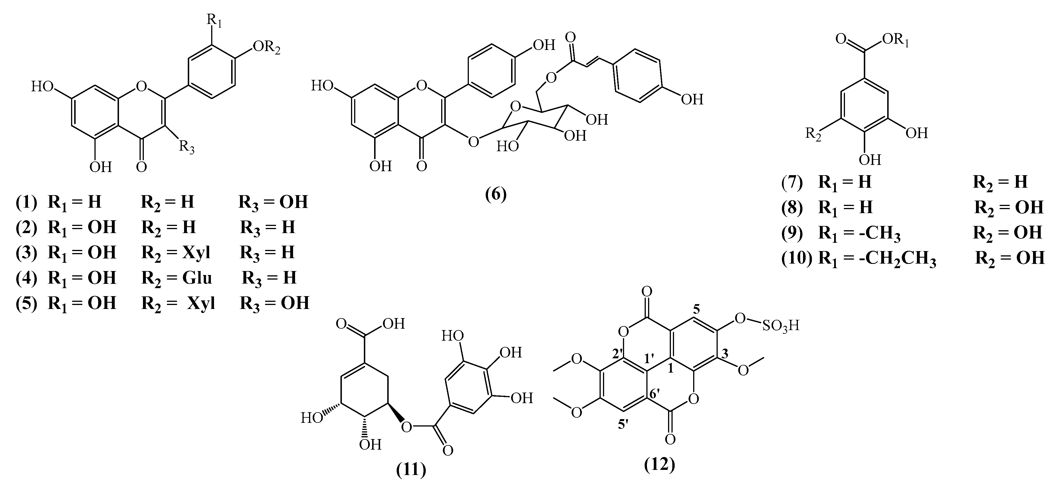
Figure 2.
Pearson correlation matrix where red colour indicates strong positive correlation and blue colour indicates strong negative correlation.
Figure 2.
Pearson correlation matrix where red colour indicates strong positive correlation and blue colour indicates strong negative correlation.
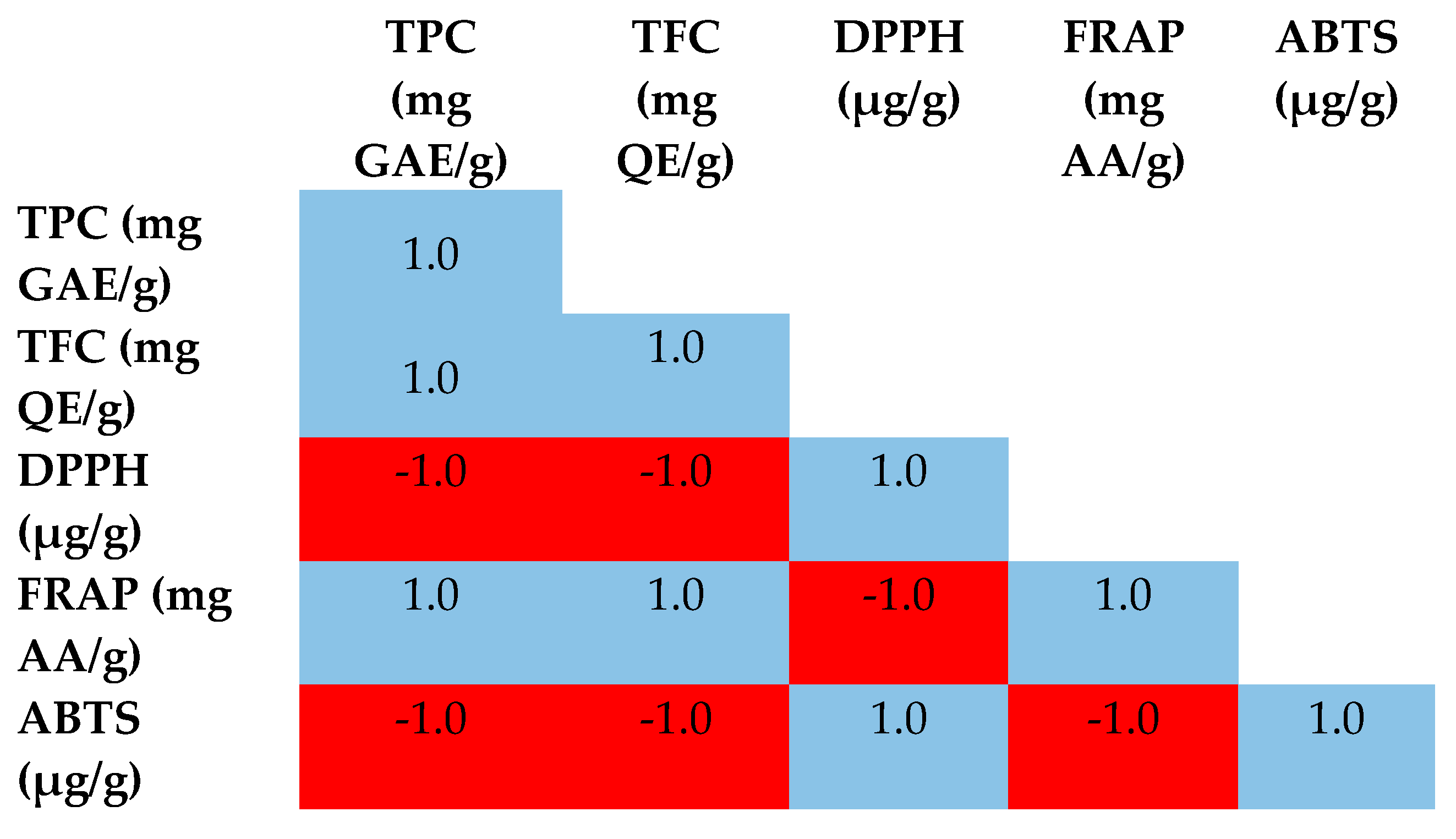
Figure 3.
Effect of trans-tiliroside and of luteolin 4'-O-β-xyloside on the viability of A549, A375, Panc-1 and Mia PaCa-2 for 72 h exposure. Data shown are mean values ± standard deviation of three independent assays. * Indicates statistical significance relative to control (p < 0.005).
Figure 3.
Effect of trans-tiliroside and of luteolin 4'-O-β-xyloside on the viability of A549, A375, Panc-1 and Mia PaCa-2 for 72 h exposure. Data shown are mean values ± standard deviation of three independent assays. * Indicates statistical significance relative to control (p < 0.005).
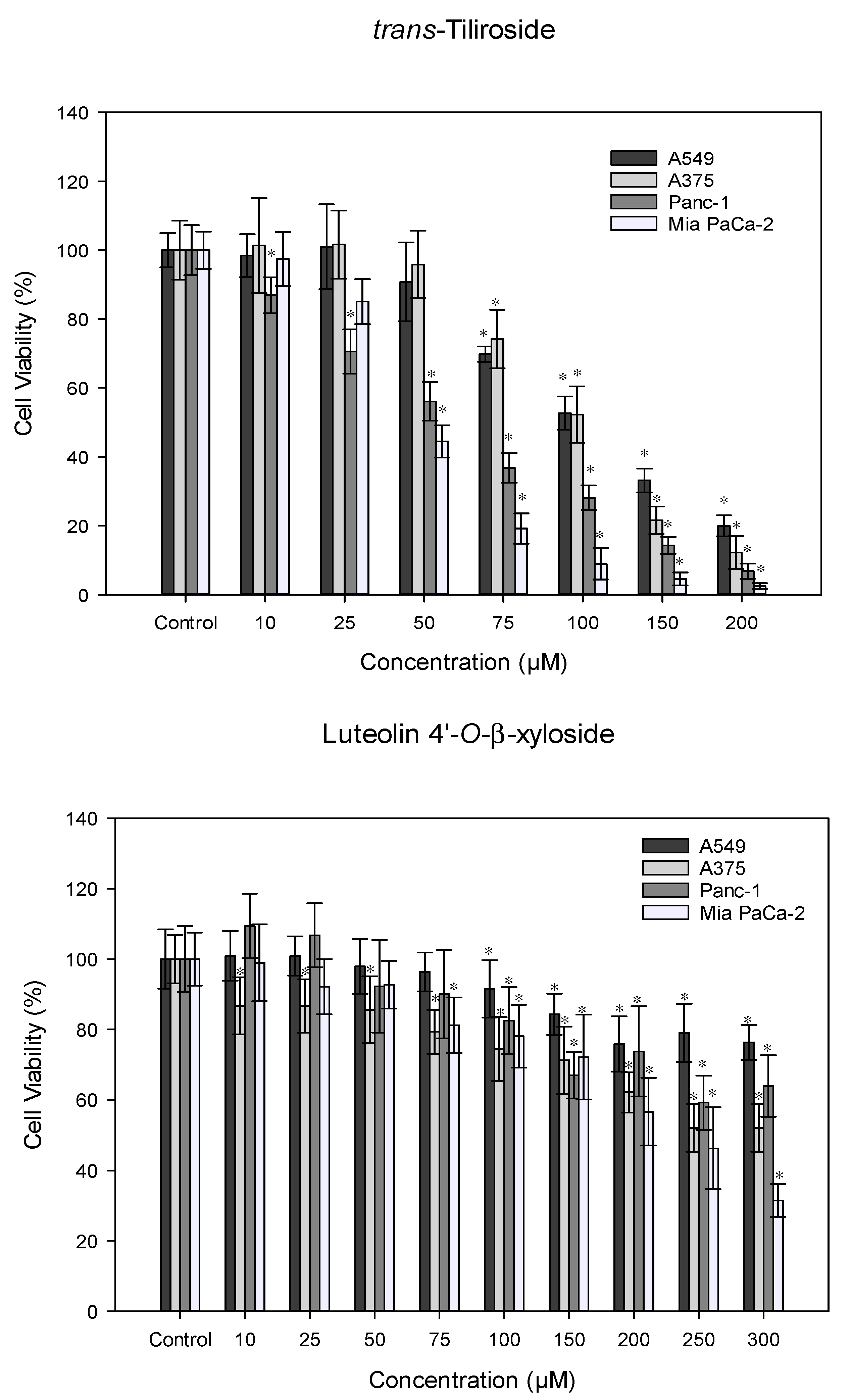

Table 1.
Total phenolic and flavonoid contents of H. cinereum extracts.
| Extracts | TPC (mg GAE/g) * | TFC (mg QE/g) ** |
|---|---|---|
| H. cinereum EtOAc | 361.51 ± 0.84 a | 148.23 ± 0.51 a |
| H. cinereum BuOH | 145.88 ± 0.63 b | 94.89 ± 0.29 b |
* GAE—gallic acid equivalents; ** QE—quercetin equivalents.
Table 2.
Antioxidant activity of H. cinereum extracts.
| Samples | DPPH (IC50 µg/g) ** | FRAP (mg AA/g) * | ABTS (IC50 µg/g) ** |
|---|---|---|---|
| H. cinereum EtOAc | 17.23 ± 0.36 a | 221.16 ± 1.03 a | 85.16 ± 1.03 a |
| H. cinereum BuOH | 24.39 ± 0.21 b | 44.69 ± 0.64 b | 121.16 ± 1.03 b |
| Trolox | 11.97 ± 0.41 | - | 23.16 ± 0.54 |
| Ascorbic acid | 3.36 ± 0.13 | - | - |
* AA- Ascorbic Acid; ** IC- Inhibition Concentration. a, b – different letters indicate significantly (p ≤ 0.05) different results according to t-test.
Disclaimer/Publisher’s Note: The statements, opinions and data contained in all publications are solely those of the individual author(s) and contributor(s) and not of MDPI and/or the editor(s). MDPI and/or the editor(s) disclaim responsibility for any injury to people or property resulting from any ideas, methods, instructions or products referred to in the content. |
© 2024 by the authors. Licensee MDPI, Basel, Switzerland. This article is an open access article distributed under the terms and conditions of the Creative Commons Attribution (CC BY) license (http://creativecommons.org/licenses/by/4.0/).
Copyright: This open access article is published under a Creative Commons CC BY 4.0 license, which permit the free download, distribution, and reuse, provided that the author and preprint are cited in any reuse.


