Preprint
Review

This version is not peer-reviewed.

Comprehensive Insights into COPD: From Pathophysiology to Immunology and Therapeutic Perspectives

Submitted:

14 November 2024

Posted:

17 November 2024

You are already at the latest version

Abstract

Chronic obstructive pulmonary disease (COPD) is a prevalent and debilitating condition with significant social and public health implications due to its high morbidity and mortality rates. The World Health Organization recognizes COPD as a major global health challenge. Despite extensive research, the complex pathophysiology of COPD has hindered the development of precise treatments. Recent advancements in understanding the gut-lung axis and immunological mechanisms have opened new avenues for COPD management. This review delves into the pathophysiological aspects of COPD, highlighting the interplay between genetic predispositions, environmental exposures, and lifestyle factors. It emphasizes the critical role of the gut-lung axis in modulating pulmonary immunity and disease progression, presenting dysbiosis as a key factor exacerbating inflammation and COPD symptoms. Emerging therapeutic approaches, including the modulation of gut microbiota through probiotics, prebiotics, and dietary changes, show promise in improving COPD outcomes. Additionally, advancements in immunotherapies, such as monoclonal antibodies targeting specific cytokines and immune checkpoint inhibitors, offer the potential for reducing inflammation and enhancing lung function. Precision medicine, which customizes treatment based on individual genetic, environmental, and lifestyle factors, represents a significant stride toward more effective COPD management. This review also identifies crucial research gaps, such as the need for a comprehensive understanding of non-smoking-related COPD, reliable biomarkers for early diagnosis, and the long-term effects of novel therapies. Future research can pave the way for innovative therapeutic strategies and improved patient care by addressing these gaps. This comprehensive analysis underscores the importance of an integrative approach to COPD, combining pathophysiological insights, immunological perspectives, and cutting-edge therapies to enhance the quality of life for individuals affected by this chronic disease.

Keywords: 
;  ;  ;  

1. Introduction

One frequent lung condition that causes breathing difficulties and reduced airflow is chronic obstructive pulmonary disease, or COPD. It is sometimes referred to as chronic bronchitis or emphysema. Phlegm buildup or lung damage can occur in individuals with COPD [1]. Globally, COPD is one of the main causes of mortality and disability. It is brought on by a mix of genetic, growing, and social variables as well as exposure to inhaled fine particles, such as air pollution and cigarette smoke [2]. Because of its high prevalence (about 10% of adults worldwide), growing incidence (partially due to population aging), and enormous emotional, societal, and financial implications, chronic obstructive pulmonary disease (COPD) is an important health issue worldwide [3]. The positive aspect is that quitting smoking is the major way to prevent COPD. However, air pollution is a significantly bigger factor in underdeveloped nations, where fifty percent of all cases are unrelated to tobacco use. The illness may also be brought on by a rare genetic disorder known as alpha-1 antitrypsin (AAT) deficiency [4]. Preventing the beginning of smoking and early illness diagnosis in the public will be the main challenges in the years to come [5]. According to epidemiological research, 20–40% of COPD sufferers worldwide never smoked [6], thus, while smoking remains the primary risk factor for COPD, other medical factors must also be taken into account. In a recent large-scale general population research conducted in Austria, Kohansal et al. [7] found several other environmental risk factors linked to poor lung function, varying significantly among age groups, and interacting and aggregating in complicated ways with aging. In addition to being the outcome of gene-environment interactions (GxE), COPD (and perhaps all human illnesses) also requires consideration of the time axis (GxExT), since the identical GxE may have distinct effects at various ages [8]. Health, illness, and life expectancy are determined by the dynamic interaction of two primary biological processes throughout a lifetime: cumulative tissue damage and aging, on the one hand, and organ development, upkeep, and repair, on the other [8]. The gut-lung axis is an important avenue of interaction where lung health is influenced by the gut microbiota and vice versa. Numerous processes, such as immune cell trafficking, synthesizing microbial metabolites including short-chain fatty acids (SCFAs), and regulating systemic inflammation, contribute to this connection [9]. Dysbiosis, or modifications in the composition of the gut microbiota, can have a major effect on respiratory health and may even play a role in the etiology of lung disorders like Chronic Obstructive Pulmonary Disease (COPD). A decreased variety of the gut microbiota is a sign of dysbiosis, which has been connected to several respiratory disorders [10]. Dysbiosis in COPD may aggravate immunological reactions and inflammation, which would worsen the course of the illness. According to studies, people with COPD frequently have altered gut microbiota profiles, which may have an impact on how quickly the disease progresses [11]. These changes include higher concentrations of harmful bacteria and decreasing levels of helpful bacteria. Changing the gut microbiome offers a treatment strategy for respiratory illnesses that shows promise [12]. Dietary changes, probiotics, and prebiotics that attempt to rebalance the gut flora have demonstrated promise in reducing symptoms and enhancing the prognosis of COPD. For example, taking certain probiotic supplements can improve lung function and lessen exacerbations in people with COPD, demonstrating the therapeutic potential of addressing the gut flora [9,13].
The objective of this review paper is to investigate the complex nature of COPD, looking at its main causes and risk factors as well as discoveries about the gut-lung axis and how they affect the course and treatment of the disease. We will go over the latest findings about the relationship between gut microbiota and lung health, the possible therapeutic advantages of adjusting the gut microbiome, and the relevance of gene-environment interactions in the lifetime context of COPD. We intend to offer a greater comprehension of the etiology of COPD and suggest novel strategies for both prevention and therapy through this thorough analysis.

2. The Role of Gut Microbiota in Lung Health

The human gut microbiota is a complex ecosystem comprised of billions of species, including bacteria, viruses, fungi, and protozoa. These microbes help maintain gut health, aid food absorption, and regulate the immune system. Diet, age, genetics, and environmental exposures all impact the composition of the gut microbiota [14]. Oropharyngeal and gut bacteria were investigated extensively before lung germs were discovered. As previously stated, unlike the lung microbiome, the oropharyngeal and gut microbiomes are persistent and strong, having significant implications for the organism's physiological and pathological status. Due to its location, researchers first assumed that the lung microbiome was identical to the oropharyngeal microbiome [15]. As the investigation progressed, scientists revised their position, noting the parallels while also emphasizing the differences between these two anatomical regions. However, it is indisputable that the oropharyngeal microbiome influences the generation, maintenance, and changes in the pulmonary microbial community. Research has linked oral health to an increased risk of developing respiratory disorders. Gastrointestinal microbes have long been recognized for their complexity and abundance. They regulate the status of a healthy organism and are linked to several types of disorders. Researchers have discovered that the impacts of the gut microbiome on the lungs, such as lung disease prevention, may be linked to the lungs' initial inhabitants. The lung microbiome has a significant impact on the gut's microbial ecosystems [14].
The gut microbiome is the most extensively researched microbial community to date, and its composition, shape, and function are well understood. Firmicutes, Bacteroides, Aspergillus, and Actinobacteria make up the majority of the gut flora, with rare appearances of Clostridium, Verruciform, and Spirochetes [16]. The core gut microbiome contains up to 14 bacterial genera and 150 bacterial "species". The lung microbiota is less diverse and abundant than the gut microbiome. Scientists have coined terminologies such as the "gut-brain axis" and the "gut-lung axis" to describe how the local microbiome affects immunity in faraway areas, as well as how the gut microbiota influences other organs. The gut-lung axis refers to the interplay between the gut and the lungs [14].
The intestinal microbiota is made up of thousands of microorganisms that can influence the pulmonary microbiota by creating ligands, metabolites, and immune cells that enter the lungs via the bloodstream and modulate pulmonary immunity. The gut microbiome may have a direct impact on pulmonary immunology and the composition of the pulmonary microbiome via these circulating cells and molecules [14,16,17]. The pulmonary microbiota is also essential for a balanced immune response. It contributes to the formation of the innate and adaptive immune response in the lung by interacting with epithelial cells and immune cells. Evidence from mice suggests that the gut-lung axis includes connections between the intestinal and pulmonary mucosa. Elevated inflammation and immunological dysregulation in the lungs can result from modifications in the makeup of the gut microbiota [14].

3. Anatomy and Physiology of the Respiratory System

Patient safety is improved when anatomical and physiological knowledge of the respiratory system is applied to medical procedures. Here is a detailed discussion of the anatomy and physiology of the airways [15]. Thoracic cage, lungs, and diaphragm are the main components of the respiratory system. Lung and chest wall compliance are included in total respiratory system compliance. Change in volume in relation to pressure change is known as compliance. The elastic load during inspiration is determined by thoracic compliance, whereas lung compliance controls the pace and force of expiration [16]. Functionally, the respiratory system is divided into two zones: the respiratory zone (alveolar duct to alveoli) is where gas exchange occurs, and the conducting zone (nose to bronchioles) forms a channel for the conduction of the inhaled gases. The respiratory tract is anatomically separated into two sections: the lower respiratory tract (organ within thorax - trachea, bronchi, bronchioles, alveolar duct, and alveoli) and the upper respiratory tract (organ outside thorax - nose, throat, and larynx) [17]. Both physiologically and functionally, the respiratory muscles are skeletal muscles.
The diaphragm, external intercostals, parasternal, sternomastoid, and scalene muscles are among the inspiratory muscles, whereas the internal intercostal, rectus abdominis, external and internal oblique, and transverse abdominal muscles are among the expiratory muscles [18]. There are three lobes in each lung; the lower lobe is found in the posterior chamber of the chest cavity, and the middle and higher lobes are placed in the anterior cavity. Air and blood make up the remaining 90% of the lung mass, with solid tissue making up just 10% of it. The structure must provide both architectural integrity and a way to move gas from the lung to the circulatory system for it to work efficiently [19]. The pharynx, larynx, nasal and oral cavities are all part of the upper airway. It extends down to the cricoid ring from the lips and anterior nares. Because the pharynx functions as a junction between the food tube and the airway, foreign objects, food, and fluids may enter the larynx and eventually make their way to the trachea and bronchi.
Nonetheless, the body has developed defense systems throughout time, such as coughing and gag reflexes. in the course of deglutition. Air enters the nose during silent breathing normally. This complicated chamber is divided medially throughout its entire length by a bony and cartilaginous septum, known as the volumer. It is enclosed inferiorly by the hard and soft palates and laterally by the inferior, middle, and superior turbinates that cover the sinus ostia before emptying into the nasopharynx. It is important to recognize that the mucosa covering these tissues is extremely vascular and innervated while doing nasopharyngeal intubation using endotracheal tubes and passing fiberoptic bronchoscopes, feeding tubes, or nasogastric sumps [20,21,22].

3.1. Normal Lung Function

The thoracic cage, lungs, and diaphragm make up the majority of the respiratory system. Lung and chest wall compliance are included in the total respiratory system compliance, which is defined as a change in volume in response to a change in pressure [23]. As the largest muscle in the respiratory system, the diaphragm is vital to inspiration. Diaphragmatic strength can only be precisely measured in vivo. Maximum voluntary ventilation, maximum inspiratory pressure, and trans-diaphragmatic pressure can all be used to quantify respiratory muscle strength [24]. Three categories may be used to categorize lung function: forced vital capacity (FVC), forced expiratory volume in one second (FEV1), and FEV-1/FVC ratio. Spirometry is used to measure the dynamic flow rates. Lung volumes affect the dynamic flow rates. Total lung capacity (TLC), vital capacity (VC), residual volume (RV), and functional residual capacity (FRC) are the four static lung volumes. The measurement of gas exchange across the alveolar-capillary membrane is accomplished by measuring the carbon monoxide (DLCO) diffusion capacity. Test results for lung function are expressed as a percentage anticipated when compared to people of the same age, gender, and height [25,26]. Lung volumes are proportional to body size, particularly height. In the lungs, gas exchange takes place across the alveolar-capillary membrane. The carbon monoxide diffusing capacity (DLCO) is used to measure it. Diffusion at the alveolar-capillary interface is inversely correlated with the thickness of the alveolar-capillary membrane and directly proportionate to the alveolar surface [27].

3.2. Pathophysiology of COPD

Chronic obstructive pulmonary disease (COPD) in particular has been a significant public health issue and will continue to be a challenge to medical professionals in the twenty-first century. Due to its high incidence, morbidity, and death, COPD is gaining attention globally and posing significant problems to healthcare systems [28]. Looking now at histological and molecular data, lung function declines due to COPD, which develops gradually over decades as tiny airways shrink and eventually vanish. Only a small percentage of vulnerable smokers see sequential, characteristic alterations in their distal airways as inflammation develops in their lungs, which accelerates the loss of lung function [29,30,31]. Changes in the morphology of the lung and chest wall in patients with chronic obstructive pulmonary disease (COPD) dictate specific and significant methods of mechanical ventilation. Emphysema of the lungs and decreased elastic rebound lengthen expiratory duration, aggravating dynamic hyperinflation; chronic inflammation of the airways increases resistances and can lead to distal air-trapping. Weakness and an early beginning of muscular exhaustion might be caused by loss of muscle and an excess of fast fibers, which can delay the weaning process [32]. This might make gas exchange worse, along with respiratory muscle exhaustion. The pulmonary vasculature's autoregulation may cause deteriorating perfusion in response to declining oxygenation. This might result in right ventricular volume overload and elevated pulmonary vasculature pressures. Exhaustion of the respiratory muscles and reduced alveolar ventilation due to blockage exacerbate hypercapnia and respiratory acidosis, which might lead to mortality in the end [28].
In the lungs of COPD patients, there are more neutrophils, macrophages, and T lymphocytes (CD8 > CD4). Many cytokines and chemotactic substances are released by these inflammatory cells, which leads to further inflammation. Leukotriene B4, which is released by macrophages, neutrophils, and epithelial cells, draws in more T cells and neutrophils. Macrophages and epithelial cells release chemotactic factors including CXC chemokines, interleukin (IL)-8, and growth-related oncogene α that promote cellular movement [29]. Through inflammation, oxidative stress, protease imbalance, and signal transduction pathways, chronic inflammation leads to metaplasia of the bronchial epithelial goblet cells. Submucosal bronchial gland hypertrophy and hyperplasia are seen.
These result in airflow restriction and a quicker deterioration of lung function, which raises the risk of acute exacerbations. The mucus that is expelled forms more plugs as a result of changes in its biochemical characteristics. These mucus plugs obstruct the airways and lead to bacterial infections continuously colonizing the airway [33]. Since it is associated with increased mortality and morbidity and usually appears later in the course of COPD, pulmonary hypertension is a significant comorbidity of the disease. Pulmonary arterial constriction is a result of persistent hypoxia. The arteries next to the bronchioles have thickened intimately as a result of the deposition of collagen and elastin in the smooth muscles. Exercise, acetylcholine, and airflow increases cannot cause these arteries to completely dilate. In COPD, long-term smoking is linked to a reduction in the nitric oxide response, which further lowers arterial vasodilatation [34,35,36].

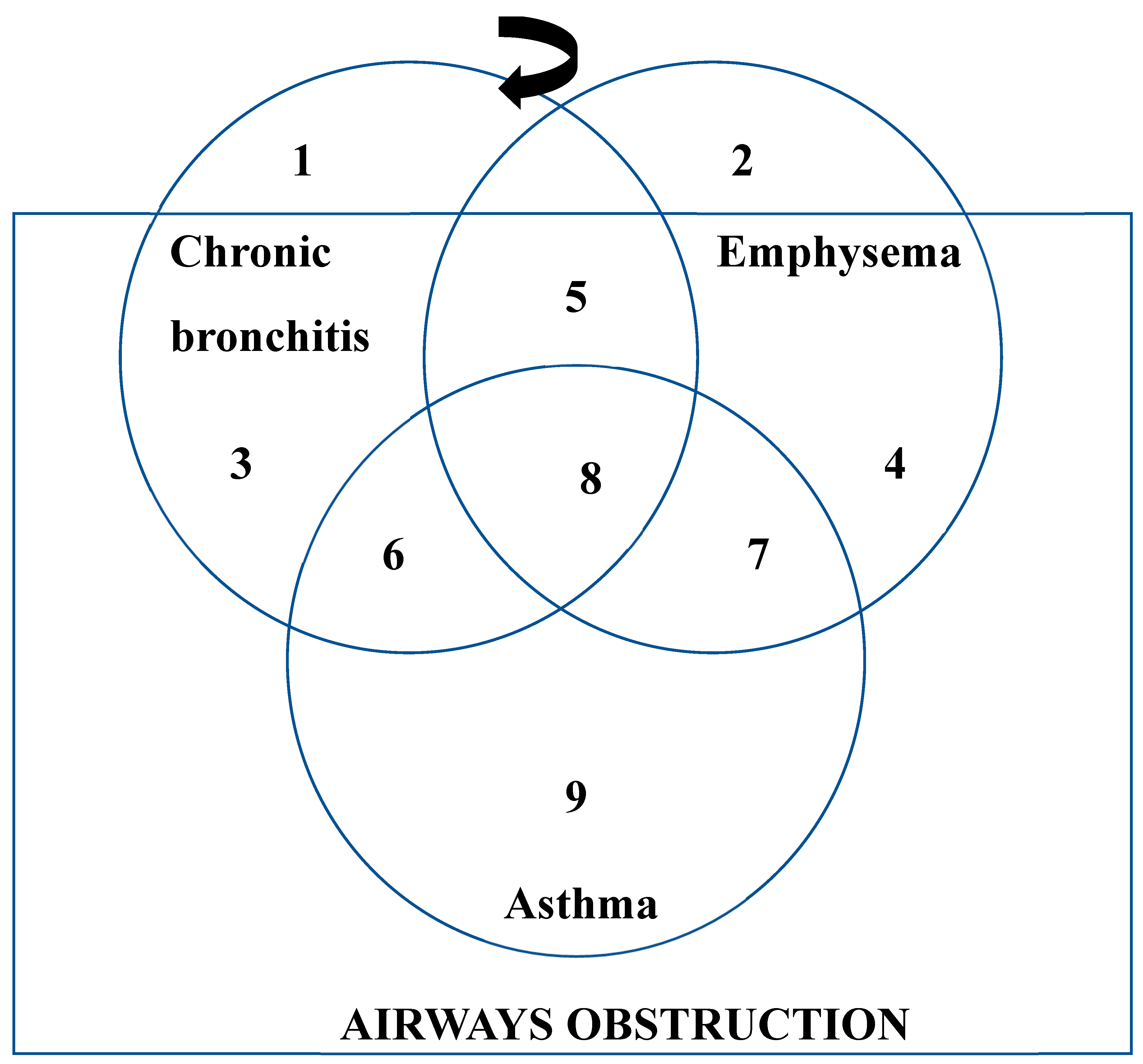
4. Types and Progression of COPD

The airflow restriction associated with Chronic obstructive pulmonary disease is mostly irreversible and advances slowly. It is caused by several reasons, with cigarette smoking being one of the primary causes [28]. COPD is a general term that includes Emphysema, Chronic bronchitis, and Asthma. Certain individuals may have COPD in addition to either emphysema or chronic bronchitis. Both chronic bronchitis and emphysema may result in airway blockage, which is frequently accompanied by airway hyperreactivity and may be partially reversible as this condition is known as COPD [19]. Asthma symptoms also include airway hyperreactivity and reversible blockage but Emphysema and chronic bronchitis are the two main components of COPD which can develop with or without airway blockage [33].
The intricate relationships between asthma, emphysema, and chronic bronchitis are illustrated in Figure 1, where three overlapping circles indicate different patient subgroups with emphysema, asthma, and chronic bronchitis. A portion of the patients positioned inside the rectangle have blocked airways. Individuals classified as having asthma, subgroup 9, fall totally inside the rectangle and are characterized by fully reversible airway obstruction. It may be challenging to determine whether patients in subgroups 6, 7, and 8 have underlying asthma or bronchial hyperreactivity as a complication of chronic bronchitis or emphysema. These patients have reversible airway obstruction with chronic productive cough or emphysema. A subgroup of 3 patients does not develop emphysema; instead, they have airway obstruction and persistent bronchitis. Subgroup 4 includes patients who just have emphysema. Subsets 5 and 8 comprise the majority of people who need medical attention for their medical condition. According to the Forced Expiratory Volume in One Second (FEV1), patients in subgroups 1 and 2 do not have airway obstruction; instead, they have radiographic or clinical signs of either emphysema or chronic bronchitis. The region shown by the shaded band, which represents COPD, includes patient subgroups 1 through 8 [33].

4.1. Chronic Bronchitis

The prevalence of chronic bronchitis is higher in the general population, and it affects 14% to 74% of patients who have already been diagnosed with the Chronic Obstructive Pulmonary Disease [21,22]. Mucus production and a persistent cough, caused by the body's immunological response to the harmful particles and gases in cigarette smoke, are the hallmarks of chronic bronchitis [23]. This coughing lasts for more than half the time for two years in a row and isn't associated with any known medical diseases like lung cancer or tuberculosis. Changes in the secretory system are seen in patients with chronic bronchitis, but they are not significant enough to be used as diagnostic markers. In chronic bronchitis, inflammation affects the epithelial lining of the central airways as well as the mucus-producing glands. This inflammatory process is associated with increased mucus production, reduced mucociliary clearance, and heightened permeability of the epithelial barrier within the airspace [24].
Smoking is the main risk factor for chronic bronchitis. As mentioned before, the 30-year cumulative incidence rate of chronic bronchitis in smokers is 42% [25]. However, it's important to note that 4% to 22% of people who have never smoked have been reported to have chronic bronchitis, suggesting the potential existence of additional risk factors. Inhaling dust, chemical fumes, and biomass fuels are a few of these possible causes [25,26].
Chronic bronchitis (CB) is caused by mucous metaplasia, characterized by excessive mucus production in response to inflammatory signals. Goblet cells overproduce and hypersecrete mucus in COPD, which is exacerbated by a reduced ability to eliminate mucus due to exposure to cigarette smoke [27,28], bacterial [29] and viral infections [30], and activation of inflammatory cells. This results in hypersecretion and overproduction of mucus, which is exacerbated by elastase release mediated by neutrophils. Mucus clearance is further hampered by impaired ciliary function, airway obstruction, and weaker respiratory muscles. Bronchoscopy and pathological investigations reveal increased mucosal metaplasia in COPD patients, especially in those with airflow obstruction [31]. These pathological alterations are closely tied to clinical consequences. Mucous metaplasia contributes to airflow obstruction through various mechanisms: increased mucus hypersecretion leads to luminal blockage [32], thickening of the epithelial layer encroaches upon the airway passage [33], and altered mucus affects airway surface tension, predisposing it to collapse during exhalation [34]. Small airway goblet cell hyperplasia negatively impacts lung function post-lung volume reduction surgery, and mucus obstruction in small airways correlates with decreased postoperative survival [35]. However, the correlation between airway pathology, physiology, and symptom severity in COPD remains weak. Understanding the inflammatory mechanisms behind mucous metaplasia in COPD is still lacking, although recent findings suggest a role for Th17 inflammation [36].
Figure 2. Factors leading to an abundance of mucus in chronic obstructive pulmonary disease. PEF (Peak Expiratory Flow).
Preprints 139689 g002

4.2. Emphysema

Emphysema typically occurs in smokers aged 45 to 60 as part of COPD [37], but can also arise in non-smoking-related conditions like HIV-1 infection or hypersensitivity pneumonitis [38]. COPD encompasses pulmonary symptoms such as airflow obstruction and lung tissue destruction, along with extrapulmonary effects like muscle wasting and anemia [39]. Breathlessness is a primary symptom. Treatment involves bronchodilators, steroids, and oxygen therapy, with no specific emphysema treatment available [40]. COPD mortality and morbidity pose significant global health and economic burdens. The characteristic features of emphysema include abnormal, chronic expansion of the airspaces distal to terminal bronchioles, along with wall damage that lacks visibly noticeable fibrosis [41]. The acinus, the main respiratory tissue unit, loses its ordered structure as a result of this damage [24]. There are several different types of emphysema: panacinar, which affects the entire acinus and is frequently observed in alpha-1 antitrypsin deficiency [42]; centrilobular, which is common in smokers and primarily affects upper lung regions; and distal acinar [43], which occurs near lung peripheries and is linked to spontaneous pneumothorax. Damage to elastic fibers reduces lung elasticity and pressure, which causes a persistent blockage of airflow. Bronchiolar luminal abnormalities are a result of alveolar wall damage and are correlated with the severity of emphysema.

4.3. Asthma

Asthma is a widespread, chronic non-communicable disease that affects both adults and children and can be fatal. Children are particularly vulnerable, with asthma being the most common non-communicable disease in childhood, it is estimated that asthma will affect an additional 100 million people, bringing the total number of individuals suffering from the condition to 300 million globally [43]. Individuals with asthma often show abnormalities in lung function. This condition increases the risk of children developing chronic obstructive pulmonary disease (COPD) later in life, a leading cause of death in adulthood. Various factors to asthma’s onset, including environmental, genetic, and allergenic influences, all of which play crucial roles in the disease’s progression and pathophysiology. Studies have long explored environmental exposures that may trigger asthma development, especially in children, who are more susceptible due to their physiology and outdoor activities. Considering the significant economic burden asthma places on the global economy, healthcare systems, and patients, effective prevention strategies are urgently needed. Common asthma triggers include dust, illnesses, smoking, temperature fluctuations, pets, odors, dust mites, exercise, viral infections, and more [44]. The disease presents in various phenotypes, with distinctions based on the age of onset, severity, and inflammatory forms (paucigranulocytic, neutrophilic, eosinophilic, and mixed granulocytic) [44].
Asthma has been categorized in children and adults by symptom severity or disease control level, following a stepwise management approach. Patients are typically divided into four or five categories based on the necessity for controller medications, such as leukotriene receptor antagonists (LTRAs), long-acting muscarinic antagonists, long-acting β2-adrenergic receptor agonists, and inhaled corticosteroids (ICSs). For the most severe cases, omalizumab, an IgE-specific monoclonal antibody, may be used. In particular, patients with non-allergic asthma often exhibit T2-high eosinophilic inflammation, which is associated with more severe asthma and frequent exacerbations [45].
Several clinical biomarkers are available for asthma diagnosis and management, such as fractional exhaled nitric oxide (FeNO), blood or sputum eosinophil counts, and IgE levels. Patients with allergic conditions may benefit from allergen immunotherapy (AIT), which involves gradually increasing doses of clinically relevant allergens to treat allergic rhinitis, allergic asthma, and hypersensitivity reactions to insect stings [46].
In addition to traditional asthma management, biologic therapies are crucial for moderate to severe cases. Monoclonal antibodies that target IgE (omalizumab) or cytokines in the Th2 pathway, such as IL-5 (mepolizumab, reslizumab) and the IL-4/IL-13 receptor (dupilumab), are used to treat severe asthma. New research into asthma pathogenesis has uncovered additional cytokine networks, with thymic stromal lymphopoietin (TSLP) and prostaglandin D2 (PGD2) emerging as potential therapeutic targets [47].
Nanotechnology is poised to revolutionize asthma treatment by improving the bioavailability of medications, reducing toxic materials, and enabling the targeted delivery of drugs or genes. This evolving field holds great promise for asthma treatment. The National Heart, Lung, and Blood Institute established a working group on nanotechnology to address the unique challenges of nanomedicine development. Various materials, including metals and polymers, are used to create nanoparticles that act as drug-delivery systems. When designing a nanocarrier for asthma treatment, several factors must be considered, such as physiological barriers posed by asthma itself. For example, the thickened mucus gel layer in the airways can prevent nanoparticles from dispersing effectively [48].
To exert therapeutic effects, nanoparticles must navigate several obstacles: they must penetrate the lung epithelium, evade phagocytosis by alveolar macrophages, and reach systemic circulation. Additionally, the immune system's neutrophils, cytokines, chemokines, and complement systems present further challenges for nanoparticles in the airways. Despite these hurdles, nanocarriers have shown promise in enhancing drug stability and efficacy, marking an exciting and challenging area of research for developing novel asthma treatments.

5. Immunomodulatory Effects of Gut Microbiota in Respiratory Diseases

Commensal microbes in the gut and lungs are necessary for immunological homeostasis to develop correctly. Unbalances in these microbial communities, known as dysbiosis, can lead to metabolic problems, aggravate autoimmune and inflammatory responses, raise the risk of neurological illnesses, and make people more susceptible to infections. Numerous tissues have been found to exhibit interactions between commensal microbes and immunological barriers, such as the urethra and the gastrointestinal mucosa. These interactions highlight the influence of dysbiosis on abnormal inflammatory responses, including bronchopulmonary dysplasia (BPD) [49].

5.1. Role of Microbiome in Innate Immunity

The body's initial line of defense against infections is the innate immune response. Particles, poisons, allergens, microorganisms, and endogenous waste from the surroundings might cause it. When pattern recognition receptors (PRRs) identify these bacteria, a signaling cascade is set off, which in turn triggers an immunological response [50]. Crucial to this process is the lung microbiome, which starts to form at birth and changes over time. Early microbial colonization affects how respiratory illnesses develop and molds immune responses throughout later life. Research indicates that early colonization by microorganisms like Staphylococcus and Ureaplasma affects an infant's immunological development [51]. As early as the first day of life, preterm babies' tracheal aspirates have been found to contain microbial DNA, indicating the early existence of microorganisms. The first three years of life are when the gut microbiome stabilizes, but it's still unknown when the respiratory microbiome matures. The development of the immune system and the course of respiratory diseases are significantly influenced by the early lung microbiome [52].

5.2. Role of Microbiome in Adaptive Immunity

Specific immune cells (cellular immunity) and antibodies (humoral immunity) are components of the adaptive immune response. The microbial environment and exposure to outside chemicals also affect this reaction. In the gut, local microbiota and dendritic cells (DCs) interact to modify DC phenotype and activate T cells. After that, activated T cells may go to the mucosa of the airways, where they may stimulate defensive and anti-inflammatory reactions [53].
Higher Th17 cell counts and the expression of inflammatory cytokines are linked to the enrichment of the oropharyngeal microbiome in the lungs, which includes Veillonella and Prevotella [54]. Proteobacterium catarrhal infections in mice cause neutrophil infiltration, increased levels of IL-6 and TNF-α, and CD4+ T-cell activation, demonstrating the influence of certain bacteria on immunological responses [55]. By encouraging advantageous immunological responses, symbiotic bacteria support immune system maintenance. For instance, the metabolites SCFAs generated by the microbiota control immunological responses and offer infection protection. Immune homeostasis depends on the relationship between the gut and lung microbiota; dysbiosis causes illnesses including allergies and asthma [56].
By encouraging advantageous immunological responses, symbiotic bacteria support immune system maintenance. For instance, the metabolites SCFAs generated by the microbiota control immunological responses and offer infection protection [57]. Immune homeostasis depends on the relationship between the gut and lung microbiota; dysbiosis causes illnesses including allergies and asthma. Variations in the microbiome can alter immune cells and chemicals, which can affect how a disease develops. Changes in the diversity of bacteria, for instance, influence immune cell populations and molecules like PD-L1, which in turn affects how the body reacts to allergens and infections [58]. Maintaining a healthy microbiome promotes health, however, dysbiosis in the lung microbiome can increase vulnerability to disease. Through cytokine signaling pathways, some bacteria, such as S. pneumoniae, improve immune responses and pathogen clearance.

6. Immunological Perspective on COPD

The hallmarks of chronic obstructive pulmonary disease (COPD) include severe immunological dysregulation and persistent inflammation. Increased neutrophils, macrophages, and T lymphocytes are seen in the lungs of COPD patients. These cells release a series of cytokines and chemokines that prolong inflammation and harm tissue. The disease's progression is greatly aided by this ongoing inflammatory state, which alters lung structure and impairs function. This condition is made worse by dysregulated immunological responses, which lead to increased oxidative stress and an imbalance in protease-antiprotease activity [59]. This promotes airway remodeling and the development of emphysema.​
The stimulation of both the innate and adaptive immune responses is one of the main inflammatory mechanisms linked to COPD. Pattern recognition receptors (PRRs) on immune cells identify infections and start inflammatory pathways, which is how innate immunity functions [60]. Pro-inflammatory cytokines like IL-6, TNF-α, and IL-1β are released during this phase and lead to chronic inflammation. Furthermore, T and B cell-mediated adaptive immune responses also have a major impact. The inflammatory environment in the lungs is maintained by CD8+ T cells and CD4+ T cells, especially Th17 cells) [61].
The fact that some COPD patients have eosinophilic inflammation, which is typically linked to asthma, emphasizes the disease's heterogeneity. Cytokines including IL-4, IL-5, and IL-13, which encourage eosinophil survival and migration to the lungs, are involved in this kind of inflammation. Compared to neutrophil-dominated inflammation, such eosinophilic COPD is frequently associated with more severe exacerbations and a different immune response profile [62]. The goal of emerging immunotherapies is to specifically target immunological mechanisms and cytokines linked to COPD. Originally created for asthma, biologic medications are now being used to treat COPD. These include monoclonal antibodies that target IL-4, IL-13 (dupilumab), IL-5 (mepolizumab, benralizumab), and IL-4, which have demonstrated promise in lowering inflammation and enhancing clinical outcomes in eosinophilic COPD patients. By counteracting the actions of cytokines implicated in the inflammatory process, these biologics lower eosinophil counts and enhance lung function [63].
Furthermore, developments in precision medicine are assisting in customizing these therapies according to unique patient profiles, taking into account elements like blood eosinophil counts and certain inflammatory indicators. By more specifically addressing the underlying inflammatory pathways, this strategy seeks to optimize therapy and may enhance both treatment efficacy and patient outcomes.​

7. Managing COPD: Treatment Approaches

Combination inhalers, bronchodilators, and corticosteroids are common therapies for COPD. These drugs aid in lung function improvement, symptom management, and preventing exacerbations. To lessen airway inflammation and improve airflow, long-acting bronchodilators like tiotropium and salmeterol as well as inhaled corticosteroids like budesonide and fluticasone are frequently utilized. Combination inhalers offer a more thorough therapy by concurrently addressing several inflammatory pathways. They contain both a corticosteroid and a long-acting bronchodilator. Novel immunotherapies are being created to specifically target inflammatory mediators linked to COPD. The goals of these therapies are to lessen chronic inflammation and regulate the immune response. Monoclonal antibodies that target cytokines like IL-5 (mepolizumab, benralizumab), IL-4, and IL-13 (dupilumab) are a few examples. Particularly in individuals with eosinophilic COPD, these biologics have demonstrated promise in lowering inflammation, enhancing lung function, and lowering the frequency of exacerbations [64].
The possibility of thymic stromal lymphopoietin (TSLP) inhibitors, like tezepelumab, to lessen airway inflammation and enhance patient outcomes for COPD patients is also being investigated. These inhibitors stop the signaling pathways mediated by TSLP, which are essential for the inflammatory response. Patients with COPD can improve their quality of life and physical fitness with pulmonary rehabilitation programs that involve education, dietary guidance, and exercise training [65]. These programs aid in symptom reduction, improve tolerance to exercise, and promote general health results. Research has demonstrated that pulmonary rehabilitation can reduce the number of hospital readmissions and enhance the psychological health of individuals with COPD. For the treatment of severe instances of COPD, oxygen therapy and mechanical ventilation are essential. Patients with advanced disease benefit from these procedures in terms of overall quality of life, alleviation of hypoxemia, and essential respiratory assistance. It has been demonstrated that long-term oxygen therapy increases survival in patients with chronic respiratory failure by preserving blood oxygen levels that are appropriate. In addition, non-invasive ventilation (NIV) can help during acute exacerbations by lessening respiratory effort and enhancing gas exchange [66].
It is critical to find treatments that can successfully lower inflammation associated with COPD and stop the disease from getting worse [67]. The development of innovative medications that specifically target inflammatory pathways implicated in COPD has been facilitated by recent advancements. The results of these trials are eagerly awaited, as the majority of these treatments are presently in preclinical or early clinical development. Not all therapeutic options may be effective due to the complexity of the inflammatory response in COPD, which is characterized by the redundancy of signal-transmitting mediators. With a wide range of endotypes and phenotypes that correspond to different pathophysiological processes, COPD is a heterogeneous illness [68]. Patients' inflammatory profiles can be used to identify more homogeneous groups, which increases the chance that therapies aimed at particular pathways will be successful. The goal of this precision medicine approach is to create treatments that specifically target the endotypes or biological processes causing the illness. Recent investigations, like the Lancet Commission, have stressed the necessity for novel COPD medicines to be customized to these particular pathways to produce a meaningful therapeutic impact and possibly even cure the condition.
Comprehending the diversity of COPD's inflammatory signature is essential for developing customized medicine and discovering new and efficient treatment methods. To assess the effects of novel compounds that target the inflammatory processes in various COPD subgroups, a number of clinical trials are presently being conducted [69].
Moreover, different strategies including regenerative therapies based on stem cells and modulators of the cystic fibrosis transmembrane conductance regulator (CFTR) might indirectly affect inflammation in COPD patients. These tactics may have a wider range of applications because they do not necessitate exact endotypic targeting. The outcomes of these ongoing trials are eagerly anticipated by the scientific community since they could lead to better treatments for this difficult condition [70].

8. Future Directions and Research Implications

8.1. Emerging Therapeutic Targets

It is vital to work toward creating treatments that efficiently lower inflammation associated with COPD and stop the illness from getting worse. Novel medications have been developed as a result of recent findings that have discovered various possible targets directly implicated in the inflammatory process [71]. With outcomes from ongoing clinical studies being anxiously awaited, the majority of these medicines are presently in preclinical or early clinical stages.
Numerous signal-transmitting mediators drive the intricate and redundant inflammatory response in COPD, implying that several treatment modalities may be required to provide meaningful clinical improvements [72]. Novel treatments are being developed to focus on particular inflammatory mediators and molecular pathways, such as:
-
Cytokines: Treatments aimed at IL-5, IL-4, and IL-13 have demonstrated potential in lowering inflammation and enhancing lung capacity. In individuals with eosinophilic COPD, for instance, mepolizumab and benralizumab (anti-IL-5) and dupilumab (anti-IL-4/IL-13) have shown promise [73].
-
Inhibitors of Thymic Stromal Lymphopoietin (TSLP): The anti-TSLP monoclonal antibody tezepelumab has demonstrated promise in lowering airway inflammation through the blockage of TSLP-mediated signaling pathways [74].
-
Immune Checkpoint Inhibitors: By focusing on immune checkpoints like PD-1 and PD-L1, one can alter immunological responses, which may lessen inflammation and enhance COPD results [75].
To lower chronic inflammation and modify the immune response, these targeted treatments provide fresh hope for individualized therapy plans catered to the unique inflammatory profiles of COPD patients.

8.2. Advances in Precision Medicine for COPD

With its emphasis on customizing medicines based on individual genetic, environmental, and lifestyle factors, precision medicine is becoming more and more acknowledged as a vital strategy in the management of COPD. This approach takes into account the distinctive qualities of every patient to deliver more efficient and individualized care [76].
With a variety of endotypes and symptoms that represent distinct pathophysiological pathways, COPD is a diverse illness. Researchers can identify more homogeneous populations and increase the likelihood of therapy success by focusing on certain pathways by analyzing the inflammatory profiles of patients [77]. To have the biggest impact, COPD treatments must be precise and target the precise biological processes or endotypes that cause the disease to develop, according to the Lancet Commission. The current body of study centers on:
-
Characterization of Endotypes and Phenotypes: By identifying particular endotypes and phenotypes, targeted treatments that target the underlying inflammatory mechanisms causing COPD can be developed [78].
-
Biomarker identification: By identifying biomarkers linked to particular endotypes, targeted medicines can be chosen with more efficacy, leading to better patient outcomes [79].
-
Customized Treatment Plans: Based on lifestyle, genetic, and environmental information, customized treatment regimens are developed for each patient.
Precision medicine has the potential to revolutionize the treatment of COPD by providing novel and efficient treatment alternatives that can greatly enhance both the prognosis and quality of life for patients [80].

8.3. Unexplored Areas of Gut-Lung Immunology

An important factor in respiratory health is the gut-lung axis, a bidirectional communication channel between the gut and lung microorganisms. Further research is needed to completely understand the particular processes by which gut microbes influence lung immunity and disease progression [81]​. Examining these fields may reveal novel treatment approaches for the treatment of respiratory conditions, such as:
-
Microbiome Modulation: Investigating the potential for microbial balance restoration and improved lung health through the use of probiotics, prebiotics, and dietary adjustments [82,83,84,85,86,87,88,89,90,91,92,93,94,95,96,97].
-
Immune Modulation: Comprehending how microbial metabolites, like short-chain fatty acids (SCFAs), influence immune responses and mitigate lung inflammation.
-
Microbiota Transplantation: Examining the possibility of microbiota transplantation as a therapeutic approach to improve immunological function and restore healthy microbial communities.

9. Conclusion

Chronic obstructive pulmonary disease (COPD) represents a significant global health challenge due to its high morbidity, mortality, and the complexity of its pathophysiology. This comprehensive review highlights the intricate interplay between genetic predispositions, environmental exposures, and lifestyle factors in COPD development and progression. The emerging understanding of the gut-lung axis underscores the role of gut microbiota in modulating pulmonary immunity and influencing disease outcomes, presenting a promising avenue for therapeutic intervention. Current COPD management strategies focus on symptom alleviation and exacerbation prevention through the use of bronchodilators, corticosteroids, and pulmonary rehabilitation. However, the persistent need for more effective treatments has driven research towards innovative approaches, including immunotherapies targeting specific cytokines and precision medicine tailored to individual patient profiles. These advancements hold the potential to significantly enhance treatment efficacy and patient quality of life. Despite these promising developments, several research gaps remain. A deeper understanding of non-smoking-related COPD, the mechanistic insights into the gut-lung axis, the identification of reliable biomarkers for early diagnosis, and the long-term effects of novel immunotherapies are critical areas for future research. Addressing these gaps through interdisciplinary collaboration and innovative research is essential for advancing COPD management.
In conclusion, the multifaceted nature of COPD necessitates a holistic and integrative approach to its management. By leveraging advancements in immunology, microbiology, and precision medicine, we can develop more effective therapies and improve outcomes for COPD patients. Continued research and a focus on personalized treatment strategies will be key to transforming the landscape of COPD care and enhancing the quality of life for millions affected by this debilitating disease.

References

  1. WHO. Chronic obstructive pulmonary disease (COPD) [Internet]. Available from: https://www.who.int/news-room/fact-sheets/detail/chronic-obstructive-pulmonary-disease-(copd).
  2. Poh TY, Phua J, Lee KH, Lim TK. Understanding COPD-overlap syndromes. Expert Rev Respir Med. 2017;11(4):285-98. [CrossRef]
  3. Singh D, Agusti A, Anzueto A, Barnes PJ, Bourbeau J, Celli BR, et al. Global strategy for the diagnosis, management, and prevention of chronic obstructive lung disease: the GOLD science committee report 2019. Eur Respir J. 2019;53(5):1-18. [CrossRef]
  4. National Heart, Lung, and Blood Institute (NHLBI). Chronic obstructive pulmonary disease (COPD) [Internet]. Available from: https://www.nhlbi.nih.gov/health/copd.
  5. Raherison C, Girodet PO. Epidemiology of COPD. Eur Respir Rev. 2009;18(114):213-21. [CrossRef]
  6. Salvi SS, Barnes PJ. Chronic obstructive pulmonary disease in non-smokers. Lancet. 2009;374(9691):733-43.
  7. Breyer-Kohansal R, Hartl S, Burghuber OC, Wouters EF. The LEAD (Lung, Heart, Social, Body) study: objectives, methodology, and external validity of the population-based cohort study. J Epidemiol. 2019;29(8):315-24. [CrossRef]
  8. Agustí A, Faner R. COPD beyond smoking: new paradigm, novel opportunities. Lancet Respir Med. 2018;6(5):324-6. [CrossRef]
  9. Postma DS, Siafakas N, editors. Epidemiology of chronic obstructive pulmonary disease. In: Management of Chronic Obstructive Pulmonary Disease. Eur Respir Mon. 1998;7:41–73.
  10. Gilliland FD, Li YF, Peters JM. Effects of glutathione S-transferase M1, maternal smoking during pregnancy, and environmental tobacco smoke on asthma and wheezing in children. Am J Respir Crit Care Med. 2002;166(4):457-63. [CrossRef]
  11. Keatings VM, Collins PD, Scott DM, Barnes PJ. Differences in interleukin-8 and tumor necrosis factor-alpha in induced sputum from patients with chronic obstructive pulmonary disease or asthma. Am J Respir Crit Care Med. 1996;153(2):530-4. [CrossRef]
  12. Lim SA, Roche WR, Belvisi MG, Walls AF, Holgate ST, Macedo P, et al. Balance of matrix metalloprotease-9 and tissue inhibitor of metalloprotease-1 from alveolar macrophages in cigarette smokers: regulation by interleukin-10. Am J Respir Crit Care Med. 2000;162(4):1355-60.
  13. Takizawa H, Tanaka M, Takami K, Ohtoshi T, Ito K, Satoh M, et al. Increased expression of transforming growth factor-β 1 in small airway epithelium from tobacco smokers and patients with chronic obstructive pulmonary disease (COPD). Am J Respir Crit Care Med. 2001;163(6):1476-83. [CrossRef]
  14. Zhu J, Li Y, Xu Y, Zhang G, Zhang L, Huang J, et al. Emerging therapeutic approaches for chronic obstructive pulmonary disease: focus on immunotherapy and precision medicine. Signal Transduct Target Ther. 2023;8(1):172. [CrossRef]
  15. Chung KF. The role of airway smooth muscle in the pathogenesis of airway wall remodeling in chronic obstructive pulmonary disease. Proc Am Thorac Soc. 2005;2(4):347-54. [CrossRef]
  16. Anatomy and Physiology of Respiratory System Relevant to Anaesthesia [Internet]. [cited 2017 Nov 29]. Available from: http://www.ijaweb.org.
  17. Mittman C, Edelman NH, Norris AH. Relationship between chest wall and pulmonary compliance with age. J Appl Physiol. 1965;20:211–16. [CrossRef]
  18. Ahmed-Nusrath A, Tong JL, Smith JE. Pathways through the nose for nasal intubation: A comparison of three endotracheal tubes. Br J Anaesth. 2008;100:269–74. [CrossRef]
  19. Ratnovsky A, Elad D, Halpern P. Mechanics of respiratory muscles. Respir Physiol Neurobiol. 2008;163:82–89. [CrossRef]
  20. Stevens JJWM, Jones JG. Functional anatomy and pathophysiology of the upper airway. Baillière’s Clin Anaesthesiol. 1995;9:213–34. [CrossRef]
  21. Hudgel DW, Hendricks C. Palate and hypopharynx – sites of inspiratory narrowing of the upper airway during sleep. Am Rev Respir Dis. 1988;138:1542–7. [CrossRef]
  22. Wheatley JR, Kelly WT, Tully A, Engel LA. Pressure-diameter relationships of the upper airway in awake supine subjects. J Appl Physiol. 1991;70(5):2242–51.
  23. Mittman C, Edelman NH, Norris AH, et al. Relationship between chest wall and pulmonary compliance with age. J Appl Physiol. 1965;20:1211–16. [CrossRef]
  24. Tolep K, Higgins N, Muza S, et al. Comparison of diaphragm strength between healthy adult elderly and young men. Am J Respir Crit Care Med. 1995;152:677–82. [CrossRef]
  25. Knudson RJ, Lebowitz MD, Holberg CJ, et al. Changes in the normal maximal expiratory flow-volume curve with growth and aging. Am Rev Respir Dis. 1983;127:725–34.
  26. Zeleznik J. Normative aging of the respiratory system. Clin Geriatr Med. 2003;19:1–18. [CrossRef]
  27. Stam H, Hrachovina V, Stijnen T, et al. Diffusing capacity dependent on lung volume and age in normal subjects. J Appl Physiol. 1994;76:2356–63. [CrossRef]
  28. MacNee W. Pathogenesis of chronic obstructive pulmonary disease. Proc Am Thorac Soc. 2005;2(4):258–66. [CrossRef]
  29. Bates DV. Chronic bronchitis and emphysema. N Engl J Med. 1968;278(11):600–5. [CrossRef]
  30. Snider GL. Chronic obstructive pulmonary disease: Risk factors, pathophysiology, and pathogenesis. Annu Rev Med. 1989;40:411–29.
  31. Burgel PR, Nesme-Meyer P, Chanez P, Lacoste JY, Roche N, Perez T, et al. Cough and sputum production are associated with frequent exacerbations and hospitalizations in COPD subjects. Chest. 2009;135(4):975–82. [CrossRef]
  32. Kim V, Han MK, Vance GB, Make BJ, Newell JD, Hokanson JE, et al. The chronic bronchitic phenotype of COPD: an analysis of the COPDGene Study. Chest. 2011;140(3):626–33. [CrossRef]
  33. Kim V, Criner GJ. Chronic Bronchitis and Chronic Obstructive Pulmonary Disease. Am J Respir Crit Care Med. 2013;187(3):228–37. [CrossRef]
  34. Hogg JC. Pathophysiology of airflow limitation in chronic obstructive pulmonary disease. Lancet. 2004;364(9435):709–21. [CrossRef]
  35. Pelkonen M, Notkola IL, Nissinen A, Tukiainen H, Koskela H. Thirty-year cumulative incidence of chronic bronchitis and COPD in relation to 30-year pulmonary function and 40-year mortality: a follow-up in middle-aged rural men. Chest. 2006;130(4):1129–37. [CrossRef]
  36. Miravitlles M, Soriano JB, García-Río F, Muñoz L, Duran-Tauleria E, Sánchez G, et al. Chronic respiratory symptoms, spirometry and knowledge of COPD among general population. Respir Med. 2006;100(11):1973–80. [CrossRef]
  37. Ebert RV, Terracio MJ. The bronchiolar epithelium in cigarette smokers. Observations with the scanning electron microscope. Am Rev Respir Dis. 1975;111(1):4–11.
  38. Deshmukh HS, Shaver CM, Case LM, Dietsch M, Wesselkamper SC, Hardie WD, et al. Metalloproteinases mediate mucin 5AC expression by epidermal growth factor receptor activation. Am J Respir Crit Care Med. 2012;171(4):305–14. [CrossRef]
  39. Burgel PR, Nadel JA. Roles of epidermal growth factor receptor activation in epithelial cell repair and mucin production in airway epithelium. Thorax. 2004;59(11):992–6. [CrossRef]
  40. Holtzman MJ, Byers DE, Alexander-Brett JM, Wang X. Acute and chronic airway responses to viral infection: implications for asthma and chronic obstructive pulmonary disease. Proc Am Thorac Soc. 2005;2(2):132–40. [CrossRef]
  41. Innes AL, Carrington SD, Thornton DJ, Kirkham S, Rousseau K, Dougherty RH, et al. Epithelial mucin stores are increased in the large airways of smokers with airflow obstruction. Chest. 2006;130(4):1102–8. [CrossRef]
  42. Hogg JC, Chu FS, Utokaparch S, Woods R, Elliott WM, Buzatu L, et al. The nature of small-airway obstruction in chronic obstructive pulmonary disease. N Engl J Med. 2004;350(26):2645–53. [CrossRef]
  43. James AL, Wenzel S. Clinical relevance of airway remodelling in airway diseases. Eur Respir J. 2007;30(1):134–55. [CrossRef]
  44. Macklem PT, Proctor DF, Hogg JC. The stability of peripheral airways. Respir Physiol. 1970;8(2):191–203. [CrossRef]
  45. Kim V, Criner GJ. Chronic bronchitis and chronic obstructive pulmonary disease. Am J Respir Crit Care Med. 2013;187(3):228–37. [CrossRef]
  46. Filley GF. Emphysema and chronic bronchitis: clinical manifestations and their physiologic significance. Med Clin North Am. 1967;51(2):283–92. [CrossRef]
  47. Sahebjami H, Diaz PT, Clanton TL, Pacht ER. Emphysema-like changes in HIV. Ann Intern Med. 2008;116(10):876. [CrossRef]
  48. Finkelstein R, Da Ma H, Ghezzo H, Whittaker K, Fraser RS, Cosio MG. Morphometry of small airways in smokers and its relationship to emphysema type and hyperresponsiveness. Am J Respir Crit Care Med. 1995;152(1):267–76. [CrossRef]
  49. Celli BR. Chronic obstructive pulmonary disease: from unjustified nihilism to evidence-based optimism. Proc Am Thorac Soc. 2006;3(1):58–65. [CrossRef]
  50. The definition of emphysema. Report of a National Heart, Lung, and Blood Institute, Division of Lung Diseases workshop. Am Rev Respir Dis. 1985;132(1):182–5.
  51. Hayes JA, Korthy A, Snider GL. The pathology of elastase-induced panacinar emphysema in hamsters. J Pathol. 1975;117(1):1–14. [CrossRef]
  52. Takahashi M, Yamada G, Koba H, Takahashi H. Classification of centrilobular emphysema based on CT-pathologic correlations. Open Respir Med J. 2012;6(1):155. [CrossRef]
  53. López-Campos JL, Tan W, Soriano JB. Global burden of COPD. Respirology. 2016;21(1):14–23. [CrossRef]
  54. Pons J, Sauleda J, Ferrer JM, et al. Blunted γδ T-lymphocyte response in chronic obstructive pulmonary disease. Eur Respir J. 2005;25:441–6.
  55. McDonough JE, Yuan R, Suzuki M, et al. Small-airway obstruction and emphysema in chronic obstructive pulmonary disease. N Engl J Med. 2011;365:1567–75. [CrossRef]
  56. Scaramuzzo G, Rossi A, De Falco S, et al. Mechanical ventilation and COPD: from pathophysiology to ventilatory management. Minerva Med. 2022;113(3):460–70. [CrossRef]
  57. Leap J, Hamilton D, Wilkerson P, et al. Pathophysiology of COPD. Crit Care Nurs Q. 2021;44(1):2–8. [CrossRef]
  58. MacNee W. ABC of chronic obstructive pulmonary disease: pathology, pathogenesis, and pathophysiology. BMJ. 2006;332(7551):1202–4.
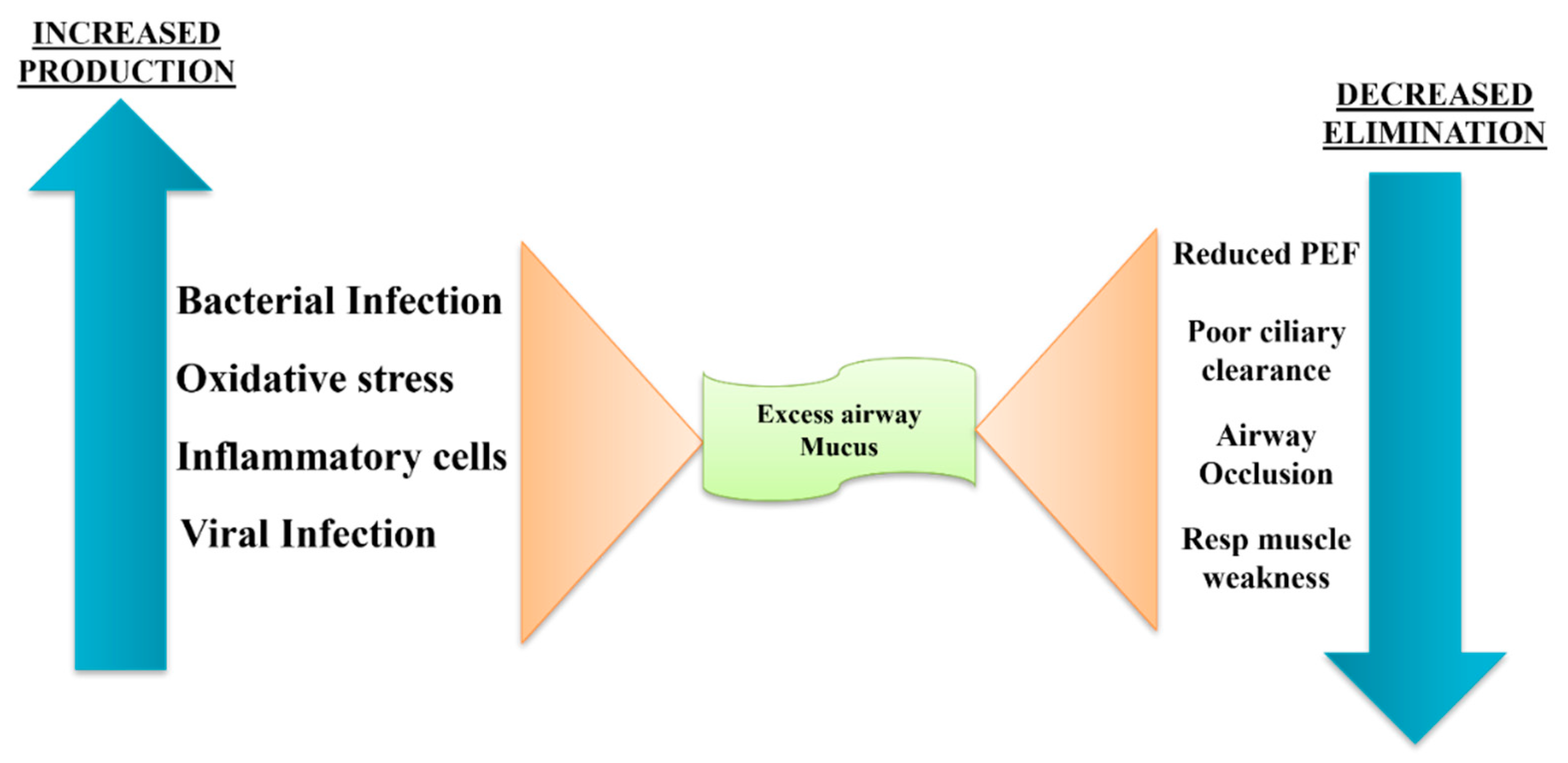
  59. Tian P, Wen F. Clinical significance of airway mucus hypersecretion in chronic obstructive pulmonary disease. J Transl Int Med. 2015;3(3):89–92. [CrossRef]
  60. Berg K, Wright JL. The pathology of chronic obstructive pulmonary disease: progress in the 20th and 21st centuries. Arch Pathol Lab Med. 2016;140(12):1423–8. [CrossRef]
  61. Kiowski W, Linder L, Stoschitzky K, et al. Diminished vascular response to inhibition of endothelium-derived nitric oxide and enhanced vasoconstriction to exogenously administered endothelin-1 in clinically healthy smokers. Circulation. 1994;90(1):27–34. [CrossRef]
  62. Masoli, M., Fabian, D., Holt, S., Beasley, R. and (2004), The global burden of asthma: executive summary of the GINA Dissemination Committee Report. Allergy, 59: 469-478. [CrossRef]
  63. Better understanding of childhood asthma, towards primary prevention: are we there yet? Consideration of pertinent literature.
  64. 64. Patterns of growth and decline in lung function in persistent childhood asthma.
  65. GBD 2015 Mortality and Causes of Death Collaborators,Global, regional, and national life expectancy, all-cause mortality, and cause-specific mortality for 249 causes of death, 1980–2015: a systematic analysis for the Global Burden of Disease Study 2015.Lancet. 2016; 388: 1459-1544.
  66. Global Initiative for Asthma. Global strategy for asthma management and prevention GINA, (2015).
  67. Lange P, Parner J, Vestbo J, Schnohr P, Jensen G. A 15-year follow-up study of ventilatory function in adults with asthma. N. Engl. J. Med. 1998;339:1194–1200. [CrossRef]
  68. Gerth van Wijk RG, de Graaf-in’t Veld C, Garrelds IM. Nasal hyperreactivity. Rhinology. 1999;37(2):50–5.
  69. Van Gerven L, Steelant B, Hellings PW. Nasal hyperreactivity in rhinitis: a diagnostic and therapeutic challenge. Allergy. 2018;73:1784. [CrossRef]
  70. Schleich, F.; Brusselle, G.; Louis, R.; Vandenplas, O.; Michils, A.; Pilette, C.; Peche, R.; Manise, M.; Joos, G. Heterogeneity of phenotypes in severe asthmatics. The Belgian Severe Asthma Registry (BSAR). Respir. Med. 2014, 108, 1723–1732. [CrossRef]
  71. Nelson, R.K.; Bush, A.; Stokes, J.; Nair, P.; Akuthota, P. Eosinophilic asthma. J. Allergy Clin. Immunol. 2020, 8, 465–473.
  72. Diamant, Z.; Vijverberg, S.; Alving, K.; Bakirtas, A.; Bjermer, L.; Custovic, A.; Dahlen, S.; Gaga, M.; van Wijk, R.G.; Del Giacco, S.; et al. Toward clinically applicable biomarkers for asthma: An EAACI position paper. Allergy 2019, 74, 1835–1851. [CrossRef]
  73. J. Bousquet, R. Lockey, H.J. MallingAllergen immunotherapy: therapeutic vaccines for allergic diseases. A WHO position paper J Allergy Clin Immunol, 102 (1998), pp. 558-562.
  74. K. Okubo, Y. Kurono, K. Ichimura, M. Enomoto, Y. Okamoto, Y. Kawauchi, et al. (Eds.), [Practical Guideline for the Management of Allergic Rhinitis in Japan] (8th ed.), Life Science, Tokyo (2016)(in Japanese).
  75. Haahtela, T, Jarvinen, M, Kava, T, et al. Comparison of a β2-agonist, terbutaline, with an inhaled corticosteroid, budesonide, in newly detected asthma. N Engl J Med 1991;325:388-392.
  76. Takaku, Y.; Nakagome, K.; Kobayashi, T.; Yamaguchi, T.; Nishihara, F.; Soma, T.; Hagiwara, K.; Kanazawa, M.; Nagata, M. Changes in airway inflammation and hyperresponsiveness after inhaled corticosteroid cessation in allergic asthma. Int. Arch. Allergy Immunol. 2010 . [CrossRef]
  77. Erminia Ridolo, Francesco Pucciarini, Maria Cristina Nizi, Eleni Makri, Paola Kihlgren, Lorenzo Panella & Cristoforo Incorvaia (2020) Mabs for treating asthma: omalizumab, mepolizumab, reslizumab, benralizumab, dupilumab, Human Vaccines & Immunotherapeutics, 16:10, 2349-2356. [CrossRef]
  78. Patra JK, Das G, Fraceto LF, et al, 2018. Nano-based drug delivery systems: recent developments and future prospects. Journal of Nanobiotechnology, 16, Pages- 1-33. [CrossRef]
  79. Buxton DB, Lee SC, Wickline SA, et al, 2003. Recommendations of the National Heart, Lung, and Blood Institute Nanotechnology Working Group. Circulation, 108(22), Pages- 2737-2742. [CrossRef]
  80. Dwivedi YK, Hughes L, Baabdullah AM, et al, 2022. Metaverse beyond the hype: Multidisciplinary perspectives on emerging challenges, opportunities, and agenda for research, practice and policy. International Journal of Information Management, 66, Pages- 102542. [CrossRef]
  81. Lane S, Molina J, Plusa T. An international observational prospective study to determine the cost of asthma exacerbations (COAX).Respir Med2006;100:434–450. [CrossRef]
  82. Hoskins G, McCowan C, Neville RG, Thomas GE, Smith B, Silverman S. Risk factors and costs associated with an asthma attack.Thorax2000;55:19–24. [CrossRef]
  83. Chowdhury NK, Choudhury R, Gogoi B, Chang CM, Pandey RP. Microbial Synthesis of Gold Nanoparticles and their Application. Curr Drug Targets. 2022;23(7):752-760. [CrossRef]
  84. Pandey RP, Mukherjee R, Chang CM. Emerging Concern with Imminent Therapeutic Strategies for Treating Resistance in Biofilm. Antibiotics (Basel). 2022 Apr 2;11(4):476. [CrossRef]
  85. Pandey RP, Mukherjee R, Priyadarshini A, Gupta A, Vibhuti A, Leal E, Sengupta U, Katoch VM, Sharma P, Moore CE, Raj VS, Lyu X. Potential of nanoparticles encapsulated drugs for possible inhibition of the antimicrobial resistance development. Biomed Pharmacother. 2021 Sep;141:111943. [CrossRef]
  86. Dutt, Y., Pandey, R. P., Dutt, M., Gupta, A., Vibhuti, A., Raj, V. S., ... & Priyadarshini, A. (2023). Liposomes and phytosomes: Nanocarrier systems and their applications for the delivery of phytoconstituents. Coordination Chemistry Reviews, 491, 215251. [CrossRef]
  87. Pandey RP, Vidic J, Mukherjee R, Chang CM. Experimental Methods for the Biological Evaluation of Nanoparticle-Based Drug Delivery Risks. Pharmaceutics. 2023 Feb 11;15(2):612. [CrossRef]
  88. Kumar S, Dhiman R, Prudencio CR, da Costa AC, Vibhuti A, Leal E, Chang CM, Raj VS, Pandey RP. Chitosan: Applications in Drug Delivery System. Mini Rev Med Chem. 2023;23(2):187-191. [CrossRef]
  89. Ruby M, Gifford CC, Pandey R, Raj VS, Sabbisetti VS, Ajay AK. Autophagy as a Therapeutic Target for Chronic Kidney Disease and the Roles of TGF-β1 in Autophagy and Kidney Fibrosis. Cells. 2023 Jan 26;12(3):412. [CrossRef]
  90. Gunjan, Vidic J, Manzano M, Raj VS, Pandey RP, Chang CM. Comparative meta-analysis of antimicrobial resistance from different food sources along with one health approach in Italy and Thailand. One Health. 2022 Dec 22;16:100477. [CrossRef]
  91. Khatri P, Rani A, Hameed S, Chandra S, Chang CM, Pandey RP. Current Understanding of the Molecular Basis of Spices for the Development of Potential Antimicrobial Medicine. Antibiotics (Basel). 2023 Jan 29;12(2):270. [CrossRef]
  92. Pandey RP, Mukherjee R, Chang CM. Antimicrobial resistance surveillance system mapping in different countries. Drug Target Insights. 2022 Nov 30;16:36-48. [CrossRef]
  93. Himanshu, R Prudencio C, da Costa AC, Leal E, Chang CM, Pandey RP. Systematic Surveillance and Meta-Analysis of Antimicrobial Resistance and Food Sources from China and the USA. Antibiotics (Basel). 2022 Oct 25;11(11):1471. [CrossRef]
  94. Tripathi S, Khatri P, Fatima Z, Pandey RP, Hameed S. A Landscape of CRISPR/Cas Technique for Emerging Viral Disease Diagnostics and Therapeutics: Progress and Prospects. Pathogens. 2022 Dec 29;12(1):56. [CrossRef]
  95. Tinker RJ, da Costa AC, Tahmasebi R, Milagres FAP, Dos Santos Morais V, Pandey RP, José-Abrego A, Brustulin R, Rodrigues Teles MDA, Cunha MS, Araújo ELL, Gómez MM, Deng X, Delwart E, Sabino EC, Leal E, Luchs A. Norovirus strains in patients with acute gastroenteritis in rural and low-income urban areas in northern Brazil. Arch Virol. 2021 Mar;166(3):905-913. [CrossRef]
  96. Nguyen TTH, Pandey RP, Parajuli P, Han JM, Jung HJ, Park YI, Sohng JK. Microbial Synthesis of Non-Natural Anthraquinone Glucosides Displaying Superior Antiproliferative Properties. Molecules. 2018 Aug 28;23(9):2171. [CrossRef]
  97. Tyagi R, Srivastava M, Singh B, Sharma S, Pandey RP, Asthana S, Kumar D, Raj VS. Identification and validation of potent Mycobacterial proteasome inhibitor from Enamine library. J Biomol Struct Dyn. 2022;40(19):8644-8654. [CrossRef]
Figure 1. The complex interrelations among chronic bronchitis, emphysema, and asthma by G. L. Snider [20] representation of chronic obstructive pulmonary disease.
Preprints 139689 g001
Disclaimer/Publisher’s Note: The statements, opinions and data contained in all publications are solely those of the individual author(s) and contributor(s) and not of MDPI and/or the editor(s). MDPI and/or the editor(s) disclaim responsibility for any injury to people or property resulting from any ideas, methods, instructions or products referred to in the content.
Copyright: This open access article is published under a Creative Commons CC BY 4.0 license, which permit the free download, distribution, and reuse, provided that the author and preprint are cited in any reuse.
Prerpints.org logo

Preprints.org is a free preprint server supported by MDPI in Basel, Switzerland.

Subscribe

Disclaimer

Terms of Use

Privacy Policy

Privacy Settings

© 2025 MDPI (Basel, Switzerland) unless otherwise stated