Submitted:
10 November 2024
Posted:
12 November 2024
You are already at the latest version
Abstract
This study systematically analyzed the research literature in the field of mechanobiology from 2010 to 2024 using the PubMed database and the MedSci bibliometric analysis platform. By applying bibliometric methods, we explored annual publication volume, average citation counts, key journals, research institutions, author collaboration networks, and the distribution of high-frequency keywords. The findings reveal research trends and hotspots within the field, highlighting the notable interdisciplinary nature of mechanobiology. Additionally, through the construction of knowledge maps and collaboration networks, this study provides valuable insights for future academic developments. The analysis also identifies current research gaps and proposes potential directions for theoretical advancement and application expansion in mechanobiology.
Keywords:
Mechanobiology
; Bibliometrics
1. Introduction
This study conducts a systematic review and analysis of research literature in mechanobiology from 2010 to March 2024 using the PubMed database and MedSci bibliometric analysis platform. The year 2010 was chosen as a starting point because it marks a period of rapid advancement in modern analytical methods, such as single-cell sequencing and microfluidics, which propelled mechanobiology into a new phase of development[1,2,3,4]. By employing bibliometric methods and visual analysis, this study aims to uncover research trends, hotspots, and dynamic developments in the field, providing a reference for future research directions.
Mechanobiology is an interdisciplinary field that combines insights from biology, engineering, and chemistry.The concept dates back to the 1970 s and 1980 s when researchers first began studying how cells respond to physical stimuli such as pressure and stretching[5,6].With advancements in cell biology and materials science, researchers recognized the critical role of the extracellular matrix (ECM) in mechanotransduction, understanding that its mechanical properties profoundly influence cellular behavior—laying the foundation for subsequent mechanobiology studies. By the 21st century, mechanobiology had emerged as an independent field of study, attracting researchers from biology, materials science, engineering, and medicine[7,8,9,10] .Ample evidence now demonstrates that mechanical signals play a pivotal role in processes like embryonic development, tissue regeneration, and tumor metastasis[11,12,13].
Over the past decade, mechanobiology research has seen significant progress.Advanced technologies—such as single-cell sequencing, atomic force microscopy, microfluidics, and 3D bioprinting—have enabled researchers to explore cellular responses to mechanical forces at the molecular level with greater precision [14,15,16].These technological advancements have fostered a deeper understanding of the role of mechanical signals in cell fate determination, tissue morphogenesis, and disease progression. Mechanobiology has thus evolved from early, simple observations of cellular mechanical responses to comprehensive studies of the complex interaction networks between cells and their microenvironment [17,18,19,20] .
This study employs a multidimensional analytical approach to innovatively reveal shifts in research intensity through annual publication volume analysis, identify core academic platforms through journal distribution analysis, pinpoint key research teams via author collaboration network analysis, and capture research frontiers through high-frequency keyword analysis. Unlike existing literature reviews, this study places particular emphasis on mechanobiology’s recent progress in emerging fields like regenerative medicine and tissue engineering. Furthermore, through social network analysis, we delve into the interdisciplinary characteristics of the field. These findings offer researchers a deeper understanding of the field, encourage cross-disciplinary collaboration, and provide valuable references for policy-making and resource allocation.
2. Statistical Data Visualization Methods
2.1. Data Sources and Research Methods
Data source: Pubmed database.
Time span: 2010 to 2024
Document type: Article and Review
Language: English
The number of articles retrieved: 669
Search term: Mechanobiology
2.2. Research Methodology
This study utilizes the bibliometric analysis tool R package “bibliometrix” (version 4.2.3) [21] to systematically analyze the global distribution network of literature in the field of mechanobiology. Specifically, a complete set of metadata was exported from the PubMed database, and in-depth data mining was conducted using the bibliometrix package within the R programming environment (version 4.2.0) [22]. The “biblioAnalysis” and “summary” functions provided an initial overview of the data, yielding multidimensional insights into temporal distribution, document types, citation patterns, author collaboration networks, and country distribution .
Following this, the “metaTagExtraction,” “Biblionetwork,” and “Networkplot” commands were applied to construct and visualize collaboration networks. Additionally, the “Biblioshiny” command facilitated an in-depth analysis of international research collaborations, institutional cooperation networks, keyword co-occurrences, and thematic evolution, offering a comprehensive view of the research landscape and emerging trends in mechanobiology.
3. Results
3.1. Search Strategy, Analytical Approach, and General Data Statistics
Using bibliometric methods, this study systematically analyzed 669 mechanobiology publications indexed under the topic term “Mechanics” in the PubMed database from 2010 to 2024. As shown in (Figure 1), data mining with the Bibliometrix software revealed that these publications span 270 journals, involve 2,358 authors, and encompass 1,353 keywords, contributing to the construction of knowledge maps and collaboration networks in this field. The analysis found that multi-center collaborations account for 14.5% of the publications, with an average citation frequency of 1.335 per document, indicating an established scholarly community with notable interdisciplinary characteristics in mechanobiology. However, institutional collaboration and academic influence still have room for growth. This bibliometric analysis offers researchers valuable insights into the research hotspots and emerging trends in the field, providing an essential reference for strategic decision-making.
3.2. Analysis of Annual Publishing Trends
A bibliometric analysis of publications from 2010 to 2024 with the keyword “Mechanobiology” reveals two distinct development phases in this field: (Figure 2A) an initial stable period from 2010 to 2018 and a rapid growth period from 2019 to 2024. During the initial phase, annual publication volume remained relatively steady, ranging between 10-20 articles per year, with a gradual increase from 19 publications in 2010 to 35 in 2018. Entering the rapid growth phase, publication volume rose significantly, reaching 44 articles in 2019 (a 25.71% year-over-year increase) and peaking at 102 articles in 2021-representing a 2.91-fold increase compared to 2018. Although publication volume slightly declined in 2022 and 2023 (71 and 76 articles, respectively), it remained at a relatively high level. However, citation analysis reveals a different trend: while citations increased from 2010 to 2012, reaching a peak of approximately 55.56 citations per article in 2012, they then experienced a notable decline.This decrease was particularly marked between 2018 and 2024, with citations dropping to 1.32 citations per article in 2023. This contrast between rising publication volume and declining citation frequency reflects the field,s growing research interest, yet indicates that its academic impact could benefit from further enhancement.
3.3. Three-Field Plot (Sankey Diagram)
Based on the bibliometric analysis of mechanobiology literature from 2010 to 2024, a three-field plot (Sankey Diagram) visualizes the knowledge flow relationships among authors, affiliations, and journal sources. As shown in the data in (Figure 2B), from the author dimension, the knowledge flow at Columbia University is particularly obvious, highlighting the university’s strong attraction and concentration for mechanobiology researchers. This suggests Columbia University’s pivotal role in fostering and connecting academic networks within this field. This knowledge flow analysis underscores the strategic importance of specific institutions in advancing the field and offers valuable bibliometric insights for researchers in choosing potential collaborators and institutions. It enhances our understanding of the distribution of academic resources and the dynamic development within the mechanobiology field.
3.4. Most Relevant Sources and Temporal Distribution of Journals
A bibliometric analysis of 669 core mechanobiology publications from 2010 to 2024 highlights distinct characteristics in journal distribution within the field. As shown in (Figure 3A,B), in terms of the number of publications, Frontiers in Bioengineering and Biotechnology ranked first with 19 articles, followed by Frontiers in Cell and Developmental Biology with 16 articles. APL Bioengineering and Biomechanics and Modeling in Mechanobiology are tied for third, each with 15 articles. This distribution pattern reflects a balance between engineering-focused and biology-focused journals, while the prominence of interdisciplinary journals underscores the collaborative and multi-disciplinary nature of mechanobiology research.
Additionally, the continued interest of high-impact journals in mechanobiology research not only provides researchers with clear publication targets but also highlights mechanobiology’s potential for innovation and growth as an emerging interdisciplinary field. This analysis of journal distribution serves as a valuable reference for identifying research frontiers and offers strong bibliometric support for forecasting future directions in the field.
3.5. Most Relevant Authors and Authors’ Production Over Time
An analysis of author distribution from 2010 to 2024 in the mechanobiology field reveals a community of 2,358 researchers, creating a substantial scholarly network. Among the 669 core publications, 14.5% were international collaborative papers, indicating a strong trend of global scientific cooperation (Figure 4A). Regarding prolific authors, A.G. Papavassiliou leads with 8 research articles, followed by J. Fu with 7 publications, and A. Gefen with 6 articles (Figure 4B). This author distribution pattern not only highlights key figures in mechanobiology but also establishes a knowledge dissemination network centered around high-output scholars. These core authors serve as crucial connectors within the field, reflecting its academic influence structure and facilitating international collaboration, which is instrumental for sustained innovation.
Further analysis of global publication frequency from 2010 to 2024 (Figure 4C) shows a typical Lotka’s Law distribution, though with distinct characteristics in mechanobiology. The frequency of single-authored publications (n=1) reaches 0.8668, significantly higher than the theoretical expectation of 0.6494, illustrating a pronounced “long-tail effect” in this field. Among prolific authors, A.G. Papavassiliou leads with 8 publications, followed by other influential authors such as J. Fu, S. Gabriele, A.N. Gargalionis, A. Gefen, F. Guilak, C.T. Lim, K.A. Papavassiliou, and C.A. Simmons, who each have produced between 5 and 7 papers. This “pyramid-shaped” distribution of author output validates the applicability of Lotka’s Law within mechanobiology and underscores the core authors’ leadership role. Through consistent scholarly contributions, these researchers provide critical intellectual support, fostering theoretical innovation and practical advancement within the field.
3.6. Institutional Distribution
The bibliometric analysis of mechanobiology from 2010 to 2024 indicates participation by 2,358 independent researchers, with 14.5% of publications involving international collaboration, emphasizing the field’s cross-national cooperation. As shown in (Figure 5A) , from an institutional perspective, the University of California ranked first with 41 publications, highlighting its leadership in the field of mechanobiology. It is followed by the University of Michigan with 29 publications and the University of Pennsylvania with 27 publications. These top research universities not only excel in publication volume but also play crucial roles in advancing the field and building a global academic network. This distribution highlights the spatial distribution of research hubs and reveals the significant contributions of high-impact institutions to the field’s growth.
3.7. Institutional Output Over Time
Examining the publication trends of the top ten research institutions from 2010 to 2024, there is a notable upward trajectory. As shown in (Figure 5B), 2016 marked a turning point, after which the annual publication rates of these institutions soared, reflecting the increase in research interest.Particularly noteworthy is the University of California, which increased its output to 41 publications by 2024, a 2.56-fold increase from 2016. This remarkable growth underscores the university’s sustained investment and leadership in mechanobiology. The upward trend among top institutions suggests two core trends: the deepening research themes within mechanobiology and strategic investments by world-class institutions.
3.8. Corresponding Authors’ Country Distribution
As shown in (Figure 5C), from 2010 to 2024, corresponding authors in the field of mechanobiology came from 38 countries or regions, and 14.5% of the papers involved international collaboration. Analyzing publication volume by country, the United States leads with 168 papers, comprising 151 single-country publications (SCP) and 17 multi-country publications (MCP). Australia and China tie for second with 29 papers each, with Australia’s SCP and MCP at 16 and 13, respectively, and China’s at 23 and 6. France follows with 24 publications (18 SCP and 7 MCP). Notably, the U.S. has a high SCP ratio of 43.6%, reflecting a strong tendency toward independent research, while Australia and China’s SCP ratios of 0.046% and 0.07% indicate a greater inclination for international collaborations.
3.9. Authors’ Country Distribution
The 2010-2024 period shows a clear regional concentration of mechanobiology research output. As shown in (Figure 5D) , the United States dominates with 446 publications (40.92%), far exceeding other countries. Australia and China are second and third, with 78 (7.16%) and 75 (6.88%) publications, respectively. This distribution not only highlights the U.S.‘s leadership but also the rise of Australia and China as emerging research powers in mechanobiology.
3.10. Evolution of National Output Over Time
As shown in (Figure 5E), from 2010 to 2024, global mechanobiology research output showed significant dynamic growth, with 2015 being a key turning point. Since then, the leading countries have demonstrated accelerated publication rates. The United States maintains a commanding lead with a cumulative 2,507 publications, while Australia follows with 420. In terms of impact, the citation performance of these countries mirrors their publication volume rankings, with the United States achieving the highest cumulative citations (1,545) and Italy ranking second with 665 citations. These metrics reflect not only the U.S.‘s output advantage but also its central influence in advancing mechanobiology.
3.11. Most Cited Countries
As shown in (Figure 5F), the United States holds the top position in citation volume for mechanobiology research, significantly ahead of other countries. Italy, followed by France, Switzerland, and the Netherlands, also make notable contributions, indicating the influential role of these European countries in the field. China and Australia are emerging as impactful contributors, reflecting their increasing investment and involvement in mechanobiology research. In contrast, Colombia, Germany, and Singapore have relatively lower citation volumes, suggesting potential opportunities for these countries to enhance their impact through focused collaborations and specialization within niche areas. Overall, this data underscores the interdisciplinary and international nature of mechanobiology, with future progress likely driven by enhanced global cooperation.
3.12. Analysis of Highly Cited Publications Worldwide
Based on a bibliometric analysis of the mechanobiology field from 2010 to 2024, several research contributions have demonstrated significant academic impact. As shown in (Figure 6) , Panciera Tito’s (2017) seminal work ranks first with 504 citations, highlighting its fundamental contribution to the field. Ladoux Benoit (2017) follows with 208 citations, underscoring the theoretical innovation and importance of this research. In third place, the study by Swarta Melody A (2012) has garnered 205 citations, indicating its enduring influence on the advancement of the discipline.
The citation distribution of these highly cited papers reveals several important characteristics. First, the notable differences in citation frequency indicate a clear hierarchy of influence among research outcomes. Second, the publication period spanning from 2012 to 2017 reflects the sustained academic value of these works. Finally, the concentration of highly cited papers points to breakthrough developments within specific research areas in mechanobiology. These high-impact publications have not only provided a crucial theoretical foundation and methodological reference for subsequent studies but have also significantly shaped the research paradigms and development directions of the field.
3.13. Keyword Distribution Analysis
Based on an analysis of author keywords in the mechanobiology literature, the top 50 most frequently appearing keywords are visualized through a word cloud (Figure 7A) and a dendrogram (Figure 7B), illustrating the thematic distribution in this fieldAs shown in (Figure 7C) , the analysis showed that “mechanobiology” appeared 174 times, dominating the keyword landscape, followed by “mechanotransduction” (83 times) and “extracellular matrix” (22 times), the second and third most common keywords, respectively. Observed over time, “Mechanobiology” has consistently appeared frequently since 2010, underscoring its central role in the field’s development. This keyword distribution pattern not only reflects current research focuses but also provides a valuable quantitative basis for tracking the evolution of this discipline.
3.14. Temporal Evolution of Keywords
A bibliometric analysis from January 2010 to October 2024 highlights the dynamic progression of mechanobiology research. The data (Figure 7D) show that “Mechanobiology” as a core concept began receiving significant attention starting in 2013. The year 2019 marked a pivotal point, with accelerated growth in the usage frequency of core terms “Mechanobiology” and “Mechanotransduction.” By 2024, the cumulative occurrences of these terms reached 174 and 83, respectively. This upward trend not only reflects the vigorous expansion of mechanobiology research but also underscores its rising academic prominence within life sciences.
3.15. Comprehensive Analysis of Research Trends
According to information spanning from 2010, to 2024 the field of mechanobiology research has seen a shift from principles to practical applications over time. As depicted in (Figure 7E) used terms such, as “mechanobiology “ and “mechanotransduction “ suggest that studies have mainly centered on investigating how physical forces influence activities and communication pathways which serve as the foundation of this area of study. On the hand the appearance of terms, like “Cancer,” “Metastasis,” and “Fibrosis” shows the application of this field in investigating illnesses indicating researchers dedication to understanding how mechanical signals play a role in disease situations and unveiling fresh perspectives for treatments. Furthermore the extensive adoption of resources and materials, like hydrogels, microfluidics and atomic force microscopy proves that cross disciplinary technologies are hastening advancements in mechanobiology. In the direction of mechanobiology research should encompass a profound exploration of mechanical signals associated with diseases while also concentrating on the advancement of novel materials and their impact, on stem cell development processes as well as fostering interdisciplinary partnerships to facilitate the transition, from fundamental research to practical clinical implementations that could potentially lead to groundbreaking advancements in the fields of biomedical and regenerative medicine.
3.16. Coupling and Clustering Analysis of Research Themes
As shown in (Figure 8A), the Time Evolution Map illustrates the evolution of research themes over time within the mechanobiology field. The horizontal distribution represents the timeline, with nodes of different colors indicating research hotspots or keywords that emerged during specific periods. This visualization aids in understanding the dynamic development of the field’s knowledge structure, revealing the progression of research themes and the emergence sequence of new directions.
Figure 8B presents a complex Co-citation Network, displaying citation relationships and academic influence across the literature. In this network, each node represents an individual paper, with node size possibly reflecting citation frequency, while the connections between nodes indicate co-citation relationships. Different color clusters represent distinct subfields or thematic communities. Dense central areas highlight core literature clusters within the field, while peripheral branches depict the development of various subfields. This network structure provides a clear depiction of the knowledge map and academic community structure within mechanobiology.
Comprehensive analysis reveals that mechanobiology, as a rapidly developing interdisciplinary field, has established several stable research communities, characterized by distinct thematic trajectories and a tightly knit citation network. The Time Evolution Map uncovers the field’s development history and innovation pathways, while the Co-citation Network demonstrates the distribution of academic influence and the knowledge dissemination patterns. This multidimensional bibliometric analysis offers valuable insights into the current state of research, helps identify focal points, and provides a reference for predicting future trends within the field.
3.17. Multidimensional Knowledge Structure Analysis in Mechanobiology
Based on bibliometric analysis of the mechanobiology field from January 2010 to October 2024, this study systematically reveals the knowledge structure of the field through multiple dimensions, including a keyword co-occurrence network (Figure 9A,B), thematic spatial distribution (Figure 9C) , knowledge flow network (Figure 9D,E), and cross-disciplinary characteristics (Figure 9F). The findings indicate that the keyword co-occurrence network exhibits a typical power-law distribution, reflecting the formation of core concept clusters. The quadrant distribution in the thematic map clearly displays a strategic layout of various research themes categorized as hot, foundational, peripheral, and niche topics. Large-scale network visualizations and Sankey diagrams reveal the dynamic features and evolutionary pathways of knowledge flow, while factor analysis further highlights the deep interdisciplinary integration within this field. This multidimensional bibliometric analysis not only systematically presents the knowledge architecture and evolutionary patterns in mechanobiology but also provides essential quantitative insights for understanding research frontiers and forecasting future trends. This analysis offers valuable guidance for comprehending the field’s development dynamics and formulating strategic research directions.
3.18. Revealing Relationships Between Documents and Authors in the Field
Based on co-citation network analysis of mechanobiology literature from 2010 to 2024, the results reveal significant knowledge structure characteristics within this field. The degree distribution of nodes shown in (Figure 10A) indicates a typical power-law distribution in co-citation relationships, underscoring the critical role of a few core documents in knowledge dissemination. These core works not only have high citation frequency within the field but also play a pivotal role in linking different themes and facilitating knowledge diffusion.
The network visualization in (Figure 10B), featuring clusters of nodes, distinguishes multiple sub-themes within mechanobiology research through color-coded clusters, showing differentiation and interrelations among them. The strength of connections between nodes represents the knowledge flow between different research topics, reflecting the pathways of knowledge transfer and the degree of thematic association within the field. The existence of these clusters suggests that mechanobiology encompasses multiple highly related research directions and shows trends of cross-disciplinary collaborative research.
Furthermore, the heat map in (Figure 10C) highlights the temporal and spatial evolution of co-citation relationships among key literature. High-intensity deep red regions, along with specific year markers (e.g., 2014), display the temporal distribution patterns of influential research contributions, emphasizing the accumulation and developmental trajectory of seminal works within the field. These heat-intensive areas help identify trending themes and their evolution over time, outlining the knowledge development trajectory of mechanobiology.
In sum, this multidimensional bibliometric analysis not only uncovers the knowledge system and organizational structure of mechanobiology but also provides valuable quantitative insights into its evolutionary patterns and future trends. This analysis serves as a scientific reference for researchers seeking a comprehensive understanding of the field’s knowledge framework and identifying emerging frontiers, aiding further innovation and interdisciplinary integration in mechanobiology.
3.19. Collaboration Network
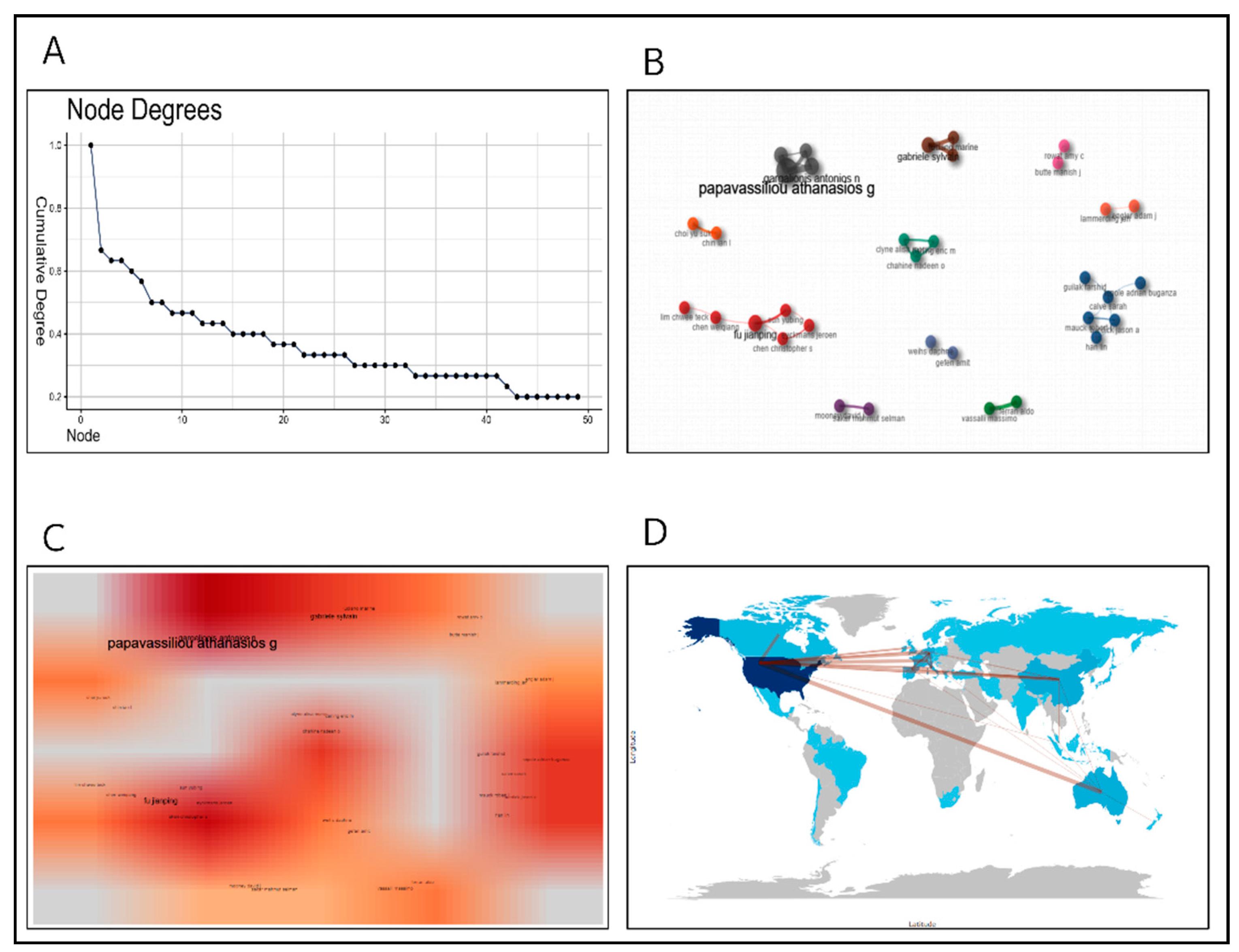
This study delves into the multidimensional characteristics of academic collaboration networks from a bibliometric perspective. Node degree distribution analysis (Figure 11A) reveals a significant “Matthew Effect” in collaborative relationships: a small number of core scholars connect a large network of researchers through extensive collaborations, while the majority have relatively limited collaborative reach. This distribution pattern not only aligns with the general trend in scientific collaboration networks but also highlights the critical role of core scholars in knowledge dissemination and in building academic influence.
The microstructure of author collaboration groups (Figure 11B) shows distinct research teams or academic groups through color-coded nodes. The formation of these groups stems primarily from three factors: shared research interests, institutional affiliation, and geographic proximity. These intra-group networks form distinctive research communities, underscoring the active collaboration among different research teams within the field.
The heat map (Figure 11C) visually represents the influence distribution of prolific and core authors. High-intensity regions indicate both the substantial academic impact of these researchers and the dense networks they establish, underscoring their central role in advancing the field and fostering collaboration. The presence of these core authors significantly enhances the efficiency of knowledge dissemination and encourages broader academic collaboration.
On a macro level, the geographic distribution of the international collaboration network (Figure 11D) illustrates a dynamic landscape of global academic exchange. The density of connecting lines reflects the strength of cross-national collaborations, with North American, European, and Asian research institutions forming robust collaborative ties, highlighting the globalization of research within this field. In terms of key research themes, academic institutions from various regions have significantly improved resource-sharing efficiency and innovation capacity through deep collaboration.
In summary, this multilayered collaboration network structure not only reflects the internationalization of contemporary academic research but also reveals hierarchical and regional characteristics in scientific collaboration. The findings suggest that cross-national collaboration and the “core scholar effect” play a pivotal role in advancing cutting-edge fields like mechanobiology, providing a strong empirical basis for future interdisciplinary partnerships. These insights hold significant implications for understanding and optimizing global scientific collaboration models.
4. Recommended Literature
4.1. High-impact Literature
Based on the data in (Table 1), this study analyzed 10 high-impact publications in the field of mechanobiology from 2010 to 2024. These publications are primarily featured in top-tier journals with high impact factors, such as Nature Reviews Molecular Cell Biology, Nature Reviews Materials, Nature Reviews Cancer, and Cell, with impact factors ranging from 45.5 to 81.3. Citation counts for these articles vary widely, from zero to 504 citations, reflecting the varying levels of attention these works have received within the research community.
The analysis indicates that highly cited articles tend to focus on widely applicable or forward-looking research themes, such as mechanobiology in cellular behavior, mechanisms involving YAP and TAZ, and the mechanobiology of the tumor microenvironment. These articles not only advance foundational research in mechanobiology but also highlight potential applications in disease treatment, which has garnered them significant attention and higher citation rates. Some of the most recent publications have not yet accumulated high citation counts, suggesting that their impact may emerge more prominently over time.
The research topics covered in these influential articles are diverse, ranging from the mechanical roles of single-protein elasticity to mechanobiology in tumor growth and cancer cachexia, as well as the mechanical properties of protein droplets[23,24,25]. This diversity reflects the wide scope and depth of research themes within the field of mechanobiology. Drawing on global “mechanobiology” literature data from January 2010 to October 2024 and using impact factor criteria, 10 recommended articles are listed in Table 1.

Table 1.
High-scoring articles.
| Title of Article | Release Date | Journals | Impact Factor | Citations | Title of Article |
| Mechanobiology by the numbers: a close relationship between biology and physics. |
2017/11/3 |
NATURE REVIEWS MOLECULAR CELL BIOLOGY |
81.3 |
18 |
[26] |
| Mechanobiology of collective cell behaviours. |
2017/11/9 |
NATURE REVIEWS MOLECULAR CELL BIOLOGY |
81.3 |
488 |
[27] |
| Mechanobiology of YAP and TAZ in physiology and disease. |
2017/9/28 |
NATURE REVIEWS MOLECULAR CELL BIOLOGY |
81.3 |
504 |
[28] |
| The role of single protein elasticity in mechanobiology. |
2023/7/20 |
NATURE REVIEWS MATERIALS |
79.8 |
16 |
[29] |
| Lymphatic and interstitial flow in the tumour microenvironment: linking mechanobiology with immunity. |
2012/2/25 |
NATURE REVIEWS CANCER |
72.5 |
411 |
[30] |
| Exploiting the tumor microenvironment and tumor mechanobiology for the treatment of cancer cachexia. |
2024/7/25 |
ANNALS OF ONCOLOGY |
56.7 |
0 |
[31] |
| Mechanobiology of Tumor Growth. |
2018/6/22 |
CHEMICAL REVIEWS |
51.4 |
28 |
[32] |
| Mechanobiology: A measure of molecular muscle. | 2017/4/14 | NATURE | 50.5 | 5 | [33] |
|
Engineered hydrogels for mechanobiology. |
2023/7/18 |
NATURE REVIEWS METHODS PRIMERS |
50.1 |
11 |
[34] |
| Mechanobiology of Protein Droplets: Force Arises from Disorder. |
2018/12/1 |
CELL |
45.5 |
6 |
[35] |
4.2. Highly Cited Literature
The highly cited articles listed in the data table reveal that foundational publications in mechanobiology have appeared in several high-impact journals (Table 2), including Nature Reviews Molecular Cell Biology, Nature Reviews Cancer, and Developmental Cell. These journals have impact factors ranging from 10.7 to 81.3, and the most highly cited publication has been referenced up to 504 times, underscoring its substantial influence in the mechanobiology research field.
The themes of these frequently cited papers encompass a range of topics such as cellular behavior, the tumor microenvironment, YAP/TAZ regulatory mechanisms, and applications of mechanobiology in brain function. This diversity highlights the central role of mechanobiology in various biological and medical research areas. Notably, the most cited paper, “Mechanobiology of YAP and TAZ in physiology and disease,” focuses on the role of YAP and TAZ in physiological and pathological contexts[36]. Its high citation count reflects its importance in advancing our understanding of cellular mechanics regulation.
Additionally, other foundational studies on materials and techniques have also garnered significant attention. For instance, a widely cited article on the development of polydimethylsiloxane (PDMS) substrates with tunable elastic moduli for studying cell mechanics in muscle and nerve Tissues—”Development of polydimethylsiloxane substrates with tunable elastic modulus to study cell mechanobiology in muscle and nerve”—provides a critical experimental tool for mechanobiology research[37]. Such studies have enabled researchers to explore cellular responses to mechanical environments with greater precision. Overall, these highly cited publications illustrate the diversity and impact of mechanobiology research across various biomedicine fields, as well as its foundational contributions to both theoretical understanding and practical applications in experimental biology.
Table 2.
Highly Cited Literature.
| Title of Article | Release Date | Journals | Impact Factor | Citations | Title of Article |
| Mechanobiology of YAP and TAZ in physiology and disease. | 2017/9/28 | NATURE REVIEWS MOLECULAR CELL BIOLOGY | 81.3 | 504 | [38] |
| Mechanobiology of collective cell behaviours. | 2017/11/9 | NATURE REVIEWS MOLECULAR CELL BIOLOGY | 81.3 | 208 | [27] |
| Lymphatic and interstitial flow in the tumour microenvironment: linking mechanobiology with immunity. | 2012/2/25 | NATURE REVIEWS CANCER | 72.5 | 205 | [30] |
| A hitchhikers guide to mechanobiology. | 2011/7/19 | DEVELOPMENTAL CELL | 10.7 | 201 | [39] |
| Development of polydimethylsiloxane substrates with tunable elastic modulus to study cell mechanobiology in muscle and nerve. | 2012/12/15 | PLOS ONE | 2.9 | 164 | [40] |
| Mechanobiology and developmental control. | 2013/10/9 | ANNUAL REVIEW OF CELL AND DEVELOPMENTAL BIOLOGY | 11.4 | 146 | [41] |
| Elucidating the mechanobiology of malignant brain tumors using a brain matrix-mimetic hyaluronic acid hydrogel platform. | 2011/8/9 | BIOMATERIALS | 12.8 | 137 | [42] |
| The mechanobiology of brain function. | 2014/11/21 | NATURE REVIEWS NEUROSCIENCE | 28.7 | 130 | [43] |
| Biomechanics and mechanobiology of trabecular bone: a review. | 2014/11/21 | JOURNAL OF BIOMECHANICAL ENGINEERING | 1.7 | 117 | [44] |
| Assaying stem cell mechanobiology on microfabricated elastomeric substrates with geometrically modulated rigidity. | 2011/2/5 | NATURE PROTOCOLS | 13.1 | 115 | [45] |
5. Discussion
This research employed analysis to uncover the evolving patterns and popular research areas, in mechanobiology between 2010 and 2024. Insights reveal an expansion, in this domain during the last ten years distinguished at the junction of biomechanics and regenerative medicine[46,47]. Highlighted keyword examination revealed “cell mechanics,” “mechanotransduction,” and “tissue regeneration” as points of investigation. The growing trend highlights how mechanobiology is closely linked with biomechanics and materials science, in the field of medicine. Emphasizing the role that mechanical microenvironments play in influencing cell behavior and tissue function[48,49,50,51].
The collaborative network analysis highlights a shift from early single-institution research to a broader international collaboration network, laying a strong foundation for technology sharing and interdisciplinary innovation within the field. However, limitations remain. For example, the concentration of research outputs in developed regions such as North America and Europe reveals an uneven geographical distribution of resources, potentially narrowing the diversity of perspectives. Encouraging greater participation and exchange with developing countries could broaden research perspectives and foster a more inclusive scientific community.
Furthermore, with advancements in single-cell analysis and 3D bioprinting, future research should focus on more detailed analysis of the cellular microenvironment and dynamic mechanical behaviors[52,53]. This would help reveal the precise roles of mechanical forces in both physiological and pathological cellular processes.
6. Conclusions
In summary, this study’s bibliometric analysis comprehensively highlights the research trends and development characteristics of mechanobiology, including publication volume, keyword distribution, and collaboration networks. Results indicate that the role of biomechanics in regulating cell function and tissue regeneration has received widespread attention, with interdisciplinary collaboration driving significant advancements in the field. These findings not only provide valuable data support for academic research within the field but also offer guidance for future research directions and academic collaborations. Moving forward, mechanobiology research should focus on understanding the mechanisms by which mechanical microenvironments regulate cell behavior, leveraging innovative techniques to deepen insights into the biological effects of mechanotransduction.
Data Availability Statement
The datasets generated and analyzed during this study are available from the corresponding author upon reasonable request.
Author Contributions
Da-Long Dong drafted the initial version of the manuscript. Da-Long Dong and Yan-Yan Zhou completed data collection, while Da-Long Dong, Yan-Yan Zhou, and Guang-Zhen Jin conducted data analysis. Da-Long Dong and Guang-Zhen Jin revised the manuscript.
Acknowledgments
This research was funded by the author’s personal resources.
References
- Macosko, E.Z.; Basu, A.; Satija, R.; Nemesh, J.; Shekhar, K.; Goldman, M.; Tirosh, I.; Bialas, A.R.; Kamitaki, N.; Martersteck, E.M.; et al. Highly Parallel Genome-Wide Expression Profiling of Individual Cells Using Nanoliter Droplets. Cell 2015, 161, 1202–1214. [Google Scholar] [CrossRef] [PubMed]
- Tang, F.; Barbacioru, C.; Wang, Y.; Nordman, E.; Lee, C.; Xu, N.; Wang, X.; Bodeau, J.; Tuch, B.B.; Siddiqui, A.; et al. mRNA-Seq Whole-Transcriptome Analysis of a Single Cell. Nat Methods 2009, 6, 377–382. [Google Scholar] [CrossRef] [PubMed]
- Rothbauer, M.; Zirath, H.; Ertl, P. Recent Advances in Microfluidic Technologies for Cell-to-Cell Interaction Studies. Lab Chip 2018, 18, 249–270. [Google Scholar] [CrossRef] [PubMed]
- Yu, L.; Chen, M.C.W.; Cheung, K.C. Droplet-Based Microfluidic System for Multicellular Tumor Spheroid Formation and Anticancer Drug Testing. Lab Chip 2010, 10, 2424–2432. [Google Scholar] [CrossRef]
- Clarke, G.D.; Davison, J.S. Mucosal Receptors in the Gastric Antrum and Small Intestine of the Rat with Afferent Fibres in the Cervical Vagus. J Physiol 1978, 284, 55–67. [Google Scholar] [CrossRef]
- Blackshaw, S.E.; Nicholls, J.G.; Parnas, I. Physiological Responses, Receptive Fields and Terminal Arborizations of Nociceptive Cells in the Leech. J Physiol 1982, 326, 251–260. [Google Scholar] [CrossRef]
- Nelson, C.M.; Xiao, B.; Wickström, S.A.; Dufrêne, Y.F.; Cosgrove, D.J.; Heisenberg, C.-P.; Dupont, S.; Shyer, A.E.; Rodrigues, A.R.; Trepat, X.; et al. Mechanobiology: Shaping the Future of Cellular Form and Function. Cell 2024, 187, 2652–2656. [Google Scholar] [CrossRef]
- Wang, J.; Lü, D.; Mao, D.; Long, M. Mechanomics: An Emerging Field between Biology and Biomechanics. Protein Cell 2014, 5, 518–531. [Google Scholar] [CrossRef]
- Cox, C.D.; Bavi, N.; Martinac, B. Biophysical Principles of Ion-Channel-Mediated Mechanosensory Transduction. Cell Rep 2019, 29, 1–12. [Google Scholar] [CrossRef]
- Shamsan, G.A.; Odde, D.J. Emerging Technologies in Mechanotransduction Research. Curr Opin Chem Biol 2019, 53, 125–130. [Google Scholar] [CrossRef]
- Marturano, J.E.; Arena, J.D.; Schiller, Z.A.; Georgakoudi, I.; Kuo, C.K. Characterization of Mechanical and Biochemical Properties of Developing Embryonic Tendon. Proc Natl Acad Sci U S A 2013, 110, 6370–6375. [Google Scholar] [CrossRef] [PubMed]
- Gensbittel, V.; Kräter, M.; Harlepp, S.; Busnelli, I.; Guck, J.; Goetz, J.G. Mechanical Adaptability of Tumor Cells in Metastasis. Dev Cell 2021, 56, 164–179. [Google Scholar] [CrossRef] [PubMed]
- Vining, K.H.; Mooney, D.J. Mechanical Forces Direct Stem Cell Behaviour in Development and Regeneration. Nat Rev Mol Cell Biol 2017, 18, 728–742. [Google Scholar] [CrossRef] [PubMed]
- Kurth, F.; Eyer, K.; Franco-Obregón, A.; Dittrich, P.S. A New Mechanobiological Era: Microfluidic Pathways to Apply and Sense Forces at the Cellular Level. Curr Opin Chem Biol 2012, 16, 400–408. [Google Scholar] [CrossRef] [PubMed]
- Zhou, W.-M.; Yan, Y.-Y.; Guo, Q.-R.; Ji, H.; Wang, H.; Xu, T.-T.; Makabel, B.; Pilarsky, C.; He, G.; Yu, X.-Y.; et al. Microfluidics Applications for High-Throughput Single Cell Sequencing. J Nanobiotechnology 2021, 19, 312. [Google Scholar] [CrossRef]
- Matellan, C.; del Río Hernández, A.E. Engineering the Cellular Mechanical Microenvironment – from Bulk Mechanics to the Nanoscale. Journal of Cell Science 2019, 132, jcs229013. [Google Scholar] [CrossRef]
- Nelson, C.M.; Xiao, B.; Wickström, S.A.; Dufrêne, Y.F.; Cosgrove, D.J.; Heisenberg, C.-P.; Dupont, S.; Shyer, A.E.; Rodrigues, A.R.; Trepat, X.; et al. Mechanobiology: Shaping the Future of Cellular Form and Function. Cell 2024, 187, 2652–2656. [Google Scholar] [CrossRef]
- Hoffman, B.D.; Grashoff, C.; Schwartz, M.A. Dynamic Molecular Processes Mediate Cellular Mechanotransduction. Nature 2011, 475, 316–323. [Google Scholar] [CrossRef]
- Darling, E.M.; Di Carlo, D. High-Throughput Assessment of Cellular Mechanical Properties. Annu Rev Biomed Eng 2015, 17, 35–62. [Google Scholar] [CrossRef]
- Zuela-Sopilniak, N.; Lammerding, J. Can’t Handle the Stress? Mechanobiology and Disease. Trends Mol Med 2022, 28, 710–725. [Google Scholar] [CrossRef]
- Aria, M.; Cuccurullo, C. Bibliometrix: An R-Tool for Comprehensive Science Mapping Analysis. Journal of Informetrics 2017, 11, 959–975. [Google Scholar] [CrossRef]
- Gutiérrez-Sacristán, A.; Guedj, R.; Korodi, G.; Stedman, J.; Furlong, L.I.; Patel, C.J.; Kohane, I.S.; Avillach, P. Rcupcake: An R Package for Querying and Analyzing Biomedical Data through the BD2K PIC-SURE RESTful API. Bioinformatics 2017, 34, 1431. [Google Scholar] [CrossRef] [PubMed]
- Zhang, H. The Glassiness of Hardening Protein Droplets. Science 2020, 370, 1271–1272. [Google Scholar] [CrossRef] [PubMed]
- Chuang, H.-Y.; He, R.-Y.; Huang, Y.-A.; Hsu, W.-T.; Cheng, Y.-J.; Guo, Z.-R.; Wali, N.; Hwang, I.-S.; Shie, J.-J.; Huang, J.J.-T. Engineered Droplet-Forming Peptide as Photocontrollable Phase Modulator for Fused in Sarcoma Protein. Nat Commun 2024, 15, 5686. [Google Scholar] [CrossRef]
- Wu, J.; Li, P.; Dong, C.; Jiang, H.; Bin Xue; Gao, X.; Qin, M.; Wang, W.; Bin Chen; Cao, Y. Rationally Designed Synthetic Protein Hydrogels with Predictable Mechanical Properties. Nat Commun 2018, 9, 620. [CrossRef]
- Schwarz, U.S. Mechanobiology by the Numbers: A Close Relationship between Biology and Physics. Nat Rev Mol Cell Biol 2017, 18, 711–712. [Google Scholar] [CrossRef]
- Ladoux, B.; Mège, R.-M. Mechanobiology of Collective Cell Behaviours. Nat Rev Mol Cell Biol 2017, 18, 743–757. [Google Scholar] [CrossRef]
- Panciera, T.; Azzolin, L.; Cordenonsi, M.; Piccolo, S. Mechanobiology of YAP and TAZ in Physiology and Disease. Nat Rev Mol Cell Biol 2017, 18, 758–770. [Google Scholar] [CrossRef]
- Beedle, A.E.M.; Garcia-Manyes, S. The Role of Single-Protein Elasticity in Mechanobiology. Nat Rev Mater 2023, 8, 10–24. [Google Scholar] [CrossRef]
- Swartz, M.A.; Lund, A.W. Lymphatic and Interstitial Flow in the Tumour Microenvironment: Linking Mechanobiology with Immunity. Nat Rev Cancer 2012, 12, 210–219. [Google Scholar] [CrossRef]
- Papavassiliou, K.A.; Papavassiliou, A.G. Exploiting the Tumor Microenvironment and Tumor Mechanobiology for the Treatment of Cancer Cachexia. Ann Oncol 2024, 35, 914–915. [Google Scholar] [CrossRef] [PubMed]
- Chaudhuri, P.K.; Low, B.C.; Lim, C.T. Mechanobiology of Tumor Growth. Chem Rev 2018, 118, 6499–6515. [Google Scholar] [CrossRef] [PubMed]
- Eisenstein, M. A Measure of Molecular Muscle. Nature 2017, 544, 255–257. [Google Scholar] [CrossRef] [PubMed]
- Blache, U.; Ford, E.M.; Ha, B.; Rijns, L.; Chaudhuri, O.; Dankers, P.Y.W.; Kloxin, A.M.; Snedeker, J.G.; Gentleman, E. Engineered Hydrogels for Mechanobiology. Nat Rev Methods Primers 2022, 2, 1–22. [Google Scholar] [CrossRef] [PubMed]
- Welsh, T.J.; Shen, Y.; Levin, A.; Knowles, T.P.J. Mechanobiology of Protein Droplets: Force Arises from Disorder. Cell 2018, 175, 1457–1459. [Google Scholar] [CrossRef]
- Jafarinia, H.; Khalilimeybodi, A.; Barrasa-Fano, J.; Fraley, S.I.; Rangamani, P.; Carlier, A. Insights Gained from Computational Modeling of YAP/TAZ Signaling for Cellular Mechanotransduction. npj Syst Biol Appl 2024, 10, 1–14. [Google Scholar] [CrossRef]
- Palchesko, R.N.; Zhang, L.; Sun, Y.; Feinberg, A.W. Development of Polydimethylsiloxane Substrates with Tunable Elastic Modulus to Study Cell Mechanobiology in Muscle and Nerve. PLoS ONE 2012, 7, e51499. [Google Scholar] [CrossRef]
- Panciera, T.; Azzolin, L.; Cordenonsi, M.; Piccolo, S. Mechanobiology of YAP and TAZ in Physiology and Disease. Nature reviews. Molecular cell biology 2017, 18, 758. [Google Scholar] [CrossRef]
- Eyckmans, J.; Boudou, T.; Yu, X.; Chen, C.S. A Hitchhiker’s Guide to Mechanobiology. Developmental Cell 2011, 21, 35–47. [Google Scholar] [CrossRef]
- Palchesko, R.N.; Zhang, L.; Sun, Y.; Feinberg, A.W. Development of Polydimethylsiloxane Substrates with Tunable Elastic Modulus to Study Cell Mechanobiology in Muscle and Nerve. PLoS ONE 2012, 7, e51499. [Google Scholar] [CrossRef]
- Mammoto, T.; Mammoto, A.; Ingber, D.E. Mechanobiology and Developmental Control. Annu Rev Cell Dev Biol 2013, 29, 27–61. [Google Scholar] [CrossRef] [PubMed]
- Ananthanarayanan, B.; Kim, Y.; Kumar, S. Elucidating the Mechanobiology of Malignant Brain Tumors Using a Brain Matrix-Mimetic Hyaluronic Acid Hydrogel Platform. Biomaterials 2011, 32, 7913–7923. [Google Scholar] [CrossRef] [PubMed]
- Tyler, W.J. The Mechanobiology of Brain Function. Nat Rev Neurosci 2012, 13, 867–878. [Google Scholar] [CrossRef] [PubMed]
- Oftadeh, R.; Perez-Viloria, M.; Villa-Camacho, J.C.; Vaziri, A.; Nazarian, A. Biomechanics and Mechanobiology of Trabecular Bone: A Review. J Biomech Eng 2015, 137, 0108021–01080215. [Google Scholar] [CrossRef] [PubMed]
- Yang, M.T.; Fu, J.; Wang, Y.-K.; Desai, R.A.; Chen, C.S. Assaying Stem Cell Mechanobiology on Microfabricated Elastomeric Substrates with Geometrically Modulated Rigidity. Nat Protoc 2011, 6, 187–213. [Google Scholar] [CrossRef]
- Lin, X.; Yang, H.; Xia, Y.; Wu, K.; Chu, F.; Zhou, H.; Gao, H.; Yang, L. Mechanobiomaterials: Harnessing Mechanobiology Principles for Tissue Repair and Regeneration. Mechanobiology in Medicine 2024, 2, 100079. [Google Scholar] [CrossRef]
- Li, H.; Xu, B.; Zhou, E.H.; Sunyer, R.; Zhang, Y. Multiscale Measurements of the Mechanical Properties of Collagen Matrix. ACS Biomater. Sci. Eng. 2017, 3, 2815–2824. [Google Scholar] [CrossRef]
- Hu, D.; Dong, Z.; Li, B.; Lu, F.; Li, Y. Mechanical Force Directs Proliferation and Differentiation of Stem Cells. Tissue Eng Part B Rev 2023, 29, 141–150. [Google Scholar] [CrossRef]
- Xie, N.; Xiao, C.; Shu, Q.; Cheng, B.; Wang, Z.; Xue, R.; Wen, Z.; Wang, J.; Shi, H.; Fan, D.; et al. Cell Response to Mechanical Microenvironment Cues via Rho Signaling: From Mechanobiology to Mechanomedicine. Acta Biomaterialia 2023, 159, 1–20. [Google Scholar] [CrossRef]
- Bakhshandeh, B.; Sorboni, S.G.; Ranjbar, N.; Deyhimfar, R.; Abtahi, M.S.; Izady, M.; Kazemi, N.; Noori, A.; Pennisi, C.P. Mechanotransduction in Tissue Engineering: Insights into the Interaction of Stem Cells with Biomechanical Cues. Experimental Cell Research 2023, 431, 113766. [Google Scholar] [CrossRef]
- Sonam, S.; Malmstrom, J.; Kamei, K.-I.; Dalby, M.J. Editorial: Materials for Mechanotransduction and Beyond. Front Cell Dev Biol 2023, 11, 1222957. [Google Scholar] [CrossRef] [PubMed]
- Sharma, Y.; Shankar, V. Technologies for the Fabrication of Crosslinked Polysaccharide-Based Hydrogels and Its Role in Microbial Three-Dimensional Bioprinting—A Review. Int J Biol Macromol 2023, 250, 126194. [Google Scholar] [CrossRef] [PubMed]
- Lu, Z.; Jiang, W.; Zhao, W.; Zhao, J.; Dai, K. Fabrication of 3D Matrix Microenvironment by Two-Photon Lithography for Mechanobiology Study. Mechanobiology in Medicine 2023, 1, 100010. [Google Scholar] [CrossRef]
Figure 1.
Search strategies and analysis routes as well as overall indicative statistics of the data.
Figure 1.
Search strategies and analysis routes as well as overall indicative statistics of the data.
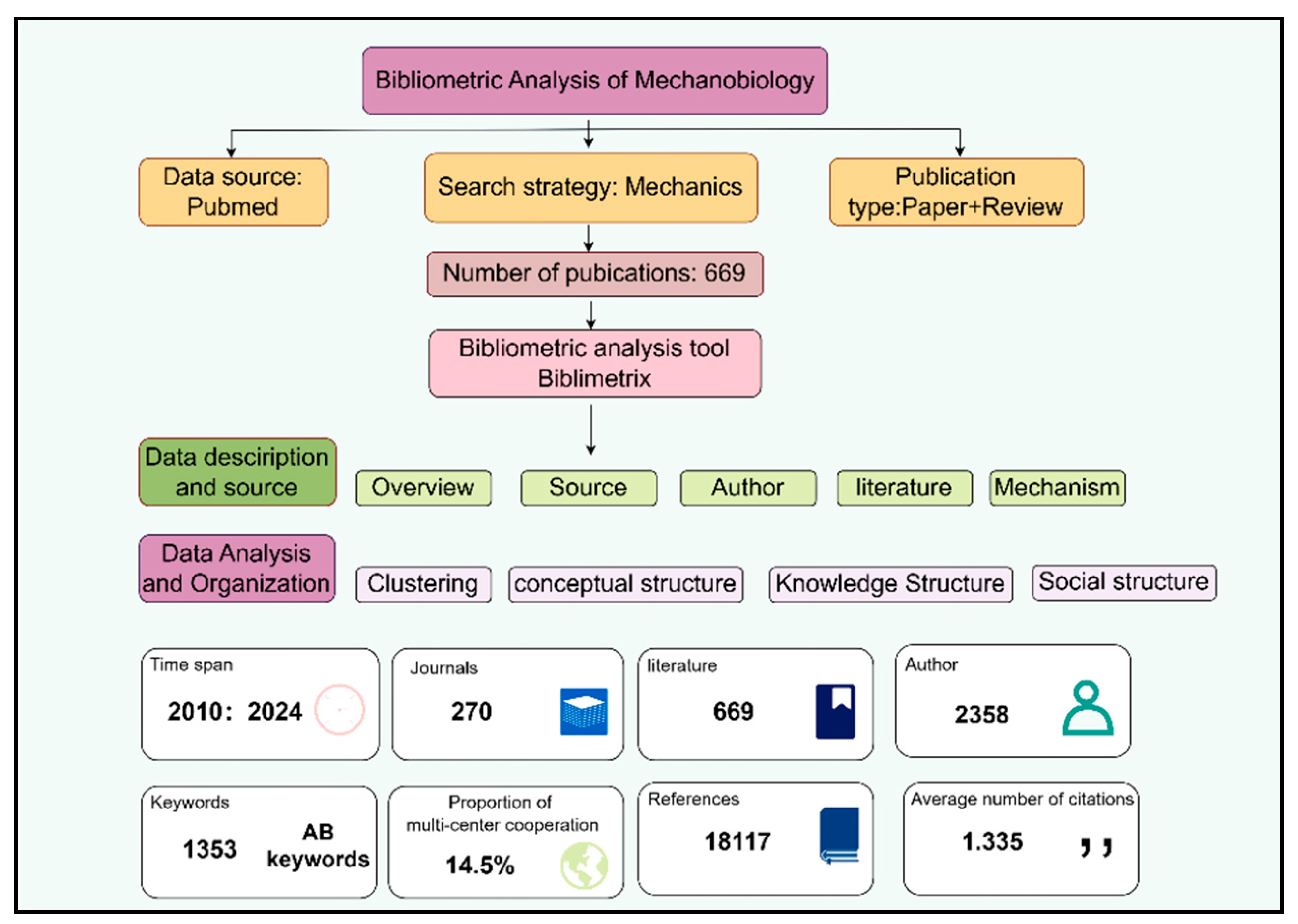
Figure 2.
(A) Annual publication trends and average citations per year for literature related to ‘Mechanobiology’ from 2010 to 2024. (B) A three-field Sankey diagram of relevant literature (AU—AU_UN—SO).
Figure 2.
(A) Annual publication trends and average citations per year for literature related to ‘Mechanobiology’ from 2010 to 2024. (B) A three-field Sankey diagram of relevant literature (AU—AU_UN—SO).
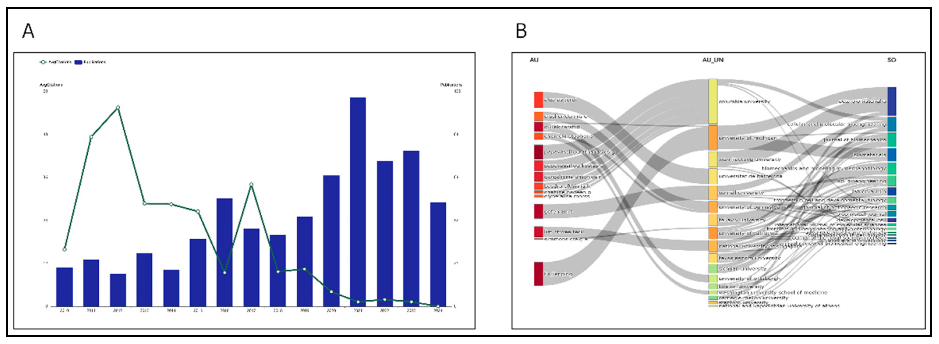
Figure 3.
(A) Bar chart of sources for literature related to ‘Mechanobiology’ from 2010 to 2024. (B) Cumulative publication statistics by source for literature related to ‘Mechanobiology’ from 2010 to 2024.
Figure 3.
(A) Bar chart of sources for literature related to ‘Mechanobiology’ from 2010 to 2024. (B) Cumulative publication statistics by source for literature related to ‘Mechanobiology’ from 2010 to 2024.
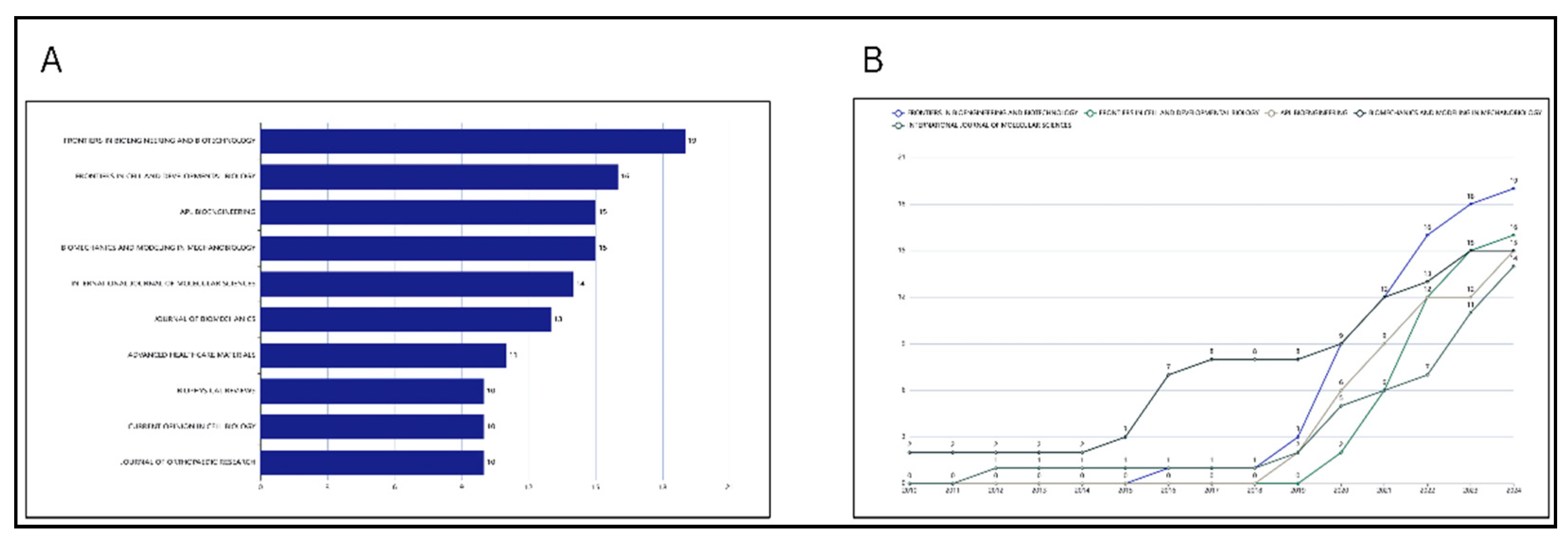
Figure 4.
(A)Institutional publication rankings in the field of ‘Mechanobiology’ from January 2010 to May 2024. (B) Publication volume by authors in the field across different years. (C) Evaluation of author productivity using Lotka’s Law.
Figure 4.
(A)Institutional publication rankings in the field of ‘Mechanobiology’ from January 2010 to May 2024. (B) Publication volume by authors in the field across different years. (C) Evaluation of author productivity using Lotka’s Law.
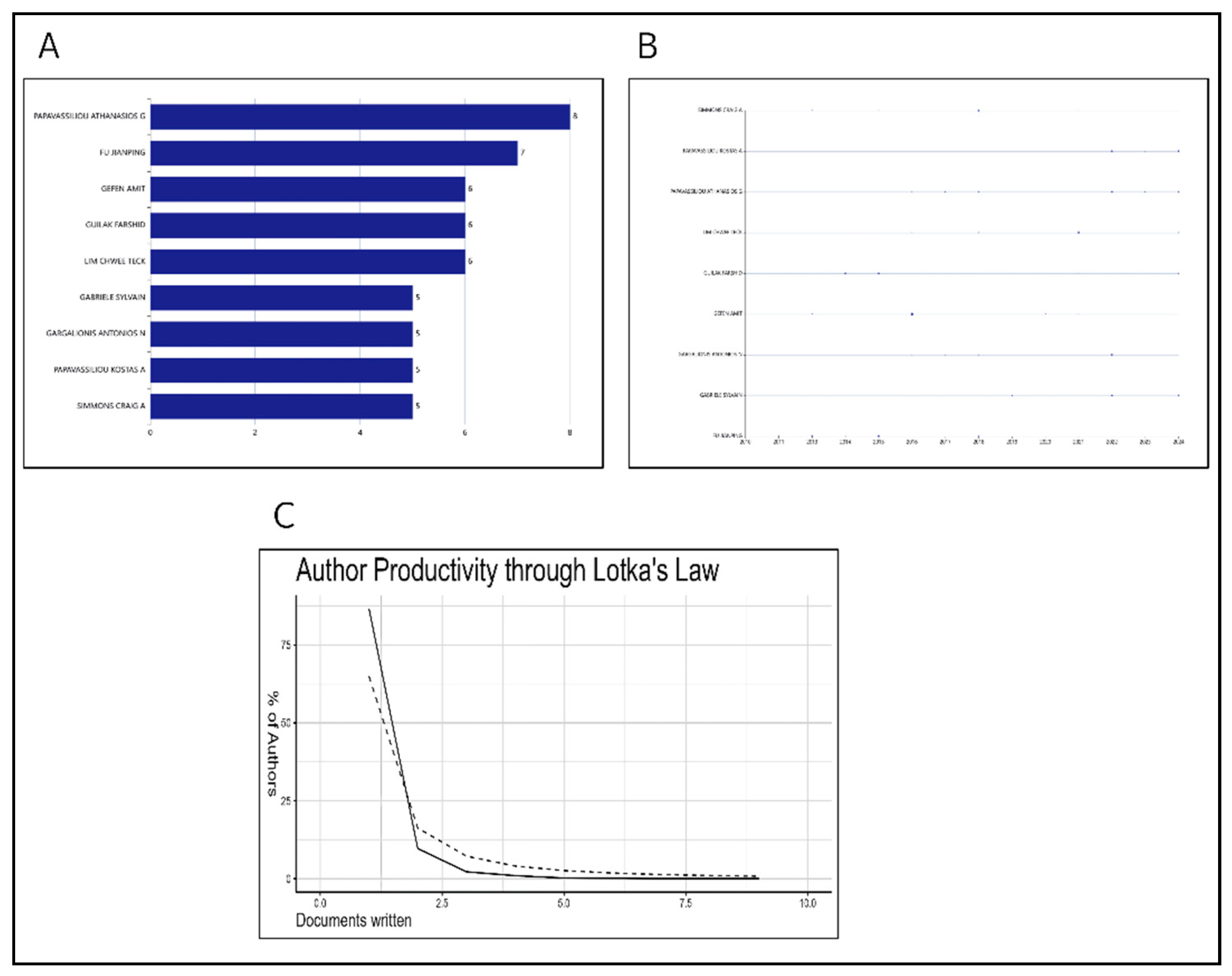
Figure 5.
Multi-dimensional Data Visualization Analysis: Trends, Distribution and Composition. (A) Institutional publication rankings in the relevant field. (B) Temporal publication output of research institutions in the field. (C) Publication rankings by country of corresponding authors and the proportion of international collaborations. (D) Scientific output by country in the relevant field. (E) Temporal publication output and citation rankings by country in the relevant field. (F) Ranking of the most cited countries.
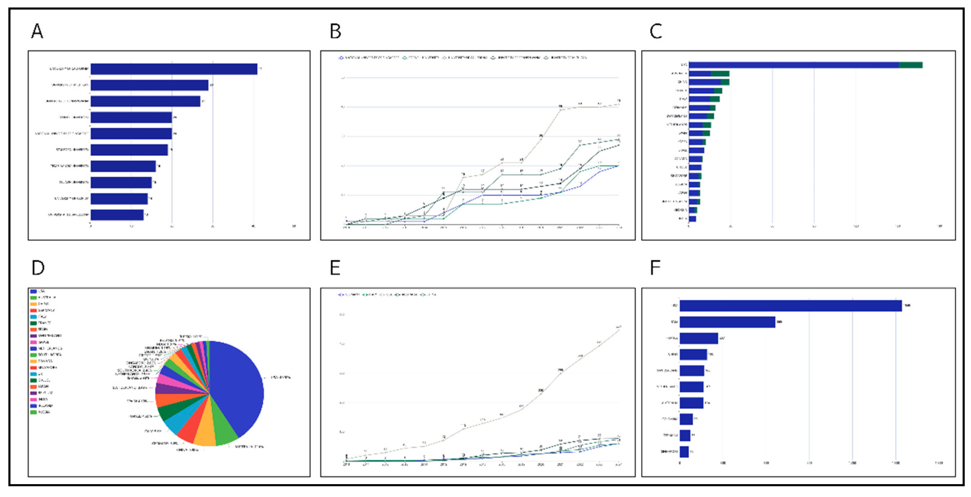
Figure 6.
Citation rankings of literature related to ‘Mechanobiology’ from 2010 to 2024.
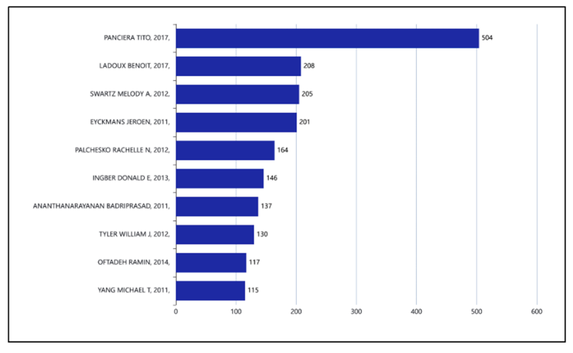
Figure 7.
(A) Word cloud displaying the top 50 most frequent keywords in the relevant literature. (B) Treemap visualization of keyword frequency. (C) Bar chart showing changes in word frequency (Most Frequent Words). (D) Temporal variation of keyword frequency in the relevant literature. (E) Research theme trends in the relevant field.
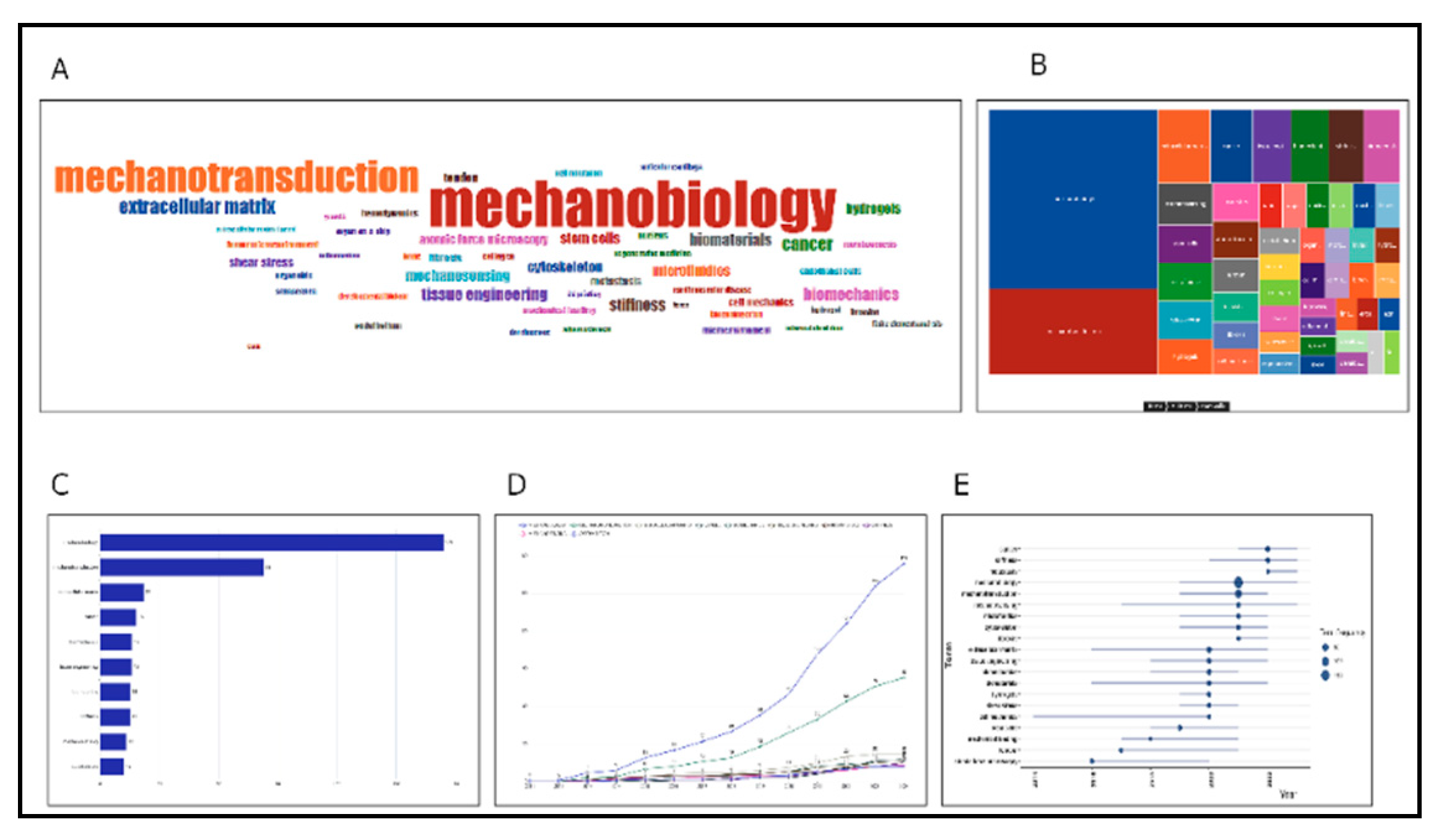
Figure 8.
Bibliographic coupling clustering of literature in the field of ‘Mechanobiology’ from 2010 to 2024. (A) the Time Evolution Map illustrates the evolution of research themes over time within the mechanobiology field. (B) presents a complex Co-citation Network, displaying citation relationships and academic influence across the literature.
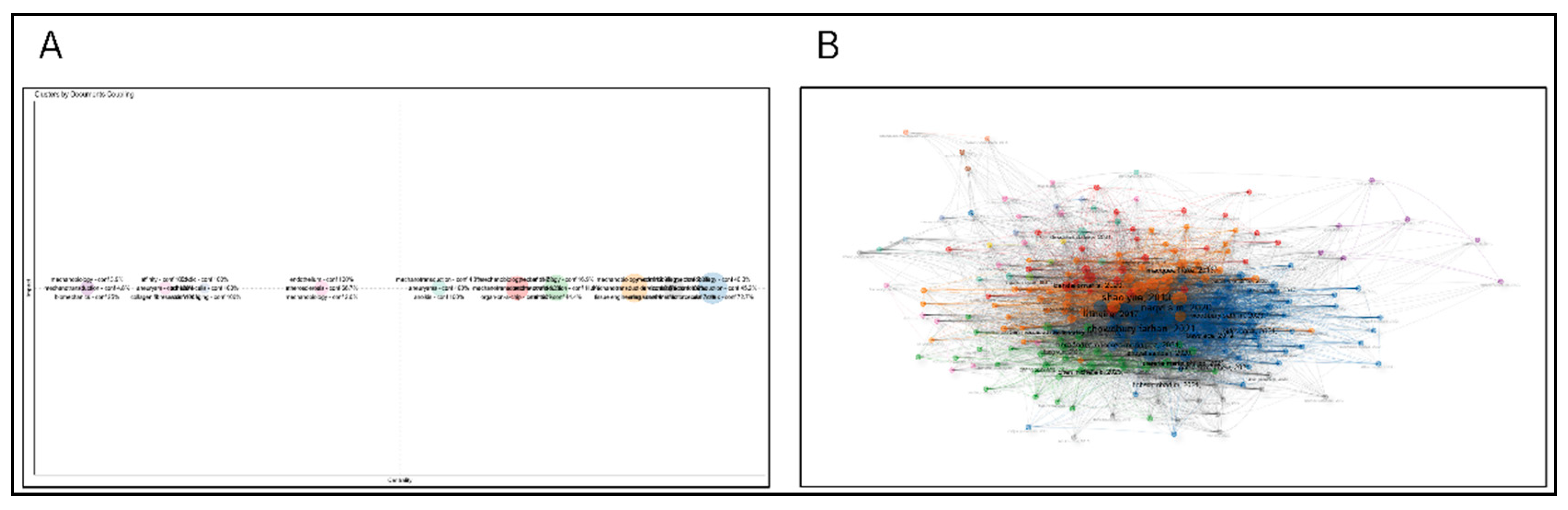
Figure 9.
Mechanobiology Multidimensional Knowledge Structure Analysis. (A) Typical power-law distribution characteristics. (B) Co-occurrence network of keywords exhibiting typical power-law distribution characteristics. (C) A thematic map of the field of “Mechanobiology” and the evolution of research topics over time. (D) Network visualization. (E) Sankey diagram clearly illustrating the dynamic knowledge flow between different research themes. (F) Factor analysis in the relevant field.
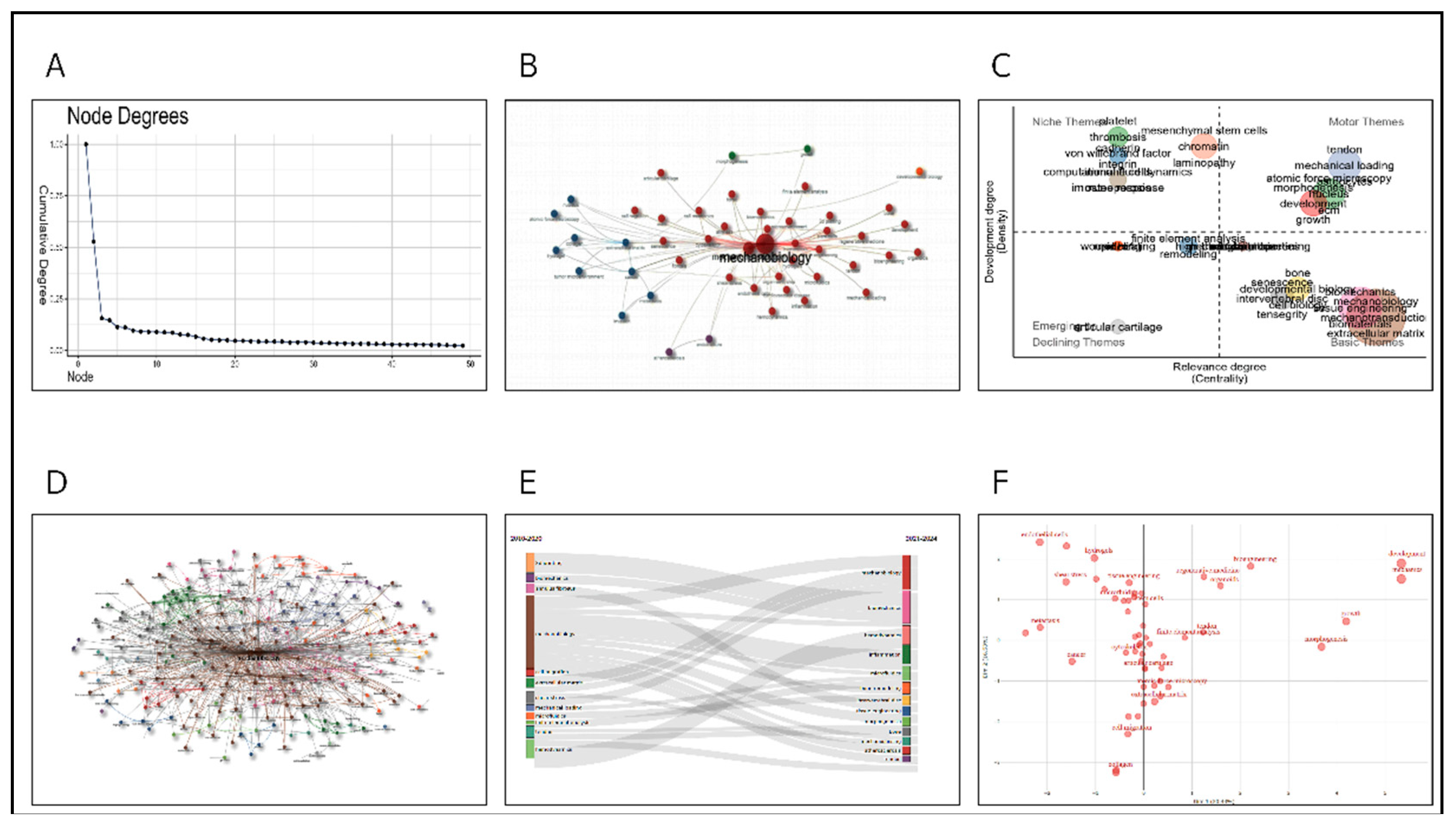
Figure 10.
The co-citation network analysis of the literature in the field of mechanobiology is presented collectively. (A) shows the degree distribution of nodes (papers) in the co-citation network. Node degree indicates the number of co-citation relationships a paper shares with others. This distribution exhibits a “long-tail” characteristic, indicating that most papers have co-citation relationships with only a few others, while a few core papers have dense co-citation connections with many others. (B) is a visualization of the co-citation network. Nodes in different colors represent research topics or groups, and the thickness of the connecting lines indicates the strength of associations between these topics/groups. The network reveals a clear clustering structure, with closely related papers forming groups, suggesting that multiple relatively independent research directions and academic communities exist within the field of mechanobiology. (C) displays the distribution of research topics in the form of a heatmap. Hotspot areas represent high-interest research directions, such as cellular mechanics and tissue engineering, while peripheral areas indicate less popular topics.
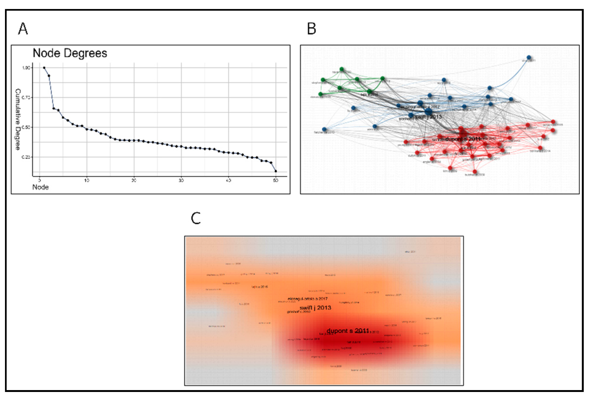
Figure 11.
Collaborative Network. (A) Node degree distribution diagram. It shows the degree distribution of each node in the network. (B) Scatter plot. It shows the connection relationship between nodes in the network. Each point represents a node, and the position and color of the point represent the properties of the node. (C) Heat map. It uses hot spots of different colors to indicate the distribution density of data, and is usually used to show spatial distribution or correlation, etc. (D) World map. It shows the distribution of network nodes in geographical locations, and uses lines to indicate the relationship between nodes.
Disclaimer/Publisher’s Note: The statements, opinions and data contained in all publications are solely those of the individual author(s) and contributor(s) and not of MDPI and/or the editor(s). MDPI and/or the editor(s) disclaim responsibility for any injury to people or property resulting from any ideas, methods, instructions or products referred to in the content. |
© 2024 by the authors. Licensee MDPI, Basel, Switzerland. This article is an open access article distributed under the terms and conditions of the Creative Commons Attribution (CC BY) license (http://creativecommons.org/licenses/by/4.0/).
Copyright: This open access article is published under a Creative Commons CC BY 4.0 license, which permit the free download, distribution, and reuse, provided that the author and preprint are cited in any reuse.


