
Submitted:
18 October 2024
Posted:
18 October 2024
You are already at the latest version
Abstract
Polyhydroxyalkanoates (PHAs) are a family of polyesters produced by many microbial species. These naturally occurring polymers are widely used in tissue engineering because of their in vivo degradability and excellent biocompatibility. The most well-studied among them is poly(3-hydroxybutyrate) (PHB) and its copolymer with 3-hydroxyvaleric acid (PHBV). Despite their superior properties, PHB and PHBV suffer from high crystallinity, poor mechanical properties, slow resorption rate, and inherent hydrophobicity. Not only PHB and PHBV are hydrophobic, but almost all members of the PHA family struggle because of this characteristic. One can overcome the limitations of microbial polyesters by modifying their bulk or surface chemical composition. Therefore, researchers have put much effort into developing the methods for chemical modification of PHAs. This paper reviews the chemical modification methods of PHAs that make them more suitable for scaffold fabrication. Different chemical strategies for improving the wettability and mechanical properties of the PHA scaffolds are discussed in this review. Emphasis is placed on the methods used to modify the chemical structure of PHB and PHBV, the most important polymers of the PHA family. The properties of PHAs that are important for their applications in tissue engineering are also discussed.
Keywords:
polyhydroxyalkanoates (PHAs)
; chemical modifications
; surface modifications
; polymer grafting
; functionalized oligomers
; tissue engineering
1. Introduction
Polyhydroxyalkanoates (PHAs) is a term given to a family of aliphatic polyesters synthesized by numerous bacteria and some archaea. Microorganisms accumulate PHAs in the form of intercellular granules that serve as carbon and energy storage material [1,2,3]. The accumulation of PHAs usually takes place when there is an excess of carbon sources with a concomitant deficiency of one (or more) of the following essential nutrients: nitrogen, oxygen, phosphorus, sulfur, or magnesium [4,5]. However, some microorganisms store PHAs even under non-limiting nutrient conditions [5]. So far, more than 300 strains of prokaryotes have been reported to produce biopolyesters [3,6]. Using the most diverse strains and growth media, it was possible to obtain bacterial polyesters, which together consist of more than 160 different monomeric units: starting from simple and saturated, through branched, unsaturated, and aromatic, and ending with those with additional functional groups, such as epoxy, halogen or hydroxyl groups [7,8,9]. In general, bacteria store homo- or copolymers of hydroxycarboxylic acids, although terpolymers also occur [10].
From the chemical point of view, microbial polyesters are polymers in which the repeating units are derived from 3-, 4-, 5-, and 6-hydroxycarboxylic acids (Figure 1a). These units (the same or different) are linked to each other by ester bonds, which determine the chemical reactions that PHAs will undergo, including their biodegradation mechanism. It is also worth noting that it is mainly the presence of ester bonds in the main chain that makes biopolyesters biodegradable. Due to the differences in their chemical structure, biopolyesters can be divided into three groups: short-chain PHAs (scl-PHAs), medium-chain PHAs (mcl-PHAs), and long-chain PHAs (lcl-PHAs) [3,7,11]. In the first group, the chemical structure of the monomers consists of no more than 5 carbon atoms. In the second group, there are 6 to 14 carbon atoms, while the monomers of lcl-PHAs contain more than 14 carbon atoms [3,12]. Scl-PHAs include, among others, poly(3-hydroxybutyrate) (PHB) and its copolymer with 3-hydroxyvaleric acid (3HV), that is poly(3-hydroxybutyrate-co-3-hydroxyvalerate) (PHBV), which are two of the most important polymers among all PHAs. It is significant to note that PHB and PHBV are some of the best-studied, most widespread in nature, and the most commercially available representatives of bacterial polyesters. Scl-PHAs also include poly(3-hydroxybutyrate-co-4-hydroxybutyrate) (P34HB) and poly(4-hydroxybutyrate) (P4HB), while mcl-PHAs include those biopolyesters that among others consist of repeating units derived from 3-hydroxyhexanoic acid (3HHx) and 3-hydroxyoctanoic acid (3HO) (Figure 1b).
The most attractive feature of microbial polyesters is their outstanding biocompatibility. Numerous studies have shown that PHAs elicit a milder immune response than such widely used polyesters as polylactide (PLA), poly(lactide-co-glycolide) (PLGA), and polycaprolactone (PCL) [13,14,15,16]. The non-toxic nature of PHAs, combined with their ability to degrade under in vivo conditions, makes them highly promising materials for a range of biomedical applications.
These features have inspired researchers to explore the use of biopolyesters as matrices for controlled drug delivery and tissue engineering [17,18,19,20,21,22,23,24,25]. In this context, PHAs have proven useful in the engineering of, among others, soft tissues, blood vessels, heart valves, as well as cartilage, bone, and nerve tissues [20,21,26]. Moreover, biopolyesters are of particular interest in applications related to bone tissue regeneration due to their piezoelectric properties [27,28]. In addition to tissue scaffolds, PHAs could be used, for example, in the production of articular cartilage repair devices, cardiovascular patches, meniscus repair devices, orthopedic pins, screws, stents, sutures, and surgical meshes [20,25].
Although PHAs have many potential applications, products made from them are rarely found in everyday life. One of the few examples of the actual use of biopolyesters is a product called ‘TephaFLEX’, a resorbable surgical suture made of P4HB [20]. The reason for the lack of wider industrial application of PHAs is still the high cost of their production, which is several times higher than that of petrochemical polymers [29]. Moreover, in medical applications, biopolyesters often do not exhibit adequate degradability and cytocompatibility. Therefore, PHAs undergo physical or chemical modifications to improve their biological properties.
There are many review articles about PHAs, but they mainly deal with their biosynthesis in bacteria [8,9,30,31,32]. Relatively few reviews have been devoted to the chemical modification of microbial polyesters and even fewer to their modifications for tissue engineering purposes [33,34,35,36,37,38]. This paper aims to shed more light on this topic. In some regards, chemical methods are more reliable and beneficial than biotechnological and physical ones. Here, we summarize the literature describing the chemical methods used to improve the performance of PHAs as scaffold materials for tissue regeneration. We also provide basic information about the properties of PHAs that are essential for their application in regenerative medicine.
2. Properties of PHAs Important for Their Applications in Tissue Engineering
2.1. Mechanical Properties and Piezoelectricity
The mechanical properties of a scaffold are of great importance for its potential application in tissue engineering [39]. Such parameters of a scaffold as tensile strength and Young’s modulus dictate the types of tissues in which regeneration that scaffold can participate. The ideal scaffold should have mechanical properties close to those of regenerated tissue. A rigid scaffold, for example, in contact with soft tissues can cause damage to them and subsequent onset of chronic inflammation in the host [40]. The reason for this is the lack of compatibility in terms of mechanical properties between the scaffold and the tissue. Therefore, flexible and elastic materials are preferentially used for soft tissue engineering, whereas stiff polymers are almost solely restricted to the regeneration of cartilage and bone tissues.
As mentioned earlier, PHAs are a group of polyesters exhibiting great structural diversity. Depending on bacterial culture conditions, PHA-producing strains can synthesize polyesters that consist of over 160 different monomeric units. This is a significant advantage of biopolyesters as it gives more options in selecting a polymer for a given application. However, only a few PHA polymers, such as PHB, PHBV, P34HB, P4HB, and PHBH, are commercially available and produced on a large scale [41]. This means that the range of mechanical properties offered by the most available PHA family members is often insufficient to meet demands in some tissue engineering approaches. Therefore, modifications of the chemical structure of PHAs are a way to expand their applications in regenerative medicine. The most important thermal and mechanical properties of selected representatives of PHAs are shown in Table 1.
When discussing the thermomechanical properties of microbial polyesters, it is typical to relate to their classification based on the chemical structure of the mers of which they are composed. Scl-PHAs are generally stiff and brittle and exhibit a relatively high degree of crystallinity (50–80%) [26,42,43]. On the other hand, mcl-PHAs are soft and flexible and less crystalline than scl-PHAs (Xc < 40%). Compared with scl-PHAs, mcl-PHAs are characterized by lower glass transition and melting temperatures, lower Young’s modulus values, and higher elongation at break [26]. In other words, mcl-PHAs are more elastic than scl-PHAs, and hence, they are more frequently used in soft tissue engineering [44,45]. At the same time, stiff and highly crystalline PHB and PHBV are usually employed as scaffold materials for bone repair.

| Biopolyester | Tg [°C] | Tm [°C] | Xc [%] | E [GPa] | Rm [Mpa] | A [%] |
|---|---|---|---|---|---|---|
| PHB | 5–10 | 173–180 | 60–80 | 3.5–4 | 20–40 | 3–8 |
| PHBV 3% | 8 | 170 | 59 | 2.9 | 38 | 5 |
| PHBV 9% | N/A | 162 | N/A | 1.9 | 37 | N/A |
| PHBV 14% | N/A | 150 | N/A | 1.5 | 35 | N/A |
| PHBV 20% | –1 | 145 | 56 | 1.2 | 32 | 50 |
| PHBV 25% | –6 | 137 | 54 | 0.7 | 30 | 100 |
| PHBHHx 10% | –1 | 127 | 35 | 0.23 | 21 | 400 |
| PHO | –38 | 49 | 25 | 0.017 | N/A | 300 |
| P4HB | –50 | 53 | < 40 | 149 | 104 | 1000 |
| P34HB 19–94% | –4–(–46) | 52–158 | 18–54 | N/A | N/A | N/A |
| P34HB 3% | N/A | 166 | N/A | N/A | 28 | 45 |
| P34HB 10% | N/A | 159 | N/A | N/A | 24 | 242 |
| P34HB 16% | –7 | 130 | 43 | N/A | 26 | 444 |
| P34HB 64% | –35 | 50 | N/A | 30 | 17 | 591 |
| P34HB 90% | –42 | 50 | N/A | 100 | 65 | 1080 |
The percentage value given next to a specific PHA polymer indicates the molar content of the non-3HB monomer; Tg, glass transition temperature; Tm, crystal melting temperature; Xc, degree of crystallinity; E, Young’s modulus; Rm, tensile strength; A, elongation at break; N/A, data not available.
PHAs exhibit a piezoelectric effect [50,51,52]. It is a desirable feature of biomaterials when they are utilized in regenerative medicine, especially in bone tissue engineering, since bone itself exhibits piezoelectric properties [53,54,55]. Piezoelectricity is a phenomenon that manifests itself by the generation of electrical charge on the surface of a material subjected to compressive or tensile forces [56]. It is a simple way to provide electrical stimuli to cells. Studies have shown that piezoelectric scaffolds, including those made of PHAs, promote bone tissue regeneration and formation [53,55,57,58].
2.2. In Vivo Degradability
Resorbability refers to the ability of a material to dissolve or disintegrate in physiological environments without causing any long-term complications [59]. Polymers resorb in the body either due to actions taken by the cells or through chemical dissolution. In-vivo degradation of the material is an important property to consider during scaffold design. To achieve the therapeutic goal, the resorption rate of the scaffold should be similar to the regrowth rate of a tissue of interest [60]. Moreover, using a resorbable implant in tissue reconstruction allows avoiding the additional surgery required to remove the implant from the body once it fulfills its function.
Degradation of biomaterials inside the body can occur enzymatically or non-enzymatically [61]. One of the most common non-enzymatic reactions that lead to bond breaking in a polymer is hydrolysis. The susceptibility of a particular polymer to hydrolysis is most often studied through in vitro tests conducted under conditions that simulate physiological environments. Phosphate-buffered saline (PBS) or simulated body fluid (SBF) is usually used to mimic in vivo conditions. Studies using these buffers are very important—an absence of in vitro hydrolysis alongside in vivo degradation may suggest that the degradation within the body is due to increased cellular activity, including the presence of enzymes.
The degradation of biomaterials, both in vitro and in vivo, may be accompanied by changes in average molecular weight as well as in the thermal and mechanical properties of the material. Additionally, as degradation progresses, a change in the mass of the biomaterial sample is observed, often due to surface erosion [62,63].
Doi et al. were among the first to study the hydrolytic degradation of PHAs using a phosphate buffer with a pH of 7.4 as the degradation medium [62]. They hydrolyzed polymer films made of various PHAs, carrying out the degradation reaction at 37 °C for 200 days. The largest relative decrease in the number-average molecular weight (Mn) was observed for a film produced from P34HB containing 16 mol% 4-hydroxybutyrate units (referred to as P34HB 16%); a smaller decrease was recorded for P34HB 9%, while the smallest decrease was observed for PHB and PHBV 68%. Thus, these researchers showed that the degree of hydrolytic degradation of a biopolyester depends on its chemical composition. By conducting a similar degradation study, Freier et al. determined that changes in weight-average molecular weight (Mw) for hydrolyzed PHB have a half-life (t1/2) of approximately 56 weeks [64]. Moreover, Zhuikov et al. investigated the effect of molecular weight on the hydrolytic degradation rate of films produced from PHB and PHBV with a 9 mol % content of 3-hydroxyvalerate (PHBV 9%) [65]. These films were incubated at 37 °C for 183 days using PBS as a degradation medium. The decrease in molecular weight for PHBV 9% was greater than that for PHB. The differences in the rate of hydrolysis between the homopolymer of 3-hydroxybutyric acid and its copolymers are explained by the fact that the copolymers are characterized by much lower crystallinity than PHB. Studies have shown that hydrolysis in the amorphous regions of PHAs is up to 20 times faster than in the crystalline part [66].
In contrast to the average molecular weight, non-enzymatic hydrolysis of PHB under neutral conditions (pH ~7) is usually not accompanied by mass loss (erosion) of the product made from this polymer [62,63,64,67]. A change in mass is not observed even when hydrolytic degradation is carried out for 730 days [68]. For this reason, bacterial polyesters are considered to be highly resistant to non-enzymatic hydrolysis.
Studies indicate that the rate of non-enzymatic hydrolytic degradation of biopolyesters increases significantly when hydrolysis is carried out in highly acidic (pH < 2) or alkaline (pH > 12) solutions, as well as at elevated temperatures [67,69,70,71,72]. Bonartsev et al. investigated the hydrolysis of a PHB film in a phosphate buffer at pH 7.4. They observed that degradation carried out at 70 °C was accompanied by a mass loss in the polymer film, with the loss being greater as the average molecular weight of PHB decreased [69]. After 83 days of degradation, the largest mass loss (38%) was observed for PHB with an initial Mw of 170,000 g/mol, and the lowest (7.5%) for the biopolyester with an initial Mw of 950,000 g/mol. Mass loss was also observed when synthetic gastric juice (pH 1.6) was used as a degradation medium [67]. Hydrolysis in the presence of this simulated physiological fluid was carried out at 37 °C for 12 weeks. Interestingly, mass loss was noted only for the film made from P34HB, while no change in the mass of the PHB film was observed.
It should be noted that products made from bacterial polyesters degrade to a greater extent when implanted into animals than when hydrolyzed in a PBS solution, which may indicate that enzymes are involved in the degradation of biopolyesters in vivo [63,73]. This can be investigated by performing the degradation of PHAs in vitro by adding an enzyme of animal origin to a phosphate buffer and comparing the results of hydrolysis conducted in the presence of this enzyme with those of non-enzymatic hydrolysis. Studies of the progress of enzymatic degradation are most often carried out by monitoring changes in the average molecular weight of biopolyesters, as well as changes in the mass of the sample made from it [63].
A different methodology for studying the progress of enzymatic hydrolysis of PHA was proposed by Sudesh et al. The method they developed is based on observing changes in the transparency of a polymer film under the influence of a solution containing a selected enzyme [71,74]. Enzymatic degradation occurring in an initially transparent area of the thin film makes this region opaque. The occurrence of such a change indicates that hydrolysis is taking place. Interestingly, this method can assess the hydrolytic activity of the enzyme both qualitatively and quantitatively.
Yet another method for studying the enzymatic hydrolysis of PHA was employed by Tarazona et al. [75]. This method involved producing a thin polymer film called a Langmuir-Blodgett layer and comparing the surface properties of this film (surface tension, surface rheology, and surface potential values) before and after exposure to a specific enzyme. The effectiveness of this method is based on the observation that enzyme-catalyzed hydrolysis is an example of heterogeneous degradation confined to the surface of the material under study [75].
The degradation of PHAs caused by the presence of animal-origin enzymes can therefore be studied in several ways. Although the results of such studies are not entirely conclusive, they tend to lean toward the fact that PHAs, with few exceptions, do not undergo hydrolysis catalyzed by animal enzymes [64,75,76,77]. It is important to highlight that, so far, no enzymes with PHA depolymerase activity—namely, enzymes exhibiting substrate specificity toward biopolyesters—have been found in human body fluids and tissues. Therefore, it has been assumed that non-specific lipases—a group of enzymes that break down fatty acid esters—may be responsible for the possible hydrolysis of PHAs in vivo [78]. These enzymes are commonly found in the human body, with pancreatic lipase being a typical representative [79]. For research, pancreatic lipase is most often obtained from porcine pancreas and used in its pure form or as a mixture of enzymes called pancreatin, which contains lipase, amylase, and proteases [78].
Studies have shown that lipases produced by eukaryotic organisms significantly degrades only those PHAs which are derived from ω-hydroxyalkanoic acids, such as 3-hydroxypropionic acid (3HP), 4-hydroxybutyric acid (4HB), 5-hydroxyvaleric acid (5HV), and 6-hydroxyhexanoic acid (6HHx) [17,67,80,81]. Steric factors thus determine the substrate specificity of lipases—the polymers of the aforementioned acids do not contain side chains, which serve as substituents of the main chain (Figure 2). Among commercially available biopolyesters, only P34HB undergoes significant hydrolysis when catalyzed by animal-derived lipases. During the lipolytic hydrolysis of this copolymer, unlike the PHB homopolymer, a loss of mass in the degradable film is observed [67,71,81].
As mentioned earlier, PHAs degrade faster in vivo than in vitro. The results of in vivo degradation tests depend on the monomeric composition of the biopolyester, the type of tissue in which the polymer is implanted, and the form of the implanted device (for example, whether the material is implanted as a film or fibers) [82]. In general, PHA copolymers, which have a lower degree of crystallinity than PHB homopolymer, are more susceptible to in vivo degradation. This susceptibility is characterized by increased erosion and a greater decrease in the average molecular weight of the implanted copolymer [13,44,63,83]. Shishatskaya et al. studied the degradation of PHB and PHBV by implanting these polyester fibers into the muscular tissue of rats [63]. After 180 days, the average mass of the implanted fibers decreased to 74% of the initial mass for PHB and 65% for PHBV. Moreover, the authors indicated that enzymes in tissue fluid and immune cells, such as macrophages and foreign body giant cells (FBGCs), are responsible for the in vivo degradation of PHAs.
Volova et al. also investigated the resorbability of bacterial polyesters using polymer films obtained from five different PHAs, namely: PHB, PHBV 13%, PHBV 27.6%, P34HB, and PHBHHx [83]. In these studies, a film made of PLA, used as a positive control, was also subjected to degradation. However, the publication does not specify whether it was PLLA or PDLLA. All films were implanted subcutaneously in rats, and the tests were conducted over 180 days. The measure of degradation was based on the change in the mass of the implanted film and the average molecular weight of the polyester from which the film was made. PLA products placed inside the body (both PLLA and PDLLA) undergo significant hydrolytic degradation, unlike PHAs, which results in their faster in vivo degradation [13,14]. Of the PHAs studied, the film made from PHBHHx showed the most degradation, followed by those made from P34HB and PHBV. The highest resistance to in vivo degradation was observed in the most crystalline of the biopolyesters, PHB homopolymer (with a degree of crystallinity of 76%, compared to no higher than 50% for the copolymers studied).
Studies indicate that macrophages and FBGCs are primarily responsible for the in vivo degradation of PHAs [14,63,82,84,85]. Some review publications suggest that biopolyesters are also degraded by osteoclasts (the bone-resorbing cells), which, like FBGCs, are derived from macrophages [82,86]. However, research conducted by Cool et al. indicates that while osteoclasts adhere to the surface of the PHA material, they do not cause the formation of resorption pits on its surface [87]. The formation of resorption pits would indicate that osteoclasts degrade bacterial polyesters.
Placing biomaterials inside the body results in the recruitment of immune system cells to the implantation site, primarily neutrophils and macrophages [40]. The former remain in the area of the foreign material for a longer period when it proves to be toxic [14,88,89]. However, PHA implantation is not accompanied by prolonged neutrophil infiltration, indicating that these polyesters can be safely used as implants [14]. The degradation of biopolyesters by macrophages and FBGCs is most often associated with the phagocytic function of these cells. Some researchers also suggest that reactive oxygen species (ROS) released by inflammatory cells, such as superoxide anion (O2–), hydrogen peroxide (H2O2), or nitric oxide (NO), may be responsible for the degradation of PHAs in vivo [75,90,91]. Lysosomal enzymes produced by macrophages and FBGCs are also indicated when considering the resorbability of PHAs [92,93].
In summary, bacterial polyesters are considered to be slow-degrading polymers under in vivo conditions. For this reason, it is suggested to use PHAs in applications where relatively high in vivo stability of the biodegradable polymer is required [75]. As mentioned earlier, the degradation time of the material used to design the scaffold should be consistent with the natural regeneration time of the target tissue. In most cases, however, the degradation time of commercially available biopolyesters is often too long, making their use in tissue engineering much more complicated [18,65].
2.3. Biocompatibility
The most important feature of polymeric materials that find application in medicine is biocompatibility. Biocompatible materials do not induce an adverse immune-related reaction in contact with body fluids or tissues [94]. The lack of toxicity applies not only to the biocompatible polymer but also to its degradation products in vivo. Moreover, the body’s immune response cannot be strong enough to prevent the biomaterial from performing its therapeutic function. This property is especially important for PHAs, as these polyesters are characterized by very good biocompatibility compared to other polymers.
Given the prevalence of biopolyesters in the natural environment, it is not surprising that they are very well tolerated by living organisms. It turns out that bacterial cells are not the only places where these polymers naturally occur. Macromolecules containing no more than 200 3HB units has also been found in the cells of eukaryotic organisms [82,95]. This oligomeric PHB is referred to in the literature as “complexed PHB” (cPHB) [86]. It has been discovered in many tissues and organs of higher mammals (including humans), such as blood, brain, heart, kidneys, nerves, eyes, and liver [82,95,96]. It has been shown that cPHB forms non-covalent complexes with inorganic polyphosphates, calcium ions, and proteins. These complexes function as ion channels embedded in the cell membrane, allowing inorganic ions to pass into the eukaryotic cell [82,96,97]. Due to the ubiquity of cPHB, many researchers suggest that it is a product of eukaryote metabolism or their symbiosis with gut bacteria [95]. However, the mechanism underlying the synthesis of cPHB is not yet well-understood.
For a polymeric material to be described as biocompatible, the compounds formed as a result of its in vivo degradation (or the metabolites of these compounds) must also not be toxic to the host organism. The final product of PHB degradation, (R)-3-hydroxybutyric acid (3HB), similar to its oligomeric counterpart (cPHB), is found in the blood and tissues of both animals and plants [95,98,99]. The 3HB content in human blood serum ranges from very low levels, measured in µmol/L, to as high as 8 mmol/L [98,100]. It is also worth noting that in animals, this compound is a metabolite of fatty acid oxidation, which becomes the primary energy source in the case of blood glucose deficiency [98,100,101]. In other words, 3HB is classified as a so-called ketone body. Studies indicate that 3HB is not only a backup source of energy for animals, but also has many important functions as a regulatory molecule [97,98]. The most important of these functions are the inhibition of histone deacetylase and NLRP3 inflammasome activity [98,102,103]. (R)-3-Hydroxybutyric acid is also involved in ROS metabolism, acting as an antioxidant by neutralizing hydroxyl radicals (OH•) [104]. Moreover, it inhibits the production of ROS by mitochondria [104]. It is also worth noting that the degradation products of other biopolyesters, such as PHBV, P34HB, and PHBHHx, are also non-toxic [42,105,106].
The fact that PHAs usually degrade slowly in the body has positive consequences. For example, PLA and PLGA degrade more rapidly, so that lactic acid (or glycolic acid), the end product of their in vivo degradation, begins to accumulate within the implant soon after implantation [107,108,109]. As a consequence, the tissue surrounding the implant becomes acidic, and a decrease in pH may accompany the implantation of PLA until it is completely absorbed [110,111,112,113]. This leads to a dangerous defensive reaction in the body, which is chronic inflammation (necrosis of host cells is also observed) [112,114,115]. Interestingly, the water-soluble oligomers formed during the degradation of PLA and PGA may also contribute to the aforementioned pathological process [116,117]. Inflammation caused by the presence of acidic degradation products is a significant medical problem, often limiting the use of PLA and its copolymers for therapeutic purposes [95]. In the case of bacterial polyesters, this problem is unlikely to occur. The use of PHB as a material for tissue scaffolds is also supported by the fact that the final product of its degradation, 3HB, is less acidic (pKa = 4.70) than lactic acid (pKa = 3.86) or glycolic acid (pKa = 3.87) [118].
Tests to assess the biocompatibility of a given polymer in a specific application are carried out either under laboratory conditions (in vitro) or on animal models (in vivo) [94,119]. The first of these is usually done before the second; they are easier to perform, faster, and spare animals from suffering [119]. One of the most basic in vitro tests is the cytotoxicity test. This test determines whether the material causes cell death due to direct contact (direct contact assay) or as a result of the release of toxic substances (extract test) [119,120]. The method of conducting a cytotoxicity assay is described in ISO 10993-5. Cytotoxicity tests are performed using cells, with fibroblasts and osteoblasts most often used for this purpose. Both direct contact and extract tests have shown that biopolyesters are non-toxic to the two types of cells mentioned above [121,122,123]. Other in vitro studies have revealed that PHAs are not teratogenic or carcinogenic [42,124,125,126].
In vivo biocompatibility studies involve implanting material into a model animal organism and characterizing the immune system’s response to the implant, known as the tissue response [13,14,19,94,127,128,129,130]. Although there are countless combinations of tissue types and polymers that can be implanted in their vicinity, the tissue response usually comes down to the same sequence of events in each case [94,127,130]. A review of several scientific publications suggests that the immune response to implants is predominantly attributed to tissue damage incurred during the implantation process [40,94,129,131,132]. However, this reaction is the result of both an immune response to tissue damage and the presence of a foreign body [94,133]. It should be noted that the implantation of different polymers, even when following the same procedure, is accompanied by varying tissue responses [14,82]. Moreover, there are no polymers that may go unrecognized by the host’s immune system and avoid a reaction from it [134]. When characterizing the tissue response to implant materials, the intensity of the immune response is assessed. The occurrence or non-occurrence of certain events (and possibly their duration) is also taken into account [94,129,132]. For example, the presence of chronic inflammation at the implantation site—a pathological process that can lead to degenerative diseases, including Alzheimer’s disease and periodontitis—is highly undesirable [135,136,137].
In the immune response to the implant, there is a cascade of events that follow one another, although they can also co-occur. These stages include, among others, the onset of acute inflammation, the onset of chronic inflammation, the reaction to a foreign body, and the encapsulation of the implant with connective tissue (formation of a fibrous capsule around the foreign body) [94,131,138]. It should be noted that the duration and intensity of these processes determine the biocompatibility of the implant [94].
The duration and intensity of the individual stages of the tissue response are determined by histopathological examination. To do this, the implant and the surrounding tissue are removed from the model organism, and the fragment showing the boundary between the polymer and the biological material is examined under a microscope. Such examinations are facilitated by the fact that each of the aforementioned stages is characterized by the presence of different types of cells [40,94,127,131]. During acute inflammation, primarily neutrophils, but also monocytes and macrophages, are observed. In chronic inflammation, lymphocytes, monocytes, and a greater number of macrophages appear. In a process called the foreign body reaction, macrophages and FBGCs adhere to almost the entire surface of the implant. The primary function of neutrophils, macrophages, and FBGCs is first to phagocytose the foreign material and then its degradation [40]. The microscopic image of the final stage of tissue response shows a capsule surrounding the implant, with macrophages present on its surface. This capsule, composed of fibroblasts and collagen fibers, also serves as a barrier separating the implant from the body’s internal environment [127].
Long-term encapsulation of a material is often a medical issue, as it prevents the biomaterial from fulfilling its therapeutic functions [40,139,140]. This is particularly evident in controlled drug delivery systems (e.g., insulin pumps), where the formation of a fibrous capsule around such devices makes it much more difficult to release medicinal substances from them. The impact of the capsule on the effectiveness of a biosensor can be observed as early as 1–3 days after implantation [140,141].
The intensity of the immune response to PHAs depends on the chemical purity of the implanted polyester [142,143]. Biopolyesters produced by bacteria may contain cell residues, such as bacterial lipopolysaccharides and proteins, which are endotoxins that can cause fever, a systemic symptom of inflammation [18,142,143]. Studies indicate that PHAs of high purity induce a milder immune response than PLA, PLGA, or PCL [13,14,15,16]. This less intense tissue response is characterized by (i) reduced immune cell infiltration, (ii) the induction of cytokine expression that promotes angiogenesis and implant acceptance, for example, by macrophage polarization towards the M2 phenotype, and (iii) the absence of a fibrous capsule or a smaller capsule thickness [40,82,131]. It should also be emphasized that PHAs have better biocompatibility, even though their in vivo degradation proceeds more slowly than that of PLA and PLGA. This remains true despite the fact that the capsule formed around the PHA implant may persist until the material is completely degraded [82].
Studies using PHB homopolymer and its copolymers indicate that these generally do not differ in tissue response [83,93]. Some reports, however, suggest that PHA copolymers are better tolerated by the host than the PHB homopolymer [14,93,129]. What distinguishes PHB from its copolymers is the in vivo degradation rate and mechanical properties. Neither of these differences is beneficial for PHB, as it degrades more slowly and is less flexible than its copolymers [14]. Sometimes the slowly degrading polymer, remaining at the implantation site for a relatively long time, leads to chronic inflammation [140]. The adverse effect of the prolonged presence of PHB implants is most often explained by the fact that they are too rigid, thus subjecting the adjacent tissues to constant mechanical stimulation [14,93]. This stimulation arises from a lack of compatibility in mechanical properties between the implant and the tissue and often results in damage to the latter, which also has an impact on the immune system [40].
It should be noted that even though PHA copolymers degrade faster than PHB, which is characterized by high crystallinity, their implantation is almost always accompanied by the production of a collagen capsule [13,14,83,93,129]. In some cases, however, the capsule dissipates quickly [82]. In one study, the authors even suggested that the capsule around the fiber mesh of PHB did not form at all [15].
Studies have shown that bacterial polyesters, in addition to very good biocompatibility, also exhibit high hemocompatibility [143]. Therefore, they could potentially be used for the production of medical devices that come into contact with blood.
2.4. Cytocompatibility
Cytocompatibility is a concept related to the assessment of the biocompatibility of a material. However, it has a broader meaning than cytotoxicity. Cytocompatible materials do not cause cell death or negatively affect the structure and function of the tissues with which they come into direct contact [144]. A polymer can therefore be non-cytotoxic and at the same time non-cytocompatible, but not the other way around; it may not cause cell death while preventing the cells from performing certain functions. The basic tests assessing cytocompatibility in relation to a specific type of cells are the test of adhesion of these cells to the surface of the tested material and the test of their multiplication (proliferation) [144]. When examining cytocompatibility, attention is also paid to the morphology of cells adhering to the material’s surface. The closer their shape is to the natural, the more cytocompatible the material is [144].
Cytocompatibility is a property of polymers that is extremely important in the context of their use in the production of tissue scaffolds. It defines the types of tissues in which the polymer can participate in the regeneration process. Studies described in the literature have shown that PHAs promote the adhesion and proliferation of many types of animal cells. These include fibroblasts, osteoblasts, chondrocytes, cardiomyocytes, Schwann cells, mesenchymal stem cells, smooth muscle cells, keratinocytes, and neural stem cells [15,121,122,145,146,147,148,149,150,151].
3. Chemical Modifications of PHAs
Biopolyesters possess many desirable properties for tissue engineering applications. However, they are not free of drawbacks that limit their utility in regenerative medicine. The scaffolds made from PHB and PHBV, i.e., from the most important members of the PHA family, are brittle and stiff. Implantation of these scaffolds in an animal model can result in tissue damage and chronic inflammation due to the mismatch of mechanical properties between the tissue and the implant. The brittleness and stiffness of PHB and PHBV are due to their high degree of crystallinity (50–80%). The high content of the crystalline phase in PHB and PHBV also results in lowering their resorption rate. Fortunately, the crystallinity of PHAs can be reduced by modifying their chemical structure.
The problem with the crystallization behavior is particularly evident in the case of PHB scaffolds. The high tendency of PHB to crystallize is due to its orderly and regular chemical structure. This polyester, synthesized by many bacterial species, is an isotactic homopolymer; that is, it is made of repeating units of one type, all of which have the same (R) configuration on the tertiary carbon atom [152]. Thus, by disrupting the repetitive structure of PHB, it is possible to reduce its degree of crystallinity and improve its mechanical properties and degradability in vivo. This can be done through various chemical methods, which aim to modify the chemical structure of PHB. An illustrative example of this method is the copolymerization reaction involving PHB oligomers with non-PHB-derived monomers, such as 1,6-hexamethylene diisocyanate and poly(ethylene glycol) (PEG) (Figure 3a) [153,154]. The overall effect of this process is the introduction of other monomers into the backbone of the discussed homopolymer. This leads to the formation of multiblock copolymers containing structural segments derived from PHB. These copolymers have significantly lower crystallization ability than the PHB homopolymer [153,154].
Functional oligomers for polycondensation reactions can be obtained, among other ways, by conducting the degradation of PHA chains [153,154,155]. Interestingly, the degradation reaction itself is an example of a chemical modification method, as the chemical structure of the degraded polymer changes as a consequence of this reaction. In addition to their use in the synthesis of multiblock polymers, PHA oligomers can also be utilized to obtain di- and triblock copolymers [156,157].
Another drawback of microbial polyesters, which hinders their widespread use in tissue engineering, is the lack of structural features that promote an appropriate cellular response upon contact with them [36,158,159,160]. The term “cellular response” refers to biological processes such as cell adhesion, proliferation, migration, and differentiation. In other words, PHAs are hydrophobic [161]. Given that the surface properties of scaffolds significantly impact cell behavior, a simple and effective way to improve the cytocompatibility of PHAs is to modify only the surface of the medical devices made from them (Figure 3b) [35,162]. Many surface modification methods have been developed to improve the hydrophilicity of PHA scaffolds [35].
Surface modification techniques are generally divided into chemical and physical methods [162,163,164]. The second ones are based on various physical phenomena, such as adsorption from a solution onto a solid surface and gas ionization by electrical discharge, occurring during a transition into the plasma state. The physical methods include, among others, dip coating, the formation of self-assembled monolayers (SAMs), and the production of plasma by dielectric barrier discharge and corona discharge [162,165]. These methods are commonly used to improve the biological properties of PHAs [35,166,167,168]. However, they have at least one serious drawback: the low structural stability of the physically modified surfaces [164,169]. Chemically modified surfaces, on the contrary, are characterized by high long-term stability [170]. Moreover, the functional groups formed due to chemical reactions can serve as substrates for further chemical modifications, including protein immobilization [162,171]. Studies have shown that the binding of specific proteins or signaling molecules to the surface of a scaffold can considerably improve its therapeutic efficacy [35,162,172]. The graft copolymerization reaction is one of the most commonly used reactions for chemical surface modification.
3.1. Graft Copolymers of PHAs
Graft copolymerization is a reaction in which the main chain of one polymer is attached to the chain of another polymer. Subjecting microbial polyesters to grafting results in branched or crosslinked copolymers that differ significantly in physicochemical properties from their linear precursors. Grafting reactions are widely used to modify the hydrophilicity of PHAs by introducing polar groups into the main chain, such as –COOH, –OH, and –NH2. They can also be used to covalently bond synthetic polymers, leading to entirely new types of materials with various properties [173].
The combination of two polymers and the formation of a graft copolymer occur through so-called active centers [174]. These centers are located along the chains, where branches are attached. Active centers can be of two types: ionic or radical [174,175,176]. For this reason, graft copolymerization is classified into radical, controlled (living) radical polymerization, anionic, and cationic types [175,177]. In this study, only the grafting of polymers proceeding through radical mechanisms will be discussed, as the vast majority of graft copolymerization reactions described in the literature, involving the grafting of PHA chains, occur through radicals.
To generate radical active centers, radiation of a specific wavelength or chemical reagents, known as radical reaction initiators, are most commonly used [174,175]. Ultraviolet (UV) light, electron beams, or gamma rays can be utilized as radiation source [174]. Without going into details, it can be stated that the interaction of radiation with polymers leads to the homolytic cleavage of their bonds and the formation of macroradicals [177,178]. A chemical property of PHAs that allows them to participate in radical reactions is the relatively easy removal of a hydrogen atom from their structure. The cleavage of the C–H bond is associated with the formation of radicals [179].
In the chemical structure of the repeating unit of PHB, three types of hydrogen atoms can be distinguished: primary (hydrogen atoms of the methyl group, –CH3), secondary (hydrogen atoms of the methylene group, –CH2–), and tertiary (the hydrogen atom of the methine group, >CH–). It has been shown that the removal of a hydrogen atom is inversely proportional to the number of hydrogen atoms bonded to a carbon atom, meaning that tertiary hydrogen atoms are the most reactive [180]. This can be reflected in the stability of each radical, with tertiary radicals being the most stable, followed by secondary, and primary radicals being the least stable [181]. This theory has been proven using electron paramagnetic resonance (EPR) spectroscopy [182]. It is worth mentioning that a small amount of secondary carbon radicals is produced during the reaction [35,183].
Tertiary PHB radicals, formed during the initiation reaction, subsequently react with monomer molecules that typically contain a double bond. This leads to the formation of a C–C bond between PHB and the monomer, as well as the generation of a new radical that can react with another alkene molecule (undergoing propagation) [178,183,184,185,186]. As the propagation step is repeated multiple times, a graft copolymer is formed, with a chain of another polymer attached to the main chain of PHB. The stages of initiation, propagation, and termination of radical reactions induced by ionizing radiation, in which PHB serves as the polymer onto which various types of alkenes are grafted, are presented in Figure 4.
We can highlight two methods of grafting induced by ionizing radiation: the pre-irradiation method and the simultaneous method [35,174,177]. In the first method, only the polymer is irradiated to create active centers, after which the monomer is added (a two-step synthesis). In the second method, both the polymer and the monomer are in the solution or suspension while being irradiated (a one-pot synthesis). Both methods possess their advantages and disadvantages, as described by Pino-Ramos et al. [174].
To initiate the radical reaction, instead of using ionizing radiation, a chemical compound containing a chemical bond that relatively easily undergoes homolytic cleavage can be used [186]. The most typical examples of such compounds are peroxides, which contain labile –O–O– bonds. These bonds break at elevated temperatures, resulting in two radicals that initiate the graft copolymerization of PHB, primarily by abstracting a tertiary hydrogen atom from its macromolecules (Figure 5) [182,185,187].
Methods that use UV radiation to initiate graft copolymerization are, in a sense, a combination of the previously described methods for initiating radical reactions, namely radiation methods and those using chemical polymerization initiators. Unlike electron beams and gamma radiation, UV light is not ionizing radiation [178]. This means it does not carry enough energy to break any of the C–H bonds in PHB and initiate graft copolymerization. For this reason, UV radiation is used in conjunction with so-called photoinitiators [178].
The most commonly used free radical initiator is benzophenone. After absorbing UV light, benzophenone gets excited into a singlet state, which then transforms into a triplet state due to intersystem crossing [188,189]. The excited molecule is capable of abstracting a hydrogen atom from the PHB chain, creating an active center, as shown in Figure 6.
In the synthesis of graft copolymers, in addition to typical laboratory glassware, extruders are also used, with their hoppers being filled with polymers, monomers, and radical initiators [182,190,191,192]. The chemical process taking place in the extruder is referred to as reactive extrusion. The biggest advantage of this method is that it does not require the use of solvents. Another advantage is good temperature control and excellent mixing of the reaction mixture compared to bulk grafting [192]. A graft copolymer of PHB and cellulose was synthesized using the reactive extrusion method [182]. The modification in the molten phase was carried out in the presence of dicumyl peroxide. PHB grafted onto a cellulose chain exhibited better thermal stability and lower crystallinity compared to its substrates [182]. XRD measurements also showed that the crystalline regions decreased in size. Large crystalline regions of PHB are the main cause of its brittleness [193,194].
A major advantage of methods based on graft copolymerization is that they can be relatively easily applied to modify the chemical structure of the surfaces of all types of polymer materials. Radical polymerizations can occur under such mild conditions that it is possible to modify the surface of electrospun fibers [35,159,195,196]. The aminolysis and acid hydrolysis reactions of biopolyesters, as discussed in the following paragraphs, are carried out at relatively high temperatures and over extended periods of time, which may negatively impact the fibers’ morphological and mechanical properties [33,171,197]. It is also possible to destroy the fibrous structure under these conditions [198]. Graft copolymerizations are a few examples of chemical reactions through which polar functional groups can be introduced onto the surface of PHA fibers.
Chen and co-workers employed radical reactions for surface modification in a slightly different way. In their method, the surface of a fibrous PHB membrane is activated using a radical initiator (hydrogen peroxide); however, the generated radicals undergo hydrolysis instead of copolymerization [172]. This method allows for the introduction of more hydroxyl moieties on the surface of the membrane. These groups were then used to initiate a series of polar reactions, resulting in the binding of platelet-rich plasma proteins to the surface of the PHB fibers. It was found that such modified membranes better support the proliferation of rabbit tenocytes compared to unmodified membranes [172].
Surfaces of PHAs modified with grafting reactions have gained increasing attention in recent years [35]. Using gamma radiation to generate PHA macroradicals, surface modification was applied to PHB and PHBV in the form of powders, PHB in the form of films, and electrospun PHBV fibers [183,195,199,200,201,202,203,204]. Modification of the powders was achieved using both the pre-irradiation method and the simultaneous radiation method. It was shown that the pre-irradiation method achieves better yields for homopolymers, whereas the simultaneous radiation method is more effective for copolymers [199,200,201]. Styrene (ST), acrylic acid (AA), methyl methacrylate (MMA), 2-hydroxyethyl methacrylate (HEMA), N-(2-hydroxyethyl)acrylamide (HEAA), N-isopropylacrylamide (NIPAM), and vinyl acetate (VAc) were used for the radiation-induced grafting of PHB and PHBV surfaces. The structures of the monomers and poly(3-hydroxybutyrate)-g-poly(2-hydroxyethyl methacrylate) (PHB-g-PHEMA) are shown in Figure 7.
Grafting PHAs with monomers such as AA, HEMA, and HEAA allows for the introduction of polar functional groups, such as hydroxyl and carboxyl groups, onto their surfaces. This results in a significant increase in the hydrophilicity of the modified surfaces, which, in turn, is expected to lead to better adhesion and proliferation of cells cultured on them [130]. Among the products mentioned in the previous paragraph, however, only PHBV fibers grafted with NIPAM were used for cell adhesion and viability studies [195]. It was found that adipose-derived stem cells cultured in the presence of NIPAM-modified fibers adhered more strongly to their surfaces than to unmodified fibers [195]. Nevertheless, viability studies showed that NIPAM-grafted fibers supported the proliferation of these cells only slightly better than non-grafted fibers.
The copolymer PHB-g-PHEAA underwent indirect biological studies, as it was chemically modified before these experiments. The modification led to the production of polyurethane foam [183]. However, these studies were somewhat limited; the viability of human fibroblast cell lines was assessed solely for the polyurethane scaffold, without evaluating the viability of the unmodified PHB. Consequently, the effects of these modifications on cell-surface interactions were not examined. Despite this, the authors concluded that PHB-based polyurethane foams are suitable for scaffolds in skin defect regeneration [183].
A major disadvantage of surfaces modified by grafting, particularly relevant to the needs of tissue engineering, is the reduced degradability of the materials [199,200,203]. Wada et al. showed that PHB grafted with AA or VAc completely lost its ability to undergo enzymatic degradation when the degree of grafting exceeded 5% [203,204]. However, it was possible to restore the biodegradability of the grafted copolymers. In the case of PHB-g-PAA, remelting and forming a thin film allowed enzymes to degrade the polymer, while in the case of PHB-g-PVAc, a saponification reaction was needed to transform PVAc into polyvinyl alcohol (PVA) [203,204]. Another disadvantage of graft copolymerization is that the amorphous phase of the polymer undergoes grafting much more easily than the crystalline phase [200,201]. Mitomo et al. investigated the degree of crystallinity of PHB films before and after grafting with MMA, showing that the degree of crystallinity was not affected, regardless of the grafting degree [200]. This suggests that the crystalline part of PHB hardly undergoes copolymerisation with MMA [200,203,205]. Grafting of the crystalline phase should result in a reduction in the proportion of this phase in the polymer, but this was not observed in the case of grafting with MMA [200]. Graft copolymerization proceeding via radical mechanisms is also accompanied by two undesired reactions: degradation of polyester chains and crosslinking [35,199,200,205,206,207].
UV light is non-ionizing radiation that carries too little energy to penetrate deeply, primarily interacting with the surface of the material, unlike gamma radiation [35,208]. Chen et al. performed surface grafting reactions on PHBV films using a method that involved simultaneous UV irradiation and a mixture of benzophenone and a compound marketed under the name Irgacure 907 as photoinitiators [35]. They used N-vinylpyrrolidone (VP) as the monomer containing a double bond, resulting in a surface with significantly reduced hydrophilicity compared to the unmodified film. By using the pre-irradiation method, the surface of PHBV films was also successfully grafted with PHEMA, poly(acrylic acid) (PAA), and polyacrylamide (PAM) [208,209,210,211,212,213]; the chemical structures of VP and acrylamide (AM) are shown in Figure 7. Benzophenone was used as the photoinitiator for grafting these polymers. However, none of the PHBV films modified in this way were used for biological studies.
In addition to films, the surfaces of porous tissue scaffolds made from PHBV using the solvent casting and particulate leaching method were also modified with UV radiation [214,215]. These scaffolds were grafted with AM in the presence of benzophenone, resulting in modified materials with improved hydrophilic properties compared to the original ones. The grafted scaffolds were subjected to cytocompatibility tests, which revealed that the introduction of amide groups onto the PHBV surface promoted the adhesion and proper reorganization of the cytoskeleton in already adhered chondrocytes [215]. Unfortunately, the biological studies did not assess the viability of chondrocytes in response to exposure to PAM-modified scaffolds. However, viability tests were conducted using bone mesenchymal stem cells, which showed that the PHBV scaffold grafted with AM supported the proliferation of these cells to a similar extent as the unmodified scaffold [214].
Using UV light, it was also possible to modify the surface of electrospun PHB fibers. For this purpose, the fibers were subjected to a reaction with 3,4-dicarboxybenzenediazonium tosylate, which decomposes under UV light, forming an aryl radical [159,216]. Such radicals couple with the main chain of PHB, resulting in a scaffold containing surface carboxyl groups (Figure 8). The presence of these groups made the originally hydrophobic PHB fibers become hydrophilic. Scaffolds modified with the diazonium compound, used in in vitro culture, were found to better support osteoblast adhesion and proliferation compared to unmodified fibers [159].
. Surface modification of PHA products through chemical methods to initiate graft copolymerization is relatively uncommon. Nonetheless, benzoyl peroxide was successfully used in the grafting of AM and HEMA onto PHBV surfaces [205,217]. In both cases, it was noted that the grafting process occurred not only on the surface of the film but also in the bulk. In addition, it was shown that grafting the PHBV film with HEMA resulted in improved hydrophilicity [265]. Thermal properties studies by differential scanning calorimetry (DSC) showed that the degree of crystallinity of the modified film decreases with increasing grafting efficiency [205].
Chen and co-workers first grafted methacrylic acid (MAA) (Figure 7) onto electrospun PHBV fibers, using benzoyl peroxide, and then utilized the carboxyl groups introduced by the MAA for covalent binding of quercetin (QUE) to the fiber surface [196]. In this way, they obtained the PHBV-g-QUE graft copolymer. The modified fibers were subjected to detailed biological studies, which showed that these chemical modifications had a positive effect on the ability of the fibers to promote chondrocyte proliferation [196].
3.2. Degradation Reactions of PHA Chains
Polymer degradation is a reaction in which the bonds in the main chain of macromolecules are broken. In the case of PHA, these reactions are primarily used to obtain their oligomers, which most often act as intermediates in the synthesis of di-, tri- and multiblock copolymers. Degradation reactions do not produce monomers, which is why depolymerization reactions are necessary. The cleavage of ester bonds in the PHA backbone typically occurs through one of two reaction mechanisms: nucleophilic substitution of the acyl group or β-elimination. Degradation reactions are always accompanied by a decrease in the average molecular weight of the polymer and often also by a change in the chemical structure of its end units.
The most common reactions for obtaining PHA oligomers from their high-molecular-weight counterparts are nucleophilic acyl substitution reactions. These reactions include hydrolysis, aminolysis, and alcoholysis (transesterification). The structural feature of PHAs that determines their reactivity in the above-mentioned types of reactions is the presence of an electrophilic carbon atom of the ester group in their chemical structure. Nucleophiles such as primary amines and alcohols can add to this carbon atom, giving a tetrahedral intermediate (Figure 9) [218]. The intermediate can react further after proton transfer. This reaction results in the release of a leaving group and the formation of two new poly- and/or oligoester chains.
The carbon atom of the PHB ester group is not electrophilic enough to react effectively with water and alcohol molecules even at elevated temperatures [219]. For this reason, hydrolysis and alcoholysis of PHB are carried out in the presence of a catalyst. Carboxylic acids and alcohols are the products of ester hydrolysis. As a result, macromolecules containing a carboxyl and hydroxyl end group should be obtained. In the case of PHA, such macromolecules can be synthesized by hydrolyzing them in an acidic environment (a schematic summary of the degradation reactions used in the synthesis of PHA oligomers is presented in Figure 10). Alkaline hydrolysis is associated with the occurrence of an undesirable β-elimination reaction [220]. Lauzier and co-workers studied the hydrolysis reaction of PHB by carrying it out at the boiling point of 3 M aqueous HCl solution [221]. They showed that this reaction proceeds according to the mechanism of random chain scission. After 14 hours of hydrolysis, they obtained oligomers with a number-average molecular weight of 1400 g/mol and a relatively low mass distribution (Mw/Mn = 1.4) [221].
The most common reaction used in the synthesis of PHA oligomers is transesterification. Alcoholysis of biopolyesters is typically performed in two ways. The first method involves dissolving the high-molecular-weight polymer in chloroform and carrying out transesterification at its boiling point (approximately 61 °C) in the presence of p-toluenesulfonic acid (PTSA) as a catalyst [219,222,223,224,225,226,227]. In the second method, diethylene glycol dimethyl ether is used as a solvent, and alcoholysis is conducted at a temperature of 140 °C with an organometallic catalyst, i.e. dibutyltin dilaurate (DBTDL) [153,228,229,230,231]. However, in some cases, the reaction is carried out without the use of any solvent at all [232]. If a low molecular weight monohydroxy compound, such as methanol or 1-hexanol, is used as the nucleophilic agent, transesterification will yield PHA oligomers that theoretically contain only one reactive functional group—a secondary hydroxyl group [230,233]. Monohydroxylated biopolyester oligomers containing an acetate group at one of the chain ends can be obtained by reacting PHA with methanol at 100 °C using H2SO4 as a catalyst (Figure 10) [156,197,233,234]. Dihydroxyl compounds (diols), on the other hand, lead to oligomers with two different terminal hydroxyl groups (oligoester diols)—a primary and a secondary group (Figure 10) [219]. The most commonly used diols are ethylene glycol and 1,4-butanediol [219,222,226,227,228,231,232,235]. Among triols, glycerol is the most frequently used [219,227]. Transesterification of PHB carried out at the boiling point of chloroform for 6 hours with a 10-fold molar excess of ethylene glycol yields oligoester diols with Mn = 4000 g/mol and Mw/Mn = 1.5 [219]. Hirt et al. obtained oligomers with Mn = 2300 g/mol and Mw/Mn ≈ 2 using diglyme and the same excess of ethylene glycol [228].
To obtain biopolyester oligomers containing hydroxyl end groups, alcoholates are not used as nucleophilic agents. These compounds are sufficiently basic that, in addition to attacking the carbonyl carbon of PHB, they can also abstract a hydrogen atom from the α-carbon, resulting in a β-elimination reaction product—a chain with an unsaturated terminal group (derived from crotonic acid). It can be assumed that the reaction between PHB and a relatively strong base proceeds via the E1cB mechanism (Figure 11) [236]. The structural features of PHB support this reaction mechanism: the presence of an acidic α proton, the electron-withdrawing character of the ester group, which stabilizes the carbanion formed after proton detachment, and the separation of the carboxylate ion as a leaving group (weak leaving group) [237,238]. If the leaving group is a weak leaving group, the rate-limiting step in the E1cB reaction is the cleavage of this group from the tetrahedral intermediate [239]. At sufficiently high temperatures, the E1cB elimination reaction is also initiated by weak bases such as sodium or potassium carboxylate [236,240].
PHA degradation in alkaline environments has been the subject of several studies [220,241,242,243,244,245]. As a result, oligomers containing a carboxylate group at one end of the chain and a mer with a double bond at the other end are produced. Iwata and co-workers were among the first to conduct alkaline hydrolysis of PHB, obtaining oligomers with Mn = 2600 g/mol and Mw/Mn = 1.5 [241]. The reaction was carried out in a chloroform-5 M aqueous KOH solution system, using 18-crown-6 (18C6) as an auxiliary substance. Crown ethers, by forming stable complexes with metal cations such as sodium or potassium, significantly increase the reactivity (including basicity) of their counteranions [246,247]. The presence of unsaturated and carboxylate end groups in the oligomers obtained from the reaction of biopolyesters with KOH was later confirmed by electrospray ionization mass spectrometry (ESI-MS) [220,243]. These oligomers were used, among other applications, in the synthesis of diblock copolymers with synthetic poly([R,S]-3-hydroxybutyrate) [242,244].
Oligomers of biopolyesters with unsaturated and carboxyl end groups can be also obtained by the thermal degradation of their high-molecular-weight counterparts [191,248,249,250]. This degradation occurs at temperatures as low as 160 °C and, similar to the alkaline hydrolysis of PHA, is an example of a β-elimination reaction [193]. However, it does not proceed according to the E1cB mechanism but involves the formation of a six-membered ring as a transition state [43,193,249,251]. Thermal degradation is preferably carried out in an extruder (reactive extrusion) using biopolyester and a suitable base (e.g., sodium bicarbonate) [252]. This designed process combines the E1cB reaction with the thermal decomposition of PHA. Using reactive extrusion, Kawalec and co-workers synthesized 3HB oligomers containing an unsaturated (crotonate) and a carboxylate end group [252]. These researchers carried out the degradation in an extruder at a temperature of 170 °C in the presence of a salt of a weak acid and a strong base, i.e., sodium bicarbonate or sodium carbonate, obtaining compounds with Mn = 750 g/mol and Mw/Mn = 1.47 after just 15 minutes.
The presence of a base in the reaction medium significantly accelerates the degradation reaction of bacterial polyesters. Nguyen and co-workers performed the thermal decomposition of PHB and PHBV without using any excipients [248]. They heated the mentioned polyesters to 190 °C, obtaining oligomers with number-average molecular weight in the range of 1200–1600 g/mol after only 9 hours of heating. Noteworthy in this method is the high yield of oligomers, which amounts to over 80%. Additionally, by varying the duration of thermal degradation, the average molecular weight of the products can be controlled [248]. Oligomers containing double bonds are very useful in organic synthesis because they can be used in various radical reactions, including polymer grafting processes [191,253].
Amines react with biopolyesters to form oligomers that are products of nucleophilic acyl substitution and E1cB-type elimination reactions [254]. Boyandin and co-workers carried out the aminolysis of PHB by first dissolving it in N,N-dimethylformamide (DMF) or 1,4-dioxane and then reacting it with ethylenediamine (EDA) or 1,4-diaminobutane; the reaction was performed at 100 °C for 10 hours. Among the products of PHB degradation by aliphatic amines, they identified oligomers with a crotonate terminal group (β-elimination reaction products) as well as products of nucleophilic substitution reactions, i.e., oligomers containing a terminal hydroxyl group, an amide group, and a terminal amine group [254]. The reactions were carried out without the use of a catalyst because primary amines are sufficiently nucleophilic to attack the carbon atom of the ester group and abstract the hydrogen atom of PHB in the α-position under appropriate conditions. The oligomer mixture obtained using EDA and DMF was characterized by an average mass of Mn = 2100 g/mol and a Mw/Mn ratio of about 1.2.
An example of nucleophilic acyl substitution is the reduction of biopolyesters by lithium aluminium hydride (LiAlH4) or lithium borohydride (LiBH4). This reaction, similar to the transesterification with a diol, leads to the formation of compounds containing two terminal hydroxyl groups. The nucleophile that attacks the carbonyl carbon atom to form a tetrahedral intermediate is the hydride anion (H−) in the case of the reaction with LiAlH4 (or LiBH4) [218]. However, after cleavage of the alkoxide ion from the intermediate formed with H−, a new ester group is not obtained, as in the case of alcoholysis of polyesters, but rather an aldehyde group (Figure 12). The newly formed aldehyde group is immediately reduced to a primary hydroxyl group in the next step. Following the reduction reaction, no substitution is observed in the ester group of the PHA ester; rather, there is a transformation of this group into a hydroxyl group.
Kwiecień and co-workers performed the reduction of P34HB using LiBH4 as the reducing agent [255]. After dissolving P34HB in tetrahydrofuran (THF), the reduction reaction was carried out at room temperature for 4 hours. As a result, they obtained oligomers with Mn = 1300 g/mol and Mw/Mn = 1.7, which were terminated with primary and secondary hydroxyl groups. The chemical structure of the synthesized oligomers was confirmed by proton magnetic resonance spectroscopy (1H NMR) and ESI-MS mass spectrometry. A limitation of the method developed by Kwiecień is that it can only be effectively applied to biopolyesters that dissolve in specific solvents. Reduction using LiBH4 can only be carried out in solvents in which LiBH4 dissolves and that are not themselves reduced (such as diethyl ether, THF, or toluene). To avoid competitive and undesirable β-elimination reactions, reduction reactions of bacterial polyesters using LiBH4 should not be performed at temperatures higher than room temperature. Both PHB and PHBV have been shown to be insoluble in ether, tetrahydrofuran, and toluene at room temperature. PHB-derived oligomers with relatively low average molecular weight (Mn = 2800 g/mol, Mw/Mn = 1.49) that are terminated with functional groups of only one type (hydroxyl groups), can be obtained using LiBH4 according to the modified method developed by Chaber et al. [256]. In this method, the reduction reaction is carried out in a heterogeneous system in which PHB powder constitutes the solid phase and the ethereal LiBH4 solution constitutes the liquid phase.
Sodium borohydride (NaBH4) was also used to degrade bacterial polyesters. Bergamaschi et al. carried out the degradation reaction with NaBH4 in the presence of methanol, having previously dissolved PHB in chloroform [257]. The method they developed proved to be effective but not very selective. In addition to oligomers terminated with hydroxyl groups, they also obtained oligomers terminated with a hydroxyl group on one side and a carboxyl group on the other. These oligomers were also produced in relatively large quantities.
Degradation reactions are also used to chemically modify the surface of PHA films [160,171,258,259,260,261,262,263]. These reactions lead to the cleavage of ester bonds and the formation of surface polar groups, which turns hydrophobic biopolyesters into hydrophilic ones [171,260,261,262]. Studies indicate that moderately hydrophilic surfaces promote adhesion and proliferation of various cell types to a greater extent than hydrophobic surfaces [130,264]. In the literature, there are surface modifications of PHB, PHBV, and PHBHHx films that are based on the alkaline hydrolysis or aminolysis reactions.
Karahaliloğlu et al. treated PHB films with an aqueous NaOH solution, generating surface carboxyl groups [260]. These modified film surfaces were shown to better support the adhesion and proliferation of human fibroblasts and osteoblasts than the unmodified PHB surface [260]. García-García et al. introduced amino groups onto the surface of a PHBHHx film by treating it with an aqueous solution of EDA at 50 °C [171]. These groups were then used to covalently link a protein containing the amino acid sequence Tyr-Ile-Gly-Ser-Arg (YIGSR). Using scanning electron microscopy (SEM), they showed that films modified with EDA and the YIGSR sequence induce a more accurate reorganization of the cytoskeleton of porcine urothelial cells following adhesion, compared to cells adsorbed on the surface of unmodified PHBHHx [171].
The heterogeneous reduction method developed by Chaber et al. was used not only to synthesize oligoester diols but also to modify the chemical structure of the surface of products made from PHBV [123]. The advantage of the LiBH4 method is that it increases the concentration of surface polar groups under very mild conditions—in a short time (5–20 minutes) and at room temperature. As a result, the hydrophilicity of mats spun from PHBV solutions was increased without affecting the morphological properties of the fibers that make them up. Viability tests conducted using the MTS method showed that fibers modified with a LiBH4 solution at a concentration of 2.5 or 5 mmol/dm3 stimulated the proliferation of osteoblast-like SaOS-2 cells three times more strongly than unmodified fibers, after just 5 minutes of exposure. It is worth noting that the LiBH4 method is the first chemical method described in the literature that is not based on free-radical mechanisms and allows for the modification of the surface chemical structure of PHA fibers.
3.3. Block Copolymers Containing Blocks Derived from PHAs
Oligomers obtained through the degradation of high-molecular-weight PHAs are most commonly used as macromonomers in various copolymerization reactions. This approach leads to the production of di-, tri-, and multiblock copolymers containing structural segments derived from biopolyesters. The first two of these copolymers are often synthesized to be amphiphilic, consisting of at least one hydrophilic block and one hydrophobic block. To obtain such copolymers, PHA oligomers are usually reacted with PEG [33,265,266,267,268,269]. It is important to emphasize that obtaining a copolymer with PHA-PEG blocks is possible not only through the reaction of oligomers with appropriate functional groups but also by transesterification of a long-chain biopolyester with oligomeric ethylene glycol [270,271]. A characteristic feature of di- and triblock amphiphilic copolymers is that they self-organize in water, forming micelles, among other structures [157,266,268,270,272]. As such, PHA-PEG copolymers are most commonly used as matrices for controlled release systems of active substances [24,157,270]. They are rarely used for the fabrication of tissue scaffolds.
In the synthesis of PHA di- and triblock copolymers, oligomers of other biodegradable polyesters, such as PLA or PCL, are also used [33,156,234,269,273,274]. Wu and co-workers synthesized a triblock copolymer of the ABC type, consisting of blocks derived from PHB, PDLLA, and PCL, respectively (PHB-b-PDLLA-b-PCL) [234]. They observed that this copolymer undergoes microphase separation: the blocks derived from PHB and PDLLA formed the so-called hard segment, while the block derived from PCL formed the soft segment. DSC and XRD studies showed that the crystallinity of PHB decreases as a result of its incorporation into the triblock copolymer [234]. Furthermore, it was found that the film made from PHB-b-PDLLA-b-PCL promotes osteoblast flattening and proliferation better than films made from PHB, PDLLA, or PCL alone [234].
Another example of a triblock polymer used in tissue engineering is a copolymer consisting of a central PHB block flanked by two poly(N-isopropylacrylamide) (PNIPAAm) blocks (PNIPAAm-PHB-PNIPAAm). It was synthesized in two steps, using PHB oligodiols as the starting substrates [275]. The first step involved the esterification of the PHB oligomers with 2-bromoisobutyryl bromide, while the second step employed atom transfer radical polymerization (ATRP) of the NIPAAm monomer with the esterified oligomers (Figure 13). The resulting triblock copolymers exhibited thermoresponsive behavior and were used as coating agents for thermally induced cell detachment in tissue engineering. Surfaces coated with PNIPAAm-PHB-PNIPAAm copolymers were effectively used to obtain sheets of embryonic stem cells and human mesenchymal stem cells [276,277].
Multiblock copolymers of biopolyesters are most often obtained from their oligomers containing two terminal hydroxyl groups. These oligomers are generally subjected to a one-step reaction with another diol and diisocyanate, resulting in poly(ester urethane)s composed of randomly occurring structural segments derived from PHA and the two other comonomers (Figure 14) [153,224,225,226,232,235,278,279,280,281,282,283,284,285,286,287,288,289]. PHA-based poly(ester urethane)s typically have relatively high Mn values, ranging from 25,000 g/mol to over 50,000 g/mol. A distinguishing feature of polyurethanes, compared to other types of polymers, is the microphase separation caused by the incompatibility between the building blocks [153,290]. Blocks derived from scl-PHAs (e.g., PHB and PHBV) usually form crystalline and hydrophobic hard (rigid) segments, while those derived from mcl-PHAs (e.g., PHO) form amorphous and hydrophobic soft (flexible) segments [153,278,287].
In the vast majority of cases, when synthesizing poly(ester urethane)s based on PHA, non-toxic hexamethylene diisocyanate (HDI) is used as a coupling agent [153,224,225,226,235,279,280,281,282,283,284,285,286,288,289]. Hirt and co-workers synthesized PHBV-based polyurethanes using two other isocyanates, namely trimethylhexamethylene diisocyanate (TMDI) and L-lysine methyl ester diisocyanate (LDI). However, they demonstrated that the mechanical properties of poly(ester urethane)s are largely independent of the type of chain extender used in their synthesis [278]. In the synthesis, in addition to PHBV oligoester diols (PHBV-diol; Mn = 1000 g/mol), they also used other oligomers with hydroxyl end groups to serve as components of the soft segments in the structure of the obtained polymers. These included oligoester diols derived from PCL (PCL-diol; Mn = 1200 or 2000 g/mol) and oligoester diols composed of mers derived from ethylene glycol, diethylene glycol, 1,4-butanediol, and adipic acid (Diorez®; Mn = 1000 g/mol) (Figure 15). Using PHBV-diol, PCL-diol, or Diorez® along with TMDI or LDI in a single-step reaction, they obtained a series of poly(ester urethane)s, which they co-named DegraPol. DegraPol polyurethanes were used for the production of tissue scaffolds, specifically three-dimensional porous foams, which were found to promote the proliferation of chondrocytes and osteoblasts, among others [291,292].
When synthesizing poly(ester urethanes) based on PHB, the following are used as soft segments: PCL oligoester diols, poly(butylene adipate) oligoester diols, poly(diethylene glycol adipate) oligodiols, PEG, poly(propylene glycol), oligoester diols derived from biopolyesters such as PHO, and a hydroxyl-terminated triblock copolymer PCL-b-PEG-b-PCL [153,225,279,281,282,283,285,286,287,288,289]. Poly(ester urethane)s incorporating blocks derived from PEG are particularly favored by researchers in the scientific community. The introduction of hydrophilic soft segments into the PHB main chain increases its degradation rate and hydrophilicity, with both susceptibility to hydrolytic degradation and hydrophilicity being greater the higher the PEG content in the multiblock copolymer [285,288]. Liu and co-workers produced electrospun fibers from polyurethanes obtained from PHB and PEG and then mineralized them by immersing them in an artificial body fluid [285]. They showed that mineral substances cover fibers made of poly(ester urethane) more than those made from PHBV. Fibers covered with inorganic calcium compounds are used in bone tissue engineering [293].
By changing the molar fraction of individual blocks in the copolymer or their length (average molecular weight), one can determine the effect of the chemical structure of polyurethane on its mechanical and thermal properties [153,228,279,283]. In the case of poly(ester urethane)s based on PHB, it can be concluded that the higher the PHB content in the copolymer, the higher the glass transition temperature of the poly(ester urethane). An increase in the melting point and the degree of crystallinity of the blocks derived from this homopolymer is also observed [153,279,283,285,289]. If the mass fraction of PHB in poly(ester urethane) containing PEG-derived blocks is relatively small (less than 13%), the thermogram obtained by the DSC method does not show the melting of its crystalline phase [153]. It is also worth mentioning that the degree of crystallinity and the melting point of PHB decrease as a result of the poly(ester urethane) formation reaction [153,285].
Certain relationships related to the change in the chemical structure of poly(ester urethanes) based on PHB are also observed in their mechanical properties. Generally speaking, an increase in PHB content in poly(ester urethane) is accompanied by an increase in Young’s modulus and tensile strength, along with a decrease in the elongation at break of the produced poly(ester urethane) film [278,285]. Liu and co-workers, previously mentioned, synthesized a poly(ester urethane) with PHB macrodiols at Mn = 1200 g/mol and PEG at Mn = 2000 g/mol, resulting in a poly(ester urethane) with Mn = 22,700 g/mol, in which the PHB content was about 50% [285]. The film made of such polyurethane had an elongation at break of 1090%, which is more than 100 times higher than that of the PHB homopolymer [285].
Poly(ester urethane)s derived from PHB and PEG, as well as from P34HB and PEG, were found to be hemocompatible and non-toxic to rat aortic smooth muscle cells (RaSMCs), supporting their growth and proliferation [283,284]. They could therefore serve as materials for the production of vascular grafts.
In addition to diisocyanates, carboxylic acids and their derivatives are also used as coupling agents. Using these compounds, copolyesters or terpolyesters can be obtained, in which individual mers are connected by ester bonds [154,229,294,295,296]. This is important because ester bonds are highly susceptible to abiotic and enzymatic hydrolysis [297]. Polyesters based on PHA, obtained through polycondensation, are characterized by low average molecular weight; the Mn values typically do not exceed 20,000 g/mol and are often even lower than 10,000 g/mol [154,229,294].
Andrade and co-workers synthesized a terpolyester with a molecular weight at peak maximum (Mp) of 7200 g/mol by using PHB oligoester diols with Mp = 3200, PHO oligoester diols with Mp = 2100, and terephthalic acid dichloride [229]. Using NMR spectroscopy, they demonstrated that the obtained terpolyester consists on average of two blocks derived from PHO, one from PHB, and two from terephthalic acid. This polymer exhibited good thermoplastic properties, with two melting temperatures (129 °C and 140 °C) and a glass transition temperature of −41 °C [229].
Li and co-workers, using PHB oligoester diols and carboxyl-terminated PEGs as comonomers, prepared a series of PHB-based alternating copolyesters that were characterized by Mn values in the range of 7800–18,900 g/mol [154]. The copolymerization was carried out at room temperature for two days, by dissolving the substrates in advance in methylene chloride. As auxiliary reagents, they used 4-dimethylaminopyridine (nucleophilic catalyst) and N,N’-dicyclohexylcarbodiimide (condensing agent). Copolyesters of PHB with PEG exhibited a lower degree of crystallinity and a lower melting point compared to the original PHB oligoester diols [154]. By varying the average molar mass of the starting oligomers, these researchers showed that the lower the PEG content of the copolymer, the higher the melting point and crystallinity of the PHB-derived block. From the obtained copolyesters, they produced thin films, which they subjected to the study of the wetting angle. It turned out that the increase in PEG content in the copolymer is accompanied by an increase in the hydrophilicity of the film surface [154].
An increase in melting point, degree of crystallinity, and glass transition temperature as a result of increased PHA content in polyester was also observed by Kwiecień and co-workers [294]. These researchers synthesized terpolyesters from PHBHHx oligoester diols and sebacyl dichloride through melt polycondensation at 120 °C. From one of these polyesters, they produced a porous tissue scaffold using the solvent casting and particulate leaching technique. This scaffold was found to be non-toxic to the HEK 293 cell line and human fibroblasts [294].
4. Conclusions
PHAs, a group of naturally occurring polyesters, are gaining increasing attention for their potential as tissue engineering scaffolds. However, microbial polyesters must fulfill numerous requirements to be successfully utilized as scaffold materials. Fortunately, PHAs fulfill the most critical requirements due to their biocompatibility and degradability in physiological conditions. The remarkable biocompatibility of microbial polyesters results partly from the fact that oligomeric PHB (known in the literature as cPHB) and the end product of its in vivo degradation, namely, 3-hydroxybutyric acid (3HB), are commonly found in many animal tissues. cPHB forms ion channels in the cell membrane of eukaryotic cells, while 3HB is classified as a ketone body. Unlike lactic acid, a degradation product of PLA, 3HB is not acidic enough to cause chronic inflammation and necrosis. The in vivo implantation of PHA materials is accompanied by a foreign body reaction, but this immune response often subsides quickly, leading to the acceptance of the implant. Consequently, PHA implants elicit a weaker immune response than those made from PLA, PLGA, and PCL, that is, from the polyesters more typically used in tissue engineering than biopolyesters.
Despite their favorable properties, microbial polyesters are not widely used as scaffold materials due to several inherent limitations. Many studies have shown that mammalian cells prefer moderately hydrophilic surfaces for their attachment, proliferation, and growth. However, PHAs are not hydrophilic but hydrophobic. Moreover, because PHB and PHBV, the most prominent members of the PHA family, are stiff and inflexible, their scaffolds can cause chronic inflammation upon implantation. This is especially true if these scaffolds are used for soft tissue regeneration. PHAs also suffer from poor resorption rates, which are often too slow to match the formation rate of the tissue being the therapeutic target. Chemical methods used for tissue engineering purposes aim to overcome these limitations.
Generally, the chemical methods used for structure modification of PHAs may be divided into two groups: those that modify the surface chemistry and those that alter the bulk chemistry of biomaterials. In particular, surface modification methods are commonly used during the scaffold preparation process. This is because these methods are relatively simple and easy to employ, but at the same time, they are very effective in improving cell-scaffold interactions. Probably the most essential surface property of scaffolds is their hydrophilicity. Since PHAs are hydrophobic, most chemical methods aim to make the surface of their scaffolds more hydrophilic.
Four types of chemical reactions have been used to modify the surface chemistry of PHA scaffolds: aminolysis, alkaline hydrolysis, borohydride reduction, and graft copolymerization. Usually, the surface of scaffolds made from PHA polymers is enriched with hydroxyl groups. These groups can be introduced into the surface of PHA scaffolds through base hydrolysis with KOH, reduction reaction with LiBH4, and graft copolymerization reaction using HEMA and HEAA. Generally, the PHA surfaces enriched with polar groups show increased wettability and improved cell-material interactions. Most of the chemical methods have been employed to alter the surface properties of PHA films. Only borohydride reduction and graft copolymerization are used to improve the biological performance of PHA electrospun fibers. These two types of reactions can proceed under very mild conditions, which is crucial for maintaining the morphological features of the fibers. The surface-modified PHA scaffolds have been used to enhance the viability and proliferation of such cells as fibroblasts, osteoblasts, chondrocytes, tenocytes, adipose-derived stem cells, and bone mesenchymal stem cells.
It is impossible to enhance the mechanical properties of microbial polyesters by modifying only their surface chemistry. Modifications in bulk chemistry of PHAs are the subject of the second group of methods. These methods are based on obtaining PHA oligomers and using them to synthesize their multiblock copolymers. By employing such approaches, various monomers can be incorporated into the PHA chain. The most commonly used comonomer in the polymerization of PHA oligomers is PEG. It is usually used to prepare polyesters and polyurethanes containing PHB blocks. In general, copolymers of PEG and PHB exhibit lower crystallinity, improved mechanical properties, faster degradation rate, and increased hydrophilicity than the homopolymer of 3HB. Therefore, they are more suitable as scaffold materials for tissue regeneration. Such synthetic copolymers of PHB have been successfully used to promote the survival and proliferation of osteoblasts, chondrocytes, and fibroblasts. The other way to modify the bulk composition of PHAs is to carry out grafting reactions to prepare their graft copolymers. For example, PHB and cellulose components of the graft copolymer both showed lower crystallinity than their separated counterparts. It should be noted, however, that graft copolymerization is much more frequently used to modify the surface properties of PHA scaffolds than to alter their bulk chemical compositions. Improvements in the mechanical properties of PHAs are usually achieved through the methods described at the beginning of this paragraph, that is, through polymerization of the PHA oligomers with carefully selected monomers.
Certainly, PHAs have many advantages that make them very promising for use in tissue engineering applications. However, they are not free of drawbacks and limitations. Fortunately, researchers have developed many chemical methods that allow overcoming these limitations. As a result, PHAs are still finding applications in different areas of regenerative medicine.
Author Contributions
Conceptualization, P.C.; writing—original draft praparation, P.C., S.A., N.G., M.Z. and K.D.; visualization, P.C.; writing—review and editing, P.C., M.M.M. and G.A. All authors have read and agreed to the published version of the manuscript.
Funding
This research received no external funding.
Institutional Review Board Statement
Not applicable.
Informed Consent Statement
Not applicable.
Data Availability Statement
No new data were created or analyzed in this study. Data sharing is not applicable to this article.
Conflicts of Interest
The authors declare no conflicts of interest.
References
- Castro-Sowinski Susana and Burdman, S. and Burdman, S. and M.O. and O.Y. Natural Functions of Bacterial Polyhydroxyalkanoates. In Plastics from Bacteria: Natural Functions and Applications; Chen, G.G.-Q., Ed.; Springer Berlin Heidelberg: Berlin, Heidelberg, 2010; ISBN 978-3-642-03287-5. [Google Scholar]
- Anjum, A.; Zuber, M.; Zia, K.M.; Noreen, A.; Anjum, M.N.; Tabasum, S. Microbial Production of Polyhydroxyalkanoates (PHAs) and Its Copolymers: A Review of Recent Advancements. Int J Biol Macromol 2016, 89, 161–174. [Google Scholar] [CrossRef]
- Leong, Y.K.; Show, P.L.; Ooi, C.W.; Ling, T.C.; Lan, J.C.-W. Current Trends in Polyhydroxyalkanoates (PHAs) Biosynthesis: Insights from the Recombinant Escherichia Coli. J Biotechnol 2014, 180, 52–65. [Google Scholar] [CrossRef]
- Rai, R.; Roy, I. Polyhydroxyalkanoates: The Emerging New Green Polymers of Choice. In A Handbook of Applied Biopolymer Technology: Synthesis, Degradation and Applications; The Royal Society of Chemistry, 2011; pp. 79–101 ISBN 978-1-84973-151-5.
- Anderson, A.J.; Dawes, E.A. Occurrence, Metabolism, Metabolic Role, and Industrial Uses of Bacterial Polyhydroxyalkanoates. Microbiol Rev 1990, 54, 450–472. [Google Scholar] [CrossRef] [PubMed]
- Serafim Luísa, S. and Xavier, A.M.R.B. and L.P.C. Storage of Hydrophobic Polymers in Bacteria. In Biogenesis of Fatty Acids, Lipids and Membranes; Geiger, O., Ed.; Springer International Publishing: Cham, 2018; ISBN 978-3-319-43676-0. [Google Scholar]
- Steinbüchel, A.; Valentin, H.E. Diversity of Bacterial Polyhydroxyalkanoic Acids. FEMS Microbiol Lett 1995, 128, 219–228. [Google Scholar] [CrossRef]
- Lu, J.; Tappel, R.C.; Nomura, C.T. Mini-Review: Biosynthesis of Poly(Hydroxyalkanoates). Polymer Reviews 2009, 49, 226–248. [Google Scholar] [CrossRef]
- Ishii-Hyakutake, M.; Mizuno, S.; Tsuge, T. Biosynthesis and Characteristics of Aromatic Polyhydroxyalkanoates. Polymers (Basel) 2018, 10. [Google Scholar] [CrossRef] [PubMed]
- Zhao, W.; Chen, G.-Q. Production and Characterization of Terpolyester Poly(3-Hydroxybutyrate-Co-3-Hydroxyvalerate-Co-3-Hydroxyhexanoate) by Recombinant Aeromonas Hydrophila 4AK4 Harboring Genes PhaAB. Process Biochemistry 2007, 42, 1342–1347. [Google Scholar] [CrossRef]
- Kunasundari, B.; Sudesh, K. Isolation and Recovery of Microbial Polyhydroxyalkanoates. Express Polym Lett 2011, 5, 620–634. [Google Scholar] [CrossRef]
- Vigneswari, S.; Noor, M.S.M.; Amelia, T.S.M.; Balakrishnan, K.; Adnan, A.; Bhubalan, K.; Amirul, A.-A.A.; Ramakrishna, S. Recent Advances in the Biosynthesis of Polyhydroxyalkanoates from Lignocellulosic Feedstocks. Life 2021, 11. [Google Scholar] [CrossRef]
- Gogolewski, S.; Jovanovic, M.; Perren, S.M.; Dillon, J.G.; Hughes, M.K. Tissue Response and in Vivo Degradation of Selected Polyhydroxyacids: Polylactides (PLA), Poly(3-Hydroxybutyrate) (PHB), and Poly(3-Hydroxybutyrate-Co-3-Hydroxyvalerate) (PHB/VA). J Biomed Mater Res 1993, 27, 1135–1148. [Google Scholar] [CrossRef]
- Qu, X.-H.; Wu, Q.; Zhang, K.-Y.; Chen, G.Q. In Vivo Studies of Poly(3-Hydroxybutyrate-Co-3-Hydroxyhexanoate) Based Polymers: Biodegradation and Tissue Reactions. Biomaterials 2006, 27, 3540–3548. [Google Scholar] [CrossRef] [PubMed]
- Castellano, D.; Blanes, M.; Marco, B.; Cerrada, I.; Ruiz-Saurí, A.; Pelacho, B.; Araña, M.; Montero, J.A.; Cambra, V.; Prosper, F.; et al. A Comparison of Electrospun Polymers Reveals Poly(3-Hydroxybutyrate) Fiber as a Superior Scaffold for Cardiac Repair. Stem Cells Dev 2014, 23, 1479–1490. [Google Scholar] [CrossRef] [PubMed]
- Han, J.; Wu, L.-P.; Liu, X.-B.; Hou, J.; Zhao, L.-L.; Chen, J.-Y.; Zhao, D.-H.; Xiang, H. Biodegradation and Biocompatibility of Haloarchaea-Produced Poly(3-Hydroxybutyrate-Co-3-Hydroxyvalerate) Copolymers. Biomaterials 2017, 139, 172–186. [Google Scholar] [CrossRef] [PubMed]
- Ang, S.L.; Sivashankari, R.; Shaharuddin, B.; Chuah, J.-A.; Tsuge, T.; Abe, H.; Sudesh, K. Potential Applications of Polyhydroxyalkanoates as a Biomaterial for the Aging Population. Polym Degrad Stab 2020, 181, 109371. [Google Scholar] [CrossRef]
- Chen, G.-Q.; Zhang, J. Microbial Polyhydroxyalkanoates as Medical Implant Biomaterials. Artif Cells Nanomed Biotechnol 2018, 46, 1–18. [Google Scholar] [CrossRef]
- Kaniuk, Ł.; Stachewicz, U. Development and Advantages of Biodegradable PHA Polymers Based on Electrospun PHBV Fibers for Tissue Engineering and Other Biomedical Applications. ACS Biomater Sci Eng 2021, 7, 5339–5362. [Google Scholar] [CrossRef]
- Wu, Q.; Wang, Y.; Chen, G.-Q. Medical Application of Microbial Biopolyesters Polyhydroxyalkanoates. Artificial Cells, Blood Substitutes, and Biotechnology 2009, 37, 1–12. [Google Scholar] [CrossRef]
- Guo, W.; Yang, K.; Qin, X.; Luo, R.; Wang, H.; Huang, R. Polyhydroxyalkanoates in Tissue Repair and Regeneration. Engineered Regeneration 2022, 3, 24–40. [Google Scholar] [CrossRef]
- Prakash, P.; Lee, W.-H.; Loo, C.-Y.; Wong, H.S.J.; Parumasivam, T. Advances in Polyhydroxyalkanoate Nanocarriers for Effective Drug Delivery: An Overview and Challenges. Nanomaterials 2022, 12. [Google Scholar] [CrossRef]
- Michalak, M.; Kurcok, P.; Hakkarainen, M. Polyhydroxyalkanoate-Based Drug Delivery Systems. Polym Int 2017, 66, 617–622. [Google Scholar] [CrossRef]
- Barouti, G.; Jaffredo, C.G.; Guillaume, S.M. Advances in Drug Delivery Systems Based on Synthetic Poly(Hydroxybutyrate) (Co)Polymers. Prog Polym Sci 2017, 73. [Google Scholar] [CrossRef]
- Ray, S.; Kalia, V.C. Biomedical Applications of Polyhydroxyalkanoates. Indian J Microbiol 2017, 57, 261–269. [Google Scholar] [CrossRef] [PubMed]
- Muthuraj, R.; Valerio, O.; Mekonnen, T.H. Recent Developments in Short- and Medium-Chain- Length Polyhydroxyalkanoates: Production, Properties, and Applications. Int J Biol Macromol 2021, 187, 422–440. [Google Scholar] [CrossRef] [PubMed]
- Doyle, C.; Tanner, E.T.; Bonfield, W. In Vitro and in Vivo Evaluation of Polyhydroxybutyrate and of Polyhydroxybutyrate Reinforced with Hydroxyapatite. Biomaterials 1991, 12, 841–847. [Google Scholar] [CrossRef]
- Lim, J.; You, M.; Li, J.; Li, Z. Emerging Bone Tissue Engineering via Polyhydroxyalkanoate (PHA)-Based Scaffolds. Materials Science and Engineering: C 2017, 79, 917–929. [Google Scholar] [CrossRef]
- Sabapathy, P.C.; Devaraj, S.; Meixner, K.; Anburajan, P.; Kathirvel, P.; Ravikumar, Y.; Zabed, H.M.; Qi, X. Recent Developments in Polyhydroxyalkanoates (PHAs) Production – A Review. Bioresour Technol 2020, 306, 123132. [Google Scholar] [CrossRef]
- Obulisamy, P.K.; Mehariya, S. Polyhydroxyalkanoates from Extremophiles: A Review. Bioresour Technol 2021, 325, 124653. [Google Scholar] [CrossRef]
- Fu, X.; Xu, H.; Zhang, Q.; Xi, J.; Zhang, H.; Zheng, M.; Xi, B.; Hou, L. A Review on Polyhydroxyalkanoates Production from Various Organic Waste Streams: Feedstocks, Strains, and Production Strategy. Resour Conserv Recycl 2023, 198, 107166. [Google Scholar] [CrossRef]
- Fukala, I.; Kučera, I. Natural Polyhydroxyalkanoates—An Overview of Bacterial Production Methods. Molecules 2024, 29. [Google Scholar] [CrossRef]
- Kai, D.; Loh, X.J. Polyhydroxyalkanoates: Chemical Modifications Toward Biomedical Applications. ACS Sustain Chem Eng 2014, 2, 106–119. [Google Scholar] [CrossRef]
- Li, Z.; Yang, J.; Loh, X.J. Polyhydroxyalkanoates: Opening Doors for a Sustainable Future. NPG Asia Mater 2016, 8, e265–e265. [Google Scholar] [CrossRef]
- Ke, Y.; Liu, C.; Zhang, X.; Xiao, M.; Wu, G. Surface Modification of Polyhydroxyalkanoates toward Enhancing Cell Compatibility and Antibacterial Activity. Macromol Mater Eng 2017, 302, 1700258. [Google Scholar] [CrossRef]
- Raza, Z.A.; Riaz, S.; Banat, I.M. Polyhydroxyalkanoates: Properties and Chemical Modification Approaches for Their Functionalization. Biotechnol Prog 2018, 34, 29–41. [Google Scholar] [CrossRef] [PubMed]
- El-malek, F.A.; Steinbüchel, A. Post-Synthetic Enzymatic and Chemical Modifications for Novel Sustainable Polyesters. Front Bioeng Biotechnol 2022, 9. [Google Scholar] [CrossRef] [PubMed]
- Jaffur, B.N.; Kumar, G.; Jeetah, P.; Ramakrishna, S.; Bhatia, S.K. Current Advances and Emerging Trends in Sustainable Polyhydroxyalkanoate Modification from Organic Waste Streams for Material Applications. Int J Biol Macromol 2023, 253, 126781. [Google Scholar] [CrossRef]
- Cai, S.; Wu, C.; Yang, W.; Liang, W.; Yu, H.; Liu, L. Recent Advance in Surface Modification for Regulating Cell Adhesion and Behaviors. Nanotechnol Rev 2020, 9, 971–989. [Google Scholar] [CrossRef]
- Carnicer-Lombarte, A.; Chen, S.-T.; Malliaras, G.G.; Barone, D.G. Foreign Body Reaction to Implanted Biomaterials and Its Impact in Nerve Neuroprosthetics. Front Bioeng Biotechnol 2021, 9. [Google Scholar] [CrossRef]
- Vandi, L.-J.; Chan, C.M.; Werker, A.; Richardson, D.; Laycock, B.; Pratt, S. Wood-PHA Composites: Mapping Opportunities. Polymers (Basel) 2018, 10. [Google Scholar] [CrossRef]
- Li, J.; Zhang, X.; Udduttula, A.; Fan, Z.S.; Chen, J.H.; Sun, A.R.; Zhang, P. Microbial-Derived Polyhydroxyalkanoate-Based Scaffolds for Bone Tissue Engineering: Biosynthesis, Properties, and Perspectives. Front Bioeng Biotechnol 2021, 9. [Google Scholar] [CrossRef]
- Mehrpouya, M.; Vahabi, H.; Barletta, M.; Laheurte, P.; Langlois, V. Additive Manufacturing of Polyhydroxyalkanoates (PHAs) Biopolymers: Materials, Printing Techniques, and Applications. Materials Science and Engineering: C 2021, 127, 112216. [Google Scholar] [CrossRef]
- Pulingam, T.; Appaturi, J.N.; Parumasivam, T.; Ahmad, A.; Sudesh, K. Biomedical Applications of Polyhydroxyalkanoate in Tissue Engineering. Polymers (Basel) 2022, 14. [Google Scholar] [CrossRef]
- Gregory, D.A.; Taylor, C.S.; Fricker, A.T.R.; Asare, E.; Tetali, S.S.V.; Haycock, J.W.; Roy, I. Polyhydroxyalkanoates and Their Advances for Biomedical Applications. Trends Mol Med 2022, 28, 331–342. [Google Scholar] [CrossRef] [PubMed]
- Puppi, D.; Pecorini, G.; Chiellini, F. Biomedical Processing of Polyhydroxyalkanoates. Bioengineering 2019, 6. [Google Scholar] [CrossRef] [PubMed]
- McAdam, B.; Brennan Fournet, M.; McDonald, P.; Mojicevic, M. Production of Polyhydroxybutyrate (PHB) and Factors Impacting Its Chemical and Mechanical Characteristics. Polymers (Basel) 2020, 12. [Google Scholar] [CrossRef] [PubMed]
- Mitomo, H.; Hsieh, W.-C.; Nishiwaki, K.; Kasuya, K.; Doi, Y. Poly(3-Hydroxybutyrate-Co-4-Hydroxybutyrate) Produced by Comamonas Acidovorans. Polymer (Guildf) 2001, 42, 3455–3461. [Google Scholar] [CrossRef]
- Hazer, B.; Steinbüchel, A. Increased Diversification of Polyhydroxyalkanoates by Modification Reactions for Industrial and Medical Applications. Appl Microbiol Biotechnol 2007, 74, 1–12. [Google Scholar] [CrossRef]
- Holmes, P.A. Applications of PHB—A Microbially Produced Biodegradable Thermoplastic. Physics in Technology 1985, 16. [Google Scholar] [CrossRef]
- Fukada, E.; Ando, Y. Piezoelectric Properties of Poly-β-Hydroxybutyrate and Copolymers of β-Hydroxybutyrate and β-Hydroxyvalerate. Int J Biol Macromol 1986, 8, 361–366. [Google Scholar] [CrossRef]
- Pryadko, A.; Surmeneva, M.A.; Surmenev, R.A. Review of Hybrid Materials Based on Polyhydroxyalkanoates for Tissue Engineering Applications. Polymers (Basel) 2021, 13. [Google Scholar] [CrossRef]
- Tandon, B.; Blaker, J.J.; Cartmell, S.H. Piezoelectric Materials as Stimulatory Biomedical Materials and Scaffolds for Bone Repair. Acta Biomater 2018, 73, 1–20. [Google Scholar] [CrossRef]
- Khare, D.; Basu, B.; Dubey, A.K. Electrical Stimulation and Piezoelectric Biomaterials for Bone Tissue Engineering Applications. Biomaterials 2020, 258, 120280. [Google Scholar] [CrossRef]
- Yang, C.; Ji, J.; Lv, Y.; Li, Z.; Luo, D. Application of Piezoelectric Material and Devices in Bone Regeneration. Nanomaterials 2022, 12. [Google Scholar] [CrossRef] [PubMed]
- Tichý, J.; Erhart, J.; Kittinger, E.; Přívratská, J. Principles of Piezoelectricity. In Fundamentals of Piezoelectric Sensorics: Mechanical, Dielectric, and Thermodynamical Properties of Piezoelectric Materials; Tichý, J., Erhart, J., Kittinger, E., Přívratská, J., Eds.; Springer Berlin Heidelberg: Berlin, Heidelberg, 2010; ISBN 978-3-540-68427-5. [Google Scholar]
- Carter, A.; Popowski, K.; Cheng, K.; Greenbaum, A.; Ligler, F.S.; Moatti, A. Enhancement of Bone Regeneration through the Converse Piezoelectric Effect, A Novel Approach for Applying Mechanical Stimulation. Bioelectricity 2021, 3. [Google Scholar] [CrossRef] [PubMed]
- Chernozem, R.V.; Surmeneva, M.A.; Shkarina, S.N.; Loza, K.; Epple, M.; Ulbricht, M.; Cecilia, A.; Krause, B.; Baumbach, T.; Abalymov, A.A.; et al. Piezoelectric 3-D Fibrous Poly(3-Hydroxybutyrate)-Based Scaffolds Ultrasound-Mineralized with Calcium Carbonate for Bone Tissue Engineering: Inorganic Phase Formation, Osteoblast Cell Adhesion, and Proliferation. ACS Appl Mater Interfaces 2019, 11, 19522–19533. [Google Scholar] [CrossRef]
- Rizwan, M.; Basirun, W.J.; Razak, B.A.; Alias, R. Bioinspired Ceramics for Bone Tissue Applications. In Ceramic Science and Engineering: Basics to Recent Advancements; Misra, K.P., Misra, R.D.K., Eds.; Elsevier, 2022; pp. 111–143 ISBN 978-0-323-89956-7.
- O’Brien, F.J. Biomaterials & Scaffolds for Tissue Engineering. Materials Today 2011, 14, 88–95. [Google Scholar] [CrossRef]
- Piskin, E. Biodegradable Polymers as Biomaterials. J Biomater Sci Polym Ed 1995, 6, 775–795. [Google Scholar] [CrossRef]
- Doi, Y.; Kanesawa, Y.; Kawaguchi, Y.; Kunioka, M. Hydrolytic Degradation of Microbial Poly(Hydroxyalkanoates). Die Makromolekulare Chemie, Rapid Communications 1989, 10, 227–230. [Google Scholar] [CrossRef]
- Shishatskaya, E.I.; Volova, T.G.; Gordeev, S.A.; Puzyr, A.P. Degradation of P(3HB) and P(3HB-Co-3HV) in Biological Media. J Biomater Sci Polym Ed 2005, 16, 643–657. [Google Scholar] [CrossRef]
- Freier, T.; Kunze, C.; Nischan, C.; Kramer, S.; Sternberg, K.; Saß, M.; Hopt, U.T.; Schmitz, K.-P. In Vitro and in Vivo Degradation Studies for Development of a Biodegradable Patch Based on Poly(3-Hydroxybutyrate). Biomaterials 2002, 23, 2649–2657. [Google Scholar] [CrossRef]
- Zhuikov, V.A.; Bonartsev, A.P.; Makhina, T.K.; Myshkina, V.L.; Voinova, V.V.; Bonartseva, G.A.; Shaitan, K. V Hydrolytic Degradation of Poly(3-Hydroxybutyrate) and Its Copolymer with 3-Hydroxyvalerate of Different Molecular Weights in Vitro. Biophysics (Oxf) 2018, 63, 169–176. [Google Scholar] [CrossRef]
- Sudesh, K.; Abe, H.; Doi, Y. Synthesis, Structure and Properties of Polyhydroxyalkanoates: Biological Polyesters. Prog Polym Sci 2000, 25, 1503–1555. [Google Scholar] [CrossRef]
- Vodicka, J.; Wikarska, M.; Trudicova, M.; Juglova, Z.; Pospisilova, A.; Kalina, M.; Slaninova, E.; Obruca, S.; Sedlacek, P. Degradation of P(3HB-Co-4HB) Films in Simulated Body Fluids. Polymers (Basel) 2022, 14. [Google Scholar] [CrossRef] [PubMed]
- Kunze, C.; Edgar Bernd, H.; Androsch, R.; Nischan, C.; Freier, T.; Kramer, S.; Kramp, B.; Schmitz, K.-P. In Vitro and in Vivo Studies on Blends of Isotactic and Atactic Poly (3-Hydroxybutyrate) for Development of a Dura Substitute Material. Biomaterials 2006, 27, 192–201. [Google Scholar] [CrossRef] [PubMed]
- Bonartsev, A.P.; Boskhomodgiev, A.P.; Iordanskii, A.L.; Bonartseva, G.A.; Rebrov, A.V.; Makhina, T.K.; Myshkina, V.L.; Yakovlev, S.A.; Filatova, E.A.; Ivanov, E.A.; et al. Hydrolytic Degradation of Poly(3-Hydroxybutyrate), Polylactide and Their Derivatives: Kinetics, Crystallinity, and Surface Morphology. Molecular Crystals and Liquid Crystals 2012, 556, 288–300. [Google Scholar] [CrossRef]
- Deroiné, M.; Le Duigou, A.; Corre, Y.-M.; Le Gac, P.-Y.; Davies, P.; César, G.; Bruzaud, S. Accelerated Ageing and Lifetime Prediction of Poly(3-Hydroxybutyrate-Co-3-Hydroxyvalerate) in Distilled Water. Polym Test 2014, 39, 70–78. [Google Scholar] [CrossRef]
- Mok, P.-S.; Ch’ng, D.H.-E.; Ong, S.-P.; Numata, K.; Sudesh, K. Characterization of the Depolymerizing Activity of Commercial Lipases and Detection of Lipase-like Activities in Animal Organ Extracts Using Poly(3-Hydroxybutyrate-Co-4-Hydroxybutyrate) Thin Film. AMB Express 2016, 6, 97. [Google Scholar] [CrossRef]
- Arbeiter, D.; Lebahn, K.; Reske, T.; Senz, V.; Eickner, T.; Schmitz, K.-P.; Grabow, N.; Oschatz, S. Comparison of Accelerated and Enzyme-Associated Real-Time Degradation of HMW PLLA and HMW P3HB Films. Polym Test 2022, 107, 107471. [Google Scholar] [CrossRef]
- Boskhomdzhiev, A.P.; Bonartsev, A.P.; Makhina, T.K.; Myshkina, V.L.; Ivanov, E.A.; Bagrov, D.V.; Filatova, E.V.; Iordanskii, A.L.; Bonartseva, G.A. Biodegradation Kinetics of Poly(3-Hydroxybutyrate)-Based Biopolymer Systems. Biochem Mosc Suppl B Biomed Chem 2010, 4, 177–183. [Google Scholar] [CrossRef]
- Ch’ng, D.H.-E.; Sudesh, K. Densitometry Based Microassay for the Determination of Lipase Depolymerizing Activity on Polyhydroxyalkanoate. AMB Express 2013, 3, 22. [Google Scholar] [CrossRef]
- Tarazona, N.A.; Machatschek, R.; Lendlein, A. Influence of Depolymerases and Lipases on the Degradation of Polyhydroxyalkanoates Determined in Langmuir Degradation Studies. Adv Mater Interfaces 2020, 7, 2000872. [Google Scholar] [CrossRef]
- Zhuikov, V.A.; Zhuikova, Y.V.; Makhina, T.K.; Myshkina, V.L.; Rusakov, A.; Useinov, A.; Voinova, V.V.; Bonartseva, G.A.; Berlin, A.A.; Bonartsev, A.P.; et al. Comparative Structure-Property Characterization of Poly(3-Hydroxybutyrate-Co-3-Hydroxyvalerate)s Films under Hydrolytic and Enzymatic Degradation: Finding a Transition Point in 3-Hydroxyvalerate Content. Polymers (Basel) 2020, 12. [Google Scholar] [CrossRef] [PubMed]
- Zhuikov, V.A.; Akoulina, E.A.; Chesnokova, D.V.; Wenhao, Y.; Makhina, T.K.; Demyanova, I.V.; Zhuikova, Y.V.; Voinova, V.V.; Belishev, N.V.; Surmenev, R.A.; et al. The Growth of 3T3 Fibroblasts on PHB, PLA and PHB/PLA Blend Films at Different Stages of Their Biodegradation In Vitro. Polymers (Basel) 2021, 13. [Google Scholar] [CrossRef] [PubMed]
- Melani, N.B.; Tambourgi, E.B.; Silveira, E. Lipases: From Production to Applications. Separation & Purification Reviews 2020, 49, 143–158. [Google Scholar] [CrossRef]
- Mukherjee, M. Human Digestive and Metabolic Lipases—a Brief Review. J Mol Catal B Enzym 2003, 22, 369–376. [Google Scholar] [CrossRef]
- Mukai, K.; Doi, Y.; Sema, Y.; Tomita, K. Substrate Specificities in Hydrolysis of Polyhydroxyalkanoates by Microbial Esterases. Biotechnol Lett 1993, 15, 601–604. [Google Scholar] [CrossRef]
- Chuah, J.-A.; Yamada, M.; Taguchi, S.; Sudesh, K.; Doi, Y.; Numata, K. Biosynthesis and Characterization of Polyhydroxyalkanoate Containing 5-Hydroxyvalerate Units: Effects of 5HV Units on Biodegradability, Cytotoxicity, Mechanical and Thermal Properties. Polym Degrad Stab 2013, 98, 331–338. [Google Scholar] [CrossRef]
- Bonartsev, A.P.; Bonartseva, G.A.; Reshetov, I.V.; Kirpichnikov, M.P.; Shaitan, K. V Application of Polyhydroxyalkanoates in Medicine and the Biological Activity of Natural Poly(3-Hydroxybutyrate). Acta Naturae 2019, 11, 4–16. [Google Scholar] [CrossRef]
- Volova, T.G.; Shishatskaya, E.I.; Nikolaeva, E.D.; Sinskey, A.J. In Vivo Study of 2D PHA Matrices of Different Chemical Compositions: Tissue Reactions and Biodegradations. Materials Science and Technology 2014, 30, 549–557. [Google Scholar] [CrossRef]
- Saad, B.; Ciardelli, G.; Matter, S.; Welti, M.; Uhlschmid, G.K.; Neuenschwander, P.; Suter, U.W. Degradable and Highly Porous Polyesterurethane Foam as Biomaterial: Effects and Phagocytosis of Degradation Products in Osteoblasts. J Biomed Mater Res 1998, 39, 594–602. [Google Scholar] [CrossRef]
- Xiong, Y.-C.; Yao, Y.-C.; Zhan, X.-Y.; Chen, G.-Q. Application of Polyhydroxyalkanoates Nanoparticles as Intracellular Sustained Drug-Release Vectors. J Biomater Sci Polym Ed 2010, 21, 127–140. [Google Scholar] [CrossRef]
- Bonartsev, A.P.; Voinova, V.V.; Bonartseva, G.A. Poly(3-Hydroxybutyrate) and Human Microbiota (Review). Appl Biochem Microbiol 2018, 54, 547–568. [Google Scholar] [CrossRef]
- Cool, S.M.; Kenny, B.; Wu, A.; Nurcombe, V.; Trau, M.; Cassady, A.I.; Grøndahl, L. Poly(3-Hydroxybutyrate-Co-3-Hydroxyvalerate) Composite Biomaterials for Bone Tissue Regeneration: In Vitro Performance Assessed by Osteoblast Proliferation, Osteoclast Adhesion and Resorption, and Macrophage Proinflammatory Response. J Biomed Mater Res A 2007, 82A, 599–610. [Google Scholar] [CrossRef] [PubMed]
- Pol, B.J.M.; van der Does, L.; Bantjes, A.; van Wachem, P.B.; van Luyn, M.J.A. In Vivo Testing of Crosslinked Polyethers. I. Tissue Reactions and Biodegradation. J Biomed Mater Res 1996, 32, 307–320. [Google Scholar] [CrossRef]
- van Wachem, P.B.; van Luyn, M.J.A.; Damink, L.H.H.O.; Dijkstra, P.J.; Feijen, J.; Nieuwenhuis, P. Biocompatibility and Tissue Regenerating Capacity of Crosslinked Dermal Sheep Collagen. J Biomed Mater Res 1994, 28, 353–363. [Google Scholar] [CrossRef] [PubMed]
- Sheikh, Z.; Brooks, P.J.; Barzilay, O.; Fine, N.; Glogauer, M. Macrophages, Foreign Body Giant Cells and Their Response to Implantable Biomaterials. Materials 2015, 8, 5671–5701. [Google Scholar] [CrossRef]
- Moore, L.B.; Kyriakides, T.R. Molecular Characterization of Macrophage-Biomaterial Interactions. In Proceedings of the Immune Responses to Biosurfaces; Lambris, J.D., Ekdahl, K.N., Ricklin, D., Nilsson, B., Eds.; Springer International Publishing: Cham, 2015; pp. 109–122. [Google Scholar]
- Artsis, M.I.; Bonartsev, A.P.; Iordanskii, A.L.; Bonartseva, G.A.; Zaikov, G.E. Biodegradation and Medical Application of Microbial Poly(3-Hydroxybutyrate). Molecular Crystals and Liquid Crystals 2012, 555, 232–262. [Google Scholar] [CrossRef]
- Ying, T.H.; Ishii, D.; Mahara, A.; Murakami, S.; Yamaoka, T.; Sudesh, K.; Samian, R.; Fujita, M.; Maeda, M.; Iwata, T. Scaffolds from Electrospun Polyhydroxyalkanoate Copolymers: Fabrication, Characterization, Bioabsorption and Tissue Response. Biomaterials 2008, 29, 1307–1317. [Google Scholar] [CrossRef]
- Anderson, J.M. 9.19—Biocompatibility. In Polymer Science: A Comprehensive Reference; Matyjaszewski, K., Möller, M., Eds.; Elsevier: Amsterdam, 2012; ISBN 978-0-08-087862-1. [Google Scholar]
- Iordanskii, A.L.; Bonartseva, G.A.; Pankova, Y.N.; Rogovina, S.Z.; Gumargalieva, K.Z.; Zaikov, G.E.; Berlin, A.A. Current Status and Biomedical Applications of Poly(3-Hydroxybutyrate) as a Bacterial Biodegradable Polymer. Journal of Information, Intelligence and Knowledge 2014, 6, 479. [Google Scholar]
- Reusch, R.N. Physiological Importance of Poly-(R)-3-Hydroxybutyrates. Chem Biodivers 2012, 9, 2343–2366. [Google Scholar] [CrossRef]
- Dedkova, E.N.; Blatter, L.A. Role of β-Hydroxybutyrate, Its Polymer Poly-β-Hydroxybutyrate and Inorganic Polyphosphate in Mammalian Health and Disease. Front Physiol 2014, 5. [Google Scholar] [CrossRef]
- Mierziak, J.; Burgberger, M.; Wojtasik, W. 3-Hydroxybutyrate as a Metabolite and a Signal Molecule Regulating Processes of Living Organisms. Biomolecules 2021, 11. [Google Scholar] [CrossRef] [PubMed]
- Wiggam, M.I.; O’Kane, M.J.; Harper, R.; Atkinson, A.B.; Hadden, D.R.; Trimble, E.R.; Bell, P.M. Treatment of Diabetic Ketoacidosis Using Normalization of Blood 3-Hydroxybutyrate Concentration as the Endpoint of Emergency Management: A Randomized Controlled Study. Diabetes Care 1997, 20, 1347–1352. [Google Scholar] [CrossRef] [PubMed]
- Kolb, H.; Kempf, K.; Röhling, M.; Lenzen-Schulte, M.; Schloot, N.C.; Martin, S. Ketone Bodies: From Enemy to Friend and Guardian Angel. BMC Med 2021, 19, 313. [Google Scholar] [CrossRef] [PubMed]
- Laffel, L. Ketone Bodies: A Review of Physiology, Pathophysiology and Application of Monitoring to Diabetes. Diabetes Metab Res Rev 1999, 15, 412–426. [Google Scholar] [CrossRef]
- Shimazu, T.; Hirschey, M.D.; Newman, J.; He, W.; Shirakawa, K.; Le Moan, N.; Grueter, C.A.; Lim, H.; Saunders, L.R.; Stevens, R.D.; et al. Suppression of Oxidative Stress by β-Hydroxybutyrate, an Endogenous Histone Deacetylase Inhibitor. Science (1979) 2013, 339, 211–214. [Google Scholar] [CrossRef]
- Youm, Y.-H.; Nguyen, K.Y.; Grant, R.W.; Goldberg, E.L.; Bodogai, M.; Kim, D.; D’Agostino, D.; Planavsky, N.; Lupfer, C.; Kanneganti, T.D.; et al. The Ketone Metabolite β-Hydroxybutyrate Blocks NLRP3 Inflammasome–Mediated Inflammatory Disease. Nat Med 2015, 21, 263–269. [Google Scholar] [CrossRef]
- Rojas-Morales, P.; Pedraza-Chaverri, J.; Tapia, E. Ketone Bodies, Stress Response, and Redox Homeostasis. Redox Biol 2020, 29, 101395. [Google Scholar] [CrossRef]
- Chang, H.M.; Wang, Z.H.; Luo, H.N.; Xu, M.; Ren, X.Y.; Zheng, G.X.; Wu, B.J.; Zhang, X.H.; Lu, X.Y.; Chen, F.; et al. Poly(3-Hydroxybutyrate-Co-3-Hydroxyhexanoate)-Based Scaffolds for Tissue Engineering. Brazilian Journal of Medical and Biological Research 2014, 47, 533–539. [Google Scholar] [CrossRef]
- Sun, J.; Dai, Z.; Zhao, Y.; Chen, G.Q. In Vitro Effect of Oligo-Hydroxyalkanoates on the Growth of Mouse Fibroblast Cell Line L929. Biomaterials 2007, 28. [Google Scholar] [CrossRef]
- Ormiston, J.A.; Webster, M.W.I.; Armstrong, G. First-in-Human Implantation of a Fully Bioabsorbable Drug-Eluting Stent: The BVS Poly-L-Lactic Acid Everolimus-Eluting Coronary Stent. Catheterization and Cardiovascular Interventions 2007, 69, 128–131. [Google Scholar] [CrossRef]
- Schliephake, H.; Weich, H.A.; Schulz, J.; Gruber, R. In Vitro Characterization of a Slow Release System of Polylactic Acid and RhBMP2. J Biomed Mater Res A 2007, 83A, 455–462. [Google Scholar] [CrossRef] [PubMed]
- Böstman, O.; Pihlajamäki, H. Clinical Biocompatibility of Biodegradable Orthopaedic Implants for Internal Fixation: A Review. Biomaterials 2000, 21, 2615–2621. [Google Scholar] [CrossRef] [PubMed]
- Kum, C.H.; Cho, Y.; Seo, S.H.; Joung, Y.K.; Ahn, D.J.; Han, D.K. A Poly(Lactide) Stereocomplex Structure with Modified Magnesium Oxide and Its Effects in Enhancing the Mechanical Properties and Suppressing Inflammation. Small 2014, 10, 3783–3794. [Google Scholar] [CrossRef] [PubMed]
- Pistner, H.; Gutwald, R.; Ordung, R.; Reuther, J.; Mühling, J. Poly(l-Lactide): A Long-Term Degradation Study in Vivo: I. Biological Results. Biomaterials 1993, 14, 671–677. [Google Scholar] [CrossRef] [PubMed]
- Feng, G.; Nguyen, T.D.; Yi, X.; Lyu, Y.; Lan, Z.; Xia, J.; Wu, T.; Jiang, X. Evaluation of Long-Term Inflammatory Responses after Implantation of a Novel Fully Bioabsorbable Scaffold Composed of Poly-L-Lactic Acid and Amorphous Calcium Phosphate Nanoparticles. J Nanomater 2018, 2018, 1519480. [Google Scholar] [CrossRef]
- Agrawal, C.M.; Athanasiou, K.A. Technique to Control PH in Vicinity of Biodegrading PLA-PGA Implants. J Biomed Mater Res 1997, 38, 105–114. [Google Scholar] [CrossRef]
- Murakami, T.; Otsuki, S.; Nakagawa, K.; Okamoto, Y.; Inoue, T.; Sakamoto, Y.; Sato, H.; Neo, M. Establishment of Novel Meniscal Scaffold Structures Using Polyglycolic and Poly-l-Lactic Acids. J Biomater Appl 2017, 32, 150–161. [Google Scholar] [CrossRef]
- Manavitehrani, I.; Fathi, A.; Wang, Y.; Maitz, P.K.; Dehghani, F. Reinforced Poly(Propylene Carbonate) Composite with Enhanced and Tunable Characteristics, an Alternative for Poly(Lactic Acid). ACS Appl Mater Interfaces 2015, 7, 22421–22430. [Google Scholar] [CrossRef]
- Ignatius, A.A.; Claes, L.E. In Vitro Biocompatibility of Bioresorbable Polymers: Poly(L, DL-Lactide) and Poly(L-Lactide-Co-Glycolide). Biomaterials 1996, 17, 831–839. [Google Scholar] [CrossRef]
- Ceonzo, K.; Gaynor, A.; Shaffer, L.; Kojima, K.; Vacanti, C.A.; Stahl, G.L. Polyglycolic Acid-Induced Inflammation: Role of Hydrolysis and Resulting Complement Activation. Tissue Eng 2006, 12, 301–308. [Google Scholar] [CrossRef]
- Majid, Q.A.; Fricker, A.T.R.; Gregory, D.A.; Davidenko, N.; Hernandez Cruz, O.; Jabbour, R.J.; Owen, T.J.; Basnett, P.; Lukasiewicz, B.; Stevens, M.; et al. Natural Biomaterials for Cardiac Tissue Engineering: A Highly Biocompatible Solution. Front Cardiovasc Med 2020, 7. [Google Scholar] [CrossRef] [PubMed]
- Li, W.; Zhou, J.; Xu, Y. Study of the in Vitro Cytotoxicity Testing of Medical Devices. Biomed Rep 2015, 3, 617–620. [Google Scholar] [CrossRef] [PubMed]
- Ramakrishna, S.; Tian, L.; Wang, C.; Liao, S.; Teo, W.E. 6 - Safety Testing of a New Medical Device. In Medical Devices; Ramakrishna, S., Tian, L., Wang, C., Liao, S., Teo, W.E., Eds.; Woodhead Publishing, 2015; pp. 137–153 ISBN 978-0-08-100289-6.
- Sombatmankhong, K.; Sanchavanakit, N.; Pavasant, P.; Supaphol, P. Bone Scaffolds from Electrospun Fiber Mats of Poly(3-Hydroxybutyrate), Poly(3-Hydroxybutyrate-Co-3-Hydroxyvalerate) and Their Blend. Polymer (Guildf) 2007, 48, 1419–1427. [Google Scholar] [CrossRef]
- Suwantong, O.; Waleetorncheepsawat, S.; Sanchavanakit, N.; Pavasant, P.; Cheepsunthorn, P.; Bunaprasert, T.; Supaphol, P. In Vitro Biocompatibility of Electrospun Poly(3-Hydroxybutyrate) and Poly(3-Hydroxybutyrate-Co-3-Hydroxyvalerate) Fiber Mats. Int J Biol Macromol 2007, 40, 217–223. [Google Scholar] [CrossRef] [PubMed]
- Chaber, P.; Tylko, G.; Włodarczyk, J.; Nitschke, P.; Hercog, A.; Jurczyk, S.; Rech, J.; Kubacki, J.; Adamus, G. Surface Modification of PHBV Fibrous Scaffold via Lithium Borohydride Reduction. Materials 2022, 15. [Google Scholar] [CrossRef]
- Kovalcik, A.; Obruca, S.; Kalina, M.; Machovsky, M.; Enev, V.; Jakesova, M.; Sobkova, M.; Marova, I. Enzymatic Hydrolysis of Poly(3-Hydroxybutyrate-Co-3-Hydroxyvalerate) Scaffolds. Materials 2020, 13. [Google Scholar] [CrossRef]
- Peng, S.-W.; Guo, X.-Y.; Shang, G.-G.; Li, J.; Xu, X.-Y.; You, M.-L.; Li, P.; Chen, G.-Q. An Assessment of the Risks of Carcinogenicity Associated with Polyhydroxyalkanoates through an Analysis of DNA Aneuploid and Telomerase Activity. Biomaterials 2011, 32, 2546–2555. [Google Scholar] [CrossRef]
- Li, X.; Li, X.; Chen, D.; Guo, J.-L.; Feng, D.-F.; Sun, M.-Z.; Lu, Y.; Chen, D.-Y.; Zhao, X.; Feng, X.-Z. Evaluating the Biological Impact of Polyhydroxyalkanoates (PHAs) on Developmental and Exploratory Profile of Zebrafish Larvae. RSC Adv. 2016, 6, 37018–37030. [Google Scholar] [CrossRef]
- Frazão, L.P.; Vieira de Castro, J.; Neves, N.M. In Vivo Evaluation of the Biocompatibility of Biomaterial Device. In Biomimicked Biomaterials: Advances in Tissue Engineering and Regenerative Medicine; Chun, H.J., Reis, R.L., Motta, A., Khang, G., Eds.; Springer Singapore: Singapore, 2020; ISBN 978-981-15-3262-7. [Google Scholar]
- Volova, T.; Shishatskaya, E.; Sevastianov, V.; Efremov, S.; Mogilnaya, O. Results of Biomedical Investigations of PHB and PHB/PHV Fibers. Biochem Eng J 2003, 16, 125–133. [Google Scholar] [CrossRef]
- Brigham, C.J.; Sinskey, A.J. Applications of Polyhydroxyalkanoates in the Medical Industry. Int J Biotechnol Wellness Ind 2012, 1, 52–60. [Google Scholar] [CrossRef]
- Saltzman, W.M.; Kyriakides, T.R. Chapter 16 - Cell Interactions with Polymers. In Principles of Tissue Engineering (Fifth Edition); Lanza, R., Langer, R., Vacanti, J.P., Atala, A., Eds.; Academic Press, 2020; pp. 275–293 ISBN 978-0-12-818422-6.
- Taraballi, F.; Sushnitha, M.; Tsao, C.; Bauza, G.; Liverani, C.; Shi, A.; Tasciotti, E. Biomimetic Tissue Engineering: Tuning the Immune and Inflammatory Response to Implantable Biomaterials. Adv Healthc Mater 2018, 7, 1800490. [Google Scholar] [CrossRef] [PubMed]
- Shishatskaya, E.I.; Voinova, O.N.; Goreva, A.V.; Mogilnaya, O.A.; Volova, T.G. Biocompatibility of Polyhydroxybutyrate Microspheres: In Vitro and in Vivo Evaluation. J Mater Sci Mater Med 2008, 19, 2493–2502. [Google Scholar] [CrossRef] [PubMed]
- Korkusuz, F.; Korkusuz, P.; Ekşioĝlu, F.; Gürsel, İ.; Hasırcı, V. In Vivo Response to Biodegradable Controlled Antibiotic Release Systems. J Biomed Mater Res 2001, 55, 217–228. [Google Scholar] [CrossRef] [PubMed]
- Love, R.J.; Jones, K.S. The Recognition of Biomaterials: Pattern Recognition of Medical Polymers and Their Adsorbed Biomolecules. J Biomed Mater Res A 2013, 101A, 2740–2752. [Google Scholar] [CrossRef] [PubMed]
- Franceschi, C.; Campisi, J. Chronic Inflammation (Inflammaging) and Its Potential Contribution to Age-Associated Diseases. The Journals of Gerontology: Series A 2014, 69, S4–S9. [Google Scholar] [CrossRef]
- Zięba, M.; Chaber, P.; Duale, K.; Martinka Maksymiak, M.; Basczok, M.; Kowalczuk, M.; Adamus, G. Polymeric Carriers for Delivery Systems in the Treatment of Chronic Periodontal Disease. Polymers (Basel) 2020, 12. [Google Scholar] [CrossRef]
- Kinney, J.W.; Bemiller, S.M.; Murtishaw, A.S.; Leisgang, A.M.; Salazar, A.M.; Lamb, B.T. Inflammation as a Central Mechanism in Alzheimer’s Disease. Alzheimer’s & Dementia: Translational Research & Clinical Interventions 2018, 4, 575–590. [Google Scholar] [CrossRef]
- Raut, H.K.; Das, R.; Liu, Z.; Liu, X.; Ramakrishna, S. Biocompatibility of Biomaterials for Tissue Regeneration or Replacement. Biotechnol J 2020, 15, 2000160. [Google Scholar] [CrossRef]
- Love, R.J.; Jones, K.S. Biomaterials, Fibrosis, and the Use of Drug Delivery Systems in Future Antifibrotic Strategies. Crit Rev Biomed Eng 2009, 37. [Google Scholar] [CrossRef]
- Jones, K. Chapter 9 - Fibrotic Response to Biomaterials and All Associated Sequence of Fibrosis. In Host Response to Biomaterials; Badylak, S.F., Ed.; Academic Press: Oxford, 2015; ISBN 978-0-12-800196-7. [Google Scholar]
- Mou, X.; Lennartz, M.R.; Loegering, D.J.; Stenken, J.A. Long-Term Calibration Considerations during Subcutaneous Microdialysis Sampling in Mobile Rats. Biomaterials 2010, 31, 4530–4539. [Google Scholar] [CrossRef]
- Williams, S.F.; Martin, D.P.; Horowitz, D.M.; Peoples, O.P. PHA Applications: Addressing the Price Performance Issue: I. Tissue Engineering. Int J Biol Macromol 1999, 25, 111–121. [Google Scholar] [CrossRef] [PubMed]
- Sevastianov, V.I.; Perova, N.V.; Shishatskaya, E.I.; Kalacheva, G.S.; Volova, T.G. Production of Purified Polyhydroxyalkanoates (PHAs) for Applications in Contact with Blood. J Biomater Sci Polym Ed 2003, 14, 1029–1042. [Google Scholar] [CrossRef] [PubMed]
- Sigot-Luizard, M.F.; Warocquier-Clerout, R. In Vitro Cytocompatibility Tests. In Test Procedures for the Blood Compatibility of Biomaterials; Dawids, S., Ed.; Springer Netherlands: Dordrecht, 1993; ISBN 978-94-011-1640-4. [Google Scholar]
- Wang, Y.; Bian, Y.Z.; Wu, Q.; Chen, G.Q. Evaluation of Three-Dimensional Scaffolds Prepared from Poly(3-Hydroxybutyrate-Co-3-Hydroxyhexanoate) for Growth of Allogeneic Chondrocytes for Cartilage Repair in Rabbits. Biomaterials 2008, 29. [Google Scholar] [CrossRef] [PubMed]
- Li, G.; Fu, N.; Xie, J.; Fu, Y.; Deng, S.; Cun, X.; Wei, X.; Peng, Q.; Cai, X.; Lin, Y. Poly(3-Hydroxybutyrate-Co-4-Hydroxybutyrate) Based Electrospun 3D Scaffolds for Delivery of Autogeneic Chondrocytes and Adipose-Derived Stem Cells: Evaluation of Cartilage Defects in Rabbit. J Biomed Nanotechnol 2015, 11. [Google Scholar] [CrossRef] [PubMed]
- Ji, Y.; Li, X.-T.; Chen, G.-Q. Interactions between a Poly(3-Hydroxybutyrate-Co-3-Hydroxyvalerate-Co-3-Hydroxyhexanoate) Terpolyester and Human Keratinocytes. Biomaterials 2008, 29, 3807–3814. [Google Scholar] [CrossRef] [PubMed]
- Xu, X.-Y.; Li, X.-T.; Peng, S.-W.; Xiao, J.-F.; Liu, C.; Fang, G.; Chen, K.C.; Chen, G.-Q. The Behaviour of Neural Stem Cells on Polyhydroxyalkanoate Nanofiber Scaffolds. Biomaterials 2010, 31, 3967–3975. [Google Scholar] [CrossRef]
- Kaniuk, Ł.; Krysiak, Z.J.; Metwally, S.; Stachewicz, U. Osteoblasts and Fibroblasts Attachment to Poly(3-Hydroxybutyric Acid-Co-3-Hydrovaleric Acid) (PHBV) Film and Electrospun Scaffolds. Materials Science and Engineering: C 2020, 110, 110668. [Google Scholar] [CrossRef]
- Qu, X.-H.; Wu, Q.; Liang, J.; Zou, B.; Chen, G.-Q. Effect of 3-Hydroxyhexanoate Content in Poly(3-Hydroxybutyrate-Co-3-Hydroxyhexanoate) on in Vitro Growth and Differentiation of Smooth Muscle Cells. Biomaterials 2006, 27, 2944–2950. [Google Scholar] [CrossRef]
- Lee, I.S.; Kwon, O.H.; Meng, W.; Kang, I.-K.; Ito, Y. Nanofabrication of Microbial Polyester by Electrospinning Promotes Cell Attachment. Macromol Res 2004, 12, 374–378. [Google Scholar] [CrossRef]
- Kessler, B.; Witholt, B. Synthesis, Recovery and Possible Application of Medium-Chain-Length Polyhydroxyalkanoates: A Short Overview. Macromol Symp 1998, 130, 245–260. [Google Scholar] [CrossRef]
- Li, X.; Loh, X.J.; Wang, K.; He, C.; Li, J. Poly(Ester Urethane)s Consisting of Poly[(R)-3-Hydroxybutyrate] and Poly(Ethylene Glycol) as Candidate Biomaterials: Characterization and Mechanical Property Study. Biomacromolecules 2005, 6, 2740–2747. [Google Scholar] [CrossRef] [PubMed]
- Li, X.; Liu, K.L.; Li, J.; Tan, E.P.S.; Chan, L.M.; Lim, C.T.; Goh, S.H. Synthesis, Characterization, and Morphology Studies of Biodegradable Amphiphilic Poly[(R)-3-Hydroxybutyrate]-Alt-Poly(Ethylene Glycol) Multiblock Copolymers. Biomacromolecules 2006, 7, 3112–3119. [Google Scholar] [CrossRef] [PubMed]
- Ekere, A.I.; Radecka, I.; Kurcok, P.; Adamus, G.; Konieczny, T.; Zięba, M.; Chaber, P.; Tchuenbou-Magaia, F.; Kowalczuk, M. Bioactive and Functional Oligomers Derived from Natural PHA and Their Synthetic Analogs. In The Handbook of Polyhydroxyalkanoates: Postsynthetic Treatment, Processing and Application; Koller, M., Ed.; CRC Press: Boca Raton, 2020. [Google Scholar]
- Don, T.M.; Liao, K.H. Studies on the Alcoholysis of Poly(3-Hydroxybutyrate) and the Synthesis of PHB-b-PLA Block Copolymer for the Preparation of PLA/PHB-b-PLA Blends. Journal of Polymer Research 2018, 25. [Google Scholar] [CrossRef]
- Domiński, A.; Krawczyk, M.; Konieczny, T.; Kasprów, M.; Foryś, A.; Pastuch-Gawołek, G.; Kurcok, P. Biodegradable PH-Responsive Micelles Loaded with 8-Hydroxyquinoline Glycoconjugates for Warburg Effect Based Tumor Targeting. European Journal of Pharmaceutics and Biopharmaceutics 2020, 154, 317–329. [Google Scholar] [CrossRef]
- Insomphun, C.; Chuah, J.-A.; Kobayashi, S.; Fujiki, T.; Numata, K. Influence of Hydroxyl Groups on the Cell Viability of Polyhydroxyalkanoate (PHA) Scaffolds for Tissue Engineering. ACS Biomater Sci Eng 2017, 3, 3064–3075. [Google Scholar] [CrossRef]
- Chernozem, R.V.; Guselnikova, O.; Surmeneva, M.A.; Postnikov, P.S.; Abalymov, A.A.; Parakhonskiy, B.V.; De Roo, N.; Depla, D.; Skirtach, A.G.; Surmenev, R.A. Diazonium Chemistry Surface Treatment of Piezoelectric Polyhydroxybutyrate Scaffolds for Enhanced Osteoblastic Cell Growth. Appl Mater Today 2020, 20, 100758. [Google Scholar] [CrossRef]
- Pompe, T.; Keller, K.; Mothes, G.; Nitschke, M.; Teese, M.; Zimmermann, R.; Werner, C. Surface Modification of Poly(Hydroxybutyrate) Films to Control Cell–Matrix Adhesion. Biomaterials 2007, 28, 28–37. [Google Scholar] [CrossRef]
- Hazer, D.B.; Kılıçay, E.; Hazer, B. Poly(3-Hydroxyalkanoate)s: Diversification and Biomedical Applications: A State of the Art Review. Materials Science and Engineering: C 2012, 32, 637–647. [Google Scholar] [CrossRef]
- Vasita, R.; Shanmugam, K.; Katti, D.S. Improved Biomaterials for Tissue Engineering Applications: Surface Modification of Polymers. Curr Top Med Chem 2008, 8, 341–353. [Google Scholar] [CrossRef]
- Fabbri, P.; Messori, M. 5 - Surface Modification of Polymers: Chemical, Physical, and Biological Routes. In Modification of Polymer Properties; Jasso-Gastinel, C.F., Kenny, J.M., Eds.; William Andrew Publishing, 2017; pp. 109–130 ISBN 978-0-323-44353-1.
- Nemani, S.K.; Annavarapu, R.K.; Mohammadian, B.; Raiyan, A.; Heil, J.; Haque, Md.A.; Abdelaal, A.; Sojoudi, H. Surface Modification of Polymers: Methods and Applications. Adv Mater Interfaces 2018, 5, 1801247. [Google Scholar] [CrossRef]
- Amani, H.; Arzaghi, H.; Bayandori, M.; Dezfuli, A.S.; Pazoki-Toroudi, H.; Shafiee, A.; Moradi, L. Controlling Cell Behavior through the Design of Biomaterial Surfaces: A Focus on Surface Modification Techniques. Adv Mater Interfaces 2019, 6, 1900572. [Google Scholar] [CrossRef]
- Kawalec, M.; Sitkowska, A.; Sobota, M.; Sieroń, A.L.; Komar, P.; Kurcok, P. Human Procollagen Type I Surface-Modified PHB-Based Non-Woven Textile Scaffolds for Cell Growth: Preparation and Short-Term Biological Tests. Biomedical Materials 2014, 9, 65005. [Google Scholar] [CrossRef] [PubMed]
- You, M.; Peng, G.; Li, J.; Ma, P.; Wang, Z.; Shu, W.; Peng, S.; Chen, G.-Q. Chondrogenic Differentiation of Human Bone Marrow Mesenchymal Stem Cells on Polyhydroxyalkanoate (PHA) Scaffolds Coated with PHA Granule Binding Protein PhaP Fused with RGD Peptide. Biomaterials 2011, 32, 2305–2313. [Google Scholar] [CrossRef] [PubMed]
- Esmail, A.; Pereira, J.R.; Zoio, P.; Silvestre, S.; Menda, U.D.; Sevrin, C.; Grandfils, C.; Fortunato, E.; Reis, M.A.M.; Henriques, C.; et al. Oxygen Plasma Treated-Electrospun Polyhydroxyalkanoate Scaffolds for Hydrophilicity Improvement and Cell Adhesion. Polymers (Basel) 2021, 13, 1056. [Google Scholar] [CrossRef]
- Jones, J.A.; Qin, L.A.; Meyerson, H.; Kwon, I.K.; Matsuda, T.; Anderson, J.M. Instability of Self-Assembled Monolayers as a Model Material System for Macrophage/FBGC Cellular Behavior. J Biomed Mater Res A 2008, 86A, 261–268. [Google Scholar] [CrossRef]
- Poncin-Epaillard, F.; Vrlinic, T.; Debarnot, D.; Mozetic, M.; Coudreuse, A.; Legeay, G.; El Moualij, B.; Zorzi, W. Surface Treatment of Polymeric Materials Controlling the Adhesion of Biomolecules. J Funct Biomater 2012, 3, 528–543. [Google Scholar] [CrossRef]
- García-García, J.M.; Quijada-Garrido, I.; López, L.; París, R.; Núñez-López, M.T.; de la Peña Zarzuelo, E.; Garrido, L. The Surface Modification of Poly(3-Hydroxybutyrate-Co-3-Hydroxyhexanoate) Copolymers to Improve the Attachment of Urothelial Cells. Materials Science and Engineering: C 2013, 33, 362–369. [Google Scholar] [CrossRef]
- Chen, W.C.; Chen, C.H.; Tseng, H.W.; Liu, Y.W.; Chen, Y.P.; Lee, C.H.; Kuo, Y.J.; Hsu, C.H.; Sun, Y.M. Surface Functionalized Electrospun Fibrous Poly(3-Hydroxybutyrate) Membranes and Sleeves: A Novel Approach for Fixation in Anterior Cruciate Ligament Reconstruction. J Mater Chem B 2017, 5. [Google Scholar] [CrossRef]
- Kumar, D.; Pandey, J.; Raj, V.; Kumar, P. A Review on the Modification of Polysaccharide Through Graft Copolymerization for Various Potential Applications. Open Med Chem J 2017, 11, 109–126. [Google Scholar] [CrossRef]
- Pino-Ramos, V.H.; Ramos-Ballesteros, A.; López-Saucedo, F.; López-Barriguete, J.E.; Varca, G.H.C.; Bucio, E. Radiation Grafting for the Functionalization and Development of Smart Polymeric Materials. Top Curr Chem 2016, 374. [Google Scholar] [CrossRef]
- Bhattacharya, A.; Misra, B.N. Grafting: A Versatile Means to Modify Polymers: Techniques, Factors and Applications. Progress in Polymer Science (Oxford) 2004, 29. [Google Scholar]
- Bhatia, J.K.; Kaith, B.S.; Kalia, S. Recent Developments in Surface Modification of Natural Fibers for Their Use in Biocomposites. In Biodegradable Green Composites; 2016.
- Ashfaq, A.; Clochard, M.-C.; Coqueret, X.; Dispenza, C.; Driscoll, M.S.; Ulański, P.; Al-Sheikhly, M. Polymerization Reactions and Modifications of Polymers by Ionizing Radiation. Polymers (Basel) 2020, 12. [Google Scholar] [CrossRef] [PubMed]
- Padeste, C.; Neuhaus, S. Polymer-on-Polymer Structures Based on Radiation Grafting. In Polymer Micro- and Nanografting; 2015.
- Saito, K.; Fujiwara, K.; Sugo, T. Fundamentals of Radiation-Induced Graft Polymerization. In Innovative Polymeric Adsorbents; 2018.
- Russell, K.E. Free Radical Graft Polymerization and Copolymerization at Higher Temperatures. Progress in Polymer Science (Oxford) 2002, 27. [Google Scholar] [CrossRef]
- Hioe, J.; Zipse, H. Radical Stability and Its Role in Synthesis and Catalysis. Org Biomol Chem 2010, 8. [Google Scholar] [CrossRef] [PubMed]
- Wei, L.; McDonald, A.G.; Stark, N.M. Grafting of Bacterial Polyhydroxybutyrate (PHB) onto Cellulose via In Situ Reactive Extrusion with Dicumyl Peroxide. Biomacromolecules 2015, 16. [Google Scholar] [CrossRef]
- Ochoa-Segundo, E.I.; González-Torres, M.; Cabrera-Wrooman, A.; Sánchez-Sánchez, R.; Huerta-Martínez, B.M.; Melgarejo-Ramírez, Y.; Leyva-Gómez, G.; Rivera-Muñoz, E.M.; Cortés, H.; Velasquillo, C.; et al. Gamma Radiation-Induced Grafting of n-Hydroxyethyl Acrylamide onto Poly(3-Hydroxybutyrate): A Companion Study on Its Polyurethane Scaffolds Meant for Potential Skin Tissue Engineering Applications. Materials Science and Engineering C 2020, 116. [Google Scholar] [CrossRef]
- Ang, C.H.; Davis, N.P.; Garnett, J.L.; Yen, N.T. The Nature of Bonding during Film Formation of Polyolefins and Cellulose Using UV and Ionizing Radiation Initiation. Radiation Physics and Chemistry 1977, 9. [Google Scholar] [CrossRef]
- Li, J.; Sun, C.R.; Zhang, X.Q. Preparation, Thermal Properties, and Morphology of Graft Copolymers in Reactive Blends of PHBV and PPC. Polym Compos 2012, 33. [Google Scholar] [CrossRef]
- Gnanou, Y.; Fontanille, M. Chain Polymerizations. In Organic and Physical Chemistry of Polymers; 2008; pp. 249–356 ISBN 9780470238127.
- Tamizifar, M.; Sun, G. Control of Surface Radical Graft Polymerization on Polyester Fibers by Using Hansen Solubility Parameters as a Measurement of the Affinity of Chemicals to Materials. RSC Adv. 2017, 7, 13299–13303. [Google Scholar] [CrossRef]
- Deng, J.; Wang, L.; Liu, L.; Yang, W. Developments and New Applications of UV-Induced Surface Graft Polymerizations. Progress in Polymer Science (Oxford) 2009, 34. [Google Scholar] [CrossRef]
- Tsuji, K.; Maeda, T.; Hotta, A. 9 - Polymer Surface Modifications by Coating. In Printing on Polymers; Izdebska, J., Thomas, S., Eds.; William Andrew Publishing, 2016; pp. 143–160 ISBN 978-0-323-37468-2.
- Moad, G. Synthesis of Polyolefin Graft Copolymers by Reactive Extrusion. Progress in Polymer Science (Oxford) 1999, 24. [Google Scholar] [CrossRef]
- Yang, X.; Clénet, J.; Xu, H.; Odelius, K.; Hakkarainen, M. Two Step Extrusion Process: From Thermal Recycling of PHB to Plasticized PLA by Reactive Extrusion Grafting of PHB Degradation Products onto PLA Chains. Macromolecules 2015, 48. [Google Scholar] [CrossRef]
- Formela, K.; Hejna, A.; Haponiuk, J.; Tercjak, A. 8 - In Situ Processing of Biocomposites via Reactive Extrusion. In Biocomposites for High-Performance Applications; Ray, D., Ed.; Woodhead Publishing, 2017; pp. 195–246 ISBN 978-0-08-100793-8.
- Rudnik, E. Chapter 4 - Thermal and Thermooxidative Degradation. In Compostable Polymer Materials; Rudnik, E., Ed.; Elsevier: Amsterdam, 2008; ISBN 978-0-08-045371-2. [Google Scholar]
- de Koning, G.J.M.; Lemstra, P.J. Crystallization Phenomena in Bacterial Poly[(R)-3-Hydroxybutyrate]: 2. Embrittlement and Rejuvenation. Polymer (Guildf) 1993, 34. [Google Scholar] [CrossRef]
- Ko, Y.-G.; Kim, Y.-J.; Park, W.H.; Cho, D.; Chung, H.Y.; Kwon, O.H. Surface Modification of PHBV Nanofiber Mats for Rapid Cell Cultivation and Harvesting. J Biomater Sci Polym Ed 2018, 29, 1026–1041. [Google Scholar] [CrossRef] [PubMed]
- Chen, W.; Li, Y.; Huang, Y.; Dai, Y.; Xi, T.; Zhou, Z.; Liu, H. Quercetin Modified Electrospun PHBV Fibrous Scaffold Enhances Cartilage Regeneration. J Mater Sci Mater Med 2021, 32, 92. [Google Scholar] [CrossRef]
- Timbart, L.; Renard, E.; Tessier, M.; Langlois, V. Monohydroxylated Poly(3-Hydroxyoctanoate) Oligomers and Its Functionalized Derivatives Used as Macroinitiators in the Synthesis of Degradable Diblock Copolyesters. Biomacromolecules 2007, 8, 1255–1265. [Google Scholar] [CrossRef]
- Niemczyk-Soczynska, B.; Gradys, A.; Sajkiewicz, P. Hydrophilic Surface Functionalization of Electrospun Nanofibrous Scaffolds in Tissue Engineering. Polymers (Basel) 2020, 12. [Google Scholar] [CrossRef]
- Mitomo, H.; Watanabe, Y.; Yoshii, F.; Makuuchi, K. Radiation Effect on Polyesters. Radiation Physics and Chemistry 1995, 46, 233–238. [Google Scholar] [CrossRef]
- Mitomo, H.; Enjôji, T.; Watanabe, Y.; Yoshii, F.; Makuuchi, K.; Saito, T. Radiation-Induced Graft Polymerization of Poly(3-Hydroxybutyrate) and Its Copolymer. Journal of Macromolecular Science, Part A 1995, 32, 429–442. [Google Scholar] [CrossRef]
- Mitomo, H.; Sasaoka, T.; Yoshii, F.; Makuuchi, K.; Saito, T. Radiation-Induced Graft Polymerization of Acrylic Acid onto Poly(3-Hydroxybutyrate) and Its Copolymer. Journal of Fiber Science and Technology 1996, 52. [Google Scholar] [CrossRef]
- Bahari, K.; Mitomo, H.; Enjoji, T.; Hasegawa, S. Radiation-Induced Graft Polymerization of Styrene onto Poly(3-Hydroxybutyrate) and Its Copolymer with 3-Hydroxyvalerate. Angewandte Makromolekulare Chemie 1997, 250. [Google Scholar] [CrossRef]
- Wada, Y.; Mitomo, H.; Kasuya, K.I.; Nagasawa, N.; Seko, N.; Katakai, A.; Tamada, M. Control of Biodegradability of Poly(3-Hydroxybutyric Acid) Film with Grafting Acrylic Acid and Thermal Remolding. J Appl Polym Sci 2006, 101. [Google Scholar] [CrossRef]
- Wada, Y.; Seko, N.; Nagasawa, N.; Tamada, M.; Kasuya, K. ichi; Mitomo, H. Biodegradability of Poly(3-Hydroxybutyrate) Film Grafted with Vinyl Acetate: Effect of Grafting and Saponification. Radiation Physics and Chemistry 2007, 76. [Google Scholar] [CrossRef]
- Lao, H.K.; Renard, E.; Linossier, I.; Langlois, V.; Vallée-Rehel, K. Modification of Poly(3-Hydroxybutyrate-Co-3-Hydroxyvalerate) Film by Chemical Graft Copolymerization. Biomacromolecules 2007, 8. [Google Scholar] [CrossRef]
- Lim, J.; Kim, J. UV-Photodegradation of Poly(3-Hydroxybutyrate-Co-3-Hydroxyhexanoate) (PHB-HHx). Macromol Res 2016, 24. [Google Scholar] [CrossRef]
- Fastow, E.; Cook, S.; Dean, P.; Ott, P.; Wilson, J.; Yoon, H.; Dietz, T.; Bateman, F.; Al-Sheikhly, M. Single-Step Synthesis of Atmospheric CO2 Sorbents through Radiation-Induced Graft Polymerization on Commercial-Grade Fabrics. Radiat Res 2019, 192. [Google Scholar] [CrossRef]
- Lao, H.K.; Renard, E.; Langlois, V.; Vallée-Rehel, K.; Linossier, I. Surface Functionalization of PHBV by HEMA Grafting via UV Treatment: Comparison with Thermal Free Radical Polymerization. J Appl Polym Sci 2010, 116. [Google Scholar] [CrossRef]
- Ke, Y.; Wang, Y.; Ren, L.; Lu, L.; Wu, G.; Chen, X.; Chen, J. Photografting Polymerization of Polyacrylamide on PHBV Films (I). J Appl Polym Sci 2007, 104. [Google Scholar] [CrossRef]
- Wang, Y.; Ke, Y.; Ren, L.; Wu, G.; Chen, X. Photografting Polymerization of Polyacrylamide on Poly(3-Hydroxybutyrate- Co-3-Hydroxyvalerate) Films. II. Wettability and Crystallization Behaviors of Poly(3-Hydroxybutyrate-Co-3-Hydroxyvalerate)-Graft-Polyacrylamide Films. J Appl Polym Sci 2008, 107. [Google Scholar] [CrossRef]
- Ke, Y.; Wang, Y.; Ren, L.; Wu, G.; Xue, W. Surface Modification of PHBV Films with Different Functional Groups: Thermal Properties and in Vitro Degradation. J Appl Polym Sci 2010, 118. [Google Scholar] [CrossRef]
- Ke, Y.; Jia, H.; Wu, G.; Xue, W.; Wang, Y. Biomimetic Ca-P Coatings on Polyacrylic Acid Modified Poly(3- Hydroxybutyrate- Co -3-Hydroxyvalerate) Films. Soft Mater 2013, 11. [Google Scholar] [CrossRef]
- Ke, Y.; Qu, Z.; Wu, G.; Wang, Y. Thermal and in Vitro Degradation Properties of the NH2- Containing PHBV Films. Polym Degrad Stab 2014, 105. [Google Scholar] [CrossRef]
- Ke, Y.; Wang, Y.J.; Ren, L.; Zhao, Q.C.; Huang, W. Modified PHBV Scaffolds by in Situ UV Polymerization: Structural Characteristic, Mechanical Properties and Bone Mesenchymal Stem Cell Compatibility. Acta Biomater 2010, 6. [Google Scholar] [CrossRef] [PubMed]
- Ke, Y.; Wang, Y.; Ren, L. Surface Modification of PHBV Scaffolds via UV Polymerization to Improve Hydrophilicity. J Biomater Sci Polym Ed 2010, 21. [Google Scholar] [CrossRef] [PubMed]
- Hari, D.P.; König, B. The Photocatalyzed Meerwein Arylation: Classic Reaction of Aryl Diazonium Salts in a New Light. Angewandte Chemie - International Edition 2013, 52. [Google Scholar] [CrossRef]
- Lee, H.S.; Lee, T.Y. Graft Polymerization of Acrylamide onto Poly(Hydroxybutyrate-Co-Hydroxy- Valerate) Films. Polymer (Guildf) 1997, 38, 4505–4511. [Google Scholar] [CrossRef]
- Ouellette, R.J.; Rawn, J.D. 218. Ouellette, R.J.; Rawn, J.D. 21 - Carboxylic Acid Derivatives. In Organic Chemistry Study Guide; Ouellette, R.J., Rawn, J.D., Eds.; Elsevier: Boston, 2015; ISBN 978-0-12-801889-7. [Google Scholar]
- Špitalský, Z.; Lacík, I.; Lathová, E.; Janigová, I.; Chodák, I. Controlled Degradation of Polyhydroxybutyrate via Alcoholysis with Ethylene Glycol or Glycerol. Polym Degrad Stab 2006, 91. [Google Scholar] [CrossRef]
- Adamus, G.; Sikorska, W.; Kowalczuk, M.; Montaudo, M.; Scandola, M. Sequence Distribution and Fragmentation Studies of Bacterial Copolyester Macromolecules: Characterization of PHBV Macroinitiator by Electrospray Ion-Trap Multistage Mass Spectrometry. Macromolecules 2000, 33. [Google Scholar] [CrossRef]
- Lauzier, C.; Revol, J.F.; Debzi, E.M.; Marchessault, R.H. Hydrolytic Degradation of Isolated Poly(β-Hydroxybutyrate) Granules. Polymer (Guildf) 1994, 35. [Google Scholar] [CrossRef]
- Abdelwahab, M.A.; El-Barbary, A.A.; El-Said, K.S.; El Naggar, S.A.; ElKholy, H.M. Evaluation of Antibacterial and Anticancer Properties of Poly(3-Hydroxybutyrate) Functionalized with Different Amino Compounds. Int J Biol Macromol 2019, 122. [Google Scholar] [CrossRef]
- Chen, Z.; Cheng, S.; Xu, K. Block Poly(Ester-Urethane)s Based on Poly(3-Hydroxybutyrate-Co-4-Hydroxybutyrate) and Poly(3-Hydroxyhexanoate-Co-3-Hydroxyoctanoate). Biomaterials 2009, 30. [Google Scholar] [CrossRef] [PubMed]
- Ou, W.; Qiu, H.; Chen, Z.; Xu, K. Biodegradable Block Poly(Ester-Urethane)s Based on Poly(3-Hydroxybutyrate-Co-4-Hydroxybutyrate) Copolymers. Biomaterials 2011, 32. [Google Scholar] [CrossRef] [PubMed]
- Naguib, H.F.; Aziz, M.S.A.; Sherif, S.M.; Saad, G.R. Synthesis and Thermal Characterization of Poly(Ester-Ether Urethane)s Based on PHB and PCL-PEG-PCL Blocks. Journal of Polymer Research 2011, 18. [Google Scholar] [CrossRef]
- Saha, P.; Khomlaem, C.; Aloui, H.; Kim, B.S. Biodegradable Polyurethanes Based on Castor Oil and Poly (3-Hydroxybutyrate). Polymers (Basel) 2021, 13. [Google Scholar] [CrossRef] [PubMed]
- Xue, D.; Lv, W.; Zhang, H. Telechelic Diols from Polyhydroxybutyrate via Alcoholysis with Ethylene Glycol or Glycerol. In Proceedings of the Proceedings of the 2015 2nd International Conference on Machinery, Materials Engineering, Chemical Engineering and Biotechnology; Atlantis Press, December 2015; pp. 259–263.
- Hirt, T.D.; Neuenschwander, P.; Suter, U.W. Telechelic Diols from Poly[(R)-3-Hydroxybutyric Acid] and Poly{[(R)-3-Hydroxybutyric Acid]-Co-[(R)-3-Hydroxyvaleric Acid]}. Macromol Chem Phys 1996, 197, 1609–1614. [Google Scholar] [CrossRef]
- Andrade, A.P.; Witholt, B.; Chang, D.; Li, Z. Synthesis and Characterization of Novel Thermoplastic Polyester Containing Blocks of Poly[(R)-3-Hydroxyoctanoate] and Poly[(R)-3-Hydroxybutyrate]. Macromolecules 2003, 36. [Google Scholar] [CrossRef]
- Jiang, L.; Liow, S.S.; Loh, X.J. Synthesis of a New Poly([R]-3-Hydroxybutyrate) RAFT Agent. Polym Chem 2016, 7. [Google Scholar] [CrossRef]
- Mai, J.; Chan, C.M.; Colwell, J.; Pratt, S.; Laycock, B. Characterisation of End Groups of Hydroxy-Functionalised Scl-PHAs Prepared by Transesterification Using Ethylene Glycol. Polym Degrad Stab 2022, 205, 110123. [Google Scholar] [CrossRef]
- Tran, M.H.; Choi, T.-R.; Yang, Y.-H.; Lee, E.Y. Synthesis and Characterization of Self-Healing Bio-Based Polyurethane from Microbial Poly(3-Hydroxybutyrate) Produced in Methanotrophs. Int J Biol Macromol 2024, 281, 136533. [Google Scholar] [CrossRef]
- Timbart, L.; Renard, E.; Langlois, V.; Guerin, P. Novel Biodegradable Copolyesters Containing Blocks of Poly(3- Hydroxyoctanoate) and Poly(ε-Caprolactone): Synthesis and Characterization. Macromol Biosci 2004, 4. [Google Scholar] [CrossRef]
- Wu, L.; Chen, S.; Li, Z.; Xu, K.; Chen, G.Q. Synthesis, Characterization and Biocompatibility of Novel Biodegradable Poly[((R)-3-Hydroxybutyrate)-Block-(D,L-Lactide-Block- (ε-Caprolactone)] Triblock Copolymers. Polym Int 2008, 57. [Google Scholar] [CrossRef]
- Loh, X.J.; Goh, S.H.; Li, J. Hydrolytic Degradation and Protein Release Studies of Thermogelling Polyurethane Copolymers Consisting of Poly[(R)-3-Hydroxybutyrate], Poly(Ethylene Glycol), and Poly(Propylene Glycol). Biomaterials 2007, 28. [Google Scholar] [CrossRef] [PubMed]
- Kawalec, M.; Adamus, G.; Kurcok, P.; Kowalczuk, M.; Foltran, I.; Focarete, M.L.; Scandola, M. Carboxylate-Induced Degradation of Poly(3-Hydroxybutyrate)s. Biomacromolecules 2007, 8. [Google Scholar] [CrossRef] [PubMed]
- Anslyn, E.V.; Dougherty, D.A. Organic Reaction Mechanisms, Part 1: Reactions Involving Additions and/or Eliminations. In Modern Physical Organic Chemistry; University Science, 2006; pp. 537–617 ISBN 9781891389313.
- Smith, M.B. Elimination Reactions. In March’s Advanced Organic Chemistry: Reactions, Mechanisms, and Structure; John Wiley & Sons, 2020; pp. 1273–1333.
- Bunnett, J.F.; Kearley, F.J. Comparative Mobility of Halogens in Reactions of Dihalobenzenes with Potassium Amide in Ammonia. Journal of Organic Chemistry 1971, 36, 184–186. [Google Scholar] [CrossRef]
- Kwiecień, M.; Kawalec, M.; Kurcok, P.; Kowalczuk, M.; Adamus, G. Selective Carboxylate Induced Thermal Degradation of Bacterial Poly(3-Hydroxybutyrate-Co-4-Hydroxybutyrate) - Source of Linear Uniform 3HB4HB Oligomers. Polym Degrad Stab 2014, 110. [Google Scholar] [CrossRef]
- Iwata, T.; Doi, Y.; Kasuya, K.I.; Inoue, Y. Visualization of Enzymatic Degradation of Poly[(R)-3-Hydroxybutyrate] Single Crystals by an Extracellular PHB Depolymerase. Macromolecules 1997, 30. [Google Scholar] [CrossRef]
- Jedliński, Z.; Kowalczuk, M.; Adamus, G.; Sikorska, W.; Rydz, J. Novel Synthesis of Functionalized Poly(3-Hydroxybutanoic Acid) and Its Copolymers. In Proceedings of the International Journal of Biological Macromolecules; 1999; Vol. 25.
- Žagar, E.; Kržan, A.; Adamus, G.; Kowalczuk, M. Sequence Distribution in Microbial Poly(3-Hydroxybutyrate-Co-3-Hydroxyvalerate) Co-Polyesters Determined by NMR and MS. Biomacromolecules 2006, 7. [Google Scholar] [CrossRef]
- Adamus, G.; Sikorska, W.; Janeczek, H.; Kwiecień, M.; Sobota, M.; Kowalczuk, M. Novel Block Copolymers of Atactic PHB with Natural PHA for Cardiovascular Engineering: Synthesis and Characterization. Eur Polym J 2012, 48. [Google Scholar] [CrossRef]
- Adamus, G.; Sikorska, W.; Kowalczuk, M.; Noda, I.; Satkowski, M.M. Electrospray Ion-Trap Multistage Mass Spectrometry for Characterisation of Co-Monomer Compositional Distribution of Bacterial Poly(3-Hydroxybutyrate-Co-3-Hydroxyhexanoate) at the Molecular Level. Rapid Communications in Mass Spectrometry 2003, 17. [Google Scholar] [CrossRef]
- De Jong, F.; Reinhoudt, D.N. Stability and Reactivity of Crown-Ether Complexes. Adv Phys Org Chem 1980, 17. [Google Scholar] [CrossRef]
- Stolarzewicz, A.; Neugebauer, D.; Grobelny, Z. Influence of the Kind of Crown Ether on the Anionic Polymerization of (Phenoxymethyl)Oxirane Initiated by Potassium Tert-butoxide. Macromol Chem Phys 1995, 196. [Google Scholar] [CrossRef]
- Nguyen, S.; Yu, G.E.; Marchessault, R.H. Thermal Degradation of Poly(3-Hydroxyalkanoates): Preparation of Well-Defined Oligomers. Biomacromolecules 2002, 3. [Google Scholar] [CrossRef] [PubMed]
- Wang, S.; Chen, W.; Xiang, H.; Yang, J.; Zhou, Z.; Zhu, M. Modification and Potential Application of Short-Chain-Length Polyhydroxyalkanoate (SCL-PHA). Polymers (Basel) 2016, 8. [Google Scholar] [CrossRef] [PubMed]
- Ramier, J.; Grande, D.; Langlois, V.; Renard, E. Toward the Controlled Production of Oligoesters by Microwave-Assisted Degradation of Poly(3-Hydroxyalkanoate)s. Polym Degrad Stab 2012, 97. [Google Scholar] [CrossRef]
- Turco, R.; Santagata, G.; Corrado, I.; Pezzella, C.; Di Serio, M. In Vivo and Post-Synthesis Strategies to Enhance the Properties of PHB-Based Materials: A Review. Front Bioeng Biotechnol 2021, 8. [Google Scholar] [CrossRef] [PubMed]
- Kawalec, M.; Sobota, M.; Scandola, M.; Kowalczuk, M.; Kurcok, P. A Convenient Route to PHB Macromonomers via Anionically Controlled Moderate-Temperature Degradation of PHB. J Polym Sci A Polym Chem 2010, 48. [Google Scholar] [CrossRef]
- Nguyen, S.; Marchessault, R.H. Atom Transfer Radical Copolymerization of Bacterial Poly(3-Hydroxybutyrate) Macromonomers and Methyl Methacrylate. Macromolecules 2005, 38, 290–296. [Google Scholar] [CrossRef]
- Boyandin, A.N.; Bessonova, V.A.; Ertiletskaya, N.L.; Sukhanova, A.A.; Shalygina, T.A.; Kondrasenko, A.A. Aminolysis of Poly-3-Hydroxybutyrate in N,N-Dimethylformamide and 1,4-Dioxane and Formation of Functionalized Oligomers. Polymers (Basel) 2022, 14. [Google Scholar] [CrossRef]
- Kwiecień, M.; Adamus, G.; Kowalczuk, M. Selective Reduction of PHA Biopolyesters and Their Synthetic Analogues to Corresponding PHA Oligodiols Proved by Structural Studies. Biomacromolecules 2013, 14, 1181–1188. [Google Scholar] [CrossRef]
- Chaber, P.; Kwiecień, M.; Zięba, M.; Sobota, M.; Adamus, G. The Heterogeneous Selective Reduction of PHB as a Useful Method for Preparation of Oligodiols and Surface Modification. RSC Adv. 2017, 7, 35096–35104. [Google Scholar] [CrossRef]
- Bergamaschi, J.M.; Pilau, E.J.; Gozzo, F.C.; Felisberti, M.I. Synthesis of Polyurethane from Poly(3-Hydroxybutyrate) and Poly(p-Dioxanone): Molar Mass Reduction via Sodium Borohydrate. In Proceedings of the Macromolecular Symposia; January 2011; Vol. 299–300, pp. 10–19.
- Rouxhet, L.; Duhoux, F.; Borecky, O.; Legras, R.; Schneider, Y.-J. Adsorption of Albumin, Collagen, and Fibronectin on the Surface of Poly(Hydroxybutyrate-Hydroxyvalerate) (PHB/HV) and of Poly(ε-Caprolactone) (PCL) Films Modified by an Alkaline Hydrolysis and of Poly(Ethylene Terephtalate) (PET) Track-Etched Membranes. J Biomater Sci Polym Ed 1998, 9, 1279–1304. [Google Scholar] [CrossRef]
- Li, J.; Yun, H.; Gong, Y.; Zhao, N.; Zhang, X. Effects of Surface Modification of Poly (3-Hydroxybutyrate-Co-3-Hydroxyhexanoate) (PHBHHx) on Physicochemical Properties and on Interactions with MC3T3-E1 Cells. J Biomed Mater Res A 2005, 75A, 985–998. [Google Scholar] [CrossRef]
- Karahaliloğlu, Z.; Ercan, B.; Taylor, E.N.; Chung, S.; Denkbaş, E.B.; Webster, T.J. Antibacterial Nanostructured Polyhydroxybutyrate Membranes for Guided Bone Regeneration. J Biomed Nanotechnol 2015, 11, 2253–2263. [Google Scholar] [CrossRef]
- Boyandin, A.N.; Sukhanova, A.A.; Nikolaeva, E.D.; Nemtsev, I.V. Chemical Modification of Films from Biosynthetic Poly-3-Hydroxybutyrate Aimed to Improvement of Their Surface Properties. Macromol Symp 2021, 395. [Google Scholar] [CrossRef]
- Keen, I.; Broota, P.; Rintoul, L.; Fredericks, P.; Trau, M.; Grøndahl, L. Introducing Amine Functionalities on a Poly(3-Hydroxybutyrate-Co-3-Hydroxyvalerate) Surface: Comparing the Use of Ammonia Plasma Treatment and Ethylenediamine Aminolysis. Biomacromolecules 2006, 7. [Google Scholar] [CrossRef]
- Yang, X.; Zhao, K.; Chen, G.-Q. Effect of Surface Treatment on the Biocompatibility of Microbial Polyhydroxyalkanoates. Biomaterials 2002, 23, 1391–1397. [Google Scholar] [CrossRef]
- Arima, Y.; Iwata, H. Effect of Wettability and Surface Functional Groups on Protein Adsorption and Cell Adhesion Using Well-Defined Mixed Self-Assembled Monolayers. Biomaterials 2007, 28, 3074–3082. [Google Scholar] [CrossRef]
- Babinot, J.; Renard, E.; Langlois, V. Controlled Synthesis of Well Defined Poly(3-Hydroxyalkanoate)s-Based Amphiphilic Diblock Copolymers Using Click Chemistry. Macromol Chem Phys 2011, 212. [Google Scholar] [CrossRef]
- Babinot, J.; Guigner, J.M.; Renard, E.; Langlois, V. A Micellization Study of Medium Chain Length Poly(3-Hydroxyalkanoate)-Based Amphiphilic Diblock Copolymers. J Colloid Interface Sci 2012, 375. [Google Scholar] [CrossRef] [PubMed]
- Li, J.; Li, X.; Ni, X.; Leong, K.W. Synthesis and Characterization of New Biodegradable Amphiphilic Poly(Ethylene Oxide)-b-Poly [(R)-3-Hydroxy Butyrate]-b-Poly(Ethylene Oxide) Triblock Copolymers. Macromolecules 2003, 36. [Google Scholar] [CrossRef]
- Li, X.; Mya, K.Y.; Ni, X.; He, C.; Leong, K.W.; Li, J. Dynamic and Static Light Scattering Studies on Self-Aggregation Behavior of Biodegradable Amphiphilic Poly(Ethylene Oxide)-Poly[(R)-3-Hydroxybutyrate]- Poly(Ethylene Oxide) Triblock Copolymers in Aqueous Solution. Journal of Physical Chemistry B 2006, 110. [Google Scholar] [CrossRef] [PubMed]
- Ke, Y.; Zhang, X.Y.; Ramakrishna, S.; He, L.M.; Wu, G. Synthetic Routes to Degradable Copolymers Deriving from the Biosynthesized Polyhydroxyalkanoates: A Mini Review. Express Polym Lett 2016, 10. [Google Scholar] [CrossRef]
- Jeong, K.H.; Kwon, S.H.; Kim, Y.J. Characterizations and Release Behavior of Poly [(R)-3-Hydroxy Butyrate]-Co-Methoxy Poly(Ethylene Glycol) with Various Block Ratios. Macromol Res 2008, 16. [Google Scholar] [CrossRef]
- Chaturvedi, K.; Ganguly, K.; Kulkarni, A.R.; Nadagouda, M.N.; Stowbridge, J.; Rudzinski, W.E.; Aminabhavi, T.M. Ultra-Small Fluorescent Bile Acid Conjugated PHB-PEG Block Copolymeric Nanoparticles: Synthesis, Characterization and Cellular Uptake. RSC Adv 2013, 3. [Google Scholar] [CrossRef]
- Ravenelle, F.; Marchessault, R.H. Self-Assembly of Poly([R]-3-Hydroxybutyric Acid)-Block-Poly(Ethylene Glycol) Diblock Copolymers. Biomacromolecules 2003, 4. [Google Scholar] [CrossRef]
- Reeve, M.S.; McCarthy, S.P.; Gross, R.A. Preparation and Characterization of (R)-Poly(.Beta.-Hydroxybutyrate)-Poly(.Epsilon.-Caprolactone) and (R)-Poly(.Beta.-Hydroxybutyrate)-Poly(Lactide) Degradable Diblock Copolymers. Macromolecules 1993, 26, 888–894. [Google Scholar] [CrossRef]
- Aluthge, D.C.; Xu, C.; Othman, N.; Noroozi, N.; Hatzikiriakos, S.G.; Mehrkhodavandi, P. PLA–PHB–PLA Triblock Copolymers: Synthesis by Sequential Addition and Investigation of Mechanical and Rheological Properties. Macromolecules 2013, 46, 3965–3974. [Google Scholar] [CrossRef]
- Loh, X.J.; Zhang, Z.-X.; Wu, Y.-L.; Lee, T.S.; Li, J. Synthesis of Novel Biodegradable Thermoresponsive Triblock Copolymers Based on Poly[(R)-3-Hydroxybutyrate] and Poly(N-Isopropylacrylamide) and Their Formation of Thermoresponsive Micelles. Macromolecules 2009, 42, 194–202. [Google Scholar] [CrossRef]
- Loh, X.J.; Gong, J.; Sakuragi, M.; Kitajima, T.; Liu, M.; Li, J.; Ito, Y. Surface Coating with a Thermoresponsive Copolymer for the Culture and Non-Enzymatic Recovery of Mouse Embryonic Stem Cells. Macromol Biosci 2009, 9, 1069–1079. [Google Scholar] [CrossRef]
- Loh, X.J.; Cheong, W.C.D.; Li, J.; Ito, Y. Novel Poly(N-Isopropylacrylamide)-Poly[(R)-3-Hydroxybutyrate]-Poly(N-Isopropylacrylamide) Triblock Copolymer Surface as a Culture Substrate for Human Mesenchymal Stem Cells. Soft Matter 2009, 5, 2937–2946. [Google Scholar] [CrossRef]
- Hirt, T.D.; Neuenschwander, P.; Suter, U.W. Synthesis of Degradable, Biocompatible, and Tough Block-Copolyesterurethanes. Macromol Chem Phys 1996, 197, 4253–4268. [Google Scholar] [CrossRef]
- Saad, G.R.; Lee, Y.J.; Seliger, H. Synthesis and Characterization of Biodegradable Poly(Ester-Urethanes) Based on Bacterial Poly(R-3-Hydroxybutyrate). J Appl Polym Sci 2001, 83, 703–718. [Google Scholar] [CrossRef]
- Lin, H.R.; Kuo, C.J.; Lin, Y.J. Synthesis and Characterization of Biodegradable Polyhydroxy Butyrate-Based Polyurethane Foams. Journal of Cellular Plastics 2003, 39. [Google Scholar] [CrossRef]
- Saad, G.R.; Seliger, H. Biodegradable Copolymers Based on Bacterial Poly((R)-3-Hydroxybutyrate): Thermal and Mechanical Properties and Biodegradation Behaviour. Polym Degrad Stab 2004, 83. [Google Scholar] [CrossRef]
- Loh, X.J.; Goh, S.H.; Li, J. New Biodegradable Thermogelling Copolymers Having Very Low Gelation Concentrations. Biomacromolecules 2007, 8, 585–593. [Google Scholar] [CrossRef] [PubMed]
- Li, Z.; Cheng, S.; Li, S.; Liu, Q.; Xu, K.; Chen, G.Q. Novel Amphiphilic Poly(Ester-Urethane)s Based on Poly[(R)-3-Hydroxyalkanoate]: Synthesis, Biocompatibility and Aggregation in Aqueous Solution. Polym Int 2008, 57, 887–894. [Google Scholar] [CrossRef]
- Pan, J.; Li, G.; Chen, Z.; Chen, X.; Zhu, W.; Xu, K. Alternative Block Polyurethanes Based on Poly(3-Hydroxybutyrate-Co-4-Hydroxybutyrate) and Poly(Ethylene Glycol). Biomaterials 2009, 30. [Google Scholar] [CrossRef]
- Liu, K.L.; Choo, E.S.G.; Wong, S.Y.; Li, X.; He, C. Bin; Wang, J.; Li, J. Designing Poly[(R)-3-Hydroxybutyrate]-Based Polyurethane Block Copolymers for Electrospun Nanofiber Scaffolds with Improved Mechanical Properties and Enhanced Mineralization Capability. Journal of Physical Chemistry B 2010, 114, 7489–7498. [Google Scholar] [CrossRef]
- Xue, D.; Fan, X.; Zhang, Z.; Lv, W. The Synthesis of Hydroxybutyrate-Based Block Polyurethane from Telechelic Diols with Robust Thermal and Mechanical Properties. J Chem 2016, 2016. [Google Scholar] [CrossRef]
- Andrade, A.P.; Neuenschwander, P.; Hany, R.; Egli, T.; Witholt, B.; Li, Z. Synthesis and Characterization of Novel Copoly(Ester-Urethane) Containing Blocks of Poly-[(R)-3-Hydroxyoctanoate] and Poly- [(R)-3-Hydroxybutyrate]. Macromolecules 2002, 35. [Google Scholar] [CrossRef]
- Loh, X.J.; Tan, K.K.; Li, X.; Li, J. The in Vitro Hydrolysis of Poly(Ester Urethane)s Consisting of Poly[(R)-3-Hydroxybutyrate] and Poly(Ethylene Glycol). Biomaterials 2006, 27, 1841–1850. [Google Scholar] [CrossRef] [PubMed]
- Loh, X.J.; Wang, X.; Li, H.; Li, X.; Li, J. Compositional Study and Cytotoxicity of Biodegradable Poly(Ester Urethane)s Consisting of Poly[(R)-3-Hydroxybutyrate] and Poly(Ethylene Glycol). Materials Science and Engineering: C 2007, 27, 267–273. [Google Scholar] [CrossRef]
- Cheng, B.X.; Gao, W.C.; Ren, X.M.; Ouyang, X.Y.; Zhao, Y.; Zhao, H.; Wu, W.; Huang, C.X.; Liu, Y.; Liu, X.Y.; et al. A Review of Microphase Separation of Polyurethane: Characterization and Applications. Polym Test 2022, 107, 107489. [Google Scholar] [CrossRef]
- Yang, L.; Korom, S.; Welti, M.; Hoerstrup, S.P.; Zünd, G.; Jung, F.J.; Neuenschwander, P.; Weder, W. Tissue Engineered Cartilage Generated from Human Trachea Using DegraPol® Scaffold. European Journal of Cardio-Thoracic Surgery 2003, 24, 201–207. [Google Scholar] [CrossRef] [PubMed]
- Saad, B.; Matter, S.; Ciardelli, G.; Uhlschmid, G.K.; Welti, M.; Neuenschwander, P.; Suter, U.W. Interactions of Osteoblasts and Macrophages with Biodegradable and Highly Porous Polyesterurethane Foam and Its Degradation Products. J Biomed Mater Res 1996, 32, 355–366. [Google Scholar] [CrossRef]
- Wu, X.; Walsh, K.; Hoff, B.L.; Camci-Unal, G. Mineralization of Biomaterials for Bone Tissue Engineering. Bioengineering 2020, 7, 132. [Google Scholar] [CrossRef]
- Kwiecień, M.; Kwiecień, I.; Radecka, I.; Kannappan, V.; Morris, M.R.; Adamus, G. Biocompatible Terpolyesters Containing Polyhydroxyalkanoate and Sebacic Acid Structural Segments – Synthesis and Characterization. RSC Adv 2017, 7, 20469–20479. [Google Scholar] [CrossRef]
- Tang, B.-C.; Yao, C.-L.; Xieh, K.-Y.; Hong, S.-G. Improvement of Physical Properties of Poly(Glycerol Sebacate) by Copolymerization with Polyhydroxybutyrate-Diols. Journal of Polymer Research 2017, 24, 215. [Google Scholar] [CrossRef]
- Nasiri, S.S.; Ahmadi, Z.; Afshar-Taromi, F. Design and Characterization of Bio-Elastomers Containing Biomaterials for Tissue Engineering Application. Life Sci 2023, 312, 121203. [Google Scholar] [CrossRef]
- Tokiwa, Y.; Suzuki, T. Hydrolysis of Polyesters by Lipases. Nature 1977, 270, 76–78. [Google Scholar] [CrossRef]
Figure 1.
(a) General chemical structure of PHAs; * = chiral center, x = 1–4, R = hydrogen atom or an alkyl group. (b) The chemical structure of mers that make up scl-PHAs and mcl-PHAs; 3HB: (R)-3-hydroxybutyric acid (C4), 3HV: (R)-3-hydroxyvaleric acid (C5), 3HHx: (R)-3-hydroxyhexanoic acid (C6), 3HO: (R)-3-hydroxyoctanoic acid (C8), 3HD: (R)-3-hydroxydecanoic acid (C10), 3HDD: (R)-3-hydroxydodecanoic acid (C12).
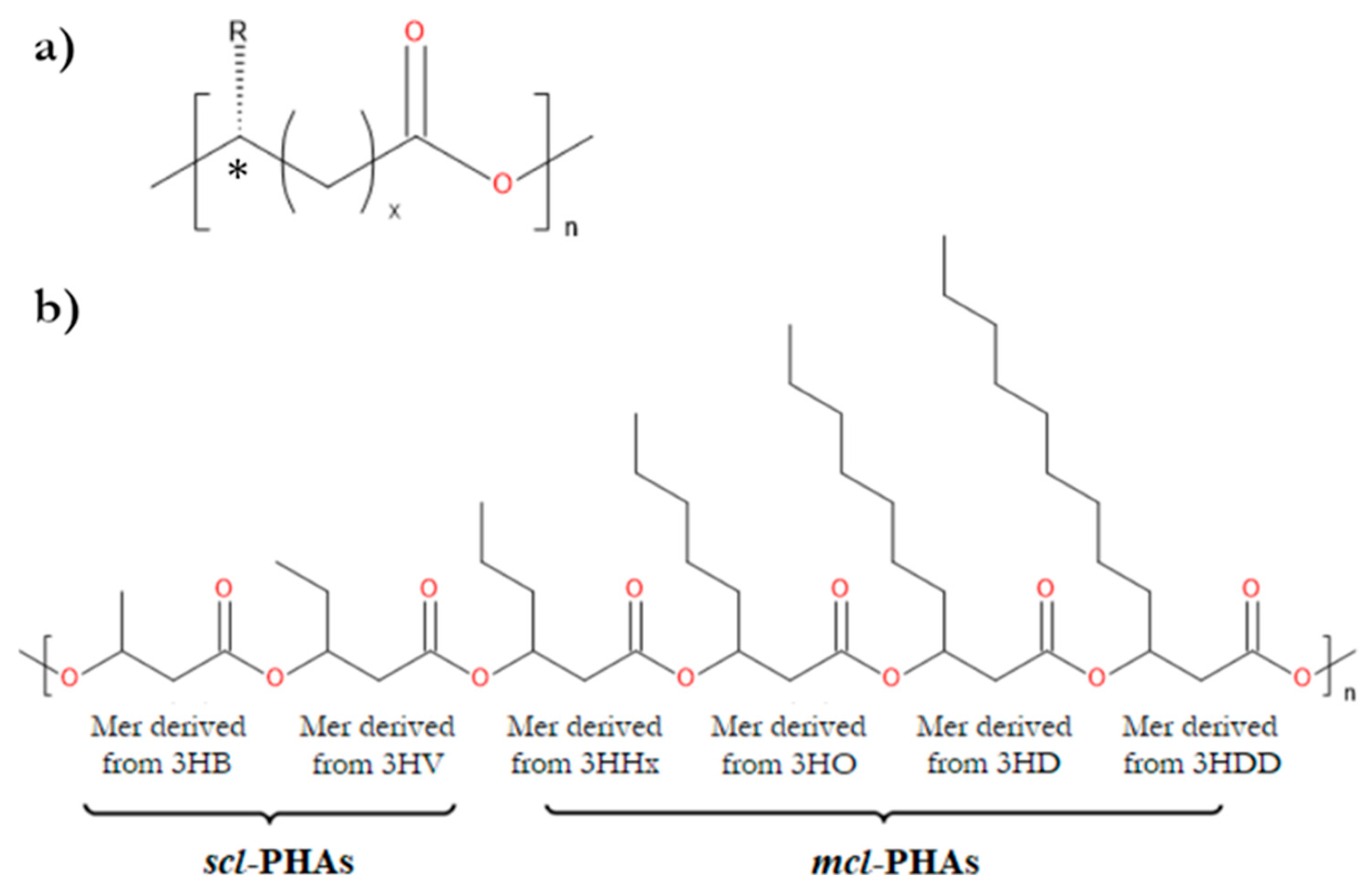
Figure 2.
Chemical structure of biopolyesters that undergo significant hydrolysis catalyzed by lipases of animal origin.
Figure 2.
Chemical structure of biopolyesters that undergo significant hydrolysis catalyzed by lipases of animal origin.
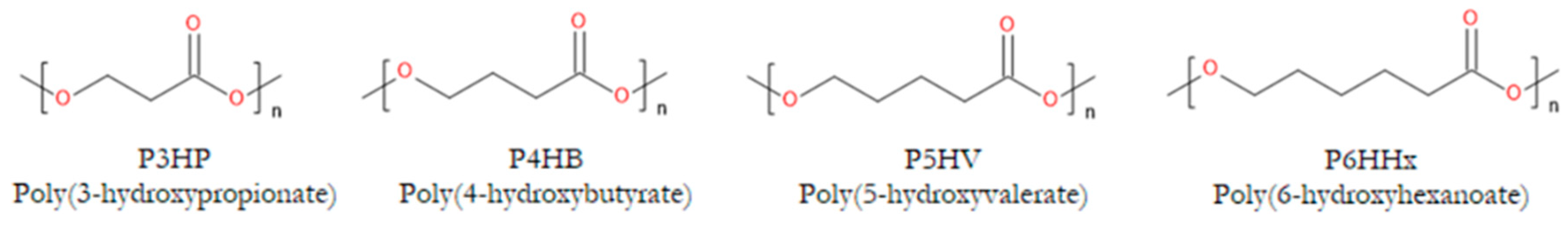
Figure 3.
Two approaches are used for altering the chemical structure of microbial polyesters. (a) One utilizes polymerization of the PHB oligomers to obtain its multiblock copolymers, (b) and the second focuses on modifying the surface chemistry of the scaffold.
Figure 3.
Two approaches are used for altering the chemical structure of microbial polyesters. (a) One utilizes polymerization of the PHB oligomers to obtain its multiblock copolymers, (b) and the second focuses on modifying the surface chemistry of the scaffold.
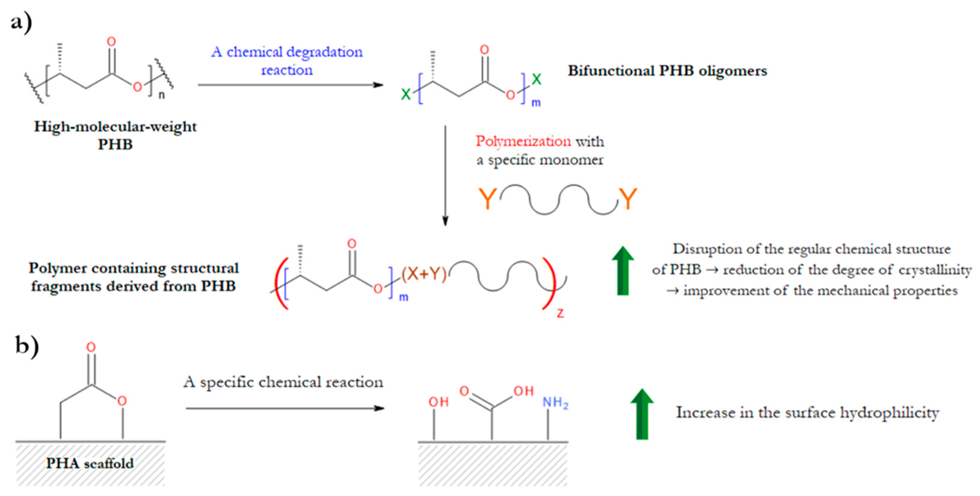
Figure 4.
Scheme illustrating the stages of initiation, propagation, and termination of radical reactions involving PHB, which are induced by ionizing radiation. For simplicity, only one of the possible termination reactions is presented in the diagram.
Figure 4.
Scheme illustrating the stages of initiation, propagation, and termination of radical reactions involving PHB, which are induced by ionizing radiation. For simplicity, only one of the possible termination reactions is presented in the diagram.
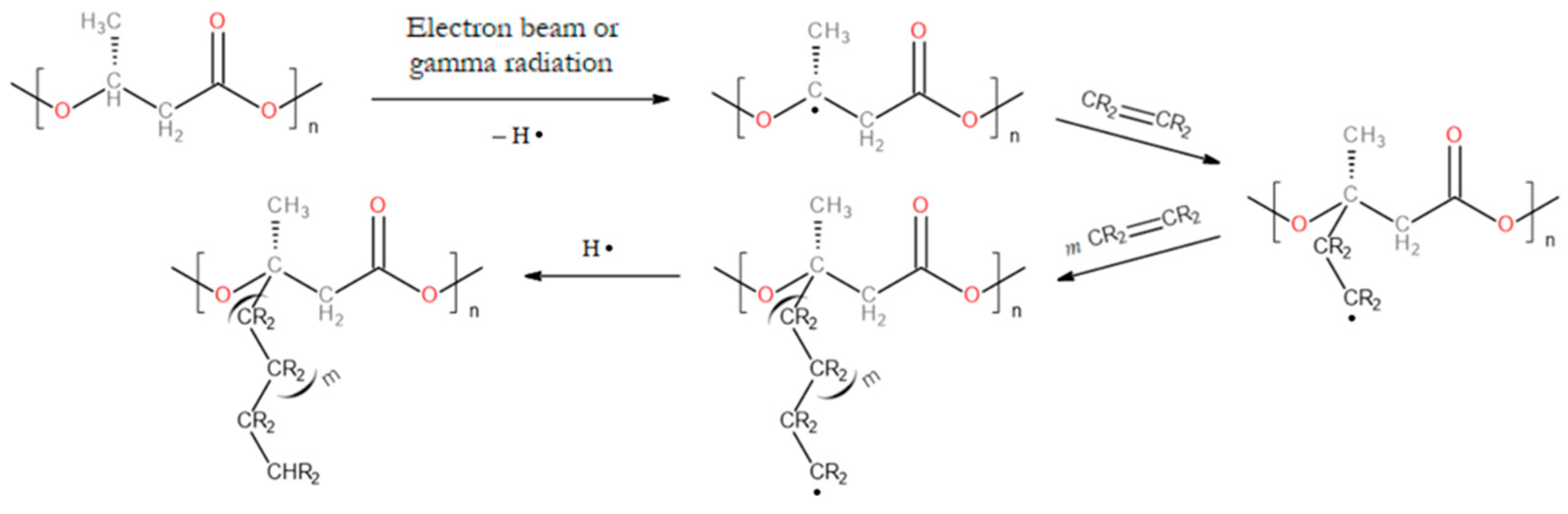
Figure 5.
Simplified scheme of PHB grafting reaction initiated by peroxide compound.
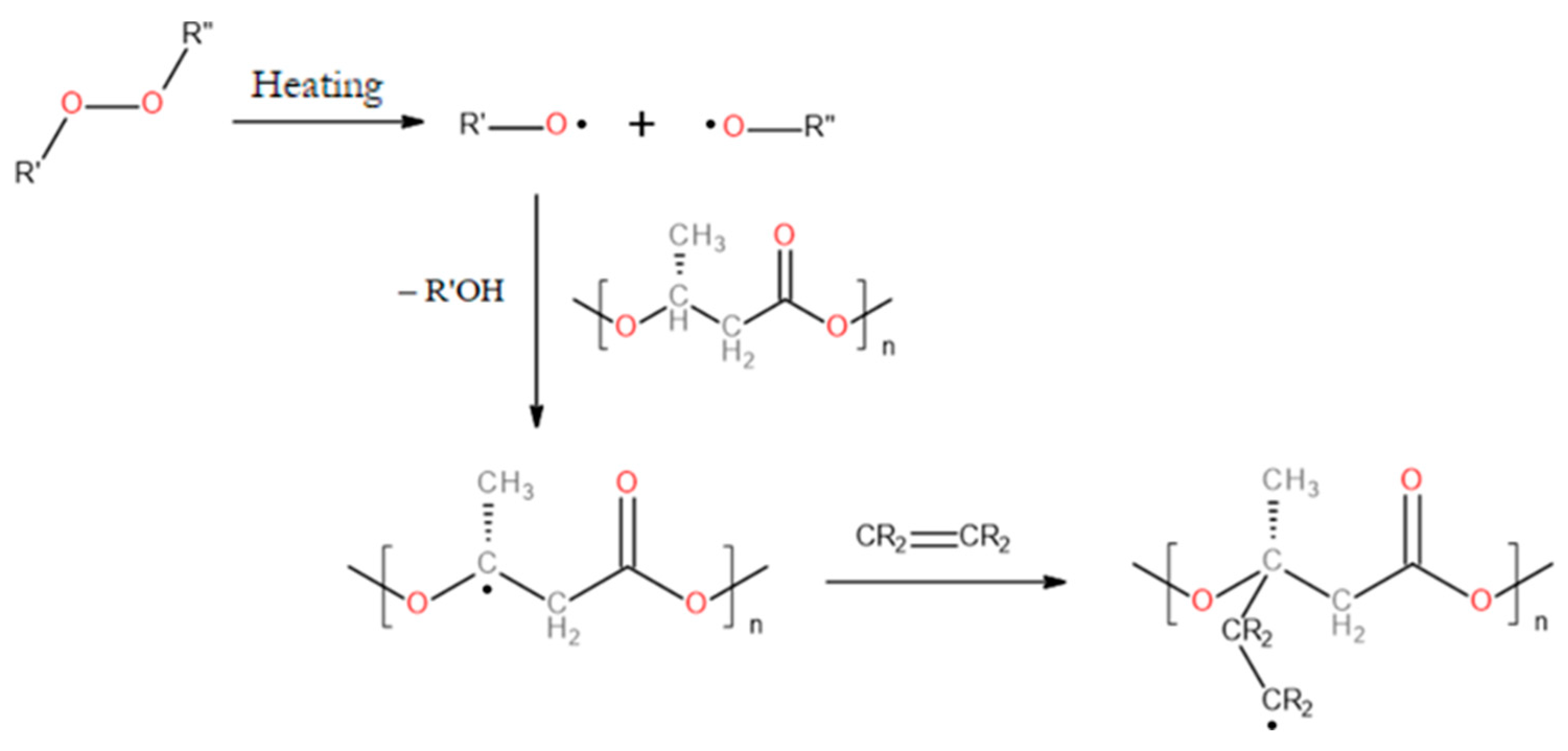
Figure 6.
Simplified scheme of PHB grafting initiated by benzophenone and UV light.
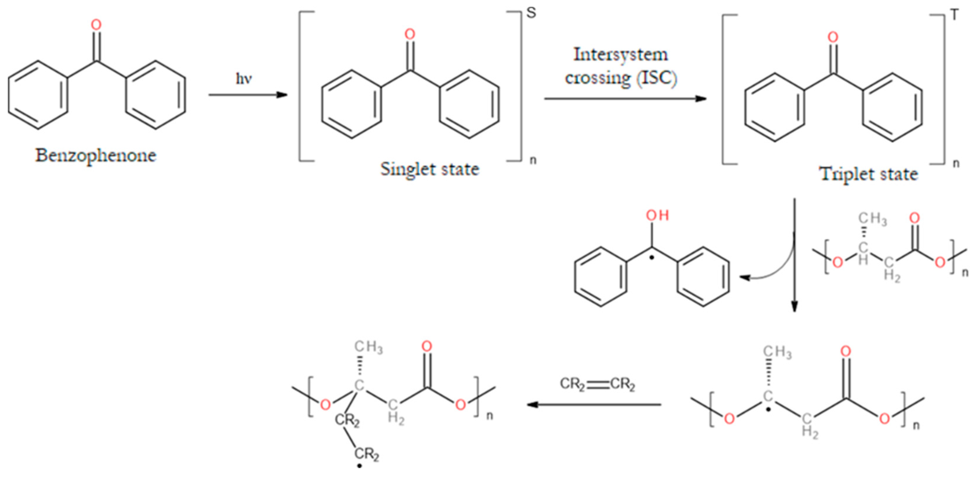
Figure 7.
Chemical structures of the monomers used for grafting the surfaces of PHB and PHBV products, and the structure of the PHB-g-PHEMA copolymer.
Figure 7.
Chemical structures of the monomers used for grafting the surfaces of PHB and PHBV products, and the structure of the PHB-g-PHEMA copolymer.
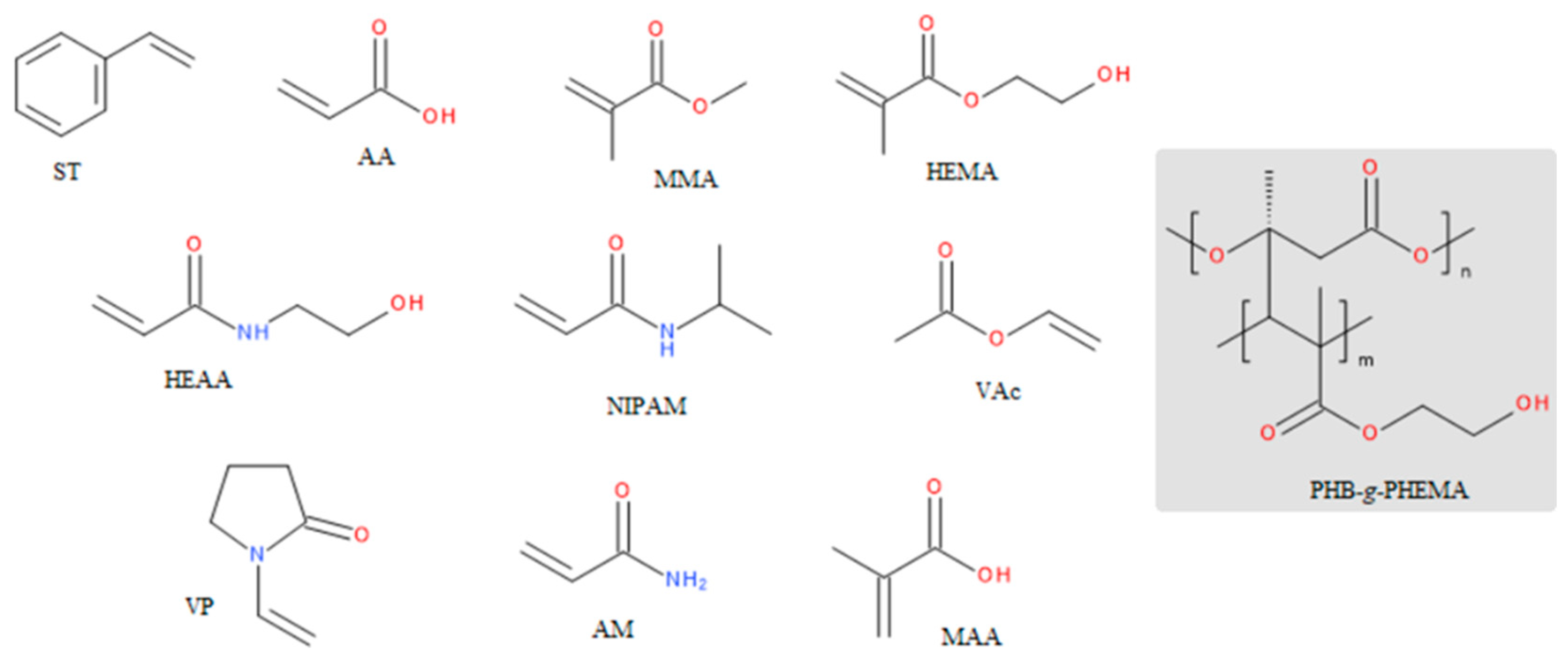
Figure 8.
Simplified scheme for chemical surface modification using UV light and 3,4-dicarboxybenzenediazonium tosylate.
Figure 8.
Simplified scheme for chemical surface modification using UV light and 3,4-dicarboxybenzenediazonium tosylate.
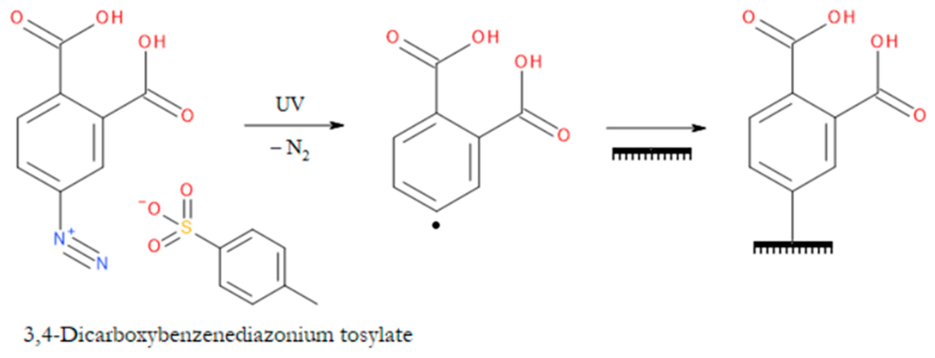
Figure 9.
Mechanism of nucleophilic substitution of an acyl group, exemplified by the glycolysis of biopolyesters; R = hydrogen atom or an alkyl group.
Figure 9.
Mechanism of nucleophilic substitution of an acyl group, exemplified by the glycolysis of biopolyesters; R = hydrogen atom or an alkyl group.
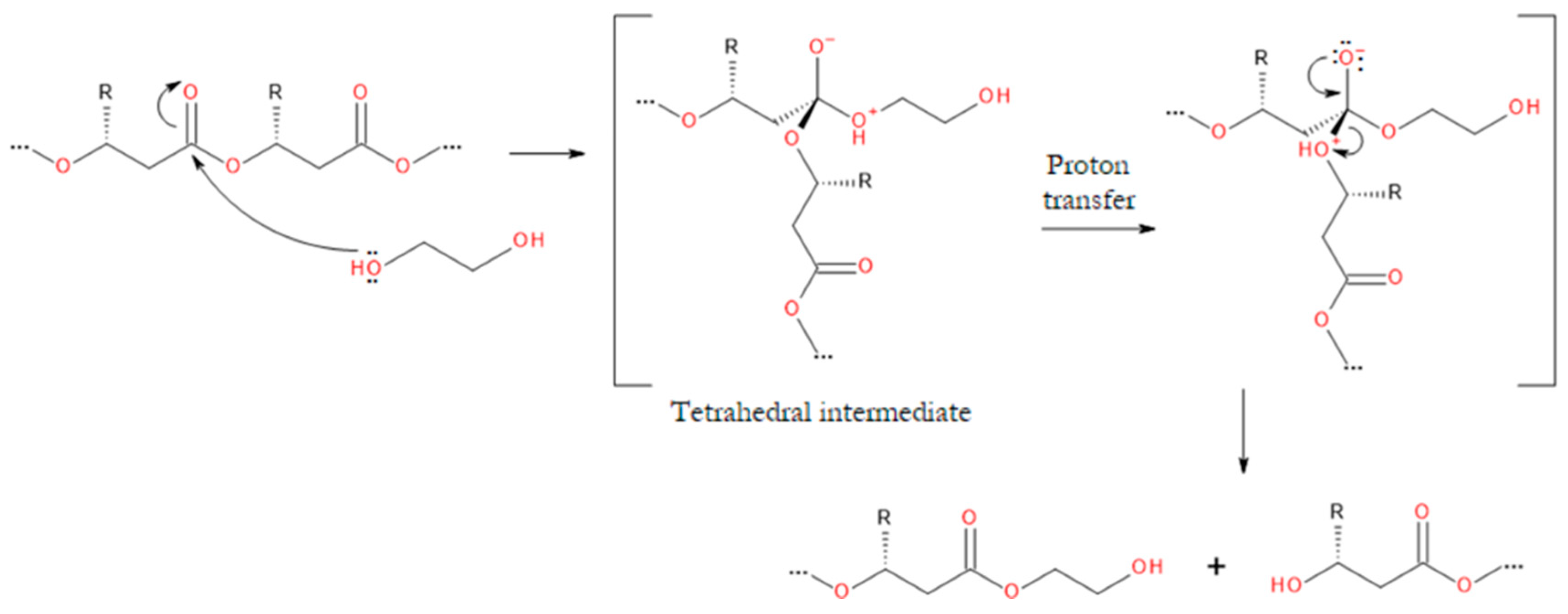
Figure 10.
Schematic overview of the degradation reactions to which biopolyesters are subjected in order to obtain their oligomers. For simplicity, only those oligomers that are the main products of the specified reactions are shown; R = hydrogen atom or an alkyl group.
Figure 10.
Schematic overview of the degradation reactions to which biopolyesters are subjected in order to obtain their oligomers. For simplicity, only those oligomers that are the main products of the specified reactions are shown; R = hydrogen atom or an alkyl group.
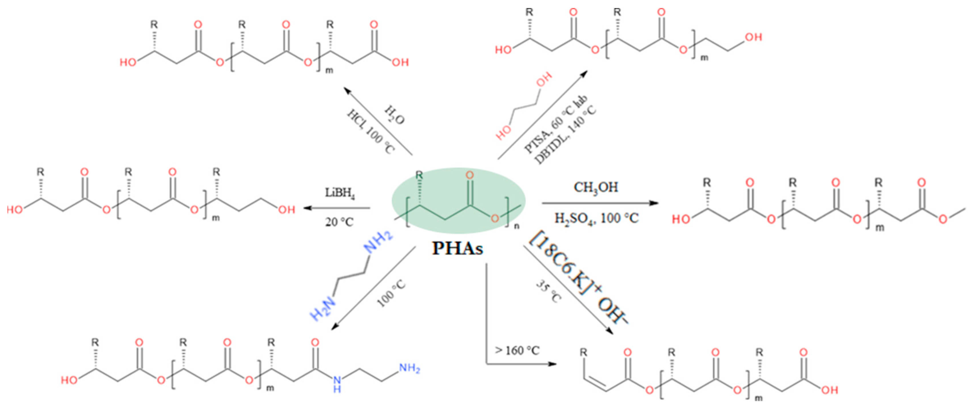
Figure 11.
Mechanism of the strong base-initiated E1cB elimination reaction of biopolyesters (counterion omitted for clarity); R = hydrogen atom or an alkyl group.
Figure 11.
Mechanism of the strong base-initiated E1cB elimination reaction of biopolyesters (counterion omitted for clarity); R = hydrogen atom or an alkyl group.
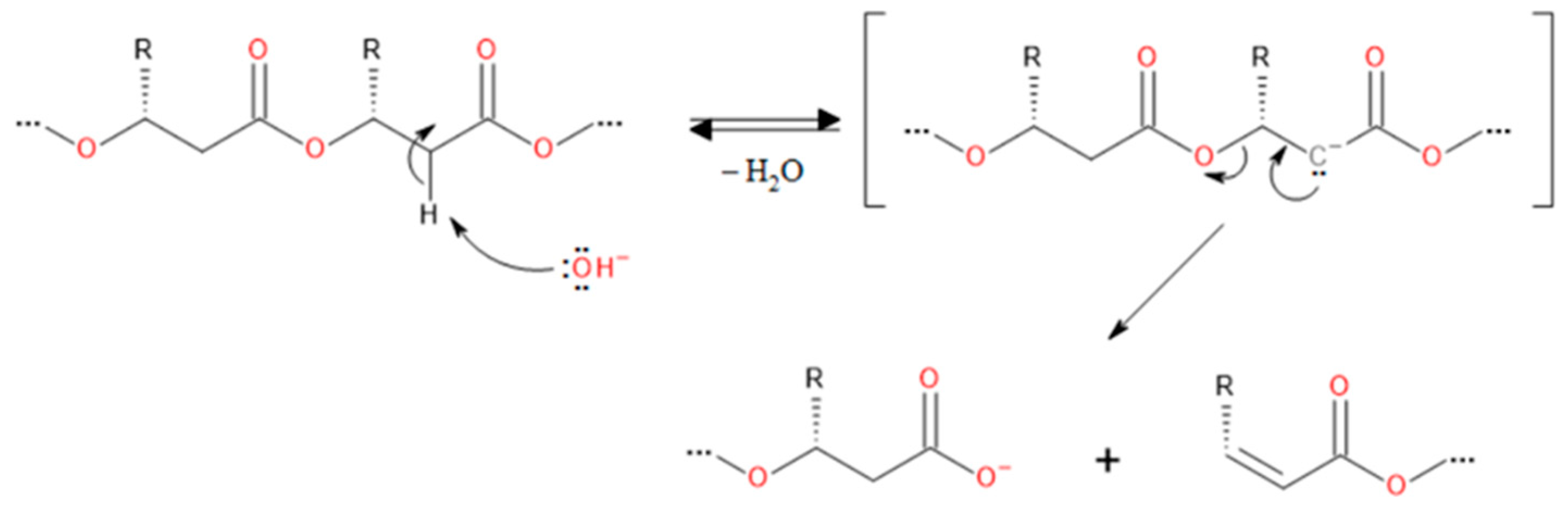
Figure 12.
Mechanism of biopolyester reduction using LiAlH4 (the counterion has been omitted for clarity); R = hydrogen atom or an alkyl group.
Figure 12.
Mechanism of biopolyester reduction using LiAlH4 (the counterion has been omitted for clarity); R = hydrogen atom or an alkyl group.
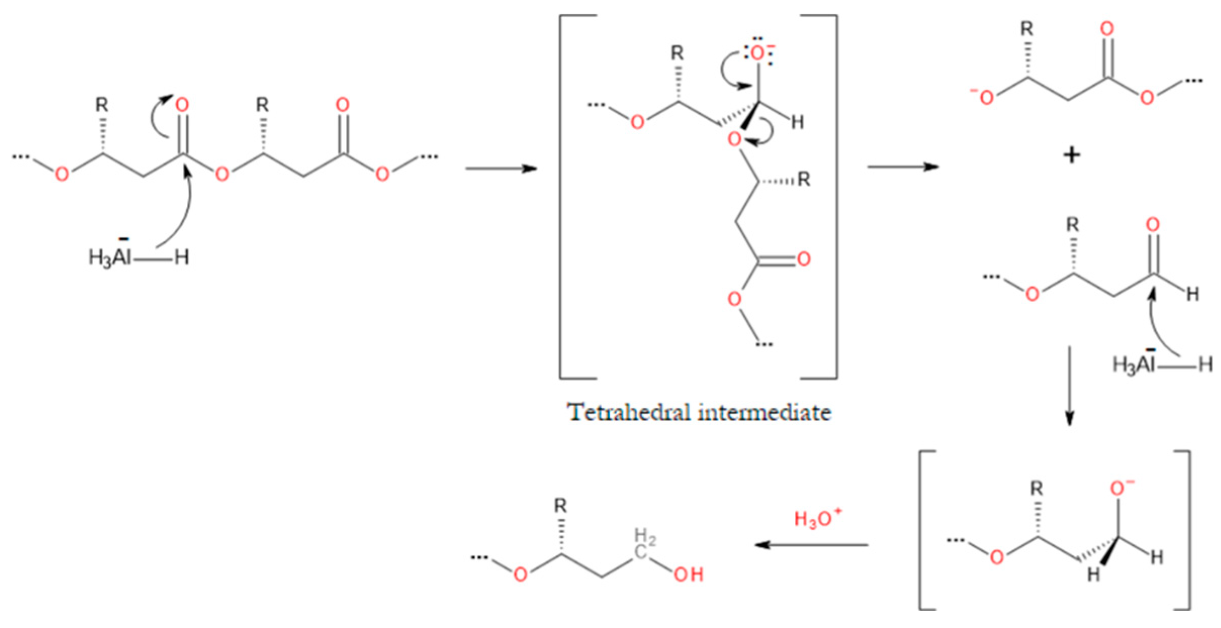
Figure 13.
Schematic of the synthesis of thermoresponsive PNIPAAm-PHB-PNIPAAm triblock copolymer via ATRP.
Figure 13.
Schematic of the synthesis of thermoresponsive PNIPAAm-PHB-PNIPAAm triblock copolymer via ATRP.
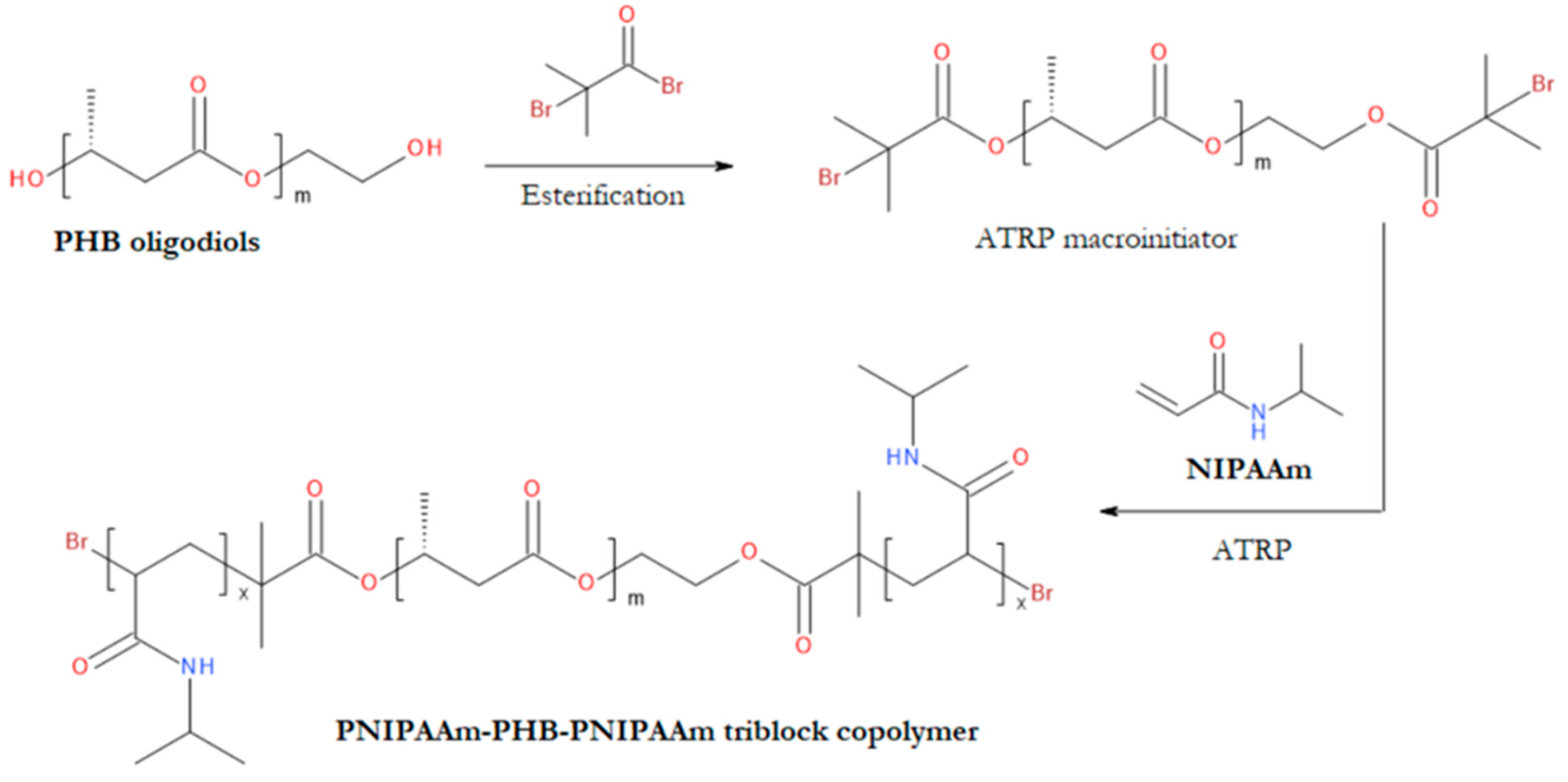
Figure 14.
Schematic of the synthesis of poly(ester urethane) from PHB oligoester diols, oligomeric PEG, and HDI [344].
Figure 14.
Schematic of the synthesis of poly(ester urethane) from PHB oligoester diols, oligomeric PEG, and HDI [344].
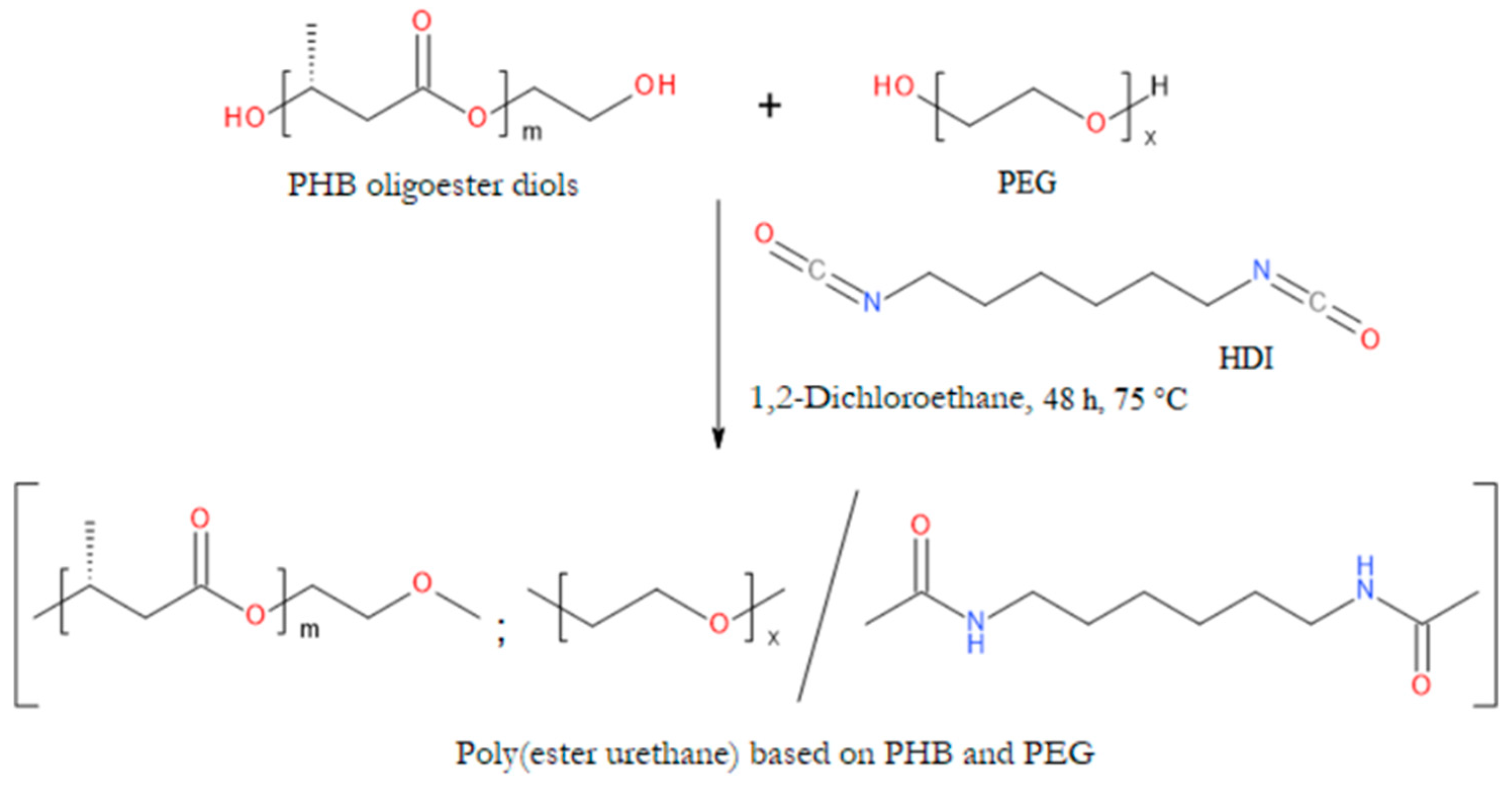
Figure 15.
Chemical structure of oligoester diols used in the synthesis of DegraPol polyurethanes.
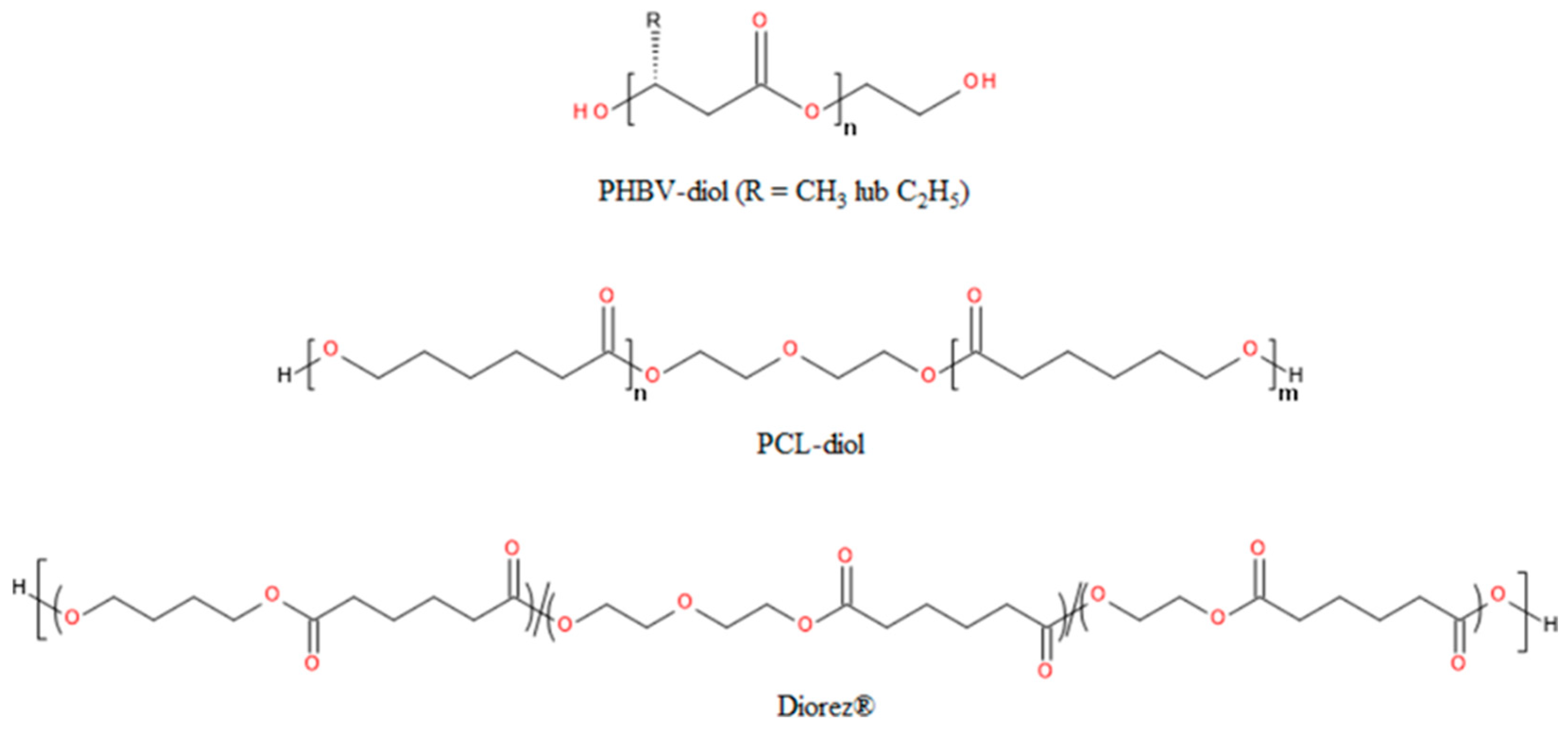
Disclaimer/Publisher’s Note: The statements, opinions and data contained in all publications are solely those of the individual author(s) and contributor(s) and not of MDPI and/or the editor(s). MDPI and/or the editor(s) disclaim responsibility for any injury to people or property resulting from any ideas, methods, instructions or products referred to in the content. |
© 2024 by the authors. Licensee MDPI, Basel, Switzerland. This article is an open access article distributed under the terms and conditions of the Creative Commons Attribution (CC BY) license (http://creativecommons.org/licenses/by/4.0/).
Copyright: This open access article is published under a Creative Commons CC BY 4.0 license, which permit the free download, distribution, and reuse, provided that the author and preprint are cited in any reuse.


